Article

Diazotrophic Bacteria and Nitrogen Fertilization on ATPase Activity in Micropropagated Pineapple Plantlets During Acclimatization

by
Aurilena de Aviz Silva
1,
Almy Junior Cordeiro de Carvalho
1,
Paulo Cesar dos Santos
2,*,
Rômulo André Beltrame
3,
Marta Simone Mendonça Freitas
1,
Flávia Paiva de Freitas
1,
Roberto Rivelino do Nascimento Barbosa
1,
Alessandro Coutinho Ramos
4,
Fabio Lopes Olivares
5,
Stella Arndt
2,
Leandro Pin Dalvi
2,
Moises Zucoloto
2,
Orlando Carlos Huertas Tavares
6 and
Mírian Peixoto Soares da Silva
7
1
Plant Science Laboratory, Center for Agricultural Science and Technologies, Northern Fluminense State University Darcy Ribeiro (UENF), Campos dos Goytacazes 28013-602, RJ, Brazil
2
Center for Agricultural Sciences and Engineering, Department of Agronomy, Federal University of Espírito Santo (UFES), Alegre Campus, Alegre 29500-000, ES, Brazil
3
Department of Soil Science, Institute of Agronomy, Federal Rural University of Rio de Janeiro (UFRRJ), Seropédica 23890-000, RJ, Brazil
4
Laboratory of Environmental Microbiology and Biotechnology, Vila Velha University (UVV), Vila Velha 29102-920, ES, Brazil
5
Cell and Tissue Biology Laboratory, Center for Biosciences and Biotechnology, Northern Fluminense State University Darcy Ribeiro (UENF), Campos dos Goytacazes 28013-602, RJ, Brazil
6
Department of Technical Education in Agriculture, Fluminense Federal Institute of Education, Science and Technology (IFF), Cambuci Campus, Cambuci 28430-000, RJ, Brazil
7
Agricultural Coordination, Federal Institute of Education, Science and Technology of Tocantins (IFTO), Pedro Afonso Campus, Pedro Afonso 77710-000, TO, Brazil
*
Author to whom correspondence should be addressed.
Horticulturae 2026, 12(3), 374; https://doi.org/10.3390/horticulturae12030374

Submission received: 21 January 2026 / Revised: 27 February 2026 / Accepted: 2 March 2026 / Published: 18 March 2026

Abstract

Micropropagated plantlets, after removal from controlled laboratory conditions, require an acclimatization period. Adaptation to the new environment induces anatomical and physiological changes controlled by cellular processes. This study investigated the involvement of the primary proton transport systems of total membranes in pineapple root colonization by diazotrophic bacteria and in the development of plantlets treated with different nitrogen doses, allowing an understanding of nutrient absorption and accumulation dynamics. The experiment followed a randomized block design (RBD) in a factorial scheme (2 × 3 × 2), with two inocula (a mixture of diazotrophic bacteria containing Burkholderia sp. UENF 114111, Burkholderia silvatlantica UENF 117111, and Herbaspirillum seropedicae HRC 54, and another without bacteria), three urea doses (0, 5, and 10 g L−1), and two evaluation (90 and 150 days) and bacterial counting times (30 and 150 days), with three blocks. Diazotrophic bacterial populations were lower in older plantlets. H+ transport mediated by P H+-ATPases changed with acclimatization time. Inoculation did not induce transport; however, the Fmax of V H+-ATPase was lower without nitrogen fertilization. Nitrogen fertilization affected V H+-ATPase proton transport activity in root membranes. The presence of diazotrophic bacteria did not induce proton transport. On the other hand, nitrogen fertilization and acclimatization time affected the proton transport activity mediated by H+-ATPases isolated from roots of micropropagated pineapple.

1. Introduction

The pineapple (Ananas comosus var. comosus) is cultivated in more than 50 countries, with the Philippines, Brazil, Costa Rica, Thailand, and China standing out as the largest producers of the fruit. Pineapple is one of the most widely crop fruit in Brazil, cultivated in virtually all states, especially in the North, Northeast, and Southeast regions [1]. The state of Rio de Janeiro stands out with 6390 hectares harvested in 2024, and its edaphoclimatic characteristics combined with the proximity to major consumer markets create favorable conditions and strong prospects for the expansion of pineapple cultivation. This interaction between environmental conditions and human factors reinforces the natural suitability and economic potential of the region for pineapple production [2].
However, the pineapple production chain faces constraints generated by the scarcity of information on the adaptability of potential new pineapple cultivars in producing regions due to the lack of studies involving productive aspects of the fruit, notably regarding seedling quality [3]. Consequently, micropropagation has been widely used in the production of pineapple plantlets, mainly aiming at the release of new cultivars. This technique has the advantage of producing plantlets free of pathogens on a large scale [4], with high vigor and uniformity [5]. However, micropropagated plantlets must be acclimatized before being transferred to the field; a long process, necessary to promote morphological, anatomical, and physiological changes in the plantlets which favor their survival after transplanting [4,6].
Additionally, nitrogen fertilization in pineapple is related to vegetative growth and plantlet quality, since nitrogen is involved both in plantlet production and growth, including stages associated with in vitro propagation, as well as in crop development, yield, and quality, as reported in studies by [7,8,9,10,11]. Nitrogen uptake occurs mainly in inorganic forms such as ammonium (NH4+) or as nitrate (NO3) [12]. Absorbed NO3 can be transported within the plant and accumulated in the vacuole for later use.
In the process of nitrogen absorption and accumulation, two enzymes of great importance in plants are involved: P H+-ATPase and V H+-ATPase. P H+-ATPase is an enzyme present in the plasma membrane that hydrolyzes ATP and transports H+ ions to the exterior of the cell, resulting in an electrochemical potential gradient [13]. H+-transporting pumps are involved in nutrient uptake [14] and several metabolic processes such as cell elongation, stomatal opening and closing, and cellular homeostasis [12,15,16].
In plant cells, the activation and stimulation of H+ pumps may occur due to several factors such as growth regulators, light, pathogen attack, hormones, inoculation with mycorrhizal fungi, or diazotrophic bacteria [17,18,19]. The use of inoculation with diazotrophic bacteria in plants has contributed to increasing N and other nutrients in the plant, in addition to improving adaptation to the ex vitro environment and reducing the acclimatization period [20].
This study provides both scientific and applied contributions by advancing the understanding of plant–microorganism interactions during the acclimatization of micropropagated pineapple plantlets. Specifically, the work clarifies whether diazotrophic bacterial inoculation modulates proton pump activity under different nitrogen fertilization regimes. From an applied perspective, these findings contribute to improving acclimatization management strategies, particularly regarding the integration of microbial inoculants and nitrogen fertilization in commercial seedling production systems.
Thus, the objective of this study was to evaluate the participation of primary H+ transporters in total membranes isolated from the root system of micropropagated pineapple plantlets inoculated with diazotrophic bacteria or fertilized with nitrogen during the acclimatization period, to understand the dynamics of absorption as well as nitrogen accumulation by the plant.

2. Materials and Methods

2.1. Experimental Design

The experiment was conducted in a greenhouse located on the campus of the Darcy Ribeiro State University of Northern Fluminense (UENF) (Latitude = 21°19′23″, Longitude = 41°10′40″; Altitude = 14 m). The experiment followed a randomized block design (RBD) arranged in a 2 × 3 × 2 factorial scheme, with three blocks. The experimental unit consisted of tray cells containing one micropropagated plantlet per cell. From each experimental unit, plantlets were randomly sampled for bacterial quantification and ATPase analyses according to the requirements of each evaluation.
The factors were represented by two types of inocula, a mixture of diazotrophic bacteria containing Burkholderia sp. UENF 114111, Burkholderia silvatlantica UENF 117111, and Herbaspirillum seropedicae HRC 54, and the other without bacteria; three urea doses (0, 5, and 10 g L−1); and two evaluation times (90 and 150 days) for proton pumps and bacterial counting (30 and 150 days), with three blocks. The experimental unit consisted of fifty plantlets grown in Styrofoam trays with 200 cells, properly separated by treatment, one designated for bacterial counting and the remainder designated for evaluation of primary H+ transporters.
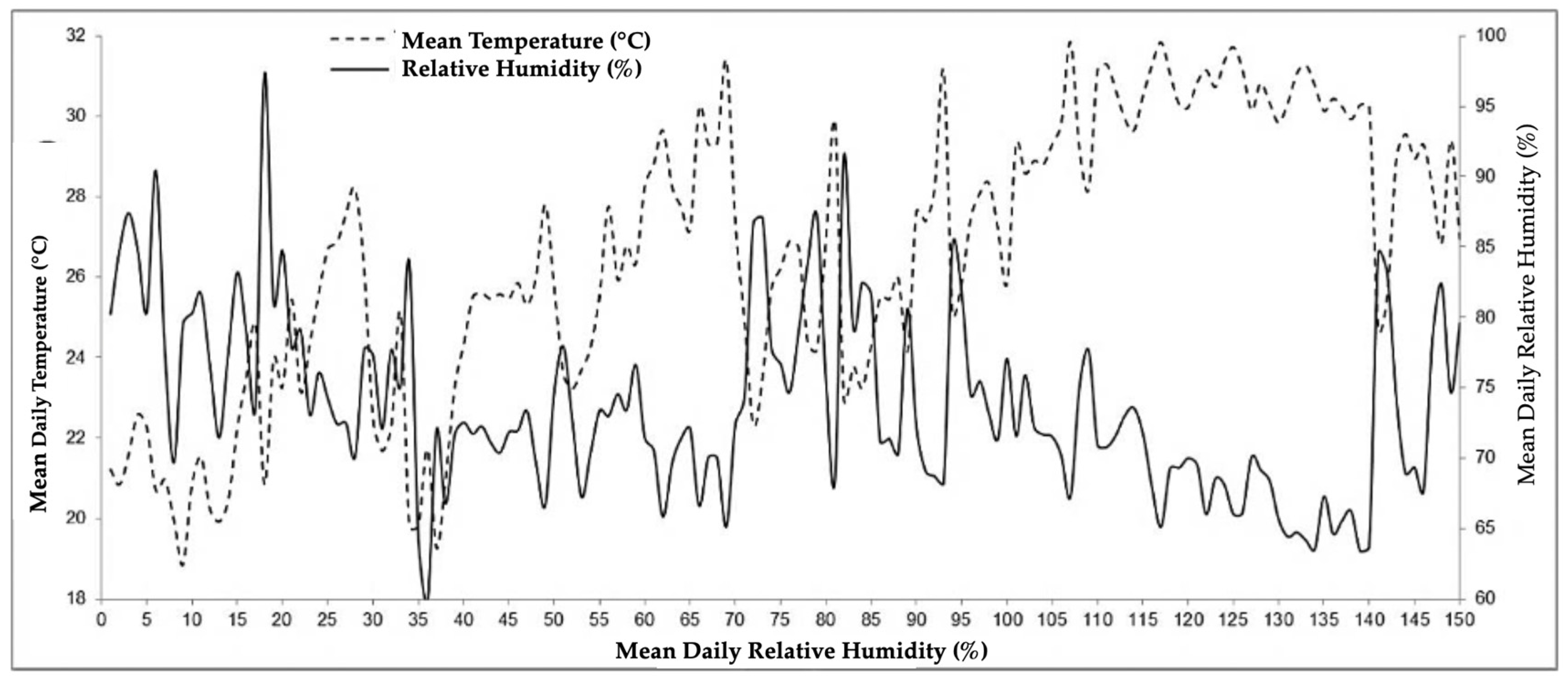
The data on daily mean temperature (°C) and daily mean relative humidity (%) recorded in the greenhouse during the experimental period were monitored using a Kimo Kistock data logger (KIMO Instruments, Montpon-Ménestérol, France), and data were processed with KILOG software (version 4.7), programmed to perform measurements at two-hour intervals and are presented in Figure 1.

2.2. Plant Material and Nitrogen Fertilization

Nitrogen fertilization was carried out by foliar application of urea (50 mL per experimental unit) in split applications: after 25 days of cultivation, the plantlets received 20% of the dose; at 50 days, 50% of the dose; and from 75 days onward, 100% of the dose was applied weekly. The ‘BRS Vitória’ pineapple plantlets used in the experiment were obtained from the Biomudas Tissue Culture Laboratory, derived from in vitro propagation, with an average fresh mass of 0.30 g.

2.3. Bacterial Inoculum

The diazotrophic bacterial strains were obtained from the bacterial collection of the Cell and Tissue Biology Laboratory at UENF. The pre-inoculum containing 20 μL of stock of each bacterium was diluted in DYG’S liquid culture medium [21] and maintained under agitation (140 rpm, at 30 °C) for 24 h. The pre-inoculum obtained was used to produce a larger volume of inoculant under the same conditions, corresponding to a final volume of 100 mL of each inoculum, with a density of 108 cells per mL.
The inocula were dissolved in 5.7 L of distilled water, and inoculation was performed by immersing the plantlets in this mixture for 30 min (Figure S1). Plantlets from the control treatment were immersed in distilled water. Subsequently, the plantlets were transplanted into trays filled with the commercial substrate Vivatto Slim Plus®, which showed the following chemical characteristics: pH = 5.9; S–SO4 = 1090 mg dm−3; P = 400 mg dm−3; K = 29.7 mmolc dm−3; Ca = 96.6 mmolc dm−3; Mg = 58.0 mmolc dm−3; Al = 0 mmolc dm−3; H+Al = 34.6 mmolc dm−3; Na = 13.2 mmolc dm−3; organic matter = 130.85 g dm−3; CEC (pH = 7) = 231.50 mmolc dm−3; SB = 196.90 mmolc dm−3; V = 85%; Fe = 155.90 mg dm−3; Cu = 1.71 mg dm−3; Zn = 32.50 mg dm−3; Mn = 84.20 mg dm−3; and B = 4.18 mg dm−3.
Fifteen days after the first inoculation, the substrate was inoculated with the same bacterial mixture, and each tray cell received 2 mL of the bacterial mixture. After transplanting, using a backpack sprayer, the plantlets were sprayed with water to prevent leaf dehydration.
At 15 days after transplanting, foliar fertilization began and was scheduled on a weekly basis, applying a complete nutrient solution (2 mL plant−1) prepared according to Döbereiner et al. [22]. From the third week onward, the plantlets no longer received nitrogen fertilization. Plantlets from the control treatment received the complete solution at 121 days to avoid plantlet loss due to N deficiency. All fertilizations were performed after 5:00 p.m., using a disinfected Styrofoam cabinet to avoid drift and contamination among plantlets from different treatments.

2.4. Bacterial Quantification

Bacterial counting was performed at two time points (30 and 150 days after planting) using root samples obtained according to Canellas et al. [21]. Root subsamples (0.1 g) were aseptically collected and individually macerated in 99.9 mL of sterile distilled water. Serial dilutions were prepared from 10−3 to 10−7 by transferring 1 mL of the previous dilution into 9 mL of sterile distilled water. Aliquots (100 μL) from selected dilutions (10−5, 10−6, and 10−7) were inoculated into the center of semisolid JNFb (nitrogen-free semi-solid) medium contained in glass vials with 5 mL of medium. The vials were incubated at 30 °C for seven days.
Bacterial growth was evaluated based on pellicle formation at the medium surface, a characteristic descriptor of diazotrophic bacterial development under nitrogen-free conditions. Bacterial identification was considered presumptive and based on colony color, morphology, pellicle formation, and growth characteristics in the semisolid JNFb medium, following the diagnostic descriptors proposed by Döbereiner et al. [22]. Colonies presenting morphological patterns inconsistent with the expected diazotrophic profile were excluded from MPN estimation.
The differentiation between target diazotrophic bacteria and potential endophytic or contaminant microorganisms was performed considering colony appearance, pellicle formation, and typical growth behavior in nitrogen-free medium. For all treatments, the minimum number of cells per gram of root was inferred based on dilution criteria (10−1).
MPN indices were obtained using the McCrady probability table for three replicates per dilution. It is important to note that MPN values represent probabilistic estimates rather than direct counts of viable cells.

2.5. Membrane Isolation

The isolation of the plasma membrane was also performed at two time points, at 90 and 150 days after planting, using the methodology described by Giannini and Briskin [23], with adaptations by Façanha and De Meis, 1998 [18]. Additionally, the roots were collected and, after weighing, were macerated using a mortar and pestle in buffered medium at pH 8.0, at a 1:1 ratio (buffer:fresh root mass). The final concentrations of the reagents in the extraction buffer were as follows: sucrose 250 mM, glycerol 10%, DTT 2 mM, EDTA 5 mM, PVP-40 0.5%, KCl 150 mM, BSA 0.13%, PMSF 2 mM, and Tris-HCl (pH 8.0) 0.1 M. After maceration, the homogenate was filtered through four layers of gauze and subjected to the first centrifugation in a HITACHI himac CP centrifuge at 30,000 rpm for 15 min at 4 °C to remove unbroken cells, nuclei, and mitochondria. The supernatant was collected and subjected to a new centrifugation in a HITACHI himac CP 85b centrifuge at 9100 rpm for 10 min. The supernatant was collected and subjected to a new centrifugation at 30,000 rpm at 2 °C for 35 min. The precipitate (pellet), corresponding to the unpurified microsomal fraction, was resuspended with 3 mL of a buffer solution at pH 7.5 (resuspension medium) containing glycerol 15%, DTT 1 mM, PMSF 1 mM, HEPES-KOH 10 mM (pH 7.6), and EDTA 1 mM, thus obtaining the resuspended microsomal fraction. The material was frozen in liquid nitrogen and stored at −70 °C until use.

2.6. Proton Transport Assay

For the determination of H+ transport, total membrane vesicles were added to two types of incubation media, one at pH 6.5 and the other at pH 7.0, both containing 100 mM KCl, 5 mM MgSO4, 10 mM HEPES-BTP buffer (bis-tris propane), 250 mM sucrose, and 1 μM 9-amino-6-chloro-2-methoxyacridine (ACMA). The inhibitors sodium orthovanadate (300 μM) (P H+-ATPase inhibitor) and concanamycin A (22 nM) (V H+-ATPase inhibitor) were used in the pH 6.5 and pH 7.0 incubation media, respectively. After 3 min of incubation, 1 mM ATP, pH 7.2, was added and H+ transport was monitored by ACMA fluorescence decay in a Shimadzu RF-530 1PC fluorimeter (Shimadzu Corporation, Kyoto, Japan). NH4Cl (final concentration 20 mM) was added after fluorescence quenching (~400-600 s) to dissipate the proton gradient. P H+-ATPase activity was evaluated using the pH 6.5 buffer and was sodium orthovanadate-sensitive. V H+-ATPase activity was evaluated using the pH 7.0 buffer and was concanamycin-sensitive (22 nM). From the data obtained, the initial velocity (V0) and the maximum fluorescence variation (Fmax) were determined.

2.7. Statistical Analysis

The results obtained from each analysis were subjected to analysis of variance (ANOVA) to evaluate the effects of the inocula factor (Factor A), urea doses factor (Factor B), time (Factor C), and their interaction (A × B × C) [24]. The F-test was used to assess the significance of the effects (p < 0.05). To verify the assumptions of ANOVA, the Shapiro–Wilk test (p > 0.05) was performed to assess residuals normality, and the Bartlett test (p > 0.05) was used to evaluate the homogeneity of variances [25,26]. When F-test was significant, the variables were subjected to the Tukey test (p ≤ 0.05) to compare means. For variables associated with bacterial quantification, values obtained by the Most Probable Number (MPN) method were log10-transformed and expressed as log10(MPN g−1 root) to improve variance homogeneity and approximate normality. Statistical analyses were performed using SANEST Version 3.0 software.

3. Results

3.1. Bacterial Quantification (MPN)

Considering that MPN represents a probabilistic estimate rather than a direct quantification of viable cells, the observed differences should be interpreted as variations in inferred bacterial population density rather than absolute cell counts. Bacterial counting revealed an interaction between the sampling time and the bacteria. After 30 days of planting, the bacterial population increased by 3 logarithmic units compared to the non-inoculated population. However, after 150 days there was a decrease in the population, with no statistical difference between inoculated and non-inoculated plantlets, as shown in Table 1.
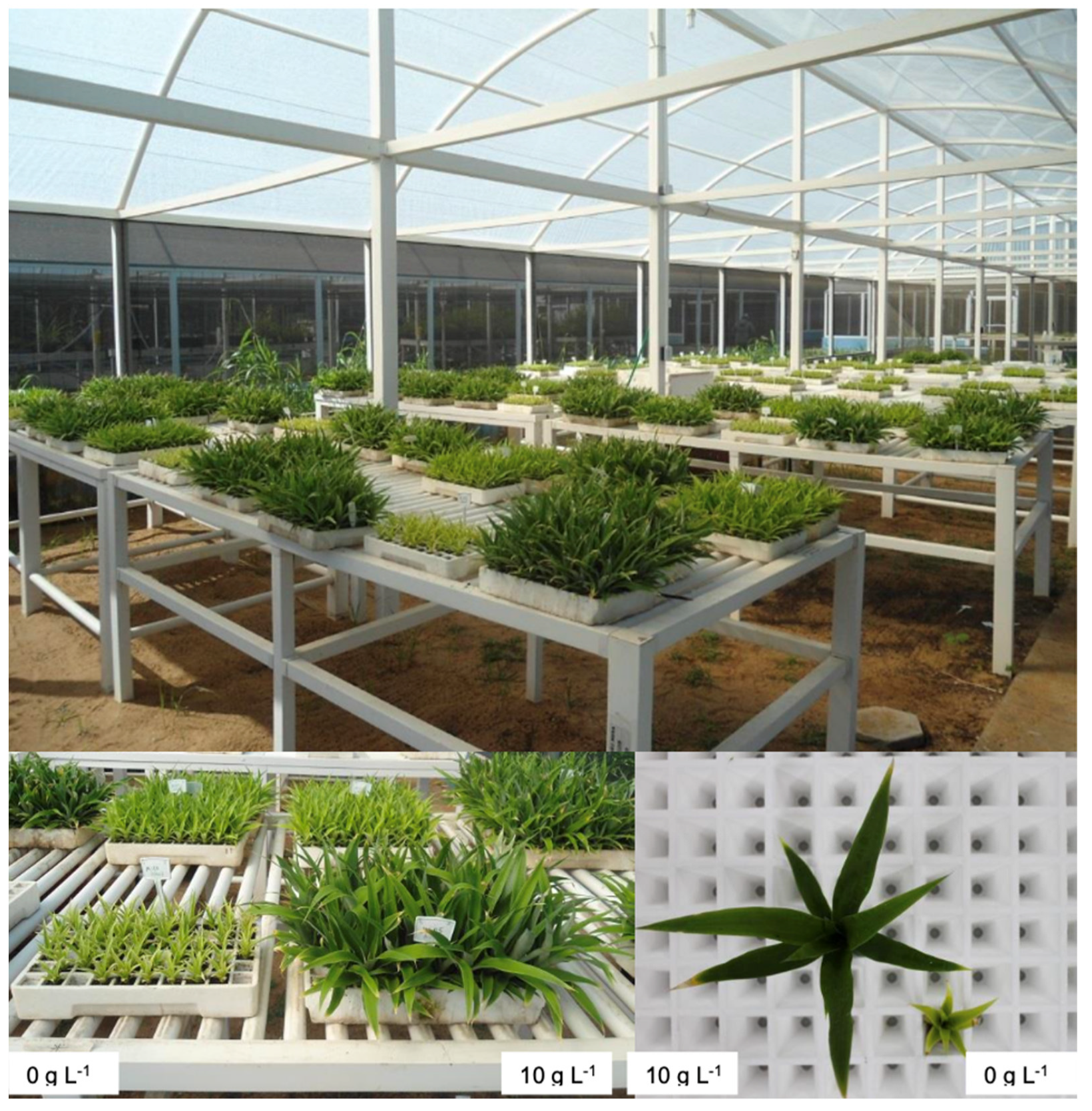
In addition to the microbiological responses, visual differences in plantlet development were evident at 150 days of acclimatization. Plantlets cultivated without urea exhibited reduced growth and vigor compared with those receiving the highest nitrogen dose. These morphological differences were clearly observable, particularly between the 0 and 10 g L−1 urea treatments (Figure 2).

3.2. Effect of Inoculation on H+-ATPases

Additionally, in Table 2, H+ transport by P H+-ATPases and V H+-ATPases in response to bacterial inoculation can be observed. Maximum fluorescence (Fmax) was significantly higher in the absence of bacteria for both H+-ATPases. On the other hand, the initial velocity of H+ transport (V0) also showed higher activity in roots isolated from non-inoculated plants; however, significant differences were observed only for V H+-ATPase.
The effect of acclimatization time on H+ transport demonstrates that Fmax of P H+-ATPase was not influenced by acclimatization (Table 3). However, the total V0 of the enzyme at pH 6.5 and the V0 of P H+-ATPase were lower after 90 days of acclimatization when compared to V0 after 150 days. On the other hand, the Fmax and V0 of V H+-ATPase showed significantly higher activity (p < 0.05) after 90 days of acclimatization when compared to the activity after 150 days.
The relationship between acclimatization times and urea doses was also analyzed (Table 4). The Fmax of H+ transport by V H+-ATPase showed the greatest increase at the concentration of 5 g L−1 urea after 90 days of acclimatization. The lowest V H+-ATPase activity was observed at 10 g L−1 urea, suggesting inhibition possibly due to the high urea concentration. Furthermore, Fmax and V0 were higher at 90 days of acclimatization in all urea doses.
Table 5 shows the activity of V H+-ATPase (proton pump) in pineapple roots, measured by the maximum fluorescence (Fmax) of H+ transport, under different urea and diazotrophic bacterial inoculation conditions. H+ transport was maximum at the concentration of 5 g L−1 urea, regardless of the presence of bacteria, indicating that this nitrogen level optimizes enzyme activity. However, in the absence of urea, inoculation with bacteria significantly reduced Fmax (from 148 to 57), suggesting a complex interaction in the regulation of vacuolar acidification by V H+-ATPase.
The V0 of H+ transport in V H+-ATPase was higher in non-inoculated plants after 90 days of acclimatization (Table 6). After 150 days, there were no significant differences among the microbiological treatments. On the other hand, the V0 activity of the enzyme was reduced after 150 days when compared to 90 days. The strains inoculated in this experiment maintained the same V0 of the V H+-ATPase reaction at the two evaluation times (Table 6).

4. Discussion

Changes in the population number of diazotrophic bacteria may decrease or increase as a function of environmental factors, such as different times of the year, water deficit, low soil moisture, and high temperatures [27,28,29].
Considering that MPN represents a probabilistic estimate rather than a direct quantification of viable cells, the observed differences should be interpreted as variations in inferred bacterial population density rather than absolute cell counts. The detection of diazotrophic bacteria in non-inoculated plantlets may be attributed to latent endophytic populations or to microbial persistence originating from the micropropagation process, as previously reported for pineapple.
The relatively high coefficient of variation observed for MPN-derived data is consistent with the intrinsic variability associated with probabilistic microbiological estimates. It is necessary to distinguish between microbial colonization, activation of physiological signaling, and functional benefits to the host plant. The detection of diazotrophic bacteria in plant tissues does not necessarily imply metabolic activation or stimulation of plant physiological processes. In the present study, bacterial presence was not consistently associated with enhanced H+-ATPase activity, indicating that colonization alone did not translate into measurable functional responses.
This pattern, combined with the reduction in MPN values over time, supports the interpretation of transient microbial colonization. Importantly, this represents a biologically relevant finding, indicating that microbial presence does not necessarily result in sustained physiological activation or functional metabolic benefits to the plant.
H+-ATPases have several functions in plant cell physiology; therefore, interactions with microorganisms may influence the activity of these enzymes. According to Janicka-Russak, 2011 [30], H+-ATPases contribute to plant immune responses and may represent important targets during plant–microbe interactions. The author also report that alterations in P H+-ATPase activity may cause significant effects on cellular functions [30]. In the present study, the decrease in enzyme activity observed in association with bacterial inoculation may reflect regulatory adjustments rather than stimulatory effects. Importantly, bacterial inoculation did not result in measurable stimulation of H+-ATPase activity, as no consistent increases in Fmax or V0 were observed. However, contrasting responses have been reported in the literature. For example, Olivares et al. [19] observed increased H+-ATPase activity in sugarcane roots inoculated with endophytic diazotrophic bacteria, which was attributed to auxin production by the microorganisms.
The growth of the plantlets after 150 days may be justified by the increase in P H+-ATPase activity. The H+ transport caused by this enzyme promotes cell wall acidification, inducing wall plasticity and cell expansion, which is essential for vegetative growth. P H+-ATPase also removes excess H+ from the cytosol [31,32].
The transfer of plantlets from in vitro culture to ‘ex vitro’ conditions is a critical step, due to the physiological and morphological stress caused by the new environment. Thus, changes in H+-ATPase activity may occur, as observed for P H+-ATPase. This enzyme, in addition to regulating physiological processes, also plays an important role in plant adaptation to changing conditions, especially under stress. Furthermore, changes in expression levels of the P H+-ATPase gene have been reported in response to a variety of environmental factors, such as high salinity, dehydration, and low temperatures [30].
According to Finbow and Harrison [33], when NO3 is present at high concentrations in the cytosol, it acts as a chaotropic anion on V H+-ATPases, causing an uncoupling between the integral and peripheral domains of the protein, rendering it unable to hydrolyze ATP and translocate protons. Urea caused distinct responses during the acclimatization period of the plantlets, because the Fmax of H+ transport decreased after 150 days of acclimatization when compared to 90 days, and there were no significant differences among urea concentrations (Table 4). The application of 5 g L−1 urea stimulated the V0 of H+ transport of the enzyme after 90 days. In addition, the V0 of H+ transport also decreased at the concentrations of 0 and 5 g L−1 urea after 150 days (Table 4).
A plausible interpretation for the absence of stimulatory effects from diazotrophic bacteria involves a physiological trade-off regulated by plant nitrogen status. Under conditions of sufficient mineral nitrogen availability, plants may preferentially invest in direct nitrogen assimilation pathways rather than maintaining energetically costly microbial associations. In such scenarios, microbial colonization may occur without triggering metabolic or enzymatic adjustments, particularly in processes related to proton transport and membrane energization.
Under nitrogen deficiency conditions, the remobilization of nitrogen stored in the vacuole may have occurred. When accumulated in the vacuole, NO3 can be released when its cytosolic concentration declines or when plant demand for nitrogen increases during growth and reproductive development. The influx and efflux of NO3 across the tonoplast are driven by H+-coupled transport systems energized by vacuolar V-H+-ATPases and H+-PPases, which generate the electrochemical proton gradient required for secondary active transport [34,35].
The presence of bacteria, as well as urea concentrations, influenced H+ transport by V H+-ATPases. Transport Fmax was stimulated in the presence of 5 g L−1 urea, even in the absence of diazotrophic bacteria. The presence of bacteria maintained enzyme activity similar to non-inoculated cultivation, except in non-fertilized plantlets (0 g L−1 urea). Thus, V H+-ATPase activity can be maintained through bacterial inoculation, provided that the plant receives nitrogen fertilization (Table 5).
The acidification of intracellular compartments by the enzyme V H+-ATPases is responsible for energizing the transport of ions and other metabolites [36]. In the case of micropropagated plantlets during the acclimatization period, H+ transport is important, because the plantlets are in the adaptation phase to “ex vitro” conditions and require adequate physiological conditions for growth.
Future investigations employing functional approaches, such as isotopic tracers (e.g., 15N), would allow a more precise assessment of the metabolic contribution of diazotrophic bacteria to plant nitrogen economy and physiological responses, particularly under contrasting nitrogen fertilization regimes [37].

5. Conclusions

The presence of diazotrophic bacteria did not induce proton transport mediated by H+-ATPases.
The use of nitrogen fertilization with urea positively influences the Fmax of V H+-ATPase.
H+ transport mediated by H+-ATPases in total membranes isolated from roots of micropropagated pineapple shows changes during acclimatization.
Nitrogen fertilization affected the proton transport activity of V H+-ATPase in root-isolated membranes of micropropagated pineapple plantlets.
These findings contribute to a better understanding of plant–diazotrophic bacteria interactions during acclimatization and highlight the importance of nitrogen management in modulating physiological responses. Future studies integrating functional microbial activity measurements, such as isotopic tracing, may further elucidate the ecological significance of transient colonization.

Supplementary Materials

The following supporting information can be downloaded at: https://www.mdpi.com/article/10.3390/horticulturae12030374/s1, Figure S1: Inoculation procedure of diazotrophic bacteria by immersion of pineapple plantlets. Figure S2: (A) Transplanting phase of micropropagated ‘BRS Vitória’ pineapple plantlets. (B) BRS Vitória’ pineapple plantlets on the third day after transplanting into trays.

Author Contributions

Conceptualization, A.d.A.S., A.J.C.d.C., P.C.d.S., F.P.d.F., R.R.d.N.B., A.C.R., F.L.O., S.A. and M.Z.; Formal analysis, A.d.A.S., A.J.C.d.C., P.C.d.S. and M.S.M.F.; Methodology, A.d.A.S., A.J.C.d.C. and P.C.d.S.; Project administration, A.J.C.d.C.; Supervision, A.J.C.d.C.; Validation, A.d.A.S., A.J.C.d.C., P.C.d.S. and M.S.M.F.; Visualization, A.J.C.d.C.; Writing—original draft, A.d.A.S., P.C.d.S., R.A.B., M.S.M.F., F.P.d.F., R.R.d.N.B., F.L.O., S.A., L.P.D., M.Z., O.C.H.T. and M.P.S.d.S.; Writing—review and editing, A.d.A.S., A.J.C.d.C., P.C.d.S., R.A.B., M.S.M.F., A.C.R., S.A., L.P.D., O.C.H.T. and M.P.S.d.S. All authors have read and agreed to the published version of the manuscript.

Funding

The research received support from FAPERJ (Fundação Carlos Chagas Filho de Amparo à Pesquisa do Estado do Rio de Janeiro) and FAPES (Fundação de Amparo à Pesquisa e Inovação do Espírito Santo).

Data Availability Statement

The original contributions presented in this study are included in the article/Supplementary Material. Further inquiries can be directed to the corresponding author.

Acknowledgments

The authors would like to thank the Northern Fluminense State University Darcy Ribeiro (UENF) and Federal University of Espírito Santo (UFES) for providing infrastructure and institutional support for the development of this study.

Conflicts of Interest

The authors declare that they have no known competing financial interests or personal relationships that could have appeared to influence the work reported in this paper.

References

  1. FAO (Food and Agriculture Organization of the United Nations). FAOSTAT: Crops and Livestock Products—Pineapple (Ananas comosus); FAO: Rome, Italy; Available online: https://www.fao.org/faostat/en/#data/QCL (accessed on 13 January 2026).
  2. De Faria, D.C.; Beltrame, R.A.; Cunha, J.M.; Arndt, S.; Moraes, S.d.P.C.B.; Dos Santos, P.C.; Freitas, M.S.M.; Zucoloto, M.; Freitas, S.d.J.; Moraes, W.B.; et al. Production and Quality of ‘Smooth Cayenne’ Pineapple as Affected by Nitrogen Fertilization and Types of Plantlets in the Northern Region of Rio de Janeiro State, Brazil. Agronomy 2026, 16, 153. [Google Scholar] [CrossRef]
  3. Hung, N.Q.; Ha, L.T.M.; Lien, D.T.; Nga, N.T.T.; Lam, V.P. Optimal Shoot Mass for Propagation to Increase the Yield and Quality of Pineapple. Sustainability 2024, 16, 5729. [Google Scholar] [CrossRef]
  4. Alavijeh, M.K.; Zarei, A. Introducing an efficient protocol for micropropagation of high-quality ‘MD2’ pineapple: Novel insights into iron form effects on in vitro plantlet quality. Biocatal. Agric. Biotechnol. 2025, 68, 103720. [Google Scholar] [CrossRef]
  5. Teixeira, J.B.; Cruz, A.R.R.; Ferreira, F.R.; Cabral, J.R. Biotecnologia aplicada à produção de mudas: Produção de mudas micropropagadas de abacaxi. Biotecnol. Ciênc. Desenvolv. 2001, 3, 42–47. [Google Scholar]
  6. Dos Santos, P.B.; Barbosa, F.S.; Vieira, C.F.; Carvalho, A.C.P. Número de explantes, meio de cultura e fotoperíodo na micropropagação de abacaxizeiro ornamental. Rev. Ciênc. Agron. 2015, 46, 749–754. [Google Scholar] [CrossRef][Green Version]
  7. Francisco-Rodríguez, J.A.; Chalchi-Martínez, M.; López-Arjona, H.; Pérez-Molphe-Balch, E.; Morales-Serna, J.A.; Villalobos-Amador, E. A pilot-scale micropropagation plant for two commercial varieties of pineapple Ananas comosus (L.) Merr. reveals shortcuts in a complex system through the recycling of fruit. Plant Cell Tissue Organ Cult. 2025, 160, 21. [Google Scholar] [CrossRef]
  8. Lima, R.P.; Sousa, A.S.B.; Dantas, R.L.; Dantas, A.L.; Mendonça, R.M.N.; Souza, A.P. Nitrogen and potassium balance as key regulators of antioxidant metabolism in pineapple fruit. J. Plant Nutr. 2025, 48, 2911–2926. [Google Scholar] [CrossRef]
  9. Sadikan, S.F.N.; Zahari, M.K.; Steven, E.J.; Pebrian, D.E.; Ismail, S.A.; Othman, N.M.I. Precision nitrogen management in pineapple cultivation through synergizing real-time sensing and laboratory-based analysis. Appl. Fruit Sci. 2025, 67, 431. [Google Scholar] [CrossRef]
  10. Fikry, A.M.; Radhi, K.S.; Abourehab, M.A.S.; Sayed-Ahmed, T.A.M.A.; Ibrahim, M.M.M.; Mohsen, F.S.; Abdou, N.A.; Omar, A.A.; Elesawi, I.E.I.; El-Saadony, M.T. Effect of inorganic and organic nitrogen sources and biofertilizer on Murcott mandarin fruit quality. Life 2022, 12, 2120. [Google Scholar] [CrossRef]
  11. Taiz, L.; Zeiger, E.; Møller, I.M.; Murphy, A. Fundamentals of Plant Physiology, 7th ed.; Artmed: Porto Alegre, Brazil, 2024; 834p. [Google Scholar]
  12. Glass, A.D.M.; Shaff, J.E.; Kochian, L.V. Studies of the Uptake of Nitrate in Barley: IV. Electrophysiology. Plant Physiol. 1992, 99, 456–463. [Google Scholar] [CrossRef]
  13. Morsomme, P.; Boutry, M. The plant plasma membrane H+-ATPase: Structure, function and regulation. Biochim. Biophys. Acta 2000, 1465, 1–16. [Google Scholar] [CrossRef]
  14. Serrano, R. Structure and function of plasma membrane ATPase. Annu. Rev. Plant Physiol. Plant Mol. Biol. 1989, 40, 61–94. [Google Scholar] [CrossRef]
  15. Schroeder, J.I.; Allen, G.J.; Hugouvieux, V.; Kwak, J.M.; Waner, D. Guard cell signal transduction. Annu. Rev. Plant Physiol. Plant Mol. Biol. 2001, 52, 627–658. [Google Scholar] [CrossRef]
  16. Brummel, D.A.; Hall, J.L. Rapid cellular response to auxin and the regulation of growth. Plant Cell Environ. 1987, 10, 523–543. [Google Scholar] [CrossRef]
  17. Deitz, K.J.; Tavakoli, N.; Klunge, C.; Mimura, T.; Sharma, S.S.; Harris, G.C.; Chardonnens, A.N.; Golldack, D. Significance of the V-type ATPase for the adaptation to stressful growth conditions and its regulation on the molecular and biochemical level. J. Exp. Bot. 2001, 52, 1969–1980. [Google Scholar] [CrossRef]
  18. Façanha, A.R.; De Meis, L. Reversibility of H+-ATPase and H+-Pyrophosphatase in tonoplast vesicles from maize coleoptiles and seeds. Plant Physiol. 1998, 116, 1487–1495. [Google Scholar] [CrossRef]
  19. Olivares, F.L.; Ferreira, F.P.; Silva, L.G.; Façanha, A.R.; Ramos, A.C.; Netto, A.T.; Campostrini, E.; Reis, V.M.; Miguens, F.C. Physiological changes induced on the host plant during the endophytic interaction between sugar cane and diazotrophic bacteria. In Proceedings of the 9th International Symposium on Nitrogen Fixation with Non-Legumes, Leuven, Belgium, 1–5 September 2002. [Google Scholar]
  20. Ramos, A.C.; Martins, M.C.; Façanha, A.R. Atividade ATPásica e pirofosfatásica em microssomos de raízes de milho colonizadas com fungos micorrízicos arbusculares. Rev. Bras. Ciênc. Solo 2005, 29, 207–213. [Google Scholar] [CrossRef][Green Version]
  21. Canellas, L.P.; Silva, R.M.; Barbosa, L.J.D.S.; Sales, F.S.; Ribeiro, R.C.; Mota, G.P.; Olivares, F.L. Co-Inoculation with Bradyrhizobium and Humic Substances Combined with Herbaspirillum seropedicae Promotes Soybean Vegetative Growth and Nodulation. Agronomy 2023, 13, 2660. [Google Scholar] [CrossRef]
  22. Döbereiner, J.; Baldani, V.L.D.; Baldani, J.I. Como Isolar e Identificar Bactérias Diazotróficas de Plantas Não Leguminosas; Embrapa-SPI/Embrapa-CNPAB: Brasília, Brazil, 1995; p. 60. [Google Scholar]
  23. Giannini, J.L.; Briskin, D.P. Proton transport in plasma membrane and tonoplast vesicles from red beet (Beta vulgaris L.) storage tissue. Plant Physiol. 1987, 84, 613–618. [Google Scholar] [CrossRef]
  24. Vargas, M.; Glaz, B.; Alvarado, G.; Pietragalla, J.; Morgounov, A.; Zelenskiy, Y.; Crossa, J. Analysis and interpretation of interactions in agricultural research. Agron. J. 2015, 107, 748–762. [Google Scholar] [CrossRef]
  25. Steel, R.G.D.; Torrie, J.H. Principles and Procedures of Statistics: A Biometrical Approach, 2nd ed.; McGraw-Hill: New York, NY, USA, 1980. [Google Scholar]
  26. Pimentel-Gomes, F.; Garcia, C.H. Statistics Applied to Agronomic and Forestry Experiments: Presentation with Examples and Guidelines for the Use of Applications; FEALQ: Piracicaba, Brazil, 2002; 309p. [Google Scholar]
  27. Bellone, C.H.; Belone, S.C.; Pedraza, R.O. Hydric deficiency and acetylene reduction in sugar-cane roots. In Proceedings of the 7th International Symposium on Nitrogen Fixation with Non-Legumes, Faisalabad, Pakistan, 16–21 October 1996; pp. 125–126. [Google Scholar]
  28. Reis Junior, F.B.; Silva, M.F.; Teixeira, K.R.S.; Urquiaga, S.; Döbereiner, J. Identificação de bactérias diazotróficas endofíticas em cana-de-açúcar. Pesq. Agropecu. Bras. 2004, 39, 247–255. [Google Scholar] [CrossRef]
  29. Brasil, M.S.; Baldani, J.I.; Baldani, V.L.D. Ocorrência e diversidade de bactérias diazotróficas associadas a gramíneas forrageiras do Pantanal Sul Matogrossense. Rev. Bras. Ciênc. Solo 2005, 29, 179–190. [Google Scholar] [CrossRef][Green Version]
  30. Janicka-Russak, M. Plant Plasma Membrane H+-ATPase in Adaptation of Plants to Abiotic Stresses. In Abiotic Stress Response in Plants—Physiological, Biochemical and Genetic Perspectives; Shanker, A., Ed.; IntechOpen: Rijeka, Croatia, 2011; pp. 197–218. [Google Scholar] [CrossRef]
  31. Rayle, D.L.; Cleland, R.E. The acid growth theory of auxin-induced cell elongation is alive and well. Plant Physiol. 1992, 99, 1271–1274. [Google Scholar] [CrossRef] [PubMed]
  32. Serrano, R. The Plant Plasma Membrane-Structure, Function and Molecular Biology. In The Plant Plasma Membrane; Larsson, C., Moller, I.M., Eds.; Springer: Berlin/Heidelberg, Germany, 1990; pp. 127–153. [Google Scholar]
  33. Finbow, M.E.; Harrison, M.A. The vacuolar H+-ATPase: A universal proton pump of eukaryotes. Biochem. J. 1997, 324, 697–712. [Google Scholar] [CrossRef]
  34. Fernandes, M.S.; Souza, S.R. Absorção de Nutrientes. In Nutrição Mineral de Plantas; Fernandes, M.S., Ed.; Sociedade Brasileira de Ciência do Solo: Viçosa, Brazil, 2006; pp. 115–152. [Google Scholar]
  35. Martinoia, E.; Maeshima, M.; Neuhaus, H.E. Vacuolar transporters and their essential role in plant metabolism. J. Exp. Bot. 2007, 58, 83–102. [Google Scholar] [CrossRef]
  36. Padmanaban, S.; Lin, X.; Perera, I.; Kawamura, Y.; Sze, H. Differential expression of vacuolar H+-ATPase subunit c genes in tissues active in membrane trafficking and their roles in plant growth as revealed by RNAi. Plant Physiol. 2004, 134, 1514–1526. [Google Scholar] [CrossRef]
  37. Herridge, D.F.; Peoples, M.B.; Boddey, R.M. Global inputs of biological nitrogen fixation in agricultural systems. Plant Soil 2008, 311, 1–18. [Google Scholar] [CrossRef]
Figure 1. Mean daily temperature (°C) and mean daily relative humidity (%) recorded in the greenhouse during the experimental period.
Horticulturae 12 00374 g001
Figure 2. General view of the experiment at 150 days of acclimatization of micropropagated ‘BRS Vitória’ pineapple plantlets under different urea doses in Campos dos Goytacazes, RJ, Brazil. Visual differences in plantlet size are noticeable, particularly between the 0 and 10 g L−1 urea treatment.
Horticulturae 12 00374 g002
Table 1. Most Probable Number (MPN), representing a probabilistic estimate of bacterial population density per gram of root of ‘Vitória’ pineapple in response to diazotrophic bacterial inoculation as a function of sampling times.
Diazotrophic BacteriaTime
30 Days150 Days
MPN g−1 of Root (Log10)
Absence3.91 bA2.72 aA
Presence6.96 aA2.84 aB
Means followed by the same lowercase letter in the column and uppercase letter in the row do not differ from each other at the 5% significance level using Tukey’s test.
Table 2. H+ transport by P H+-ATPases and V H+-ATPases isolated from total membranes of pineapple roots inoculated or non-inoculated with diazotrophic bacteria after 150 days of acclimatization.
Diazotrophic BacteriapH 6.5P-Type H+-ATPasepH 7.0V-Type H+-ATPase
Total FmaxTotal V0FmaxV0Total FmaxTotal V0FmaxV0
Absence399 a125 a188 a54 a364 a128 a148 a45 a
Presence269 b91 a112 b32 a322 a124 a57 b28 b
Mean3341081504334312611836.4
CV (%)50516984645619.240.2
Maximum fluorescence (Fmax) and initial velocity (V0) of H+ transport by P and V H+-ATPases. Means from the evaluation of two sampling times. Means followed by the same lowercase letter in the column do not differ from each other at the 5% significance level using Tukey’s test.
Table 3. H+ transport by P H+-ATPases and V H+-ATPases isolated from total membranes of pineapple roots inoculated or non-inoculated with diazotrophic bacteria.
Acclimatization Time (Days)pH 6.5P-Type H+-ATPasepH 7.0V-Type H+-ATPase
Total FmaxTotal V0FmaxV0Total FmaxTotal V0FmaxV0
90291 a83 b141 a29 b333 a133 a184 a49 a
150377 a133 a160 a57 a353 a119 a51 b23 b
Mean3341081504334312611836
CV (%)5051698464561940
Maximum fluorescence (Fmax) and initial velocity (V0) of H+ transport by P and V H+-ATPases. Means followed by the same lowercase letter in the column do not differ from each other at the 5% significance level using Tukey’s test.
Table 4. H+ transport by V H+-ATPase isolated from total membranes of pineapple roots at different acclimatization times and urea concentrations.
Urea
(g L−1)
Fmax
(Relative Fluorescence Units)
V0
(Relative Fluorescence Units)
90 Days150 DaysMean90 Days150 DaysMean
0158 bA48 aB10332 bA10 aB21
5306 aA63 aB18583 aA32 aB58
1088 cA44 aB6634 bA28 aA31
Mean18452118502336
CV (%)19.240.2
Means followed by the same lowercase letter in the column and uppercase letter in the row do not differ from each other at the 5% significance level using Tukey’s test.
Table 5. H+ transport by V H+-ATPase isolated from total membranes of pineapple roots inoculated or non-inoculated with diazotrophic bacteria and fertilized with different urea concentrations.
Urea
(g L−1)
Fmax
(Relative Fluorescence Units)
Absence of BacteriaPresence of BacteriaMean
0148 bA57 bB103
5187 aA181 aA184
1065 cA67 bA66
Mean133102
CV (%)19.2
Maximum fluorescence (Fmax) of H+ transport by V H+-ATPase. Means followed by the same lowercase letter in the column and uppercase letter in the row do not differ from each other at the 5% significance level using Tukey’s test.
Table 6. H+ transport by V H+-ATPases isolated from total membranes of pineapple roots inoculated or non-inoculated with diazotrophic bacteria and different urea concentrations at different acclimatization times.
Diazotrophic BacteriaV0 (Relative Fluorescence Units)
90 Days150 DaysMean
Absence69 aA21 aB45
Presence30 bA26 aA28
Mean5024
CV (%)4.2
Initial velocity (V0) of H+ transport by V H+-ATPase. Means followed by the same lowercase letter in the column and uppercase letter in the row do not differ from each other at the 5% significance level using Tukey’s test.
Disclaimer/Publisher’s Note: The statements, opinions and data contained in all publications are solely those of the individual author(s) and contributor(s) and not of MDPI and/or the editor(s). MDPI and/or the editor(s) disclaim responsibility for any injury to people or property resulting from any ideas, methods, instructions or products referred to in the content.