Article

New Approaches on Micropropagation of Arracacia xanthorrhiza (“Arracacha”): In Vitro Establishment, Senescence Reduction and Plant Growth Regulators Balance

by
Patrick Dias Marques
1,
Thiago Sanches Ornellas
1,
Yohan Fritsche
1,
Ingrilore Flores Mund
2,
Clarissa Alves Caprestano
1,
Valdir Marcos Stefenon
1,
Marcelo F. Pompelli
3,4,* and
Miguel Pedro Guerra
1,5,*
1
Graduate Program in Plant Genetic Resources, Universidade Federal de Santa Catarina, Florianópolis 88034-000, SC, Brazil
2
Agronomy Department, Universidade Federal de Santa Catarina, Florianópolis 88034-000, SC, Brazil
3
Facultad de Ciencias Agricolas, Universidad de Córdoba, Montería 230002, CO, Colombia
4
Department of Botany, Federal University of Pernambuco, Recife 50670-901, PE, Brazil
5
Research and Innovation Support Foundation of Santa Catarina State—Fapesc, Regional University of Blumenau, Graduate Program in Forest Engineering, Blumenau 89030-903, SC, Brazil
*
Authors to whom correspondence should be addressed.
Horticulturae 2025, 11(9), 1134; https://doi.org/10.3390/horticulturae11091134
Submission received: 15 August 2025 / Revised: 6 September 2025 / Accepted: 16 September 2025 / Published: 18 September 2025
(This article belongs to the Section Propagation and Seeds)

Abstract

The present study is part of the efforts to develop a micropropagation protocol for Arracacia xanthorrhiza, focusing on improving in vitro establishment, reducing senescence, and balancing plant growth regulators. To control bacterial contamination during culture initiation, ampicillin and tetracycline were tested using impregnated paper disks. Ampicillin at 100 mg·L−1 achieved 92.4% survival and reduced bacterial contamination to 25.2%, compared to 65.6% in the untreated control, confirming its effectiveness as a low-cost and non-toxic solution. Senescence reduction was evaluated through the addition of activated charcoal and silver nitrate (AgNO3); the latter, at 26 µM, significantly enhanced explant survival, reduced leaf senescence, and promoted shoot and sprout formation. Three plant growth regulators—6-benzylaminopurine (BAP), kinetin (KIN), and meta-topolin (mT)—were tested at multiple concentrations. Meta-topolin at 1 µM produced 3.5 sprouts and 7.2 leaves per plant, demonstrating three times greater biological activity than BAP and optimal morphogenetic response. The integration of antimicrobial control, ethylene inhibition, and cytokinin optimization resulted in a reliable and scalable protocol for A. xanthorrhiza micropropagation. As a concluding remark, these findings provide a practical and efficient framework for clean plant production, with direct applications in conservation, breeding, and commercial propagation of this underutilized Andean crop, while highlighting the need for further validation across genotypes.

Graphical Abstract

1. Introduction

The arracacha (Arracacia xanthorrhiza Bancroft), also known as Peruvian carrot, is a root crop native to the Andes and cultivated for around 3000 years for its nutritional and cultural importance [1]. It is considered one of the earliest domesticated plants in the Americas, with colonial records describing its use by Indigenous peoples. A FAO report even suggested its domestication may predate the potato, though its spread has been limited by specific growth requirements [2]. In its native regions, arracacha is sold fresh and prepared in diverse ways—boiled, fried, or in soups, purées, salads, and cakes—similar to potato or cassava. Research centers such as the International Center for Tropical Agriculture and the International Potato Center have studied this crop [1]. Its tuberous roots are rich in starch (>70%), dietary fiber (4.07%), protein (9.64%), potassium (3.32%), magnesium (1.12%), calcium, and vitamin A [3]. They also show high swelling capacity (3.86 g g−1), making them suitable for pasta and baked goods. Additionally, their phenolic profile, including 1-caffeoylquinic acid, quercetin derivatives, and d-viniferin, provides antioxidant, anticancer, and gut health benefits, reinforcing their potential for functional and health-oriented foods [3]. In Brazil, as well as in Andean and Caribbean countries, A. xanthorrhiza holds significant cultural and economic value, serving as an essential source of income for family farmers. Modern cultivars emerged in the 1990s, particularly in Brazil and Colombia. At the same time, traditional landraces continue to be preserved in local communities. They are actively used in modern breeding programs through germplasm banks in Peru, Bolivia, Ecuador, Colombia, and Brazil [1]. In recent years, the species has also attracted industrial interest, especially for the development of biodegradable materials such as biofilms [4], which has further expanded its economic potential and international relevance. Despite its recognized value, A. xanthorrhiza remains underrepresented in biotechnology-driven research, particularly in areas related to propagation techniques and germplasm conservation strategies [5]. Brazil is currently the world’s largest producer of the tuber, followed by Colombia, Venezuela, Peru, Ecuador, and Bolivia [6]. The country produces approximately 250,000 tons annually [7], across nearly 20,000 hectares, with Santa Catarina, Paraná, and the Federal District reporting yields of 10.5 tons ha−1, compared to the national average of 9.0 tons ha−1 [8]. This production generates an annual revenue of USD 360 million and sustains employment for over five thousand families [9]. With expanding cultivation areas [10], the crop’s outlook is positive, driving increased demand for high-quality seedling production.
Arracacia xanthorrhiza is still mainly propagated using cornels taken directly from field-grown plants. While this method helps preserve important genetic and physical traits, it also serves as a major route for the spread of diseases caused by bacteria, fungi, and viruses [11]. Over time, this leads to declining productivity and the gradual loss of valuable cultivars. Another challenge is the lack of standardized propagation methods, which limits the large-scale production of healthy seedlings and makes it difficult to share germplasm between breeding programs and conservation centers [12]. In this context, in vitro culture techniques offer a modern solution. They help ensure plant health, reduce the risk of contamination, and support both large-scale propagation and the long-term conservation of genetic resources.
Tissue culture techniques, particularly micropropagation, offer numerous advantages for clonal multiplication, including high multiplication rates, minimal space requirements, and the potential to obtain pathogen-free plants. Yet, several challenges persist in applying these techniques to arracacha. Among them are the difficulties in establishing aseptic cultures, the physiological responses related to senescence, and the often limited efficiency of conventional plant growth regulators in inducing organogenesis [5]. Although previous studies have focused on the role of cytokinins such as benzylaminopurine (BAP) in shoot proliferation [13,14], there remains a need to explore alternative regulators and optimize media compositions to improve the species’ in vitro performance.
In addition to hormonal regulation, the accumulation of growth-inhibitory compounds like ethylene in closed culture environments has been recognized as a major factor contributing to early senescence and reduced plant vigor during micropropagation. Various strategies have been proposed to address this issue, including the supplementation of silver-based compounds and activated charcoal to counteract ethylene activity and phenolic accumulation [15]. Likewise, bacterial contamination represents a significant barrier during the in vitro establishment phase, mainly when explants are sourced from field-grown plants. The use of broad-spectrum antibiotics, although controversial, has shown promise in some protocols when combined with appropriate delivery systems that minimize phytotoxicity [16].
Given these challenges, the present study aims to contribute to the development of a micropropagation protocol for A. xanthorrhiza that is not only efficient and cost-effective but also adaptable to the biological particularities of the species. The central hypothesis of this work is that specific adjustments in antimicrobial treatments, ethylene modulation, and cytokinin selection can significantly enhance the success of in vitro culture. By grounding these adjustments in both theoretical and empirical knowledge, the study seeks to support broader applications in conservation, breeding, and commercial production of this underutilized yet highly valuable crop.

2. Materials and Methods

2.1. Plant Material

Mother plants were kept in greenhouse conditions, originating from cornels of healthy and vigorous plants selected from production fields of family farmers in Angelina County (27°27′22.5″ S; 49°3′47.0″ E), at Santa Catarina State, South Brazil.
The cultivar “Amarela de Senador Amaral” was selected to accomplish the tests that compose the basic data to support protocol development, due to its representativeness in crop fields. This cultivar was considered superior to other evaluated cultivars, producing 34.71 tons ha−1 (28.8 t h−1 roots) with 1139 g roots per plant [14].

2.2. In Vitro Introduction, Basal Medium, and Growing Conditions

The disinfestation procedure consisted of three steps. Primarily, cormels with 3 cm of basal reserve tissue, and 3 cm of petioles were excised from the mother plants, washed under constant water flow for 10 min., and then immersed in 70% ethanol (Sigma-Aldrich, St.Louis, MO, USA, part number 1.00983) for 3 min., followed by immersion in sodium hypochlorite 4% (v./v.) and polysorbate-20 (Sigma-Aldrich, St. Louis, MO, USA, part number W291501) (0.1%; w./v.) for 20 min. Aiming to obtain maximum vigor, establishment rate, sanity, and genetic fidelity, only apical sprouts were utilized in this process.
Thereafter, the explants were reduced to dimensions about 1 to 1.5 cm high, leaving only 5 mm of basal tissue and, thenceforth, sent to a laminar flow cabinet to accomplish a new disinfection of the tissues. This disinfection was performed by immersion of the explants in ethanol 70% for 1 min., followed by sodium hypochlorite 2% (v./v.) and polysorbate-20 (3 drops per 100 mL) for 20 min., rinsed three times in sterile deionized water. The tissues with visual chlorine damage in the extremities were excised with a scalpel, leaving the final explant with sizes from 0.7 to 1 cm high before the inoculation into the culture medium.
Based on the previous study of Madeira [17] and Matos et al. [18], the culture medium was Gamborg’s B-5 basal medium with vitamins from Sigma-Aldrich (part number G5768) was selected, and supplemented with sucrose 3% (w./v.) (Sigma-Aldrich; part number S5016), 0.5 µM of 1-naphthaleneacetic acid (NAA; Sigma-Aldrich; part number N0640), and 3 µM of 6-benzylaminopurine (BAP; Sigma-Aldrich; part number B3408), gelled with 0.2% (w./v.) Phytagel™; Sigma-Aldrich; part number P8169). The pH was adjusted to 5.8 with 1 M KOH before sterilization at 121 °C for 15 min.
Each explant was inoculated in 25 mm diameter test tubes containing 10 mL of basal culture medium and incubated in a growth room at 25 ± 2 °C, 60 ± 10% of relative humidity, and a photoperiod of 16 h under 50 µM photons m−2 s−1, supplied by white spectrum LED lamps (GreenPower TLED W, Signify N.V., Eindhoven, The Netherlands).

2.3. Antibiotic Testing

To avoid contamination with associated bacteria in the newly introduced cultures, two wide-spectrum antibiotics were tested: ampicillin sodium salt (Sigma-Aldrich, part number A9393) and tetracycline hydrochloride (Sigma-Aldrich, part number T8032). Before use, sterile disks (Sigma-Aldrich, part number 74146) were imbibed with 100 μL of ampicillin and tetracycline solution so that each disc contained 1 mg and 0.2 mg of ampicillin and tetracycline, respectively. The two antibiotics, plus one control without biocides, constituted the three treatments, 4 repetitions each, 10 plants per repetition, totaling 120 plant units in a randomized complete block design (RCBD). After 21 days in culture, survival and contamination rates were quantified.

2.4. Plantlet Senescence Reduction and Silver Nitrate Effects over Plantlet Morphogenesis and Resilience

Aiming to reduce the leaf senescence and the viability of the cultures, a preliminary test was made, adding compounds to absorb and/or restrain the action of growth inhibitors, such as phenols and ethylene. Therefore, 1.75 g L−1 activated charcoal (Sigma-Aldrich, part number 242276) or 10 µM silver nitrate (AgNO3) (Sigma-Aldrich, part number S8157) was added to the basal culture medium supplemented with modified STABA vitamins (BMS) from Scribd, Inc. (San Francisco, CA, USA, part number S743) [19] and contrasted with the control.
The experiment consisted of a randomized block design with 5 replicates and a sample unit, constituting 15 sample units with 10 essay tubes containing one in vitro plantlet with one leaf, totalizing 150 plantlets tested in an RCBD. After 30 days, the frequencies of survival, sprouts, and shoots were quantified.
To evaluate the effects of different levels of AgNO3 on the plants’ morphogenesis and estimate its optimal concentration, the BMS was supplemented with 0, 5, 15, or 45 µM of filter-sterilized AgNO3 solutions. Along these, an experiment with four treatments and five replications in RCBD was carried out, containing 20 sample units with 10 test tubes with one in vitro plantlet with one leaf inoculated in 10 mL of culture medium each, totalizing 200 plantlets tested. The frequencies of survival, sprouts, shoots, and dead leaves were quantified in all treatments after 30 days of cultivation.

2.5. Different Exogenous Cytokinins and Determination of Optimal Concentrations in the Culture Medium

The evaluation of different cytokinins was performed by testing BAP, KIN, and 6-(3-hydroxybenzylamino)purine → Meta-topolin—mT → Duchefa Biochemie B.V., Haarlem, The Netherlands, CAS number 75737-38-1) at different concentrations (0, 1, 3, 9, and 27 µM), jointly with 0.5 µM of NAA and 26 µM of AgNO3 in supplementation of the BMS culture medium. These concentrations were based on previous essays established in order to define activity ranges [20].
The experiment was assembled in a two-way ANOVA design with 12 treatments, repeated five times, arranged into an RCBD, with 5 test tubes each repetition containing one in vitro plantlet with one leaf and 1 cm height, inoculated in 10 mL of culture medium, comprising 300 sample units. After 30 days in culture, the frequency of sprouts, shoots, dead leaves, and height was quantified per treatment. The morphogenetic traits were observed using a stereomicroscope (SHZ10®, Olympus™, Tokyo, Japan) and registered through a photographic device (DP71®, Olympus™, Tokyo, Japan) paired with the equipment.

2.6. Statistical Analysis

To reduce the potentiality of multiplicative effects and/or scalar distortions that inflate the variance, data measured as percentages (such as survival and contamination rates) were submitted to transformation through the A r c s e n   Y i + 0.5 100 . In the same approach, counting data (such as sprouts and shoots frequencies) were transformed by the log 10 Y i + 1 .
All the quantitative data were submitted to normality and homoscedasticity analysis and tested in an ANOVA with Scott–Knott hierarchical clustering of the means at 5% probability of error. In cases with compound levels (tests with AgNO3 and cytokine), the ANOVA was performed along with a regression analysis inside the qualitative factor(s), to estimate optimal dosages to each evaluated character following instructions reviewed by Vasconcellos et al. [21]. All statistical analyses were performed through data processing in R studio 8.01 [22]. The regression analysis graphics were produced using the Origin Pro 8.0 software (OriginLab Corporation, Northampton, MA, USA).

3. Results and Discussion

3.1. Ampicillin Promotes Efficient Explant Asepsis Without Toxicity

The results showed that both ampicillin and tetracycline promoted the highest survival rate—92.4% and 92.8%, respectively, while the control promoted only 69.3% survival (Table 1). Most of the contamination was due to associated bacteria (50.7 ± 19.3%), contrasting with the very low rate of fungal contamination (4.1 ± 2.9%). Few samples (4.7 ± 3.0) presented contamination by fungi and bacteria occurring together (Table 1). Tetracycline showed no effect to avoid fungal contamination, while ampicillin and tetracycline resulted in a lesser positive effect against fungi and bacteria together. Different from fungal contamination, the bacterial growth in the culture medium did not necessarily predict the culture’s death during the experimental time. The ampicillin-treated cultures (Figure 1) showed significant effectiveness in preventing and eliminating bacterial contamination (25.2%) against 61.2% and 65.6% of bacterial-contaminated cultures in the tetracycline-treated cultures and the negative control, respectively. Ampicillin and tetracycline are broad-spectrum antibiotics effective against Gram-positive and Gram-negative bacteria, which justified their use in this experiment [16,23]. Ampicillin, a β-lactam aminopenicillin, blocks the enzyme D-Ala-D-Ala carboxypeptidase, preventing cross-linking of the peptidoglycan cell wall [24] while tetracycline, a naphthacene compound, binds to the 30S ribosomal subunit, inhibiting tRNA attachment and halting protein synthesis [25].
Broad-spectrum antibiotics offer a fast, efficient, and low-cost way to prevent microbial contamination in plant cultures [26]. In bacterial cases, 16S rDNA sequencing helps define the most effective treatment [27,28]. Filter paper use protects antibiotics from heat degradation during sterilization and allows easy replacement of doses without transferring cultures, reducing costs and handling. In a similar manner to our results, Fagonia indica cultures treated against bacterial contaminants, most of which are Gram-positive, such as Bacillus spp., showed low efficacy with tetracycline compared to other shorter-spectrum antibiotics, including teicoplanin and ciprofloxacin [29]. Although mortality and morphogenesis did not differ in our data, tetracycline has long been reported to induce phytotoxicity in plant tissue culture. According to Palú et al. [30], tetracycline was highly toxic to Ficus carica cultures at 500 mg L−1, contrasting with a high survival and decontamination of cultures treated with a high dose of ampicillin (250 mg L−1). Pollock et al. [31] examined the toxicity of over 20 antibiotics to protoplast-derived cells of Nicotiana plumbaginifolia. Tetracycline was found to be one of the most toxic antibiotics tested, whereas β-lactams (e.g., ampicillin) showed minimal toxicity and were therefore suitable for providing broad-spectrum antimicrobial activity without significant damage to plant cells. The ampicillin treatment was also reported as showing an absence of toxicity effects in Solanum tuberosum cultures, in contrast to tetracycline treatment [32]. Salehi and Khosh-Khui [33] reported 84% of asepsis without phytotoxicity effects in Rosa chinensis cultures under 100 mg L−1 ampicillin, similar to the results reported in this study. However, the use of antibiotics in plant tissue cultures is controversial in the literature, as reported by Leone et al. [27], where reduction in in vitro Ananas comosus growth was attributed to the elimination of important endophytes by antibiotic treatment. On the other hand, in vitro recalcitrance of Citrullus lanatus associated with endophytes, not manifested by bacterial growth in the culture medium, was bypassed with periodical antibiotic treatments [34]. All these examples show the difficulties in balancing in vitro asepsis and the plant’s inner microbiome ecology. In our case, no growth or multiplication abnormalities were observed in any of the decontaminated A. xanthorrhiza cultures, even when subcultured over 24 months after the antibiotic therapy. Considering this, shoot tips are easier for in vitro introduction in industrial contexts, where the technical staff do not always have the necessary training and qualification for meristem cultures. Therefore, using ampicillin emerges as a cheaper and faster strategy to establish and multiply cultures with lower technological skills.

3.2. Silver Nitrate Reduces In Vitro Premature Culture Senescence

The preliminary essay with silver nitrate (AgNO3) and activated charcoal (AC) showed apparent differences (Table 2). Plants grown in basal medium with AC (1.75 mg L−1) performed worse than control in survival, sprouting, and shoot production. In contrast, cultures treated with AgNO3 (10 µM) had the best results, with higher survival rates (97.2 ± 2.4%), more sprouts (3.0 ± 0.4), and shoots (4.6 ± 0.2). AC had some positive effect, though weaker than AgNO3, while the control was intermediate.
The initial hypothesis was that premature senescence of cultures (21–30 days) resulted from phenol or ethylene accumulation in the in vitro environment. A suitable option was associated with the use of AC due to its well-documented ability to adsorb inhibitory molecules such as phenols, ethylene, and hydroxymethylfurfural [15,35,36]. The AC porous microcrystalline structure provides high surface area and affinity for polar and aromatic compounds. However, AC can also bind essential growth regulators and micronutrients, which may explain the growth inhibition and defoliation observed in this study [37].
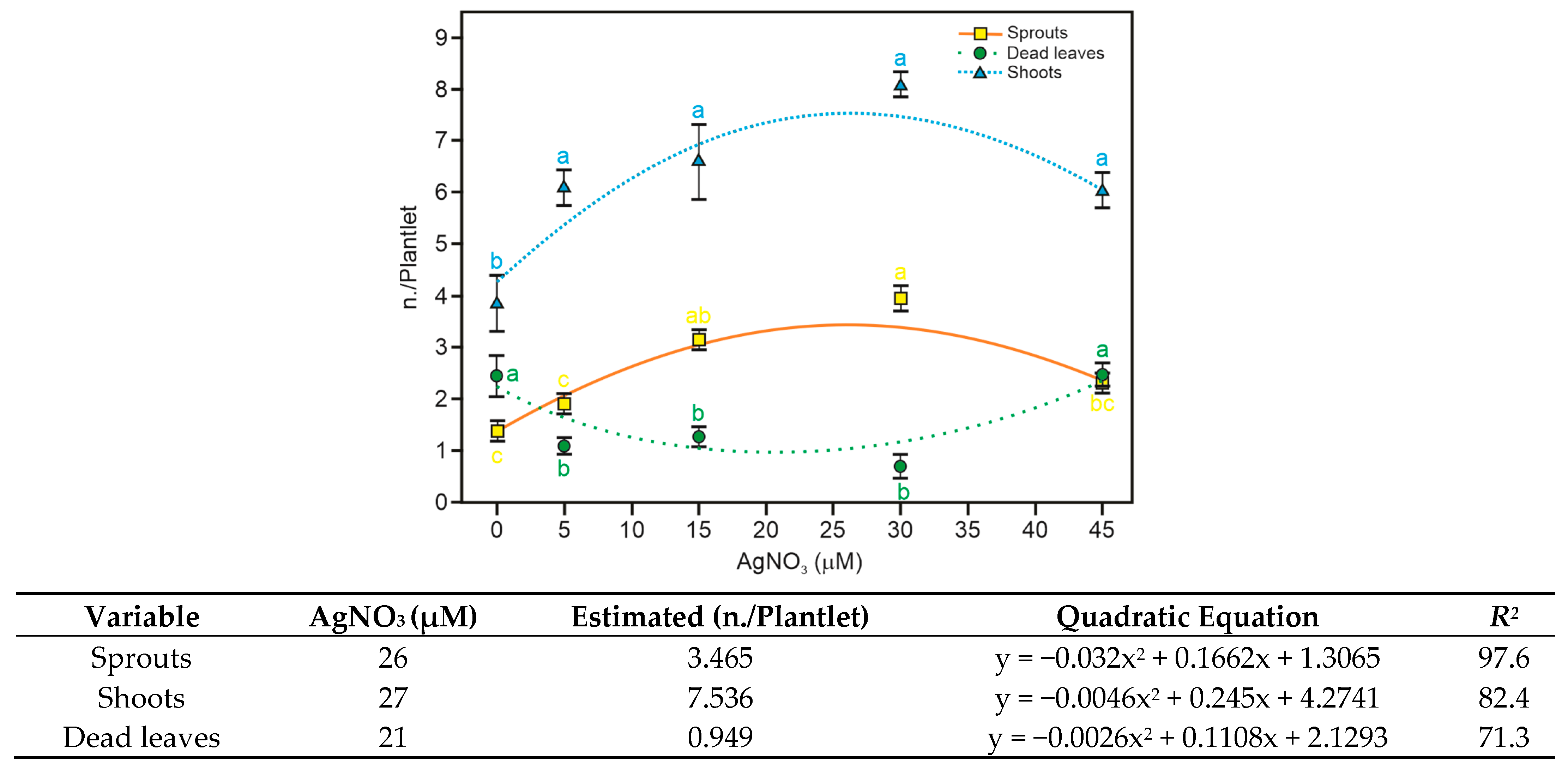
Based on the preliminary results, a second experiment was established to investigate the optimal concentration of AgNO3. The results revealed that this compound increased survival and sprouting in all tested concentrations. Even the highest concentration tested (45 µM) could not overcome plant resilience to the point of lethality (Figure 2) because the higher dosage was ineffective in reducing leaf senescence when compared to the experimental control (Figure 2). The AgNO3 significantly increased the shoot number up to a concentration of 15 µM, decreasing when the cultures were exposed to 45 µM (Figure 2). It also reduced the number of necrotic leaves at 5 and 15 µM. The regression analysis determined the ideal levels to reduce leaf senescence and to increase shoot regeneration rate, ranging from 21 µM to 27 µM. The results found in the present study are consistent with the theoretical models and practical results obtained by other scientific papers and reviews on the subject.
The silver ions are mainly referred to for their capability to inhibit the action of ethylene, which is also involved in the action of calcium signaling ions and polyamines related to organogenesis and embryogenesis [38]. Also, according to Gao et al. [39], silver ions, such as silver nitrate (AgNO3) or silver thiosulphate (STS), have been identified as inhibitors that block the action of ethylene receptors without affecting ethylene biosynthesis. The AgNO3 and other compounds can donate silver ions (like silver thiosulfate or silver nanoparticles) directly inhibiting the ethylene synthesis and accumulation over the whole plant, but are capable of generating insensitivity of the vegetal organism to it [40]. However, there is no consensus about the mode of action of silver-mediated ethylene blocking. Still, the most accepted hypothesis is that the silver ions might replace copper ions in the binding section of the ethylene receptor protein (ETR1), present in the endoplasmic reticulum of the cells, preventing the ethylene binding and signaling [41]. Therefore, the promotion of plant regeneration and growth described in the literature probably does not occur as a direct action of the silver ions, but as a mediation of the events that allow the signaling triggered for secondary metabolites once inhibited by the ethylene [42].
The addition of 2.0 mg·L−1 AgNO3 significantly enhanced shoot regeneration frequency, shoot number, and shoot length, indicating a promotion of in vitro plant regeneration [43]. Similar results were observed in tomato under 1.0 mg L−1 AgNO3, suggesting its positive role in enhancing both callus induction and plant regeneration [44]. In another way, Zhang et al. [45], supplementing the organogenesis medium with 12 mg L−1, sharply curtailed callusing in cassava, where callus frequency fell from 100% to 5% and large calli, with a similar dose-dependent suppression observed across cultivars. Coffea arabica in vitro plants exposed to 10 µM of AgNO3 showed enhanced shoot emission, leaf area, and chlorophyll content [46]. In Musa spp., shoot induction and rooting were improved in response to the addition of 59 µM of AgNO3 to the culture medium [47], while in Capsicum frutescens, the flowering, shoot length, and the number of shoots were promoted by 30 µM of AgNO3 [48]. To summarize, due to the improvements to in vitro regeneration and multiplication, its low cost, easy preparation, high solubility in water, as well as its low phytotoxicity, the use of 26 µM AgNO3 (Figure 2) to improve in vitro cultures in A. xanthorrhiza is the best result of our study.

3.3. Meta-Topolin Performs Better than 6-Benzilaminopurine in A. xanthorrhiza In Vitro Organogenesis

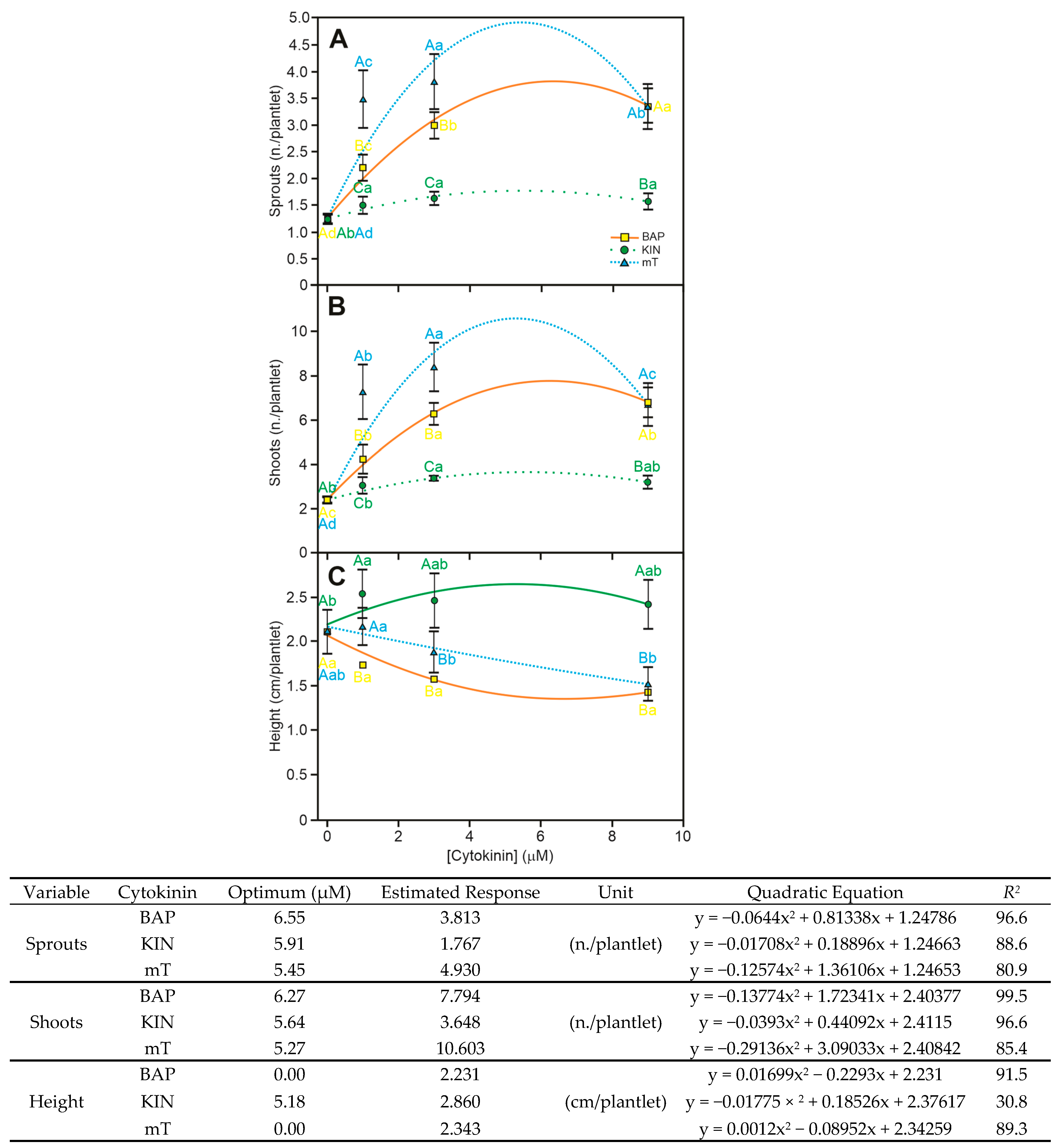
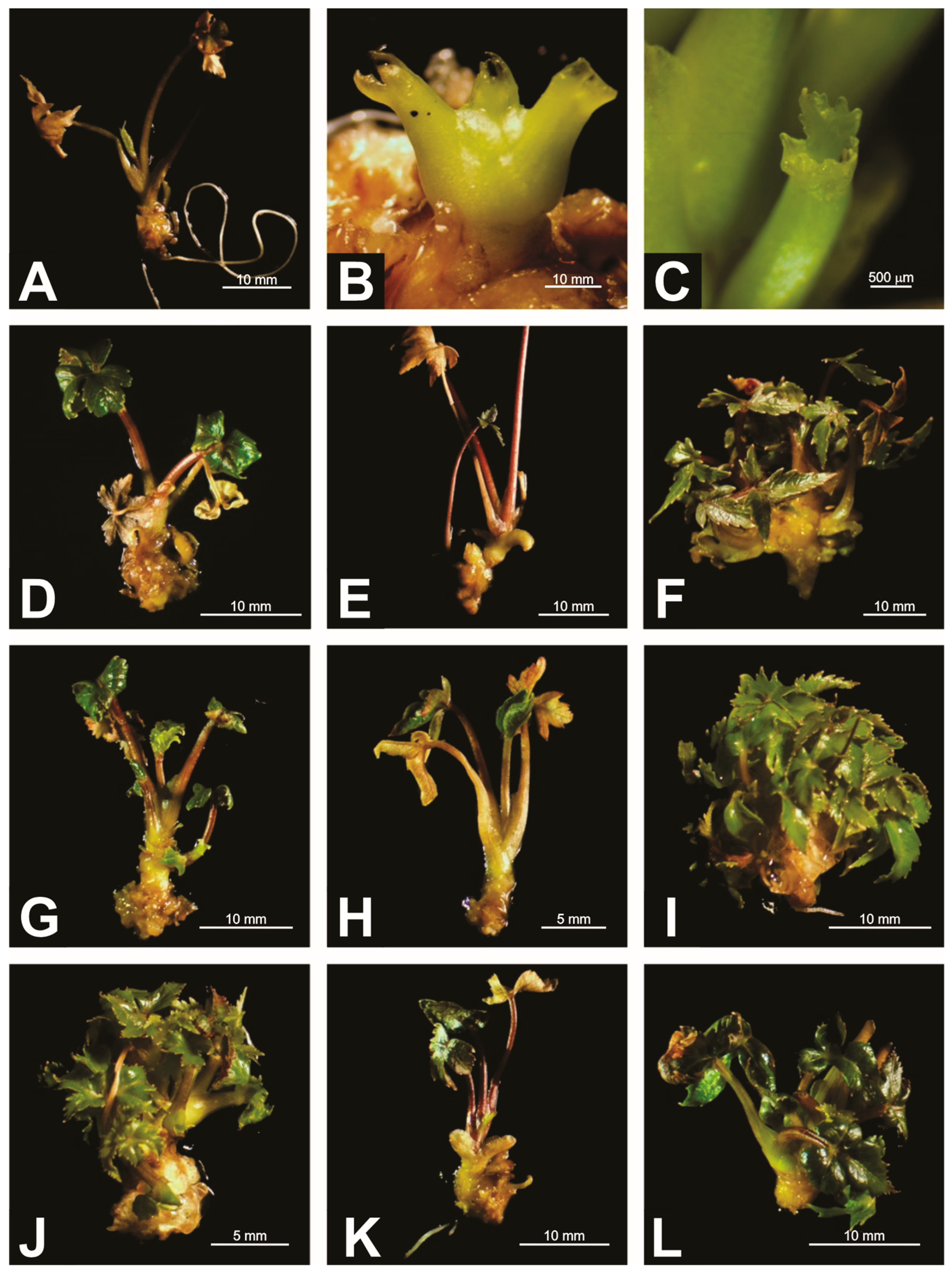
To induce organogenesis in A. xanthorrhiza, we tested four concentrations (0, 1, 3, and 9 µM) of BAP, KIN, and mT (Figure 3). A fifth concentration (27 µM) of the three growth regulators was also tested but discontinued due to the malformations induced in mT and BAP treatments, showing a predominance of leaves with a dramatic reduction of limb (Figure 4C). Regarding 0 to 9 µM, any impairments of survival were observed in any of the growth regulators under analysis in any of the tested dosages (general average of 98.76%).
Shoot development was significantly increased using mT (Figure 4B,F,I,L). The lower dosage (1 µM) resulted in the development of 3.5 sprouts (Figure 3A) and 7.2 leaves (Figure 3B) per plant, from which a concentration increase led to a minor enhancement in the regeneration rates without statistical differences ranging from 1 µM to 9 µM for both variables (Figure 3), demonstrating a dose-response pattern of saturation. The regression analysis inferred an optimal dosage of 4.45 µM of mT (y = −0.29136x2 + 3.09033x + 2.40842), associated with the mean development of 4.9 sprouts and 5.27 µM for 10.6 shoots regenerated.
To obtain statistically comparable results with mT, the BAP-treated cultures required a concentration three times higher (Figure 4D,G,J). The effects did not differ regarding the number of shoots regenerated after 3 µM (3 sprouts and 6.2 shoots). Still, they presented a reasonable fitness to the regression analysis, with an R2 of 96.6% for new sprouts and 99.52% for new shoots. The interpolation of the optimal concentration was calculated as 6.27 µM for a mean of 7.7 new shoots. The use of KIN (Figure 4E,H,K) did not equal or overcome the effectiveness of mT or BAP in the sprouting or number of leaves in any of the tested concentrations, except for the control concentration (PGR-free), with low statistical fitness for sprouting (R2 = 88.6%), only showing significance over the regression curve for shoot emissions (R2 = 96.6%).
With exceptions [49] focused on the use of auxins for in vitro cultures of A. xanthorrhiza, the investigations with this species employed BAP as the main plant growth regulator. The first reported best regeneration rate in response to 0.44 µM of BA and 0.11 µM of NAA [50]. Slíva et al. [13] reported for this species that in the multiplication phase, MS medium supplemented with 0.1 mg L−1 NAA + 1 mg L−1 BAP resulted in the highest shoot proliferation—an average of 4.2 ± 0.73 new shoots per explant within four weeks, significantly outperforming other treatments. In comparison, MS medium with 0.1 mg L−1 NAA + 2 mg L−1 BAP generated about 3 ± 0.63 shoots per explant, while 1 mg L−1 BAP alone yielded only 2.2 ± 0.37 shoots.
It is interesting to note that those first reports were centered on establishing a regeneration protocol from meristems to guarantee pathogen-free plants. Other researchers [13,14,18,51] focused on developing in vitro protocols for A. xanthorrhiza mass propagation. Madeira et al. [14] and Matos et al. [18], studying Brazilian and Venezuelan genotypes, respectively, reached the same conclusions about the most adequate concentration of BAP at 1.33 µM, supplemented with 0.54 µM of NAA, to induce a pattern of morphogenesis with low callus formation and maximum regeneration rate. Madeira et al. [14] also used GA3 (0.72 µM) in the culture medium, seeking to alleviate the dwarf pattern in plants cultured under BAP. Slíva et al. [13] showed that A. xanthorrhiza cultures reached the maximum regeneration rate in culture medium supplemented with BAP (4.44 µM) and NAA (0.54 µM).
The results presented in the present study reveal important points of consonance and deviation with those reported in the previous studies regarding the effects of cytokinin types and levels and their impact on A. xanthorrhiza in vitro morphogenesis (Table 3). Granting that, in a first effort, a curve of cytokinin concentrations was developed in an attempt to interpolate all the previous results in the literature, covering from 0 µM to 27 µM. It is remarkable that toxic levels of BAP and mT were encountered at the higher dosage. Vitamvas et al. [52] described that the combination of 0.1 mg L−1 NAA + 1 mg L−1 BAP yielded the highest multiplication rate, with an average of 4.2 ± 0.73 new shoots per explant in four weeks, and 100% of explants producing shoots. Also, 0.1 mg L−1 NAA + 2 mg L−1 BAP produced approximately 3 ± 0.63 shoots, while 1 mg L−1 BAP alone gave 2.2 ± 0.37 shoots. Regarding malformation, higher auxin levels, particularly 2,4-D at the rooting stage, led to excessive callus, poor leaf growth, browning, and eventual death, indicating malformation and reduced vigor. Still, Vitamvas et al. [52] demonstrated NAA treatments (1–2 mg L−1) that similarly produced excessive callus and up to 50% plantlet mortality within four weeks. For Slíva et al. [13], the highest shoot multiplication in A. xanthorrhiza was achieved with 1.0 mg L−1 BAP, inducing an average of 3.5 shoots per explant; however, increasing BAP to 2.0 mg L−1 or combining with NAA led to malformations such as hyperhydricity and abnormal leaf development. Treatments with 2,4-D, especially at higher concentrations, promoted excessive callus formation and inhibited organized shoot development, contributing to morphological disorders and loss of regenerative capacity. Madeira et al. [14] reported limitations in their protocol when using BAP levels higher than 1.33 µM with the same cultivar tested in this study, which leads to two possible and non-excluding interpretations. There is a positive interaction in the proposed salt supplementation, and/or there are possible epigenetic or even genetic differences between the used accessions. The major difference in detail from the past studies with micropropagation of A. xanthorrhiza is the regeneration rate in response to mT to induce the same number of sprouting and shoots with one-third of the BAP dosage. The mT and the BAP are aromatic cytokinins. Still, mT differs in the presence of a hydroxyl group bound to the benzyl ring, which is credited as the main factor for its enhanced biological activity in planta [53].
At the present time, the topolins are still costly, as compared to other aromatic and isoprenoid cytokinins; however, they have demonstrated superior performance in micropropagation by promoting efficient shoot multiplication and minimizing adverse effects typically caused by BAP. Unlike BAP, which is associated with hyperhydricity, phytotoxicity, chimera formation, and histogenetic instability, topolins maintain physiological stability and shoot quality. For instance, mT at concentrations around 1.0 to 2.0 mg L−1 has been shown to induce high shoot proliferation while significantly reducing the incidence of hyperhydricity and abnormal morphogenesis, offering a more stable alternative for in vitro culture systems [55]. In Piper hispidinervum, high concentrations of BAP (5.0 mg L−1) induced marked phytotoxic effects, including hyperhydricity and callus formation, while meta-topolin (mT) at the same concentration promoted better shoot morphology with reduced malformations. The use of 2.5 mg L−1 mT provided an optimal balance, yielding well-formed shoots with a lower incidence of physiological disorders compared to equivalent BAP treatments [56]. In Dianthus caryophyllus, the application of 1.0 mg L−1 mT significantly enhanced shoot height, leaf length, chlorophyll content, and dry matter index while preventing vitrification. In the same way, silica nanoparticles (SiNPs) at 1.0–1.5 mg L−1 promoted root development and plantlet survival, but higher concentrations (>2.0 mg L−1) induced stress and growth inhibition. Those advantages cannot be generalized to the A. xanthorrhiza micropropagation under mT stimulation, as malformations and height inhibition were observed in a very similar pattern to BAP after 3 µM. However, mT showed slightly higher averages and larger confidence intervals of response. The main benefit in the present work was observed at low dosages, with benefits associated with less epigenetic variation and reduced costs for regenerated clones.

4. Conclusions

The proposed improvements effectively addressed key challenges in the micropropagation of Arracacia xanthorrhiza through targeted interventions. Ampicillin (100 mg·L−1) achieved a 70% aseptic culture rate without phytotoxicity, confirming its low-cost effectiveness for bacterial control. Silver nitrate (AgNO3) at 26 µM improved shoot and sprout formation while reducing leaf senescence, supporting its role in ethylene inhibition and plantlet vigor. Meta-topolin (mT) at 1 µM outperformed BAP, producing three times higher organogenic activity, validating the potential of non-conventional cytokinins. However, further studies are needed to assess genetic fidelity and adapt the protocol to diverse genotypes. Future work should include tests on landraces and breeding lines, along with flow cytometry and molecular analyses, to ensure high-yield, true-to-type regeneration. These findings confirm that optimizing antimicrobial use, ethylene suppression, and cytokinin selection can significantly advance micropropagation protocols for this underutilized crop.

Author Contributions

P.D.M.: conceptualization, methodology, investigation, data curation, formal analysis, writing—original draft preparation; T.S.O.: methodology, data curation, formal analysis; Y.F.: methodology, data curation, formal analysis, writing—original draft preparation; C.A.C.: methodology, investigation; writing—original draft preparation; I.F.M.: methodology, investigation; V.M.S.: validation, visualization, writing—review and editing; M.F.P.: data curation, visualization, formal analysis, writing—review and editing, supervision; M.P.G.: conceptualization, methodology, data curation, validation, writing—review and editing, supervision, project administration, funding acquisition. All authors have read and agreed to the published version of the manuscript.

Funding

Brazilian National Council for Scientific and Technological Development (CNPq). Grants 304522/2023-6, and 407974/2018-0.

Data Availability Statement

All data were uploaded and are available for unrestricted access at https://1drv.ms/x/c/90670d53d2ee7f18/ER9_zdGJmP9Khy3D1-OR4V0B9kuDQqBrwkiEzBhKhcTaeg?e=dvF06l, accessed on 15 September 2025.

Acknowledgments

To the Research and Innovation Support Foundation of Santa Catarina State—Fapesc, SC, Brazil, for the grant awarded to MPG.

Conflicts of Interest

The authors declare no conflicts of interest.

References

  1. Andrade, N.J.P.; Sevillano, R.H.B.; Sørensen, M. Traditional uses, processes, and markets: The case of arracacha (Arracacia xanthorrhiza Bancroft). In Traditional Starch Food Products: Application and Processing, 4th ed.; Cereda, M.P., Vilpoux, O.F., Eds.; Academic Press: London, UK, 2025; pp. 325–332. [Google Scholar]
  2. EFSA. EFSA NDA Panel (EFSA Panel on Dietetic Products, Nutrition and Allergies). Scientific Opinion on the safety of Arracacia xanthorrhiza as a novel food. EFSA J. 2015, 13, 3954. [Google Scholar] [CrossRef]
  3. Sanchez-Portillo, S.; Salazar-Sánchez, M.D.R.; Campos-Muzquiz, L.G.; Ascacio-Valdés, J.A.; Solanilla-Duque, J.F.; Lopez Badillo, C.M.; Flores-Gallegos, A.C.; Rodríguez-Herrera, R. Proximal characteristics, phenolic compounds profile, and functional properties of Ullucus tuberosus and Arracacia xanthorrhiza. Explor. Foods Foodomics 2024, 2, 672–686. [Google Scholar] [CrossRef]
  4. Lescano, M.; Vásquez, N.; Vásquez, S.; Yoplac, I.; Velásquez-Barreto, F.K. Development and optimization of biofilms made from potato or arracacha starch. Starch 2021, 73, 2100075. [Google Scholar] [CrossRef]
  5. Morillo, E.; Sécond, G. Tracing the domestication of the Andean root crop arracacha (Arracacia xanthorrhiza Bancr.): A molecular survey confirms the selection of a wild form apt to asexual reproduction. Plant Genet. Resour. 2017, 15, 380–387. [Google Scholar] [CrossRef]
  6. Madeira, N.R.; Carvalho, A.D.F.; Silva, G.O.; Brotel, N.; Bortoletto, A.C. Mandioquinha-Salsa Arracacia xanthorrhiza Bancroft; Embrapa: Brasília, DF, Brazil, 2021. [Google Scholar]
  7. Embrapa. Mandioquinha-Salsa (Arracacia xanthorrhiza). Embrapa Hortaliças, Sistemas de Produção, 4. 2008. Available online: https://sistemasdeproducao.cnptia.embrapa.br/FontesHTML/Mandioquinha/MandioquinhaSalsa/apresentacao.html (accessed on 15 September 2025).
  8. Mesquita Filho, M.V.; Souza, A.F.; Silva, H.R.; Santos, F.F.; Oliveira, S.A. Adubação nitrogenada e fosfatada para produção comercializável de mandioquinhasalsa em latossolo vermelho amarelo. Hortic. Bras. 1996, 14, 211–215. [Google Scholar]
  9. Alencar, G. Cientistas Desenvolvem Variedades de Mandioquinha-Salsa Que Produzem até 80% Mais Que a Cultivar Tradicional. Embrapa News. 2018. Available online: https://encurtador.com.br/hkYnL (accessed on 15 September 2025).
  10. Macedo, A. Mandioquinha-Salsa: Embrapa Multiplica Ações em Novas Áreas de Cultivo no Paraná. Embrapa News. 2019. Available online: https://encurtador.com.br/zwKVC (accessed on 15 September 2025).
  11. Fletcher, P.J.; Fletcher, J.D. In vitro virus elimination in three Andean root crops: Oca (Oxalis tuberosa), ulluco (Ullucus tuberosus), and arracacha (Arracacia xanthorrhiza). N. Z. J. Crop Hort. Sci. 2001, 29, 23–27. [Google Scholar] [CrossRef]
  12. Abdalla, N.; El-Ramady, H.; Seliem, M.K.; El-Mahrouk, M.E.; Taha, N.; Bayoumi, Y.; Shalaby, T.A.; Dobránszki, J. An academic and technical overview on plant micropropagation challenges. Horticulturae 2022, 8, 677. [Google Scholar] [CrossRef]
  13. Slíva, Š.; Viehmannová, I.; Vítámvás, J. Micropropagation and morphogenesis of arracacha (Arracacia xanthorrhiza Bancroft). Agric. Trop. Subtrop. 2010, 43, 206–211. [Google Scholar]
  14. Madeira, N.R.; Santos, F.F.; Souza, R.J. Desempenho de clones de mandioquinha-salsa (Arracacia xanthorrhiza Bancroft) na região de Lavras-MG. Ciênc. Agrotec. 2002, 26, 711–718. [Google Scholar]
  15. Manchanda, P.; Gosal, S.S. Effect of activated charcoal, carbon sources and gelling agents on direct somatic embryogenesis and regeneration in sugarcane via leaf roll segments. Sugar Tech. 2012, 14, 168–173. [Google Scholar] [CrossRef]
  16. Khatoon, N.; Alam, H.; Khan, A.; Raza, K.; Sardar, M. Ampicillin silver nanoformulations against multidrug resistant bacteria. Sci. Rep. 2019, 9, 6848. [Google Scholar] [CrossRef] [PubMed]
  17. Madeira, N.R. Micropropagação e Indexação de Mandioquinha-Salsa. Ph.D. Thesis, Universidade Federal de Lavras, Lavras, MG, Brazil, 2004. [Google Scholar]
  18. Matos, E.; Marcano, M.; Azócar, C.J.; Mora, A. Establecimiento y multiplicación in vitro de cinco cultivares de apio (Arracacia xanthorrhiza Bancroft) colectados en Venezuela. Bioagro 2015, 27, 121–130. [Google Scholar]
  19. Staba, E.J. Plant tissue culture as a technique for the phytochemist. In Recent Advances in Phytochemistry; Seikel, M.K., Runcekles, V.C., Eds.; Appleton-Century-Crofts: New York, NY, USA, 1969; Volume 2, pp. 75–106. [Google Scholar]
  20. Vrundha, C.P.K.; Aswathi, N.V.; Thomas, T.D. The role of meta-topolin in plant morphogenesis in vitro. In Meta-Topolin: A Growth Regulator for Plant Biotechnology and Agriculture; Ahmad, N., Strnad, M., Eds.; Springer Nature Singapore Pte Ltd.: Singapore, 2021; pp. 93–118. [Google Scholar]
  21. Vasconcellos, E.S.; Cruz, C.D.; Regazzi, A.J.; Bhering, L.L.; Rosado, T.B.; Vasconcelos, F.S. Regression models grouping for genotype adaptability and stability analysis. Pesqui. Agropecu. Bras. 2010, 45, 1357–1362. [Google Scholar]
  22. Lozano-Isla, F.; Benites Alfaro, O.; Santana, D.G.; Ranal, M.A.; Pompelli, M.F. Germination Indexes for Seed Germination Variables for Ecophysiological Studies [R Package GerminaR Version 1.4.2]; R Development Core Team: Kaysville, UT, USA, 2020. [Google Scholar]
  23. Rusu, A.; Buta, E.L. The development of third-generation tetracycline antibiotics and new perspectives. Pharmaceutics 2021, 13, 2085. [Google Scholar] [CrossRef] [PubMed]
  24. Mitchell, A.S.M.; Ullman, J.L.; Teel, A.L.; Watts, R.J. pH and temperature effects on the hydrolysis of three β-lactam antibiotics: Ampicillin, cefalotin and cefoxitin. Sci. Total Environ. 2014, 466–467, 547–555. [Google Scholar] [CrossRef] [PubMed]
  25. Guerra, W.; Silva-Caldeira, P.P.; Terenzi, H.; Pereira-Maia, E.C. Impact of metal coordination on the antibiotic and non-antibiotic activities of tetracycline-based drugs. Coord. Chem. Rev. 2016, 327–328, 188–199. [Google Scholar] [CrossRef]
  26. Romadanova, N.V.; Tolegen, A.B.; Kushnarenko, S.V.; Zholdybayeva, E.V.; Bettoni, J.C. Effect of plant preservative mixtureTM on endophytic bacteria eradication from in vitro-grown apple shoots. Plants 2022, 11, 2624. [Google Scholar] [CrossRef]
  27. Leone, G.F.; Almeida, C.V.; Abreu-Tarazi, M.F.; Batagin-Piotto, K.D.; Artioli-Coelho, F.A.; Almeida, M.D. Antibiotic therapy in Pineapple (Ananas comosus) microplants. Ciên. Rural 2016, 46, 89–94. [Google Scholar] [CrossRef]
  28. El-Banna, A.N.; El-Mahrouk, M.E.; Dewir, Y.H.; Farid, M.A.; Abou Elyazid, D.M.; Schumacher, H.M. Endophytic bacteria in banana in vitro cultures: Molecular identification, antibiotic susceptibility, and plant survival. Horticulturae 2021, 7, 526. [Google Scholar] [CrossRef]
  29. Khan, T.; Abbasi, B.H.; Iqrar, I.; Khan, M.A.; Shinwari, Z.K. Molecular identification and control of endophytic contamination during in vitro plantlet development of Fagonia indica. Acta Physiol. Plant 2018, 40, 150. [Google Scholar] [CrossRef]
  30. Palú, E.G.; Corrêa, L.S.; Suzuki, A.N.; Boliani, A.C. Uso de antibióticos para o controle de bactérias endógenas visando à micropropagação da figueira (Ficus carica L.). Rev. Bras. Frutic. 2011, 33, 587–592. [Google Scholar] [CrossRef]
  31. Pollock, K.; Barfield, D.G.; Shields, R. The toxicity of antibiotics to plant cell cultures. Plant Cell Rep. 1983, 2, 36–39. [Google Scholar] [CrossRef]
  32. Pereira, J.E.S.; Fortes, G.R.L. Antibiotics toxicity on the in vitro potato cultivation in semi-solid and liquid media. Pesqui. Agropecu. Bras. 2003, 38, 1273–1279. [Google Scholar] [CrossRef]
  33. Salehi, H.; Khosh-Khui, M. A simple procedure for disinfection of ‘Baby Masquerade’ miniature rose explants. Sci. Hort. 1997, 68, 145–148. [Google Scholar] [CrossRef]
  34. Thomas, P. In vitro decline in plant cultures: Detection of a legion of covert bacteria as the cause for degeneration of long-term micropropagated triploid watermelon cultures. Plant Cell Tiss. Org. Cult. 2004, 77, 173–179. [Google Scholar] [CrossRef]
  35. Najafi, F.; Mohammadi, S.; Asgharian, P.; Kosari-Nasab, M. The impact of activated charcoal and graphite on growth parameters and production of secondary metabolites of Plantago maritima through in vitro culture. BMC Chem. 2025, 19, 222. [Google Scholar] [CrossRef] [PubMed]
  36. Sáenz, L.; Herrera-Herrera, G.; Uicab-Ballote, F.; Chan, J.L.; Oropeza, C. Influence of form of activated charcoal on embryogenic callus formation in coconut (Cocos nucifera). Plant Cell Tiss. Organ. Cult. 2010, 100, 301–308. [Google Scholar] [CrossRef]
  37. Thomas, T.D. The role of activated charcoal in plant tissue culture. Biotechnol. Adv. 2008, 26, 618–631. [Google Scholar] [CrossRef]
  38. Kumar, V.; Parvatam, G.; Ravishankar, G.A. AgNO3: A potential regulator of ethylene activity and plant growth modulator. Electron. J. Biotechnol. 2009, 12, 1–15. [Google Scholar] [CrossRef]
  39. Gao, H.; Xia, X.; An, L.; Xin, X.; Liang, Y. Reversion of hyperhydricity in pink (Dianthus chinensis L.) plantlets by AgNO3 and its associated mechanism during in vitro culture. Plant Sci. 2017, 254, 1–11. [Google Scholar] [CrossRef]
  40. Husak, V.V.; Stambulska, U.Y.; Pitukh, A.M.; Lushchak, V.I. Exposure of Paulownia seedlings to silver nitrate improves growth parameters via stimulation of mild oxidative stress. Agric. Conspec. Sci. 2024, 89, 209–218. [Google Scholar]
  41. Schaller, G.E.; Binder, B.M. Inhibitors of ethylene biosynthesis and signaling. In Ethylene Signaling. Methods in Molecular Biology; Binder, B., Eric Schaller, G., Eds.; Humana Press: New York, NY, USA, 2017; Volume 1573, pp. 223–235. [Google Scholar]
  42. Mahendran, D.; Geetha, N.; Venkatachalam, P. Role of silver nitrate and silver nanoparticles on tissue culture medium and enhanced the plant growth and development. In In vitro Plant Breeding Towards Novel Agronomic Traits; Kumar, M., Muthusamy, A., Kumar, V., Bhalla-Sarin, N., Eds.; Springer: Singapore, 2019; pp. 59–74. [Google Scholar]
  43. Türkoğlu, A.; Haliloğlu, K.; Demirel, F.; Aydin, M.; Çiçek, S.; Yiğider, E.; Demirel, S.; Piekutowska, M.; Szulc, P.; Niedbała, G. Machine learning analysis of the impact of silver nitrate and silver nanoparticles on wheat (Triticum aestivum L.): Callus induction, plant regeneration, and DNA methylation. Plants 2023, 12, 4151. [Google Scholar] [CrossRef] [PubMed]
  44. Enfeshi, N.; Abdulali, E.; Salama, M.; Geath, Z.; Shaaban, A.; Ben Saad, Z.; Abughnia, E. Effect of silver nitrate (AgNO3) and copper sulphate (CuSO4) on callus formation and plant regeneration from two pepper varieties (Chile Ancho and Misraty) in vitro. Sci. J. Fac. Sci. Sirte Univ. 2023, 3, 150–157. [Google Scholar]
  45. Zhang, P.; Phansiri, S.; Puonti-Kaerlas, J. Improvement of cassava shoot organogenesis by the use of silver nitrate in vitro. Plant Cell Tiss. Org. Cult. 2001, 67, 47–54. [Google Scholar] [CrossRef]
  46. Giridhar, P.; Indu, E.P.; Vijaya Ramu, D.; Ravishankar, G.A. Effect of silver nitrate on in vitro shoot growth of Coffee. Trop. Sci. 2003, 43, 144–146. [Google Scholar] [CrossRef]
  47. Tamimi, S.M. Effects of ethylene inhibitors, silver nitrate (AgNO3), cobalt chloride (CoCl2) and aminooxyacetic acid (AOA), on in vitro shoot induction and rooting of banana (Musa acuminata L.). Afr. J. Biotechnol. 2015, 14, 2511–2516. [Google Scholar] [CrossRef]
  48. Sharma, A.; Kumar, V.; Giridhar, P.; Ravishankar, G.A. Induction of in vitro flowering in Capsicum frutescens under the influence of silver nitrate and cobalt chloride and pollen transformation. Electron. J. Biotechnol. 2008, 11, 84–89. [Google Scholar] [CrossRef]
  49. Lopes, F.S. Morfogênese In Vitro Como Auxílio ao Melhoramento da Mandioquinha-Salsa (Arracacia xanthorrhiza Bancroft). Master’s Thesis, Universidade Federal do Espírito Santo, Vitória, ES, Brazil, 2009. [Google Scholar]
  50. Senna Neto, N. Micropropagação de Mandioquinha-Salsa (Arracacia xanthorrhiza Bancroft); Universidade Federal de Viçosa: Viçosa, MG, Brazil, 1990. [Google Scholar]
  51. Landazuri, P. Introduccion In Vitro, Micropropagacion, Conservacion y Aclimatizacion de Zanahoria Blanca (Arracacia xanthorrhiza) y Achira (Canna edulis); Pontificia Universidad Catolica del Ecuador: Quito, Ecuador, 1996. [Google Scholar]
  52. Vitamvas, J.; Viehmannova, I.; Cepkova, P.H.; Mrhalova, H.; Eliasova, K. Assessment of somaclonal variation in indirect morphogenesis-derived plants of Arracacia xanthorrhiza. Pesqui. Agropecu. Bras. 2019, 54, e00301. [Google Scholar] [CrossRef]
  53. Karwasra, R.K.; Nazir, R.; Pandey, D.K. Meta-topolin mediated improved micropropagation, phytochemical profiling, and assessment of genetic fidelity in Chlorophytum borivilianum Sant. et Fernand., an endangered medicinal herb. Ind. Crop. Prod. 2024, 214, 118463. [Google Scholar] [CrossRef]
  54. Luz, J.M.Q. Obtenção In Vitro de Plantas de Mandioquinha-Salsa (Arracacia xanthorrhiza Bancroft) via Cultura de Meristemas. Master’s Thesis, Universidade Federal de Lavras, Lavras, MG, Brazil, 1993. [Google Scholar]
  55. Abdouli, D.; Placková, L.; Dolezal, K.; Bettaieb, T.; Werbrouck, S.P.O. Topolin cytokinins enhanced shoot proliferation, reduced hyperhydricity and altered cytokinin metabolism in Pistacia vera L. seedling explants. Plant Sci. 2022, 322, 111360. [Google Scholar] [CrossRef]
  56. Ríos-Ríos, A.M.; Silva, J.V.S.; Fernandes, J.V.M.; Batista, D.S.; Silva, T.D.; Chagas, K.; Pinheiro, M.V.M.; Faria, D.V.; Otoni, W.C.; Fernandes, S.A. Micropropagation of Piper crassinervium: An improved protocol for faster growth and augmented production of phenolic compounds. Plant Cell Tiss. Organ. Cult. 2019, 137, 495–509. [Google Scholar] [CrossRef]
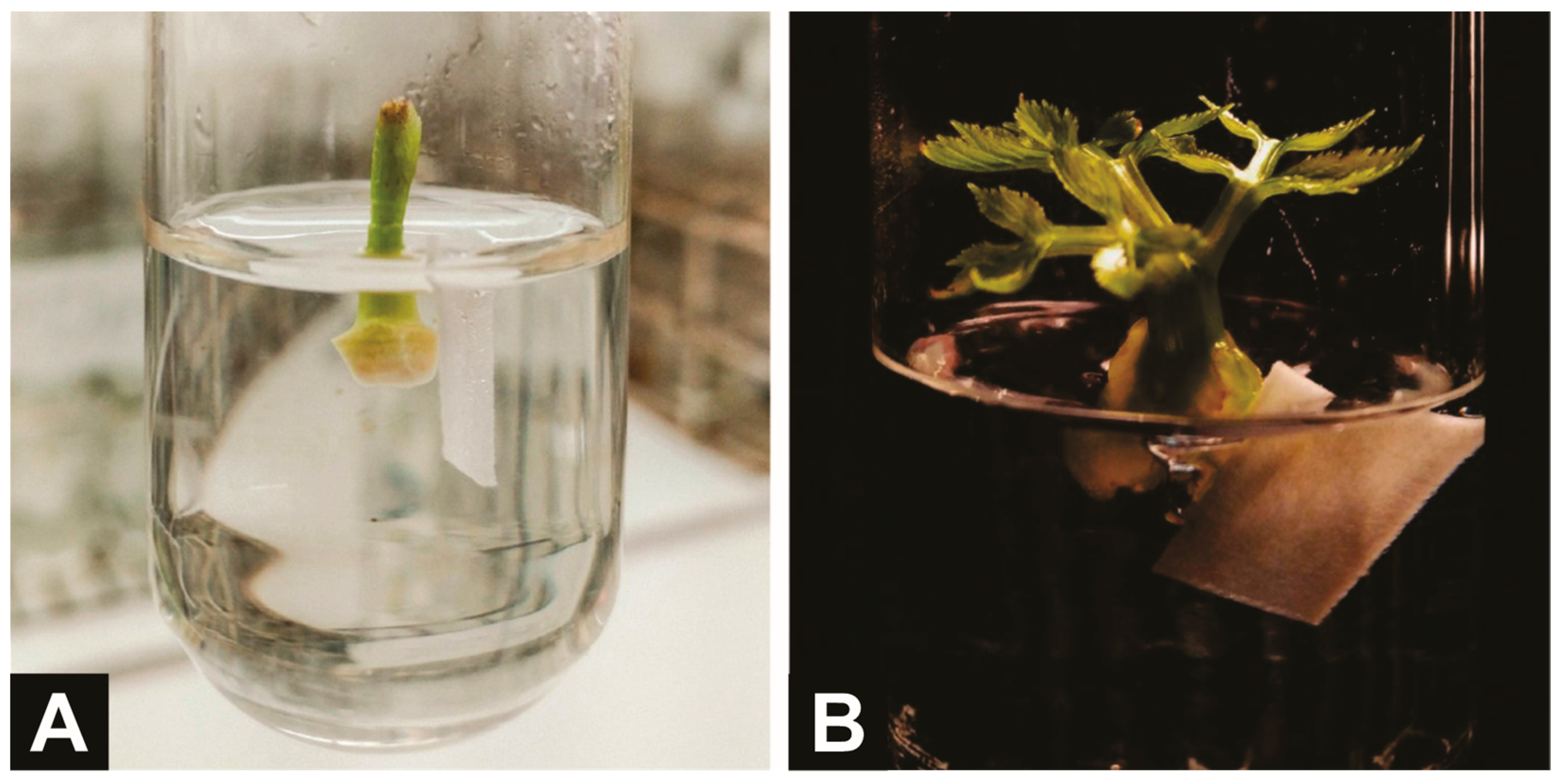
Figure 1. Cormels of A. xanthorrhiza inoculated in vitro under 100 mg L−1 ampicillin at time zero (A), and aseptic cornels regenerating new A. xanthorrhiza plants after 21 days of culture (B).
Horticulturae 11 01134 g001
Figure 2. Regression analysis with estimated response to sprouts, shoots (B), and dead leaves, and quadratic equation, coefficient of determination, and ANOVA’s coefficient of variation (Table) of A. xanthorrhiza exposed different doses (µM) of AgNO3 in the culture medium. Means followed by the same letters in the same plant growth regulator do not differ according to the Newman–Keuls mean separation test at 5% probability of error.
Horticulturae 11 01134 g002
Figure 3. Regression analysis with estimated response to sprouts (A) shoots (B), and height (C), and quadratic equation, coefficient of determination, and ANOVA’s coefficient of variation (Table) of in vitro cultures of A. xanthorrhiza exposed to distinct cytokinins—6-benzylaminopurine (BAP), kinetin (KIN) and meta-topolin (mT)—different doses (µM) in the culture medium. Means followed by the same letters in the same column do not differ according to the Newman–Keuls mean separation test at 5% probability of error.
Horticulturae 11 01134 g003
Figure 4. Morphological aspects of A. xanthorrhiza in vitro plantlets cultivated under different dosages 6-benzylamylopurine (BAP), kinetin (KIN) and meta-topolin (mT): (A) experimental control (no cytokinins supplementation); (B) emerging sprout feasible of isolation to form a new in vitro propagule; (C) limb malformation induced by 27 µM BAP; (D) 1 µM BAP; (E) 1 µM KIN; (F) 1 µM mT; (G) 3 µM BAP; (H) 3 µM KIN; (I) 3 µM mT; (J) 9 µM BAP; (K) 9 µM KIN; (L) 9 µM mT.
Horticulturae 11 01134 g004
Table 1. Averages of A. xanthorrhiza newly in vitro introduced cultures exposed to different antibiotics: survival (%), and contamination with bacteria, fungi, and total (%).
TreatmentSurvival (%) **Bacteria (B %) **Fungi (F %) **B + F (%) **
Control69.3 ± 5.2 b *65.6 ± 5.8 a6.0 ± 3.5 a8.2 ± 1.0 a
Ampicillin92.4 ± 7.8 a25.2 ± 1.8 b4.1 ± 1.9 a1.3 ± 0.1 c
Tetracycline92.8 ± 6.7 a61.2 ± 6.5 a2.5 ± 1.6 a4.6 ± 0.3 b
Average (%)84.8 ± 1350.7 ± 19.34.1 ± 2.94.7 ± 3.0
CV%15.338.168.963.7
* Means followed by the same letters in the same column do not differ according to the Newman–Keuls mean separation test at 5% probability of error. ** Variable data transformed to A r c s e n Y i + 0.5 100 during analysis.
Table 2. Survival (%), sprouts, and shoots (n./plantlet) of in vitro cultures of A. xanthorrhiza with supplementation of different growth inhibitors suppressors in the culture medium.
TreatmentSurvival (%)Sprouts (n./Plantlet)Shoots (n./Plantlet)
Control78.5 ± 3.5 b *2.1 ± 0.1 b2.7 ± 0.1 b
AgNO3 (10 µM)97.2 ± 2.4 a3.0 ± 0.4 a4.6 ± 0.2 a
Activated charcoal (1.75 g L−1)70.2 ± 5.5 c1.2 ± 0.3 c1.7 ± 0.3 c
General Average (%)82.0 ± 12.22.1 ± 0.83.0 ± 1.2
CV (%)14.935.641.4
* Means followed by the same letters in the same column do not differ according to the Newman–Keuls mean separation test at 5% probability of error.
Table 3. Saline formulations and growth regulators are reported as the best combinations for the establishment and multiplication of A. xanthorrhiza organogenic cultures compared to the presented results.
ReportSaline FormulationNAABAPmTGA3
Senna Neto (1990) [50]B50.110.44--
Luz (1993) [54]B50.570.88-0.72
Landázuri (1996) [51]MS0.2724.86--
Madeira (2002) [14]B50.541.33-0.72
Slíva et al. (2010) [13]MS0.544.44--
Matos et al. (2015) [18]B50.541.33--
Presented resultsB50.51--
Disclaimer/Publisher’s Note: The statements, opinions and data contained in all publications are solely those of the individual author(s) and contributor(s) and not of MDPI and/or the editor(s). MDPI and/or the editor(s) disclaim responsibility for any injury to people or property resulting from any ideas, methods, instructions or products referred to in the content.