Article

Exploring the Role of Vertical and Horizontal Pathways in the Formation of Lettuce Plant Endospheric Bacterial Communities: A Comparative Study of Hydroponic and Soil Systems

by
Polina Kuryntseva
*,
Nataliya Pronovich
*,
Gulnaz Galieva
,
Polina Galitskaya
and
Svetlana Selivanovskaya
Institute of Ecology, Biotechnology and Nature Management, Kazan (Volga Region) Federal University, Kazan 420008, Russia
*
Authors to whom correspondence should be addressed.
Horticulturae 2025, 11(7), 762; https://doi.org/10.3390/horticulturae11070762
Submission received: 20 May 2025 / Revised: 19 June 2025 / Accepted: 25 June 2025 / Published: 2 July 2025
(This article belongs to the Section Vegetable Production Systems)

Abstract

Plant-associated microbiomes play a critical role in plant health, nutrition, growth, and adaptation. This study aimed to investigate the formation pathways of the endospheric microbiome in lettuce (Lactuca sativa) through vertical (seed) and horizontal (substrate) transmission in hydroponic and soil environments. The bacterial microbiomes from the seeds, roots, leaves, and substrates were analyzed via 16S rRNA gene sequencing. The seed microbiome contained 236 OTUs dominated by Verrucomicrobia (31%) and Firmicutes (29%). Rhizospheric soil contained 1594 OTUs, while the hydroponic solution had 448 OTUs. The root endosphere from soil-grown lettuce contained 295 OTUs, compared with 177 in hydroponic conditions, and the leaf microbiome contained 43 OTUs in soil and 115 OTUs in hydroponics. In total, 30–51% of the leaf and root microbiomes originated from the seed microbiota, while 53–65% of the root microbiome originated from the substrate. Microbiome overlap was observed between the rhizospheric soil and the root microbiome. This study provides new insights into the microbiome of lettuce seeds and the pathways of formation of the endospheric microbiome in adult plants. These findings lay the groundwork for future research aimed at better understanding microbiome dynamics in leafy crops and plant protection.

Graphical Abstract

1. Introduction

Microorganisms (fungi, bacteria, and archaea [1]) play a significant role in the development and functioning of plants, leading to the recent conceptual emergence of the holobiont—a system comprising a plant host and microbial consortia residing inside and on its surfaces [2]. Microorganisms play a role in plant nutrition, adaptation to environmental conditions, disease control, growth and maturation, and reproduction [3,4,5]. Plant-associated microbial communities are divided into the endospheric microbiome, which may differ depending on plant compartment, and the epiphytic microbiome, located in the above-ground (phylosphere) or underground (rhizosphere) parts of the plant.
Recently, it has been shown that the composition and network of plant-associated microbial communities is not random [6]. Thus, plants of the same species growing in different conditions have been found to have the same microorganisms—the so-called core microbiome [7,8]. At the same time, the same microorganisms are found in the endophytic microbiomes of plants of different species growing under the same conditions, such as salinity or pollution, and apparently play a role in adaptation to such conditions [9]. Many questions about the formation of microbial communities associated with plants still remain open. The generally accepted idea is that there are two main pathways [10]—vertical transfer of part of the parental microbiome (parental) through seeds or vegetative organs [11], or horizontal transfer from the environment, subdivided into random (through surface damage) or directed (a result of targeted recruitment of microorganisms from the environment) [12]. These communities’ compositions from both horizontal and vertical sources are subject to abiotic factors like the physical and chemical composition of the substrate, temperature, humidity, light exposure, atmospheric dust, and wind [1,13,14,15].
Omics technologies have debunked the earlier hypothesis that the endosphere of seeds is sterile [16]. Some species from seeds (temporary, occasional) are not transmitted to seedlings, while others (persistent) are transmitted [17,18]. The endospheric microbiome of a plant (Arabidopsis thaliana, wheat, tomato, maize, rice, barley, sunflower, beans) originates from the endophytic microbiome of its seeds [19,20]. Notably, bacterial endophytes in maize seeds not only colonize other plant tissues but traverse the endosphere, exiting roots to colonize the rhizosphere [21,22]. Some authors hypothesize that in cases of vertical symbiont transmission, if the entire life cycle occurs within the host plant, the symbiont may not be adapted to survive in the external environment, leading to mutualistic relationships [23]. It is noteworthy variability in microbial communities occurs among different seed tissues (e.g., seed coat, embryo, endosperm, perisperm), explaining differences in the endospheric microbiome among various organs and tissues of a single plant [20]. For instance, certain authors report that endophytes of the genera Pantoea, Enterobacter, and Pseudomonas are characteristic of the seed embryo [15,22,24,25,26,27,28,29].
Horizontal transfer can occur both randomly, such as in cases of plant injury, and deterministically, facilitated by the attraction of microorganisms possessing specific properties via plant root exudates or endospheric species [30]. Endophytes can be horizontally acquired from soil, air, and water environments. Additionally, they may be introduced by animals during seed transport and, further, by humans during seed processing [15,31,32]. The proportion of endophytes entering plants through horizontal pathways is a subject of debate. Some researchers argue that horizontal transfer is the primary colonization route for plants [33]. This assertion is supported by experiments involving plant growth in a sterile environment and an environment inoculated, for example, with soil microbial communities [34]. However, a sterile gnotobiotic environment represents a highly simplified system that does not account for the competitive dynamics between endophytic microorganisms and the biome of the surrounding environment [35,36,37]. The ability to recruit a diverse set of symbionts from the environment may confer advantages for sessile organisms like plants, providing adaptive responses to changing environmental conditions [38]. Indeed, plants appear to harbor a wide array of universal endophytes, the presence or absence of which depends more significantly on the plant’s environment (e.g., soil type) than its genotype at a given time [39,40,41,42,43,44].
However, an opposing viewpoint exists, suggesting that the horizontal colonization path necessitates intricate mechanisms due to the need to find entry points for microorganisms into the internal portions of the host plant (such as wounds, cracks, stomata, penetration paths through root hairs, and between epidermal cells), along with signaling molecule systems and quorum sensing systems [14]. Additionally, plants exhibit biological filtration capabilities, restricting the penetration of soil microbes [15].
Distinguishing between endospheric and exospheric microorganisms associated with plants involves categorization based on their localization—whether in underground or aboveground parts—and the specific plant tissues (and the type of plant tissues). Some microorganisms inhabit both the interior and exterior of the plant simultaneously (arbuscular mycorrhiza) or migrate between these environments [45].
Not all plants have been studied in terms of the composition and formation of their microbiome. Furthermore, the scrutiny of plant-associated bacteria has been considerably lower compared with fungi, given the higher prevalence of uncultured species in the latter group. The focus of research has primarily concentrated on rhizospheric bacteria and fungi, with limited diversity across plant species. Among the well-studied species are rice [46,47], grapevine [48], tomato [49], aloe [50], and peony [51].
The methods of studying the endosphere microbiome are divided into two groups, based on the study of the cultivated microbiome and the use of omics approaches [52,53]. Most studies have focused on the cultured microbiome, driven by the notion of subsequent application of microorganisms as biocontrol agents or growth stimulators, thus rendering non-cultured species less relevant for research. The uncultured microbiome also warrants investigation, as it constitutes a substantial portion of the microbial community. Consequently, only 1% of endophytic microbes have been cultivated and biotechnologically explored to date [52,54].
There is a paucity of data on the endophytic microbiome of plants consumed by humans in raw (unprocessed) form. However, the presence of microorganisms from fruits and vegetables in the human gut microbiome has been demonstrated [55]. Lettuce is one of the most frequently consumed fresh vegetables (as a source of fiber, vitamins, antioxidants, and other nutrients); it is easily grown in open and closed ground on various substrates [56]. However, its endosphere microbiome has not been sufficiently studied [34,57,58,59].
To identify the dominant plant colonization pathway, researchers conduct two fundamental types of experiments, growing plants in a sterile environment (followed by inoculation with a known consortium of species) and comparing endospheric microbiomes of offspring plants from the same parent grown in different conditions (different soil types), albeit without considering the parent’s seed microbiome [60,61]. Both approaches have their advantages and drawbacks. The former allows the identification of temporary and persistent species in the seed microbiome and the determination of which seed tissues serve as sources of the microbiome for various plant organs. However, it does not consider competitive interactions between vertically and horizontally acquired communities. In contrast, the latter assesses the endospheric microbiome under natural conditions, including its shared component among related plants cultivated under different conditions (the so-called core microbiome). Nevertheless, it does not distinguish between vertical and horizontal sources.
This study aimed to design an experiment that combined the analysis of the microbiome of seeds with the study of the endospheric microbiome of plants grown on various substrates, as well as an examination of the microbiomes of these substrates themselves. This work employed lettuce plants, which can be consumed raw and potentially influence the human gut microbiome and human health.

2. Materials and Methods

2.1. Seed Preparation

Lettuce seeds (Lactuca sativa var. crispa L.), cultivar Ozornik, produced by Agrofirm AELITA LLC (Moscow, Russia), were selected for the experiment. To assess the endophytic microbiome of the seeds, surface sterilization was carried out. For this, the lettuce seeds were treated with 2.5% sodium hypochlorite for 2 min, rinsed in sterilized tap water, and immersed in 70% ethanol for 3 min. Sterile seeds were thoroughly washed five times with sterile tap water. The last wash water was sown on Petri dishes with LB agar [62]. In the absence of growth of bacterial or fungal colonies, the seed sterilization process was considered successful and these seeds were used for subsequent DNA extraction from the endosphere microbiome.

2.2. Vegetation Experiment

The experiments were conducted in a greenhouse at the Kazan Federal University, Russia. The process of formation of the endosphere microbiome was analyzed in two different substrates—hydroponic solution and soil. In both cases, mineral fertilizers were added to maintain the optimal composition of micro- and macroelements in the soil and hydroponic solution. The hydroponic system was made up of three separate 12 L plastic tanks (water depth 19 cm) for each variant, with a polystyrene tray hosting 9 lettuce plants(18 plants in total). The hydroponic water contained the following macronutrients: N-NO3 (14.0 mM), N-NH4+ (2.0 mM), P (2.0 mM), K (10.0 mM), Ca (4.5 mM), Mg (2.0 mM), and S-SO4. (5.0 mM) [59]. The following salts were used to prepare the hydroponic solution: Ca(NO3)2∙2H2O, NH4NO3, KH2PO4, MgSO4∙7H2O, KNO3, and K2SO4. Every 7 days, the hydroponic mixtures were replaced with fresh ones. Micronutrients were added to the hydroponic system in the form of mineral salts, including iron (II) sulfate heptahydrate, zinc (II) sulfate heptahydrate, copper (II) sulfate pentahydrate, manganese (II) sulfate monohydrate, orthoboric acid, and hexaammonium molybdate. The micronutrients’ content in the solution was B—53.6, Cu—4.6, Fe—27.9, Mn—22.1, Mo—1.2, and Zn—6.3 μM [59].
The soil was taken from a backyard garden (Kazan, Russia) (55.790934°N, 49.120356°E) from a depth of 20–40 cm. Characteristics of the soil used in the experiment were as follows: pH—5.86 ± 0.115, EC 117.47 ± 15.016 mS/cm, TN 1.63 ± 0.057%, TC 2.31 ± 0.028%, acid-soluble forms of microelements: B 1.0 mg/kg, Cu 9.2 mg/kg, Fe 11941.7 mg/kg, Mn 57.5 mg/kg, Mo—n.a. mg/kg, Zn 29.5 mg/kg, mobile forms of microelements: B 0.24 mg/kg, Cu 0.51 mg/kg, Fe 23.22 mg/kg, Mn 4.08 mg/kg, Mo n.a., Zn 10.99 mg/kg. Soil pH [63] and EC [64] were determined for a pre-prepared aqueous extract (1:10 w/v). The contents of total carbon (tC) and total nitrogen (tN) were determined on a Vario Max Cube elemental analyzer (Elementar Analysen systeme GmbH, Langenselbold, Germany) according to the Pregl–Dumas method [65,66] (ISO 10694:1995, ISO 13878:1998). The analysis of total and mobile microelement content was carried out by extraction with nitric acid and ammonium buffer, respectively, and subsequent analysis of the extracts with ICPE 9000 ICP spectrometer (Shimadzu, Kyoto, Japan). The soil used in the experiment was a clay type (sand 39.81 ± 0.95%, silt 4.63 ± 1.73%, clay 55.50 ± 1.78%). The granulometric composition of the soil was determined on a Microtrack Bluewave laser diffractometer (Microtrac, Inc., Montgomeryville, PA, USA) [67] (ISO 13320: 2009). The soil type was determined according to the Ferret triangle.
The experiment with model soil was organized as follows: 10 kg of soil was placed in black plastic containers (3 containers for each test sample). To supply plants with NPK elements, 0.133 g/kg soil of superphosphate and 0.35 g/kg soil of potassium nitrate were introduced into the soil once, at the beginning of the vegetation experiment. Treatment with mineral fertilizers was carried out according to the FAO recommendations for specific plant species (EMSP for greenhouse vegetable crops in south-eastern Europe) [68]. Microelements in chelated form were added to the soil in the amounts B—129, Cu—11, Fe—67, Mn—53, Mo—3, and Zn—15 μM.
Lettuce seeds were sown in a tray filled with vermiculite and raised for 10 days, and the lettuce seedlings were then planted in hydroponic and soil systems. Plants were grown in a greenhouse with a light regime (light/night) of 16:8 h at 22 °C for 28 days. On the 28th day of the experiment, the plants were removed from the hydroponic solution and soil, divided into 2 parts—underground (root) and aboveground, washed of soil particles, and surface sterilized. Additionally, samples were taken of the rhizospheric soil and hydroponic solution in which plants had grown. Roots and leaves were placed by sequentially soaking in 2.5% sodium hypochlorite for 2 min, rinsing in sterilized tap water, immersing in 70% ethanol for 3 min, and rinsing 5 times in sterile tap water. The presence of bacteria in the final eluent was checked by monitoring colony formation on an LB agar plate after incubation at 37 °C for 24 h. Then, sterilized leaves and roots, 0.3 g each, were ground in sterile mortars in liquid nitrogen.

2.3. DNA Extraction, Amplicon Library Preparation and Sequencing

For DNA extraction, the MoBio Power Soil DNA Isolation Kit (MoBio Laboratories, Carlsbad, CA, USA) was used according to the manufacturer’s instructions. The DNA concentration and quality were estimated using a NanoDrop 2000 spectrophotometer (Thermo Fisher Scientific Inc., Waltham, MA, USA).
Analysis of the bacterial endophytic community association was performed using an Illumina MiSeq platform (Illumina, San Diego, CA, USA). The procedures for genomic library preparation, 16S rRNA gene sequencing, and data processing followed the procedures described by Danilova N. [69]. The number of raw reads ranged between 12,947 and 50,326 for 16S rRNA amplicons. 16S rRNA sequencing data were analyzed using Quantitative Insights Into Microbial Ecology (QIIME) platform software (Version 1.9.1), and a table of sequence variants with taxonomy based on the Silva reference database version 132 was created [70]. Operational taxonomic units (OTUs) were grouped at a similarity threshold of 97%. Before subsequent analyses, all OTUs identified as “chloroplasts” or “mitochondria”, as well as singletones, were removed. For bacterial community analysis, data on relative abundance of different OTUs were used. Venn diagrams presented in the study were generated using the Bioinformatics and Evolutionary Genomics tool (https://bioinformatics.psb.ugent.be/webtools/Venn/ (accessed on 10 August 2021)). Alpha diversity of the bacterial communities was assessed using the Chao1 [71], Shannon, and Simpson criteria [72,73]. Microbiome taxonomic profiling was performed using the service EzBioCloud [74].

2.4. Statistical Analysis

All measurements were carried out at least in triplicate. The obtained data were processed using the Microsoft program Office Excel 2016 (Redmond, WA, USA) and R.vegan package. To assess the significance of differences, the nonparametric Mann–Whitney test at p ≤ 0.05 was used.

3. Results and Discussion

From all samples (hydroponic solution—HS, endosphere microbiome of leaves (HL) and roots (HR) of lettuce plants grown under hydroponic experiment conditions, endosphere microbiome of leaves (SL) and roots (SR) of lettuce plants grown under soil experiment conditions, rhizosphere soil—R, seed—S), 577,488 high-quality non-chimeric sequences for the 16S rRNA region were obtained, the average number of sequences per sample was 33,969. However, the endosphere microbiome samples of the seeds, leaves, and roots contained a large proportion of chloroplast (65–95%) and mitochondrial (4–34%) sequences, which were removed before further analysis.

3.1. Endophytic Bacterial Microbiome of Lettuce Seeds

The endophytic bacterial microbiome of lettuce seeds contained 236 OTUs, including 216 genera, 134 families, 87 orders, 41 classes, 18 phyla. Figure 1 shows the microbiome taxonomic profiling. It was observed that the dominant phyla included Verrucomicrobia (with 31% relative abundance), Firmicutes (29%), Bacteroidetes (12%), Actinobacteria (12%), Proteobacteria (7%). At the same time, the Verrucomicrobia phylum was represented by 97% of one OTU belonging to the genus Akkermansia. The Firmicutes phylum included 3 classes: Clostridiales (19% of the relative abundance of bacteria), Bacilli (7%), Erysipelotrichaeceae (5%). The Bacteroidetes phylum was represented mainly by the class Bacteroidales (11% of the relative abundance of bacteria), as well as Sphingobacteriia (0.7%). The Actinobacteria phylum included 5 classes: Actinobacteria (6%), Rubrobacteria (7%), Coriobacteriaceae (1%), Thermoleophilia (1%), Acidimicrobiales (0.9%).
There are limited data on the endospheric seed microbiome of lettuce. Acuna et al. [13] showed that unsprouted lettuce seeds were dominated by the phylum Pseudomonadota (Proteobacteria)—40–89%) and Bacillota (Firmicutes)—10–60%. A significantly larger number of studies have been devoted to the analysis of the endospheric seed microbiome of other plants: rice [28,53,75], beans [61], cucumber [76], barley [27], wheat [77,78], soybean [79]. Many studies devoted to the study of the endophytic seed microbiome were carried out using cultivation methods, since before the development of omics approaches, such studies were of an applied nature and included the search for plant-growth-promoting bacteria. In recent years, an increasing number of studies have incorporated metagenomic analysis data into the study of seed microbiomes [79,80,81,82]. The number of OTUs of the lettuce endospheric microbiome obtained in the present study is comparable to data for larger seeds (rice, bean, soybean, wheat, barley), which included from 36 to 310 OTUs [28,53,62,75,76,77,78]. It is worth noting that metagenomic analysis can identify a greater number of genera than cultivation-based methods; therefore, the results obtained from these approaches for the same microbiome often do not coincide. Using cultivation methods, representatives of the genera Alcaligenes, Bacillus, Brachybacterium, Curtobacterium, Enterococcus, Erwinia, Firmicutes, Methylobacterium, Microbacterium, Microbacterium, Micrococcus, Paenibacillus, Pantoea, Phyllobacterium, Pseudomonas, Rhodococcus, Roseomonas, Tsukamurella have most frequently been identified in the seed endophytic microbiome [19,77,83,84,85,86,87,88,89]. Metagenomic sequencing results of the seed endophytic microbiome show greater diversity and identify genera such as Acinetobacter, Atlantibacter, Bifidobacterium, Brevundimonas, Chelativorans, Corynebacterium, Dialister, Dietzia, Enterobacter, Escherichia-Shigella, Faecalibacterium, Flavobacterium, Franconibacter, Halomonas, Halomonas, Lactobacillus, Neorhizobium, Nesterunkonia, Pelagibacterium, Pseudomonas, Pseudonocardia, Quadrisphaera, Raoultella, Rhizobium, Sphingomonas, Stenotropomonas, Streptomyces, Xanthomonas [19,28,53,62,75,78,79].
Of the 236 OTUs found in the seeds, 13 were dominant (with abundance > 1%) and belonged to the genera Bacillus, Lactobacillus, Butyrivibrio, Lachnospiraceae NK4A136 group, Ruminococcus, [Eubacterium] coprostanoligenes group, Turicibacter, Akkermansia, families Muribaculaceae, Simkaniaceae, and orders Gaiellales, Bacteroidales, Tepidisphaerales (Table 1). It is worth noting that 12 of the 13 OTUs included uncultured bacteria.
The highest relative abundance was characterized by OTU belonging to the genus Akkermansia—31%. According to the literature data, bacteria of this genus can decompose mucin and are present in the gastrointestinal tract of mammals, as well as in environmental objects [90,91]. The second dominant OTU of the seed was a strain belonging to the Muribaculaceae family (6%). It is known that bacteria of this family are found in the guts of animals [92,93], as well as in soil, rhizosphere, and plant tissues [94,95]. Representatives of the Simkaniaceae family were found by researchers inside animal cells, as well as in the endosphere and rhizosphere of plants, in the soil [96,97,98,99,100]. It is worth noting that bacteria from this family were previously identified in the lettuce rhizosphere [101]. The next dominant OTU belonged to the genus Turicibacter (4%). According to the literature, representatives of this genus have also been identified as dominants in the endophytic root microbiome of Dendrobium officinale [102] and in the endophytic microbiome of the Sargassum horneri stratum [103], and they may be present in apples [104], soil [105], in the excrement of mammals [106].
The alpha diversity of the seed’s endospheric community was assessed using Chao1, Simpson, and Shannon indices (Figure 2). This analysis was used to measure the diversity of microbial communities within a single sample, focusing on species richness (number of taxa) and evenness (distribution of abundances). The values obtained were 395, 0.89, and 3.6, respectively. The values obtained are comparable to those presented in the literature for lettuce seeds and other plants. Thus, the Chao1 index of the seed microbiome of lettuce varied in the range of 10–110, the Shannon index—3–7, and the Simpson index—0.93–0.99 [13]; the Shannon index of the rice seed microbiome varied in the range 0.5–2.8 [28,107], cucumber—2.1–2.8 [76], wheat—3.0–4.5 [77,78]. The Chao1 index of the seed microbiome of cucumber varied in the range 58–90 [76], wheat—450–540 [77]. The Simpson index of the seed microbiome of wheat varied in the range 0.948–0.954 [77]. There is limited information available on the alpha diversity indices of lettuce. By comparing our data with alpha diversity indices reported for other plant species, we were able to contextualize the range observed in lettuce.

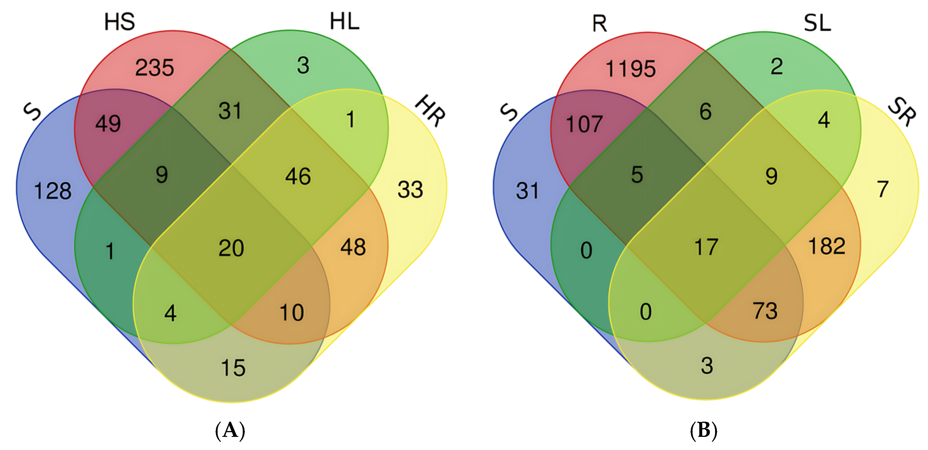
3.2. Composition of the Endospheric Bacterial Microbiome of Lettuce Plants and Substrates of Its Growth

DNA was extracted from all the samples, and the composition of the community was evaluated by sequencing 16S rRNA amplicons on the Illumina MiSeq platform. The largest number of OTU was characterized by rhizospheric soil (1594), followed by a hydroponic solution (448), the endospheric microbiome of the root (295 and 177 for samples grown in soil and hydroponics, respectively) and the endospheric microbiome of the leaves (43 and 115 for samples grown in soil and hydroponics, respectively).
Figure 3 shows data on the relative abundance of OTUs belonging to different phylum. It can be seen that the dominant phyla in all samples included Proteobacteria, Actinobacteria, Bacteroidetes, and Firmicutes. The dominant phyla in the endospheric microbiome of the seed included Verrucomicobia, which was present in all samples, but was not dominant in endospheric microbiome of the seed. The largest number of phylls was identified for the soil sample (37), the smallest for the endospheric microbiome of leaves (8–12). Among the dominant OTU (>1%) in the endosphere of lettuce plants were Rhodococcus erythropolis, Lactobacillus salivarius, as well as bacteria of genera Nocardia, Pseudarthrobacter, Parasegetibacter, Pedobacter, Lachnoclostridium, uncultured Candidatus Saccharibacteria bacterium, Reyranella, Methylobacterium, Devosia, Allorhizobium-Neorhizobium-Pararhizobium-Rhizobium, Pseudaminobacter, Bradyrhizobium, Sphingobium, Sphingomonas, Advenella, Pseudomonas (leaf) and Rhodococcus erythropolis, Streptomyces griseoruber, Lactobacillus salivarius, and also bacteria of genera Pseudarthrobacter, Flavobacterium, Lachnoclostridium, Peptoclostridium, Terrisporobacter, uncultured Candidatus Saccharibacteria bacterium, Devosia, Allorhizobium–Neorhizobium–Pararhizobium–Rhizobium, Sphingomonas, Advenella, Duganella, Limnohabitans, Methylobacillus, and Pseudomonas (root). The dominant OTUs (>1%) in substrates was Streptomyces griseoruber and bacteria included uncultured genera Acidobacterium, Pseudarthrobacter, Nocardioides, Devosia, Sphingomonas, Pseudomonas, Lysobacter, Bacillus (soil), Glutamicibacter arilaitensis, bacteria of genera Methylobacillus, Pseudarthrobacter, Flavobacterium, Synechococcus CC9902, Candidatus Saccharimonas, Brevundimonas, Reyranella, Devosia, Allorhizobium-Neorhizobium-Pararhizobium-Rhizobium, Sphingobium, Sphingomonas, and Novosphingobium (hydroponic solution) [69]. The alpha diversity of plant endosphere communities and substrates was assessed using Chao1, Simpson, and Shannon indices (Figure 3).
As expected, the soil was characterized by the greatest diversity. The Chao1 index was 1280, the Simpson index was 0.989, the Shannon index was 5.647. The hydroponic solution was characterized by lower values of the Chao1 (484), Simpson (0.956), and Shannon (4.030) indices. The most diverse endospheric microbiome was established for the roots under the conditions of the soil experiment, and no significant differences were found under the conditions of the hydroponic experiment. The calculated values of the α-diversity indices for the seed did not significantly differ from those for the endospheric microbiome, with the exception of the SL sample. The data obtained were comparable with the data of the α-diversity indices obtained for other plants. The Chao1 index estimated for the endospheric microbiome of the root was 320 for olive [108], 393 for rice [75]; the Shannon index was 3.7 for olive [108], 1.6 for lettuce [34]. A similar decrease in α-diversity from the endospheric microbiome of the root to the endospheric microbiome of the shoot when growing plants on soil has been shown for lettuce [34], and rice [75]. Low diversity characterized the xylem of grapes; the Chao1 index was 20–90, the Shannon index—0.5–4.5 [109].

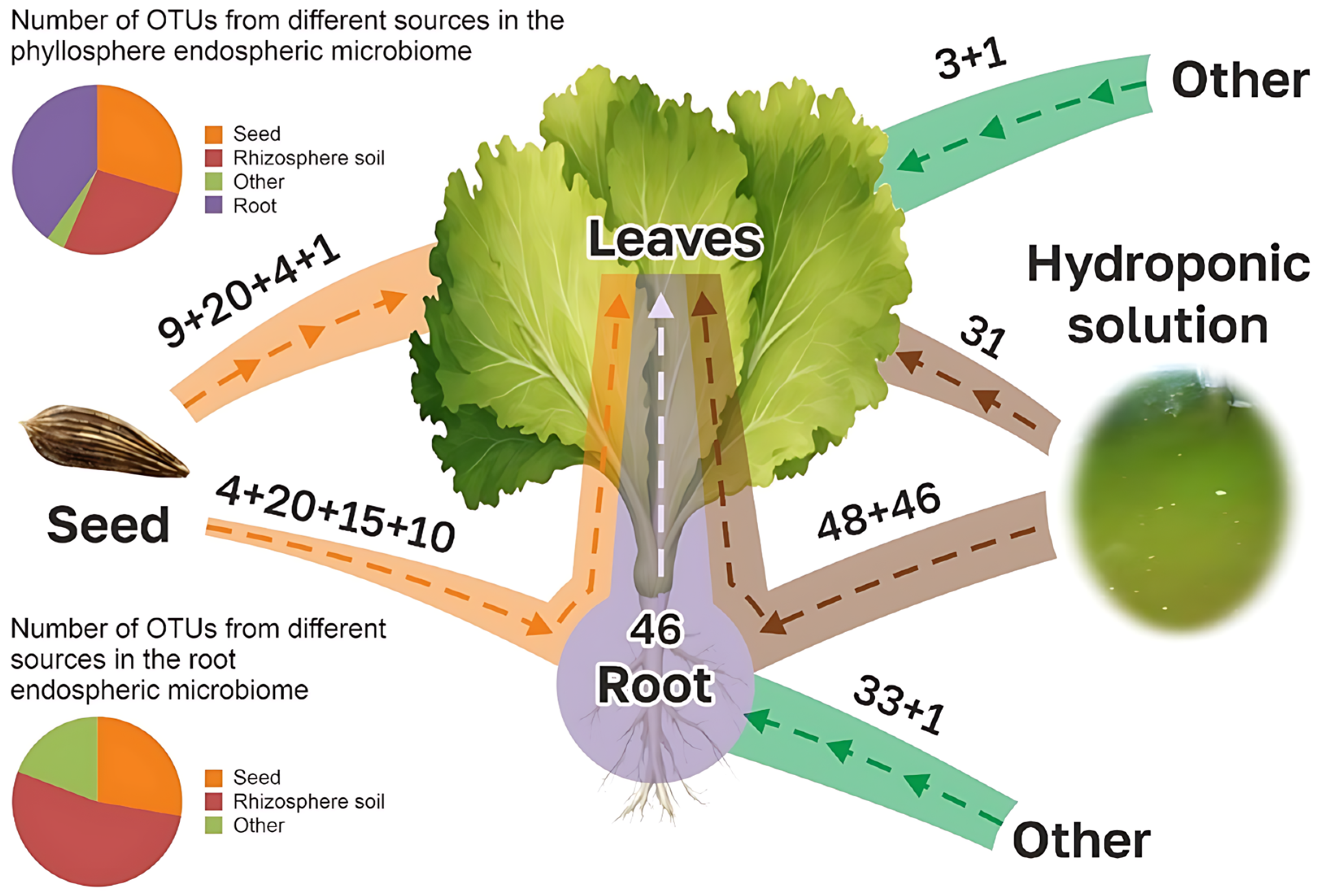
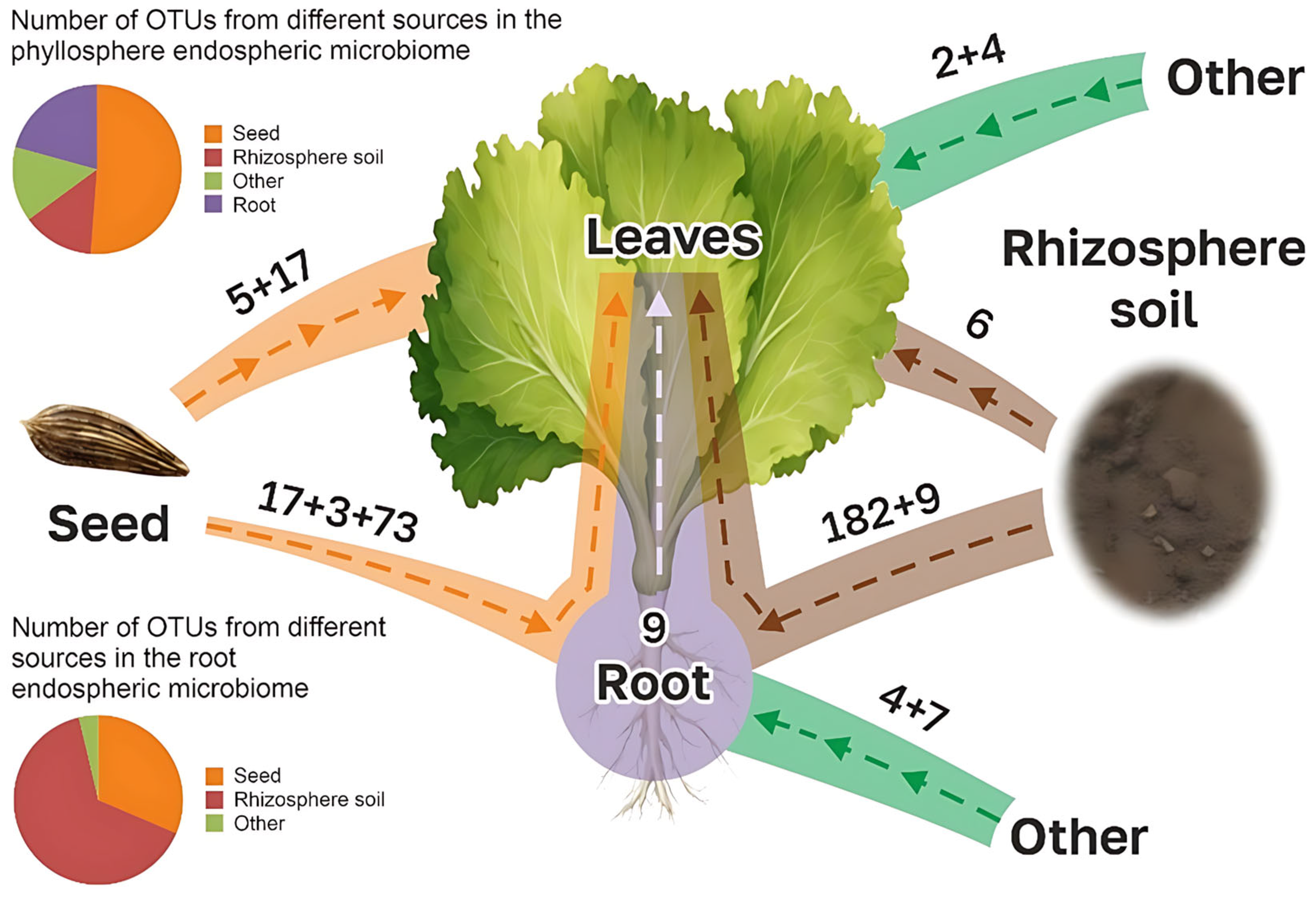
3.3. Comparison of Microbiomes of Lettuce Plants with Microbes of Seeds and Substrates

Comparing the microbiomes of seed and adult plants grown under different conditions can offer insights into processes of endospheric plant microbiome formation and transformation [15]. Therefore, at the next stage, we examined the common and different OTUs in the seed and the endosphere of plants and the substrate, aiming to explain the transfer of OTUs from one compartment to another.
Figure 4 shows Venn diagrams visualizing common and different OTU samples (separately for lettuce plants grown on soil and hydroponics). In hydroponic cultivation, 49 OTUs were common to the seeds and roots of plants, 34 OTUs to the seeds and leaves, and 24 OTUs to the seeds, the roots, and the leaves at the same time. Common to the hydroponic solution and the seed, there were 88 OTUs, and to the hydroponic solution and the root, there were 124 OTUs. In total, 128 OTUs were unique (not found in other samples) for the seed. When plants were grown in soil, 73 OTUs were common to the seeds and roots of plants, 22 OTUs to the seeds and leaves, and 17 OTUs to for the seed of the root and leaf at the same time. Common to rhizospheric soil and seed were 202 OTUs, and 281 OTUs were common to rhizospheric soil and roots. Overall, 31 OTUs were unique (not found in other samples) to the seed. It was shown that in both experiments, the seed microbiome contained unique OTUs that had not passed into the endospheric microbiome of the roots and leaves or into the substrate. A lot of work has been undertaken on comparing the endophytic microbiome of rice seeds and adult rice plants. In the work of Wang and co-authors, it was shown that the endosphere of the seed contained 78 actinobacterial OTUs, the root contained 393, and the shoot contained 195. At the same time, the share of seed OTU in the endospheric microbiome of the stem was 23% (44 OTUs) and in the roots, it was is 9% (30 OTU) [75]. In the work of Kim and co-authors [80] it was shown that the total number of bacterial OTUs in rice seed was 93, the number of OTUs in the endospheric microbiome of aboveground plant parts (shoot, stem, leaf) varied from 15 to 20 on days 7–14, while the number of total OTUs (included in both the seed and the aboveground part) ranged from 9 up to 14, which was 60–70% of the total number of OTUs in the aboveground endosphere. When analyzing 18 plant species (both monocotyledonous and dicotyledonous), it was shown that the proportion of total bacterial OTUs entering from the soil into the endosphere of the roots and shoots was 15–58 and 9–48%, respectively, while the proportion of total bacterial OTUs entering from the seed into the endosphere of the roots and shoots was 14–89 and 15–86% [15]. For Potentilla fruticosa var. albicans, it was shown that the proportion of OTUs found in both the rhizosphere and the endosphere of the root (23.5%) was higher than the proportion of OTUs found in the rhizosphere and endosphere of the leaf (13.9%) [110].
Among the vertically transmitted OTUs (seed-derived), the dominant key microbial taxa were identified. In lettuce plants cultivated in soil, representatives of the family Simkaniaceae and the genera Sphingomonas and Akkermansia predominated, whereas in hydroponically grown lettuce, the dominant OTUs included Rhodococcus erythropolis and members of the genera Sphingomonas, Pseudomonas, and Akkermansia. Among the horizontally transmitted OTUs (environmentally acquired), the dominant key taxa for soil-grown lettuce included representatives of the genera Glutamicibacter, Allorhizobium–Neorhizobium–Pararhizobium–Rhizobium, Sphingomonas, Massilia, and Methylophilus, as well as members of the order Saccharimonadales. In hydroponically cultivated lettuce, the predominant horizontally transmitted OTUs comprised Rhodococcus erythropolis and bacteria belonging to the genera Pseudarthrobacter, Sphingomonas, and Pseudomonas.
Based on data on common and unique species in samples of seeds, lettuce plants, and substrates, it is possible to make an assumption about the role of seeds and substrates in the formation of the microbiome of an adult plant. Figure 5 shows a potential scheme of such formation based on research data, as well as compiled taking into account the time sequence of events (for example, if there are common species in the seed and rhizosphere soil, the seed is considered the source and the soil as the recipient, since the seed microbiome is initially formed, and the rhizosphere microbiome results from interactions among bulk soil, seed microbiome, and plant recruitment).
(1)
Vertical colonization (seed -> root)
The pathway of direct transmission from seed to root is widely described in the literature [19,20,44,75,111,112,113,114,115,116]. At the initial stages of development, the seedling consists entirely of the species present in the seed, but does not include all of them [116,117,118,119,120,121,122,123]. As the plant develops, the proportion of the seed microbiome decreases [79,124,125].
(2)
Vertical colonization (seed -> leaf)
Direct transfer of microorganisms from the seed to the leaf and root endosphere is considered one of the main ways of forming the endospheric microbiomes of leaves and shoots [78,124,125]. Because the endospheric microbiome of the leaves does not come into direct contact with environments characterized by a high abundance and diversity of microorganisms (rhizosphere and rhizoplane), it is characterized by an actively functioning and diverse microbiome.
(3)
Vertical colonization (seed -> substrate)
Defoliation of cotyledons and the development of a seedling, in particular the root, leads to the possibility of migration of microorganisms from the seed to the substrate on which the plant grows (soil, hydroponic solution) [19,22,113]. However, competition for nutrients, space, and antagonism between the seed microbiota and the local soil microbiota limit this colonization pathway [126].
(4)
Horizontal colonization (substrate -> root)
Transfer from the substrate in which the root develops to the root endosphere is considered the main route. Studies show that most of the microbiome of plants is collected from the environment [23,61,127,128,129]. The routes of bacterial migration into the rhizosphere and then into the rhizoplane of host plants have been illustrated [5,126,130,131].
(5)
Horizontal colonization (substrate -> leaf)
To date, no specific examples based on metagenomic analyses demonstrating the transfer of particular OTUs or ASVs from the substrate to the leaves have been reported in the literature. However, the transmission of microorganisms is likely to be facilitated by environmental factors such as wind, irrigation, and rain. The colonization path has been described, including an intermediate stage of colonization, “Rhizosphere—Root—Leaf” [36,112,132,133,134,135].
(6)
Horizontal colonization (leaf -> root)
Due to the large pressure of the microbial community of the rhizosphere, colonization of the root along the leaf–root path is difficult. Data on this type of colonization are not currently available in the literature.
(7)
Horizontal colonization (other sources -> root and leaf)
Bacteria from other environmental components (atmosphere, water, insects) can colonize the endosphere through stochastic (and possibly mechanical) processes, penetrating through wounds or ruptured tissue [136]. Meanwhile, microorganisms that have thus entered the plant endosphere may be present there for a short time [5,137], or establish long-term relationships throughout the plant’s life cycle and beyond [138]. Furthermore, such microorganisms can become part of the main microbiome [139,140,141,142].
Based on the presented schematic diagram, the amount of OTU transferred vertically or horizontally to the leaf and root of an adult lettuce plant (Appendix A) was determined. Comparing the numbers of OTUs acquired in one way or another with the total numbers of OTUs in the roots and leaves, we calculated the proportion of the endospheric microbiome in the leaves and roots of adult plants of seed and soil origin. It was found that, depending on the cultivation conditions (hydroponic system or soil), the endospheric microbiome of the leaf and root consisted of 30–51% of the endospheric microbiome of the seed.
Regarding the endospheric root microbiome, the contribution of the seed microbiome did not differ significantly between plants grown in hydroponic conditions and those cultivated in soil, amounting to 28–32%, respectively (Figure 6).
Interestingly, not all microorganisms from the seed persisted in the adult plant (25% and 42%, respectively), which generally aligns with the existing literature [124]. The contribution of the substrate microbiome (hydroponic solution and soil) to the formed endospheric root microbiome was 53% and 65%, respectively, while the contribution to the phyllosphere microbiome was 27% and 14%. Similarly, it has been demonstrated that 17% of OTUs are common to both the endosphere of the seed and the shoot Citrus limon [124].

4. Conclusions

This study provides valuable insights into the bacterial endospheric microbiome of lettuce seeds and the processes involved in the formation of the endospheric microbiome in grown lettuce plants, a widely consumed leafy vegetable. By using 16S rRNA gene sequencing, it was demonstrated that both vertical (seed) and horizontal (environmental) pathways play significant roles in shaping the endophytic bacterial communities in lettuce grown in hydroponic and soil systems.
The findings revealed that a substantial portion of the root and leaf microbiome (30–51%) originated from the seed microbiome, while 53–65% was recruited from the surrounding substrate, whether hydroponic solution or soil. The presence of distinct microbiome compositions between the two cultivation methods emphasizes the influence of the environment in microbiome assembly. Rhizospheric soil, in particular, contributed more diverse microbial communities than the hydroponic solution, suggesting that soil environments may facilitate more complex interactions between the plant and its microbiome.
Importantly, this study addresses the gap in knowledge regarding lettuce seed microbiomes, which had previously been understudied compared to other crops. These findings lay the groundwork for future research aimed at better understanding microbiome dynamics in leafy crops and plant protection. The results underline the necessity of further research into seed microbiomes and their impact on plant health, growth, and resilience. Such knowledge can lead to improved agricultural practices, with potential applications in microbiome management for enhancing crop yield, disease resistance, and environmental adaptability in lettuce and other crops.

Author Contributions

Conceptualization, methodology, P.K. and P.G.; validation, P.K. and P.G.; formal analysis, N.P. and G.G.; investigation, N.P. and G.G.; data curation, N.P. and G.G.; writing—original draft preparation, P.K. and S.S.; writing—review and editing, P.G. and S.S.; supervision, S.S.; project administration, P.K. and S.S.; funding acquisition, S.S. All authors have read and agreed to the published version of the manuscript.

Funding

The work was carried out in accordance with the Strategic Academic Leadership Program “Priority 2030” of the Kazan Federal University of the Government of the Russian Federation. The funder—Ministry of science and higher education of the Russian Federation.

Data Availability Statement

The original data presented in the study are openly available in NCBI SRA at https://www.ncbi.nlm.nih.gov/sra/PRJNA1279616 or PRJNA1279616 (accessed on 10 August 2021).

Conflicts of Interest

The authors declare no conflicts of interest.

Appendix A

Figure A1. The numbers of OTUs from various sources contributing to the endospheric microbiome of the leaves and roots of lettuce grown under hydroponic conditions (OTUs absent from both the seed and substrate samples are classified as “Other.” The numbers above the arrows represent the number of OTUs transitioning between environments according to the pathways depicted in the diagram. These values correspond to the shared OTU counts derived from the Venn diagrams shown in Figure 4A).
Horticulturae 11 00762 g0a1
Figure A2. The numbers of OTUs from various sources contributing to the endospheric microbiome of the leaves and roots of lettuce grown under soil conditions (OTUs absent from both the seed and substrate samples are classified as “Other.” The numbers above the arrows represent the number of OTUs transitioning between environments according to the pathways depicted in the diagram. These values correspond to the shared OTU counts derived from the Venn diagrams shown in Figure 4B).
Horticulturae 11 00762 g0a2

References

  1. Adeleke, B.S.; Muller, D.; Babalola, O.O. A Metagenomic Lens into Endosphere Microbial Communities, Promises, and Discoveries. Lett. Appl. Microbiol. 2023, 76, ovac030. [Google Scholar] [CrossRef]
  2. Vandenkoornhuyse, P.; Quaiser, A.; Duhamel, M.; Le Van, A.; Dufresne, A. The Importance of the Microbiome of the Plant Holobiont. New Phytol. 2015, 206, 1196–1206. [Google Scholar] [CrossRef]
  3. Trivedi, P.; Leach, J.E.; Tringe, S.G.; Sa, T.; Singh, B.K. Plant–Microbiome Interactions: From Community Assembly to Plant Health. Nat. Rev. Microbiol. 2020, 18, 607–621. [Google Scholar] [CrossRef] [PubMed]
  4. Afzal, I.; Shinwari, Z.K.; Sikandar, S.; Shahzad, S. Plant Beneficial Endophytic Bacteria: Mechanisms, Diversity, Host Range and Genetic Determinants. Microbiol. Res. 2019, 221, 36–49. [Google Scholar] [CrossRef]
  5. Compant, S.; Clément, C.; Sessitsch, A. Plant Growth-Promoting Bacteria in the Rhizo- and Endosphere of Plants: Their Role, Colonization, Mechanisms Involved and Prospects for Utilization. Soil Biol. Biochem. 2010, 42, 669–678. [Google Scholar] [CrossRef]
  6. Amine Hassani, M.; Durán, P.; Hacquard, S. Microbial Interactions within the Plant Holobiont. Microbiome 2018, 6, 58. [Google Scholar]
  7. Macaya-Sanz, D.; Witzell, J.; Collada, C.; Gil, L.; Martín, J.A. Core Endophytic Mycobiome in Ulmus Minor and Its Relation to Dutch Elm Disease Resistance. Front. Plant Sci. 2023, 14, 1125942. [Google Scholar] [CrossRef]
  8. Lemanceau, P.; Blouin, M.; Muller, D.; Moënne-Loccoz, Y. Let the Core Microbiota Be Functional. Trends Plant Sci. 2017, 22, 583–595. [Google Scholar] [CrossRef]
  9. Li, F.; He, X.; Sun, Y.; Zhang, X.; Tang, X.; Li, Y.; Yi, Y. Distinct Endophytes Are Used by Diverse Plants for Adaptation to Karst Regions. Sci. Rep. 2019, 9, 5246. [Google Scholar] [CrossRef]
  10. Fitzpatrick, C.R.; Salas-González, I.; Conway, J.M.; Finkel, O.M.; Gilbert, S.; Russ, D.; Pereira, P.J.; Teixeira, L.; Dangl, J.L. The Plant Microbiome: From Ecology to Reductionism and Beyond. Annu. Rev. Microbiol. 2020, 74, 81–100. [Google Scholar] [CrossRef]
  11. Newcombe, G.; Harding, A.; Ridout, M.; Busby, P.E. A Hypothetical Bottleneck in the Plant Microbiome. Front. Microbiol. 2018, 9, 1645. [Google Scholar] [CrossRef]
  12. Liu, F.; Hewezi, T.; Lebeis, S.L.; Pantalone, V.; Grewal, P.S.; Staton, M.E. Soil Indigenous Microbiome and Plant Genotypes Cooperatively Modify Soybean Rhizosphere Microbiome Assembly. BMC Microbiol. 2019, 19, 201. [Google Scholar] [CrossRef] [PubMed]
  13. Acuña, J.J.; Hu, J.; Inostroza, N.G.; Valenzuela, T.; Perez, P.; Epstein, S.; Sessitsch, A.; Zhang, Q.; Jorquera, M.A. Endophytic Bacterial Communities in Ungerminated and Germinated Seeds of Commercial Vegetables. Sci. Rep. 2023, 13, 19829. [Google Scholar] [CrossRef]
  14. Ibáñez, F.; Tonelli, M.L.; Muñoz, V.; Figueredo, M.S.; Fabra, A. Bacterial Endophytes of Plants: Diversity, Invasion Mechanisms and Effects on the Host; Springer: Cham, Switzerland, 2017; Volume 1, pp. 25–40. ISBN 9783319665405. [Google Scholar]
  15. Johnston-Monje, D.; Gutiérrez, J.P.; Lopez-Lavalle, L.A.B. Seed-Transmitted Bacteria and Fungi Dominate Juvenile Plant Microbiomes. Front. Microbiol. 2021, 12, 737616. [Google Scholar] [CrossRef] [PubMed]
  16. Poupin, M.J.; González, B. Embracing Complexity in Plant–Microbiome Systems. Environ. Microbiol. Rep. 2024, 16, e70000. [Google Scholar] [CrossRef] [PubMed]
  17. Pagán, I. Transmission through Seeds: The Unknown Life of Plant Viruses. PLOS Pathog. 2022, 18, e1010707. [Google Scholar] [CrossRef]
  18. Simonin, M.; Préveaux, A.; Marais, C.; Garin, T.; Arnault, G.; Sarniguet, A.; Barret, M. Transmission of Synthetic Seed Bacterial Communities to Radish Seedlings: Impact on Microbiota Assembly and Plant Phenotype. Peer Community J. 2023, 3, e95. [Google Scholar] [CrossRef]
  19. Nelson, E.B. The Seed Microbiome: Origins, Interactions, and Impacts. Plant Soil 2018, 422, 7–34. [Google Scholar] [CrossRef]
  20. Shade, A.; Jacques, M.A.; Barret, M. Ecological Patterns of Seed Microbiome Diversity, Transmission, and Assembly. Curr. Opin. Microbiol. 2017, 37, 15–22. [Google Scholar] [CrossRef]
  21. Johnston-Monje, D.; Mousa, W.K.; Lazarovits, G.; Raizada, M.N. Impact of Swapping Soils on the Endophytic Bacterial Communities of Pre-Domesticated, Ancient and Modern Maize. BMC Plant Biol. 2014, 14, 233. [Google Scholar] [CrossRef]
  22. Johnston-Monje, D.; Raizada, M.N. Conservation and Diversity of Seed Associated Endophytes in Zea across Boundaries of Evolution, Ethnography and Ecology. PLoS ONE 2011, 6, e20396. [Google Scholar] [CrossRef] [PubMed]
  23. Bright, M.; Bulgheresi, S. A Complex Journey: Transmission of Microbial Symbionts. Nat. Rev. Microbiol. 2010, 8, 218–230. [Google Scholar] [CrossRef] [PubMed]
  24. Barret, M.; Briand, M.; Bonneau, S.; Préveaux, A.; Valière, S.; Bouchez, O.; Hunault, G.; Simoneau, P.; Jacquesa, M.A. Emergence Shapes the Structure of the Seed Microbiota. Appl. Environ. Microbiol. 2015, 81, 1257–1266. [Google Scholar] [CrossRef]
  25. Leff, J.W.; Lynch, R.C.; Kane, N.C.; Fierer, N. Plant Domestication and the Assembly of Bacterial and Fungal Communities Associated with Strains of the Common Sunflower, Helianthus Annuus. New Phytol. 2017, 214, 412–423. [Google Scholar] [CrossRef]
  26. Yang, L.; Danzberger, J.; Schöler, A.; Schröder, P.; Schloter, M.; Radl, V. Dominant Groups of Potentially Active Bacteria Shared by Barley Seeds Become Less Abundant in Root Associated Microbiome. Front. Plant Sci. 2017, 8, 1005. [Google Scholar] [CrossRef]
  27. Rahman, M.M.; Flory, E.; Koyro, H.W.; Abideen, Z.; Schikora, A.; Suarez, C.; Schnell, S.; Cardinale, M. Consistent Associations with Beneficial Bacteria in the Seed Endosphere of Barley (Hordeum vulgare L.). Syst. Appl. Microbiol. 2018, 41, 386–398. [Google Scholar] [CrossRef]
  28. Eyre, A.W.; Wang, M.; Oh, Y.; Dean, R.A. Identification and Characterization of the Core Rice Seed Microbiome. Phytobiomes J. 2019, 3, 148–157. [Google Scholar] [CrossRef]
  29. Wassermann, B.; Rybakova, D.; Adam, E.; Zachow, C.; Bernhard, M.; Muller, M.; Mancinelli, R.; Berg, G. Studying Seed Microbiomes. In The Plant Microbiome Methods and Protocols; Humana: New York, NY, USA, 2021; pp. 1–22. [Google Scholar]
  30. Van Elsas, J.D.; Turner, S.; Bailey, M.J. Horizontal Gene Transfer in the Phytosphere. New Phytol. 2003, 157, 525–537. [Google Scholar] [CrossRef] [PubMed]
  31. Berg, G.; Raaijmakers, J.M. Saving Seed Microbiomes. ISME J. 2018, 12, 1167–1170. [Google Scholar] [CrossRef]
  32. Simonin, M.; Briand, M.; Chesneau, G.; Rochefort, A.; Marais, C.; Sarniguet, A.; Barret, M. Seed Microbiota Revealed by a Large-scale Meta-analysis Including 50 Plant Species. New Phytol. 2022, 234, 1448–1463. [Google Scholar] [CrossRef]
  33. Hardoim, P.R.; Hardoim, C.C.P.; van Overbeek, L.S.; van Elsas, J.D. Dynamics of Seed-Borne Rice Endophytes on Early Plant Growth Stages. PLoS ONE 2012, 7, e30438. [Google Scholar] [CrossRef] [PubMed]
  34. Mohamed, N.Z.; Schena, L.; Malacrinò, A. Lettuce Seedlings Rapidly Assemble Their Microbiome from the Environment through Deterministic Processes. Rhizosphere 2024, 30, 100896. [Google Scholar] [CrossRef]
  35. Ma, Y.; Rajkumar, M.; Luo, Y.M.; Freitas, H. Inoculation of Endophytic Bacteria on Host and Non-Host Plants-Effects on Plant Growth and Ni Uptake. J. Hazard. Mater. 2011, 195, 230–237. [Google Scholar] [PubMed]
  36. Compant, S.; Reiter, B.; Sessitsch, A.; Nowak, J.; Clément, C.; Barka, E.A. Endophytic Colonization of Vitis Vinifera L. by Plant Growth-Promoting Bacterium Burkholderia Sp. Strain PsJN. Appl. Environ. Microbiol. 2005, 71, 1685–1693. [Google Scholar] [CrossRef]
  37. Khan, Z.; Guelich, G.; Phan, H.; Redman, R.; Doty, S. Bacterial and Yeast Endophytes from Poplar and Willow Promote Growth in Crop Plants and Grasses. ISRN Agron. 2012, 2012, 890280. [Google Scholar]
  38. Carroll, G. Fungal Endophytes in Stems and Leaves: From Latent Pathogen to Mutualistic Symbiont. Soc. Ecol. 1986, 69, 2–9. [Google Scholar]
  39. Schlaeppi, K.; Bulgarelli, D. The Plant Microbiome at Work. Mol. Plant-Microbe Interact. 2015, 28, 212–217. [Google Scholar] [CrossRef]
  40. Bulgarelli, D.; Rott, M.; Schlaeppi, K.; Ver Loren van Themaat, E.; Ahmadinejad, N.; Assenza, F.; Rauf, P.; Huettel, B.; Reinhardt, R.; Schmelzer, E.; et al. Revealing Structure and Assembly Cues for Arabidopsis Root-Inhabiting Bacterial Microbiota. Nature 2012, 488, 91–95. [Google Scholar] [CrossRef]
  41. Lundberg, D.S.; Lebeis, S.L.; Paredes, S.H.; Yourstone, S.; Gehring, J.; Malfatti, S.; Tremblay, J.; Engelbrektson, A.; Kunin, V.; Del Rio, T.G.; et al. Defining the Core Arabidopsis Thaliana Root Microbiome. Nature 2013, 501, 86–90. [Google Scholar] [CrossRef]
  42. Peiffer, J.A.; Spor, A.; Koren, O.; Jin, Z.; Tringe, S.G.; Dangl, J.L.; Buckler, E.S.; Ley, R.E. Diversity and Heritability of the Maize Rhizosphere Microbiome under Field Conditions. Proc. Natl. Acad. Sci. USA 2013, 110, 6548–6553. [Google Scholar] [CrossRef]
  43. Schlaeppi, K.; Dombrowski, N.; Oter, R.G.; Ver Loren Van Themaat, E.; Schulze-Lefert, P. Quantitative Divergence of the Bacterial Root Microbiota in Arabidopsis Thaliana Relatives. Proc. Natl. Acad. Sci. USA 2014, 111, 585–592. [Google Scholar] [CrossRef] [PubMed]
  44. Edwards, J.; Johnson, C.; Santos-Medellín, C.; Lurie, E.; Podishetty, N.K.; Bhatnagar, S.; Eisen, J.A.; Sundaresan, V.; Jeffery, L.D. Structure, Variation, and Assembly of the Root-Associated Microbiomes of Rice. Proc. Natl. Acad. Sci. USA 2015, 112, E911–E920. [Google Scholar] [CrossRef]
  45. He, J.; Zhang, L.; Van Dingenen, J.; Desmet, S.; Goormachtig, S.; Calonne-Salmon, M.; Declerck, S. Arbuscular Mycorrhizal Hyphae Facilitate Rhizobia Dispersal and Nodulation in Legumes. ISME J. 2024, 18, wrae185. [Google Scholar] [CrossRef]
  46. Sengupta, S.; Ganguli, S.; Singh, P.K. Metagenome Analysis of the Root Endophytic Microbial Community of Indian Rice (O. sativa L.). Genomics Data 2017, 12, 41–43. [Google Scholar] [CrossRef] [PubMed]
  47. Kim, H.; Jeon, J.; Lee, K.K.; Lee, Y.H. Longitudinal Transmission of Bacterial and Fungal Communities from Seed to Seed in Rice. Commun. Biol. 2022, 5, 772. [Google Scholar] [CrossRef] [PubMed]
  48. Campisano, A.; Antonielli, L.; Pancher, M.; Yousaf, S.; Pindo, M.; Pertot, I. Bacterial Endophytic Communities in the Grapevine Depend on Pest Management. PLoS ONE 2014, 9, e112763. [Google Scholar] [CrossRef]
  49. Tian, B.Y.; Cao, Y.; Zhang, K.Q. Metagenomic Insights into Communities, Functions of Endophytes, and Their Associates with Infection by Root-Knot Nematode, Meloidogyne Incognita, in Tomato Roots. Sci. Rep. 2015, 5, 17087. [Google Scholar] [CrossRef]
  50. Akinsanya, M.A.; Goh, J.K.; Lim, S.P.; Ting, A.S.Y. Diversity, Antimicrobial and Antioxidant Activities of Culturable Bacterial Endophyte Communities in Aloe Vera. FEMS Microbiol. Lett. 2015, 362, 184. [Google Scholar] [CrossRef]
  51. Yang, R.; Liu, P.; Ye, W. Illumina-Based Analysis of Endophytic Bacterial Diversity of Tree Peony (Paeonia Sect. Moutan) Roots and Leaves. Braz. J. Microbiol. 2017, 48, 695–705. [Google Scholar] [CrossRef]
  52. Hong, C.E.; Kim, J.U.; Lee, J.W.; Bang, K.H.; Jo, I.H. Metagenomic Analysis of Bacterial Endophyte Community Structure and Functions in Panax Ginseng at Different Ages. 3Biotech 2019, 9, 300. [Google Scholar] [CrossRef]
  53. Zhang, J.; Zhang, C.; Yang, J.; Zhang, R.; Gao, J.; Zhao, X.; Zhao, J.; Zhao, D.; Zhang, X. Insights into Endophytic Bacterial Community Structures of Seeds Among Various Oryza sativa L. Rice Genotypes. J. Plant Growth Regul. 2019, 38, 93–102. [Google Scholar] [CrossRef]
  54. Abedinzadeh, M.; Etesami, H.; Alikhani, H.A. Characterization of Rhizosphere and Endophytic Bacteria from Roots of Maize (Zea mays L.) Plant Irrigated with Wastewater with Biotechnological Potential in Agriculture. Biotechnol. Reports 2019, 21, e00305. [Google Scholar] [CrossRef]
  55. Wicaksono, W.A.; Cernava, T.; Wassermann, B.; Abdelfattah, A.; Soto-Giron, M.J.; Toledo, G.V.; Virtanen, S.M.; Knip, M.; Hyöty, H.; Berg, G. The Edible Plant Microbiome: Evidence for the Occurrence of Fruit and Vegetable Bacteria in the Human Gut. Gut Microbes 2023, 15, 2258565. [Google Scholar] [CrossRef]
  56. Žiarovská, J.; Urbanová, L.; Moravčíková, D.; Artimová, R.; Omelka, R.; Medo, J. Varieties of Lettuce Forming Distinct Microbial Communities Inhabiting Roots and Rhizospheres with Various Responses to Osmotic Stress. Horticulturae 2022, 8, 1174. [Google Scholar] [CrossRef]
  57. Damerum, A.; Arnold, E.; Bernard, V.; Smith, H.; Taylor, G. Good and Bad Lettuce Leaf Microbes? Unravelling the Genetic Architecture of the Microbiome to Inform Plant Breeding for Enhanced Food Safety and Reduced Food Waste. bioRxiv, 2021; bioRxiv: 2021.08.06.455490. [Google Scholar]
  58. Vlasselaer, L.; Crauwels, S.; Lievens, B.; De Coninck, B. Unveiling the Microbiome of Hydroponically Cultivated Lettuce: Impact of Phytophthora Cryptogea Infection on Plant-Associated Microorganisms. FEMS Microbiol. Ecol. 2024, 100, fiae010. [Google Scholar] [CrossRef] [PubMed]
  59. Galieva, G.; Kuryntseva, P.; Selivanovskaya, S.; Brusko, V.; Garifullin, B.; Dimiev, A.; Galitskaya, P. Glutamic-N,N-Diacetic Acid as an Innovative Chelating Agent in Microfertilizer Development: Biodegradability, Lettuce Growth Promotion, and Impact on Endospheric Bacterial Communities. Soil Syst. 2024, 8, 67. [Google Scholar] [CrossRef]
  60. Moroenyane, I.; Tremblay, J.; Yergeau, E. Soybean Microbiome Recovery After Disruption Is Modulated by the Seed and Not the Soil Microbiome. Phytobiomes J. 2021, 5, 418–431. [Google Scholar] [CrossRef]
  61. Bintarti, A.F.; Sulesky-Grieb, A.; Stopnisek, N.; Shade, A. Endophytic Microbiome Variation Among Single Plant Seeds. Phytobiomes J. 2022, 6, 45–55. [Google Scholar] [CrossRef]
  62. Barra, P.J.; Inostroza, N.G.; Acuña, J.J.; Mora, M.L.; Crowley, D.E.; Jorquera, M.A. Formulation of Bacterial Consortia from Avocado (Persea americana Mill.) and Their Effect on Growth, Biomass and Superoxide Dismutase Activity of Wheat Seedlings under Salt Stress. Appl. Soil Ecol. 2016, 102, 80–91. [Google Scholar] [CrossRef]
  63. ISO 10390:2021; Soil, Treated Biowaste and Sludge—Determination of PH. International Organization for Standardization: Geneva, Switzerland, 2021; Volume 2021, p. 10.
  64. ISO 11265:1994; ISO Soil Quality—Determination Specific Electrical Conductivity. International Organization for Standardization: Geneva, Switzerland, 1994.
  65. ISO 10694:1995; ISO Soil Quality—Determination of Organic and Total Carbon After Dry Combustion (Elementary Analysis). International Organization for Standardization: Geneva, Switzerland, 1995.
  66. ISO 13878:1998; Soil Quality—Determination of Total Nitrogen Content by Dry Combustion (“Elemental Analysis”). International Organization for Standardization: Geneva, Switzerland, 1998.
  67. ISO 13320: 2009; Particle Size Analysis—Laser Diffraction Methods. International Organization for Standardization: Geneva, Switzerland, 2009.
  68. Baudoin, W.; Nono-Womdim, R.; Lutaladio, N.; Hodder, A. Good Agricultural Practices for Greenhouse Vegetable Crops Principles for Mediterranean Climate Areas; Food and Agriculture Organization of the United Nations: Rome, Italy, 2013; ISBN 9789251076491. [Google Scholar]
  69. Danilova, N.; Galieva, G.; Kuryntseva, P.; Selivanovskaya, S.; Galitskaya, P. Influence of the Antibiotic Oxytetracycline on the Morphometric Characteristics and Endophytic Bacterial Community of Lettuce (Lactuca sativa L.). Microorganisms 2023, 11, 2828. [Google Scholar] [CrossRef]
  70. Caporaso, J.G.; Kuczynski, J.; Stombaugh, J.; Bittinger, K.; Bushman, F.D.; Costello, E.K. QIIME Allows Analysis of High-Throughput Community Sequencing Data. Geofis. Int. 2004, 43, 415–425. [Google Scholar] [CrossRef] [PubMed]
  71. Hughes, J.B.; Hellmann, J.J.; Ricketts, T.H.; Bohannan, B.J.M. Counting the Uncountable: Statistical Approaches to Estimating Microbial Diversity. Appl. Environ. Microbiol. 2001, 67, 4399–4406. [Google Scholar] [CrossRef] [PubMed]
  72. Shannon, C. A Mathematical Theory of Communication. J. Franklin Inst. 1948, 27, 379–423. [Google Scholar]
  73. Beisel, J.N.; Moreteau, J.C. A Simple Formula for Calculating the Lower Limit of Shannon’s Diversity Index. Ecol. Modell. 1997, 99, 289–292. [Google Scholar] [CrossRef]
  74. Chalita, M.; Kim, Y.O.; Park, S.; Oh, H.S.; Cho, J.H.; Moon, J.; Baek, N.; Moon, C.; Lee, K.; Yang, J.; et al. EzBioCloud: A Genome-Driven Database and Platform for Microbiome Identification and Discovery. Int. J. Syst. Evol. Microbiol. 2024, 74, 006421. [Google Scholar] [CrossRef]
  75. Wang, W.; Zhai, Y.; Cao, L.; Tan, H.; Zhang, R. Illumina-Based Analysis of Core Actinobacteriome in Roots, Stems, and Grains of Rice. Microbiol. Res. 2016, 190, 12–18. [Google Scholar] [CrossRef]
  76. Navya, B.; Babu, S. Comparative Metataxonamic Analyses of Seeds and Leaves of Traditional Varieties and Hybrids of Cucumber (Cucumis sativus L.) Reveals Distinct and Core Microbiome. Heliyon 2023, 9, e20216. [Google Scholar] [CrossRef]
  77. Kumar, K.; Verma, A.; Pal, G.; Anubha; White, J.F.; Verma, S.K. Seed Endophytic Bacteria of Pearl Millet (Pennisetum glaucum L.) Promote Seedling Development and Defend Against a Fungal Phytopathogen. Front. Microbiol. 2021, 12, 774293. [Google Scholar] [CrossRef]
  78. Ren, Z.; Chen, A.J.; Zong, Q.; Du, Z.; Guo, Q.; Liu, T.; Chen, W.; Gao, L. Microbiome Signature of Endophytes in Wheat Seed Response to Wheat Dwarf Bunt Caused by Tilletia controversa Kühn. Microbiol. Spectr. 2023, 11, e00390-22. [Google Scholar] [CrossRef]
  79. Chouhan, U.; Gamad, U.; Choudhari, J.K. Metagenomic Analysis of Soybean Endosphere Microbiome to Reveal Signatures of Microbes for Health and Disease. J. Genet. Eng. Biotechnol. 2023, 21, 84. [Google Scholar] [CrossRef]
  80. Kim, H.; Lee, Y.H. Spatiotemporal Assembly of Bacterial and Fungal Communities of Seed-Seedling-Adult in Rice. Front. Microbiol. 2021, 12, 708475. [Google Scholar] [CrossRef] [PubMed]
  81. Zhang, X.; Ma, Y.N.; Wang, X.; Liao, K.; He, S.; Zhao, X.; Guo, H.; Zhao, D.; Wei, H.L. Dynamics of Rice Microbiomes Reveal Core Vertically Transmitted Seed Endophytes. Microbiome 2022, 10, 216. [Google Scholar] [CrossRef] [PubMed]
  82. Rodríguez, C.E.; Antonielli, L.; Mitter, B.; Sessitsch, A. Assembly of Endophytic Communities of Setaria viridis Plants When Grown in Different Soils and Derived from Different Seeds. Phytobiomes J. 2024, 8, 34–45. [Google Scholar] [CrossRef]
  83. Abdelfattah, A.; Tack, A.J.M.; Lobato, C.; Wassermann, B.; Berg, G. From Seed to Seed: The Role of Microbial Inheritance in the Assembly of the Plant Microbiome. Trends Microbiol. 2023, 31, 346–355. [Google Scholar] [CrossRef]
  84. Kumar, A.; Solanki, M.K.; Wang, Z.; Solanki, A.C.; Singh, V.K.; Divvela, P.K. Revealing the Seed Microbiome: Navigating Sequencing Tools, Microbial Assembly, and Functions to Amplify Plant Fitness. Microbiol. Res. 2024, 279, 127549. [Google Scholar] [CrossRef] [PubMed]
  85. López-López, A.; Rogel, M.A.; Ormeño-Orrillo, E.; Martínez-Romero, J.; Martínez-Romero, E. Phaseolus Vulgaris Seed-Borne Endophytic Community with Novel Bacterial Species Such as Rhizobium endophyticum sp. nov. Syst. Appl. Microbiol. 2010, 33, 322–327. [Google Scholar] [CrossRef]
  86. Rosenblueth, M.; López-López, A.; Martínez, J.; Rogel, M.A.; Toledo, I.; Martínez-Romero, E. Seed Bacterial Endophytes: Common Genera, Seed-to-Seed Variability and Their Possible Role in Plants. Acta Hortic. 2012, 938, 39–48. [Google Scholar] [CrossRef]
  87. Sun, Z.; Adeleke, B.S.; Shi, Y.; Li, C. The Seed Microbiomes of Staple Food Crops. Microb. Biotechnol. 2023, 16, 2236–2249. [Google Scholar] [CrossRef]
  88. Solanki, M.K.; Wang, F.Y.; Wang, Z.; Li, C.N.; Lan, T.J.; Singh, R.K.; Singh, P.; Yang, L.T.; Li, Y.R. Rhizospheric and Endospheric Diazotrophs Mediated Soil Fertility Intensification in Sugarcane-Legume Intercropping Systems. J. Soils Sediments 2019, 19, 1911–1927. [Google Scholar] [CrossRef]
  89. Díaz Herrera, S.; Grossi, C.; Zawoznik, M.; Groppa, M.D. Wheat Seeds Harbour Bacterial Endophytes with Potential as Plant Growth Promoters and Biocontrol Agents of Fusarium graminearum. Microbiol. Res. 2016, 186–187, 37–43. [Google Scholar] [CrossRef]
  90. Ouwerkerk, J.P.; Aalvink, S.; Belzer, C.; de Vos, W.M. Akkermansia Glycaniphila Sp. Nov., an Anaerobic Mucin-Degrading Bacterium Isolated from Reticulated Python faeces. Int. J. Syst. Evol. Microbiol. 2016, 66, 4614–4620. [Google Scholar] [CrossRef]
  91. Schoch, C.L.; Ciufo, S.; Domrachev, M.; Hotton, C.L.; Kannan, S.; Khovanskaya, R.; Karsch-Mizrachi, I. NCBI Taxonomy: A Comprehensive Update on Curation, Resources and Tools. Available online: https://www.ncbi.nlm.nih.gov/Taxonomy/Browser/wwwtax.cgi?mode=Tree&id=512293&lvl=3&lin=f&keep=1&srchmode=1&unlock (accessed on 3 February 2021).
  92. Lagkouvardos, I.; Lesker, T.R.; Hitch, T.C.A.; Gálvez, E.J.C.; Smit, N.; Neuhaus, K.; Wang, J.; Baines, J.F.; Abt, B.; Stecher, B.; et al. Sequence and Cultivation Study of Muribaculaceae Reveals Novel Species, Host Preference, and Functional Potential of This yet Undescribed Family. Microbiome 2019, 7, 28. [Google Scholar] [CrossRef] [PubMed]
  93. Zuo, W.F.; Pang, Q.; Yao, L.P.; Zhang, Y.; Peng, C.; Huang, W.; Han, B. Gut Microbiota: A Magical Multifunctional Target Regulated by Medicine Food Homology Species. J. Adv. Res. 2023, 52, 151–170. [Google Scholar] [CrossRef] [PubMed]
  94. Dong, C.; Shao, Q.; Ren, Y.; Ge, W.; Yao, T.; Hu, H.; Huang, J.; Liang, Z.; Han, Y. Assembly, Core Microbiota, and Function of the Rhizosphere Soil and Bark Microbiota in Eucommia ulmoides. Front. Microbiol. 2022, 13, 855317. [Google Scholar] [CrossRef]
  95. Zhang, H.; Ma, L.J.; Lu, W.C.; Yu, D.; Hang, X.N.; Liao, D.X. Colonization Characteristics of Pseudomonas Poae HT1 in Roots and Stems of Faba Bean and Its Effect on Endophytic Bacterial Diversity. Microbiol. China 2021, 48, 4677–4687. [Google Scholar]
  96. Rosenberg, E.; De Long, E.F.; Lory, S.; Stackebrandt, E.; Thompson, F. The Prokaryotes: Other Major Lineages of Bacteria and the Archaea; Springer: Berlin/Heidelberg, Germany, 2014; pp. 1–1028. [Google Scholar]
  97. Grothjan, J.J.; Young, E.B. Bacterial Recruitment to Carnivorous Pitcher Plant Communities: Identifying Sources Influencing Plant Microbiome Composition and Function. Front. Microbiol. 2022, 13, 791079. [Google Scholar] [CrossRef]
  98. Bigott, A.F. A Comparison of Soils and Their Associated Microbial Communities as Affected by Sugarcane Cultivation. Master’s Theses, Louisiana State University, Baton Rouge, LA, USA, 2017. [Google Scholar]
  99. Chewning, S.S.; Grant, D.L.; Banion, B.S.O.; Gates, A.D.; Kennedy, B.J.; Campagna, S.R.; Lebeis, S.L. Root-Associated Streptomyces Isolates Harboring melC Genes Demonstrate Enhanced Plant Colonization. Phytobiomes J. 2019, 3, 165–176. [Google Scholar] [CrossRef]
  100. De Tender, C. Microbial Community Analysis in Soil (Rhizosphere) and the Marine (Plastisphere) Environment in Function of Plant Health and Biofilm Formation. Ph.D. Thesis, Ghent University, Gent, Belgium, 2017; p. 254. [Google Scholar]
  101. Debode, J.; De Tender, C.; Soltaninejad, S.; Van Malderghem, C.; Haegeman, A.; Van der Linden, I.; Cottyn, B.; Heyndrickx, M.; Maes, M. Chitin Mixed in Potting Soil Alters Lettuce Growth, the Survival of Zoonotic Bacteria on the Leaves and Associated Rhizosphere Microbiology. Front. Microbiol. 2016, 7, 565. [Google Scholar] [CrossRef]
  102. Li, X.; Hu, H.; Ren, Q.; Wang, M.; Du, Y.; He, Y.; Wang, Q. Comparative Analysis of Endophyte Diversity of Dendrobium Officinale Lived on Rock and Tree. Plant Biotechnol. 2023, 40, 145–155. [Google Scholar] [CrossRef]
  103. Mei, X.; Wu, C.; Zhao, J.; Yan, T.; Jiang, P. Community Structure of Bacteria Associated with Drifting Sargassum Horneri, the Causative Species of Golden Tide in the Yellow Sea. Front. Microbiol. 2019, 10, 1192. [Google Scholar] [CrossRef]
  104. Wassermann, B. The Seed & Fruit Microbiome: Health Implications for Crops and Consumers. Ph.D. Thesis, Graz University of Technology, Graz, Austria, 2020. [Google Scholar]
  105. Pu, Q.; Wang, H.T.; Pan, T.; Li, H.; Su, J.Q. Enhanced Removal of Ciprofloxacin and Reduction of Antibiotic Resistance Genes by Earthworm Metaphire vulgaris in Soil. Sci. Total Environ. 2020, 742, 140409. [Google Scholar] [CrossRef] [PubMed]
  106. Alrashedi, S. Effect of Endophyte-Infected Tall Fescue Toxins on Growth Performance and the Microbial Community in the Rumen and Feces in Pregnant Ewes. Ph.D. Thesis, University of Arkansas, Fayetteville, AR, USA, 2017. [Google Scholar]
  107. Guo, J.; Ling, N.; Li, Y.; Li, K.; Ning, H.; Shen, Q.; Guo, S.; Vandenkoornhuyse, P. Seed-Borne, Endospheric and Rhizospheric Core Microbiota as Predictors of Plant Functional Traits across Rice Cultivars Are Dominated by Deterministic Processes. New Phytol. 2021, 230, 2047–2060. [Google Scholar] [CrossRef]
  108. Fernández-González, A.J.; Villadas, P.J.; Gómez-Lama Cabanás, C.; Valverde-Corredor, A.; Belaj, A.; Mercado-Blanco, J.; Fernández-López, M. Defining the Root Endosphere and Rhizosphere Microbiomes from the World Olive Germplasm Collection. Sci. Rep. 2019, 9, 20423. [Google Scholar] [CrossRef]
  109. Hamaoka, K.; Aoki, Y.; Takahashi, S.; Enoki, S.; Yamamoto, K.; Tanaka, K.; Suzuki, S. Diversity of Endophytic Bacterial Microbiota in Grapevine Shoot Xylems Varies Depending on Wine Grape-Growing Region, Cultivar, and Shoot Growth Stage. Sci. Rep. 2022, 12, 15772. [Google Scholar] [CrossRef] [PubMed]
  110. Sonam, W.; Liu, Y.; Guo, L. Endophytic Bacteria in the Periglacial Plant Potentilla fruticosa var. albicans Are Influenced by Habitat Type. Ecol. Process. 2023, 12, 57. [Google Scholar] [CrossRef]
  111. Bodhankar, S.; Grover, M.; Hemanth, S.; Reddy, G.; Rasul, S.; Yadav, S.K.; Desai, S.; Mallappa, M.; Mandapaka, M.; Srinivasarao, C. Maize Seed Endophytic Bacteria: Dominance of Antagonistic, Lytic Enzyme-Producing Bacillus Spp. 3Biotech 2017, 7, 232. [Google Scholar] [CrossRef]
  112. James, E.K.; Gyaneshwar, P.; Mathan, N.; Barraquio, W.L.; Reddy, P.M.; Iannetta, P.P.M.; Olivares, F.L.; Ladha, J.K. Infection and Colonization of Rice Seedlings by the Plant Growth-Promoting Bacterium Herbaspirillum seropedicae Z67. Mol. Plant-Microbe Interact. 2002, 15, 894–906. [Google Scholar] [CrossRef]
  113. Adam, E.; Bernhart, M.; Müller, H.; Winkler, J.; Berg, G. The Cucurbita Pepo Seed Microbiome: Genotype-Specific Composition and Implications for Breeding. Plant Soil 2018, 422, 35–49. [Google Scholar] [CrossRef]
  114. Johnston-Monje, D.; Lundberg, D.S.; Lazarovits, G.; Reis, V.M.; Raizada, M.N. Bacterial Populations in Juvenile Maize Rhizospheres Originate from Both Seed and Soil. Plant Soil 2016, 405, 337–355. [Google Scholar] [CrossRef]
  115. Santoyo, G. How Plants Recruit Their Microbiome? New Insights into Beneficial Interactions. J. Adv. Res. 2022, 40, 45–58. [Google Scholar] [CrossRef]
  116. Gundel, P.E.; Rudgers, J.A.; Ghersa, C.M. Incorporating the Process of Vertical Transmission into Understanding of Host-Symbiont Dynamics. Oikos 2011, 120, 1121–1128. [Google Scholar]
  117. Hodgson, S.; de Cates, C.; Hodgson, J.; Morley, N.J.; Sutton, B.C.; Gange, A.C. Vertical Transmission of Fungal Endophytes Is Widespread in Forbs. Ecol. Evol. 2014, 4, 1199–1208. [Google Scholar] [CrossRef] [PubMed]
  118. Bergna, A.; Cernava, T.; Grosch, R.; Zachow, C.; Berg, G. Tomato Seeds Preferably Transmit Plant Beneficial Endophytes. Phytobiomes J. 2018, 2, 183–193. [Google Scholar]
  119. Shahzad, R.; Khan, A.L.; Bilal, S.; Asaf, S.; Lee, I.J. What Is There in Seeds? Vertically Transmitted Endophytic Resources for Sustainable Improvement in Plant Growth. Front. Plant Sci. 2018, 9, 319267. [Google Scholar] [CrossRef]
  120. Hardoim, P. The Ecology of Seed Microbiota. In Seed Endophytes; Springer: Cham, Switzerland, 2019; pp. 103–125. [Google Scholar]
  121. Rochefort, A.; Briand, M.; Marais, C.; Wagner, M.H.; Laperche, A.; Vallée, P.; Barret, M.; Sarniguet, A. Influence of Environment and Host Plant Genotype on the Structure and Diversity of the Brassica Napus Seed Microbiota. Phytobiomes J. 2019, 3, 326–336. [Google Scholar]
  122. Wassermann, B.; Cernava, T.; Müller, H.; Berg, C.; Berg, G. Seeds of Native Alpine Plants Host Unique Microbial Communities Embedded in Cross-Kingdom Networks. Microbiome 2019, 7, 108. [Google Scholar]
  123. Rodríguez, C.E.; Antonielli, L.; Mitter, B.; Trognitz, F.; Sessitsch, A. Heritability and Functional Importance of the Setaria viridis Bacterial Seed Microbiome. Phytobiomes J. 2020, 4, 40–52. [Google Scholar] [CrossRef]
  124. Faddetta, T.; Abbate, L.; Alibrandi, P.; Arancio, W.; Siino, D.; Strati, F.; De Filippo, C.; Fatta Del Bosco, S.; Carimi, F.; Puglia, A.M.; et al. The Endophytic Microbiota of Citrus Limon Is Transmitted from Seed to Shoot Highlighting Differences of Bacterial and Fungal Community Structures. Sci. Rep. 2021, 11, 7078. [Google Scholar] [CrossRef]
  125. Abdelfattah, A.; Wisniewski, M.; Schena, L.; Tack, A.J.M. Experimental Evidence of Microbial Inheritance in Plants and Transmission Routes from Seed to Phyllosphere and Root. Environ. Microbiol. 2021, 23, 2199–2214. [Google Scholar] [CrossRef]
  126. Kong, H.G.; Song, G.C.; Ryu, C.M. Inheritance of Seed and Rhizosphere Microbial Communities through Plant-Soil Feedback and Soil Memory. Environ. Microbiol. Rep. 2019, 11, 479–486. [Google Scholar]
  127. Wolfgang, A.; Zachow, C.; Müller, H.; Grand, A.; Temme, N.; Tilcher, R.; Berg, G. Understanding the Impact of Cultivar, Seed Origin, and Substrate on Bacterial Diversity of the Sugar Beet Rhizosphere and Suppression of Soil-Borne Pathogens. Front. Plant Sci. 2020, 11, 560869. [Google Scholar] [CrossRef] [PubMed]
  128. Walsh, C.M.; Becker-Uncapher, I.; Carlson, M.; Fierer, N. Variable Influences of Soil and Seed-Associated Bacterial Communities on the Assembly of Seedling Microbiomes. ISME J. 2021, 15, 2748–2762. [Google Scholar] [CrossRef] [PubMed]
  129. Walsh, A.; Simonin, M.; Marais, C.; Guillerm-Erckelboudt, A.-Y.; Barret, M.; Sarniguet, A. Transmission of Seed and Soil Microbiota to Seedling. mSystems 2021, 6, e0044621. [Google Scholar]
  130. Hardoim, P.R.; van Overbeek, L.S.; van Elsas, J.D. Properties of Bacterial Endophytes and Their Proposed Role in Plant Growth. Trends Microbiol. 2008, 16, 463–471. [Google Scholar] [CrossRef]
  131. Dastogeer, K.M.G.; Tumpa, F.H.; Sultana, A.; Akter, M.A.; Chakraborty, A. Plant Microbiome—An Account of the Factors That Shape Community Composition and Diversity. Curr. Plant Biol. 2020, 23, 100161. [Google Scholar] [CrossRef]
  132. Compant, S.; Kaplan, H.; Sessitsch, A.; Nowak, J.; Ait Barka, E.; Clément, C. Endophytic Colonization of Vitis Vinifera L. by Burkholderia Phytofirmans Strain PsJN: From the Rhizosphere to Inflorescence Tissues. FEMS Microbiol. Ecol. 2008, 63, 84–93. [Google Scholar] [CrossRef]
  133. Sessitsch, A.; Reiter, B.; Pfeifer, U.; Wilhelm, E. Cultivation-Independent Population Analysis of Bacterial Endophytes in Three Potato Varieties Based on Eubacterial and Actinomycetes-Specific PCR of 16S RRNA Genes. FEMS Microbiol. Ecol. 2002, 39, 23–32. [Google Scholar] [CrossRef]
  134. Berg, G.; Krechel, A.; Ditz, M.; Sikora, R.A.; Ulrich, A.; Hallmann, J. Endophytic and Ectophytic Potato-Associated Bacterial Communities Differ in Structure and Antagonistic Function against Plant Pathogenic Fungi. FEMS Microbiol. Ecol. 2005, 51, 215–229. [Google Scholar] [CrossRef]
  135. Okunishi, S.; Sako, K.; Mano, H.; Imamura, A.; Morisaki, H. Bacterial Flora of Endophytes in the Maturing Seed of Cultivated Rice (Oryza sativa). Microbes Environ. 2005, 20, 168–177. [Google Scholar] [CrossRef]
  136. Santoyo, G.; Moreno-Hagelsieb, G.; del Carmen Orozco-Mosqueda, M.; Glick, B.R. Plant Growth-Promoting Bacterial Endophytes. Microbiol. Res. 2016, 183, 92–99. [Google Scholar] [CrossRef]
  137. Glick, B.R. Plant Growth-Promoting Bacteria: Mechanisms and Applications. Scientifica 2012, 2012, 963401. [Google Scholar] [CrossRef] [PubMed]
  138. Del Carmen Orozco-Mosqueda, M.; Duan, J.; DiBernardo, M.; Zetter, E.; Campos-García, J.; Glick, B.R.; Santoyo, G. The Production of ACC Deaminase and Trehalose by the Plant Growth Promoting Bacterium pseudomonas sp. UW4 Synergistically Protect Tomato Plants against Salt Stress. Front. Microbiol. 2019, 10, 458514. [Google Scholar]
  139. Montejano-Ramírez, V.; García-Pineda, E.; Valencia-Cantero, E. Bacterial Compound N,N-Dimethylhexadecylamine Modulates Expression of Iron Deficiency and Defense Response Genes in Medicago Truncatula Independently of the Jasmonic Acid Pathway. Plants 2020, 9, 624. [Google Scholar] [CrossRef] [PubMed]
  140. González, V.; Santamaría, R.I.; Bustos, P.; Pérez-Carrascal, O.M.; Vinuesa, P.; Juárez, S.; Martínez-Flores, I.; Cevallos, M.Á.; Brom, S.; Martínez-Romero, E.; et al. Phylogenomic Rhizobium Species Are Structured by a Continuum of Diversity and Genomic Clusters. Front. Microbiol. 2019, 10, 441237. [Google Scholar] [CrossRef]
  141. Poole, P.; Ramachandran, V.; Terpolilli, J. Rhizobia: From Saprophytes to Endosymbionts. Nat. Rev. Microbiol. 2018, 16, 291–303. [Google Scholar] [CrossRef]
  142. Schulz, B.; Boyle, C. The Endophytic Continuum. Mycol. Res. 2005, 109, 661–686. [Google Scholar] [CrossRef]
Figure 1. Bacterial composition based on sequences of the 16S rRNA gene of the endophytic microbiome of lettuce seed (levels of phyla, classes, orders, families, and genera).
Horticulturae 11 00762 g001
Figure 2. Alpha diversity of the endospheric bacterial microbiome of lettuce plants and substrates of its growth: (A)—Chao1 index, (B)—Simpson index, (C)—Shannon index (HS—hydroponic solution, HL—leaves (hydroponics), HR—roots (hydroponics), SL—leaves (soil), SR—roots (soil), R—rhizosphere soil, S—seed).
Horticulturae 11 00762 g002aHorticulturae 11 00762 g002b
Figure 3. Composition of the different communities (endospheric microbiome community of root, shoot and seed of the lettuce, and substrates of the plants growing) at the phylum level (HS—hydroponic solution, HL—leaves (hydroponics), HR—roots (hydroponics), SL—leaves (soil), SR—roots (soil), R—rhizosphere soil, S—seed).
Horticulturae 11 00762 g003
Figure 4. Venn diagrams of the number of unique and shared OTUs for the endophytic microbiome of lettuce plants, seed and substrate: (A)—hydroponic solution, (B)—soil (HS—hydroponic solution, HL—leaves (hydroponics), HR—roots (hydroponics), SL—leaves (soil), SR—roots (soil), R—rhizosphere soil, S—seed).
Horticulturae 11 00762 g004
Figure 5. The pathways of seed microbiome formation [5,19,20,22,23,27,36,44,61,80,111,112,113,114,115,116,117,118,119,120,121,122,123,124,125,126,127,128,129,130,131,132,133,134,135,136].
Horticulturae 11 00762 g005
Figure 6. Contribution of different pools to the formation of plant endospheric microbiome under soil (A) and hydroponic (B) conditions. The contributions of various sources to the formation of the root and leaf microbiomes, including hydroponic solution, seed, soil, and others, along with the roles of seed and growing substrate in shaping the plant endophyte microbiome, are quantified and presented as percentages totaling 100%.
Horticulturae 11 00762 g006
Table 1. The relative abundance of dominant bacterial OTU (above 1%) of lettuce seed endophytic microbiome.
PhylumClassOrderFamilyGenusOTUR. Abundance, %
ActinobacteriaHermoleophiliaGaiellalesN/AN/AN/A1.20
BacteroidetesBacteroidiaBacteroidalesF082N/AN/A2.82
BacteroidetesBacteroidiaBacteroidalesMuribaculaceaeN/AN/A6.00
ChlamydiaeChlamydiaeChlamydialesSimkaniaceaeN/AN/A4.62
FirmicutesBacilliBacillalesBacillaceaeBacillusN/A3.90
FirmicutesBacilliLactobacillalesLactobacillaceaeLactobacillusN/A1.92
FirmicutesClostridiaClostridialesLachnospiraceaeButyrivibrioN/A1.20
FirmicutesClostridiaClostridialesLachnospiraceaeLachnospiraceae NK4A136 groupN/A2.46
FirmicutesClostridiaClostridialesRuminococcaceaeRuminococcusN/A2.10
FirmicutesClostridiaClostridialesRuminococcaceae[Eubacterium] coprostanoligenes groupN/A3.24
FirmicutesErysipelotrichiaErysipelotrichalesErysipelotrichaceaeTuricibacterN/A4.20
PlanctomycetesPhycisphaeraeTepidisphaeralesWD2101 soil group N/A0.96
VerrucomicrobiaVerrucomicrobiaeVerrucomicrobialesAkkermansiaceaeAkkermansiaN/A30.77
Disclaimer/Publisher’s Note: The statements, opinions and data contained in all publications are solely those of the individual author(s) and contributor(s) and not of MDPI and/or the editor(s). MDPI and/or the editor(s) disclaim responsibility for any injury to people or property resulting from any ideas, methods, instructions or products referred to in the content.

