Article

Functional Characterization of CpPIP1;1 and Genome-Wide Analysis of PIPs in Wintersweet (Chimonanthus praecox (L.) Link)

1
Key Laboratory of Agricultural Biosafety and Green Production of Upper Yangtze River (Ministry of Education), Chongqing Engineering Research Center for Floriculture, College of Horticulture and Landscape Architecture, Southwest University, Chongqing 400715, China
2
State Key Laboratory of Forage Breeding-by-Design and Utilization, Key Laboratory of Photobiology, Institute of Botany, Chinese Academy of Sciences, Beijing 100093, China
3
College of Horticulture and Landscape Architecture, Yangzhou University, Yangzhou 225009, China
*
Author to whom correspondence should be addressed.
Horticulturae 2025, 11(6), 581; https://doi.org/10.3390/horticulturae11060581 (registering DOI)
Submission received: 9 April 2025 / Revised: 8 May 2025 / Accepted: 22 May 2025 / Published: 24 May 2025

Abstract

:
Plant aquaporin proteins (AQPs) are categorized into seven distinct families, among which, plasma membrane intrinsic proteins (PIPs) play pivotal roles in plant growth and physiological processes. In this study, we identified 11 CpPIP genes in wintersweet (Chimonanthus praecox (L.) Link) based on bioinformatic characterization of gene structural organization, chromosomal localization, and phylogenetic relationships. Subsequent phylogenetic reconstruction resolved two evolutionarily distinct CpPIP subclasses. We focused on the isolation and characterization of CpPIP1;1, which showed the highest expression in floral organs. During flowering, a significant increase was observed in the expression of the CpPIP1;1 gene in response to a gradual reduction in environmental temperature. Moreover, the overexpression of CpPIP1;1 in Arabidopsis thaliana resulted in early flowering and an enhanced tolerance to salt, drought, and cold stress. We subsequently transcriptionally fused the CpPIP1;1 promoter containing MYC and MYB low-temperature response elements to the β-glucuronidase (GUS) reporter gene and introduced this construct into Nicotiana tabacum. GUS activity assays of the transgenic plants revealed that the CpPIP1;1 promoter was effectively expressed in flowers. Furthermore, the promoter transcriptional activity was enhanced in response to salt, drought, cold, gibberellic acid, and methyl jasmonate treatments. Collectively, our findings in this study revealed that CpPIP1;1 plays a key role in the regulation of flowering and stress tolerance in wintersweet plants.

1. Introduction

Plasma membrane intrinsic proteins (PIPs), which regulate water permeability and prevent tissue dehydration and damage, are divided into two groups, of which PIP2 proteins primarily function as water transporters, whereas PIP1 proteins facilitate CO2 diffusion and collaborate with PIP2 types to promote water permeability [1].
Numerous studies have investigated the modulation of PIP expression in transgenic plants to enhance stress resistance. For example, the expression of Malus domestica Borkh. MdPIP2;1 in transgenic Arabidopsis thaliana (L.) Heynh. increased drought and salt resistance [2]. Transgenic A. thaliana expressing PaPIP1-3 from apricot (Prunus armeniaca L.) exhibited improved cold tolerance relative to wild-type controls [3]. Furthermore, the overexpression of PIP genes contributes to enhancing drought tolerance in plants, including PvPIP2;9 in Panicum virgatum, SlPIP1;7 in tomato (Solanum lycopersicum L.), and TaPIP2;1 in wheat (Triticum aestivum L.) [4,5,6].
In ornamental plants, PIP proteins have been established to be involved in a range of developmental processes that require significant water uptake, including cellular expansion, organ movement, and elongation [7]. For example, in roses (Rosa hybrida Hort. ex Groenland and Rümpler), RhPIP1;1 has been demonstrated to play a role in ethylene-regulated petal cell expansion via interactions with RhPIP2;1 [8]. Furthermore, the transcription factor RhPTM has been found to contribute to balancing growth and drought survival via RhPIP2;1 [9], whereas in lilies (Lilium Oriental Hybrids), LoPIP2 plays a significant role in modulating the water balance within anthers [10], and in gentians (Gentiana scabra Bunge), GsPIP2;2 and GsPIP2;7 have been shown to directly regulate repeated flower opening [11]. Similarly, in carnations (Dianthus caryophyllus L.), DcaPIP1;3, DcaPIP2;2, and DcaPIP2;5 may play important roles in flower opening [12]. To date, however, most of the research conducted on PIP proteins has tended to focus on non-winter-blooming plants and, consequently, there have been relatively few studies that have examined their association with cold resistance mechanisms during flowering in winter.
As a deciduous shrub exhibiting rare winter-flowering phenology, wintersweet (Chimonanthus praecox (L.) Link) is particularly noted for its distinctive floral fragrance. [13]. To initiate normal flowering and overcome bud dormancy, wintersweet requires temperatures below 12 °C and a chilling requirement exceeding 570 chill units. Flowering in wintersweet involves the development of floral organs and the acquisition of cold tolerance, and is associated with a range of intricate activities, including the uptake of water by petal cells, in which PIPs function as channels facilitating the movement of water across cell membranes [14].
Thus, this study aims to investigate the evolutionary relationships, gene structures, and chromosomal locations of PIP genes in wintersweet plants, we sought to elucidate the function of CpPIP1;1, including its capacity to confer cold resistance in transgenic A. thaliana. Our findings will contribute to enhancing our understanding of the molecular regulation of CpPIP1;1 during floral development in wintersweet plants.

2. Materials and Methods

2.1. Plant Materials and Growth Conditions

Plants of wintersweet were cultivated on the campus of Southwest University in Chongqing, China. Tissue samples were collected from six-leaf-stage plants and floral organs were collected at different stages of flower development [15,16]. For the purposes of this study, we also used wild-type (WT) A. thaliana ecotype Columbia plants (Col-0), and all transgenic plants were generated in the Col-0 background and grown under controlled conditions at 25 °C under a 16-h light/8-h dark photoperiod and light intensity of 55 µmol·m−2·s−1. Seeds of tobacco (Nicotiana tabacum L.) were germinated on Murashige and Skoog (MS) medium at 25 °C and thereafter transferred to pots containing soil for transient transformation.

2.2. Cloning of Genes and Promoters

Total RNA isolation and cDNA synthesis for gene expression analyses were performed using modified versions of the protocols described by Chen et al. [8]. Genomic DNA was extracted using the CTAB method using a Plant Genomic DNA Kit in accordance with the manufacturer’s instructions [17], and a PrimeScript RT Reagent Kit (Takara, Dalian, China) was used for cDNA synthesis. The full-length cDNA and genomic sequence of CpPIP1;1, as well as its promoter sequence [CpPIP1pro-P1 (P1: −1961 base pairs (bp))], were cloned from wintersweet. Additionally, we generated two truncated promoter fragments [CpPIP1;1pro-P2 (P2: −1702 bp) and CpPIP1;1pro-P3 (P3: −819 bp)]. The sequences of the primers used for amplification are listed in Table S1.

2.3. Bioinformatic Analysis

The genome and protein sequences of Zea mays L., Oryza sativa L., N. tabacum, Hordeum vulgare L., and A. thaliana were retrieved from Ensembl Plant database (http://plants.ensembl.org/index.html, accessed on 1 September 2024). The genomic information and gene annotation data of C. praecox were sourced from Nanjing Agricultural University [18]. Multiple sequence alignments were conducted using ClustalW integrated within DNAMAN software 9.0 [19]. A phylogenetic tree depicting CpPIPs along with PIPs from Z. mays, O. sativa, N. tabacum, H. vulgare, and A. thaliana was constructed using MEGA 6.0 based on the maximum likelihood (ML) method in conjunction with the GTR + G + L model. In addition, we used TBtools software v2.056 to generate maps of exon–intron structures, conserved motifs, and chromosome locations. The online bioinformatic analysis tools used in this study are summarized in Table S2.

2.4. Subcellular Localization of CpPIP1;1

The coding sequence of CpPIP1;1 lacking a stop codon was cloned into a pCAMBIA1300-mCherry vector, resulting in the generation of the recombinant vector CpPIP1;1-mCherry. Onion (Allium cepa L.) epidermal cells were cultured in MS medium at 28 °C for 24 h in the absence of light. Agrobacterium tumefaciens strain GV3101 harbouring 35S::mCherry, CpPIP1;1-mCherry, or a combination of CpPIP1;1-mCherry and PM-GFP, was subsequently introduced into the onion epidermal cells, and following a 48-h incubation at 25 °C under a 16-h light/8-h dark photoperiod, mCherry and GFP fluorescence was visualized using confocal laser microscopy (Olympus, Tokyo, Japan).

2.5. Expression Patterns of CpPIP1;1 in Wintersweet

Drought and NaCl treatments were conducted by irrigating wintersweet plants with 20 mL of 20% PEG6000 and 300 mM NaCl, respectively, whereas cold stress treatment involved exposing the plants to a temperature of 4 °C. Plants were also subjected to hormonal treatment based on exogenous spray application of 70 µM methyl jasmonate (MeJA), 70 µM gibberellic acid (GA3), and 50 µM abscisic acid (ABA). Samples were subsequently collected at 0, 2, 6, 12, and 24 h post-treatment and immediately frozen in liquid nitrogen. In addition. flower buds and display petals were placed in a container with fresh water, with the water level being maintained 35 cm below the cut ends. Following a 1-h period of rehydration, 10 flower buds were collected at 0, 0.5, 24, 48, and 72 h under low temperature conditions (4 °C), and a further 10 flower buds were collected from vase cuttings at 22 °C. For each of the assessed time points, we obtained samples from three replicate treatments to ensure the reliability of results.

2.6. Extraction of RNA and Analysis of Gene Expression

Quantitative real-time PCR (qRT-PCR) was performed in a Bio-Rad instrument using a Ssofast EvaGreen Supermix (Bio-Rad, Hercules, CA, USA). Reactions underwent 40 amplification cycles (95 °C for 5 s denaturation, 58 °C for 5 s annealing, 72 °C for 5 s extension) following initial 95 °C/30 s activation, with final melt curve analysis (65–95 °C). As reference genes for wintersweet, we used CpActin and CpTubulin, whereas AtActin and EF1α were used as reference genes for A. thaliana and tobacco, respectively [20]. The levels of gene expression were quantified using the comparative 2−△△CT method. The sequences of the primers used for amplification are listed in Table S1. Three biologically independent replicates were analyzed in technical triplicate, with data reported as mean ± standard deviation SD, with significance set at p < 0.05 or p < 0.01.

2.7. Vector Construction and Genetic Transformation

The open reading frame (ORF) of CpPIP1;1 was inserted into a pCAMBIA2301G vector to generate the recombinant vector 35S::CpPIP1;1. Subsequently, 35S::CpPIP1;1 was introduced into A. tumefaciens strain GV3101, and we used the floral dip method to transfer 35S::CpPIP1;1 to A. thaliana [21], with hygromycin-resistant plants being selected for further identification using PCR. qRT-PCR analysis was used to quantify the expression of CpPIP1;1 in the transgenic plants [22], and phenotypic analysis was conducted for three selected T3 transgenic lines (OE2, OE5, and OE3), with WT A. thaliana plants being used as controls.
To generate a plant overexpression vector fused with the GUS reporter gene, the three sequenced promoter fragments (P1, P2, and P3) were, respectively, inserted into pCAMBIA1305.1 vectors, and these were used to transform tobacco leaf discs following the protocol described by Tian et al. [23]. The sequences of the primers used for vector construction are listed in Table S1.

2.8. Evaluation of Transgenic A. thaliana Plants

To evaluate drought tolerance, WT and transgenic A. thaliana plants were irrigated at 3-day intervals days with 20 mL of 20% PEG6000, whereas for determinations of salt tolerance, the plants received equal volumes of 20 mL 300 mM NaCl solution at identical intervals, whilst maintaining a consistent NaCl concentration in the growth tray. For cold tolerance analysis, the plants were exposed to a temperature of 4 °C for 3 days. Assessments of stress tolerance were based on analyses of chlorophyll levels, malondialdehyde (MDA) concentrations, proline accumulation, relative conductivity, and superoxide dismutase (SOD, EC 1.15.1.1) and peroxidase (POD, EC 1.11.1.7) activities in transgenic A. thaliana plants [24]. Three biologically independent replicates were analyzed in technical triplicate.

2.9. GUS Histochemical and Activity Assays

Histochemical staining was performed based on the method established by Jefferson et al. [25]. For GUS staining analysis, we used different tissues obtained from A. thaliana stably transformed with CpPIP1;1pro-P1::GUS, and five groups of transgenic tobacco plants characterized by consistent growth after a 1-month period of growth were selected for further analysis. Tissue samples were immersed in 1.0 mL GUS staining solution followed by overnight chlorophyll clearance in 75% (v/v) ethanol at 37 °C in darkness. Controls included GV3101 null strain (negative). Drought and NaCl treatments were applied by irrigating the plants with 20 mL 20% PEG6000 and 300 mM NaCl, respectively, and cold stress was assessed by exposing plants to a temperature of 4 °C. Plants were also treated with exogenous hormones by spraying with 70 µM MeJA and 70 µM GA3. All treated tobacco plants were incubated for 3 days. The control group comprised transgenic plants that had undergone no intervention and were used for the ensuing GUS activity assay [26].

3. Results

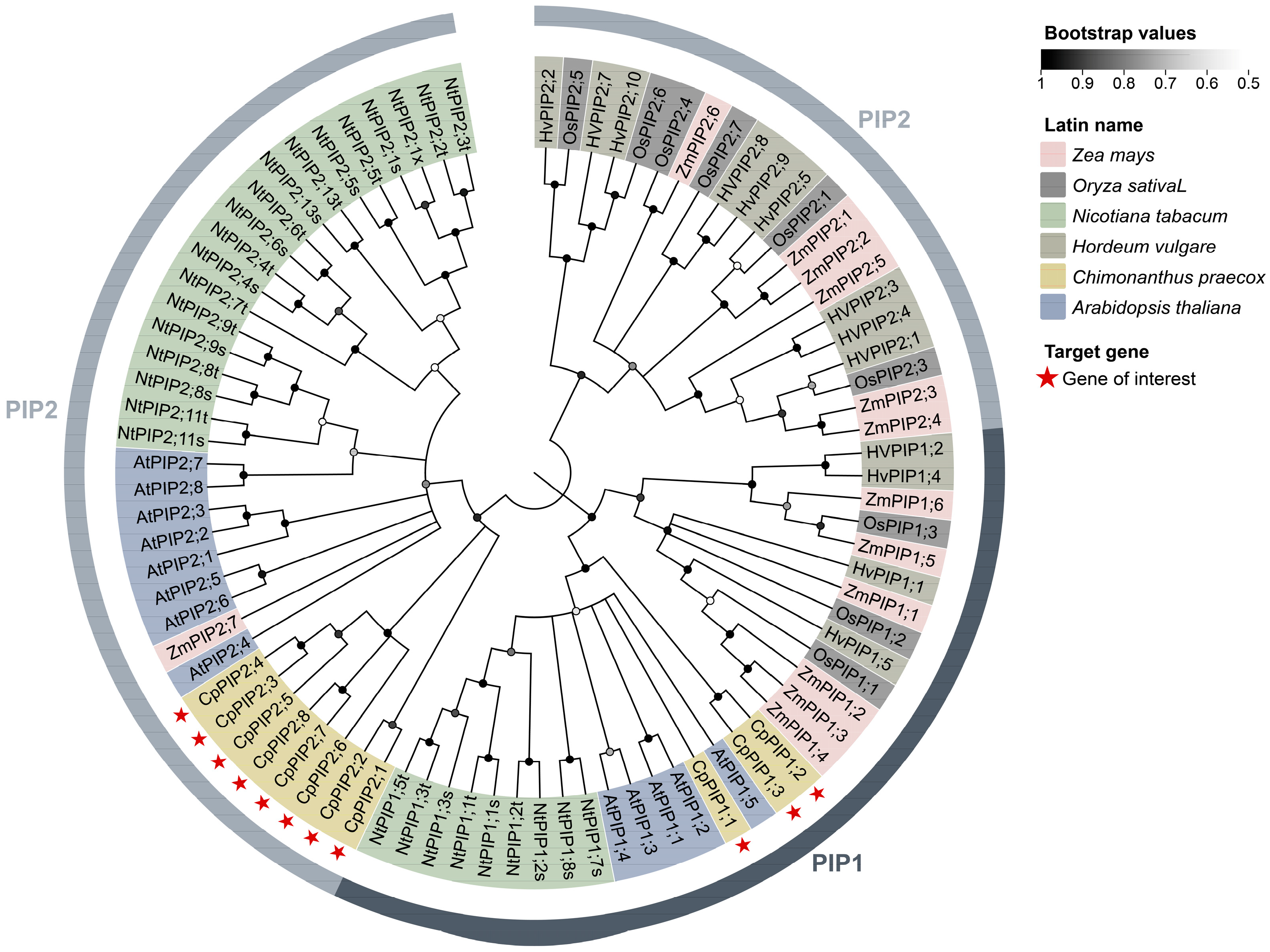
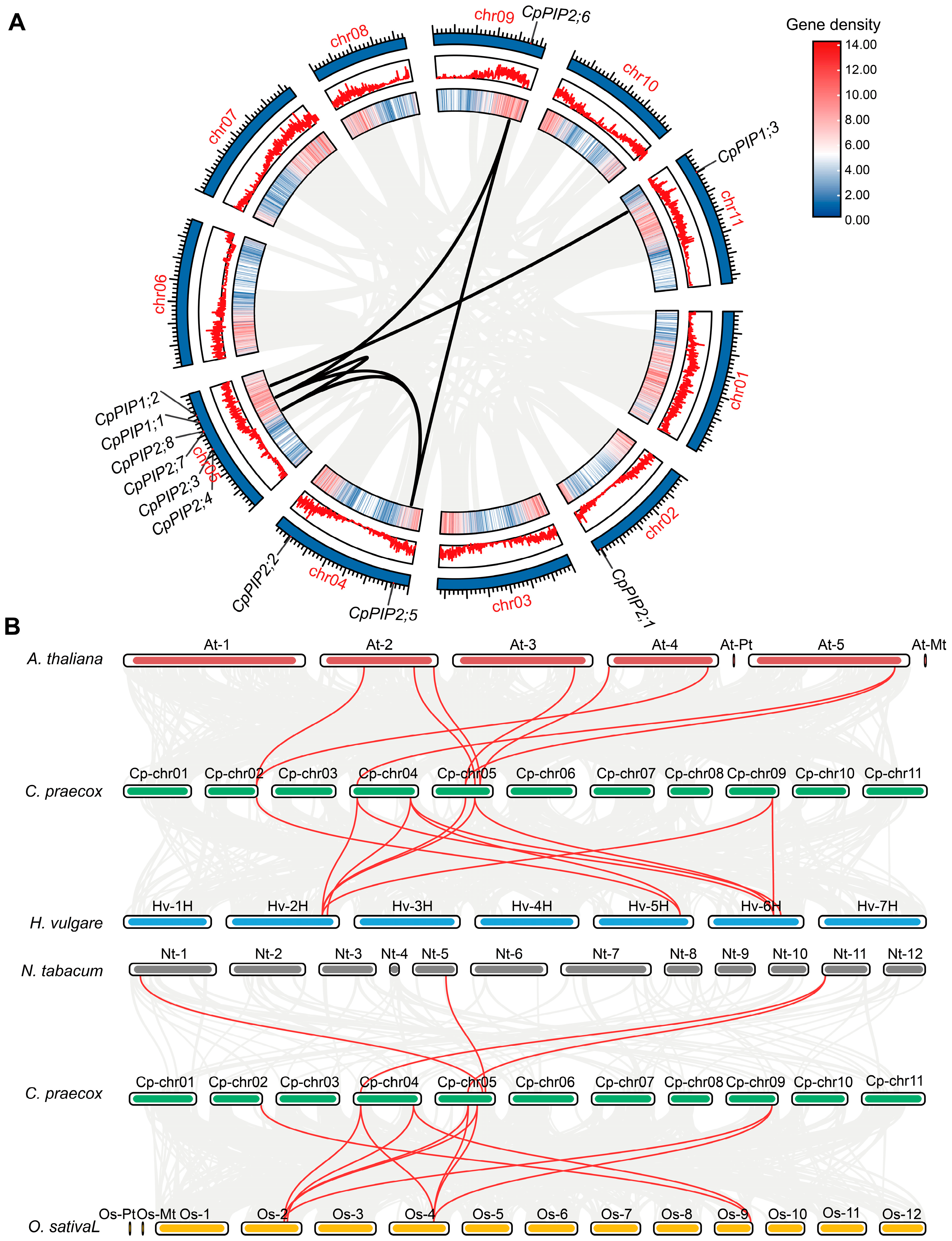
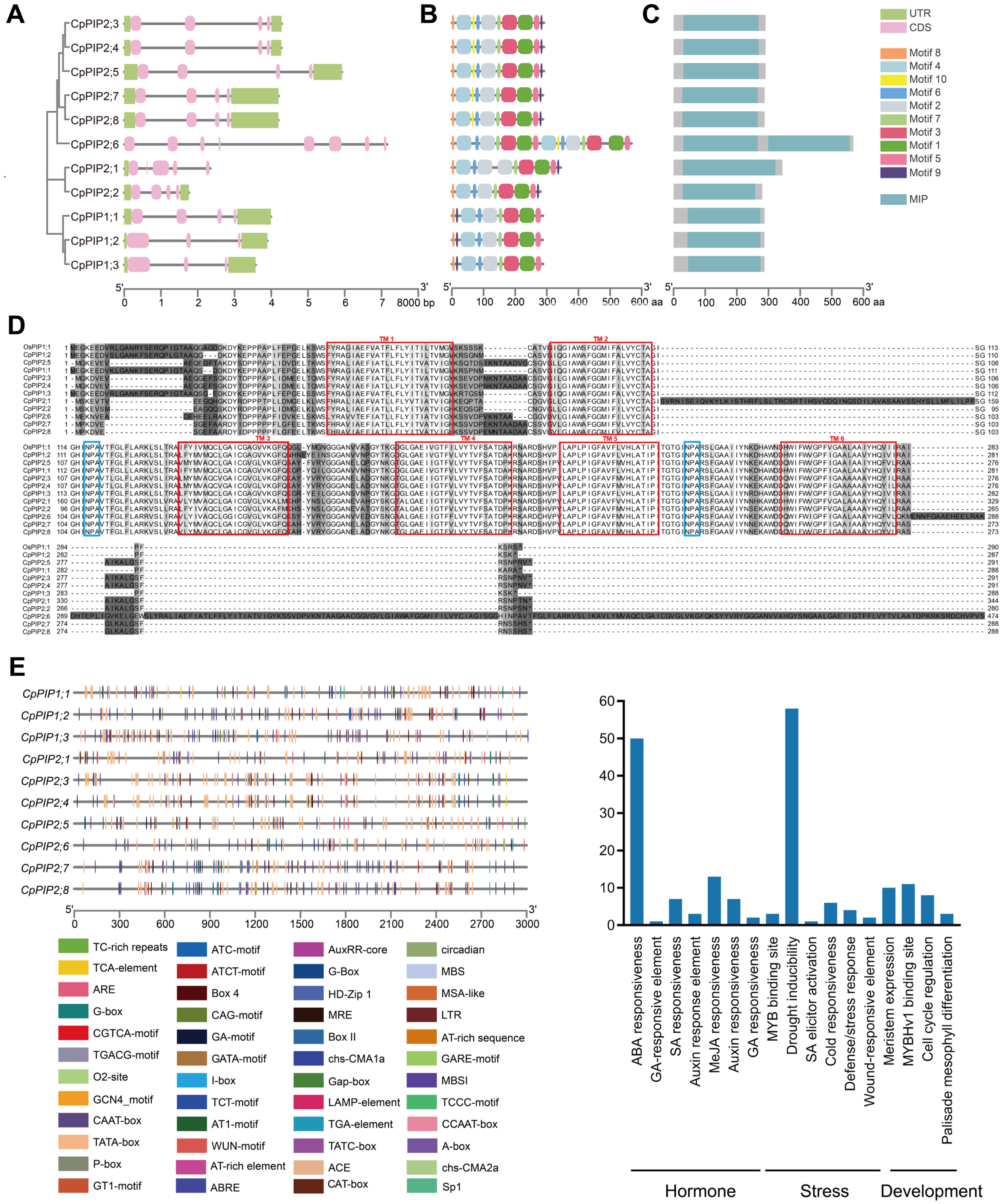
3.1. Identification, Sequence, and Phylogenetic Analysis of PIP Genes in C. praecox

In this study, we identified 11 PIP genes in the wintersweet genome. To elucidate the evolutionary relationships within the PIP gene family, we constructed a phylogenetic tree based on the full-length PIP proteins of Z. mays, O. sativa, N. tabacum, H. vulgare, A. thaliana, and C. praecox. The results revealed that the 11 CpPIPs can be classified into two subcategories, designated CpPIP1;1 to CpPIP1;3 and CpPIP2;1 to CpPIP2;8, respectively (Figure 1). Details regarding these CpPIPs, including cDNA sequences, amino acid sequences, promoter regions, chromosomal locations, ORF lengths, protein lengths, theoretical isoelectric point (pI), molecular weights, and number of introns, are presented in Tables S3 and S4. Chromosomal location analysis revealed that the CpPIPs are randomly distributed among five chromosomes, with six genes being detected on chr05, two on chr04, and one each on chr02, chr09, and chr11 (Figure 2A). Collinearity analysis of wintersweet with other species indicated that the CpPIP gene family has 8, 11, 11, and 11 pairs of collinear genes with A. thaliana, N. tabacum, O. sativa, and H. vulgare, respectively (Figure 2B).
Among the identified genes, CpPIP2;6, a tandemly duplicated gene, contains the largest number of exons (8), whereas a majority of the CpPIPs comprise four exons (Figure 3A). Motif analysis revealed that most of the CpPIPs contain motifs 1–8, whereas CpPIP1, CpPIP2;1, and CpPIP2;2 lack motif 10 (Figure 3B), and conserved domain analysis indicated that CpPIP2;6 contains two MIP domains, whereas the remaining ten CpPIP members each harbour a single MIP domain (Figure 3C). Furthermore, multiple sequence alignments of the CpPIPs revealed a high similarity between these sequences, as indicated by the presence of six conserved transmembrane domains and two nucleosome assembly Asn-Pro-Ala (NPA) motifs (Figure 3D). To assess the potential physiological roles of CpPIPs genes, we performed in silico analysis on the 3000-bp region upstream of the promoter sequences of these genes. Among the 11 CpPIP promoters, we identified a total of 219 cis-acting regulatory elements, which could be functionally categorized into hormone responsiveness, plant growth and development, and stress adaptation types (Figure 3E). Notably, all 11 CpPIPs promoters harbour at least one drought-responsive cis-element, thereby providing evidence to indicate their functional involvement in abiotic stress responses. The quantitative distributions of these regulatory elements are presented in Table S5.

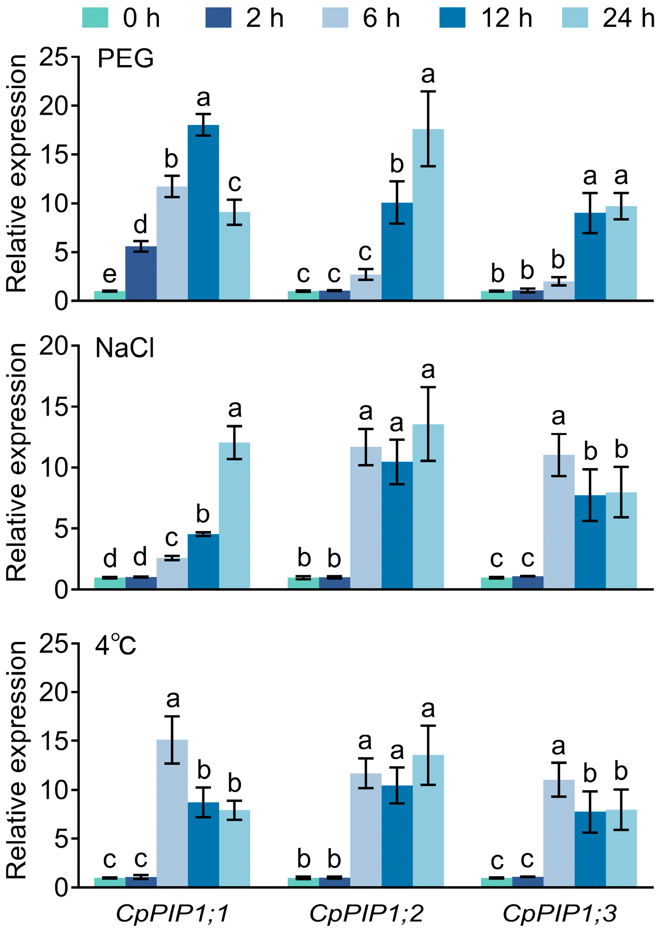
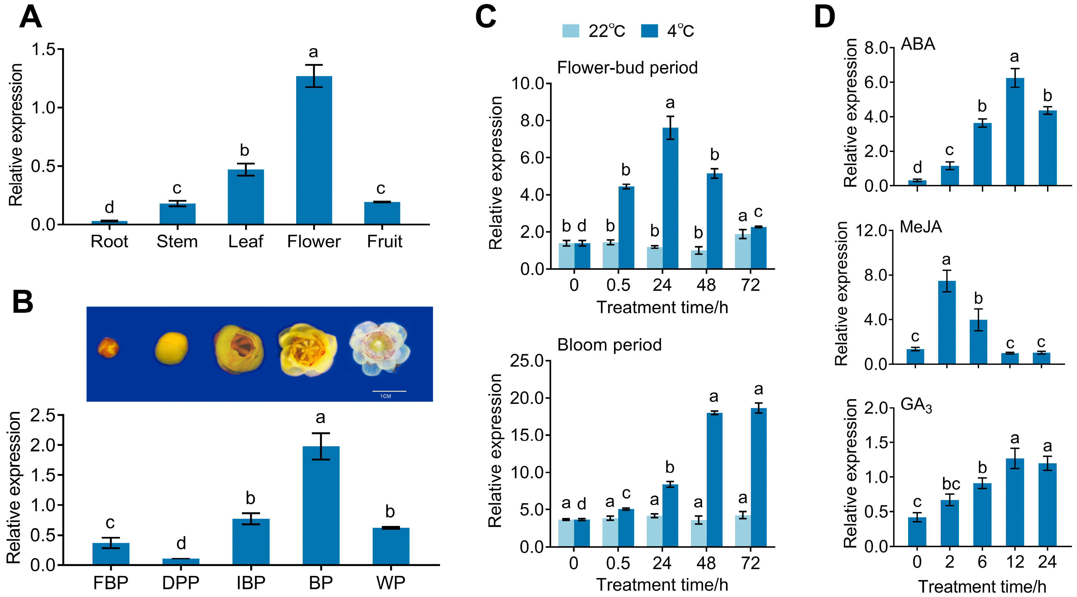
3.2. Expression Pattern of CpPIP1 Gene Family Members in Stressed Wintersweet

In terms of amino acid sequence, the three identified CpPIP1 proteins CpPIP1;1, CpPIP1;2, and CpPIP1;3 are highly similar to AtPIP1;5, which has been established to be associated with flowering and freeze–thaw tolerance (Figure 1) [27,28]. Using qRT-PCR, we assessed the expression of these three CpPIP1 genes under stress conditions, which revealed that CpPIP1;1 is characterized by a strong response to both drought and cold stress, on the basis of which we selected this gene for further investigation (Figure 4).

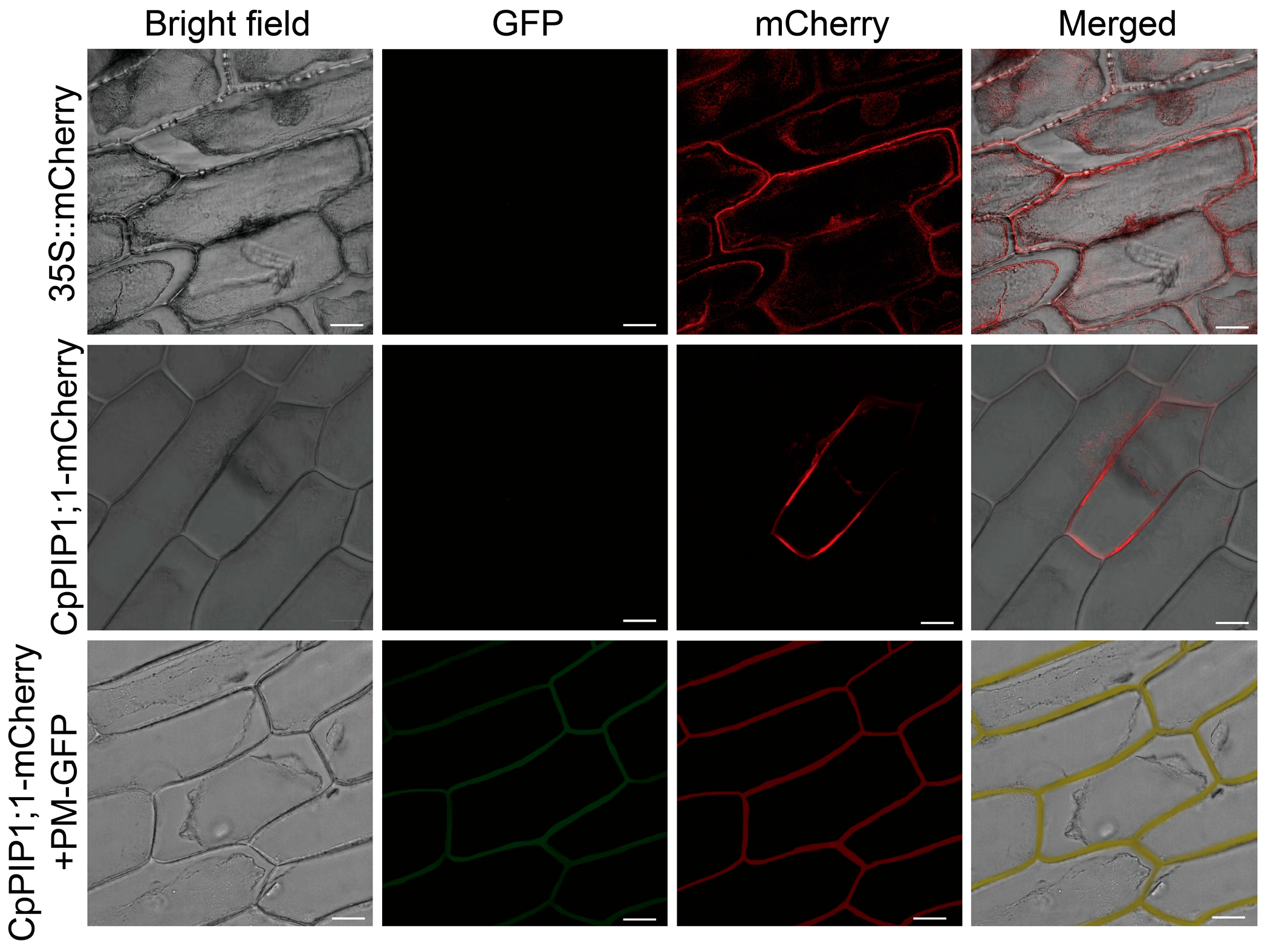
3.3. Subcellular Localization and Expression of CpPIP1;1

The full-length cDNA sequence of CpPIP1;1 (GenBank accession number OR651237) was determined, and we subsequently assessed the subcellular localization of CpPIP1;1. The detection of fluorescent signals in the membranes of A. cepa cells transiently expressing CpPIP1;1-mCherry (Figure 5).
We subsequently examined the expression of CpPIP1;1 in different organs of wintersweet plants, with the highest levels observed in flowers (Figure 6A). In response to a reduction in winter temperatures, wintersweet flowers gradually come into bloom, and we detected a corresponding gradual increase in the expression of CpPIP1;1 from the flower bud stage to the bloom period, peaking at the height of the bloom period, prior to undergoing a slight decline during subsequent senescence (Figure 6B).
To further investigate the association between CpPIP1;1 and low-temperature flowering, we exposed wintersweet vase cuttings to low-temperature treatment at 4 °C, in response to which, we detected a gradual increase in the expression of the CpPIP1;1 gene during the flower bud stage with an extension of treatment time, peaking at 24 h, prior to undergoing a subsequent decline. Similarly, during the bloom period, there was a gradual increase in the expression of CpPIP1;1 with increasing treatment time, which reached a maximum level at 72 h, at which it was approximately five-fold higher than that in the untreated samples. In contrast, during both the flower bud and bloom periods, we detected only relatively low levels of CpPIP1;1 expression at 22 °C, with no significant changes in gene expression observed over time (Figure 6C). Furthermore, in response to exogenous ABA, MeJA, and GA3 treatments, we observed an initial increase in the expression of CpPIP1;1, followed by a post-peak gradual decline (Figure 6D).

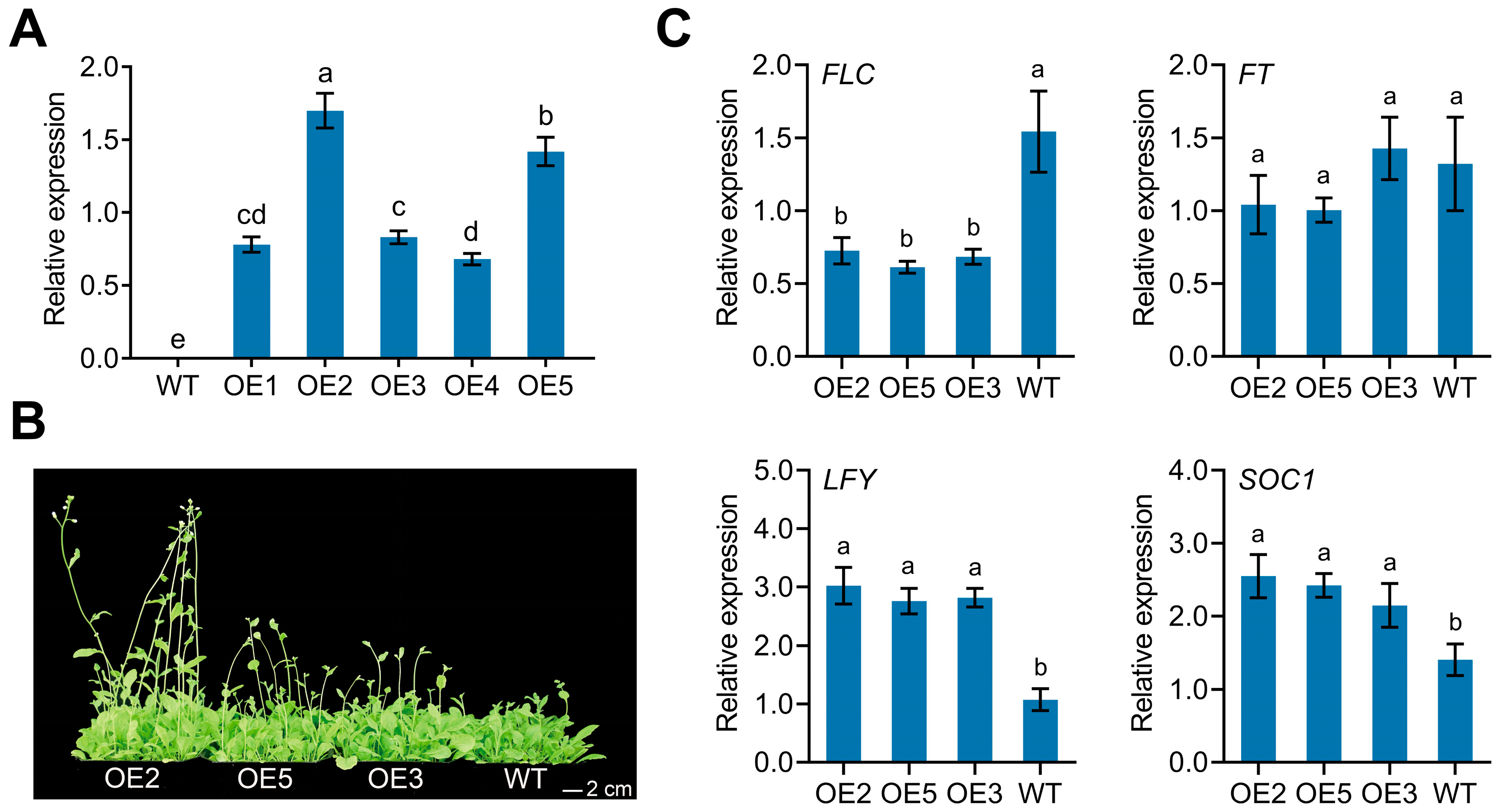
3.4. Overexpression of CpPIP1;1 Improves Flowering and Stress Tolerance in A. thaliana

Analysis of three homozygous CpPIP1;1 overexpression lines (OE2, OE5, and OE3), characterized by relatively high levels of transgene expression (Figure 7A), revealed that under standard growth conditions, compared with the WT plants, those of the OE2 line were characterized by a significantly earlier flowering phenotype (Figure 7B, Table S6). Whereas under these growth conditions, we detected no significant differences between CpPIP1;1-overexpressing and WT plants with respect to the relative expression of FLOWERING LOCUS T (FT); in contrast, there was a significantly elevated expression of SUPPRESSOR OF OVEREXPRESSION OF CO1 (SOC1) and LEAFY (LFY) in CpPIP1;1-overexpressing plants relative to that in WT plants. Conversely, there was a significant suppression of FLOWERING LOCUS C (FLC) expression in CpPIP1;1-overexpressing plants (Figure 7C).
Furthermore, in response to exposure to drought, salt, and cold stresses, transgenic plants were found to be characterized by superior growth performance compared with the WT controls (Figure 8A–C), and following 3-day post-drought and post-cold recovery periods, the survival of transgenic lines was found to be significantly greater than that of WT plants (Figure 8D). Subsequent physiological assessments revealed that compared with the WT plants, transgenic plants were characterized by a lower accumulation of MDA and less electrolyte leakage. Furthermore, under stress conditions, we detected elevated levels chlorophyll retention, an increase in proline accumulation, and enhanced SOD/POD activities in the transgenic plants (Figure 8E). In addition, following low-temperature treatment, compared with those in the WT plants, we detected a significant upregulation in the relative expression of C-REPEAT BINDING FACTOR 1/2/3 (CBF1/2/3) and COLD-REGULATED 15A/47 (COR15A/47) in the transgenic lines, whereas, contrastingly, the expression of CRPK1 was markedly downregulated in the transgenic plants (Figure 8F). Collectively, these findings thus indicate that the overexpression of CpPIP1;1 contributed to a significant enhancement of the drought, salt, and cold tolerance of transgenic A. thaliana.

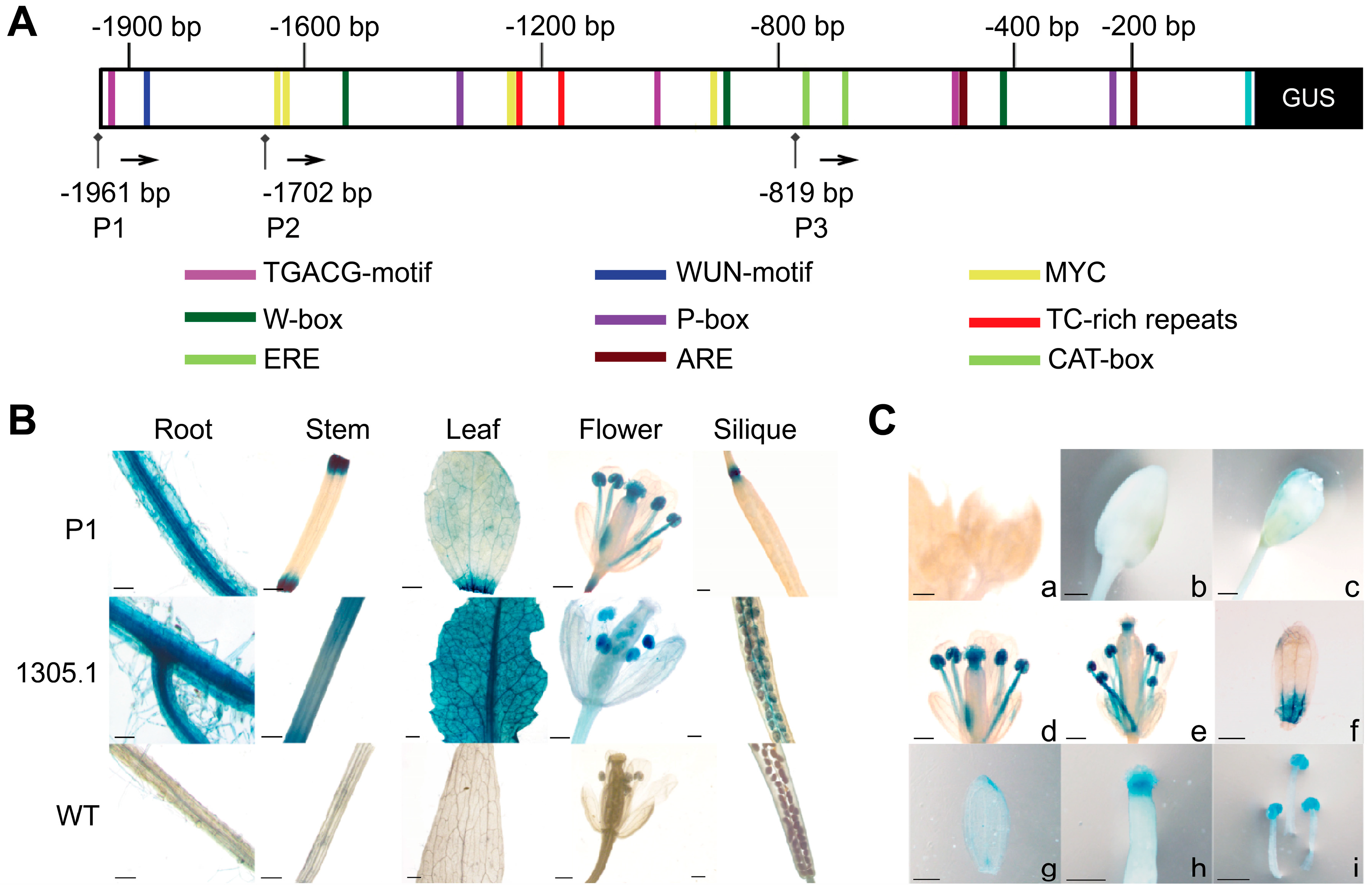
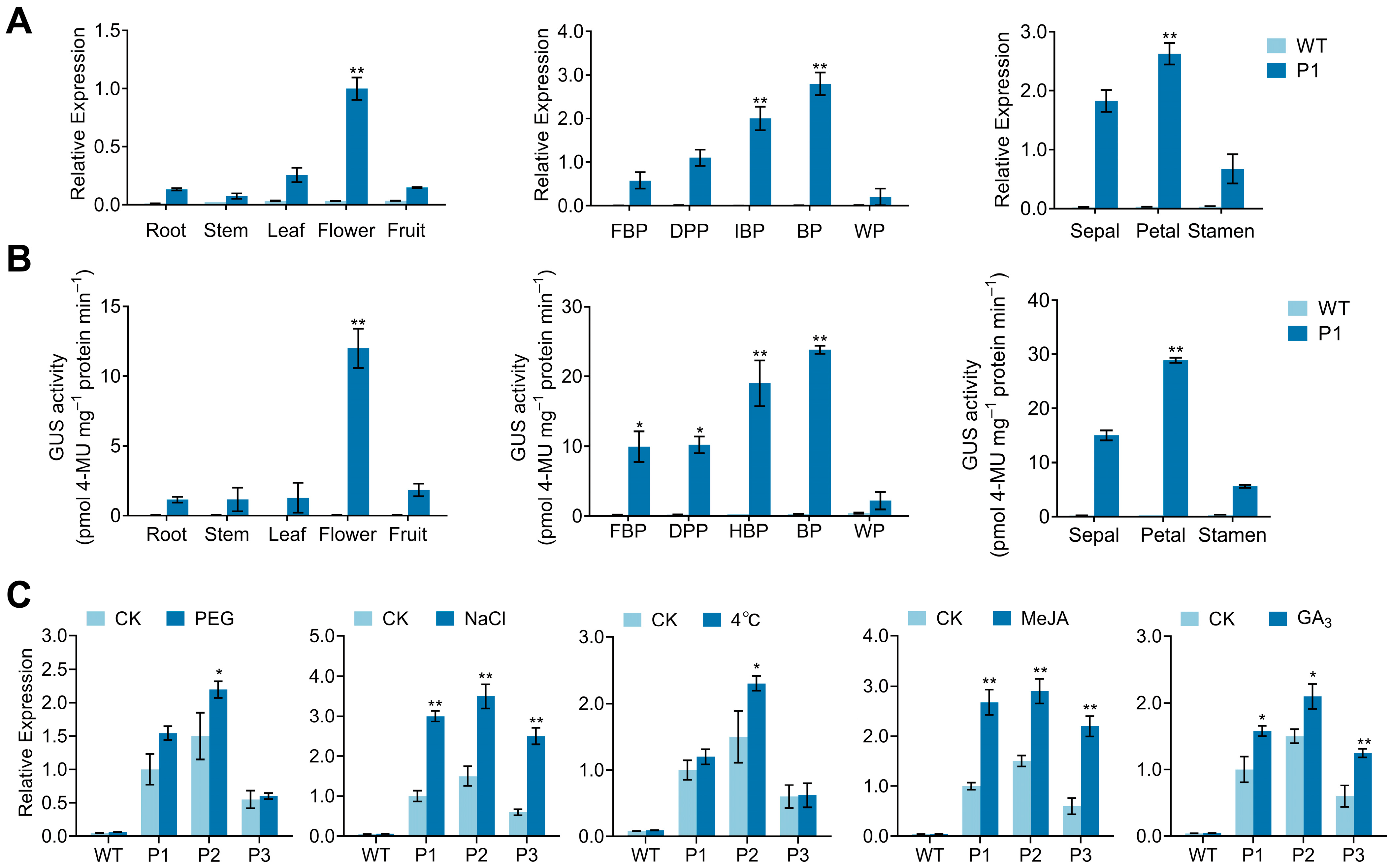
3.5. Analysis of CpPIP1;1 Promoter Cis-Acting Elements and GUS Histochemical Staining in Transgenic A. thaliana

A 1961-bp fragment upstream of the CpPIP1;1 gene (P1) and two deletions of the CpPIP1;1 promoter (P2 and P3) were cloned (Figure 9A). In the P1 region, we detected a total of 66 TATA boxes and 35 CAAT boxes, which are core promoter elements that enhance the efficiency of eukaryotic promoters. In addition, a number of hormone and stress response elements were identified, including two P-boxes, three CGTCA motifs, three MYB domains, and four MYC domains (Table S7). To assess the patterns of GUS gene expression driven by P1, we performed histochemical analyses of T3 transgenic A. thaliana lines throughout the developmental period, the results of which revealed expression of the GUS gene in the roots, leaves, and floral organs (Figure 9B). Notably, although we detected only a weak staining intensity in flower buds during the early inflorescence stage, as flowers bloomed, we observed a significant increase in GUS activity throughout the period of inflorescence development, including in the sepals, petals, stamens, and pistils (Figure 9C).

3.6. Analysis of CpPIP1;1 Promoter Expression and Activity in Tobacco

To further analyse promoter properties, we investigated the expression and activity of GUS driven by the CpPIP1;1 promoter in tobacco, the findings of which indicated that compared with other tissues, the most pronounced expression of GUS was detected in the floral organs. Notably, expression of the promoter increased gradually with a progressive opening of the flower, peaking during the bloom period, and subsequently undergoing a rapid decline during the period of senescence, with the strongest expression during the bloom period being detected in the petals, which was found to correspond to the activity of the promoter (Figure 10A,B).
Salt treatment led to a significant upregulation of GUS gene expression driven by the three assessed promoter fragments, resulting in a three-fold increase in the expression of this gene. Notably, in response to cold and drought treatment, we found that compared with that promoted by the both P1 and P3 fragments, the P2 promoter fragment induced a greater upregulation of GUS expression. In addition, all three promoter fragments were characterized by significant responses to MeJA treatment, although no substantial differences were observed. Contrastingly, although all fragments responded to exposure to GA3, whereas P1 and P2 showed similar responses, the response of P1 differed significantly from that of P3 (Figure 10C).

4. Discussion

PIPs, a family of aquaporin proteins, have been established to play essential roles in the spatial and temporal regulation of water transport across the plasma membrane [29]. A correlation exists between high levels of PIP expression and developmental processes that require significant water intake, including root elongation and expansion, anther dehiscence, and petal expansion [30]. To date, however, the direct involvement of PIPs in petal expansion under cold stress conditions has received limited attention. In this study, we identified 11 CpPIP genes within the wintersweet genome, among which CpPIP2;6 was a tandem duplication, potentially arising via evolutionary duplication events. Such mechanisms tend to be evolutionarily conserved in plant genomes, with similar patterns being observed in other AQP gene families. Notable examples include leucine-rich repeat domain expansions in asparagus and paralogue clusters of RsAQP within chromosomal linkage groups in radish (Raphanus sativus L.) [31,32,33]. Consistent with AQPs, CpPIPs contain six conserved transmembrane domains and two NPA motifs. Phylogenetic analysis revealed that CpPIP1;1 is most closely (91%) related to AtPIP1;5, a protein involved in the flowering of A. thaliana [27], and among the AtPIP1 proteins, AtPIP1;5 has been found to have the highest relative phenotype ranking for water transport efficiency, and is also associated with freeze–thaw tolerance [28].
PIPs are characterized by tissue-specific expression patterns, thereby reflecting their diverse functions [34]. During flower development, the expansion of petals is dependent on cell expansion mediated by PIP-facilitated water transport [35]. For example, in lilies, it has been found that the AqpL1 gene is highly expressed in young petals, whereas expression is significantly reduced during petal senescence [36]. In the present study, we detected the highest levels of CpPIP1;1 expression in the flowers of wintersweet, which peaked during the period blooming (Figure 6A,B). Consistent with these observations, in transgenic tobacco, we demonstrated that the CpPIP1;1 promoter drives GUS reporter gene expression predominantly in the floral organs, with peak activity being detected during flower opening (Figure 10A,B). Furthermore, we identified two P-boxes, which serve as MYB protein-binding sites, in the promoter region. P-box elements have also been established to direct flower-specific expression [37]. The potential role of these sites in floral-specific regulation is supported by their involvement in flower-related gene expression, as has been reported for FSP061 in Brassica napus L. [38].
Among the floral organs of transgenic tobacco, we detected the strongest CpPIP1;1 promoter activity in petals during the bloom period (Figure 10A,B), and compared with WT plants, A. thaliana plants transformed with CpPIP1;1 were found to be characterized by an early flowering phenotype (Figure 7B). The regulation of early floral development is governed by a number of key genes, among which, SOC1 and LFY (floral induction factors), FLC (a floral repressor), and FT (which facilitates the shift between developmental and flowering stages) are well characterized [16,39,40]. In the present study, we found that CpPIP1;1 promotes the upregulated expression of SOC1 and LFY, although had no appreciable effects on FT levels. Conversely, CpPIP1;1 was observed to downregulate FLC, a known floral development inhibitor. These findings accordingly provided evidence to indicate that in wintersweet, the expression of CpPIP1;1 is closely associated with petal expansion and flower development.
Non-winter flowering plants are often dependent on storage organs, such as corms and tubers, to withstand external cold stress, given that their floral organs have limited cold resistance and poor survival in winter. Contrastingly, wintersweet is characterized by notable cold resistance, as evidenced by the maintenance of normal blooming, even during the depths of winter. As ambient temperatures declined during winter, we observed a progressive upregulation of CpPIP1;1 expression in wintersweet floral organs, which was positively correlated with the progression of flowering, potentially reflecting a heightening of hydraulic demands during floral development (Figure 6B,C). This increased water requirement is assumed to reflect an adaptation to winter droughts and low-temperature environments. In this regard, it has been established that the regulation of PIP genes under water deficit conditions involves both ABA-dependent and -independent mechanisms [41]. In the present study, we detected the upregulated expression of CpPIP1;1 expression in wintersweet in response to ABA treatment (Figure 6D), thereby providing evidence for ABA-mediated regulation of flowering under conditions of water deficit. In this context, it has been demonstrated that the MYC/MYB pathway, an ABA-dependent signalling cascade, activates drought-induced genes [42], and, notably, we detected both MYC and MYB recognition motifs in the CpPIP1;1 promoter (Table S7), thereby implying potential regulation by MYC/MYB transcription factors.
The findings of previous studies have indicated that PIPs are also regulated by MeJA and GA3, as evidenced by the GA3-induced downregulation of RsPIP2 in radish roots and the time-dependent MeJA modulation of AtPIP1;5 expression [43,44]. GA3 has also been found to enhance RhPIP2;1 promoter activity in moonflower [45], and, consistently, in wintersweet, we detected an upregulation of CpPIP1;1 expression in response to GA3, thus indicating the involvement in GA3 signalling. Furthermore, CpPIP1;1 promoter activity has been shown to respond to MeJA and GA3 treatment (Figure 10C), with rapid changes in MeJA-induced expression being detected within 2 h, indicating potential targeting by MeJA signalling (Figure 6D).
PIP proteins have been established to play key roles in the responses of plants to abiotic stresses [46]. Consistently, we detected a significant induction of CpPIP1;1 expression and promoter activity in wintersweet plants under drought, salt, and cold stress conditions (Figure 4). We performed heterologous overexpression of CpPIP1;1 in A. thaliana, thereby generating transgenic lines for phenotypic analysis. Under stress conditions, these lines outperformed the WT plants, showing significantly lower relative conductivity and MDA contents (Figure 8E), thus indicating enhanced membrane integrity and reduced oxidative damage [47]. Concurrently, compared with the WT plants, these transgenic plants were found to accumulate higher levels of chlorophyll and proline (Figure 8E).
In the context of cold adaptation by plants, the CBF-COR pathway has been identified as a key regulatory mechanism that mediates cold tolerance via the CBF-dependent activation of COR expression in response to low-temperature stress, the activity of which is negatively regulated by CRPK1 [48]. Our experimental results revealed that in response to cold treatment, transgenic lines showed a marked upregulated expression of both CBF and COR relative to that in WT plants, accompanied by a concurrent downregulation of CRPK1 expression. Collectively, these findings indicate that transgenic A. thaliana plants overexpressing CpPIP1;1 are characterized by enhanced resistance to multiple abiotic stressors. Moreover, in response to drought and cold stress, the relative expression of GUS in plants transformed with the P2 construct was higher than that in plants transformed with P1 or P3. These findings thus provide evidence of negative regulatory elements within the 259-bp fragment between P1 and P2 and indicate that a potential region-specific enhancer may also be present within the fragment between P2 and P3.

5. Conclusions

In this study, in which we sought to characterize the regulation of flowering in the winter-flowering plant wintersweet, we identified 11 CpPIP genes, which can be classified into two subfamilies. By analysing the expression patterns of CpPIP1;1 in different tissues and overexpressing CpPIP1;1 in A. thaliana, we elucidated the involvement of CpPIP1;1 in flower development and its specific role in responses to low temperatures and osmotic stress. Our findings regarding the role played by CpPIP1;1 in enhancing cold resistance in the floral organs of wintersweet plants will contribute to broadening our understanding of the function of PIPs in floral development and stress tolerance.

Supplementary Materials

The following supporting information can be downloaded at: https://www.mdpi.com/article/10.3390/horticulturae11060581/s1, title; Table S1: Primers used for CpPIP1;1 PCR and qRT-PCR; Table S2: The online software for bioinformatics analysis; Table S3: The detailed information of CpPIP genes identified in wintersweet genome; Table S4: cDNA, amino acid sequence, and promoter region of CpPIP genes; Table S5: Cis-acting regulatory elements analysis of CpPIP genes sequence; Table S6: Phenotypic traits in T3 transgenic lines of A. thaliana overexpressing CpPIP1 and the wild type; Table S7: Cis-acting regulatory elements analysis of CpPIP1;1 gene promoter sequence.

Author Contributions

F.R.: Writing—original draft, investigation, formal analysis, conceptualization. Z.F.: Software, formal analysis, conceptualization. G.W.: Writing—review and editing. Y.L.: Investigation, data curation. J.Z.: Investigation. Y.D.: Validation. S.S.: Funding acquisition. J.M.: Writing—review and editing, funding acquisition, conceptualization, project administration. All authors have read and agreed to the published version of the manuscript.

Funding

This research was financially supported by the National Natural Science Foundation of China (Grant No. 32271937), and Chongqing Science and Technology Bureau Project (CSTB2023TIAD-KPX0039).

Data Availability Statement

Data are contained within the article.

Conflicts of Interest

The authors declare no conflicts of interest.

References

  1. Paluch-Lubawa, E.; Polcyn, W. Tissue-specific accumulation of PIP aquaporins of a particular heteromeric composition is part of the maize response to mycorrhiza and drought. Sci. Rep. 2024, 14, 21712. [Google Scholar] [CrossRef] [PubMed]
  2. Liu, H.; Yang, L.; Xin, M.; Ma, F.; Liu, J. Gene-Wide Analysis of Aquaporin Gene Family in Malus domestica and Heterologous Expression of the Gene MpPIP2;1 Confers Drought and Salinity Tolerance in Arabidposis thaliana. Int. J. Mol. Sci. 2019, 20, 3710. [Google Scholar] [CrossRef]
  3. Li, S.; Wang, L.; Zhang, Y.; Zhu, G.; Zhu, X.; Xia, Y.; Li, J.; Gao, X.; Wang, S.; Zhang, J.; et al. Genome-Wide Identification and Function of Aquaporin Genes During Dormancy and Sprouting Periods of Kernel-Using Apricot (Prunus armeniaca L.). Front. Plant Sci. 2021, 12, 690040. [Google Scholar] [CrossRef] [PubMed]
  4. Zhang, J.; Wen, W.; Li, H.; Lu, Q.; Xu, B.; Huang, B. Overexpression of an aquaporin gene PvPIP2;9 improved biomass yield, protein content, drought tolerance and water use efficiency in switchgrass (Panicum virgatum L.). Glob. Change Biol. Bioenergy 2020, 12, 979–991. [Google Scholar] [CrossRef]
  5. Ayadi, M.; Brini, F.; Masmoudi, K. Overexpression of a Wheat Aquaporin Gene, TdPIP2;1, Enhances Salt and Drought Tolerance in Transgenic Durum Wheat cv. Maali. Int. J. Mol. Sci. 2019, 20, 2389. [Google Scholar] [CrossRef]
  6. Fan, S.; Han, N.; Wu, H.; Jia, J.; Guo, J. Plasma membrane intrinsic protein SlPIP1;7 promotes root growth and enhances drought stress tolerance in transgenic tomato (Solanum lycopersicum) plants. Plant Breed. 2021, 140, 1102–1114. [Google Scholar] [CrossRef]
  7. Rabeh, K.; Sallami, A.; Gaboun, F.; Filali-Maltouf, A.; Sbabou, L.; Belkadi, B. Genome-wide analysis of aquaporin and their responses to abiotic stresses in plants: A systematic review and meta-analysis. Plant Stress 2024, 11, 100362. [Google Scholar] [CrossRef]
  8. Chen, W.; Yin, X.; Wang, L.; Tian, J.; Yang, R.; Liu, D.; Yu, Z.; Ma, N.; Gao, J. Involvement of rose aquaporin RhPIP1;1 in ethylene-regulated petal expansion through interaction with RhPIP2;1. Plant Mol. Biol. 2013, 83, 219–233. [Google Scholar] [CrossRef]
  9. Zhang, S.; Feng, M.; Chen, W.; Zhou, X.; Lu, J.; Wang, Y.; Li, Y.; Jiang, C.-Z.; Gan, S.-S.; Ma, N.; et al. In rose, transcription factor PTM balances growth and drought survival via PIP2;1 aquaporin. Nat. Plants 2019, 5, 290–299. [Google Scholar] [CrossRef]
  10. Tong, Z.; Li, Q.; Yang, Y.; Dai, F.; Gao, J.; Hong, B. Isolation and expression analysis of LoPIP2, a lily (Lilium Oriental Hybrids) aquaporin gene involved in desiccation-induced anther dehiscence. Sci. Hortic. 2013, 164, 316–322. [Google Scholar] [CrossRef]
  11. Nemoto, K.; Niinae, T.; Goto, F.; Sugiyama, N.; Watanabe, A.; Shimizu, M.; Shiratake, K.; Nishihara, M. Calcium-dependent protein kinase 16 phosphorylates and activates the aquaporin PIP2;2 to regulate reversible flower opening in Gentiana scabra. Plant Cell 2022, 34, 2652–2670. [Google Scholar] [CrossRef]
  12. Kong, W.; Bendahmane, M.; Fu, X. Genome-Wide Identification and Characterization of Aquaporins and Their Role in the Flower Opening Processes in Carnation (Dianthus caryophyllus). Molecules 2018, 23, 1895. [Google Scholar] [CrossRef]
  13. Lv, Y.; Xie, M.; Zhou, S.; Wen, B.; Sui, S.; Li, M.; Ma, J. CpCAF1 from Chimonanthus praecox Promotes Flowering and Low-Temperature Tolerance When Expressed in Arabidopsis thaliana. Int. J. Mol. Sci. 2023, 24, 12945. [Google Scholar] [CrossRef] [PubMed]
  14. Xue, J.; Yang, F.; Gao, J. Isolation of Rh-TIP1;1, an aquaporin gene and its expression in rose flowers in response to ethylene and water deficit. Postharvest Biol. Technol. 2009, 51, 407–413. [Google Scholar] [CrossRef]
  15. Wu, C.L.; Hu, N.Z. Studies on the flower form and blooming characteristics of the wintersweet. Acta Hortic. Sin. 1995, 22, 277–282. [Google Scholar]
  16. Huang, R.; Sui, S.; Liu, H.; Li, M.; Liu, D. Overexpression of CpWRKY75 from Chimonanthus praecox Promotes Flowering Time in Transgenic Arabidopsis. Genes 2022, 13, 68. [Google Scholar] [CrossRef] [PubMed]
  17. Siegel, C.S.; Stevenson, F.O.; Zimmer, E.A. Evaluation and comparison of FTA card and CTAB DNA extraction methods for non-agricultural taxa. Appl. Plant Sci. 2017, 5, 1600109. [Google Scholar] [CrossRef] [PubMed]
  18. Jiang, Y.; Chen, F.; Song, A.; Zhao, Y.; Chen, X.; Gao, Y.; Wei, G.; Zhang, W.; Guan, Y.; Fu, J. The genome assembly of Chimonanthus praecox var. concolor and comparative genomic analysis highlight the genetic basis underlying conserved and variable floral traits of wintersweet. Ind. Crops Prod. 2023, 206, 117603. [Google Scholar] [CrossRef]
  19. Li, Y.; Zhang, J.; Wang, S.; Liu, Y.; Yang, M.; Huang, Y. Genome-wide identification of the Pyrus R2R3-MYB gene family and PhMYB62 regulation analysis in Pyrus hopeiensis flowers at low temperature. Int. J. Biol. Macromol. 2024, 257, 128611. [Google Scholar] [CrossRef]
  20. Deng, Y.Y.; Lin, Y.; Wei, G.; Hu, X.Q.; Zheng, Y.H.; Ma, J. Overexpression of the CpCOR413PM1 Gene from Wintersweet (Chimonanthus praecox) Enhances Cold and Drought Tolerance in Arabidopsis. Horticulturae 2024, 10, 599. [Google Scholar] [CrossRef]
  21. Clough, S.J.; Bent, A.F. Floral dip: A simplified method for Agrobacterium-mediated transformation of Arabidopsis thaliana. Plant J. 1998, 16, 735–743. [Google Scholar] [CrossRef] [PubMed]
  22. Lin, J.; Liu, D.; Wang, X.; Ahmed, S.; Li, M.; Kovinich, N.; Sui, S. Transgene CpNAC68 from Wintersweet (Chimonanthus praecox) Improves Arabidopsis Survival of Multiple Abiotic Stresses. Plants 2021, 10, 1403. [Google Scholar] [CrossRef] [PubMed]
  23. Tian, C.; Zhang, Z.Y.; Huang, Y.; Xu, J.J.; Liu, Z.; Xiang, Z.M.; Zhao, F.L.; Xue, J.P.; Xue, T.; Duan, Y.B. Functional characterization of the Pinellia ternata cytoplasmic class II small heat shock protein gene PtsHSP17.2 via promoter analysis and overexpression in tobacco. Plant Physiol. Biochem. 2022, 177, 1–9. [Google Scholar] [CrossRef]
  24. Seliem, M.; El-Mahrouk, M.; El-Banna, A.; Hafez, Y.; Dewir, Y. Micropropagation of Philodendron selloum: Influence of copper sulfate on endophytic bacterial contamination, antioxidant enzyme activity, electrolyte leakage, and plant survival. S. Afr. J. Bot. 2021, 139, 230–240. [Google Scholar] [CrossRef]
  25. Jefferson, R.A. Assaying chimeric genes in plants: The GUS gene fusion system. Plant Mol. Biol. Report. 1987, 5, 387–405. [Google Scholar] [CrossRef]
  26. Bradford, M.M. A rapid and sensitive method for the quantitation of microgram quantities of protein utilizing the principle of protein-dye binding. Anal. Biochem. 1976, 72, 248–254. [Google Scholar] [CrossRef]
  27. Quigley, F.; Rosenberg, J.M.; Shachar-Hill, Y.; Bohnert, H.J. From genome to function: The Arabidopsis aquaporins. Genome Biol. 2001, 3, 1–17. [Google Scholar] [CrossRef]
  28. Groszmann, M.; De Rosa, A.; Chen, W.; Qiu, J.; McGaughey, S.A.; Byrt, C.S.; Evans, J.R. A high-throughput yeast approach to characterize aquaporin permeabilities: Profiling the Arabidopsis PIP aquaporin sub-family. Front. Plant Sci. 2023, 14, 1078220. [Google Scholar] [CrossRef]
  29. Hachez, C.; Moshelion, M.; Zelazny, E.; Cavez, D.; Chaumont, F. Localization and quantification of plasma membrane aquaporin expression in maize primary root: A clue to understanding their role as cellular plumbers. Plant Mol. Biol. 2006, 62, 305–323. [Google Scholar] [CrossRef]
  30. Maistriaux, L.C.; Laurent, M.J.; Jeanguenin, L.; Prado, S.A.; Nader, J.; Welcker, C.; Charcosset, A.; Tardieu, F.; Nicolas, S.D.; Chaumont, F. Genetic variability of aquaporin expression in maize: From eQTLs to a MITE insertion regulating PIP2;5 expression. Plant Physiol. 2024, 196, 368–384. [Google Scholar] [CrossRef]
  31. Yi, X.F.; Sun, X.C.; Tian, R.; Li, K.X.; Ni, M.; Ying, J.L.; Xu, L.; Liu, L.W.; Wang, Y. Genome-Wide Characterization of the Aquaporin Gene Family in Radish and Functional Analysis of RsPIP2-6 Involved in Salt Stress. Front. Plant Sci. 2022, 13, 860742. [Google Scholar] [CrossRef] [PubMed]
  32. Die, J.V.; Castro, P.; Millán, T.; Gil, J. Segmental and Tandem Duplications Driving the Recent NBS-LRR Gene Expansion in the Asparagus Genome. Genes 2018, 9, 568. [Google Scholar] [CrossRef]
  33. He, W.; Liu, M.; Qin, X.; Liang, A.; Chen, Y.; Yin, Y.; Qin, K.; Mu, Z. Genome-Wide Identification and Expression Analysis of the Aquaporin Gene Family in Lycium barbarum during Fruit Ripening and Seedling Response to Heat Stress. Curr. Issues Mol. Biol. 2022, 44, 5933–5948. [Google Scholar] [CrossRef]
  34. Shibasaka, M.; Horie, T.; Katsuhara, M. Mechanisms Activating Latent Functions of PIP Aquaporin Water Channels via the Interaction between PIP1 and PIP2 Proteins. Plant Cell Physiol. 2021, 62, 92–99. [Google Scholar] [CrossRef]
  35. Guterman, I.; Shalit, M.; Menda, N.; Piestun, D.; Dafny-Yelin, M.; Shalev, G.; Bar, E.; Davydov, O.; Ovadis, M.; Emanuel, M. Rose scent: Genomics approach to discovering novel floral fragrance–related genes. Plant Cell 2002, 14, 2325–2338. [Google Scholar] [CrossRef]
  36. Ding, X.; Iwasaki, I.; Kitagawa, Y. Overexpression of a lily PIP1 gene in tobacco increased the osmotic water permeability of leaf cells. Plant Cell Environ. 2004, 27, 177–186. [Google Scholar] [CrossRef]
  37. Kobayashi, H.; Oikawa, Y.; Koiwa, H.; Yamamura, S. Flower-specific gene expression directed by the promoter of a chalcone synthase gene from Gentiana triflora in Petunia hybrida. Plant Sci. 1998, 131, 173–180. [Google Scholar] [CrossRef]
  38. Li, Y.; Dong, C.; Hu, M.; Bai, Z.; Tong, C.; Zuo, R.; Liu, Y.; Cheng, X.; Cheng, M.; Huang, J.; et al. Identification of Flower-Specific Promoters through Comparative Transcriptome Analysis in Brassica napus. Int. J. Mol. Sci. 2019, 20, 5949. [Google Scholar] [CrossRef] [PubMed]
  39. Gu, X.F.; Le, C.; Wang, Y.Z.; Li, Z.C.; Jiang, D.H.; Wang, Y.Q.; He, Y.H. Arabidopsis FLC clade members form flowering-repressor complexes coordinating responses to endogenous and environmental cues. Nat. Commun. 2013, 4, 1947. [Google Scholar] [CrossRef]
  40. Monniaux, M.; McKim, S.M.; Cartolano, M.; Thévenon, E.; Parcy, F.; Tsiantis, M.; Hay, A. Conservation vs divergence in LEAFY and APETALA1 functions between Arabidopsis thaliana and Cardamine hirsuta. New Phytol. 2017, 216, 549–561. [Google Scholar] [CrossRef]
  41. Lian, H.-L.; Yu, X.; Lane, D.; Sun, W.-N.; Tang, Z.-C.; Su, W.-A. Upland rice and lowland rice exhibited different PIP expression under water deficit and ABA treatment. Cell Res. 2006, 16, 651–660. [Google Scholar] [CrossRef] [PubMed]
  42. Aslam, M.M.; Deng, L.; Meng, J.; Wang, Y.; Pan, L.; Niu, L.; Lu, Z.; Cui, G.; Zeng, W.; Wang, Z. Characterization and expression analysis of basic leucine zipper (bZIP) transcription factors responsive to chilling injury in peach fruit. Mol. Biol. Rep. 2023, 50, 361–376. [Google Scholar] [CrossRef]
  43. Bae, E.-K.; Lee, H.; Lee, J.-S.; Noh, E.-W. Drought, salt and wounding stress induce the expression of the plasma membrane intrinsic protein 1 gene in poplar (Populus alba× P. tremula var. glandulosa). Gene 2011, 483, 43–48. [Google Scholar] [CrossRef]
  44. Lee, S.H.; Zwiazek, J.J. Regulation of water transport in Arabidopsis by methyl jasmonate. Plant Physiol. Biochem. 2019, 139, 540–547. [Google Scholar] [CrossRef] [PubMed]
  45. Li, Y.; Wu, Z.; Ma, N.; Gao, J. Regulation of the rose Rh-PIP2;1 promoter by hormones and abiotic stresses in Arabidopsis. Plant Cell Rep. 2009, 28, 185–196. [Google Scholar] [CrossRef] [PubMed]
  46. Jiang, C.; Song, X.Q.; He, H.; Chu, L.W.; Zhou, H.J.; Zhao, Y.Q.; Xu, Y.J.; Zeng, W.; Lin, X.C.; Lu, M.Z. Genome-wide identification of plasma membrane aquaporin gene family in Populus and functional identification of PIP1;1 involved in osmotic stress. Environ. Exp. Bot. 2020, 179, 104200. [Google Scholar] [CrossRef]
  47. He, W.-D.; Gao, J.; Dou, T.-X.; Shao, X.-H.; Bi, F.-C.; Sheng, O.; Deng, G.-M.; Li, C.-Y.; Hu, C.-H.; Liu, J.-H. Early cold-induced peroxidases and aquaporins are associated with high cold tolerance in Dajiao (Musa spp.‘Dajiao’). Front. Plant Sci. 2018, 9, 282. [Google Scholar] [CrossRef]
  48. Peng, Y.; Ming, Y.H.; Jiang, B.C.; Zhang, X.Y.; Fu, D.Y.; Lin, Q.H.; Zhang, X.Y.; Wang, Y.; Shi, Y.T.; Gong, Z.Z.; et al. Differential phosphorylation of Ca2+-permeable channel CYCLIC NUCLEOTIDE-GATED CHANNEL20 modulates calcium-mediated freezing tolerance in Arabidopsis. Plant Cell 2024, 36, 4356–4371. [Google Scholar] [CrossRef]
Figure 1. Phylogenetic relationships of PIPs genes in A. thaliana, Z. mays, O. sativa, N. tabacum, H. vulgare, and C. praecox.
Horticulturae 11 00581 g001
Figure 2. Synteny analysis of PIP genes. (A) Syntenic relationship of CpPIPs in C. praecox genome. (B) Synteny relationship of PIP genes between C. praecox and A. thaliana, Z. mays, O. sativa, N. tabacum, and H. vulgare.
Horticulturae 11 00581 g002
Figure 3. The phylogenetic relationships, gene structure, conserved motifs, multiple sequence alignment, and cis-element analysis of CpPIP genes. (A) The intron–exon structure of CpPIP genes. Pink solid boxes represent exons, green boxes represent untranslated regions, and black lines represent introns. (B) Conserved motif. Motifs 1–10 are shown as differently coloured boxes, with the box length indicating the motif length. (C) MIP domains of PIP proteins in wintersweet. (D) Multiple sequence alignment of the NPA residues of CpPIP and OsPIP1;1. The dark and light grey backgrounds, showing analogous amino acids, are marked. The blue boxes indicate the positions of NPA conserved regions, whereas the red boxes show the positions of conserved transmembrane domain regions. (E) Analysis of cis-elements in the promoter sequences of CpPIP genes.
Horticulturae 11 00581 g003
Figure 4. qRT-PCR analyses of relative expression of CpPIP1 genes of wintersweet under stress. Relative expression of CpPIP1;1, CpPIP1;2, and CpPIP1;3 in leaves treated with 20% PEG6000, 300 mM NaCl, and 4 °C at 0, 2, 6, 12, and 24 h, respectively. A one-way ANOVA was conducted, followed by Duncan’s multiple range test, p < 0.01. Different lower-case letters above bars denote a significant difference between treatment time points.
Horticulturae 11 00581 g004
Figure 5. Subcellular localization of CpPIP1;1-mCherry in onion epidermal cells. The red fluorescence indicates the location of CpPIP1;1 within the epidermal cells. 35S::mCherry: positive control. White scale bars denote 50 µm.
Horticulturae 11 00581 g005
Figure 6. Relative expression level of CpPIP1;1 in wintersweet. (A) CpPIP1;1 expression in different tissues. (B) CpPIP1;1 expression during different periods of flowering. FBP, flower bud period; DPP, display petal period; IBP, bloom initiation period; BP, bloom period; WP, withering period. (C) Characterization of CpPIP1;1 expression in low-temperature treatments during flower development in wintersweet. Different lower-case letters above bars indicate significant differences between different time points for the same treatment at the p < 0.01 level. (D) The expression profiles of CpPIP1;1 in response to hormone treatments were analysed in six-leaf stage of wintersweet plants exposed to 50 µM ABA, 70 µM MeJA, and 70 µM GA3, respectively. RNA was extracted from the leaves at 0, 2, 6, 12, and 24 h post-treatment. One-way ANOVA followed by Duncan’s multiple range test, p < 0.01.
Horticulturae 11 00581 g006
Figure 7. Flower development phenotypes and profiles of gene expression induced by ectopic overexpression of CpPIP1;1 in A. thaliana. (A) Relative expression of CpPIP1;1 in leaves of transgenic A. thaliana plants. (B) The flowering phenotype of wild-type (WT) and transgenic A. thaliana lines (T3) for 25 days. White scale bars denote 2 cm. (C) Relative expression of FLC, FT, LFY, and SOC1. Different lower-case letters above bars denote significant difference between lines for the same treatment (p < 0.05).
Horticulturae 11 00581 g007
Figure 8. Overexpression of CpPIP1;1 enhances the stress tolerance of transgenic A. thaliana. (A) Performance of A. thaliana overexpressing CpPIP1;1 (OE lines) under 20% PEG6000 treatment. (B) Performance of A. thaliana overexpressing CpPIP1;1 (OE lines) under NaCl treatment. (C) Performance of A. thaliana overexpressing CpPIP1;1 (OE lines) under 4 °C treatment. (D) Survival of A. thaliana wild-type (WT) and CpPIP1;1-overexpressing transgenic lines following PEG6000 and low-temperature treatment after 3-day recovery period. (E) Chlorophyll content, MDA content, relative conductivity, proline content, and SOD and POD activities in WT and OE lines under different treatments. (F) Relative expression of endogenous cold stress-responsive genes in A. thaliana. WT: Col-0 ecotype; OE1–OE5: CpPIP1;1-overexpressing lines. Different lower-case letters above bars denote significant difference between lines for the same treatment (p < 0.05).
Horticulturae 11 00581 g008
Figure 9. GUS histochemical staining of CpPIP1;1pro-P1::GUS in transgenic A. thaliana lines. (A) Schematic representation of the CpPIP1;1 promoter and deletion fragment. P1, CpPIP1;1pro-P1; P2, CpPIP1;1pro-P2; P3, CpPIP1;1pro-P3. (B) P1, GUS activity driven by the CpPIP1;1pro-P1 in transgenic A. thaliana; 1305.1, GUS activity in transgenic A. thaliana driven by the 35S promoter of pCAMBIA1305.1; WT, wild type A. thaliana plants. (C) a, young floral buds; b, floral buds; c, flower initiation; d, blossoming flower; e, senescent flower; f, sepal of the blossoming flower; g, petal of the blossoming flower; h, pistil of the blossoming flower; i, stamen of the blossoming flower. Black scale bars denote 2 mm.
Horticulturae 11 00581 g009
Figure 10. Quantitative detection of the relative expression of GUS and GUS activity in transgenic tobacco. (A) Relative expression of the GUS gene was assessed among different tissues, at different stages of flowering, and within floral organs during the bloom period of transgenic tobacco. FBP, flower-bud period; DPP, display petal period; IBP, bloom initiation period; BP, bloom period; WP, withering period. (B) Quantitative analysis of GUS activity in different tissues of transgenic tobacco, in flowers at different stages of flowering, and in floral organs during the bloom period. (C) Expression profiles of GUS in response to abiotic stress and hormone treatments. Tobacco was exposed to 20% PEG6000, 300 mM NaCl, 4 °C, 100 µM MeJA, or 100 µM GA3. CK, transgenic plants that underwent no intervention. RNA was extracted from the leaves at 48 h after treatment. P1, CpPIP1;1pro-P1; P2, CpPIP1;1pro-P2; P3, CpPIP1;1pro-P3. (* p < 0.05, ** p < 0.01).
Horticulturae 11 00581 g010
Disclaimer/Publisher’s Note: The statements, opinions and data contained in all publications are solely those of the individual author(s) and contributor(s) and not of MDPI and/or the editor(s). MDPI and/or the editor(s) disclaim responsibility for any injury to people or property resulting from any ideas, methods, instructions or products referred to in the content.

