Article

Characterization of Plant Defensin (PDF) Genes in Banana (Musa acuminata) Reveals the Antifungal Ability of MaPDF2.2 to Fusarium Wilt Pathogens

1
College of Horticulture, Shanxi Agricultural University, Taigu 030801, China
2
College of Horticulture, Fujian Agriculture and Forestry University, Fuzhou 350002, China
*
Author to whom correspondence should be addressed.
Horticulturae 2025, 11(5), 513; https://doi.org/10.3390/horticulturae11050513
Submission received: 21 March 2025 / Revised: 29 April 2025 / Accepted: 6 May 2025 / Published: 9 May 2025

Abstract

:
Plant defensin (PDF/DEF), an important pathogenesis-related protein which widely exists in plants, displays broad-spectrum antifungal activities. To date, however, reports on the banana PDFs are very limited. In this study, we identified, cloned, and characterized the five Class I PDFs (MaPDF2.1~MaPDF2.5) in banana (Musa acuminata). Further, their expression in root, corm, leaf, and fruit were studied. MaPDFs exhibited quite different expression patterns in different organs, with MaPDF2.2 as the only member expressing in all the tested organs, and its expression levels in all organs were the highest among all MaPDFs. The MaPDF2.2 expression could be significantly upregulated by both low- and high-temperature stresses but significantly downregulated by the inoculations of plant growth promoting endophytic fungus Serendipita indica and banana Fusarium wilt (FW) pathogen Fusarium oxysporum f. sp. cubense (Foc) Tropical race 4 (FocTR4). Moreover, the S. indica pre-colonization could significantly alleviate the suppression of FocTR4 on MaPDF2.2, suggesting that this MaPDF might contribute greatly to the S. indica-enhanced FW resistance. By using tobacco leaf transient overexpression, the function of MaPDF2.2 was investigated. Its overexpression significantly inhibited the infection of Foc race 1 (Foc1) and FocTR4 in tobacco leaves. Furthermore, in vitro antifungal ability assays revealed that the recombinant His-MaPDF2.2 protein could significantly inhibit the growth of Foc1 and FocTR4, as well as the pigment accumulation of Foc1. Our study revealed the sequence and expression characteristics of banana PDFs and demonstrated the antifungal ability of MaPDF2.2 to FW pathogens.

1. Introduction

Plant defensins (PDFs/DEFs), a class of cysteine-rich low molecular weight antimicrobial peptides (AMPs, also known as host defense peptides), play essential roles in the plant immune defense system [1,2,3]. As one of the most ancient AMP members, PDFs exhibit potent broad-spectrum antimicrobial effects even at a very low concentration [4,5]. Based on their protein sequence characteristics, PDFs can be further divided into two main classes, i.e., Class I PDFs, which contain an endoplasmic reticulum (ER) signal peptide and a defensin domain consisting of approximately 45~55 amino acid residues, and Class II PDFs, which contain ER signal peptide, defensin domain and an additional C-terminal pro-peptide (CTPP) structure [6].
Accumulated research has revealed that PDFs play significant roles in counteracting insects, and in managing abiotic and biotic stresses in plants [7,8]. Transgenic tobacco [9] and maize [10] overexpressing the maize ZmDEF1 both exhibit enhanced resistance to the maize weevil larvae (Sitophilus zeamais Motsch.), and crude proteins extracted from the transgenic tobacco and maize seeds showed much stronger inhibitory effect on the alpha-amylase activity of the maize weevil larvae than non-transgenic controls. Transgenic Arabidopsis thaliana plants overexpressing A. halleri AhPDF1.1 displayed a markedly enhanced resistance to zinc stress [3]. Heterologous overexpression of Ammopiptanthus mongolicus AmDEF2.7 enhanced the low temperature and drought stress resistance of transgenic A. thaliana plants [11]. PDFs play a crucial role in plant defense against bacterial and fungal diseases. For instance, the transient overexpression of AhDef2.2 significantly inhibited the Ralstonia solanacearum infection in leaves of tobacco and peanut (Arachis hypogaea L.), resulting in a remarkable reduction of lesion areas [12]. Transgenic wheat (Triticum aestivum) plants overexpressing Medicago truncatula MtDEF4.2 showed enhanced resistance to leaf rust disease [13]. The wheat TaPDF4.9 and TaPDF2.15 transient overexpression in tobacco (Nicotiana benthamiana) significantly enhanced the leaf resistance to Phytophthora infestans strain ‘88069’ [14]. Similarly, the transient overexpression of gerbera (Gerbera hybrida) GhPDF1.5 and GhPDF2.4 inhibited the infection of P. cryptogea in tobacco leaves, and the prokaryotic expressed GhPDF2.4 could inhibit the hyphal growth of P. cryptogea and enhance the resistance of tissue-cultured gerbera seedlings to root rot disease [6].
Bananas (Musa spp.), one of the most important fruits and food crops, are widely cultivated and cater to both food security and economic livelihoods in tropical and subtropical regions worldwide [15,16]. In recent decades, the global banana industry has been severely threatened by Fusarium diseases, such as fruit rot, fruit crown, and Fusarium wilt (FW, also called Panama disease) [17,18,19]. Of them, FW, caused by the soil-borne fungus F. oxysporum f. sp. cubense (Foc), is considered to be the most destructive disease to bananas [20,21]. Unfortunately, to date, there is no effective method to completely cure this disease. PDF plays an essential role in plant resistance to fungal diseases and has been proved to have antifungal ability against Foc. Most excitingly, it has been successfully utilized to improve the FW resistance of banana through genetic transformation. In vitro antifungal assay revealed that both Medicago truncatula MsDef1 and MtDef4 could inhibit the growth of Foc [22]. Lay et al. [23] found that the ornamental tobacco (Nicotiana alata) defensin NaD1 and petunia (Petunia hybrida) PhDef1 and PhDef2, all isolated from the flowers, can markedly inhibit the growth of F. oxysporum. Notably, transgenic banana plants overexpressing PhDef1 and PhDef2 exhibited significantly enhanced Foc resistance [24]. Similarly, transgenic banana plants overexpressing a Stellaria aquatica defensin gene SmAMPD1 displayed significantly improved Foc1 resistance [25]. Although the antifungal abilities of PDFs to the FW pathogen have been demonstrated, research on banana PDFs is relatively limited. The aims of this study were as follows: (i) revealing the characteristics of the banana (Musa acuminata) PDF gene family through whole-genome identification and a series of bioinformatic analysis, (ii) exploring their expression patterns in different banana organs, in roots treated with the probiotic Serendipita indica and the FW pathogen Foc Tropical race 4 (FocTR4), and in leaves treated in 4 °C low temperature and 45 °C high temperature, and (iii) investigating the antifungal ability of MaPDF2.2 (the most highly expressed member) against Foc Race 1 (Foc1) and FocTR4 via tobacco leaf transient overexpression-based disease resistance evaluation and in vitro antifungal ability assays of the prokaryotic expressed recombinant His-MaPDF2.2 proteins. The results obtained in this study can clarify the characteristics of MaPDFs and provide a basis for their applications in future FW control and prevention.

2. Materials and Methods

2.1. Plant Materials and Treatments

Healthy, uniform four-leaf-stage ‘Tianbaojiao’ banana seedlings treated with S. indica (strain DSM11827) (Si group), FocTR4 (Foc group, isolated by Yun et al. [26]), and S. indica + FocTR4 (SF group) were obtained according to our previous studies [26,27]. Non-treated banana seedlings were used as controls (CK group). For each group, at least fifteen seedlings were used. For transient overexpression, healthy 30-day-old tobacco (Nicotiana benthamiana) seedlings were used. All plant materials were cultivated in a 28 °C climate chamber with a photoperiod of 16 h light/8 h dark.

2.2. Identification of Banana PDF Genes

The banana (Musa acuminata) gDNA, CDS, protein sequences, and genome annotation files were downloaded from https://banana-genome-hub.southgreen.fr/ (accessed on 12 August 2021). Sequences of the 13 Arabidopsis PDFs were downloaded from TAIR (https://www.arabidopsis.org) (accessed on 12 August 2021) and used as query sequences to BLASTP against the banana protein database using E ≤ 1 × 10⁻⁵ as a criterion (Supplemental Table S1). Meanwhile, the hidden Markov model file for PDF (PF00304) was downloaded from the Pfam database (http://pfam.xfam.org/) (accessed on 13 August 2021) and searched against the banana protein data using HMMER 3.0 software. Candidate PDFs identified using these two methods were further subjected to CDD (http://smart.embl-heidelberg.de/) (accessed on 13 August 2021) to verify the presence of the PDF domain, and sequences that did not contain this conserved domain were removed from further analysis.

2.3. Cloning and Bioinformatic Analysis of MaPDFs

By using TRIzol reagent (Invitrogen, Carlsbad, CA, USA), total RNA was extracted from banana leaves. The RevertAid first-strand cDNA synthesis kit (Thermo Scientific, Shanghai, China) was used to synthesize cDNA. Primer3 (https://primer3.ut.ee/) (accessed on 23 August 2021) was used to design gene-specific primers for cloning the full-length coding sequences (CDS) of MaPDFs (Table S2). The 25 μL PCR reaction system contained the following: Dream Taq™ Green PCR Master Mix (2×) 12.5 μL, ddH₂O 9.5 μL, template cDNA 1 μL, and forward and reverse primers, each 1 μL. Amplification procedures were set as follows: initial denaturation at 95 °C for 3 min, followed by 35 cycles of denaturation at 95 °C for 20 s, annealing at 56 °C for 30 s, and extension at 72 °C for 1 min, with a final extension at 72 °C for 10 min. Target PCR products were gel purified, ligated into a pMD18-T vector, and transformed into Escherichia coli DH5α competent cells. Positive clones were sent to Qingke Biotechnology (Fuzhou) Co. Ltd. for sequencing verification.
ExPASy (https://web.expasy.org/protparam/) (accessed on 26 August 2021), SignalP 3.0 Server (http://www.cbs.dtu.dk/services/SignalP-3.0/) (accessed on 26 August 2021), TMHMM Server v.2.0 (http://www.cbs.dtu.dk/services//) (accessed on 26 August 2021), and CELLO v.2.5 (http://www.cbs.dtu.dk/services/TMHMM) (accessed on 26 August 2021)were used to predict the physicochemical properties, signal peptides, transmembrane structures, and subcellular localization of MaPDFs, respectively. For the sequence alignment analysis of PDF proteins, Jalview v.2.11.1.4 software was used [28]. Weblogo 3.0 (http://weblogo.threeplusone.com/) (accessed on 27 August 2021) was used to analyze the conserved sequences Weblogo of MaPDFs [29]. MEME (http://meme-suite.org/tools/meme) (accessed on 28 August 2021) and GSDS (http://gsds.cbi.pku.edu.cn/) (accessed on 28 August 2021)were used to predict the conserved motifs of MaPDFs and gene structures of their encoding genes, respectively. For the visualization of conserved motif and gene structure, TBtools v.1.045 software was used [30].

2.4. Phylogenetic Analysis of MaPDFs and Prediction of Cis-Acting Elements in Promoters of MaPDFs

Multiple sequence alignment analysis of PDFs from banana, A. thaliana, and some other species (downloaded from NCBI, Bethesda, MD, USA) was performed using the MUSCLE method in MEGA-X v10.0.4 software. Then, a phylogenetic tree was constructed using the maximum likelihood method (Jones-Taylor-Thornton (JTT) model with complete deletion of gaps and 1000 bootstrap replicates) in MEGA-X software.
By using TBtools [30], the 2000 bp upstream sequences of the initiation codon (ATG) of MaPDFs were extracted and used as promoter sequences. For the prediction of cis-acting elements in these promoters, PlantCARE (http://bioinformatics.psb.ugent.be/webtools/plantcare/html/) (accessed on 10 September 2021) was used.

2.5. Expression Analysis of MaPDFs

Previously, by using ‘Tianbaojiao’ banana as materials, transcriptomes of CK, Si, Foc, and SF roots, 4 °C low-temperature-treated leaves, 45 °C high-temperature-treated leaves, and 28 °C control leaves, as well as healthy corm and fruit, were sequenced. To analyze the expression patterns of MaPDFs, in this study, the FPKM (Fragments per kilobase of transcript per million mapped reads) values of MaPDFs were extracted from these transcriptome data, log2 (FPKM+1) normalized, and subjected to heatmap analysis using the Heatmap module embedded in TBtools. Moreover, quantitative real time PCR (qRT-PCR) was used to verify the expression of root, highly expressed MaPDF2.1 and MaPDF2.2 in CK, Si, Foc, and SF roots according to Cheng et al. [27]. Information for primers used for qRT-PCR are listed in Supplemental Table S2.

2.6. Overexpression Vector Construction and Tobacco Leaf Transient Overexpression

MaPDF2.2 overexpression vectors were constructed referring to Wang et al. [31]. Agrobacterium tumefaciens GV3101 carrying pBI121-MaPDF2.2 and pBI121 empty vectors (EV, as control) were individually shake-cultured to OD600 = 1.5~2.0, centrifuged at 6000 rpm for 10 min to remove supernatant solutions, re-suspended with MES buffer (containing 10 mM/L MgCl2 + 10 mM/L MES + 100 µM/L acetosyringone, pH = 5.8), adjusted to OD600 = 0.8~1.0, and activated by shaking at 200 rpm for 30 min at 28 °C to obtain the Agrobacteria inoculation solution [32]. After gently pricking the underside of tobacco leaves with a needle, a 1 mL syringe was used to inject the resuspended Agrobacteria inoculation solution into tobacco leaves. For each tobacco leaf, Agrobacteria carrying pBI121-MaPDF2.2 and EV was injected on the opposite site. After keeping treated tobacco plants in a culture room (25 ± 2 °C, relative humidity 60–80%) in the dark for 2 d, leaves were harvested and used for Foc1/FocTR4 inoculation by covering the leaf injection sites with their PDA discs (with diameter of about 5 mm) [31]. Tobacco leaves were then placed in petri dishes in a constant temperature incubator (28 ± 1 °C, with a photoperiod of 12 h light/12 h dark). Three days later, fungal discs were removed, and leaves were subjected to photographing and lesion area measurement. For this experiment, three biological replicates were conducted, with at least six tobacco leaves for each replicate.

2.7. Prokaryotic Expression and Purification of MaPDF2.2

According to Wang et al. [31], the MaPDF2.2 gene with enzyme-cutting site sequences (BamHI and HindIII) was subcloned by using the TA plasmids containing the MaPDF2.2 CDS as template, introduced into the pET-32a vector, and transformed into E. coli BL21 (DE3) competent cells. According to the method of Cheng et al. [6], the His-MaPDF2.2 fusion protein was detected and purified, and adjusted to a final concentration of 1.5 mg/mL for further use. Primers used for the construction of the prokaryotic expression vector are shown in Table S2.

2.8. Antifungal Activity Assays of His-MaPDF2.2 Proteins

To verify the in vitro antifungal activity of MaPDF2.2, 100 μL of purified prokaryotic expressed His-MaPDF2.2 protein solution (1.5 mg/mL) was spread onto PDA media, following the method of Wang et al. [31]. Then, fresh Foc1 and FocTR4 fungal plugs were inoculated into the center of PDA media and incubated upside down in the dark at 28 °C. The colony areas of Foc1 and FocTR4 were determined once a day until covering the whole 90 mm plate (at approximately seven days post inoculation, dpi) to calculate the inhibitory effect of the recombinant proteins on fungal growth. Foc1 and FocTR4 fungal plugs were also inoculated into PDA media containing no recombinant protein and used as control. The antifungal activity assay experiments were repeated three times, and six plates were used for each repetition.

3. Results

3.1. Identification, Cloning, and Characterization of MaPDFs

In total, we identified five PDF genes from the banana genome data. According to their chromosome location information (Figure S1a) and names of their homologous AtPDFs, they were named as MaPDF2.1~2.5, respectively. By using reverse transcription PCR (RT-PCR), we successfully amplified the five MaPDFs from banana (Figure S1b). Sequencing results revealed that the amplified lengths of MaPDF2.1~2.5 were 240, 234, 225, 246, and 231 bp, each encoding 79, 77, 74, 81, and 76 amino acids, respectively. Their sequences were the same as the reference sequences from the genomic data. Physicochemical property analysis of the MaPDFs showed (Table 1) that the molecular weights of MaPDFs ranged from 8.05~9.04 kDa, isoelectric points (pI) ranged from 5.26~9.27, instability coefficient ranged from 37.42 to 64.58, and aliphatic index ranged from 65.81 to 79.74. MaPDF2.1, MaPDF2.2, and MaPDF2.5 are hydrophilic proteins, while MaPDF2.3 and MaPDF2.4 are hydrophobic proteins. The subcellular localization results indicate that, except for MaPDF2.5 (localized in the chloroplast), all other members are extracellular-localized.

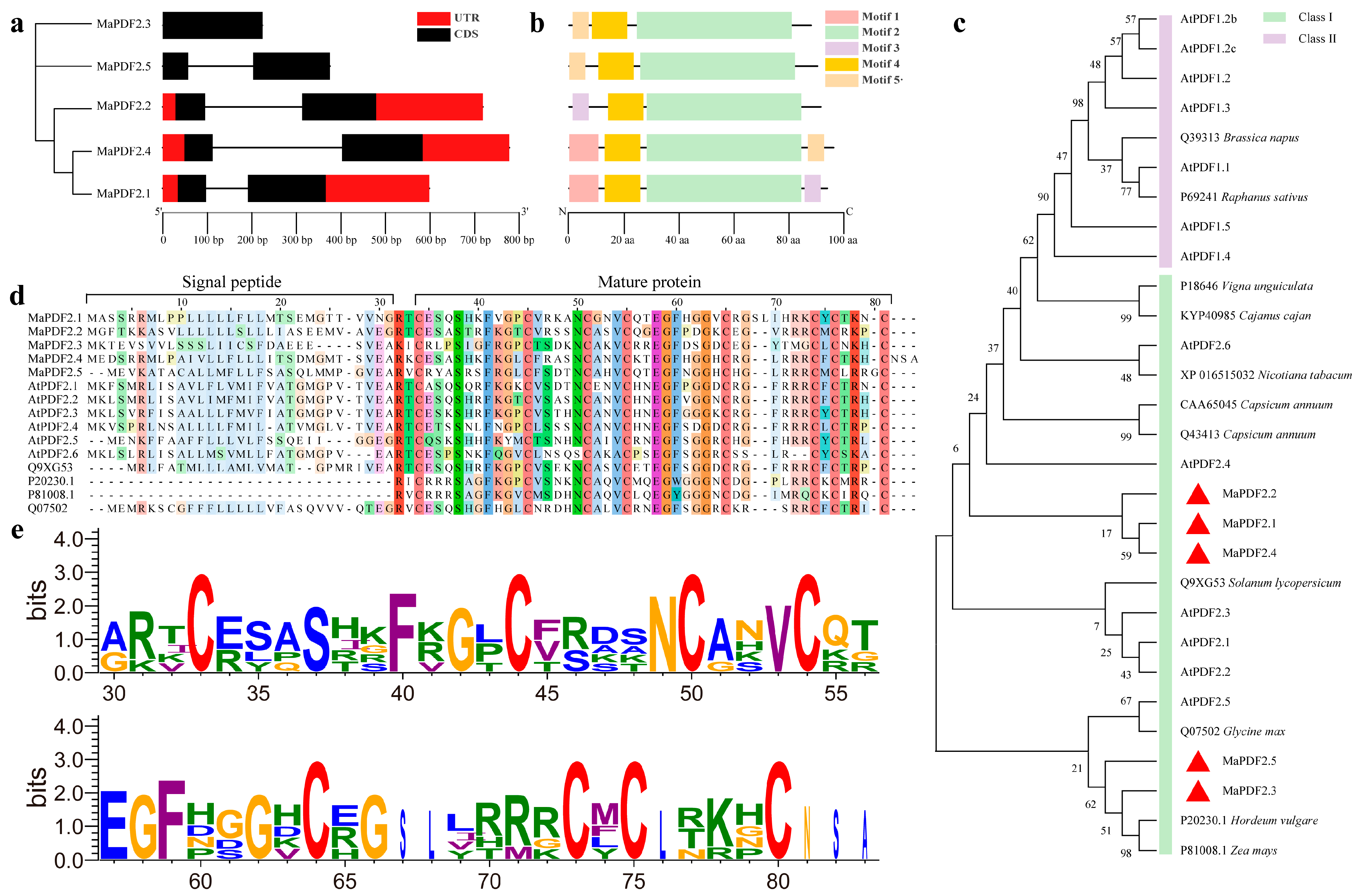
3.2. Gene Structure, Conserved Motifs, and Phylogenetic Analysis of MaPDFs

Gene structure analysis results revealed that MaPDF2.3 had one exon, while all the other MaPDFs each had two exons (Figure 1a). In total, we identified five motifs from the five MaPDFs, with Motifs 2 and 4 present in all members. Motif 1, Motif 3, and Motif 5 were present in MaPDF2.1 and MaPDF2.4, MaPDF2.1 and MaPDF2.2, and MaPDF2.3~2.5 (Figure 1b), respectively.
Phylogenetic analysis results revealed that MaPDF2.1, MaPDF2.2, and MaPDF2.4 shared a very close relationship with AtPDF2.4, while the other MaPDFs were close to AtPDF2.5 (Figure 1c). Plant PDFs can be divided into two classes, Class I and Class II. Notably, all these identified MaPDFs belong to Class I. Multiple sequence alignment analysis showed that MaPDFs and their high homologous proteins from other plants all contain eight conserved cysteine residues that can form 4 disulfide bonds (Cys1-Cys8, Cys2-Cys5, Cys3-Cys6, and Cys4-Cys7) (Figure 1d,e).

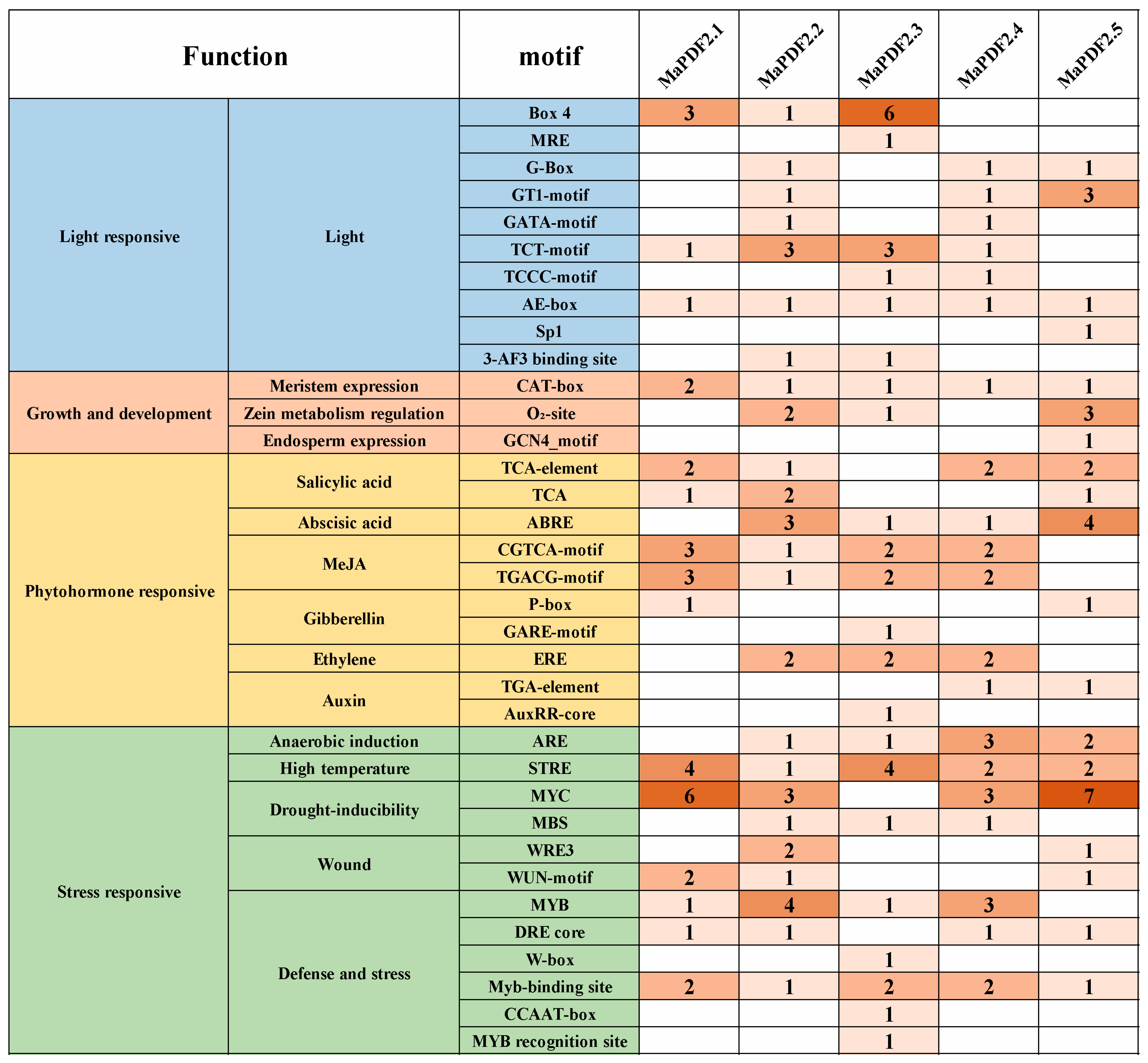
3.3. Analysis of Cis-Acting Elements in MaPDFs Promoters

Promoter analysis showed significant differences in types and numbers of the cis-acting elements among MaPDFs promoters (Figure 2). In total, we identified ten kinds of light-responsive elements in MaPDFs promoters, with the promoters of MaPDF2.1~2.5 containing three, seven, six, six, and four types of light-responsive elements, respectively. All MaPDFs promoters contain the AE-box element. Three growth and development related elements, including meristem-metabolism-related CAT-box, zein metabolism regulatory O2-site element, and endosperm-expression-related GCN4_motif elements, were identified in MaPDFs promoters. Of them, the CAT-box element was present in all MaPDFs promoters.
The promoters of MaPDFs contain ten phytohormone responsive elements involving six phytohormones, including salicylic acid (SA), abscisic acid (ABA), methyl jasmonic acid (MeJA), gibberellin (GA), ethylene (ET), and auxin. The MaPDF2.2 and MaPDF2.4 promoters contain elements responsive to all six phytohormones, and the other MaPDFs’ promoters contain elements responsive to five phytohormones. It is worth noting that the MaPDF2.2 and MaPDF2.4 promoters both contain elements responsive to all four of the stress phytohormones (SA, ABA, MeJA and ET).
The promoters of MaPDFs contained twelve kinds of defense stress-related elements involving five types of stresses, including anaerobic induction, high-temperature, drought-inducibility, wound, and defense and stress. All MaPDFs’ promoters contain high-temperature related STRE and defense and stress-related Myb-binding site elements. Both MaPDF2.2 and MaPDF2.5 promoters contain all five types of defense stress-related elements. Additionally, the MaPDF2.2 promoter contains the most kinds of defense and stress related elements, accounting for nine.

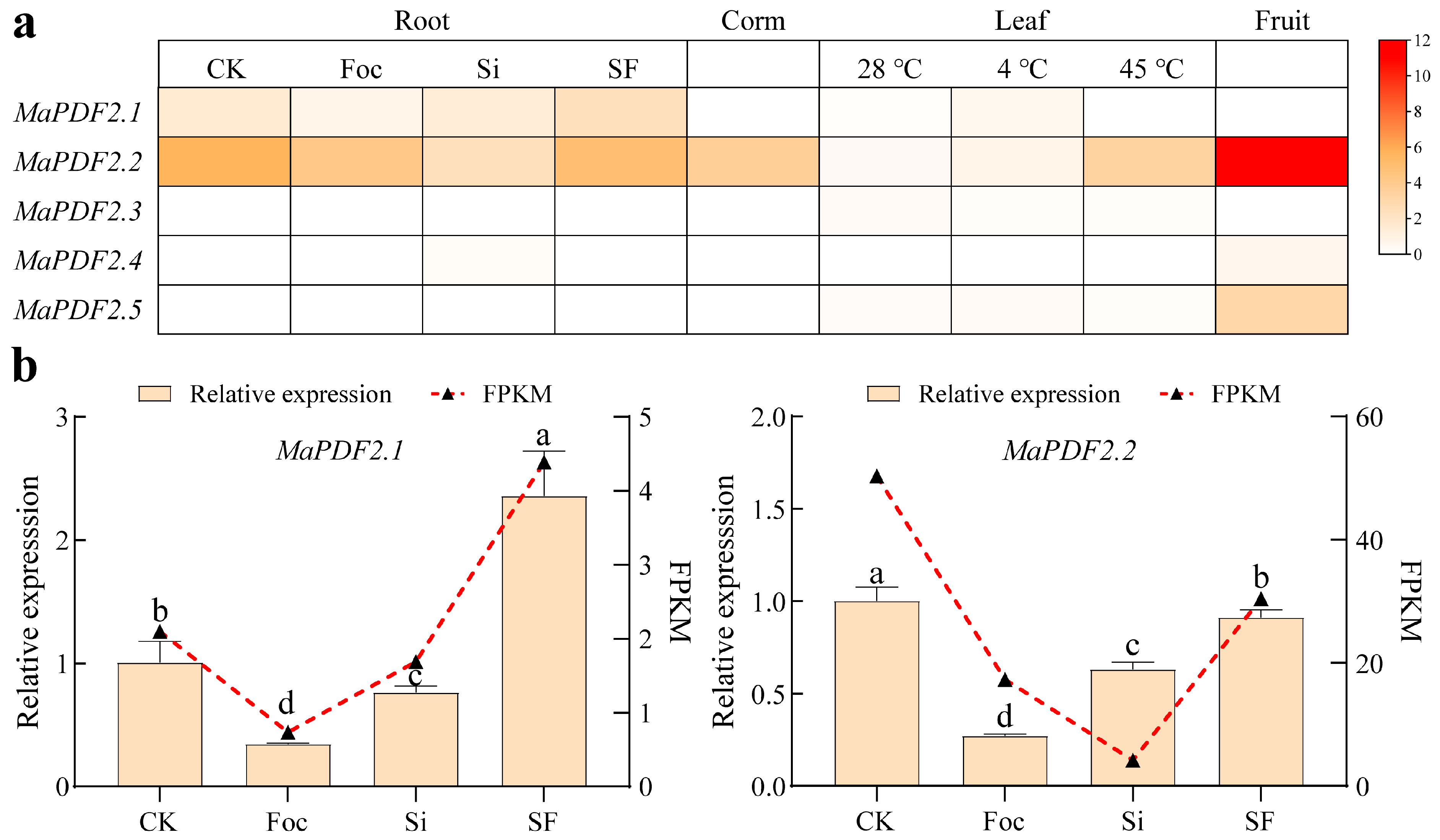
3.4. Expression Pattern Analysis of MaPDFs in Different Organs and Under Various Stress Conditions

Based on transcriptome data, the relative expression levels of MaPDFs in roots (CK, Si, Foc and SF), leaves (28 °C, 4 °C and 45 °C), corm, and fruit were first compared (Figure 3a). MaPDF2.2 was expressed highly in all tested organs, and its expression levels in all these organs were all the highest among these five MaPDFs. Only MaPDF2.1 and MaPDF2.2 were expressed in all root samples, and MaPDF2.4 was expressed slightly in root from the Si group. MaPDF2.2 is the only member that is expressed in corm. All MaPDFs except MaPDF2.4 were expressed in leaves; three MaPDFs (MaPDF2.2, MaPDF2.4, and MaPDF2.5) were expressed in fruit.
The influences of temperature stresses on MaPDF expression in banana leaf were also studied. Results showed that the expression levels of MaPDF2.1, MaPDF2.2, and MaPDF2.5 were significantly upregulated by 4 °C low-temperature stress, accounting for 5.18-fold, 2.55-fold, and 1.25-fold of the 28 °C control, respectively. However, the expression of MaPDF2.3 was significantly downregulated to 44.83% of 28 °C. Also, 45 °C high-temperature stress significantly upregulated the MaPDF2.2 expression (to approximately 31.28-fold of 28 °C), while significantly downregulating the expression of MaPDF2.3 and MaPDF2.5 in leaf.
By further comparing the MaPDF expression in banana root, we found that the expression levels of MaPDF2.1 and MaPDF2.2 in roots of Foc and Si groups were significantly lower than that in CK, indicating that their expressions were suppressed by fungal infection. The expression levels of MaPDF2.1 and MaPDF2.2 in the SF group were significantly higher, accounting for 5.95- and 1.76-fold of Foc, and 1.59-fold and 6.19-fold of Si, respectively. QRT-PCR was further used to validate the expression of MaPDF2.1 and MaPDF2.2 in banana roots of CK, Foc, Si, and SF groups (Figure 3b). Results revealed that their expression patterns were almost the same as our transcriptome data except that the MaPDF2.2 expression in Si was higher than that in Foc by qRT-PCR. The expression level of MaPDF2.1 and MaPDF2.2 in Foc root was only about 34.55% and 27.13% of CK, respectively. In the Si group, their expression levels were also significantly lower than in the CK group. The expression levels of MaPDF2.1 and MaPDF2.2 in the SF group were significantly higher, being 6.76- and 3.36-fold of the Foc group, respectively. This indicated that the S. indica pre-colonization can alleviate the inhibitory effects of FocTR4 on the expression of both MaPDF2.1 and MaPDF2.2.

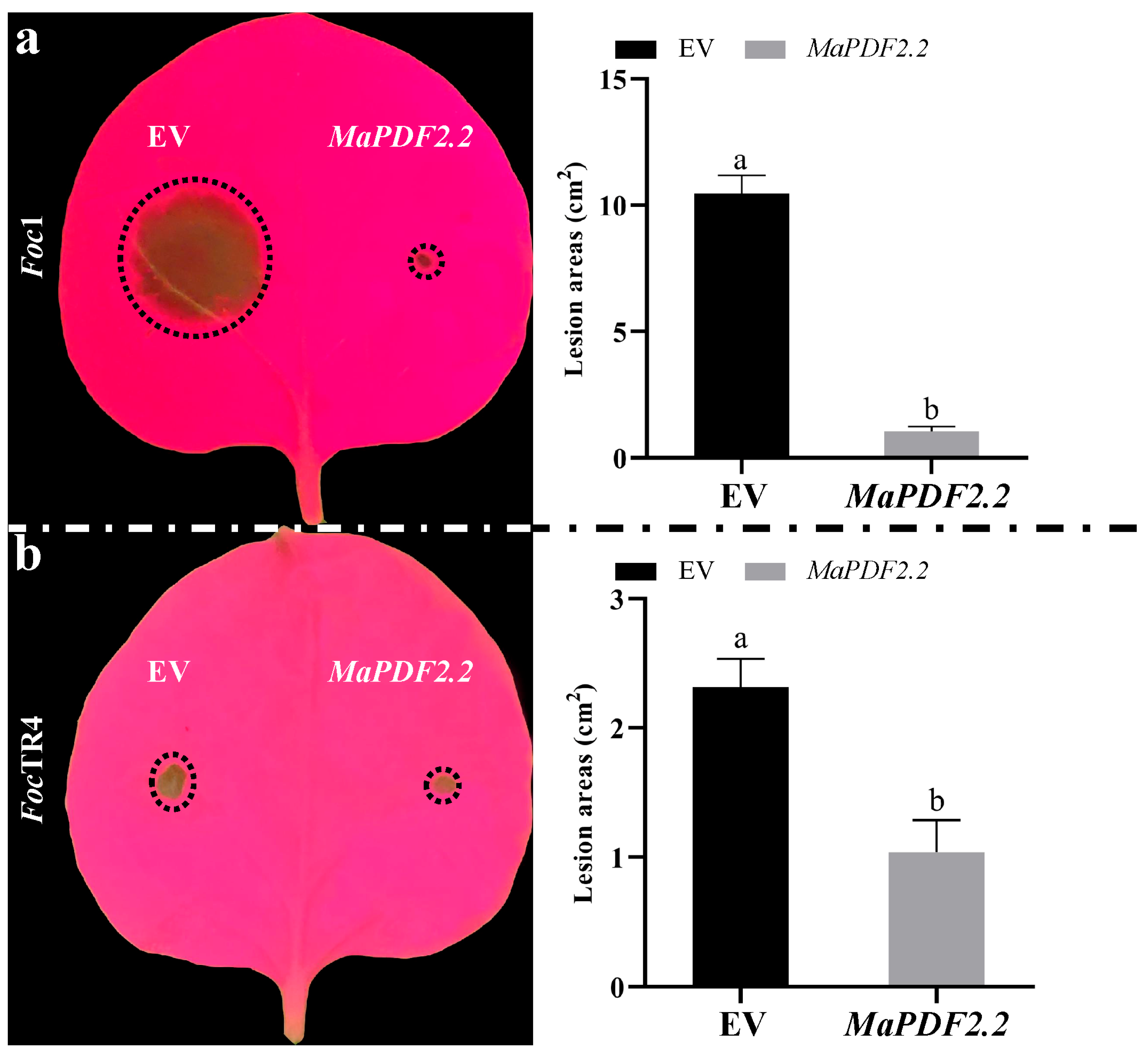
3.5. Transient Overexpression of MaPDF2.2 Can Inhibit the Growth of Foc1 and FocTR4 in Tobacco Leaves

Of the five MaPDFs, MaPDF2.2 expressed the highest in all banana organs. Its expression could be significantly downregulated by FocTR4 and S. indica, but its expression in the SF group was significantly higher than that in the Foc and Si groups. Therefore, it is hypothesized that MaPDF2.2 contributes greatly to the S. indica-enhanced FW resistance. To verify this hypothesis, tobacco-leaf-based disease resistance assays were performed. Results showed that at three days after inoculation, the lesion areas caused by Foc1 and FocTR4 infection in tobacco leaves overexpressing MaPDF2.2 were significantly smaller than that overexpressing EV (Figure 4). In tobacco leaf overexpressing MaPDF2.2, the lesion area caused by Foc1 and FocTR4 was only 10.82% (Figure 4a) and 44.79% (Figure 4b) of its corresponding EV, respectively. These results indicated that the overexpression of MaPDF2.2 can significantly inhibit the infection and the growth of Foc1 and FocTR4 pathogens in tobacco leaves.

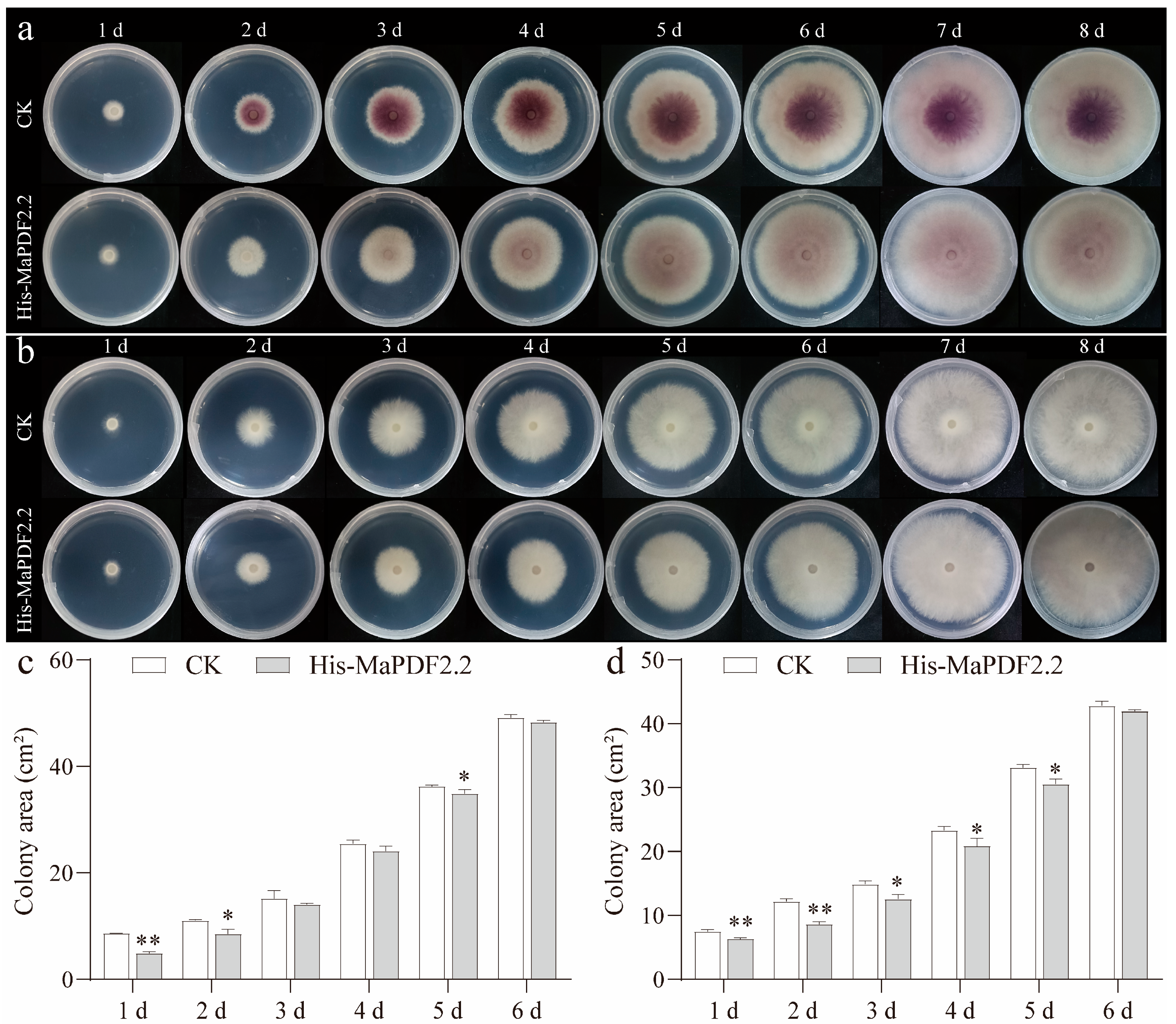
3.6. Prokaryotic Expressed Recombinant His-MaPDF2.2 Can Inhibit the Growth and Development of Both Foc1 and FocTR4 In Vitro

Antifungal ability assays of MaPDF2.2 were further conducted by inoculating Foc1 and FocTR4 fungal plugs onto PDA media spread with or without prokaryotic expressed recombinant His-MaPDF2.2 proteins (Figure 5a,b). Results showed that the addition of His-MaPDF2.2 proteins inhibited the growth of Foc1 and FocTR4 on PDA media in the early five days (Figure 5a,b). At 1 dpi, the average Foc1 colony area on His-MaPDF2.2 added PDA media was very significantly smaller than that of the normal PDA control (CK) (p < 0.01), accounting for only about 56.54% of CK (Figure 5c). At 2 and 5 dpi, compared to CK, the average Foc1 colony area on His-MaPDF2.2 added PDA media was significantly smaller (p < 0.05), accounting for 77.49% and 96.26% of it (Figure 5c), respectively. Interestingly, its addition resulted in much less pigment accumulation of the Foc1 colonies (Figure 5a).
For FocTR4, a very significant difference was identified between the His-MaPDF2.2 added and non-added controls at 1 dpi and 2 dpi (p < 0.01), with the fungal area on His-MaPDF2.2 added PDA media accounting for 70.57% and 74.5% of CK, respectively (Figure 5d). At 3, 4 and 5 dpi, the average FocTR4 colony area on His-MaPDF2.2 added PDA media was significantly smaller than that on the normal PDA media (p < 0.05), accounting for 84.5%, 89.49%, and 92.27% of CK (Figure 5d), respectively.
Collectively, this study demonstrated that the prokaryotic expressed recombinant His-MaPDF2.2 can inhibit the in vitro growth and development of both Foc1 and FocTR4. This indicates that the prokaryotic expressed proteins have potential to be used in the prevention and control of FW.

4. Discussion

PDFs are generally encoded by a multi-gene family in plant species [33,34,35]. Based on their precursor organization, PDFs can be categorized into two classes [33]. For a long time, most PDFs identified in many plant species were found to belong to Class I, and many Class II members were wrongly classified as Class I PDFs [34]. In this study, we identified five MaPDFs from banana which might be caused by the precursor sequence dependence when classifying PDFs [35]. Gene expression analysis revealed that most MaPDFs showed organ-specific expression patterns, which can also be explained by their functional redundancy. Of the five MaPDFs, MaPDF2.2 expressed the highest in all organs, suggesting that it might play a key role in banana defense responses in different organs. Evidence revealed that some PDFs also contribute greatly to plant resistance to various abiotic stresses, such as high-temperature and low-temperature resistance [36,37,38,39]. Sasaki et al. [40] reported that the Triticum aestivum defensin 1 (TAD1) is cold inducible, and its overexpression could enhance the snow mold and Fusarium head blight resistance of wheat. Consistently, in this study, the expression of MaPDF2.2 in leaf was high-temperature and low-temperature inducible, and its expression in roots was greatly influenced by both mutualistic and parasitic fungal infections. This suggested that it played an important role in banana responses to temperature stresses and fungal inoculations.
Many studies have demonstrated that the transcription of PDF genes is markedly influenced by light and stresses. For instance, the expression of Arabidopsis PDF1.2 could be downregulated by low ratios of red light/far-red light treatment [41]. The induction effects of Flg22 on the DEF gene expression in tomato leaves was reported to be dependent on daytime and ethylene [42]. Our study found that the promoters of MaPDFs all contain many light-responsive elements, indicating that their expression may be regulated by light.
In plants, the accumulations of endogenous SA and ET can activate the expression of the defense gene PDF1.2 [43,44]. In defense against necrotrophic pathogens, a set of specific defense genes, including PDF1.2, can be synergistically activated through the JA and ET signaling pathway [45]. Interestingly, in this study, we identified many phytohormone responsive elements in promoters of MaPDFs. Notably, the MaPDF2.2 promoter contains SA-, JA-, ET-, and ABA-responsive elements, and anaerobic induction-, high-temperature-, drought-inducibility-, wound-, and defense and stress-related elements, suggesting that its expression might be regulated by stress phytohormones under various stress conditions. Consistently, our gene expression analysis revealed that the expression of MaPDF2.2 could be remarkably upregulated by temperature stresses and significantly downregulated by fungal inoculations.
PDFs exhibit potent broad-spectrum antimicrobial effects [46,47,48]. Transgenic cotton overexpressing N. alata NaD1 [49] and transgenic tobacco and potato plants overexpressing a horseradish defensin gene all showed significantly enhanced resistance to F. oxysporum. Notably, transgenic banana overexpressing SmAMPD1 displayed significantly enhanced resistance to Foc1 [25]. In this study, gene expression analysis showed that the MaPDF2.2 expression could be remarkably downregulated by not only the FW pathogen FocTR4 but also plant growth promoting endophytic fungus S. indica. The downregulation of this antimicrobial gene might be helpful in facilitating fungal infection/colonization. Our study also found that the expression of MaPDF2.2 in SF was significantly higher than in Foc. It can be explained that the S. indica pre-colonization prominently weakened the downregulation effect of FocTR4 on MaPDF2.2, i.e., the S. indica-colonization-enhanced FW resistance was achieved, at least partially, by alleviating the suppression effects of FocTR4 on the expression of MaPDF2.2. Consistently, tobacco leaves transiently overexpressing MaPDF2.2 exhibited significantly reduced lesion areas caused by Foc1 and FocTR4 infection.
Many studies demonstrated the in vitro antimicrobial activities of PDFs to Fusarium pathogens. Fernández et al. [50] reported that the SmDef2 protein isolated from Silybum marianum flowers can inhibit the growth of F. graminearum in vitro. GST-MzDef could significantly inhibit the growth of F. verticillioides [51]. Synthetic Capsicum chinense defensin peptides could disrupt the cell structure and reduce the pathogenicity of F. solani and F. oxysporum [52]. In this study, we found that the His-MaPDF2.2 addition inhibited the growth of Foc1 and FocTR4 on PDA plates in the early five days. This indicated that the recombinant His-MaPDF2.2 might have great application potentials in controlling FW disease.
Fusarium species can produce a variety of secondary metabolites, such as bikaverin [53] and fusarielin [54]. The accumulation of bikaverin has been reported to be the main reason for the pigmentation of Fusarium hyphae [55]. Furthermore,, bikaverin plays a primary role in enhancing the resistance of Fusarium to various stresses [55,56]. Moreover, it was reported that the bikaverin secretion reduction would lead to decreased infection ability of Fusarium species [57]. Interestingly, our study found that His-MaPDF2.2 can significantly inhibit the pigment accumulation of Foc1 on PDA plates. This suggested that the inhibitory effect of MaPDF2.2 on the growth of Foc1 might be achieved by suppressing the biosynthesis of bikaverin.

5. Conclusions

In this study, we successfully identified five PDF genes from banana. Of them, only MaPDF2.2 was expressed highly in all banana organs and its expression was responsive to temperature stresses and fungal colonization/infection. S. indica pre-colonization can alleviate the inhibitory effect of FocTR4 on MaPDF2.2, which might be closely related to the fungal-colonization-enhanced FW resistance. MaPDF2.2 overexpression can inhibit the infection of FocTR4 and Foc1 in tobacco leaves. Additionally, the prokaryotic expressed recombinant His-MaPDF2.2 protein can inhibit the growth of Foc1 and FocTR4, and suppress the pigments accumulation of Foc1. Our study demonstrated the antifungal ability of MaPDF2.2 to FW pathogens.

Supplementary Materials

The following supporting information can be downloaded at: https://www.mdpi.com/article/10.3390/horticulturae11050513/s1, Figure S1. Chromosome location analysis of MaPDFs (a) and electrophoresis detection (b) results of amplified MaPDFs products. The 1~5 in (b) represent amplified PCR products of MaPDF2.1~MaPDF2.5, respectively. Table S1. The BLASTp identified banana PDFs by using Arabidopsis PDFs as queries. Table S2. Information for the primers used in this study.

Author Contributions

R.L.: software, formal analysis, data curation, and writing—original draft. B.W.: formal analysis, writing—original draft, and methodology. H.W.: investigation, resources, and conceptualization. C.C.: conceptualization, writing—review and editing, supervision, funding acquisition, and project administration. All authors have read and agreed to the published version of the manuscript.

Funding

This work was supported by the Fund for High-level Talents of Shanxi Agricultural University (2021XG010).

Data Availability Statement

Data are contained in the article or Supplementary Materials.

Conflicts of Interest

The authors declare that they have no known competing financial interests or personal relationships that could have appeared to influence the work reported in this paper.

Abbreviations

The following abbreviations are used in this manuscript:
ABAAbscisic acid
DpiDays post inoculation
ETEthylene
GAGibberellin
FPKMFragments Per Kilobase of transcript per Million mapped reads
Foc1Fusarium oxysporum f. sp. cubense 1
FocTR4Fusarium oxysporum f. sp. cubense Tropical Race 4
FWFusarium wilt
PRPathogenesis-related proteins
PDFPlant defensin
SASalicylic acid

References

  1. De Oliveira, S.S.S.; Cherene, M.B.; Taveira, G.B.; De Oliveira Mello, É.; De Oliveira Carvalho, A.; Gomes, V.M. Plant antimicrobial peptides and their main families and roles: A review of the literature. Curr. Issues Mol. Biol. 2024, 47, 1. [Google Scholar] [CrossRef] [PubMed]
  2. Liu, K.; Liang, Z.; Yang, A.; Yan, J.; Cong, P.; Han, X.; Zhang, C. Comparative transcriptome analysis reveals key genes and pathways in response to Alternaria alternata apple pathotype infection. Hortic. Plant J. 2024, 10, 641–656. [Google Scholar] [CrossRef]
  3. Mirouze, M.; Sels, J.; Richard, O.; Czernic, P.; Loubet, S.; Jacquier, A.; François, I.E.J.A.; Cammue, B.P.A.; Lebrun, M.; Berthomieu, P.; et al. A putative novel role for plant defensins: A defensin from the zinc hyper-accumulating plant, Arabidopsis halleri, confers zinc tolerance. Plant J. 2006, 47, 329–342. [Google Scholar] [CrossRef]
  4. Guan, L.; Cui, Q.; Li, S.; Dong, C.; Li, H.; Peng, J.; Li, X. Research advances on the structure, function and regulation of defensins gene family in plants. Biotechnol. Bull. 2018, 34, 33–39. (In Chinese) [Google Scholar] [CrossRef]
  5. Sher Khan, R.; Iqbal, A.; Malak, R.; Shehryar, K.; Attia, S.; Ahmed, T.; Ali Khan, M.; Arif, M.; Mii, M. Plant defensins: Types, mechanism of action and prospects of genetic engineering for enhanced disease resistance in plants. 3 Biotech 2019, 9, 192. [Google Scholar] [CrossRef] [PubMed]
  6. Cheng, C.; Wu, H.; Zhang, Y. Characterization and functional analysis of gerbera plant defensin (PDF) genes reveal the role of GhPDF2.4 in defense against the root rot pathogen Phytophthora cryptogea. aBiotech 2024, 5, 325–338. [Google Scholar] [CrossRef] [PubMed]
  7. Hazlett, L.; Wu, M. Defensins in innate immunity. Cell Tissue Res. 2011, 343, 175–188. [Google Scholar] [CrossRef]
  8. Lay, F.T.; Poon, S.; McKenna, J.A.; Connelly, A.A.; Barbeta, B.L.; McGinness, B.S.; Fox, J.L.; Daly, N.L.; Craik, D.J.; Heath, R.L.; et al. The C-terminal propeptide of a plant defensin confers cytoprotective and subcellular targeting functions. BMC Plant Biol. 2014, 14, 41. [Google Scholar] [CrossRef]
  9. Vi, T.X.T.; Le, H.D.; Nguyen, V.T.T.; Le, V.S.; Chu, H.M. Expression of the ZmDEF1 gene and α-amylase inhibitory activityof recombinant defensin against maize weevils. Turk. J. Biol. 2017, 41, 98–104. [Google Scholar] [CrossRef]
  10. Vi, T.X.T.; Nguyen, T.N.L.; Pham, T.T.N.; Nguyen, H.Q.; Nguyen, T.H.Y.; Tu, Q.T.; Le, V.S.; Chu, H.M. Overexpression of the ZmDEF1 gene increases the resistance to weevil larvae in transgenic maize seeds. Mol. Biol. Rep. 2019, 46, 2177–2185. [Google Scholar] [CrossRef]
  11. Liu, Q.; Yan, H.; Zhang, Z.; Zheng, L.; Zhou, Y.; Gao, F. AmDEF2.7, a tandem duplicated defensin gene from Ammopiptanthus mongolicus, activated by AmWRKY14, enhances the tolerance of Arabidopsis to low temperature and osmotic stress. Environ. Exp. Bot. 2024, 227, 105956. [Google Scholar] [CrossRef]
  12. Zhao, K.; Ren, R.; Ma, X.; Zhao, K.; Qu, C.; Cao, D.; Ma, Q.; Ma, Y.; Gong, F.; Li, Z.; et al. Genome-wide investigation of defensin genes in peanut (Arachis hypogaea L.) reveals AhDef2.2 conferring resistance to bacterial wilt. Crop J. 2022, 10, 809–819. [Google Scholar] [CrossRef]
  13. Kaur, J.; Fellers, J.; Adholeya, A.; Velivelli, S.L.S.; El-Mounadi, K.; Nersesian, N.; Clemente, T.; Shah, D. Expression of apoplast-targeted plant defensin MtDef4.2 confers resistance to leaf rust pathogen Puccinia triticina but does not affect mycorrhizal symbiosis in transgenic wheat. Transgenic Res. 2017, 26, 37–49. [Google Scholar] [CrossRef]
  14. Dong, Y.; Wang, Y.; Tang, M.; Chen, W.; Chai, Y.; Wang, W. Bioinformatic analysis of wheat defensin gene family and function verification of candidate genes. Front. Plant Sci. 2023, 14, 1279502. [Google Scholar] [CrossRef]
  15. Cheng, C.; Wu, S.; Deng, G.; Sheng, O.; Yi, G.; Yang, Q. Recent advances and future directions in banana molecular biology and breeding. Mol. Hortic. 2024, 4, 42. [Google Scholar] [CrossRef]
  16. Li, X.; Yu, S.; Cheng, Z.; Chang, X.; Yun, Y.; Jiang, M.; Chen, X.; Wen, X.; Li, H.; Zhu, W.; et al. Origin and evolution of the triploid cultivated banana genome. Nat. Genet. 2024, 56, 136–142. [Google Scholar] [CrossRef] [PubMed]
  17. Abd Murad, N.B.; Mohamed Nor, N.M.I.; Shohaimi, S.; Mohd Zainudin, N.A.I. Genetic diversity and pathogenicity of Fusarium species associated with fruit rot disease in banana across peninsular malaysia. J. Appl. Microbiol. 2017, 123, 1533–1546. [Google Scholar] [CrossRef]
  18. Uribe-Palacio, S.; Ramírez-Sánchez, M.; Umaña-Rojas, G.; Sáenz- Murillo, M.V. Studies on the potential for treatment with short wave ultraviolet light (UV-C) to reduce postharvest diseases in banana fruit crown (Musa sp., Group AAA, subgroup Cavendish). Fruits 2022, 77, 1–7. [Google Scholar] [CrossRef]
  19. Conti Taguali, S.; Riolo, M.; Dopazo, V.; Meca, G.; Cacciola, S.O. Characterization of mycotoxins produced by two Fusarium species responsible for postharvest rot of banana fruit. J. Plant Pathol. 2024, 106, 1785–1800. [Google Scholar] [CrossRef]
  20. Chen, A.; Sun, J.; Matthews, A.; Armas-Egas, L.; Chen, N.; Hamill, S.; Mintoff, S.; Tran-Nguyen, L.T.T.; Batley, J.; Aitken, E.A.B. Assessing variations in host resistance to Fusarium oxysporum f sp. Cubense Race 4 in Musa species, with a focus on the subtropical Race 4. Front. Microbiol. 2019, 10, 1062. [Google Scholar] [CrossRef]
  21. Maymon, M.; Sela, N.; Shpatz, U.; Galpaz, N.; Freeman, S. The origin and current situation of Fusarium Oxysporum f. sp. Cubense Tropical Race 4 in Israel and the Middle East. Sci. Rep. 2020, 10, 1590. [Google Scholar] [CrossRef]
  22. Radhajeyalakshmi, R.; Xia, Y.; Shah, D. Antifungal defensins in controlling panama wilt of banana Fusarium Oxysporum f. sp. Cubense. Ann. Biol. Sci. 2020, 8, 38–43. [Google Scholar]
  23. Lay, F.T.; Brugliera, F.; Anderson, M.A. Isolation and properties of floral defensins from ornamental tobacco and petunia. Plant Physiol. 2003, 131, 1283–1293. [Google Scholar] [CrossRef] [PubMed]
  24. Ghag, S.B.; Shekhawat, U.K.S.; Ganapathi, T.R. Petunia floral defensins with unique prodomains as novel candidates for development of fusarium wilt resistance in transgenic banana plants. PLoS ONE 2012, 7, e39557. [Google Scholar] [CrossRef] [PubMed]
  25. Ghag, S.B.; Shekhawat, U.K.S.; Ganapathi, T.R. Transgenic banana plants expressing a Stellaria media defensin gene (Sm-AMP-D1) demonstrate improved resistance to Fusarium Oxysporum. Plant Cell Tissue Organ Cult. 2014, 119, 247–255. [Google Scholar] [CrossRef]
  26. Yun, Y.; Song, A.; Bao, J.; Chen, S.; Lu, S.; Cheng, C.; Zheng, W.; Wang, Z.; Zhang, L. Genome data of Fusarium Oxysporum f. Sp. Cubense Race 1 and tropical race 4 isolates using long-read sequencing. Mol. Plant-Microbe Interact. 2019, 10, 1270–1272. [Google Scholar] [CrossRef]
  27. Cheng, C.; Liu, F.; Sun, X.; Wang, B.; Liu, J.; Ni, X.; Hu, C.; Deng, G.; Tong, Z.; Zhang, Y.; et al. Genome-wide identification of FAD gene family and their contributions to the temperature stresses and mutualistic and parasitic fungi colonization responses in banana. Int. J. Biol. Macromol. 2022, 204, 661–676. [Google Scholar] [CrossRef]
  28. Waterhouse, A.M.; Procter, J.B.; Martin, D.M.A.; Clamp, M.; Barton, G.J. Jalview version 2—A multiple sequence alignment editor and analysis workbench. Bioinformatics 2009, 25, 1189–1191. [Google Scholar] [CrossRef]
  29. Crooks, G.E.; Hon, G.; Chandonia, J.-M.; Brenner, S.E. WebLogo: A sequence logo generator. Genome Res. 2004, 14, 1188–1190. [Google Scholar] [CrossRef]
  30. Chen, C.; Chen, H.; Zhang, Y.; Thomas, H.R.; Frank, M.H.; He, Y.; Xia, R. TBtools: An integrative toolkit developed for interactive analyses of big biological data. Mol. Plant 2020, 13, 1194–1202. [Google Scholar] [CrossRef] [PubMed]
  31. Wang, B.; Xu, Y.; Xu, S.; Wu, H.; Qu, P.; Tong, Z.; Lü, P.; Cheng, C. Characterization of banana SNARE genes and their expression analysis under temperature stress and mutualistic and pathogenic fungal colonization. Plants 2023, 12, 1599. [Google Scholar] [CrossRef] [PubMed]
  32. Lai, G.; Fu, P.; Liu, Y.; Xiang, J.; Lu, J. Molecular characterization and overexpression of VpRPW8s from Vitis pseudoreticulata enhances resistance to Phytophthora capsici in Nicotiana benthamiana. Int. J. Mol. Sci. 2018, 19, 839. [Google Scholar] [CrossRef]
  33. Kovaleva, V.; Bukhteeva, I.; Kit, O.Y.; Nesmelova, I.V. Plant defensins from a structural perspective. Int. J. Mol. Sci. 2020, 21, 5307. [Google Scholar] [CrossRef] [PubMed]
  34. De-Paula, V.S.; Razzera, G.; Medeiros, L.; Miyamoto, C.A.; Almeida, M.S.; Kurtenbach, E.; Almeida, F.C.L.; Valente, A.P. Evolutionary relationship between defensins in the poaceae family strengthened by the characterization of new sugarcane defensins. Plant Mol. Biol. 2008, 68, 321–335. [Google Scholar] [CrossRef]
  35. Costa, L.S.M.; Pires, Á.S.; Damaceno, N.B.; Rigueiras, P.O.; Maximiano, M.R.; Franco, O.L.; Porto, W.F. In Silico characterization of Class II plant defensins from Arabidopsis thaliana. Phytochemistry 2020, 179, 112511. [Google Scholar] [CrossRef] [PubMed]
  36. De Coninck, B.M.A.; Sels, J.; Venmans, E.; Thys, W.; Goderis, I.J.W.M.; Carron, D.; Delauré, S.L.; Cammue, B.P.A.; De Bolle, M.F.C.; Mathys, J. Arabidopsis thaliana plant defensin AtPDF1.1 is involved in the plant response to biotic stress. New Phytol. 2010, 187, 1075–1088. [Google Scholar] [CrossRef]
  37. Gaudet, D.A.; Laroche, A.; Frick, M.; Huel, R.; Puchalski, B. Cold induced expression of plant defensin and lipid transfer protein transcripts in winter wheat. Physiol. Plant. 2003, 117, 195–205. [Google Scholar] [CrossRef]
  38. Domingo, G.; Locato, V.; Cimini, S.; Ciceri, L.; Marsoni, M.; De Gara, L.; Bracale, M.; Vannini, C. A Comprehensive characterization and expression profiling of defensin family peptides in Arabidopsis thaliana with a focus on their abiotic stress-specific transcriptional modulation. Curr. Plant Biol. 2024, 39, 100376. [Google Scholar] [CrossRef]
  39. Koike, M.; Okamoto, T.; Tsuda, S.; Imai, R. A novel plant defensin-like gene of winter wheat is specifically induced during cold acclimation. Biochem. Biophys. Res. Commun. 2002, 298, 46–53. [Google Scholar] [CrossRef]
  40. Sasaki, K.; Kuwabara, C.; Umeki, N.; Fujioka, M.; Saburi, W.; Matsui, H.; Abe, F.; Imai, R. The cold-induced defensin TAD1 confers resistance against snow mold and Fusarium head blight in transgenic wheat. J. Biotechnol. 2016, 228, 3–7. [Google Scholar] [CrossRef]
  41. Cerrudo, I.; Keller, M.M.; Cargnel, M.D.; Demkura, P.V.; de Wit, M.; Patitucci, M.S.; Pierik, R.; Pieterse, C.M.J.; Ballaré, C.L. Low red/far-red ratios reduce arabidopsis resistance to Botrytis cinerea and jasmonate responses via a COI1-JAZ10-dependent, salicylic acid-independent mechanism. Plant Physiol. 2012, 158, 2042–2052. [Google Scholar] [CrossRef]
  42. Czékus, Z.; Kukri, A.; Hamow, K.Á.; Szalai, G.; Tari, I.; Ördög, A.; Poór, P. Activation of local and systemic defence responses by Flg22 is dependent on daytime and ethylene in intact tomato plants. Int. J. Mol. Sci. 2021, 22, 8354. [Google Scholar] [CrossRef]
  43. Nandi, A.; Kachroo, P.; Fukushige, H.; Hildebrand, D.F.; Klessig, D.F.; Shah, J. Ethylene and jasmonic acid signaling affect the npr1-independent expression of defense genes without impacting resistance to Pseudomonas syringae and Peronospora parasitica in the Arabidopsis ssi1 mutant. Mol. Plant-Microbe Interact. 2003, 16, 588–599. [Google Scholar] [CrossRef] [PubMed]
  44. Tian, H.; Xu, L.; Li, X.; Zhang, Y. Salicylic acid: The roles in plant immunity and crosstalk with other hormones. J. Integr. Plant Biol. 2024, 67, 773–785. [Google Scholar] [CrossRef]
  45. Zarei, A.; Körbes, A.P.; Younessi, P.; Montiel, G.; Champion, A.; Memelink, J. Two GCC boxes and AP2/ERF-domain transcription factor ORA59 in jasmonate/ethylene-mediated activation of the PDF1.2 promoter in Arabidopsis. Plant Mol. Biol. 2011, 75, 321–331. [Google Scholar] [CrossRef] [PubMed]
  46. Kaewklom, S.; Wongchai, M.; Petvises, S.; Hanpithakphong, W.; Aunpad, R. Structural and biological features of a novel plant defensin from Brugmansia x candida. PLoS ONE 2018, 13, e0201668. [Google Scholar] [CrossRef]
  47. Batool, R.; Umer, M.J.; Wang, Y.; He, K.; Shabbir, M.Z.; Zhang, T.; Bai, S.; Chen, J.; Wang, Z. Myco-synergism boosts herbivory-induced maize defense by triggering antioxidants and phytohormone signaling. Front. Plant Sci. 2022, 13, 790504. [Google Scholar] [CrossRef] [PubMed]
  48. Tetorya, M.; Li, H.; Djami-Tchatchou, A.T.; Buchko, G.W.; Czymmek, K.J.; Shah, D.M. Plant defensin MtDef4-derived antifungal peptide with multiple modes of action and potential as a bio-inspired fungicide. Mol. Plant Pathol. 2023, 24, 896–913. [Google Scholar] [CrossRef]
  49. Gaspar, Y.M.; McKenna, J.A.; McGinness, B.S.; Hinch, J.; Poon, S.; Connelly, A.A.; Anderson, M.A.; Heath, R.L. Field resistance to Fusarium oxysporum and Verticillium dahliae in transgenic cotton expressing the plant defensin NaD1. J. Exp. Bot. 2014, 65, 1541–1550. [Google Scholar] [CrossRef]
  50. Fernández, A.; Colombo, M.L.; Curto, L.M.; Gómez, G.E.; Delfino, J.M.; Guzmán, F.; Bakás, L.; Malbrán, I.; Vairo-Cavalli, S.E. Peptides derived from the α-core and γ-core regions of a putative Silybum marianum flower defensin show antifungal activity against Fusarium graminearum. Front. Microbiol. 2021, 12, 632008. [Google Scholar] [CrossRef]
  51. Al Kashgry, N.A.T.; Abulreesh, H.H.; El-Sheikh, I.A.; Almaroai, Y.A.; Salem, R.; Mohamed, I.; Waly, F.R.; Osman, G.; Mohamed, M.S.M. Utilization of a recombinant defensin from Maize (Zea Mays L.) as a potential antimicrobial peptide. AMB Express 2020, 10, 208. [Google Scholar] [CrossRef] [PubMed]
  52. de Azevedo Dos Santos, L.; Taveira, G.B.; da Silva, M.S.; da Silva Gebara, R.; da Silva Pereira, L.; Perales, J.; Teixeira-Ferreira, A.; de Oliveira Mello, É.; de Oliveira Carvalho, A.; Rodrigues, R.; et al. Antimicrobial peptides from Capsicum chinense fruits: Agronomic alternatives against phytopathogenic fungi. Biosci. Rep. 2020, 40, BSR20200950. [Google Scholar] [CrossRef]
  53. Bell, A.A.; Wheeler, M.H.; Liu, J.; Stipanovic, R.D.; Puckhaber, L.S.; Orta, H. United states department of agriculture—Agricultural research service studies on polyketide toxins of Fusarium oxysporum f sp vasinfectum: Potential targets for disease control. Pest Manag. Sci. 2003, 59, 736–747. [Google Scholar] [CrossRef] [PubMed]
  54. Rajendar, G.; Rehman, M. A short approach to cytogenin and first total synthesis of aspergisocoumarin A and fusarimarin C. Tetrahedron 2024, 168, 134343. [Google Scholar] [CrossRef]
  55. Lebeau, J.; Petit, T.; Clerc, P.; Dufossé, L.; Caro, Y. Isolation of two novel purple naphthoquinone pigments concomitant with the bioactive red bikaverin and derivates thereof produced by Fusarium Oxysporum. Biotechnol. Prog. 2019, 35, e2738. [Google Scholar] [CrossRef]
  56. Wiemann, P.; Willmann, A.; Straeten, M.; Kleigrewe, K.; Beyer, M.; Humpf, H.; Tudzynski, B. Biosynthesis of the red pigment bikaverin in Fusarium fujikuroi: Genes, their function and regulation. Mol. Microbiol. 2009, 72, 931–946. [Google Scholar] [CrossRef]
  57. Amuzu, P.; Pan, X.; Hou, X.; Sun, J.; Jakada, M.A.; Odigie, E.; Xu, D.; Lai, D.; Zhou, L. Recent updates on the secondary metabolites from Fusarium fungi and their biological activities (covering 2019 to 2024). J. Fungi 2024, 10, 778. [Google Scholar] [CrossRef]
Figure 1. Sequence analysis results for MaPDFs and their encoded genes. (a): Gene structures of MaPDFs; (b,c): conserved motifs and phylogenetic analysis results for MaPDFs; (d): sequences alignment results for PDFs from banana and some other plant species; (e): Weblogo for the conserved sequences in MaPDFs. bp: base pair; UTR: untranslated region; CDS: coding sequence; N: N-terminal; C: C-terminal; aa: amino acid; At: Arabidopsis thaliana.
Horticulturae 11 00513 g001
Figure 2. The identified cis-acting elements in promoters of MaPDFs.
Horticulturae 11 00513 g002
Figure 3. Gene expression analysis of MaPDFs. (a): Expression heatmap for MaPDFs in different banana organs; (b) qRT-PCR analysis results for MaPDF2.1 and MaPDF2.2 in roots of CK, Foc, Si and SF. Different lowercase letters above columns represent significant difference at p < 0.05 level.
Horticulturae 11 00513 g003
Figure 4. Influences of the MaPDF2.2 transient overexpression on the Foc1 (a) and FocTR4 (b) infection in tobacco leaves. Different lowercase letters above columns represent significant difference at p < 0.05 level. EV: empty vector control.
Horticulturae 11 00513 g004
Figure 5. The influences of prokaryotic expressed recombinant His-MaPDF2.2 infusion proteins on the growth of Foc1 (a,c) and FocTR4 (b,d) on PDA media. “*” and “**” indicate significant difference between CK and His-MaPDF2.2 at the p < 0.05 and p < 0.01 levels, respectively. Bars above columns represent standard deviations (SD).
Horticulturae 11 00513 g005
Table 1. Physiochemical property and subcellular location prediction results for MaPDFs. pI: isoelectric point; GRAVY: Grand average of hydropathicity.
Gene IDGene NameChromosome LocationProtein Size/aaMolecular Weight/kDPIInstability CoefficientAliphatic IndexGRAVYSubcellular
Location
Ma02_g12840MaPDF2.1chr02: 21473861…21474459(−)798.679289.2742.5373.920.019Extracellular
Ma04_g36140MaPDF2.2chr04: 34629849…34630567(−)778.320868.9237.4279.740.121Extracellular
Ma06_g21420MaPDF2.3chr06: 15679425…15679649(+)748.046235.2649.8965.81−0.109Extracellular
Ma08_g13660MaPDF2.4chr08: 10784713…10785491(−)819.041639.2751.5966.3−0.105Extracellular
Ma11_g12930MaPDF2.5chr11: 16886961…16887336(−)768.468029.0664.5865.530.158Chloroplast
Disclaimer/Publisher’s Note: The statements, opinions and data contained in all publications are solely those of the individual author(s) and contributor(s) and not of MDPI and/or the editor(s). MDPI and/or the editor(s) disclaim responsibility for any injury to people or property resulting from any ideas, methods, instructions or products referred to in the content.

