Article

Preliminary Establishment of an Efficient Regeneration and Genetic Transformation System for Hemerocallis middendorffii Trautv. & C. A. Mey.

1
School of Horticulture and Landscape Architecture, Northeast Agricultural University, Harbin 150030, China
2
School of Forestry and Landscape Architecture, Anhui Agricultural University, Hefei 230036, China
*
Author to whom correspondence should be addressed.
Horticulturae 2025, 11(4), 417; https://doi.org/10.3390/horticulturae11040417
Submission received: 20 March 2025 / Revised: 8 April 2025 / Accepted: 10 April 2025 / Published: 14 April 2025
(This article belongs to the Section Propagation and Seeds)

Abstract

:
Hemerocallis middendorffii is widely used in the landscaping of Northern China for its exceptional ornamental and ecological attributes. It is also the focus of a substantial body of germplasm development and stress tolerance research. However, the absence of an efficient regeneration and genetic transformation system has been a critical barrier to conducting gene function studies on this species. In this research, the aerial parts of seed-derived H. middendorffii plantlets were used as explants, and the callus induction, proliferation, subculture, differentiation, and rooting conditions in the in vitro regeneration process were optimized. A callus induction rate of 95.6% was achieved, with a regeneration rate of 84.4%. Based on this procedure, a simple and effective Agrobacterium-mediated genetic transformation system was preliminarily developed using a hygromycin-based selection system. The system comprised an Agrobacterium tumefaciens culture solution optical density at 600 nm (OD600) of 0.6, an acetosyringone concentration of 100 μmol·L−1 in both the A. tumefaciens infection solution and the co-cultivation medium, a sterilization culture with Timentin at 300 mg·L−1, and a selection culture with hygromycin at 9 mg·L−1. Transgenic H. middendorffii T0 rooted plants were produced within a 5-month period, with a transformation rate of 11.9% and positive rate of 32.8%. The regeneration and genetic transformation system established in this study should help advance functional gene research and genetic improvement in H. middendorffii. However, the genetic transformation was only validated in the T0 plants. To confirm stable integration and long-term transgene stability, future research on the phenotypic and molecular characterization of T1 progeny, including segregation analysis and Southern blot verification, will be conducted.

1. Introduction

Hemerocallis middendorffii Trautv. & C. A. Mey., also known as Amur daylily, belongs to the Hemerocallis genus in the Asphodelaceae family. It is a herbaceous perennial that inhabits meadows, wet grasslands, forest margins, and rocky cliffs of East Asia from temperate to alpine zones [1,2]. It has linear arching leaves with golden yellow or orange flowers that are slightly fragrant. Additionally, H. middendorffii exhibits strong tolerance to drought, freezing, and saline–alkali stresses [3,4,5]. Hence, it is highly suitable for landscaping in gardens and lands with poor ecological conditions due to its high ornamental value, early flowering period, and superior abiotic stress tolerance [6,7]. Despite the above-mentioned advantages, the cultivation of H. middendorffii in China is impeded by key obstacles, such as its long growth cycles and short flowering phase, all of which limit its wider usage [8]. To address these challenges, altering these traits through genetic improvement is likely a useful approach.
Tissue culture and genetic transformation have emerged as vital tools for gene function research and the improvement of specific individual traits without altering the existing characteristics [9]. Although attempts at using explants from species in the Hemerocallis genus for proliferation cultivation date back to the 1950s, early efforts were hindered by the low proliferation rate and slow growth of the genus [10,11,12,13]. In recent decades, research on regeneration systems for H. middendorffii has made some progress. An in vitro callus induction and plant regeneration system was recently established utilizing various organs of H. middendorffii including tender scapes, leaves, and receptacles [3]. However, owing to the seasonal limitation and difficulties in maintaining sterile cultures, these explants have not been widely used in H. middendorffii tissue culture systems.
Hemerocallis is a genus of perennial monocotyledons, with a complex genetic background and heterozygosity arising from interspecific hybridization [14]. The few successful reports of genetic transformation of Hemerocallis spp. have included the use of gene guns [15], the pollen tube pathway [16], and Agrobacterium [17]. A genetic transformation system for H. middendorffii was preliminarily established by Liu [3]. However, the explants used in this system were derived from the field and were difficult to thoroughly disinfect, resulting in a contamination rate of up to 20%. In addition, genetic transformation in this system was only detected by PCR amplification, which not only had a low amplification rate (7.6%), but also could not accurately demonstrate the actual genetic transformation efficiency. At present, the lack of a reliable and efficient genetic transformation system has restricted the wider application of genetic engineering in developing new H. middendorffii varieties. Therefore, improving the genetic transformation of H. middendorffii is of great importance.
To validate the effectiveness of the developed transformation system, the FLOWERING LOCUS T (FT) gene was selected as a functional marker, which is a key regulator of flowering time and plant development [18,19,20]. Although the functional role of HmFT in H. middendorffii remains uncharacterized, its homologs in model plants were found to be pivotal regulators of flowering and stress adaptation [18,19,20,21,22]. While reporter genes like GUS could confirm transformation success, HmFT provides a functional marker that directly links genetic modification to agronomic improvement, aligning with our goal of advancing molecular breeding in H. middendorffii.
Based on the above rationale, the objective of this study was to preliminarily develop an efficient and stable regeneration and genetic transformation system for H. middendorffii using aerial parts of seed-derived plantlets as explants. First, various critical factors in the in vitro regeneration of H. middendorffii, including the selection of explants and the concentration of plant growth regulators (PGRs) were optimized. Next, factors influencing Agrobacterium-mediated transformation, including the concentrations of hygromycin, Timentin, and acetosyringone, as well as A. tumefaciens density, were explored, utilizing transformation with HmFT. The results of this research should help enable future functional genomic research and the molecular enhancement of traits in H. middendorffii. In future research, Western blot analysis of H. middendorffii T0 plants as well as segregation analysis and Southern blot verification of T1 transgene plants will be conducted to further validate the effectiveness and stability of the developed genetic transformation protocol.

2. Materials and Methods

2.1. Preparation of Media and Acquisition of Explants

Murashige and Skoog (MS) basal medium (HB8469-5, Qingdao Hope Bio-Technology, Qingdao, China) contained both inorganic salts and organic components (vitamins, myo-inositol, and glycine) as originally formulated by Murashige and Skoog (1962) [23]. The medium was supplemented with 30 g·L−1 sucrose, 7.6 g·L−1 agarose (A8190, Solarbio, Beijing, China), and various PGRs at specific concentrations. The pH value of the medium was adjusted to 5.8, followed by autoclaving at 121 °C for 20 min prior to the tissue culture experiments. Acetosyringone (AS), hygromycin (Hyg), and Timentin (TMT) were filter-sterilized (0.22 μm membrane) and added after the medium had cooled to 50–60 °C. Media compositions for the regeneration and genetic transformation procedures are presented in Table 1 and Table 2, respectively. Basal Luria–Bertani (LB) liquid medium was used for bacterial culture [24].
Seeds of H. middendorffii were collected from the Horticulture Experimental Station of Northeast Agricultural University (Harbin, China; 126.7° E, 45.7° N). Plump and uniform H. middendorffii seeds without physical damage were selected and soaked in distilled water for 36–48 h at room temperature (25 ± 2 °C). The seeds were then categorized into three groups based on the seed coat treatment using flame-sterilized forceps: M1, seed coat left intact; M2, black leathery outer seed coat removed; and M3, both the brown villous inner seed coat and the black leathery outer seed coat removed. Following this, all seeds were surface-sterilized by immersion in a 3% (v/v) sodium hypochlorite solution for 20 min and subsequently rinsed thoroughly with sterile water five to six times (2 min per rinse). Ten seeds were placed in each glass culture dish (90 mm × 15 mm), with 15 culture dishes constituting each group. Each dish contained 30 mL of MS seed germination medium (Table 1). The dishes were then incubated at 22 ± 2 °C under a 16 h/8 h (light/dark) photoperiod with a photosynthetic photon flux density of 50 μmol·m−2·s−1 to promote germination.

2.2. Optimization of Plant Regeneration Conditions

2.2.1. Callus Induction, Proliferation, and Subculture

The aerial parts of seed-derived plantlets were cultured on callus induction medium supplemented with varying concentrations of 6-benzylaminopurine (6-BA 1.0, 1.5, 2.0, 2.5, 3.0, 3.5, and 4.0 mg·L−1) and naphthaleneacetic acid (NAA 0.1, 0.2, and 0.3 mg·L−1). The medium compositions are summarized in Table 1. After the formation of calli around the explants, vigorously growing calli segments were selected and transferred to the callus proliferation medium (Table 1). The proliferation medium was formulated based on the optimized induction medium, and further supplemented with 0.1 mg·L−1 2,4-dichlorophenoxyacetic acid (2,4-D). All cultures were maintained in darkness at a steady temperature of 22 ± 2 °C, with subculturing performed every 15 days. For each treatment, there were 30 explants, with the experiment replicated in triplicate. Subsequent experiments adhered to the same experimental setup.

2.2.2. Differentiation and Rooting

Calli obtained from proliferation culture were divided into fragments measuring approximately 0.5 cm3 and then placed in the callus differentiation medium. The medium was enriched with 6-BA at various concentrations (0, 0.5, 1.0, 1.5, and 2.0 mg·L−1) and 0.2 mg·L−1 NAA (Table 1). After two cycles of subculture, with each subculture lasting 18–20 days, the cluster buds (the structure formed by the dense growth of multiple adventitious buds on the surface of calli) reached a height of 3–5 cm, at which point individual plants were isolated and transferred into the rooting medium (Table 1). After the roots reached a length of 5–10 cm, the tissue culture bottles were unsealed for a 3-day acclimatization period. Subsequently, the rooted seedlings were removed, and the agarose surrounding the roots was rinsed away with warm water. The seedlings were then transplanted into a substrate mixture composed of vermiculite, peat soil, and garden soil at a 1:1:1 ratio (v/v/v). After transplantation, the seedlings were thoroughly watered.

2.3. Construction of Overexpression Vector and A. tumefaciens Strain for Transformation

Based on the transcriptomic data of H. middendorffii in our previous study, the FLOWERING LOCUS T (FT) gene was selected as the target for vector construction. Specific primers for FT amplification were designed using Primer Premier 5.0 (Premier Biosoft, Palo Alto, CA, USA), and the sequences are presented in Supplementary Table S1. The binary vector pCAMBIA1300 (Cambia, Canberra, Australia) was modified by inserting the cauliflower mosaic virus (CaMV) 35S promoter into the EcoRI and SacI restriction sites and replacing the native CaMV polyA terminator with the nopaline synthase (NOS) terminator (Figure 1). The cloned FT gene from H. middendorffii, designated HmFT (GenBank No. KU821026, full sequence in Supplementary Figure S1), was ligated into the KpnI and PstI sites of the modified vector (pCAMBIA1300-35S) using T4 DNA ligase (Tsingke Biotechnology, Beijing, China). The resulting recombinant plasmid, pCAMBIA1300-35S-HmFT, was introduced into A. tumefaciens strain EHA105 (Tsingke Biotechnology, Beijing, China) via the freeze–thaw method [25].

2.4. Evaluation of the Factors Affecting Transformation Rate

2.4.1. Determination of the Tolerance of Calli to Hyg

Calli that exhibited healthy growth were carefully chosen and divided into small pieces measuring approximately 0.2 cm3. These calli were then inoculated onto the pre-cultivation medium (Table 2) containing Hyg at varying concentrations (0, 3, 6, 9, and 12 mg·L−1). After 30 days, the browning rate of the calli was recorded.

2.4.2. Determination of the Tolerance of A. tumefaciens to TMT

The lethality of TMT at various concentrations to A. tumefaciens was investigated to identify the minimum lethal concentration. In the experiment, 20 μL of A. tumefaciens culture solution at an optical density at 600 nm (OD600) of 1.0 was inoculated into 20 mL of LB liquid medium. This medium was supplemented with TMT at concentrations of 0, 100, 200, 300, and 400 mg·L−1, respectively. The culture was transferred to a shaker at 28 °C and 220 rpm for 48 h. After incubation, the OD600 values were measured to assess the impact of TMT concentration on A. tumefasciens.

2.4.3. Screening of AS Concentration

AS at different concentrations (0, 50, 100, 150, and 200 μmol·L−1) was added to both the A. tumefaciens infection solution and the co-cultivation medium to examine the effect of AS concentration on callus transformation. The OD600 value of the A. tumefaciens solution was calibrated to 0.5. The transformation rate utilizing this culture solution was evaluated after 35 days.

2.4.4. Screening of A. tumefaciens Optical Density

The pCAMBIA1300-HmFT plasmid was transformed into A. tumefaciens EHA105 cells and cultured in 3 mL of LB liquid medium containing 50 mg·L−1 kanamycin and 50 mg·L−1 rifampicin at 28 °C overnight. Positive transformation was confirmed by PCR using HmFT-F/R primers. A transformed bacterial strain was inoculated into a LB liquid medium at a 1:20 ratio (v/v) and transferred to a shaker at 28 °C and 220 rpm for 12 h. Then, the bacterial precipitate was collected by centrifugation at 3438× g for 10 min. The collected bacterial pellet was resuspended in a sterile resuspension solution of varying volumes, with its pH value being adjusted to 5.7, to prepare the A. tumefaciens infection solution. The OD600 value of the culture solution was calibrated to one of five different concentrations: 0.3, 0.4, 0.5, 0.6, or 0.7. The transformation rate was evaluated after 35 days.

2.5. Transformation of Calli via A. tumefaciens Cells

After 4 weeks of proliferation on medium supplemented with 0.1 mg·L−1 2,4-D, differentiated and senescent regions (identified by browning or hardening) were removed. Only the friable, pale-yellow calli from proliferation medium were selected for transformation. These calli were sectioned into 0.3 cm3 fragments and pre-cultured for 3 days prior to Agrobacterium infection. Subsequently, the calli were immersed in A. tumefaciens infection solution at varying concentrations for 25 min. During this transformation period, gentle and continuous agitation was employed to ensure the thorough penetration of the transforming agent while avoiding damage caused by excessive agitation. Following A. tumefaciens infection, the bacterial suspension was carefully decanted. Three sheets of clean filter paper were placed at the bottom of a sterile culture dish. The calli were evenly distributed on the filter papers using flame-sterilized forceps, followed by overlay with an additional sheet of sterile filter paper. This configuration enabled simultaneous absorption of residual A. tumefaciens suspension and controlled desiccation of the transformed recipient calli. After 30 min, the calli (without the filter paper) were transferred to the co-cultivation medium (Table 2) and incubated in darkness at 22 ± 2 °C for 2 days.
After co-cultivation, the transformed recipient calli were transferred to a resting medium and kept in dark conditions at 22 ± 2 °C (Table 2). After a 5-day period, these recipient calli were inoculated onto the selection medium (Table 2). Throughout the selection period, the transformed recipients were subcultured every 15 days. Initially, they were incubated in darkness at 22 ± 2 °C. After 15 days, they were kept in conditions of a 16 h/8 h (light/dark) photoperiod to encourage further growth. Preliminarily resistant buds were observed after 45 days.

2.6. Confirmation of Transgenic Plants

During the regeneration period of H. middendorffii transformants, numerous regenerative shoots were produced and then separated into individual plantlets for the purpose of subsequent subculturing. To verify the successful transformation of these plantlets, DNA was extracted from the young leaves using the CTAB method [26]. With the genomic DNA serving as the template, a set of 35S sequence-specific primers was designed based on the binary vector pCAMBIA1300-35S-HmFT, spanning a total of 801 bp, and the primer sequences are presented in Supplementary Table S1. The objective was to preliminarily evaluate the potential positive transformants among the regenerated plantlets. Non-transgenic wild-type (WT) plantlets and the overexpression plasmid vector were utilized as the negative and positive controls, respectively. PCR amplification was conducted using E × Taq enzyme (Takara, Beijing, China) with 0.2 ng of DNA template. The thermal cycling program for PCR consisted of an initial denaturation at 98 °C for 10 s, followed by annealing at 57 °C for 30 s, and then extension at 72 °C for 1 min, repeated for a total of 35 cycles, and concluding with a final extension at 72 °C for 2 min. PCR products were then resolved on a 1% agarose gel via electrophoresis and visualized using a gel documentation system (SH-510, Shenhua Science Technology, Hangzhou, China).
Quantitative real-time PCR (qRT-PCR) was further used to verify the positive plants and analyze HmFT expression in WT and transgenic H. middendorffii plantlets. The total RNA was extracted from the fresh leaves using RNAiso Plus RNA extraction reagent (Takara) according to the manufacturer’s instructions. Next, cDNA was synthesized from 1 mg of the extracted RNA after removal of genomic DNA. Then, qRT-PCR was conducted using SYBR Green qPCR Master Mix (Vazyme, Nanjing, China). The Actin gene, amplified using primers based on the H. fulva gene sequence, was used as the internal reference gene [27]. All the primer sequences used for qRT-PCR in the present studies are listed in Supplementary Table S1. Both biological and technical replicates were performed in triplicate. The delta–delta cycle threshold 2−ΔΔCt method was used to calculate the relative gene expression [28].

2.7. Formulas

The various parameters reported in this study were calculated as indicated by the following formulas:
Seed germination rate (%) = (the number of germinated seeds/the total number of tested seeds) × 100;
Seed contamination rate (%) = (the number of contaminated seeds/the total number of tested seeds) × 100;
Callus induction rate (%) = (the number of explants forming calli/the total number of explants) × 100;
Regeneration rate (%) = (the number of calli forming shoots/the total number of calli) × 100;
Callus browning rate (%) = (the number of calli turning brown/the total number of calli) × 100;
Transformation rate (%) = (the number of hygromycin-resistant plants/the total number of infectious explants) × 100;
Positive rate (%) = (the number of PCR positive plants/the number of hygromycin-resistant plants) × 100

2.8. Statistical Analysis

Data are presented as mean ± standard deviation (SD) values. SPSS statistical analysis software (Version 26, IBM Corp., Armonk, NY, USA) was utilized for one-way analysis of variance (ANOVA) followed by Duncan’s multiple range test, with the significance level determined at a threshold of p < 0.05.

3. Results

3.1. Plant Regeneration Conditions

3.1.1. Effect of Seed Coat Treatments on Primary Culture

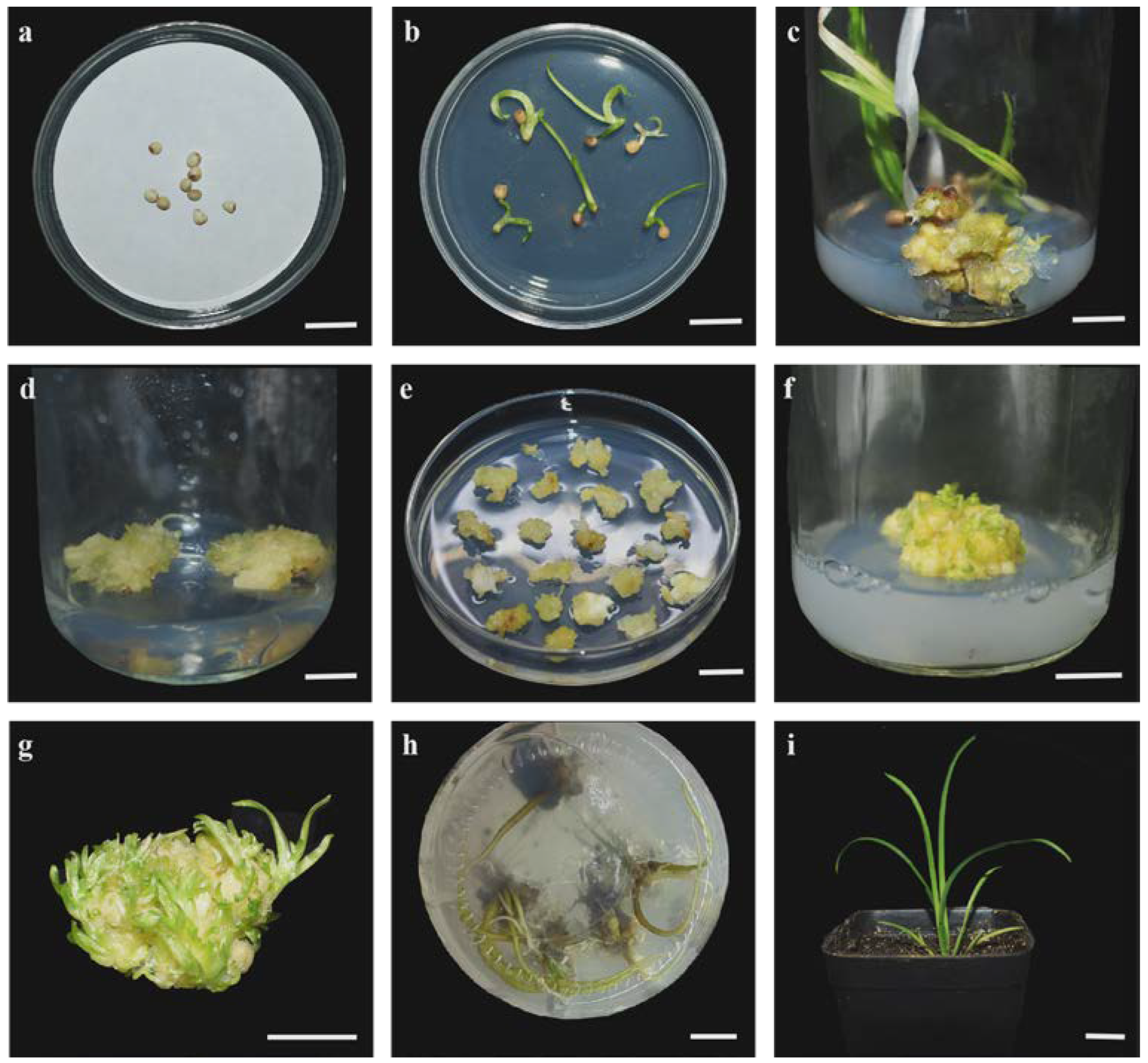
Significant variance in both contamination and germination rates existed among the three different seed coat treatments (Table 3). Notably, the treatment with both the inner and outer seed coats removed (M3) exhibited an average contamination rate of 6.7% and an impressive average germination rate of 98.0%. In comparison, the other two seed coat treatments (M1 and M2) presented much higher contamination rates and lower germination rates, offering insufficient potential in providing sterile and viable plant materials for subsequent callus induction. Consequently, the aerial parts of plantlets derived from seeds with both the inner and outer seed coats removed were utilized as proper explants (Figure 2a,b). The optimal 6-BA and NAA concentrations during the stages of callus induction and differentiation were further explored.

3.1.2. Establishment of the Callus Induction and Shoot Induction Protocol

To develop a highly efficient Agrobacterium-mediated genetic transformation protocol for H. middendorffii, eighteen different medium formulations were created (six 6-BA concentrations and three NAA concentrations) to stimulate callus formation from the aerial parts of seed-derived plantlets. After cultivation for one month, the initiation of calli was noted (Figure 2c). It was observed that 6-BA at lower concentrations slowed down the callus formation process, whereas NAA had a weak effect on the induction of calli, as shown in Table 4. The callus induction rate was 28.9% under the addition of 1.5 mg·L−1 6-BA and 0.3 mg·L−1 NAA, whereas a considerably higher induction rate of 95.6% was attained with 3.5 mg·L−1 6-BA and 0.1 mg·L−1 NAA. These results indicated that a synergistic effect involving higher concentrations of 6-BA and lower concentrations of NAA significantly boosted callus formation in H. middendorffii. Then, 2,4-D was incorporated into the subculture medium to promote the proliferation of calli (Figure 2d). Initially, the induced calli exhibited a compact and nodular morphology with a yellow and calloused appearance. As callus proliferation progressed, the texture became soft and friable, and the color transitioned to a pale yellow.
The effect of 6-BA at varying concentrations on shoot induction was assessed by altering its concentration in the callus induction medium. When the NAA concentration was 0.2 mg·L−1, the regeneration rate of shoots decreased as the 6-BA concentration increased from 1.0 to 3.0 mg·L−1. Additionally, 0.5 mg·L−1 6-BA also yielded a relatively high growth rate (Table 5). It was also observed that 6-BA and NAA at low concentrations not only promoted the growth of small shoots but also facilitated their formation and proliferation. Consequently, the optimal medium for shoot regeneration was determined to be MS medium supplemented with 1.0 mg·L−1 6-BA and 0.2 mg·L−1 NAA (Figure 2e–g).
After a 5–6-week period of shoot induction culture, the individually separated plantlets were moved to a rooting medium. The initiation of rooting commenced on the 15th day. By the 30th day, the roots had grown to a length of 3–5 cm; then, the plantlets were transplanted into pots.

3.2. Factors Influencing Agrobacterium-Mediated Transformation

3.2.1. Hyg Concentration

In order to determine the optimal Hyg concentration, a sensitivity assessment was conducted, as its effectiveness varies among species. Hyg at the appropriate concentration effectively inhibits the growth of non-transformed plants and induces their gradual death, while it only slightly impairs the normal growth of transformed recipients.
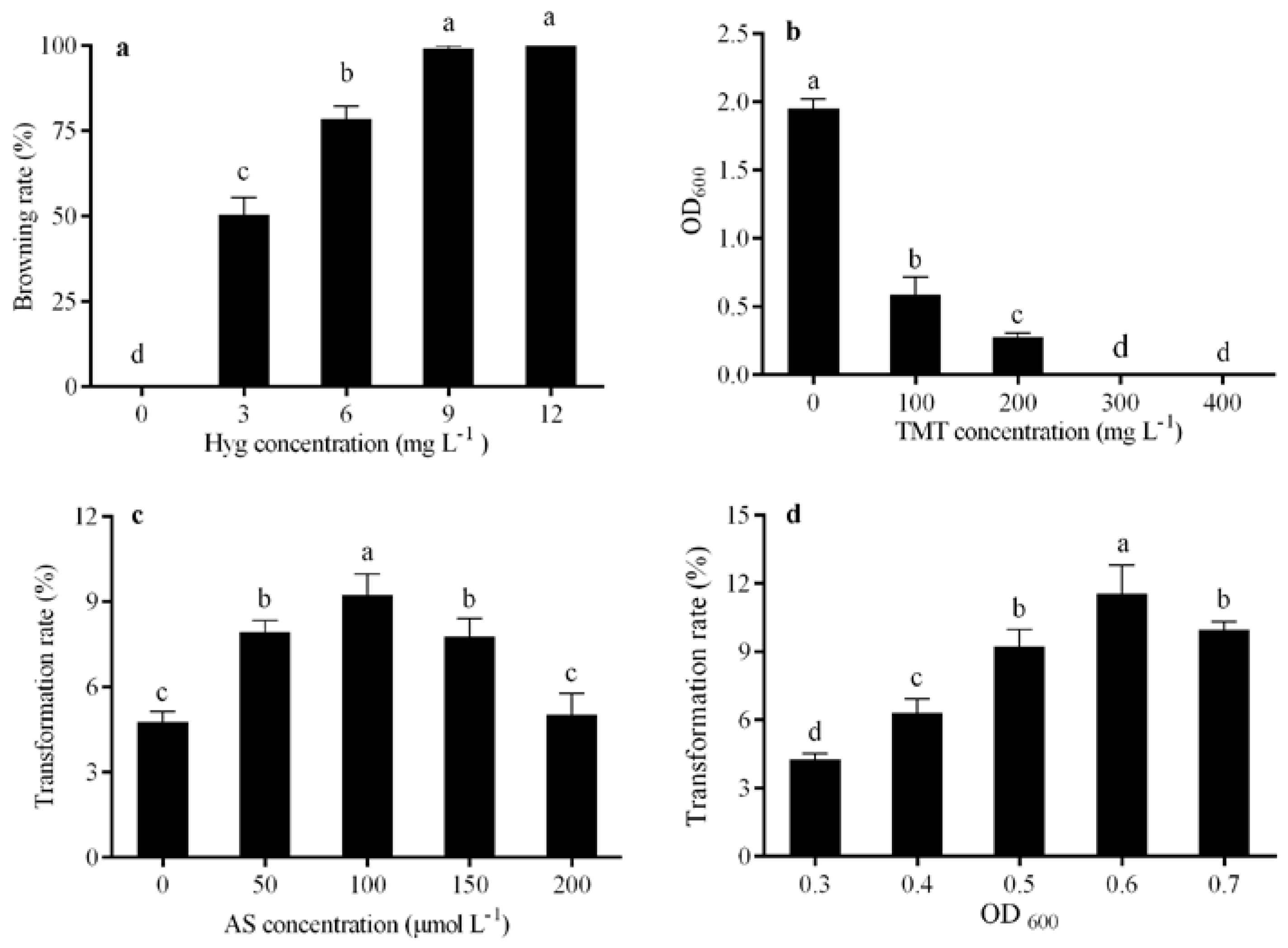
The calli were cultured on pre-cultivation medium containing Hyg at varying concentrations. There was significant variation in the browning rate of calli. Specifically, Hyg at concentrations of 9 and 12 mg·L−1 effectively hindered the differentiation of calli, leading to their browning and subsequent death (Figure 3a). Consequently, 9 mg·L−1 Hyg was determined as the threshold concentration for effectively screening calli in the genetic transformation process.

3.2.2. TMT Concentration

TMT was utilized as the antibiotic to suppress A. tumefaciens growth. After propagation culture, the bacteria were introduced into the LB liquid medium with TMT at varying concentrations for further culturing, and the change in bacterial density was monitored.
As shown in Figure 3b, the OD600 value rose to 2.0 in the LB liquid medium without TMT supplementation. The increase in TMT concentration significantly inhibited the reproduction of A. tumefaciens, resulting in a pronounced decrease in OD600 value. A. tumefaciens was essentially inactivated at a TMT concentration of 300 mg·L−1, with the OD600 value declining to nearly 0. Increasing the TMT concentration to 400 mg·L−1 did not alter the growth dynamic of A. tumefaciens as compared with that at 300 mg·L−1, suggesting that 300 mg·L−1 was the empirically supported critical concentration of TMT.

3.2.3. AS Concentration

Numerous studies have demonstrated that AS, a small phenolic compound, can induce the activation of vir genes in A. tumefaciens, thereby enhancing the successful transfer of Ti plasmids into host cells [29,30]. Among the tested concentrations of AS, the maximum transformation rate of 9.2% was observed at an AS concentration of 100 μmol·L−1. Concentrations above 100 μmol·L−1 yielded lower transformation rates (Figure 3c). Furthermore, AS at high levels was found to disrupt the normal growth of the recipients, leading to the excessive growth of A. tumefaciens and browning of the recipients. Therefore, 100 μmol·L−1 AS was determined to be the most effective concentration for enhancing transformation rate.

3.2.4. Bacterial Cell Density

The effect of A. tumefaciens infiltration solution density on the transformation rate was investigated. As shown in Figure 3d, the optimal transformation rate of 11.6% was achieved with recipients exposed to an OD600 value of 0.6. A slight decrease in the transformation rate was observed when the A. tumefaciens culture OD600 was further increased.

3.3. Stability Test of the H. middendorffii Transformation System

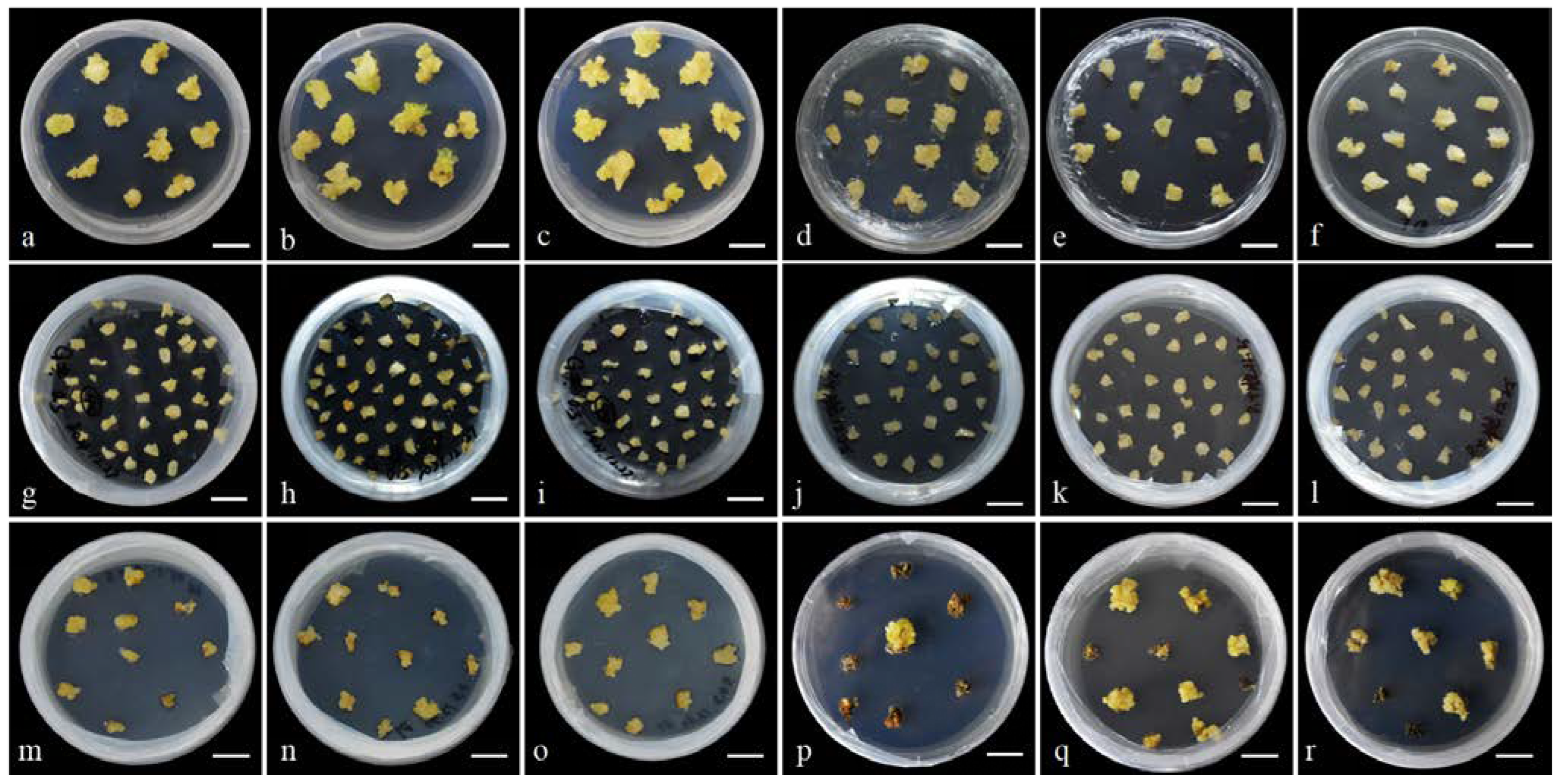
To assess the stability of the developed Agrobacterium-mediated transformation system for H. middendorffii, three experimental replicates were conducted using the optimal combination of genetic transformation factors (Figure 4). The results confirmed the system’s stability, yielding an average transformation rate of 11.9% (Table 6). In addition, it took approximately 4.5–5.0 months to obtain transgenic H. middendorffii T0 rooted plants by using this system.

3.4. Validation of Transgenic Plants

The transgenic status of regenerated plants was initially confirmed by PCR amplification using 35S sequence-specific primers. Following hygromycin selection, a total of 16 candidate positive plants were obtained and designated as lines 1–15. After differentiation, each line produced at least two replicates. Through three independent experimental replicates, PCR products corresponding to the 801 bp 35S sequence fragment were successfully amplified (Figure 5a). The PCR products were derived from transgenic lines 1-1, 1-2, 1-3, 7-1, 7-2, 8-1, 8-2, 11-2, and 13-2, resulting in a positive line rate of 32.8%. Lines 2, 3, 4, 5, 6, 9, 10, 12, 14, and 15 were identified as false positives. The Sanger sequencing validation results are presented in Supplementary Figure S2.
The growth status of the H. middendorffii plantlets indicated that the length and the number of lateral roots in WT plants were significantly lower than those in pCAMBIA1300-35S-HmFT transgenic plants under the same growth conditions (Figure 5b). Moreover, the root growth ability of the transgenic pCAMBIA1300-35S-HmFT plants was enhanced (Supplementary Table S2).
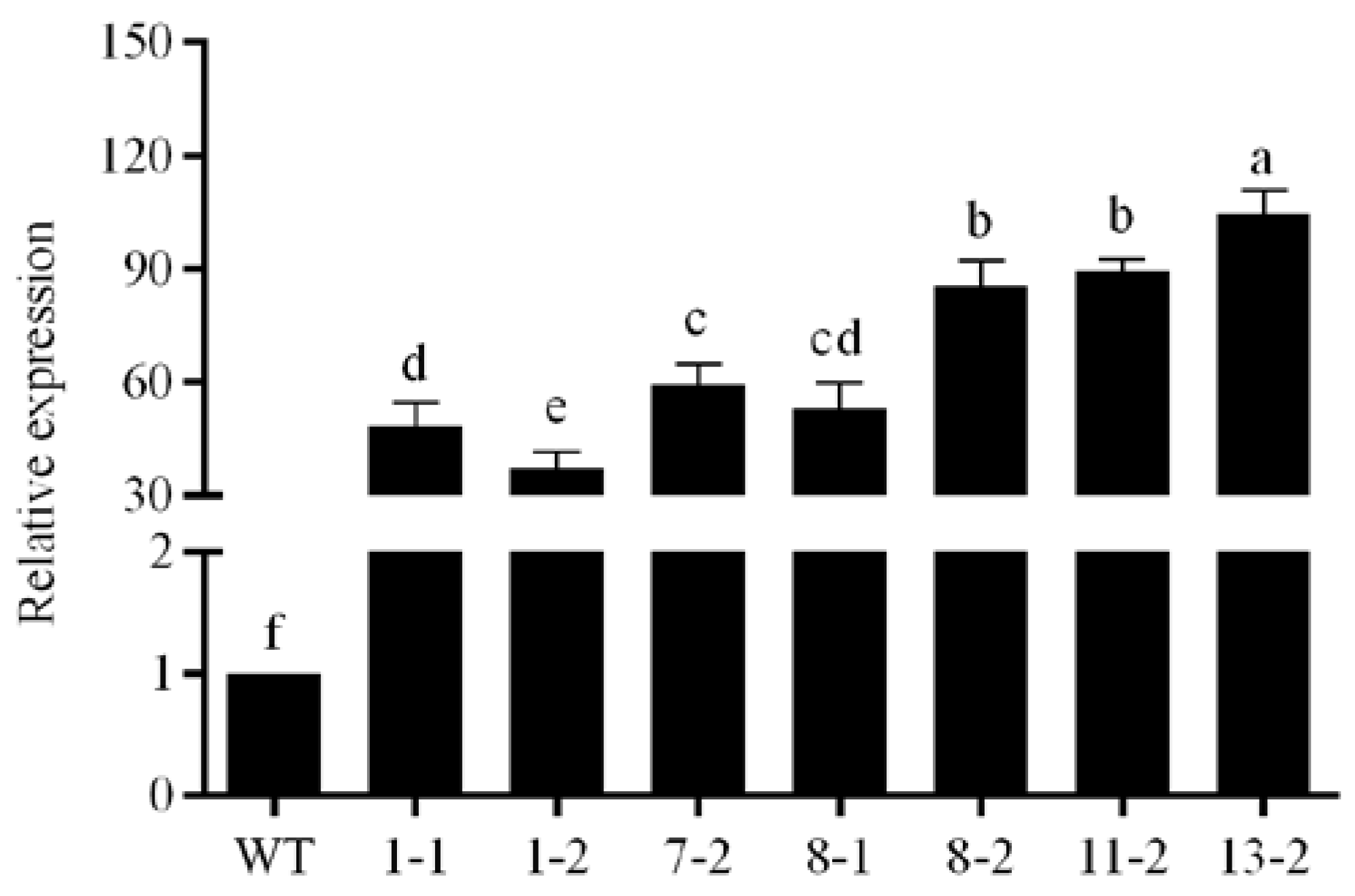
The qRT-PCR results indicated that the expression levels of HmFT in transgenic plants were significantly upregulated compared with WT plants (Figure 6). The relative expression level in the 13-2 transgenic line was 100 times higher than that in WT plants. These results demonstrated that pCAMBIA1300-35S-HmFT expression vector plasmid was successfully introduced into the H. middendorffii genome and expressed.

4. Discussion

Plant tissue culture is widely employed in large-scale plant reproduction, virus elimination, secondary metabolite production, and in vitro cloning [31,32]. Extensive studies have shown that the healing rates of Hemerocallis flower organs, including flower stems, pedicels, and petals, were superior to those of other parts in tissue culture [17]. However, the act of cutting for explant collection can activate polyphenol oxidases in these tissues, which oxidize phenolic substances into brownish quinone compounds, leading to the browning at the cut sites of the explants. This phenomenon may inhibit normal explant development or even lead to explant death [33,34]. In this study, the aerial parts of seed-derived H. middendorffii plantlets were utilized as explants, which acted as excellent hosts for Agrobacterium-mediated genetic transformation. Furthermore, the removal of both the inner and outer seed coats reduced the contamination rate. Meanwhile, this process eliminated the potential influence of trace phenolic compounds within the seed coats, rendering it the optimal source for H. middendorffii tissue culture materials. Moreover, before the seed coats were removed, the seeds were soaked in distilled water for 36–48 h. When the soaking time was shorter than 36 h, the seed coats were insufficiently softened, which hindered seed coat removal. When the soaking time was longer than 48 h, the seeds showed signs of germination and were susceptible to physical damage during the sterilization process, which affected the subsequent normal germination.
Various studies have underscored the dramatic effect of exogenous PGRs in stimulating and inducing morphogenesis [35,36,37]. The combined action of cytokinin and auxin enhanced in vitro plant regeneration [38,39]. In Dianthus chinensis, the application of 1.0 mg·L−1 6-BA along with 0.06 mg·L−1 NAA led to a higher leaf regeneration rate and the highest observed explant induction rate [9]. The optimal shoot induction in Indian red banana was obtained with 3.0 mg·L−1 6-BA and 0.2 mg·L−1 NAA [37]. In addition, the greatest number of branches was stimulated in Lallemantia iberica by application of 1.0 mg·L−1 6-BA and 0.05 mg·L−1 NAA [31]. In the present study, MS medium was enhanced with 3.5 mg·L−1 6-BA and 0.1 mg·L−1 NAA, leading to a notable increase in the induction rate. These results implied that effective explant induction may require a higher level of 6-BA and a lower level of NAA. Additionally, it was indicated that optimal explant differentiation might necessitate a balanced combination of lower concentrations of 6-BA and NAA.
Agrobacterium-mediated transformation is a complex process influenced by multiple factors, including the optical density of the A. tumefaciens culture solution and the concentrations of AS, TMT, and Hyg, among other factors [40,41,42,43,44]. In the present research, Hyg was used for the selection of transgenic plants, as the pCAMBIA1300-35S expression vector contains a gene that confers resistance to Hyg, making it an effective selection marker for identification of transgenic plants. Research has demonstrated that even among different varieties of the same species, the optimal concentrations of Hyg as a selection agent can vary. For example, 7 mg·L−1 Hyg was effective for further selection and regeneration of positive transformants in Rosa chinensis Jacq. [45]. However, research on Rosa hybrida demonstrated that 50 mg·L−1 Hyg inhibited excessive growth of calli, and 20 mg·L−1 Hyg had no dramatic impact on the later growth stages of the plants [46]. In the present study, 9 mg·L−1 Hyg efficiently eliminated most of the non-transformants and was identified as the threshold for screening transgenic H. middendorffii plants.
TMT effectively inhibits A. tumefaciens and is widely used in plant in vitro culture [47,48], with optimally effective concentrations of 300–500 mg·L−1. In the present protocol, 300 mg·L−1 TMT was added to both the sterilization medium and subsequent screening medium. It effectively inhibited A. tumefaciens growth, eliminated other bacterial contaminants, and prevented the contamination of H. middendorffii calli and regenerative shoots.
Previous research has indicated that AS can significantly increase the transformation rate of various plant species across a broad concentration range of 20–100 µmol·L−1 [49,50,51,52]. For instance, 50 µmol·L−1 AS was effective for the transformation of Cyclamen persicum [53], and 100 µmol·L−1 AS was identified as the optimal co-cultivation agent for cucumber transformation [54]. These results are consistent with previous findings in Tagetes erecta and Typha latifolia [55,56]. Similarly, this research revealed that the highest transformation rate for H. middendorffii was achieved when the AS concentration in the A. tumefaciens culture solution and co-cultivation medium was 100 µmol·L−1.
Furthermore, the optical density of the A. tumefaciens solution exerts a crucial impact on the transformation rate [54]. When the concentration is too low, A. tumefaciens cannot sufficiently adhere to recipients, leading to ineffective transformation. Conversely, when the concentration is too high, it can cause severe damage to the recipients [57]. In the present study, it was found that the transformation rate was promoted and the transformed recipients were relatively healthier and more robust as the OD600 value was increased from 0.3 to 0.6. However, when OD600 value exceeded 0.6, most of the transformed recipients exhibited browning and death, which could be attributed to the excessively high concentration of the infection solution [42].
In this study, PCR amplification using 35S promoter-specific primers successfully detected the 801 bp target fragment in the transgenic lines (Figure 5a). The absence of amplification in WT plants confirmed primer specificity. Further validation by qRT-PCR revealed that HmFT expression in transgenic lines was significantly upregulated compared to WT plants (Figure 6). Line 13-2 exhibited the highest expression level (100-fold increase), consistent with its enhanced root growth (Supplementary Table S2). These results preliminarily validate the possible utility of the established transformation system for functional genomics and trait improvement in H. middendorffii. To further confirm transgene functionality at the protein level, future studies will include Western blot analysis using HmFT-specific antibodies to detect HmFT protein accumulation in T0 plants. Additionally, Southern blot analysis of T0 plants will be performed to verify the stable integration of the transgene into the plant genome, providing critical evidence of transformation stability prior to T1 generation analysis.

5. Conclusions

In this study, an efficient and reliable protocol was developed for the regeneration and callus genetic transformation of H. middendorffii using the aerial parts of seed-derived plantlets as explants. By optimizing the callus induction, proliferation, subculture, differentiation, and rooting conditions, a callus induction rate of 95.6% and a regeneration rate of 84.4% were achieved. Based on this procedure, a refined Agrobacterium-mediated transformation system for H. middendorffii realized the generation of transgenic plants within a 5-month period, with the transformation rate reaching up to 11.9% and the positive rate of 32.8%. The established Agrobacterium-mediated transformation system for H. middendorffii lays a solid foundation for the genetic modification and breeding of this valuable ornamental species. Furthermore, this genetic transformation system is a robust and versatile tool for gene function and editing research in H. middendorffii, thereby facilitating research on trait enhancement, functional genomics, and metabolite engineering in this species.

Supplementary Materials

The following supporting information can be downloaded at: https://www.mdpi.com/article/10.3390/horticulturae11040417/s1, Figure S1: Sequence of HmFT gene from H. middendorffii; Figure S2: Sanger sequencing result using the 35S sequence-specific primers; Table S1: Sequences of primers used in the present study; Table S2: Statistics on the root growth of WT and transgenic H. middendorffii.

Author Contributions

Conceptualization, Y.L. and W.L.; methodology, J.D.; software, J.D.; validation, J.D., J.S., and N.Z.; formal analysis, J.D.; investigation, J.S.; resources, J.D.; data curation, N.Z.; writing—original draft preparation, J.D.; writing—review and editing, W.L.; visualization, J.D.; supervision, Y.L.; project administration, W.L.; funding acquisition, Y.L. and W.L. All authors have read and agreed to the published version of the manuscript.

Funding

This research was funded by “the National Natural Science Foundation of China, grant number 32102420” and “the Natural Science Foundation of Heilongjiang Province of China, grant number YQ2021C014”.

Data Availability Statement

The datasets used and/or analyzed during the current study are available from the corresponding author on reasonable request. We sincerely hope that readers can further develop a rapid and efficient system for adventitious shoot regeneration based on our research.

Conflicts of Interest

The authors declare that there are no conflicts of interest.

References

  1. Chen, X.; Liang, S.; Xu, J.; Tamura, M.N. Flora of China; Science Press: Bejing, China; Missouri Botanical Garden Press: St. Louis, SK, Canada, 2020; pp. 73–263. [Google Scholar]
  2. Masuda, K.; Setoguchi, H.; Nagasawa, K.; Ishihara, M.; Sakaguchi, S. Development and characterization of EST-SSR markers for Amur daylily, Hemerocallis middendorffii Trautv. & C. A. Mey. (Asphodelaceae). Acta Phytotax. Geobot. 2021, 72, 73–78. [Google Scholar] [CrossRef]
  3. Liu, Y. The Establishment of Hemerocallis middendorfii Callus Induction and Agrobacterium Mediated Genetic Transofrmation System. Master’s Dissertation, Jilin Agricultural University, Changchun, China, 2016. [Google Scholar]
  4. Wang, H. Alleviating Effect of Exogenous Ca(NO3)2 on Hemerocallis middendorfii Under Salt Stress. Master’s Dissertation, Northeast Agricultural University, Harbin, China, 2022. [Google Scholar]
  5. Chen, L.F.; Liu, Y.Y.H.; Li, J.J.; Bai, Y.; Meng, Y.; Zhou, Y.W. Identification and analysis of bHLH transcription factor gene family in Hemerocallis middendorffii under drought stress. Southwest China J. Agr. Sci. 2023, 36, 1027–1038. [Google Scholar]
  6. Ren, Y.; Gao, Y.K.; Liu, J.J.; Fu, M.; Zhang, Q.X. Investigation on resources of Hemerocallis in North China. Acta Hortic. 2017, 1185, 65–72. [Google Scholar] [CrossRef]
  7. Wang, R. Effects of Three Plant Growth Regulators on the Growth and Development of Hemerocallis middendorfii. Master’s Dissertation, Northeast Agricultural University, Harbin, China, 2023. [Google Scholar]
  8. Shen, P. Genetic Diversity of Hemerocallis middendorfii in North-East of China. Master’s Dissertation, Beijing Forestry University, Beijing, China, 2010. [Google Scholar]
  9. Zhang, X.; Wu, Q.; Lin, S.; Zhang, Z.; Wang, Z.; Wang, Q.; Yan, X.; Bendahmane, M.; Bao, M.; Fu, X. Regeneration and Agrobacterium-mediated genetic transformation in Dianthus chinensis. Sci. Hortic. 2021, 287, 110279. [Google Scholar] [CrossRef]
  10. Heuser, C.W.; Apps, D.A. In vitro plantlet formation from flower petal explants of Hemerocallis cv. Chipper Cherry. Can. J. Bot. 1976, 54, 616–618. [Google Scholar] [CrossRef]
  11. Li, Z.; Mize, K.; Campbell, F. Regeneration of daylily (Hemerocallis) from young leaf segments. Plant Cell Tissue Organ Cult. 2010, 102, 199–204. [Google Scholar] [CrossRef]
  12. Gao, J. Optimization of Regeneration System and Establishment of Agrobacterium-Mediated Transformation System of Daylily. Master’s Dissertation, Sichuan Agricultural University, Ya’an, China, 2012. [Google Scholar]
  13. Rodriguez-Enriquez, M.J.; Grant-Downton, R.T. A new day dawning: Hemerocallis (daylily) as a future model organism. AoB Plants 2013, 5, pls055. [Google Scholar] [CrossRef]
  14. Jin, L.M.; Zhou, Y.Z.; Guo, Z.H.; Huang, B.J. Effect of inbred, hybrid and different cultivation enviroments on seed setting characteristics of Hemerocallis hybrid. Hubei Agric. Sci. 2012, 51, 2760–2763. [Google Scholar] [CrossRef]
  15. Aziz, A.N.; Sauvé, R.J.; Zhou, S. Genetic transformation of Stella de Oro daylily by particle bombardment. Can. J. Plant Sci. 2003, 83, 873–876. [Google Scholar] [CrossRef]
  16. Ni, D.; Liang, J.; Gao, M.; Zhong, R.; Tan, Y.; Liu, X.; Yin, D.; Zhang, Z. Daylily glyoxalase gene cloning and pollen-tube pathway transformation. Plant Physiol. J. 2020, 56, 171–179. [Google Scholar] [CrossRef]
  17. Wang, X.X. The Plant Regeneration and Transformation of CHS Gene by Grobacteria-Mediated in Hemerocallis hybrida. Master’s Dissertation, Gansu Agricultural University, Lanzhou, China, 2013. [Google Scholar]
  18. Srinivasan, C.; Dardick, C.; Callahan, A.; Scorza, R. Plum (Prunus domestica) trees transformed with poplar FT1 result in altered architecture, dormancy requirement, and continuous flowering. PLoS ONE 2012, 7, e40715. [Google Scholar] [CrossRef]
  19. Heller, W.P.; Ying, Z.; Davenport, T.L.; Keith, L.M.; Matsumoto, T.K. Identification of members of the Dimocarpus longan Flowering Locus T gene family with divergent functions in fowering. Trop. Plant Biol. 2014, 7, 19–29. [Google Scholar] [CrossRef]
  20. Zhang, L.; Zhang, F.; Zhou, X.; Poh, T.X.; Xie, L.; Shen, J.; Yang, L.; Song, S.; Yu, H.; Chen, Y. The tetratricopeptide repeat protein OsTPR075 promotes heading by regulating florigen transport in rice. Plant Cell 2022, 34, 3632–3646. [Google Scholar] [CrossRef]
  21. Kinoshita, T.; Ono, N.; Hayashi, Y.; Morimoto, S.; Nakamura, S.; Soda, M.; Kato, Y.; Ohnishi, M.; Nakano, T.; Inoue, S.; et al. FLOWERING LOCUS T regulates stomatal opening. Curr. Biol. 2011, 21, 1232–1238. [Google Scholar] [CrossRef] [PubMed]
  22. Jing, S.; Sun, X.; Yu, L.; Wang, E.; Cheng, Z.; Liu, H.; Jiang, P.; Qin, J.; Begum, S.; Song, B. Transcription factor StABI5-like 1 binding to the FLOWERING LOCUS T homologs promotes early maturity in potato. Plant Physiol. 2022, 189, 1677–1693. [Google Scholar] [CrossRef] [PubMed]
  23. Murashige, T.; Skoog, F. A revised medium for rapid growth and bio assays with tobacco tissue cultures. Physiol. Plant. 1962, 15, 473–497. [Google Scholar] [CrossRef]
  24. MacWilliams, M.P.; Liao, M.K. Luria Broth (LB) and Luria Agar (LA) media and their uses protocol. Am. Soc. Microbiol. 2006, 1–4. [Google Scholar]
  25. Chen, H.; Nelson, R.; Sherwood, J. Enhanced recovery of transformants of Agrobacterium tumifaciens after freeze-thaw transformation and drug selection. Biotechniques 1994, 16, 664–670. [Google Scholar]
  26. Aljanabi, S.; Martinez, I. Universal and rapid salt-extraction of high quality genomic DNA for PCR-based techniques. Nucleic Acids Res. 1997, 25, 4692–4693. [Google Scholar] [CrossRef]
  27. Liu, Y.; Gao, Y.; Yuan, L.; Zhang, Q. Functional characterization and spatial interaction of TERMINAL FLOWER 1 in Hemerocallis. Sci. Hortic. 2019, 253, 154–162. [Google Scholar] [CrossRef]
  28. Zhang, Y.; Sun, J.; Xia, H.; Zhao, C.; Hou, L.; Wang, B.; Li, A.; Chen, M.; Zhao, S.; Wang, X. Characterization of peanut phytochromes and their possible regulating roles in early peanut pod development. PLoS ONE 2018, 13, e0198041. [Google Scholar] [CrossRef] [PubMed]
  29. Veluthambi, K.; Krishnan, M.; Gould, J.H.; Smith, R.H.; Gelvin, S.B. Opines stimulate induction of the vir genes of the Agrobacterium tumefaciens Ti plasmid. J. Bacteriol. 1989, 171, 3696–3703. [Google Scholar] [CrossRef] [PubMed]
  30. Krishnamohan, A.; Balaji, V.; Veluthambi, K. Efficient vir gene induction in Agrobacterium tumefaciens requires virA, virG, and vir box from the same Ti plasmid. J. Bacteriol. 2001, 183, 4079–4089. [Google Scholar] [CrossRef]
  31. Ebrahimzadegan, R.; Maroufi, A. In vitro regeneration and Agrobacterium-mediated genetic transformation of Dragon’s Head plant (Lallemantia iberica). Sci. Rep. 2022, 12, 1784. [Google Scholar] [CrossRef] [PubMed]
  32. Haida, Z.; Sinniah, U.R.; Nakasha, J.J.; Hakiman, M. Shoot induction, multiplication, rooting and acclimatization of black turmeric (Curcuma caesia Roxb.): An important and endangered Curcuma species. Horticulturae 2022, 8, 740. [Google Scholar] [CrossRef]
  33. Das, D.K.; Rahman, A. Phenolic compositions of litchi shoot tips and zygotic embryos collected in different months and their effects on the explant browning and its control. Adv. Anthropol. 2016, 6, 73–92. [Google Scholar] [CrossRef]
  34. Lux-Endrich, A.; Treutter, D.; Feucht, W. Influence of nutrients and carbohydrate supply on the phenol composition of apple shoot cultures. Plant Cell Tissue Organ Cult. 2000, 60, 15–21. [Google Scholar] [CrossRef]
  35. Gbadamosi, I.T.; Egunyomi, A. Micropropagation of Plumbago zeylanica L. (Plumbaginaceae) in Ibadan, Southwestern, Nigeria. J. Med. Plants Res. 2010, 4, 293–297. [Google Scholar]
  36. Siddique, I.; Javed, S.B.; Al-Othman, M.R.; Anis, M. Stimulation of in vitro organogenesis from epicotyl explants and successive micropropagation round in Cassia angustifolia Vahl.: An important source of sennosides. Agrofor. Syst. 2013, 87, 583–590. [Google Scholar] [CrossRef]
  37. Rajoriya, P.; Singh, V.K.; Jaiswal, N.; Lall, R. Optimizing the effect of plant growth regulators on in vitro micro propagation of Indian red banana (Musa acuminata). J. Pharmacogn. Phytochem. 2018, 7, 628–634. [Google Scholar]
  38. De Klerk, G.J.; Hanecakova, J.; Jasik, J. The role of cytokinins in rooting of stem slices cut from apple microcuttings. Plant Biosyst. 2001, 135, 79–84. [Google Scholar] [CrossRef]
  39. Huang, T.; Zhang, H.; Zhao, R.; Zhu, Z. Establishing an efficient regeneration system for tissue culture in Bougainvillea buttiana ‘Miss Manila’. Plants 2022, 11, 2372. [Google Scholar] [CrossRef] [PubMed]
  40. Confalonieri, M.; Balestrazzi, A.; Bisoffi, S. Genetic transformation of Populus nigra by Agrobacterium tumefaciens. Plant Cell Rep. 1994, 13, 256–261. [Google Scholar] [CrossRef]
  41. Yuan, X.; Wang, Z.; Liu, J.; She, J. Development of a plant regeneration system from seed-derived calluses of centipedegrass [Eremochloa ophiuroides (Munro.) Hack]. Sci. Hortic. 2009, 120, 96–100. [Google Scholar] [CrossRef]
  42. Chhabra, G.; Chaudhary, D.; Sainger, M.; Jaiwal, P.K. Genetic transformation of Indian isolate of Lemna minor mediated by Agrobacterium tumefaciens and recovery of transgenic plants. Physiol. Mol. Biol. Plants 2011, 17, 129–136. [Google Scholar] [CrossRef]
  43. Chen, X.A.; He, X.; Zhang, M.; Mao, X.M.; Li, Y. Q An efficient genetic transformation system for Chinese medicine fungus Tolypocladium ophioglossoides. J. Microbiol. Methods 2020, 176, 106032. [Google Scholar] [CrossRef] [PubMed]
  44. Wang, X.; Zhou, F.; Liu, J.; Liu, W.; Zhang, S.; Li, D.; Song, J.; Wang, R.; Yang, Y. Establishment of efficient callus genetic transformation system for Pyrus armeniacaefolia. Sci. Hortic. 2021, 289, 110429. [Google Scholar] [CrossRef]
  45. Chen, J.R.; Chen, Y.B.; Ziemiańska, M.; Liu, R.; Deng, Z.N.; Niedźwiecka-Filipiak, I.; Li, Y.L.; Jiao, J.X.; Xiong, X.Y. Co-expression of MtDREB1C and RcXET enhances stress tolerance of transgenic China rose (Rosa chinensis Jacq.). J. Plant Growth Regul. 2016, 35, 586–599. [Google Scholar] [CrossRef]
  46. Liu, G.; Yuan, Y.; Jiang, H.; Bao, Y.; Ning, G.; Zhao, L.; Zhou, X.; Zhou, H.; Gao, J.; Ma, N. Agrobacterium tumefaciens-mediated transformation of modern rose (Rosa hybrida) using leaf-derived embryogenic callus. Hortic. Plant J. 2021, 7, 359–366. [Google Scholar] [CrossRef]
  47. Bannikova, M.A.; Gorbatyuk, I.R.; Hnatiuk, I.S.; Malysheva-Otto, L.V.; Duplij, V. Effect of Ceftriaxone and Timentin antibiotics on morphogensis in the in vitro culture of bread wheat Triticum aestivum L. Biopolym. Cell 2016, 32, 367–376. [Google Scholar] [CrossRef]
  48. Zhang, H.; Wang, G.; Qiao, Y.; Chen, C. Effects of Timentin and other β-Lactam antibiotics on callus induction, shoot regeneration, and rooting in Anthurium andraeanum Linden ex Andre. In Vitro Cell. Dev. Biol. Plant 2017, 53, 219–225. [Google Scholar] [CrossRef]
  49. John, E.; Maqbool, A.; Malik, K.A. Optimization of Agrobacterium tumefaciens mediated transformation in Populus deltoides. Pak. J. Bot. 2014, 46, 1079–1086. [Google Scholar]
  50. Movahedi, A.; Zhang, J.; Amirian, R.; Zhuge, Q. An efficient Agrobacterium-mediated transformation system for Poplar. Int. J. Mol. Sci. 2014, 15, 10780–10793. [Google Scholar] [CrossRef]
  51. Li, S.; Zhen, C.; Xu, W.; Wang, C.; Cheng, Y. Simple, rapid and efficient transformation of genotype Nisqually-1: A basic tool for the first sequenced model tree. Sci. Rep. 2017, 7, 2638. [Google Scholar] [CrossRef]
  52. Li, X.; Li, H.; Zhao, Y.; Zong, P.; Zhan, Z.; Piao, Z. Establishment of a simple and efficient Agrobacterium-mediated genetic transformation system to Chinese Cabbage (Brassica rapa L. ssp. pekinensis). Hortic. Plant J. 2021, 7, 117–128. [Google Scholar] [CrossRef]
  53. Ratjens, S.; Mortensen, S.; Kumpf, A.; Bartsch, M.; Winkelmann, T. Embryogenic callus as target for efficient transformation of Cyclamen persicum enabling gene function studies. Front. Plant Sci. 2018, 9, 1035. [Google Scholar] [CrossRef]
  54. Tan, J.; Lin, L.; Luo, H.; Zhou, S.; Zhu, Y.; Wang, X.; Miao, L.; Wang, H.; Zhang, P. Recent progress in the regeneration and genetic transformation system of cucumber. Appl. Sci. 2022, 12, 7180. [Google Scholar] [CrossRef]
  55. Nandakumar, R.; Chen, L.; Rogers, S.M.D. Factors affecting the Agrobacterium-mediated transient transformation of the wetland monocot, Typha latifolia. Plant Cell Tissue Organ Cult. 2004, 79, 31–38. [Google Scholar] [CrossRef]
  56. Godoy-Hernández, G.; Berzunza, E.A.; Concha, L.C.; Miranda-Ham, M.d.L. Agrobacterium-mediated transient transformation of marigold (Tagetes erecta). Plant Cell Tissue Organ Cult. 2006, 84, 365–368. [Google Scholar] [CrossRef]
  57. Chen, Y.; Hou, X.; Zheng, Y.; Lyu, Y. The establishment of a genetic transformation system and the acquisition of transgenic plants of oriental hybrid lily (Lilium L.). Int. J. Mol. Sci. 2023, 24, 782. [Google Scholar] [CrossRef]
Figure 1. The pCMBIA1300-35S vector.
Horticulturae 11 00417 g001
Figure 2. Regeneration process of H. middendorffii plantlets from seeds. Seeds removed from both the inner and outer seed coats (a). Germination status of seeds after 14 days (b). Calli induced from seedlings after 6 weeks of cultivation on the callus induction medium (c). Proliferation of calli on the callus proliferation medium (d). Initiation of shoot formation from calli on the differentiation medium (e). Progress of shoot induction after 3 weeks (f). Development of shoots after 5 weeks (g). Root initiation observed in a single shoot on the rooting medium after 1 month (h). Plantlets being acclimatized in pots (i). Scale bar = 1 cm.
Horticulturae 11 00417 g002
Figure 3. Effect of hygromycin (Hyg) concentration on the callus browning rate (a). Impact of Timentin (TMT) concentration on the A. tumefaciens culture solution optical density at 600 nm (OD600) (b). Effect of OD600 value on the transformation rate (c). Influence of acetosyringone (AS) concentration on the transformation rate (d). Hyg, hygromycin; TMT, Timentin; AS, acetosyringone. Different lowercase letters indicate significant differences at the 5% level based on Duncan’s multiple range test (p < 0.05).
Horticulturae 11 00417 g003
Figure 4. Genetic transformation process of H. middendorffii. Healthy and robust transformed recipients (ac). Pre-cultivation on MS medium supplemented with 1.0 mg·L−1 6-BA, 0.2 mg·L−1 NAA, 30 g·L−1 sucrose, and 7.6 g·L−1 agarose for 3 days (df). Co-cultivation on MS medium supplemented with 1.0 mg·L−1 6-BA, 0.2 mg·L−1 NAA, 100 μmol·L−1 AS, 30 g·L−1 sucrose, and 7.6 g·L−1 agarose for 2 days (gi). Resting on MS medium supplemented with 1.0 mg·L−1 6-BA, 0.2 mg·L−1 NAA, 300 mg·L−1 TMT, 20 g·L−1 sucrose, and 7.6 g·L−1 agarose for 5 days (jl). Selection 1 phase on MS medium supplemented with 1.0 mg·L−1 6-BA, 0.2 mg·L−1 NAA, 9 mg·L−1 Hyg, 300 mg·L−1 TMT, 30 g·L−1 sucrose, and 7.6 g·L−1 agarose for 15 days (mo). Selection 2 phase for 30 days (pr). Scale bar = 1 cm.
Horticulturae 11 00417 g004
Figure 5. PCR analysis and phenotypic status of wild-type (WT) and transgenic H. middendorffii. PCR amplification of the 35S sequence (801 bp) in WT plant and five transgenic lines (a). Growth state of WT and pCAMBIA1300-35S-HmFT transgenic plants (b). Lane M, DNA maker; Lanes 1-1, 1-2, 1-3, 7-1, 7-2, 8-1, 8-2, 11-2, and 13-2, transgenic lines; Lane P, plasmid control, Lane WT, wild-type plant.
Horticulturae 11 00417 g005
Figure 6. Expression levels of HmFT in leaves of wild-type (WT) and transgenic H. middendorffii plants by qRT-PCR. The expression level of HmFT in WT plants used as a control. Transgenic lines: 1-1, 1-2, 7-2, 8-1, 8-2, 11-2, and 13-2. The Actin gene, amplified using primers based on the H. fulva gene sequence, was used as the internal reference gene. The expression level of HmFT gene in WT plant was used as a control. Different lowercase letters indicate significant differences at the 5% level based on Duncan’s multiple range test (p < 0.05).
Horticulturae 11 00417 g006
Table 1. Media compositions for the regeneration of H. middendorffii.
ProcedureMedium Composition
Seed germinationMS + 1.0 mg·L−1 6-BA + 1.0 mg·L−1 KT + 0.5 mg·L−1 IBA + 30 g·L−1 sucrose + 7.6 g·L−1 agarose
Callus inductionMS + (1.0, 1.5, 2.0, 2.5, 3.0, 3.5, 4.0) mg·L−1 6-BA + (0.1, 0.2, 0.3) mg·L−1 NAA + 30 g·L−1 sucrose + 7.6 g·L−1 agarose
Callus proliferationMS + (1.0, 1.5, 2.0, 2.5, 3.0, 3.5, 4.0) mg·L−1 6-BA + (0.1, 0.2, 0.3) mg·L−1 NAA + 0.1 mg·L−1 2,4-D + 30 g·L−1 sucrose + 7.6 g·L−1 agarose
Callus differentiationMS + (0, 0.5, 1.0, 1.5, 2.0) mg·L−1 6-BA + 0.2 mg·L−1 NAA + 30 g·L−1 sucrose + 7.6 g·L−1 agarose
Rooting1/2MS + 0.2 mg·L−1 NAA + 30 g·L−1 sucrose + 7.6 g·L−1 agarose
MS: Murashige and Skoog; 6-BA: 6-benzylaminopurine; KT: kinetin; IBA: 3-indolebutyric acid; NAA: naphthaleneacetic acid; 2,4-D: 2,4-dichlorophenoxyacetic acid.
Table 2. Media compositions for the genetic transformation of H. middendorffii.
ProcedureMedium Composition
Pre-cultivationMS + 1.0 mg·L−1 6-BA + 0.2 mg·L−1 NAA + 30 g·L−1 sucrose + 7.6 g·L−1 agarose
Agrobacterium infectionMS + (0, 50, 100, 150, 200) μmol·L−1 AS + 30 g·L−1 sucrose
Co-cultivationMS + 1.0 mg·L−1 6-BA + 0.2 mg·L−1 NAA + (0, 50, 100, 150, 200) μmol·L−1 AS + 30 g·L−1 sucrose + 7.6 g·L−1 agarose
RestingMS + 1.0 mg·L−1 6-BA + 0.2 mg·L−1 NAA + (0, 100, 200, 300, 400) mg·L−1 TMT + 20 g·L−1 sucrose + 7.6 g·L−1 agarose
SelectionMS + 1.0 mg·L−1 6-BA + 0.2 mg·L−1 NAA + (0, 3, 6, 9, 12) mg·L−1 Hyg + (0, 100, 200, 300, 400) mg·L−1 TMT+ 30 g·L−1 sucrose + 7.6 g·L−1 agarose
Rooting1/2MS + 0.2 mg·L−1 NAA + 30 g·L−1 sucrose + 7.6 g·L−1 agarose
MS: Murashige and Skoog; 6-BA: 6-benzylaminopurine; NAA: naphthaleneacetic acid; Hyg: hygromycin; TMT: Timentin; AS: acetosyringone.
Table 3. Contamination and germination rates of H. middendorffii seeds under different seed coat treatments. The number of seeds used for each treatment was 100.
Experimental TreatmentSeed Contamination Rate (%)Seed Germination Rate (%)
M192.2 ± 6.9 a32.6 ± 4.6 c
M268.9 ± 10.2 b74.5 ± 10.7 b
M36.7 ± 6.7 c98.0 ± 3.9 a
M1: seed coat left intact, M2: outer seed coat removed, and M3: both inner and outer seed coats removed. Different lowercase letters within the same column indicate significant differences at the 5% level based on Duncan’s multiple range test (p < 0.05).
Table 4. Effect of 6-BA and NAA concentrations on the callus induction from H. middendorffii. The number of explants used for each treatment was 30.
6-BA (mg·L−1)NAA (mg·L−1)Number of Calli per MediumInduction Rate (%)
1.50.111.3 ± 1.5 efg37.8 ± 5.1 efg
1.50.213.7 ± 2.5 ef45.6 ± 8.4 ef
1.50.38.7 ± 2.1 g28.9 ± 6.9 g
2.00.115.7 ± 1.5 def52.2 ± 5.1 def
2.00.215.3 ± 1.5 def51.1 ± 5.1 def
2.00.312.7 ± 1.5 ef42.2 ± 5.1 ef
2.50.118.3 ± 1.5 cd61.1 ± 5.1 cd
2.50.215.7 ± 3.0 de52.2 ± 1.0 de
2.50.314.7 ± 2.1 de48.9 ± 6.9 de
3.00.126.3 ± 1.5 ef87.8 ± 5.1 ef
3.00.224.0 ± 1.7 bc80.0 ± 5.8 bc
3.00.321.0 ± 2.7 bc70.0 ± 8.8 bc
3.50.128.5 ± 5.8 a95.6 ± 1.9 a
3.50.226.7 ± 2.5 b85.6 ± 5.1 b
3.50.322.7 ± 2.1 bc75.6 ± 6.9 bc
4.00.125.3 ± 1.5 bc84.4 ± 5.1 bc
4.00.221.0 ± 2.7 cd70.0 ± 8.8 cd
4.00.322.7 ± 2.1 bc75.6 ± 6.9 bc
6-BA: 6-benzylaminopurine, NAA: naphthaleneacetic acid. Different lowercase letters within the same column indicate significant differences at the 5% level based on Duncan’s multiple range test (p < 0.05).
Table 5. Effect of 6-BA and NAA concentrations on the shoot induction from H. middendorffii. The number of calli used for each treatment was 30.
6-BA (mg·L−1)NAA (mg·L−1)Number of Calli Forming ShootsRegeneration Rate (%)
0.50.222.0 ± 2.0 ab73.3 ± 6.7 ab
1.00.225.3 ± 3.1 a84.4 ± 10.2 a
1.50.218.7 ± 0.6 bc62.2 ± 1.9 bc
2.00.215.7 ± 1.5 cd52.2 ± 5.1 cd
2.50.212.7 ± 2.5 d42.2 ± 8.4 d
3.00.28.3 ± 1.2 e27.8 ± 3.9 e
6-BA: 6-benzylaminopurine, NAA: naphthaleneacetic acid. Different lowercase letters within the same column indicate significant differences at the 5% level based on Duncan’s multiple range test (p < 0.05).
Table 6. Stability test of the Agrobacterium-mediated transformation system for H. middendorffii.
ReplicateNumber of ExplantsTransformation Rate (%)Positive Rate (%)
14311.625.0
25012.033.3
34112.240.0
Average 11.932.8
Disclaimer/Publisher’s Note: The statements, opinions and data contained in all publications are solely those of the individual author(s) and contributor(s) and not of MDPI and/or the editor(s). MDPI and/or the editor(s) disclaim responsibility for any injury to people or property resulting from any ideas, methods, instructions or products referred to in the content.