Article

Genomic Insights into Plant Growth Promotion and Biocontrol of Bacillus velezensis Amfr20, an Olive Tree Endophyte

by
Tasos-Nektarios Spantidos
1,
Dimitra Douka
1,
Panagiotis Katinakis
1 and
Anastasia Venieraki
2,*
1
Laboratory of General and Agricultural Microbiology; Department of Crop Science, Agricultural University of Athens, Iera Odos 75, 11855 Athens, Greece
2
Laboratory of Plant Pathology, Department of Crop Science, Agricultural University of Athens, Iera Odos 75, 11855 Athens, Greece
*
Author to whom correspondence should be addressed.
Horticulturae 2025, 11(4), 384; https://doi.org/10.3390/horticulturae11040384
Submission received: 27 February 2025 / Revised: 26 March 2025 / Accepted: 2 April 2025 / Published: 4 April 2025
(This article belongs to the Section Plant Pathology and Disease Management (PPDM))

Abstract

:
The endophytic strain Amfr20 was isolated from roots of the olive tree var. Amfissa. Based on core-genome phylogenomic analyses, it was classified as Bacillus velezensis. The isolate showed positive results in numerous plant growth promoting traits, as well as in abiotic stress tolerance and in colonization related traits in vitro. Furthermore, the strain exhibited antifungal activity in vitro through diffusible and volatile compounds. Whole genome analysis revealed that the strain possesses large and various arsenals of secondary metabolite biosynthetic gene clusters involved in the bioagent’s functional properties, including plant growth promotion, colonization, and plant defense elicitation, as well as having the genomic potential for abiotic stress mediation. Based on TLC-bioautography, the ethyl acetate extracts of secreted agar-diffusible compounds from Amfr20 through single and dual cultures were found to be bioactive independently of the fungal pathogen’s interaction. The bacterial endophyte also proved efficient in suppressing the severity of anthracnose olive rot and gray mold post-harvest diseases on olive fruits and table grape berries, respectively. Lastly, Amfr20 beneficially affected Arabidopsis thaliana growth under normal and saline conditions, while boosting the plant development of Solanum lycopersicum through seed biopriming and root irrigation methods. The results of this multilevel study indicate that the novel endophyte Amfr20 Bacillus velezensis is a promising bioagent that should be exploited in the future as an ecological biopesticide and/or biostimulant.

1. Introduction

The basic principle of the agriculture sector is to meet the food security needs of the world population. The agricultural practices that have been applied until recently are mostly inextricably linked to the application of agricultural chemicals [1], causing harmful effects on human health and the ecosystem. The overuse of chemical pesticides for crop protection has led to widespread resistance to phytopathogenic microorganisms, as well as a reduction in beneficial microbial diversity, affecting both the effectiveness of plant protection and soil fertility [2]. In addition, factors such as climate change, including biotic and abiotic stresses, threaten the sustainability of agriculture [3,4]. The problem is expected to intensify soon, considering that the world population is projected to increase by 30% (9.2 billion by 2050); therefore, the need for food production is estimated to increase by 70% [5]. As can be seen, in order to meet the ever-increasing needs for quantitative and qualitative food supplies at the lowest environmental cost, research is focused on environmentally friendly practices. A promising alternative is the use of plant-associated beneficial microorganisms (Plant Growth Promoting Microbes, PGPMs) [6]. The beneficial effect of PGPMs on plants is achieved through a variety of mechanisms leading to plant protection and development [7].
Among PGPMs, endophytic bacteria gained research interest due to the direct relation with plants. Bacterial endophytes inhabit internal plant tissues without causing disease [8], making them important components of plants due to providing multiple benefits such as (i) the increase of nutrients acquired by plants, (ii) the modulation of plant development (iii) the direct suppression of diseases, (iv) the enhancement of plant defense from pathogens, (v) and the induction of abiotic stress tolerance. These various functions they perform make them potential candidates for environmental management, acting either as biofertilizers or as biological control agents [9,10,11,12,13].
The most important group of PGP bacteria commonly detected is the genus Bacillus. The dominance of this genus is based on the high colonization properties and the endospore-forming ability which helps them to persist in extreme environments [14]. Plant-associated Bacillus species can be found in the rhizosphere, the phyllosphere and the endosphere, providing several benefits to plants [15,16]. They are capable of stimulating plant growth through the increase in the availability of essential nutrients (such as phosphorus and iron) and the production of plant growth-promoting substances [16,17]. Furthermore, they can produce a vast array of secondary metabolites with antimicrobial activity, including lytic enzymes, polyketides, lipopeptides, and bacteriocins involved in direct disease suppression, as well as in the eliciting of plant defense responses [14,15,18,19]. A typical example of Bacillus species is B. velezensis FZB42, known for its ability to synthesize a vast array of antimicrobial compounds, such as difficidin and bacillaene, as well as the lipopeptides fengycin, bacillomycin D, and surfactin, conferring in plant disease management [20]. B. subtilis GB03 produced volatile organic compounds triggering induced system resistance and increased the growth of Arabidopsis thaliana plantlets [21,22]. The application of commercialized B. subtilis FZB 24® was revealed to be effective in increasing plant yield, facilitating nutrients and water uptake, and showcasing its role as a biofertilizer [23,24]. Additionally, the use of beneficial bacteria has emerged as a promising strategy for enhancing plant resistance in high salinity conditions, so investigating the salinity tolerance of bacteria is crucial, as their effectiveness may be reduced in saline soils. The contribution of beneficial Bacillus bacteria to plant growth under saline conditions is particularly significant, as they possess mechanisms that enhance plant resistance to salinity stress. Bacillus strains produce phytohormones such as indole-3-acetic acid, produce exopolysaccharides, and dissolve inorganic nutrients, thus, facilitating the availability of phosphorus and other trace elements in saline soils, stimulating the production of enzymes such as catalases, superoxide dismutases, and peroxidases, which neutralize reactive oxygen species protecting plants [25], produce antimicrobial substances, and emit volatile organic compounds (such as 2,3-butanediol and acetoin), which promote plant growth and enhance plant stress tolerance [26].
Numerous studies reported the establishment of an endophytic relationship between beneficial Bacillus species and perennial woody plants, which provide more opportunity to the bacteria for colonizing host plants growing for many years [27,28,29,30]. This underlines that the endophytic lifestyle is attributed to processes of selection or active colonization rather than passive diffusion [30]. The olive tree (Olea europaea L.) is a long-living evergreen species that holds important historical importance in the Mediterranean basin as a vital source of food and oil for centuries [31]. The endophytic bacteria of the olive tree, due to its centuries-old nature and its continuous exposure to environmental conditions, is an excellent example of investigating the most promising candidates for biostimulants and biological control agents. However, biotic and abiotic stresses affect olive tree production, so several researchers have endeavored to investigate the relationship between plants and resident microorganisms, mainly endophytic bacteria like Bacillus spp., which may help plants to alleviate the severity of many stresses [28,32,33,34,35,36,37,38].
The main objective of this study was to explore the potential of endophytic bacteria isolated from olive tree roots as biocontrol agents and plant growth promoters by integrating in vitro and in vivo evaluations with whole-genome sequencing and functional genomic analysis. In particular, we studied (i) the isolation of endophytic bacteria from asymptomatic roots of the olive tree var. Amfissa; (ii) the identification of the most antagonistic bacterial strain against various fungal plant pathogens in vitro; (iii) the bacterial potential examination of plant growth promoting and induction of abiotic stress tolerance through in vitro assays; (iv) the performance of whole-genome sequencing and genome mining analysis regarding promoting plant growth, biological control, abiotic stress tolerance, and colonization properties; (v) the in vivo testing of antagonistic activity against the post-harvest fungal pathogens Colletotrichum acutatum and Botrytis cinerea upon detached fruits.

2. Methodological Techniques and Materials

2.1. Sample Collection and Isolation of Bacterial Isolates

The collection of plant tissues (roots and leaves) was carried out from the asymptomatic olives var. Amfissa within the Agricultural University of Athens in the month of March. Surface disinfection of plant organs was performed according to the protocol of Kusari et al. [39]. Plant tissues were placed separately in falcon tubes with 70% ethanol and shaken for 1 min. The solution was discarded and washing solution (5% NaOCl and 0.1% Tween 20) was added to the falcons. Vigorous shaking was performed for 3 min, and then the solution was removed. The disinfection process continued by adding 70% ethanol to the falcons. The samples were shaken vigorously for 30 s, and the solution was removed. Finally, they were thoroughly rinsed with sterile double-distilled water (ddH2O). Using a mortar and pestle, the tissues were homogenized. A quantity of 0.1 mL from the homogenized material was spread onto petri dishes with nutrient agar (NA, Conda) where 100 μg/mL cycloheximide was supplemented. The treated plates were incubated at 30 °C for 10 days. The disinfected tissues were plated on the surface of a solid culture media of fungi, bacteria, and actinomycetes to ensure the effectiveness of surface sterilization. Incubation was performed at 30 °C in the dark. The single bacterial colonies that grew were isolated and then recultured to confirm the purity of the obtained bacterial cultures. The strains were kept cryopreserved in 2-mL vials containing 40% glycerol at −80 °C until use.

2.2. In Vitro Characterization of the Strain Amfr20

2.2.1. In Vitro Antagonistic Activity

The isolated bacteria were preliminary screened for their antifungal activity against the fast grower Rhizoctonia solani through a multiple-culture control system in vitro in order to select the most competitive strain. In this method, the fungus was placed at the center of the plate with a solid NA medium, and the testing bacteria was placed the next day in four points on the imaginary edges of a cross, 2 cm from the plate’s perimeter. Then, the most promising strain was further evaluated using the dual culture assay in vitro against important phytopathogens (R. solani, Fusarium oxysporum f.sp. radicis-lycopersici, Verticillium dahliae, and Colletotrichum acutatum). Each fungus and the bacterium were placed at opposite diametrical points on NA plates, at a distance of 3 cm from the edge of the plate. In all treatments, fungal inoculation was carried out by excising a mycelium plug (6 × 6 mm) from a 10-day fungal culture on a PDA medium (Conda), and bacterial inoculation was conducted via the application of 5 µL suspension after growth in NB at 30 °C for 15–20 h. Plates of the same composition where only the fungi were grown served as controls. The above procedure was repeated three times. Inoculation of R. solani and F. oxysporumradicis-lycopersici preceded the bacterial inoculation by 2 days, and of C. acutatum and V. dahliae by 3 days and 4 days, respectively. All the treatments were incubated at 25 °C in the dark for 5 days (for R. solani and F. oxysporum f.sp. radicis-lycopersici) or for 10 days (for C. acutatum and V. dahliae). The antagonistic activity effect of Amfr20 against the plant pathogens was assessed using the dual-culture biocontrol method and expressed as a percentage of pathogen growth inhibition compared to the controls, resulting from mycelial radius measurements. The percentage of inhibition (%I) was determined using the type I% = [(R0 − R1)/R0] × 100, where R0 represents the radial growth of fungus in the control plate, and R1 the fungal radial growth in the presence of bacteria. The experiment was conducted in triplicate. For this study, we used the fungi R. solani Kühn BPIC2531 and isolates of F. oxysporum f.sp. radicis-lycopersici, V.dahliae, and C. acutatum isolated from symptomatic tomato roots, olive branches, and olive fruits, respectively. Phytopathogenic fungi (FORL, V. dahliae and C. acutatum) were isolated using standard laboratory techniques. Samples were surface-sterilized, cultured on PDA with antibiotics, and identified based on morphology, microscopy, and ITS region sequencing. Pathogenicity was confirmed through Koch’s postulates, and isolates were preserved in glycerol stocks at −80 °C.

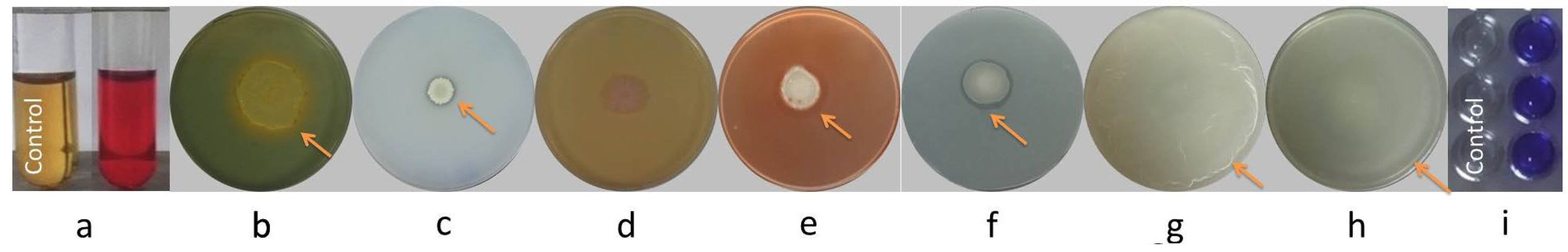
2.2.2. PGP and Environmental Fitting Traits

Amfr20 was tested for its ability to produce siderophore compounds using Chrome Azurol Sulphonate agar medium [40] (Table S1). The strain was grown overnight in nutrient broth where spotting with 10 μL culture and incubation for 72 h at 30 °C was carried out. The test allows the detection of siderophore production through the appearance orange halo around the spot.
For phosphate solubilization detection, 10 μL of overnight bacterial suspension was spotted on Pikovskaya’s (PVK) agar medium [41] (Table S2). The appearance of a clear halo around the colony after 7 days at 30 °C indicates the solubilization of precipitated phosphorus.
For cellulase production testing, 10 μL of overnight bacterial culture was spotted on the CYEA medium amended with 1% CMC (carboxymethyl cellulose) (Table S3). The plates were incubated at 30 °C for 72 h, and after that, Congo red dye (0.1% w/v) was added and remained there for 15 min. A quantity of 1M NaCl was used to flood the plates, removing dye that had been unbound. Positive results considered the formation of a clear halo around the colony.
Protease production was tested on CYEA medium (Table S3) with 7% skim milk powder via spotting with 10 μL of an overnight bacterial culture on the plate and incubating at 30 °C for 72 h [42]. The formation of a clear halo around the colony indicated positive results.
Urease production was examined via spotting with 10 μL of an overnight bacterial culture on a Urea Base Christensen ISO 6579, ISO 19,250 (Conda, Madrid, Spain) medium [43]. The ureolytic ability of the testing isolates was indicated by the formation of a pink halo around the colony after 48 h of incubation (Table S4).
Acetoin synthesis was investigated according to the Voges–Proskauer test [44] (Tables S5 and S6), where positive results were characterized by the appearance of pink coloration of medium.
The biofilm formation assay was carried out by the crystalline violet staining method on 96-well polystyrene plates [45]. Each well was inoculated with 0.1 mL of a 1:100 dilution of overnight bacterial culture, with five replicate wells. The polystyrene plates were kept non-agitated and were incubated at 30 °C for 24 h. After incubation, the plates were washed thoroughly three times with distilled water. Then, 0.2 mL of 0.1% (w/v) crystal violet solution was added to each well. The plates were incubated at room temperature for 30 min and were washed rigorously in order for the stain to be removed. Then, 0.2 mL of ethanol-acetone solution (1:4 ratio) was added, and the incubation was repeated for 30 min. The appearance of the purple color indicated the biofilm formation. The experiments were performed three times in three independent experiments. The swimming motility was examined via spotting on a 3 μL from overnight culture on NA medium (0.3% agar) and swarming motility by inoculating 5 μL on NA (0.5% agar). The incubation was carried out for 1 day at 30 °C.

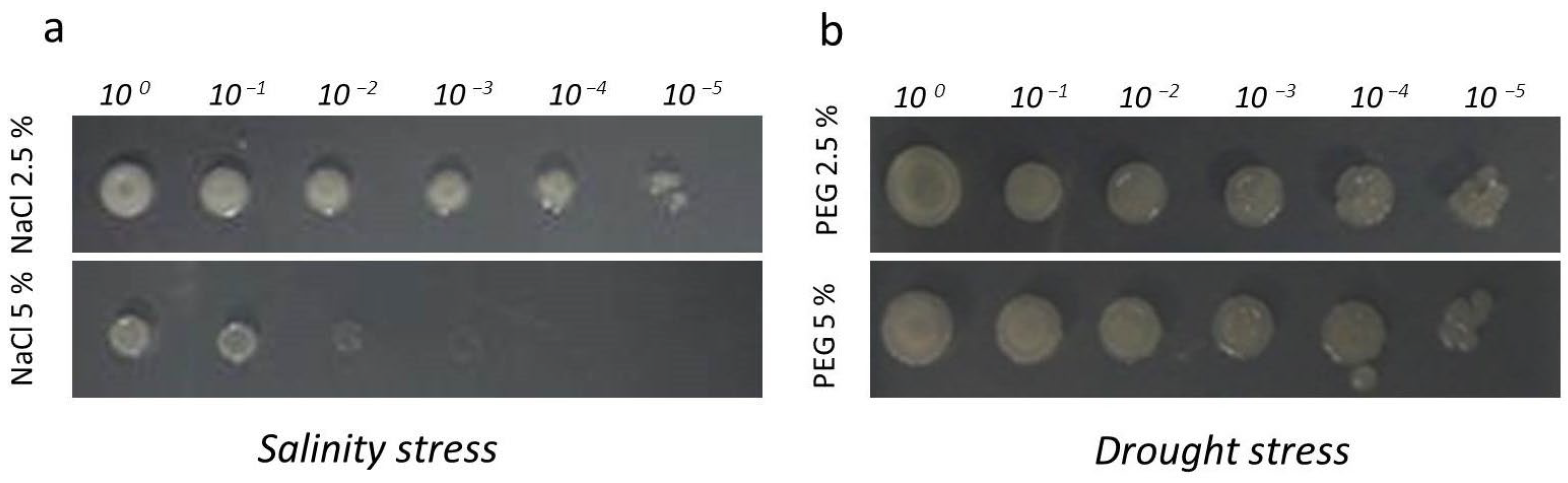
2.2.3. Abiotic Stress Traits

The evaluation of abiotic stress tolerance was performed via spotting on NA medium plates with 2.5% and 5% sodium chloride for salinity conditions, as well as NA medium plates with 2.5% and 5% polyethylene glycol (PEG3000) for drought conditions. Overnight liquid cultures were serially diluted, and 5 μL of each dilution was inoculated onto the corresponding plates, followed by incubation at 30 °C for 24 h.

2.2.4. Antibiotic Susceptibility Assay

Antibiotic susceptibility of the bacterial isolates was tested by using the Kirby-Bauer method [46]. Plates with NA medium were plated using 0.1 mL from a liquid culture grown overnight. After they were dried, paper disks (diameter 6 mm) were soaked with 20 μL of the antibiotics ampicillin, chloramphenicol, kanamycin, rifampicin, streptomycin, and tetracycline, with three tested concentrations (10, 30 and 50 μg/mL), and then placed on the medium. The plates were incubated at 30 °C for 48 h. The appearance or absence of a clear halo surrounding the disk revealed the responsiveness of the bacterium to the corresponding antibiotic.

2.3. Examination of Antagonistic Activity Against Post-Harvest Diseases upon Detached Fruits

Healthy olives (Olea europea var. Amfissa) and grape berries (Vitis vinifera var. Fraoula) were selected and soaked in dH2O. They were then immersed in an aqueous solution of commercial bleach for 4 min. The fruits were rewashed with sterilized dH2O and dried aseptically. The treated fruits were artificially wounded (wound size: 3 mm × 3 mm) using a sterilized scalpel. An aliquot 20 μL of bacterial whole culture (vegetative cells) containing 108 CFU/mL was inoculated in the wound of each fruit and transferred to sterilized transparent boxes with wet filter paper in the bottom to ensure high moisture conditions. After 3–4 h, a 20 μL inoculant (106 spores/mL) of respective fungal phytopathogen (B. cinerea or C. acutatum) was applied to the wounded fruits, and they remained at room temperature for 4 days. Fruits inoculated only using bacterial or fungal spore suspension were served as controls. To evaluate the disease incidence index (DI%), we calculated the percentage of infected fruit according to the type: Di = (A/Β) × 100, where A is the number of infected fruits and Β the total number of the fruits. The disease severity index (DS%) was calculated by using the formula Ds = [Σ(Ci)/n*Z] x 100, where Σ(Ci) is the total number of fruits in each corresponding class, n is the total number of fruits, and Z is the highest class. Classes of rating scale regarding the mycelial and rot coverage of each fruit were the following: 0 = healthy fruit, 1 = 1–20%, 2 = 20–50%, 3 = 50–75%, 4 = 75% and above according to the visible fruit surface from the observer. The estimation of DS% and DI% derived from 12 tested fruits per replicate, with 3 replicates (Total = 36 fruits), while Amfr20 population dynamics in the wound tissue of the fruits (Log10CFU/g) was derived from experimental replicates with 3 tested fruits per replicate (Total = 9 fruits).

2.4. TLC-Bioautography Assay

To isolate agar-diffusible compounds from the bacterial strain, the modified protocols of [47,48] were used. Petri dishes with a single culture of Amfr20 and dual culture of Amfr20 and Botrytis cinerea treatments were used and incubated for 7 days at 30 °C.
The inhibition zones from all treatments (single and dual cultures) were carefully removed, their weights were measured, and they were cut into small pieces. These samples were then placed in Erlenmayer flasks containing a mixture of ethyl acetate and 0.1% formic acid. The flasks were vortexed and subjected to sonication using a water bath sonicator at room temperature for 0.5 h. Following this, the resulting solution was filtered through Whatman filter paper (Whatman plc, Maidstone, UK) and evaporated to dryness using a vacuum evaporator. The dried residue was reconstituted in 0.1 mL of HPLC-grade methanol and subsequently filtered through Whatman® Uniflo® filters. Thin-layer chromatography (TLC) of the obtained extracts was performed according to Calvo et al. (2019) [49]. The stationary phase consisted of silica gel 60 F254 plates (size 20 × 20 cm, Merck, Darmstadt, Germany). A solvent system comprising chloroform, methanol, and water in a 65:25:4 (v/v/v) ratio served as the mobile phase. For the TLC-bioautography assay, the developing TLC plates were overlaid with PDA semisolid agar medium (0.8% w/v) containing B. cinerea spore suspension (107 spores/mL) and incubated at 25 °C for 3 days. After the incubation, they were sprayed with tetrazolium salt dye solution (2.5 mg/mL) (Sigma-Aldrich®, Merk KGaA, Burlington, MA, USA), which renders living cells a purple color. The formation of clear zones is considered as bioactive spots exhibiting antifungal activity. Lastly, the Rf values of bioactive spots were determined based on the formula: Rf (retention factor) = travelling distance of the solute/travelling distance of the solvent front.

2.5. Plant Growth Promoting Activity on A. thaliana Col-0 Seedlings In Vitro

The surface sterilization of A. thaliana Col-0 seeds was carried out according to the modified protocol of Palacio-Rodríguez et al. (2017) [50] and included the following steps: washing for 0.5 min in 70% ethanol, immersing for 1.5 min in washing solution containing 5% NaClO) and 0.1% Tween20, washing again for 30 s in 70% ethanol, and finally rinsing in sterile distilled water. The sterilized seeds were placed on a growth medium containing half-strength Murashige and Skoog (½MS) with vitamins (MS0222, Duchefa Biochemie, Haarlem, The Netherlands), with 0.6% agar amended with 1.5% sucrose, pH 5.8. Then, all plates were kept at 4 °C for 48 h and were positioned at an angle of 70° or horizontally (bi-plates) in a growth chamber (16-h light: 8-h dark photoperiod, 22–25 °C) until used for transplanting. To evaluate the plant growth effect, 3-day-old seedlings that emerged from the treated seeds were transplanted on growth medium containing ½ MS, 1% sucrose, and 0.8% agar (for normal conditions) or adding to growth medium 100 mM NaCl (for saline conditions). In each petri dish, 6 seedlings were used. An aliquot 10 μL of bacterial suspension (108 CFU/mL) was applied 3 cm below each root tip. The plates were positioned at a 70-degree angle for 9 days. Plants without bacterial treatment served as a control. The plants were weighed and photographed to determine growth parameters. The bacterial volatiles’ effect on plant growth was evaluated according to the modified protocol of Asari et al. (2016) [51]. Surface sterilized seeds were on one side of a bi plate containing growth medium: ½MS with vitamins, 1.5% sucrose, and 0.8% agar (for normal conditions), or supplemented with 100 mM NaCl (for saline conditions). With a pipette tip we applied 6 seedlings in each petri dish. Then, on the other half of the bi-plates, bacterial inoculation (108 CFU/mL) was conducted using triplicate spots (20 μL/spot). The plates were incubated for 2 weeks in a growth chamber at 25 °C (16-h light: 8-h dark) and positioned horizontally after sealing with double-layer parafilm. Plants without bacterial treatment served as a control. Growth parameters were determined after photographing and weighing the seedlings.

2.6. Plant Growth Promoting Activity on Solanum lycopersicum

2.6.1. Bacterial Effect on the Seeding Dynamics of Tomato Seeds Using the Biopriming Method

For surface sterilization, tomato seeds of var. Chondrocatsari Messinia were sown in 70% ethanol and washed vigorously for 1 min. After discarding the solution, a washing solution containing 5% NaOCl and 0.1% Tween 20 was added and maintained for 3 min. The procedure was completed by thoroughly rinsing the seeds with ddH2O. Then, after drying the seeds, they were immersed in a bacterial inoculum suspended with 1% CMC with two concentrations (106 and 108 CFU/mL) for one hour. Seeds treated only with CMC solution, without bacterial inoculation, were used as controls. Coated and uncoated seeds, after drying in a nematic flow chamber, were transferred to plates with filter paper soaked in ddH2O and placed at 25 °C for 5 days in the dark. The experiment was conducted in triplicate, with 15 seeds per replicate. To evaluate the effect of microorganisms, the percentage of germination was calculated with the formula GP % = (g/t) × 100. The number of seeds that successfully germinated are denoted by ’g’ and the total number of seeds by ’t’. Additionally, the radicle of the germinated seeds was measured and compared with the controls.

2.6.2. Plant Growth Promoting Effect on Plant Pots

In this experiment, the seeds coated with 108 CFU/mL and 1% CMC were placed on plates with filter paper soaked in ddH2O, and were incubated at 25 °C for 2 days in the dark to germinate. The germinated seeds were sown in plastic plant pots (8 × 8 × 8 cm) with a peat and perlite mixture at a 5:1 ratio. After a period of 15 days, an aliquot 10 mL of bacterial inoculum 108 CFU/mL was applied per plant pot via root irrigation. The plants were grown for 28 days under laboratory conditions at 25 ± 5 °C in a photoperiod (14 h light/10 h dark) and watered thrice weekly with a defined quantity of water. A total of 30 plants were used for each treatment with 3 biological replicates (10 plants/biological replicate). The plant growth effect was studied after measuring three growth characteristics: shoot height, shoot fresh weight, and shoot dry weight. Uninoculated plants were used as controls

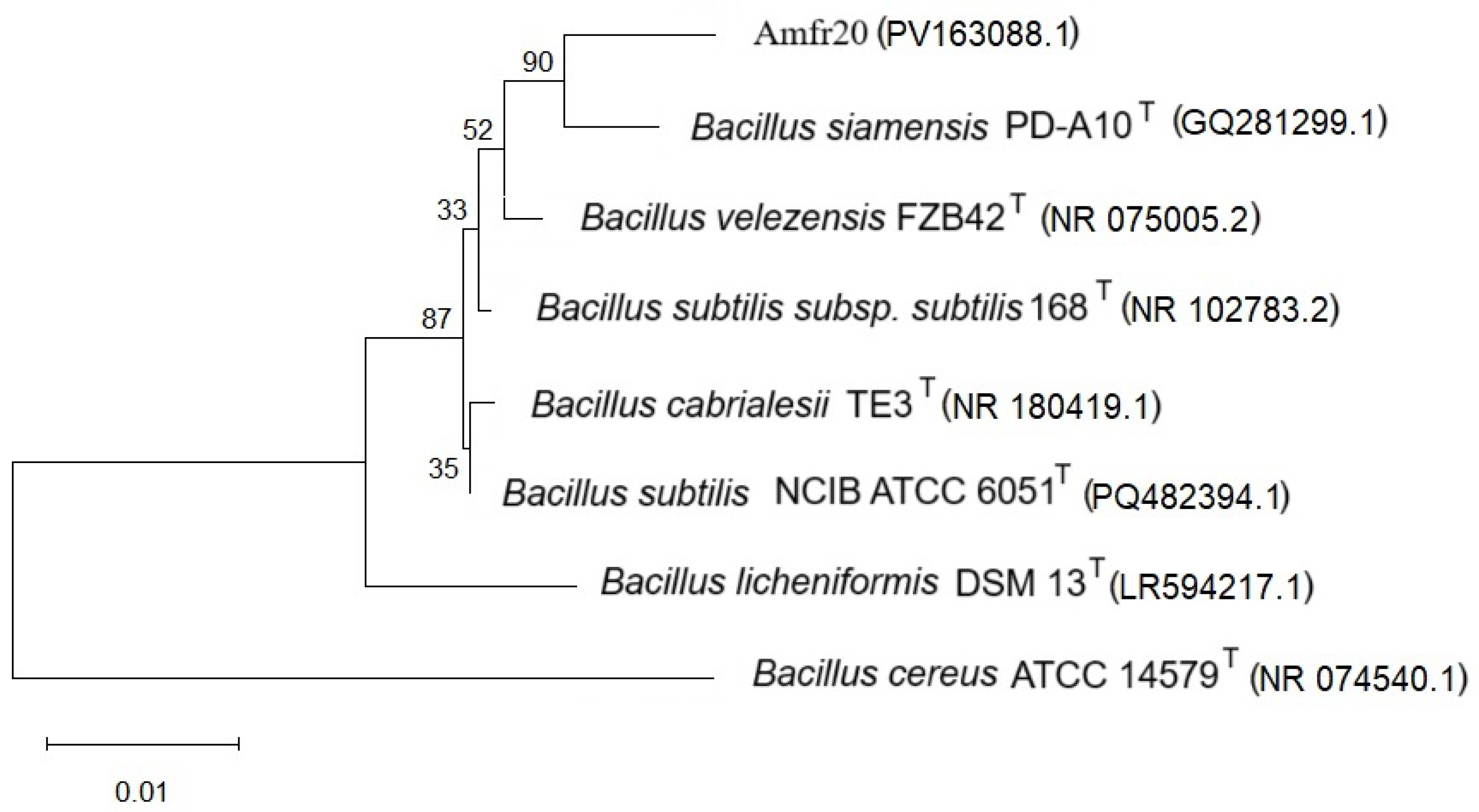
2.7. Phylogenetic Taxonomy According to 16S rRNA Amplicon

Bacterial DNA was obtained from an overnight culture using a Nucleospin® Microbial DNA (Macherey-Nagel GmbH and Co. KG, Düren, Germany) kit. The set of primers which were used for the amplification of the 16S rRNA gene fragment was: 5′-AGAGTTTGATCCTGGCTCAG-3′, R: 5′-ACGGCTACCTTGTTACGACTT-3′ [52].
The composition of the PCR reaction mixture included the following: 1 μL of forward primer (forward, 30 μM), 1 μL of reverse primer (reverse, 30 μM), 1 μL (50–100 ng) of DNA matrix, a 1 μL mixture of deoxynucleotide triphosphates (dNTPs, 10 mM), 5 μL 10× buffer (PCR buffer), 0.5 μL of DNA polymerase (5 U/μL), 2 μL of dimethyl sulfoxide (DMSO), and ddH2O to a final volume of 50 μL. The thermocycler program was set as follows: an initial denaturation at 94 °C for 10 min, followed by 35 cycles of 94 °C for 30 s, primer annealing at 48 °C for 30 s, extension at 72 °C for 30 s, and a final extension at 72 °C for 5 min. The size of the PCR product is estimated at 1450 bp.
For electrophorisis, 5 μL of the PCR product was mixed with loading buffer (0.25% w/v bromophenol blue, 0.25% w/v xylene cyanol, 30% v/v glycerol) and was loaded and visualized onto a 1.2% (w/v) agarose gel. Subsequently, the PCR product was purified using the GeneJET PCR Purification Kit (Thermo Fisher Scientific, Waltham, MA, USA), according to the manufacturer. The purified partial 16S rRNA sequence was submitted to GenBank, with accession numbers listed in Table S7. The accession number of the Amfr20 16S rRNA sequence, as well as the percentage identity of some reference strains deposited in GenBank, are listed in Table S8. A phylogenetic tree based on 16S rRNA was generated, illustrating the taxonomic position of Amfr20 and type strains of the NCBI database, using the neighbor-joining method in MEGA12 [53].

2.8. Whole-Genome Sequencing

Genomic DNA was extracted from bacterial overnight culture using the PureLink® Genomic DNA Mini Kit (Fair Lawn, NJ, USA), according to manufacturer instructions. The sequencing was carried out using SNPsaurus through the Illumina HiSeq 2000 system (Illumina, San Diego, CA, USA). The DNA library was built with the Nextera XT DNA Library Prep Kit (Illumina). The sequence was trimmed with BBDuk, and the genome was assembled using SPAdes-3.12.0 with its standard settings [54]. The final genome (accession number JAEACN000000000) is composed of 7 scaffolds and spans 3,957,523 base pairs. For functional annotation, firstly, we utilized PROKKA for the rapid and accurate annotation of coding sequences and gene functions. This analysis enabled us to correlate genomic features with the observed plant growth-promoting activities [55]. Phylogenomic analysis of the Amfr20 was performed by using the Type (Strain) Genome server (TYGS) platform (https://tygs.dsmz.de) accessed on 25 November 2024 and by calculating the average nucleotide identity (OrthoANI) and digital DNA:DNA hybridization (dDDH) values using the Genome-to-Genome Distance Calculator (GGDC 2.1). For ANI and dDDH, we obtained values based on species delineation threshold values that were suggested by default analysis (95–96% and 70%, respectively) [56,57]. A phylogenetic tree was constructedusing FastME [58] based on distances from Genome BLAST Distance Phylogeny (GBDP). Cazymes were detected using the Carbohydrate-Active enZYmes (CAZy) open database (www.cazy.org) and the Carbohydrate-active enzyme ANnotation dbCAN3 web server (https://bcb.unl.edu/dbCAN2/, accessed on 25 March 2025). Analysis of secondary metabolites was performed using antiSMASH (https://antismash.secondarymetabolites.org, accessed on 25 March 2025). The adenylation and condensation structures of Non-Ribosomal Polypeptide Synthetases (NRPSs) and their amino acid sequence were identified using antiSMASH and the PKS/NRPS Analysis Web-tool, https://nrps.igs.umaryland.edu/, accessed on 25 March 2025). Lastly, the genes related plant growth-promoting properties were identified by searching in the NCBI Sequence Set Browser.

2.9. Statistical Analysis

To compare the bacterial treatment with the control group, we applied a two-tailed independent samples Student’s t-test (using a significance threshold of p < 0.05). In our graphs, the plotted points represent the average values, the error bars show the standard deviation, and any statistically significant differences are marked with asterisks. All of the statistical analyses and graph plotting were performed using GraphPad Prism v.10.4.1 (GraphPad Software, San Diego, CA, USA).

3. Results

3.1. Isolation of Bacterial Endophytes from Olea europaea L. var. Amfissa and Selection of the Most Promising Antagonistic Strain

A total of 32 single bacterial colonies were isolated from the asymptomatic roots of the Olea europaea L. var. Amfissa. Every single colony was tested macroscopically and stereoscopically for its features. As a first step, massive screening of all bacterial isolates was performed in multiple-culture system in vitro against the plant pathogen R. solani, in order to determine the most potential antifungal antagonists (Figure 1a). Amfr20 exhibited the most promising antagonistic activity regarding the size of the inhibition zone compared to the other testing isolates, so it was selected for further examination.

3.2. In Vitro Characterization of Bacterial Strain Amfr20

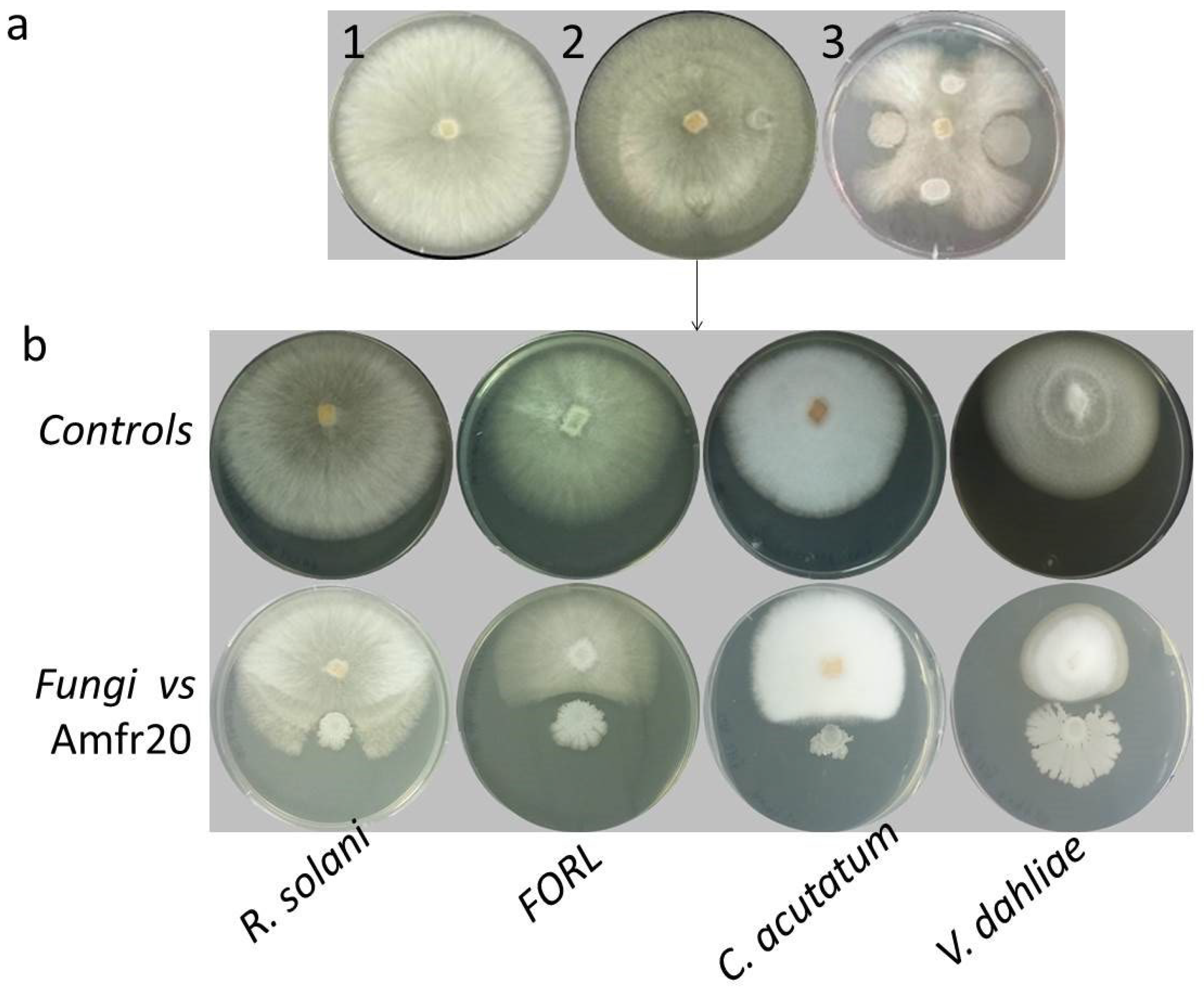
3.2.1. Biological Control Activity

The biological control activity of the strain Amfr20 was evaluated against four significant fungal plant pathogens, including R. solani, FORL, V. dahlia, and C. acutatum, using the dual culture method in vitro. The results after the calculation of the mycelial radius index revealed that the strain exhibited antagonistic activity against the fungal plant pathogens by inhibiting the mycelia radius (Figure 1b, Table 1).

3.2.2. Plant Growth Promoting Properties

Strain Amfr20 was tested in vitro for plant growth-promoting and colonization-related traits. The bacterial isolate showed positive results in multiple assays, such as acetoin and siderophores production, and precipitated solubilization of precipitated phosphorus. Furthermore, it exhibited lytic enzymes secretion such as protease and cellulase, but no production of urease was detected. Additionally, the strain exhibited exceptional swarming and swimming abilities, along with robust biofilm formation in polystyrene wells (with OD550 = 0.8) (Figure 2).

3.2.3. Abiotic Strain Tolerance

Amfr20 was evaluated for its tolerance to drought and salinity stress under laboratory conditions. In drought stress assays, the strain demonstrated robust survival, maintaining growth up to a 10⁻⁵ dilution in media containing 2.5% and 5% PEG3000. Under salinity stress, Amfr20 exhibited increased survival in 2.5% sodium chloride, with growth observed up to a 10⁻⁵ dilution, compared to growth up to a 10⁻³ dilution in 5% sodium chloride (Figure 3).

3.2.4. Antibiotic Susceptibility

The Amfr20 strain exhibited resistance to chloramphenicol and tetracycline across all tested concentrations, as well as to streptomycin and ampicillin at 10 and 30 μg/mL, with no clear inhibition zones observed. However, the strain was susceptible to kanamycin (10, 30, and 50 μg/mL), ampicillin (50 μg/mL), rifampicin (30 and 50 μg/mL), and streptomycin (50 μg/mL) (Table 2).

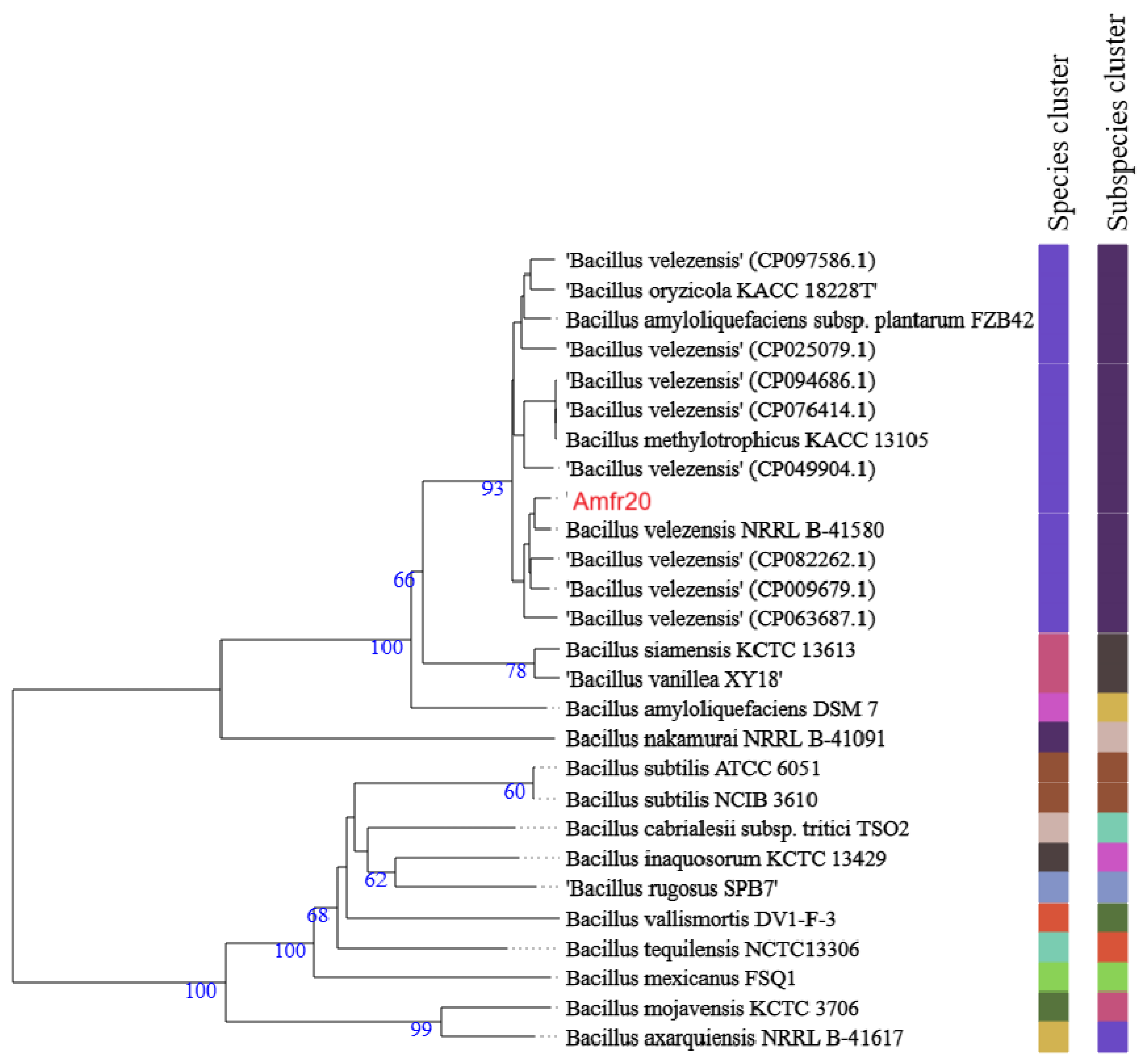
3.3. Phylogenomic Classification of Bacterial Strain Amfr20

A representative number of endophytic isolates was selected, and their 16S rRNA genes were amplified and sequenced for identification (Table S7). The 16S rRNA analysis of the strain Amfr20 (Accession number PV163088) through NCBI BLASTn algorithm revealed a high degree of similarity to B. velezensis, B. siamensis, and B. amyloliquefaciens species (Table S8). In addition, a phylogenetic tree was generated based on the 16S rRNA gene sequence of Amfr20 and type strains from the NCBI database using the neighbor-joining method (Figure 4).
However, the use of a single phylogenetic marker is not a safe option for accurate classification of the strain at the species level. In order to achieve a more accurate phylogenetic classification, a comparison of Amfr20 with other Bacillus species was performed at the whole genome level with the program Type (strain) Genome Server (http://tyg.dsmz.de). In the phylogenetic tree below, Amfr20 appears in a distinct cluster where B. velezensis species are included, observing a small phylogenetic distance of the particular strain with those of the database (Figure 5).
To further validate this classification, the genome of Amfr20 was compared to other B. velezensis strains using alignment-based Average Nucleotide Identity (ANI) and digital DNA-DNA hybridization (dDDH) methods. The dDDH and ANI values ranged from 84.8% to 94.9% and 97.99% to 99.37%, respectively, where the highest value was found when comparing Amfr20 with B. velezensis NRRL B-41580T (NZ_LLZC00000000. 1). These values were higher than the recognized lower limits of 70% for dDDH and 95–96% for ANI, which confirms that Amfr20 belongs to the species B. velezensis [59,60] (Table 3).

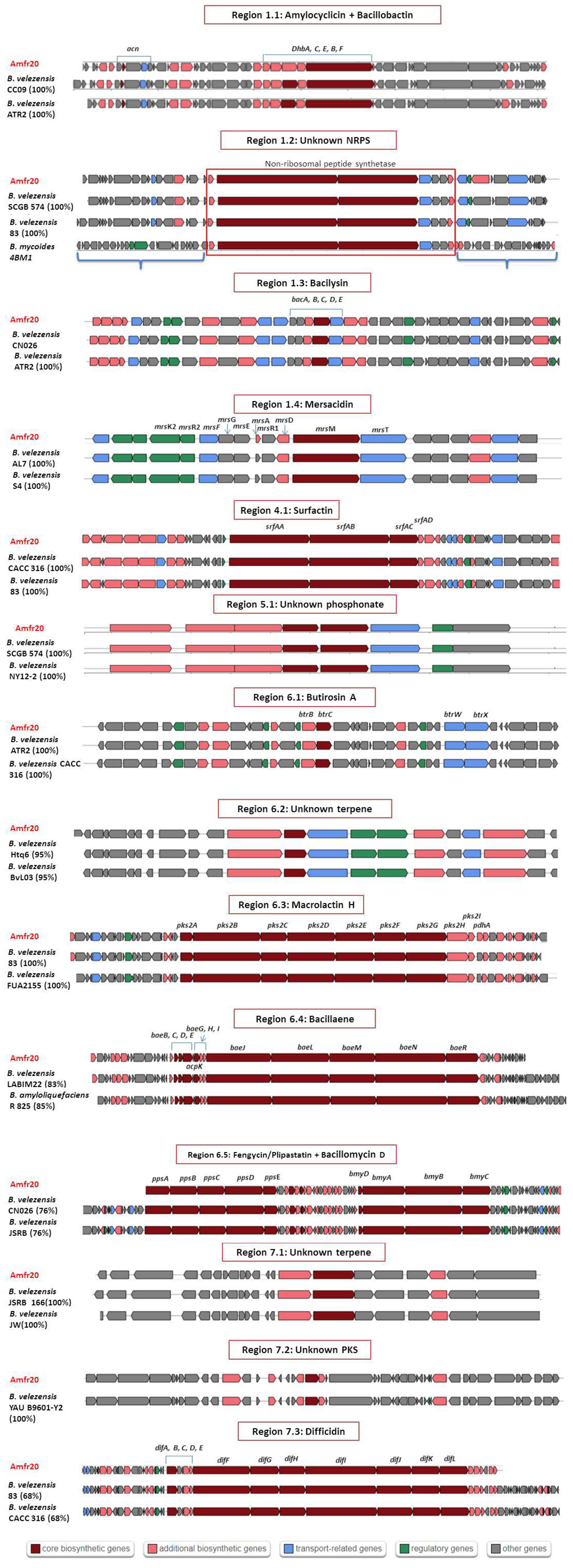
3.4. Prediction of Biosynthetic Gene Clusters of Secondary Metabolites

Genomic analysis of strain Amfr20 was carried out through the antiSMASH algorithm to predict and identify gene clusters for the biosynthesis of secondary metabolites (BGCs). According to the results, 16 individual BGCs were identified along 14 biosynthetic regions, where in two regions (regions 1.1 and 6.5), two BGCs were identified. These biosynthetic gene clusters produce either known compounds or other unknown prototypes or are not listed in the MIBIG database, yet they are common to other strains of B. velezensis (Table 4).
As far as the known clusters are concerned, strain Amfr20 showed high homology and similar gene organization with other strains of the MIBIG database, representing the biosynthetic genes of the compounds surfactin (homology 82%) and fengycin (93% homology), as well as amylocyclicin, bacilysin, mersacidin, macrolactin H, bacillaene, and difficidin, with 100% homology when comparing with reference strains. For instance, in region 1.1, 2BGCs were identified, where the first showed 100% homology with bacillibactin, while the second one showed 100% homology with amylocyclicin. In addition, in region 6.5, two biosynthetic clusters were adjacent, where the first concerned BGC fengycin biosynthesis; however, this particular BGC also encodes the synthetases of plipastatin due to the D arrangement of Tyr9. The second one showed high homology with three BGC NRPSs; bacillomycin D (100%), mycosubtilin (100%), and iturin A (88%), but the predicted amino acid sequence showed that this BGC is related to Bacillomycin D synthetases due to the modules Pro4, Glu5 and Thr7, which are unique to Bacillomycin D.
The remaining unknown compounds, two BGCs of terpenes, were identified in regions 6.2 and 7.1. Moreover, in region 1.2, a BGC of NRPS was found, and in area 7.2, a BGC of type III was found. In addition, in region 5.1, a BGC of phosphonate was detected. Lastly, in region 6.1, a gene cluster with 7% similarity was identified as encoding the polyketide synthesis of butirosin A. ClusterBlust analysis revealed that the gene cluster for butirosin A of strain Amfr20 showed high gene similarity (100%) with the PKS gene cluster found in several B. velezensis strains (strains YAU B9601-Y2, ATR2, JSRB166, CACC316, etc.) (Figure 6).

3.5. Genetic Features of Bacterial Strain Amfr20 as a Potential Plant Growth Promoter and Biological Control Agent

Analysis of the Amfr20 strain’s genome has identified genes associated with plant growth-promoting abilities, such as indoleacetic acid biosynthesis (trpA, trpΒ, trpC, trpD, trpS, ysnE, yclB, yclC, padC, ydaP, shaS), phosphorus solubilization (phoB, phyC), and acetoin production were identified, as well as 2,3-butanediol (2,3-butanediol dehydrogenase coding gene, budA, alsS, ilvN, ilvB). Genes involved in motility and colonization on surfaces were also detected: chemotaxis (cheA, cheC, cheD, cheV, cheW, cheY), swarming motility (swrA, swrB), biofilm formation (remA, wecB, csrA, rpoN, ylbF, sinR), and synthesis and function of bacterium flagella (fliT, fliS, fliW, flgL, flgK, flgN, flgB, flgC, fliG, fliF, fliH, fliI, fliJ, flgD, flgG, fliL, fliM, fliY, fliZ, fliP, fliQ, flhA, flhF, flhB). Additionally, genes detected for adaptation salinity stress tolerance include genes related to the biosynthesis of osmolytes such as proline (proA, proB, proC) and ion homeostasis (nhaC), glutamate (gltB, gltD), and glutamine (glnA).
Furthermore, the evaluation of carbohydrate active enzymes (Cazymes) was carried out in order to investigate the strain’s capacity to suppress the pathogen and its absorption of various nutrients. Cazyme analysis revealed 110 encoding genes; 40 of these encoded glycoside hydrolase families (GHs), 37 encoded glycosyl-transferases (GTs), 3 encoded lyases (PLs), 12 encoded polysaccharide esterases (CEs), 5 encoded growth factors (AAs), and 13 encoded families of carbohydrate binding proteins (CBMs) (Table 5).

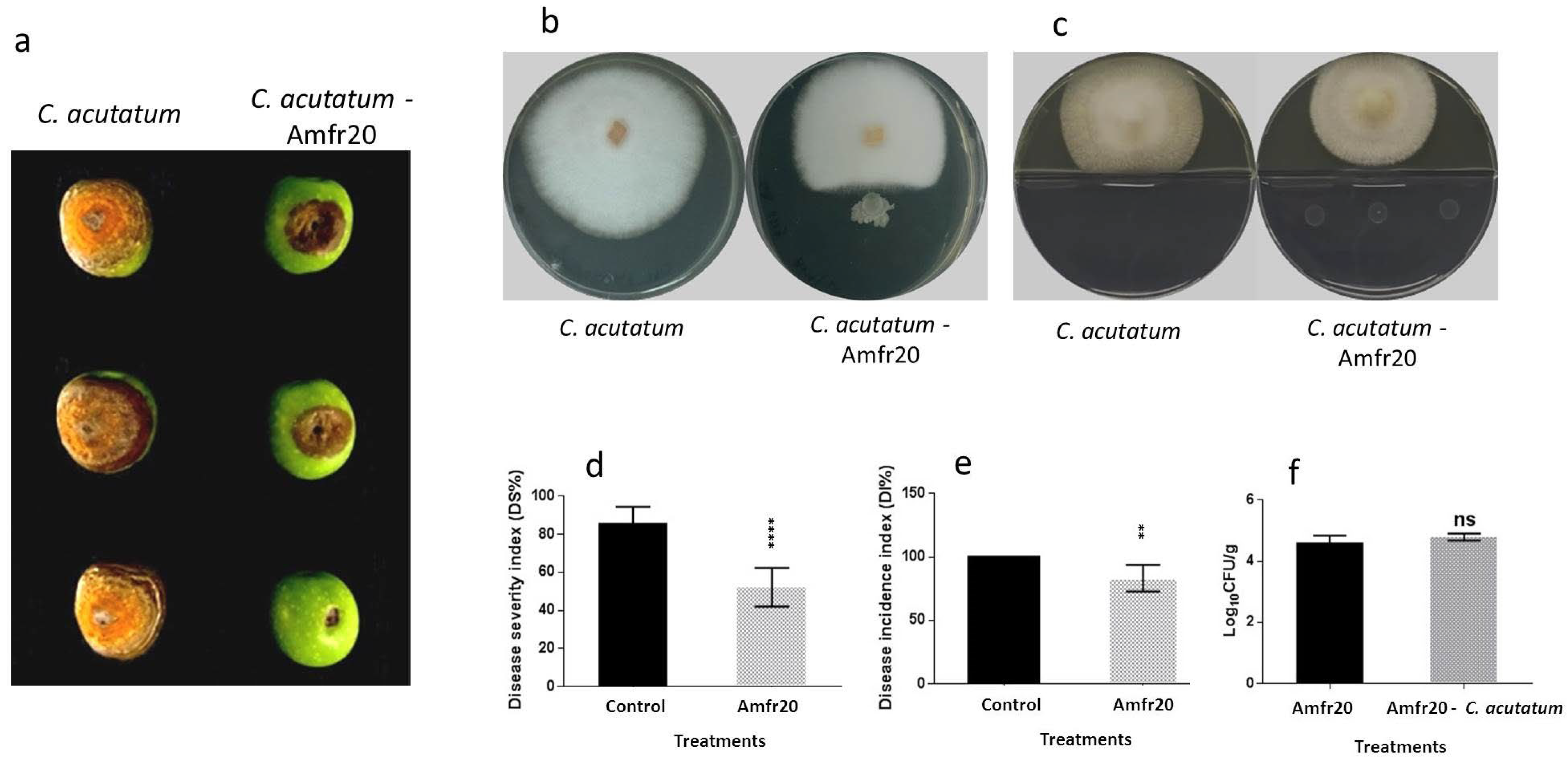
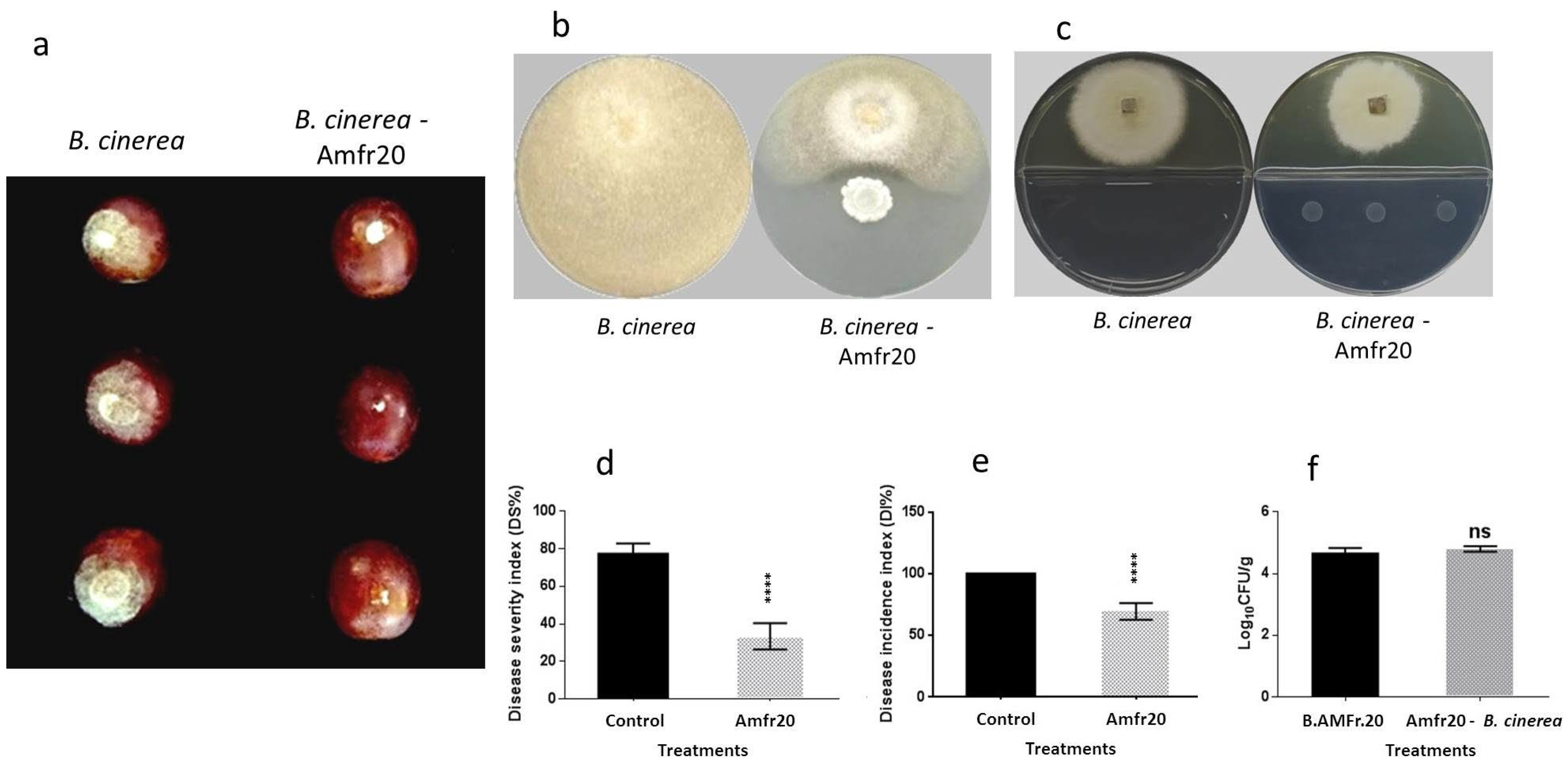
3.6. Examination of In Vivo Biological Control Activity of the Amfr20 Strain upon Artificially Infected Detached Fruits

The results of the genome mining analysis and biocontrol assays in vitro revealed the role of the Amfr20 strain as a promising BCA as a first step of examination. So, the following experiment was established in order to evaluate the in vivo biological activity of the strain against postharvest diseases upon artificially infected detached fruits. Artificially wounded olive fruits and grape berries were inoculated with 20 μL of bacterial culture suspension (108 CFU/mL), and when dried, they were treated with 20 μL of spore suspension (106 spores/mL) of the phytopathogens C. acutatum and B. cinerea. According to the results, it was revealed that the strain’s application upon the wounded fruits caused significant protection against the post-harvest fungal diseases. Specifically, the strain’s antagonistic activity against C. acutatum upon olive fruits caused a significant reduction in the disease incidence index (Di) and disease severity index (Ds), where the Di value of the strain was 83.33% and the Ds value was 52.08%, significantly lower than the control values (Figure 7). Similarly, the strain Amfr20 showed to be significantly effective against B. cinerea upon infected grape berries, where the Di and Ds values of the strain were low enough (up to 69.43% and 33.32%, respectively), significantly lower than the control values (Figure 8).

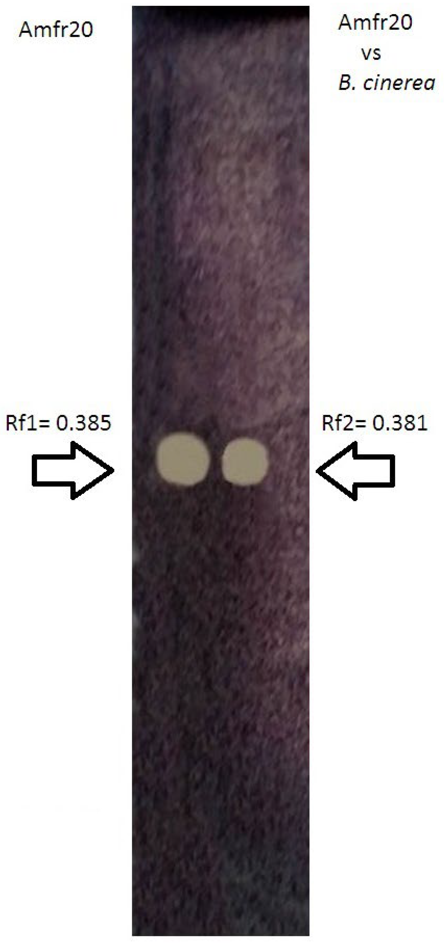
3.7. TLC-Bioautography Development of Potential Bioactive Secreted Compounds from Solid Culture Against Postharvest B. cinerea

The strain Amfr20 was highly effective against gray mold on detached grape berries, while an additional in vitro biocontrol assay also verified the strain’s antagonistic activity through diffusible metabolites against B. cinerea. The question that arose was whether the antagonistic effect that was observed was attributed to bacterial compounds from the solid culture that are constitutively secreted or are triggered by the fungal pathogen’s interaction. Therefore, ethyl acetate extracts were performed from both single bacterial culture and dual bacterial-fungal culture and evaluated through TLC-bioautography assay. The experimental results revealed that both secreted metabolites exhibited bioactive spots with the same pattern with Rf values; Rf1 = 0.385 for extracts from single cultures and Rf2 = 0.381 for extracts from dual cultures (Figure 9).

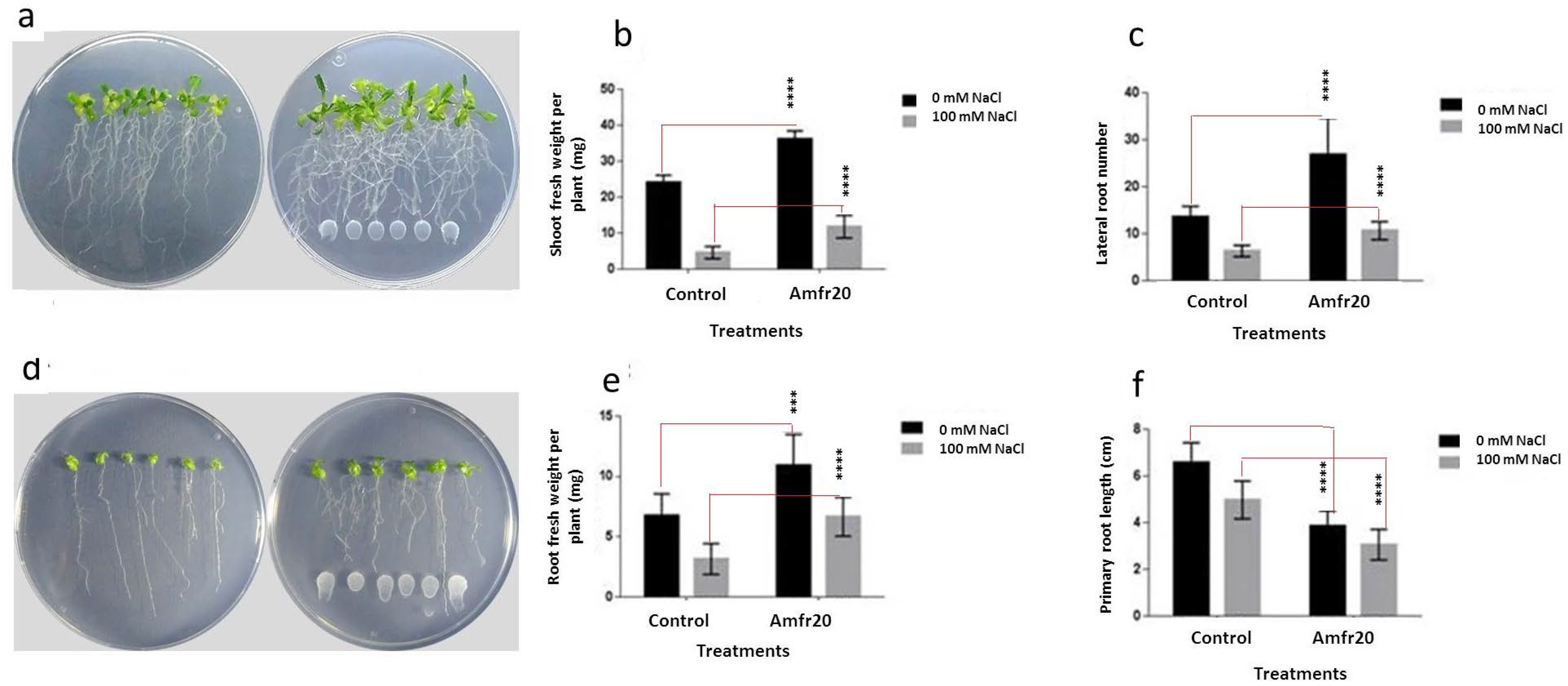
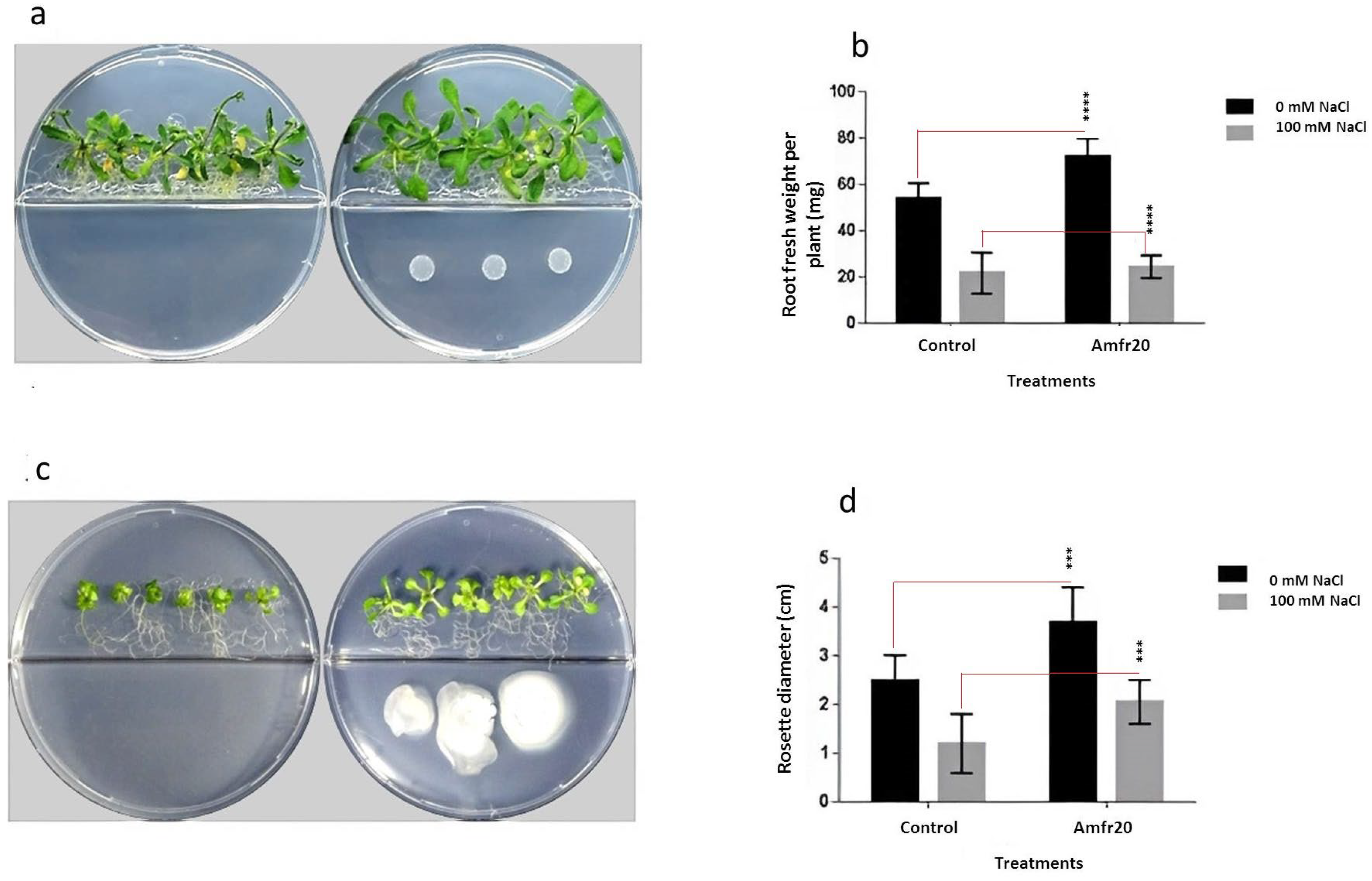
3.8. Investigation of In Vitro Plant Growth-Promoting Ability of the Amfr20 Strain on the Model Plant A. thaliana Under Normal and Saline Conditions

Beyond the above-mentioned biocontrol potential of the Amfr20, the strain also possesses promising plant growth and salt tolerance properties according to the in silico and in vitro experiments results. Thus, the plant growth-promoting effect was further examined in vitro on A. thaliana cultivated on MS agar under both normal and salt-stress conditions (amended with 100 mM NaCl). To investigate whether the effect is induced by diffusible and/or volatile compounds of the strain, petri dishes with transplanting seedlings were used with and without center partition. Interestingly, after nine days of co-culture, bacterial inoculant applied below the root tip was shown to be effective in alleviating the salt stress on the seedlings and enhancing plant biomass under normal and saline growth conditions. Moreover, the root system architecture was morphologically altered to a branching root phenotype, including the shortening of the primary root length and the increase of lateral root formation and the entire root surface area as well (Figure 10). Additionally, the volatile compounds experiment revealed that the Amfr20 strain beneficially affected the growing seedlings after 14 days of co-culture in bi-plates by increasing shoot fresh weight and leaf area under both growth conditions (Figure 11).

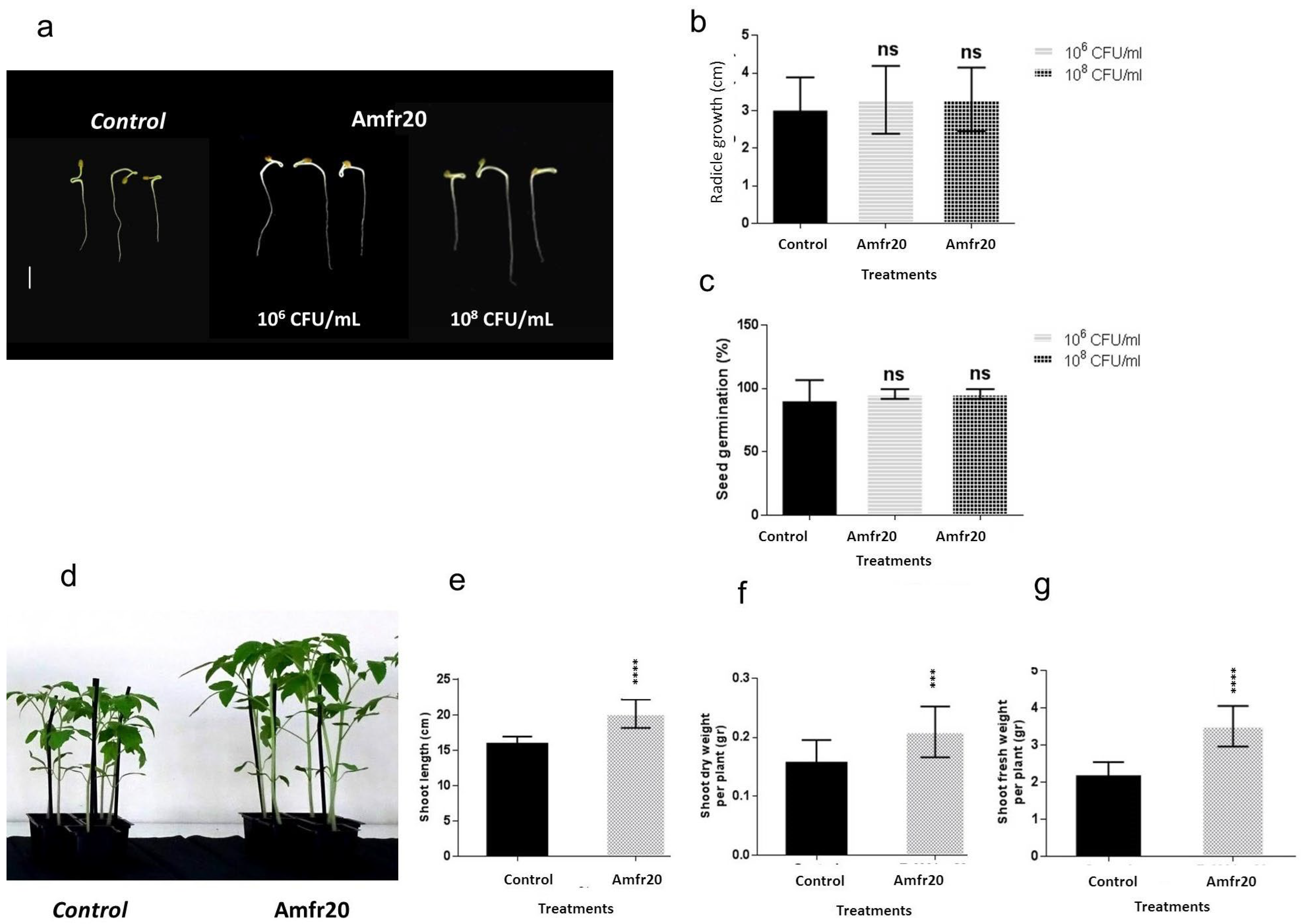
3.9. Plant Growth-Promoting Effect of the Amfr20 Strain on Solanum lycopersicum

Following the plant growth potential effect on the A. thaliana, its effect was also examined on tomato seeds and plants as well through seed biopriming and root irrigation in pots. Seeds of the native Greek variety Solanum lycopersicum var. Chondrokatsari Messinias were selected, while the bacterial inoculant was prepared using the method of seed biopriming with CMC (1%) as an adhesive with two bacterial suspensions (106 and 108 CFU/mL). The potential action of the strains was determined after calculating the percentage of seed germination and the length of the primary root. It was observed that the application of both bacterial suspensions mediated a small increase in seed germination percentage and in radicle growth compared to the control, but not in a statistically significant manner (Figure 12b,c). In addition, the combined application of biological seed coating and root irrigation with 108 CFU/mL proved to be more effective compared to that of biological seed coating. The effect of strain on plants was determined by recording three biological features of plant growth, such as shoot length, shoot fresh weight, and dry weight after 28 days of growth. According to the results, a statistically significant increase was observed in all three growth parameters of the plants treated with the Amfr20 strain compared to the control treatments (Figure 12e–g).

4. Discussion

Plants are colonized by many microorganisms on the exterior of plants, such as the rhizo- or phyllosphere, or in the interior of plants, referred to as the endosphere, developing interaction relationships and conferring beneficial effects on the growth and health of plants [61,62]. These relationships are dynamic and are shaped according to the plant tissue, the genetic profile of the host, and the prevailing environmental conditions. Numerous literature reports are concerned with the recording of the endophytic communities (such as bacterial endophytes) from many plant species and the examination of their plant beneficial functions due to the advantageous location from where they thrive and act [63,64,65].
In the current study, root tissues of the native Greek olive cultivar (cv. Amfissa) [66] were selected, where culturable endophytic bacteria were isolated. Among the isolates, the strain Amfr20 emerged for its biological activity in a first-stage in vitro screening assay. Further in vitro examination revealed positive results in biocontrol assays against four significant fungal plant pathogens, two of which (C. acutatum and V. dahliae) severely affect the olive fruit and the vascular system of the tree [67,68]. Biochemical analysis results also showed the beneficial potential of the strain Amfr20, which possesses the ability of acetoin production, chelate iron mobilization, precipitated phosphate solubilization, and lytic enzymes secretion (cellulase and protease). Lastly, Amfr20 was also shown as an excellent swarmer and swimmer, and exhibited a great biofilm formation ability, leading to a successful surface colonization.
To determine the taxonomic status of the strain Amfr20, BLASTn analysis (https://blast.ncbi.nlm.nih.gov/Blast.cgi, accessed on 25 March 2025) of 16S rRNA amplicon was carried out, and it was revealed to have high homology with Bacillus spp. strains. Nevertheless, in terms of the current classification, many Bacillus spp. (such as the species B. amyloliquefaciens, B. safensis and B. velezensis) share a high degree of similarity; thus, it is difficult to achieve safe taxonomic results with the traditional classification methods [69,70,71]. In order to be more accurate, a genome-wide phylogenomic tree was constructed, where the phylogenetic analysis revealed that the Amfr20 strain and other B. velezensis strains were in the same branch; the dDDH and ANI values also highlighted the close genomic relatedness, suggesting that Amfr20 is classified as B. velezensis.
Bacillus species are considered to be important biological agents for exerting positive effects on the enhancement of plant growth and plant disease management through multiple features [15,19,72,73]. Recently, it became increasingly evident that B. velezensis appears to be a species of great importance with diverse bioprospecting abilities, since the biosynthetic repertoire of B. velezensis is more powerful and diverse than that of other Bacillus species such as B. subtilis [74,75]. The results from genome mining shed light on beneficial aspects of the strain B. velezensis Amfr20. The presence of the phoB, phyC genes [76,77], as well as the dhbACEBF genes responsible for catecholic siderophore bacillibactin biosynthesis [78], suggests the strain’s ability to mineralize organic phosphorous and utilize and sequester iron, which has already been confirmed by in vitro examination. The genome of Amfr20 contains genes responsible for biosynthesis of the volatile acetoin and 2,3-butanediol, which can act simultaneously as plant growth regulators and plant defense elicitors [21,22]. The ability of the strain to produce the important plant hormone, auxin, was identified by the presence of genes trpA, trpB, trpC, trpD, trpE, ysnE, yclB, yclC, padC, ydaP, and shaS, which are involved in the IAA biosynthesis [79,80]. Moreover, the presence of the genes related to swarming motility [81], chemotaxis [82], and biofilm formation [83,84,85,86,87] indicated the colonization ability of the strain. The genomic analysis of the Amfr20 strain also revealed several stress-resistant genes, including nhaC (Na+/H+ antiporter) affecting the export of protons from the cytoplasm, genes responsible for biosynthesis of osmolytes, as well as dnaK, and dnaJ genes encoding for chaperone proteins which enable the strain to tolerate abiotic stress (heat, cold shock, and osmotic stress), and aid in the repair of damaged proteins [88,89]. The distribution of 110 genes encoding for CAZymes in B. velezensis Amfr20 genome indicates the ability to degrade and utilizate polymers, some of which are basic components of fungal cell walls [90,91]. Genes related to the degradation of cellulose, hemicellulose, pectin, amylase, xylan, chitosane, and peptidoglycan were detected. The endoglucanase (GH16), exo-glucanase (GH1), β-glucosidase (GH3), endo-glucanases (GH5, GH51), endo-β-1,4-xylanase (GH11), and chitosanase (GH46) have been identified, some of which are involved in endophytic colonization [92] or are considered as plant defense elicitors [93,94], suggesting that the strain uses the secretion of lytic enzymes to exhibit antagonistic activity through both direct inhibition and triggering host immunity.
Furthermore, antiSMASH analysis revealed 14 antimicrobial genomic regions with 16 BGCs, while a known commercial biological control agent, B. velezensis FZB42, possesses 13 genomic regions [20,95]. Among these gene clusters of Amfr20, 10 BGCs were detected with high homology and related to known antimicrobial compound synthesis of non-ribosomal peptides, polyketides, and RiPPs. Bacillibactin is synthesized by the dhb gene cluster and acts as a siderophore with a dual role, inhibiting microbial competitors in particular when there is an iron deficit and chelating Fe element, which can also be absorbed by plants, contributing to the plant growth promotion [92,96,97]. Bacilysin is a dipeptide which was first discovered in B. subtilis. It contains an L-Ala residue at the N-terminus and an L-anticapsin at the C-terminus and is encoded by the bacABCDE gene cluster [98]. Related studies have reported the antifungal and antibacterial activities of Bacilysin against plant pathogens, such as Erwinia amylovora, Xanthomonas oryzae, and Phytophthora spp. [99,100,101]. Bacillaene is effective against both Gram-positive and Gram-negative bacteria by inhibiting the production of prokaryotic proteins [102]. Difficidin is encoded by the gene cluster dif and inhibits protein synthesis and possibly damages cell membranes. It has been found to be active against the phytopathogen Erwinia amylovora [103,104]. Macrolactin is an antibacterial compound encoded by the mlnBCDEFGH gene cluster. It is an inhibitor of the bacterial peptide, deformylase [96], and exhibits potent antibacterial activity against Escherichia coli, B. subtilis and S. aureus. Amylocyclicin is a circular head-to-tail bacteriocin belonging to ribosomally synthesized and post-translationally modified peptides (RiPPs). It was first detected in the genome of B. amyloliquefaciens FZB42, and it exhibits antibacterial activity, particularly against Gram-positive bacteria, by perforating their cell membranes. The acn gene cluster consisting of six genes, as also detected in the Amfr20 genome, is responsible for several functions such as self-immunity, as well as the production, modification and export of the compound [20,105]. Mersacidin belongs to the type B lantibiotics of RiPPs and obstructs bacterial growth by scavenging the peptidoglycan precursor lipid. It is encoded by the mrs gene cluster consisting of 10 genes responsible for lantibiotic modification, regulation, export, and self-immunity [106,107].
Among the antimicrobial secondary metabolites synthesized by the Bacillus species, cyclic lipopeptides (CLPs) are usually emphasized, focusing on the surfactin, fengycin, and iturin families. They have an amphiphilic structure comprising a hydrophilic peptide ring structure attached to a hydrophobic β-hydroxy fatty acid moiety. Due to their amphiphilic structure, CLPs possess several biological activities, including antibacterial, antifungal, antiviral, and antitumor activities, and can induce plant systemic resistance, enhancing the plant’s defensive ability against virulent factors [108,109]. Surfactin is a known surfactant molecule encoded by srfA operon consisting of four genes (srfAA-AD). Surfactin exhibits antimicrobial activity against various pathogenic microbes, especially against bacteria, by damaging the plasma membrane through the pore-forming mechanism and eliciting the ISR-dependent plant immune resistance as well. It also plays an important role in biofilm formation, motility, and hence, in the process of colonization [110,111,112]. Plipastatin is a potent antifungal lipopeptide against a broad spectrum of filamentous fungi and is encoded by an operon consisting of five genes ppsA-E. Plipastatin is commonly associated with the fengycin family due to its structural similarity, but with an L and D-configuration of tyrosine amino acid in positions three and nine, respectively, within the peptide backbone [96,113,114]. In the genomic region where the BGC of plipastatin is located, a BGC for the synthesis of iturinic lipopeptide is is also located, showing the same high gene similarity to mycosubtilin and bacillomycin D. According to the amino acid sequence, it was noted that the strain Amfr20 harbored a gene cluster predicted to encode the iturinic heptapeptide bacillomycin D. Bacillomycin D encoded by the bmy operon that regulates the biosynthesis of the lipopeptide comprises four genes (i.e., bmyD, bmyA, bmyB, and bmyC). It exerts antifungal properties by targeting both cell walls and plasma membranes and also functions as a signaling molecule in biofilm formation. Bacillomycin D can disrupt the cell membrane by affecting the expression of genes involved in ergosterol synthesis, leading to distorting its shape, releasing the cytoplasm and organelles to the environment and forming empty holes. Previous studies have reported that Bacillomycin D inhibited the growth of several fungi such as A. flavus, F. graminearum, F. oxysporum, B. cinerea, and C. gloeosporioides, showing in some cases more potent activity than common fungicides [96,115,116]. Lastly, in the genome of the Amfr20 strain, gene clusters responsible for the synthesis of novel secondary metabolites were identified that warrant further investigation. For instance, the existence of a new NRP with an unknown structure of amino acids (Dcys)-Ser-cys-ala-asn-(D-asn). This region displays high nucleotide and amino acid sequence similarity (>96%) to the gene cluster found in other Bacillus species (e.g., B. mycoides 4BM1), which might indicate that this NRPS gene cluster was acquired through a horizontal transfer event. A novel metabolite from T3PKS, as well as a phosphonate and two terpene derivatives, may also enhance the antagonistic activity of the strain. Recent studies have shown that the phosphonate BGC is involved in the biosynthesis of phosphonopeptides with antibacterial activity against human and plant pathogens [117]. Interestingly, the phosphonate BGC is absent from most of the commercial B. velezensis strains, FZB42, QST713 and SQR9 [118].
Moreover, one antibiotic gene cluster was detected and showed only 7% similarity to Butirosin. Butirosin is an aminoglycoside antibiotic which is effective against Gram-negative bacteria [119] and was recently reported in species B. velezensis [120].
To our knowledge, strain Amfr20 exhibits potential antagonistic activity based on genomic mining and in vitro assays. Therefore, we proposed evaluating its in vivo biocontrol efficacy on detached infected fruits. Recent studies highlight the importance of such assays, as they mimic natural conditions and aid in postharvest disease management [121,122]. In this study, Amfr20 inhibited C. acutatum and B. cinerea mycelial growth through diffusible and volatile compounds in dual culture assays. Consequently, its biocontrol potential was tested in vivo on olive fruits infected with C. acutatum and grape berries infected with B. cinerea. The application of the strain’s suspension significantly reduced disease incidence and severity, particularly against these pathogens. These findings align with previous reports demonstrating the effectiveness of Bacillus cell suspensions in managing postharvest fungal diseases without affecting fruit quality [123,124,125,126]. Additionally, Bcillus sp. strains have been shown to suppress B. cinerea and C. acutatum [34,127,128], with volatile organic compounds (VOCs) reducing V. dahliae microsclerotia density in naturally infested soil [35].
Bacillus-based biological control agents (BCAs) offer an eco-friendly alternative for managing postharvest decay due to their multifunctional properties [122,123,124,125,126,127,128,129]. Their non-toxic nature [73,129], spore-forming ability [130], and diverse secondary metabolite arsenal enable both direct pathogen inhibition and host immunity induction [111,131,132]. Successful colonization is key to biocontrol, as BCAs outcompete pathogens for space and nutrients while forming biofilms that act as protective barriers [122,132,133]. Additionally, their siderophore production sequesters iron, limiting fungal pathogen growth [134,135,136].
Amfr20 exhibited strong antagonistic activity, likely due to its production of lytic enzymes such as chitinases and proteases, which degrade fungal cell walls [121,137]. It also released volatile compounds with antifungal properties, similar to those reported against postharvest pathogens like Monilinia spp. R. stolonifer and B. cinerea [138,139]. The strain’s genome revealed biosynthetic gene clusters for bacillomycin D and plipastatin, with TLC-bioautography suggesting the presence of iturinic lipopeptides [140]. Unlike Bacillus sp. LYLB4, which modulated bacillomycin D production in co-culture [141], Amfr20 appeared to constitutively express these metabolites regardless of fungal interaction. However, given the limitations of TLC for precise metabolite identification, further chemical analysis is needed to confirm the strain’s secretome.
The genomic and phenotypic analysis of strain Amfr20 suggested its plant growth-promoting potential, leading to its in vitro evaluation on Arabidopsis thaliana Col-0 seedlings. Testing biostimulants in A. thaliana is a common preliminary step before field application [140,141,142]. The strain’s inoculation at a distance from the root tip stimulated seedling growth, increasing root and shoot fresh weight while altering root architecture, suppressing primary root elongation but enhancing lateral root formation and overall root area, and improving nutrient uptake and plant biomass [143,144]. Additionally, volatile organic compounds (VOCs) emitted by Amfr20 boosted shoot fresh weight and leaf area, likely linked to acetoin and 2,3-butanediol production, which promote auxin biosynthesis [145,146]. The observed root branching aligns with auxin-mediated responses affecting primary and lateral root growth [147,148].
Bacilli are powerful allies in maintaining crop productivity in saline soils by enhancing plant growth and stress resistance. Bacillus strains offer a potent solution for enhancing tomato plant resilience against multiple stress factors, paving the way for field-applicable bio-products [149]. Strain Amfr20 displays exceptional in vitro salt tolerance. In vivo, Amfr20 enhances Arabidopsis growth up to 100 mM salinity, suggesting plant limitations and the influence of 1/2 MS medium nutrient restrictions. The discrepancy between in vitro and in vivo results highlights the importance of complex plant-bacteria interactions and nutrient availability in determining bacterial effectiveness under stress. Amfr20 also enhanced A. thaliana growth under saline stress, potentially due to VOC emission [150], osmolyte production [151], and biofilm-mediated EPS secretion, which mitigates salt stress by reducing ion toxicity and improving water retention [152,153,154]. Following these promising results, Amfr20 was further tested on the tomato variety Solanum lycopersicum var. Chondrokatsari Messinias using seed biopriming and root irrigation. Biopriming introduces beneficial microbes into the spermosphere, enhancing germination, seedling vigor, and stress tolerance [155,156]. Although biopriming alone improved germination and root growth, the effects were not significant. However, combining seed coating with root irrigation resulted in an increase in shoot length and biomass, supporting previous findings that dual application methods outperform single treatments [157,158]. These results align with studies where Bacillus strains significantly improved tomato shoot length and biomass [159,160,161]. The findings highlight Amfr20′s potential as a biostimulant, warranting further evaluation for sustainable agriculture.

5. Conclusions

This study presented the novel bacterial endophyte Amfr20 from olive tree roots, which was finally identified as B. velezensis by phylogenomic analyses. Genome mining and in vitro traits results enlightened the strain’s potential for plant growth-promoting abilities and revealed the great biocontrol capacity to secrete secondary metabolites and other new compounds that are involved in antibiosis and/or host plant systemic resistance induction. The functional genome predictions presented are based on bioinformatic analyses and require experimental validation. B. velezensis. Amfr20 showed plant growth-promoting abilities regarding A. thaliana under normal and saline conditions in vitro, using different inoculation methods. The combining bacterial application techniques on S. lycopersicum also exerted beneficial effects on the testing growth characteristics. The large repertoire of secondary metabolite biosynthetic gene clusters endowed the strain with the biocontrol efficiency, which was revealed by the minimizing of the ingression and expansion of the postharvest fungal pathogens B. cinerea and C. acutatum on infected detached fruits. Furthermore, our study on salinity effect focused on Arabidopsis thaliana and did not assess Amfr20′s effects on tomato under saline conditions, which is a target of our future experiments in planta. Overall, further studies will be needed to extend current knowledge on the field application of a promising bioagent comprised of the strain Amfr20.

Supplementary Materials

The following supporting information can be downloaded at: https://www.mdpi.com/article/10.3390/horticulturae11040384/s1, Table S1. NA medium composition. Table S2. PVK agar medium composition. Table S3. CYEA agar medium composition. Table S4. Agar medium base (CYEA). Table S5. Nutrient broth medium composition. Table S6. Composition of reagents. Table S7. Molecular identification of endophytic isolates from olive-tree roots based on 16S rRNA gene sequence. Table S8. Molecular identification of bacterial endophyte Amfr20 based on 16S rRNA gene sequence comparing with the closest species deposited in GenBank.

Author Contributions

Conceptualization, A.V. and P.K.; methodology, T.-N.S. and A.V.; investigation and formal analysis, T.-N.S. and P.K; validation: A.V. and P.K.; writing—original draft, T.-N.S. and P.K.; elaborating the research questions, analyzing the data, formal analysis, software, writing and reviewing the article: D.D., T.-N.S., A.V. and P.K.; supervision and funding acquisition, P.K. and A.V. All authors have read and agreed to the published version of the manuscript.

Funding

This research received no external funding.

Data Availability Statement

The 16S rRNA gene sequences’ accession numbers are available in the NCBI database (Table S7). The Bacillus velezensis strain Amfr20 whole genome project is available in the NCBI database under the accession number JAEACN000000000.1 (GenBank), SAMN16949414 (BioSample) and PRJNA681328 (BioProject).

Conflicts of Interest

The authors declare no conflict of interest.

References

  1. Naik, K.; Mishra, S.; Srichandan, H.; Singh, P.K.; Sarangi, P.K. Plant growth promoting microbes: Potential link to sustainable agriculture and environment. Biocatal. Agric. Biotechnol. 2019, 21, 101326. [Google Scholar]
  2. Barzman, M.; Bàrberi, P.; Birch, A.N.E.; Boonekamp, P.; Dachbrodt-Saaydeh, S.; Graf, B.; Hommel, B.; Jensen, J.E.; Kiss, J.; Kudsk, P.; et al. Eight principles of integrated pest management. Agron. Sustain. Dev. 2015, 35, 1199–1215. [Google Scholar]
  3. Miliute, I.; Buzaite, O.; Baniulis, D.; Stanys, V. Bacterial endophytes in agricultural crops and their role in stress tolerance: A review. Zemdirbyste 2015, 102, 465–478. [Google Scholar]
  4. Liu, H.; Carvalhais, L.C.; Crawford, M.; Singh, E.; Dennis, P.G.; Pieterse, C.M.J.; Schenk, P.M. Inner plant values: Diversity, colonization and benefits from endophytic bacteria. Fron. Microbiol. 2017, 8, 2552. [Google Scholar] [CrossRef]
  5. Popp, J.; Pető, K.; Nagy, J. Pesticide productivity and food security. A review. Agron. Sustain. Dev. 2013, 33, 243–255. [Google Scholar]
  6. Deng, S.; Wipf, H.M.L.; Pierroz, G.; Raab, T.K.; Khanna, R.; Coleman-Derr, D. A Plant Growth-Promoting Microbial Soil Amendment Dynamically Alters the Strawberry Root Bacterial Microbiome. Sci. Rep. 2019, 9, 17677. [Google Scholar] [CrossRef]
  7. Trivedi, P.; Trivedi, C.; Grinyer, J.; Anderson, I.C.; Singh, B.K. Harnessing host-vector microbiome for sustainable plant disease management of phloem-limited bacteria. Front. Plant Sci. 2016, 7, 1423. [Google Scholar]
  8. Reinhold-Hurek, B.; Hurek, T. Living inside plants: Bacterial endophytes. Curr. Opin. Plant Biol. 2011, 14, 435–443. [Google Scholar]
  9. Shah, A.; Nazari, M.; Antar, M.; Msimbira, L.A.; Naamala, J.; Lyu, D.; Rabileh, M.; Zajonc, J.; Smith, D.L. PGPR in agriculture: A sustainable approach to increasing climate change resilience. Front. Sustain. Food Syst. 2021, 5, 667546. [Google Scholar]
  10. El-Saadony, M.T.; Saad, A.M.; Soliman, S.M.; Salem, H.M.; Ahmed, A.I.; Mahmood, M.; El-Tahan, A.M.; Ebrahim, A.A.M.; Abd El-Mageed, T.A.; Negm, S.H.; et al. Plant growth-promoting microorganisms as biocontrol agents of plant diseases: Mechanisms, challenges and future perspectives. Front. Plant Sci. 2022, 13, 923880. [Google Scholar] [CrossRef]
  11. Kumar, M.; Ahmad, S.; Singh, R.P. Plant growth promoting microbes: Diverse roles for sustainable and ecofriendly agriculture. Energy Nexus 2022, 7, 100133. [Google Scholar] [CrossRef]
  12. Stamenković, S.; Beškoski, V.; Karabegović, I.; Lazić, M.; Nikolić, N. Microbial fertilizers: A comprehensive review of current findings and future perspectives. Span. J. Agric. Res. 2018, 16, e09R01. [Google Scholar] [CrossRef]
  13. Abhilash, P.C.; Dubey, R.K.; Tripathi, V.; Gupta, V.K.; Singh, H.B. Plant growth-promoting microorganisms for environmental sustainability. Trends Biotechnol. 2016, 34, 847–850. [Google Scholar] [CrossRef] [PubMed]
  14. Dame, Z.T.; Rahman, M.; Islam, T. Bacilli as sources of agrobiotechnology: Recent advances and future directions. Green Chem. Lett. Rev. 2021, 14, 246–271. [Google Scholar] [CrossRef]
  15. Etesami, H.; Jeong, B.R.; Glick, B.R. Biocontrol of plant diseases by Bacillus spp. Physiol. Mol. Plant Pathol. 2023, 126, 102048. [Google Scholar] [CrossRef]
  16. Etesami, H.; Jeong, B.R.; Glick, B.R. Potential use of Bacillus spp. as an effective biostimulant against abiotic stresses in crops—A review. Curr. Res. Biotechnol. 2023, 5, 100128. [Google Scholar] [CrossRef]
  17. Tsotetsi, T.; Nephali, L.; Malebe, M.; Tugizimana, F. Bacillus for plant growth promotion and stress resilience: What have we learned? Plants 2022, 11, 2482. [Google Scholar] [CrossRef]
  18. Radhakrishnan, R.; Hashem, A.; AbdAllah, E.F. Bacillus: A biological tool for crop improvement through bio-molecular changes in adverse environments. Front. Physiol. 2017, 8, 667. [Google Scholar] [CrossRef]
  19. Shafi, J.; Tian, H.; Ji, M. Bacillus species as versatile weapons for plant pathogens: A review. Biotechnol. Biotechnol. Equip. 2017, 31, 446–459. [Google Scholar] [CrossRef]
  20. Fan, B.; Wang, C.; Song, X.; Ding, X.; Wu, L.; Wu, H.; Gao, X.; Borriss, R. Bacillus velezensis FZB42 in 2018: The gram-positive model strain for plant growth promotion and biocontrol. Front. Microbiol. 2018, 9, 2491. [Google Scholar] [CrossRef]
  21. Ryu, C.M.; Farag, M.A.; Hu, C.H.; Reddy; Wei, H.X.; Paré, P.W.; Kloepper, J.W. Bacterial volatiles promote growth in Arabidopsis. Proc. Natl. Acad. Sci. USA 2003, 100, 4927–4932. [Google Scholar] [CrossRef] [PubMed]
  22. Ryu, C.M.; Farag, M.A.; Hu, C.H.; Reddy, M.S.; Kloepper, J.W.; Paré, P.W. Bacterial volatiles induce systemic resistance in Arabidopsis. Plant Physiol. 2004, 134, 1017–1026. [Google Scholar] [CrossRef] [PubMed]
  23. Kilian, M.; Steiner, U.; Krebs, B.; Junge, H.; Schmiedeknecht, G.; Hain, R. FZB24® Bacillus subtilis–mode of action of a microbial agent enhancing plant vitality. Bayer. CropSci. J. 2000, 1, 1. [Google Scholar]
  24. Yao, A.V.; Bochow, H.; Karimov, S.; Boturov, U.; Sanginboy, S.; Sharipov, A.K. Effect of FZB 24® Bacillus subtilis as a biofertilizer on cotton yields in field tests. Arch. Phytopathol. Plant Prot. 2006, 39, 323–328. [Google Scholar] [CrossRef]
  25. Wani, P.A.; Khan, M.S.; Zaidi, A. Synergistic effects of the PGPR and salt-tolerant bacteria on growth and physiological attributes of plants under salt stress. Curr. Microbiol. 2020, 77, 3654–3665. [Google Scholar]
  26. Zhang, H.; Kim, M.S.; Sun, Y.; Dowd, S.E.; Shi, H.; Paré, P.W. Soil bacteria confer plant salt tolerance by tissue-specific regulation of the sodium transporter HKT1. Mol. Plant Microbe Interact. 2007, 20, 1328–1334. [Google Scholar] [CrossRef]
  27. Bizos, G.; Papatheodorou, E.M.; Chatzistathis, T.; Ntalli, N.; Aschonitis, V.G.; Monokrousos, N. The role of microbial inoculants on plant protection, growth stimulation, and crop productivity of the olive tree (Olea europea L.). Plants 2020, 9, 743. [Google Scholar] [CrossRef]
  28. Aranda, S.; Montes-Borrego, M.; Jiménez-Díaz, R.M.; Landa, B.B. Microbial communities associated with the root system of wild olives (Olea europaea L. subsp. europaea var. sylvestris) are good reservoirs of bacteria with antagonistic potential against Verticillium dahliae. Plant Soil. 2011, 343, 329–345. [Google Scholar] [CrossRef]
  29. Etminani, F.; Harighi, B. Isolation and identification of endophytic bacteria with plant growth promoting activity and biocontrol potential from wild pistachio trees. Plant Pathol. J. 2018, 34, 208. [Google Scholar] [CrossRef]
  30. Izumi, H.; Anderson, I.C.; Killham, K.; Moore, E.R. Diversity of predominant endophytic bacteria in European deciduous and coniferous trees. Can. J. Microbiol. 2008, 54, 173–179. [Google Scholar] [CrossRef]
  31. Diez, C.M.; Trujillo, I.; Martinez-Urdiroz, N.; Barranco, D.; Rallo, L.; Marfil, P.; Gaut, B.S. Olive domestication and diversification in the Mediterranean Basin. New Phytol. 2015, 206, 436–447. [Google Scholar] [CrossRef] [PubMed]
  32. Krid, S.; Rhouma, A.; Mogou, I.; Quesada, J.M.; Nesme, X.; Gargouri, A. Pseudomonas savastanoi endophytic bacteria in olive tree knots and antagonistic potential of strains of Pseudomonas fluorescens and Bacillus subtilis. J. Plant Pathol. 2010, 92, 335–341. [Google Scholar]
  33. Müller, H.; Berg, C.; Landa, B.B.; Auerbach, A.; Moissl-Eichinger, C.; Berg, G. Plant genotype-specific archaeal and bacterial endophytes but similar Bacillus antagonists colonize Mediterranean olive trees. Front. Microbiol. 2015, 6, 138. [Google Scholar] [CrossRef] [PubMed]
  34. Cheffi, M.; Chenari Bouket, A.; Alenezi, F.N.; Luptakova, L.; Belka, M.; Vallat, A.; Rateb, M.E.; Tounsi, S.; Triki, M.A.; Belbahri, L. Olea europaea L. root endophyte Bacillus velezensis OEE1 counteracts oomycete and fungal harmful pathogens and harbours a large repertoire of secreted and volatile metabolites and beneficial functional genes. Microorganisms 2019, 7, 314. [Google Scholar] [CrossRef]
  35. Castro, D.; Torres, M.; Sampedro, I.; Martínez-Checa, F.; Torres, B.; Béjar, V. Biological control of Verticillium wilt on olive trees by the salt-tolerant strain Bacillus velezensis XT1. Microorganisms 2020, 8, 1080. [Google Scholar] [CrossRef]
  36. Zicca, S.; De Bellis, P.; Masiello, M.; Saponari, M.; Saldarelli, P.; Boscia, D.; Sisto, A. Antagonistic activity of olive endophytic bacteria and of Bacillus spp. strains against Xylella fastidiosa. Microbiol. Res. 2020, 236, 126467. [Google Scholar] [CrossRef]
  37. Azabou, M.C.; Gharbi, Y.; Medhioub, I.; Ennouri, K.; Barham, H.; Tounsi, S.; Triki, M.A. The endophytic strain Bacillus velezensis OEE1: An efficient biocontrol agent against Verticillium wilt of olive and a potential plant growth promoting bacteria. Biol. Control. 2020, 142, 104168. [Google Scholar]
  38. Dias, M.C.; Araújo, M.; Ma, Y. A bio-based strategy for sustainable olive performance under water deficit conditions. Plant Stress 2024, 11, 100342. [Google Scholar] [CrossRef]
  39. Kusari, S.; Hertweck, C.; Spiteller, M. Chemical ecology of endophytic fungi: Origins of secondary metabolites. Chem. Biol. 2012, 19, 792–798. [Google Scholar] [CrossRef]
  40. Schwyn, B.; Neilands, J.B. Universal chemical assay for the detection and determination of siderophores. Anal. Biochem. 1987, 160, 47–56. [Google Scholar] [CrossRef]
  41. Pikovskaya, R.I. Mobilization of Phosphorus in Soil Connection with the Vital Activity of Some Microbial Species. Microbiology 1948, 17, 362–370. [Google Scholar]
  42. Kumar, R.S.; Ayyadurai, N.; Pandiaraja, P.; Reddy, A.V.; Reddy, A.V.; Venkateswarlu, Y.; Prakash, O.; Sakthivel, N. Characterization of antifungal metabolite produced by a new strain Pseudomonasaeruginosa PUPa3 that exhibits broad-spectrum antifungal activity and biofertilizing traits. J. Appl. Microbiol. 2005, 98, 145–154. [Google Scholar] [PubMed]
  43. Mobley, H.L.; Hausinger, R.P. Microbial ureases: Significance, regulation, and molecular characterization. Microbiol. Mol. Biol. 1989, 53, 85–108. [Google Scholar] [CrossRef] [PubMed]
  44. Baysal, O.; Lai, D.; Xu, H.H.; Siragusa, M.; Calışkan, M.; Carimi, F.; da Silva, J.A.; Tör, M. A proteomic approach provides new insights into the control of soil-borne plant pathogens by Bacillus species. PLoS ONE 2013, 8, e53182. [Google Scholar] [CrossRef]
  45. O’Toole, G.A.; Kolter, R. Initiation of biofilm formation in Pseudomonas fluorescens WCS365 proceeds via multiple, convergent signalling pathways: A genetic analysis. Mol. Microbiol. 1998, 28, 449–461. [Google Scholar]
  46. Bauer, A.W.; Kirby, W.M.; Sherris, J.C.; Turck, M. Antibiotic susceptibility testing by a standardized single disk method. Tech. Bull. Regist. Med. Technol. 1966, 45, 493–496. [Google Scholar]
  47. Bertrand, S.; Schumpp, O.; Bohni, N.; Bujard, A.; Azzollini, A.; Monod, M.; Gindro, K.; Wolfender, J.L. Detection of metabolite induction in fungal co-cultures on solid media by high-throughput differential ultra-high pressure liquid chromatography–time-of-flight mass spectrometry fingerprinting. J. Chromatogr. A 2013, 1292, 219–228. [Google Scholar]
  48. Costa, O.Y.; Raaijmakers, J.M.; Kuramae, E.E. Microbial extracellular polymeric substances: Ecological function and impact on soil aggregation. Fron. Microbiol. 2018, 9, 1636. [Google Scholar]
  49. Calvo, H.; Mendiara, I.; Arias, E.; Gracia, A.P.; Blanco, D.; Venturini, M.E. Antifungal activity of the volatile organic compounds produced by Bacillus velezensis strains against postharvest fungal pathogens. Postharvest Biol. Technol. 2020, 166, 111208. [Google Scholar] [CrossRef]
  50. Palacio-Rodríguez, R.; Coria-Arellano, J.L.; López-Bucio, J.; Sánchez-Salas, J.; Muro-Pérez, G.; Castañeda-Gaytán, G.; Sáenz-Mata, J. Halophilic rhizobacteria from Distichlis spicata promote growth and improve salt tolerance in heterologous plant hosts. Symbiosis 2017, 73, 179–189. [Google Scholar]
  51. Asari, S.; Matzén, S.; Petersen, M.A.; Bejai, S.; Meijer, J. Multiple effects of Bacillus amyloliquefaciens volatile compounds: Plant growth promotion and growth inhibition of phytopathogens. FEMS Microbiol. Ecol. 2016, 92, fiw070. [Google Scholar] [CrossRef] [PubMed]
  52. Weisburg, W.G.; Barns, S.M.; Pelletier, D.A.; Lane, D.J. 16S ribosomal DNA amplification for phylogenetic study. J. Bacteriol. 1991, 173, 697–703. [Google Scholar] [CrossRef] [PubMed]
  53. Kumar, S.; Stecher, G.; Suleski, M.; Sanderford, M.; Sharma, S.; Tamura, K. MEGA12: Molecular Evolutionary Genetic Analysis version 12 for adaptive and green computing. Mol. Biol. Evol. 2024, 41, msae263. [Google Scholar]
  54. Bankevich, A.; Nurk, S.; Antipov, D.; Gurevich, A.A.; Dvorkin, M.; Kulikov, A.S.; Lesin, V.M.; Nikolenko, S.I.; Pham, S.; Prjibelski, A.D.; et al. SPAdes: A new genome assembly algorithm and its applications to single-cell sequencing. J. Comput. Biol. 2012, 19, 455–477. [Google Scholar] [CrossRef]
  55. Seemann, T. Prokka: Rapid prokaryotic genome annotation. Bioinformatics 2014, 30, 2068–2069. [Google Scholar]
  56. Meier-Kolthoff, J.P.; Auch, A.F.; Klenk, H.P.; Göker, M. Genome sequence-based species delimitation with confidence intervals and improved distance functions. BMC bioinform. 2013, 14, 60. [Google Scholar]
  57. Yoon, S.H.; Ha, S.M.; Kwon, S.; Lim, J.; Kim, Y.; Seo, H.; Chun, J. Introducing EzBioCloud: A taxonomically united database of 16S rRNA gene sequences and whole-genome assemblies. Int. J. Syst. Evol. Microbiol. 2017, 67, 1613. [Google Scholar]
  58. Lefort, V.; Desper, R.; Gascuel, O. FastME 2.0: A comprehensive, accurate, and fast distance-based phylogeny inference program. Mol. Biol. Evol. 2015, 32, 2798–2800. [Google Scholar]
  59. Richter, M.; Rosselló-Móra, R. Shifting the genomic gold standard for the prokaryotic species definition. Proc. Natl. Acad. Sci. USA 2009, 106, 19126–19131. [Google Scholar] [CrossRef]
  60. Wayne, L.G.; Brenner, D.; Colwell, R.R.; Grimont, P.A.D.; Kandler, O.; Krichevsky, M.I.; Moore, L.H.; Moore, W.E.C.; Murray, R.G.E.; Stackebrandt, E.; et al. Report of the ad hoc committee on reconciliation of approaches to bacterial systematics. Int. J. Sys. Evol. Microbiol. 1987, 37, 463–464. [Google Scholar]
  61. Compant, S.; Samad, A.; Faist, H.; Sessitsch, A. A review on the plant microbiome: Ecology, functions, and emerging trends in microbial application. J. Adv. Res. 2019, 19, 29–37. [Google Scholar] [PubMed]
  62. Mendes, R.; Garbeva, P.; Raaijmakers, J.M. The rhizosphere microbiome: Significance of plant beneficial, plant pathogenic, and human pathogenic microorganisms. FEMS Microbiol. Rev. 2013, 37, 634–663. [Google Scholar] [CrossRef] [PubMed]
  63. White, J.F.; Kingsley, K.L.; Zhang, Q.; Verma, R.; Obi, N.; Dvinskikh, S.; Elmore, M.T.; Verma, S.K.; Gond, S.K.; Kowalski, K.P. Endophytic microbes and their potential applications in crop management. Pest Manag. Sci. 2019, 75, 2558–2565. [Google Scholar] [PubMed]
  64. Eid, A.M.; Fouda, A.; Abdel-Rahman, M.A.; Salem, S.S.; Elsaied, A.; Oelmüller, R.; Hijri, M.; Bhowmik, A.; Elkelish, A.; Hassan, S.E.-D. Harnessing bacterial endophytes for promotion of plant growth and biotechnological applications: An overview. Plants 2021, 10, 935. [Google Scholar] [CrossRef]
  65. Lacava, P.T.; Bogas, A.C.; Cruz, F.D.P.N. Plant growth promotion and biocontrol by endophytic and rhizospheric microorganisms from the tropics: A review and perspectives. Front. Sustain. Food Syst. 2022, 6, 796113. [Google Scholar] [CrossRef]
  66. Iatrou, G.; Mourelatos, S.; Zartaloudis, Z.; Iatrou, M.; Gewehr, S.; Kalaitzopoulou, S. Remote sensing for the management of Verticillium wilt of olive. Fresenius Environ. Bull. 2016, 25, 3622–3628. [Google Scholar]
  67. Montes-Osuna, N.; Mercado-Blanco, J. Verticillium wilt of olive and its control: What did we learn during the last decade? Plants 2020, 9, 735. [Google Scholar] [CrossRef]
  68. Talhinhas, P.; Loureiro, A.; Oliveira, H. Olive anthracnose: A yield-and oil quality-degrading disease caused by several species of Colletotrichum that differ in virulence, host preference and geographical distribution. Mol. Plant Pathol. 2018, 19, 1797–1807. [Google Scholar] [CrossRef]
  69. Hamdache, A.; Azarken, R.; Lamarti, A.; Aleu, J.; Collado, I.G. Comparative genome analysis of Bacillus spp. and its relationship with bioactive nonribosomal peptide production. Phytochem. Rev. 2013, 12, 685–716. [Google Scholar]
  70. Dunlap, C.A.; Kim, S.J.; Kwon, S.W.; Rooney, A.P. Bacillus velezensis is not a later heterotypic synonym of Bacillus amyloliquefaciens; Bacillus methylotrophicus, Bacillus amyloliquefaciens subsp. plantarum and Bacillus oryzicola are later heterotypic synonyms of Bacillus velezensis based on phylogenomics. Int. J. Syst. Evol. Microbiol. 2016, 66, 1212–1217. [Google Scholar]
  71. Dunlap, C.A. Taxonomy of registered Bacillus spp. strains used as plant pathogen antagonists. Biol. Control. 2019, 134, 82–86. [Google Scholar] [CrossRef]
  72. Miljaković, D.; Marinković, J.; Balešević-Tubić, S. The significance of Bacillus spp. in disease suppression and growth promotion of field and vegetable crops. Microorganisms 2020, 8, 1037. [Google Scholar] [CrossRef] [PubMed]
  73. Khan, A.R.; Mustafa, A.; Hyder, S.; Valipour, M.; Rizvi, Z.F.; Gondal, A.S.; Yousuf, Z.; Iqbal, R.; Daraz, U. Bacillus spp. as bioagents: Uses and application for sustainable agriculture. Biology 2022, 11, 1763. [Google Scholar] [CrossRef] [PubMed]
  74. Rabbee, M.F.; Hwang, B.S.; Baek, K.H. Bacillus velezensis: A Beneficial Biocontrol Agent or Facultative Phytopathogen for Sustainable Agriculture. Agronomy 2023, 13, 840. [Google Scholar] [CrossRef]
  75. Rabbee, M.F.; Ali, M.S.; Choi, J.; Hwang, B.S.; Jeong, S.C.; Baek, K.-H. Bacillus velezensis: A valuable member of bioactive molecules within plant microbiomes. Molecules 2019, 24, 1046. [Google Scholar] [CrossRef]
  76. Pan, L.; Cai, B. Phosphate-Solubilizing Bacteria: Advances in Their Physiology, Molecular Mechanisms and Microbial Community Effects. Microorganisms 2023, 11, 2904. [Google Scholar] [CrossRef]
  77. Vuolanto, A.; von Weymarn, N.; Kerovuo, J.; Ojamo, H.; Leisola, M. Phytase production by high cell density culture of recombinant Bacillus subtilis. Biotechnol. Lett. 2001, 23, 761–766. [Google Scholar] [CrossRef]
  78. Qin, Y.; He, Y.; She, Q.; Larese-Casanova, P.; Li, P.; Chai, Y. Heterogeneity in respiratory electron transfer and adaptive iron utilization in a bacterial biofilm. Nat. Commun. 2019, 10, 3702. [Google Scholar] [CrossRef]
  79. Szigeti, R.; Milescu, M.; Gollnick, P. Regulation of the tryptophan biosynthetic genes in Bacillus halodurans: Common elements but different strategies than those used by Bacillus subtilis. J. Bacteriol. 2004, 186, 818–828. [Google Scholar] [CrossRef]
  80. Shao, J.; Li, S.; Zhang, N.; Cui, X.; Zhou, X.; Zhang, G.; Shen, Q.; Zhang, R. Analysis and cloning of the synthetic pathway of the phytohormone indole-3-acetic acid in the plant-beneficial Bacillus amyloliquefaciens SQR9. Microb. Cell Factories 2015, 14, 130. [Google Scholar] [CrossRef]
  81. Kearns, D.B.; Chu, F.; Rudner, R.; Losick, R. Genes governing swarming in Bacillus subtilis and evidence for a phase variation mechanism controlling surface motility. Mol. Microbiol. 2004, 52, 357–369. [Google Scholar] [PubMed]
  82. Tahir, U.; Aslam, F.; Nawaz, S.; Khan, U.H.; Yasmin, A. Annotation of chemotaxis gene clusters and proteins involved in chemotaxis of Bacillus subtilis strain MB378 capable of biodecolorizing different dyes. Environ. Sci. Pollut. Res. 2022, 29, 3510–3520. [Google Scholar] [CrossRef] [PubMed]
  83. Carabetta, V.J.; Tanner, A.W.; Greco, T.M.; Defrancesco, M.; Cristea, I.M.; Dubnau, D. A complex of YlbF, YmcA and YaaT regulates sporulation, competence and biofilm formation by accelerating the phosphorylation of Spo0A. Mol. Microbiol. 2013, 88, 283–300. [Google Scholar] [PubMed]
  84. Winkelman, J.T.; Bree, A.C.; Bate, A.R.; Eichenberger, P.; Gourse, R.L.; Kearns, D.B. RemA is a DNA-binding protein that activates biofilm matrix gene expression in Bacillus subtilis. Mol. Microbiol. 2013, 88, 984–997. [Google Scholar]
  85. Leiman, S.A.; Arboleda, L.C.; Spina, J.S.; McLoon, A.L. SinR is a mutational target for fine-tuning biofilm formation in laboratory-evolved strains of Bacillussubtilis. BMC Microbiol. 2014, 14, 301. [Google Scholar]
  86. Liu, Y.; Shi, H.; Wang, Z.; Huang, X.; Zhang, X. Pleiotropic control of antibiotic biosynthesis, flagellar operon expression, biofilm formation, and carbon source utilization by RpoN in Pseudomonas protegens H78. Appl. Microbiol. Biotechnol. 2018, 102, 9719–9730. [Google Scholar] [CrossRef]
  87. Pourciau, C.; Lai, Y.J.; Gorelik, M.; Babitzke, P.; Romeo, T. Diverse mechanisms and circuitry for global regulation by the RNA-binding protein CsrA. Front. Microbiol. 2020, 11, 601352. [Google Scholar] [CrossRef]
  88. Raj, C.D.; Suryavanshi, M.V.; Kandaswamy, S.; Ramasamy, K.P.; James, R.A. Whole genome sequence analysis and in-vitro probiotic characterization of Bacillus velezensis FCW2 MCC4686 from spontaneously fermented coconut water. Genomics 2023, 115, 110637. [Google Scholar]
  89. Chen, B.; Zhou, Y.; Duan, L.; Gong, X.; Liu, X.; Pan, K.; Zeng, D.; Ni, X.; Zeng, Y. Complete genome analysis of Bacillus velezensis TS5 and its potential as a probiotic strain in mice. Front. Microbiol. 2023, 14, 1322910. [Google Scholar]
  90. Xu, W.; Yang, Q.; Yang, F.; Xie, X.; Goodwin, P.H.; Deng, X.; Tian, B.; Yang, L. Evaluation and genome analysis of Bacillus subtilis YB-04 as a potential biocontrol agent against Fusarium wilt and growth promotion agent of cucumber. Front. Microbiol. 2022, 13, 885430. [Google Scholar]
  91. Yang, F.; Jiang, H.; Ma, K.; Wang, X.; Liang, S.; Cai, Y.; Jing, Y.; Tian, B.; Shi, X. Genome sequencing and analysis of Bacillus velezensis VJH504 reveal biocontrol mechanism against cucumber Fusarium wilt. Front. Microbiol. 2023, 14, 1279695. [Google Scholar] [CrossRef] [PubMed]
  92. Thomloudi, E.-E.; Tsalgatidou, P.C.; Baira, E.; Papadimitriou, K.; Venieraki, A.; Katinakis, P. Genomic and Metabolomic Insights into Secondary Metabolites of the Novel Bacillus halotolerans Hil4, an Endophyte with Promising Antagonistic Activity Against Gray Mold and Plant Growth Promoting Potential. Microorganisms 2021, 9, 2508. [Google Scholar] [CrossRef] [PubMed]
  93. Guo, J.; Cheng, Y. Advances in fungal elicitor-triggered plant immunity. Int. J. Mol. Sci. 2022, 23, 12003. [Google Scholar] [CrossRef] [PubMed]
  94. Dietrich, R.; Ploss, K.; Heil, M. Constitutive and induced resistance to pathogens in Arabidopsis thaliana depends on nitrogen supply. Plant Cell Environ. 2004, 27, 896–906. [Google Scholar] [CrossRef]
  95. Chen, X.H.; Koumoutsi, A.; Scholz, R.; Eisenreich, A.; Schneider, K.; Heinemeyer, I.; Morgenstern, B.; Voss, B.; Hess, W.R.; Reva, O.; et al. Comparative analysis of the complete genome sequence of the plant growth–promoting bacterium Bacillus amyloliquefaciens FZB42. Nat. Biotechnol. 2007, 25, 1007–1014. [Google Scholar] [CrossRef]
  96. Fazle Rabbee, M.; Baek, K.H. Antimicrobial activities of lipopeptides and polyketides of Bacillus velezensis for agricultural applications. Molecules 2020, 25, 4973. [Google Scholar] [CrossRef]
  97. Miethke, M.; Klotz, O.; Linne, U.; May, J.J.; Beckering, C.L.; Marahiel, M.A. Ferri-bacillibactin uptake and hydrolysis in Bacillus subtilis. Mol. Microbiol. 2006, 61, 1413–1427. [Google Scholar] [CrossRef]
  98. Islam, T.; Rabbee, M.F.; Choi, J.; Baek, K.H. Biosynthesis, molecular regulation, and application of bacilysin produced by Bacillus species. Metabolites 2022, 12, 397. [Google Scholar] [CrossRef]
  99. Wu, L.; Wu, H.; Chen, L.; Yu, X.; Borriss, R.; Gao, X. Difficidin and bacilysin from Bacillus amyloliquefaciens FZB42 have antibacterial activity against Xanthomonas oryzae rice pathogens. Sci. Rep. 2015, 5, 12975. [Google Scholar] [CrossRef]
  100. Chen, X.H.; Scholz, R.; Borriss, M.; Junge, H.; Mögel, G.; Kunz, S.; Borriss, R. Difficidin and bacilysin produced by plant-associated Bacillus amyloliquefaciens are efficient in controlling fire blight disease. J. Biotechnol. 2009, 140, 38–44. [Google Scholar] [CrossRef]
  101. Han, X.; Shen, D.; Xiong, Q.; Bao, B.; Zhang, W.; Dai, T.; Zhao, Y.; Borriss, R.; Fan, B. The plant-beneficial rhizobacterium Bacillus velezensis FZB42 controls the soybean pathogen Phytophthora sojae due to bacilysin production. Appl. Environ. Microbiol. 2021, 87, e01601–e01621. [Google Scholar] [PubMed]
  102. Patel, P.S.; Huang, S.; Fisher, S.; Pirnik, D.; Aklonis, C.; Dean, L.; Meyers, E.; Fernandes, P.; Mayerl, F. Bacillaene, a novel inhibitor of procaryotic protein synthesis produced by Bacillus subtilis: Production, taxonomy, isolation, physico-chemical characterization and biological activity. J. Antibiot. 1995, 48, 997–1003. [Google Scholar] [CrossRef] [PubMed]
  103. Zhao, X.; Kuipers, O.P. Identification and classification of known and putative antimicrobial compounds produced by a wide variety of Bacillales species. BMC Genom. 2016, 17, 882. [Google Scholar]
  104. Chen, X.H.; Vater, J.; Piel, J.; Franke, P.; Scholz, R.; Schneider, K.; Koumoutsi, A.; Hitzeroth, G.; Grammel, N.; Strittmatter, A.W.; et al. Structural and functional characterization of three polyketide synthase gene clusters in Bacillus amyloliquefaciens FZB42. J. Bacteriol. 2006, 188, 4024–4036. [Google Scholar]
  105. Scholz, R.; Vater, J.; Budiharjo, A.; Wang, Z.; He, Y.; Dietel, K.; Schwecke, T.; Herfort, S.; Lasch, P.; Borriss, R. Amylocyclicin, a novel circular bacteriocin produced by Bacillus amyloliquefaciens FZB42. J. Bacteriol. 2014, 196, 1842–1852. [Google Scholar]
  106. Tran, C.; Cock, I.E.; Chen, X.; Feng, Y. Antimicrobial Bacillus: Metabolites and their mode of action. Antibiotics 2022, 11, 88. [Google Scholar] [CrossRef]
  107. Herzner, A.M.; Dischinger, J.; Szekat, C.; Josten, M.; Schmitz, S.; Yakéléba, A.; Reinartz, R.; Jansen, A.; Sahl, H.G.; Piel, J.; et al. Expression of the lantibiotic mersacidin in Bacillus amyloliquefaciens FZB42. PLoS ONE 2011, 6, e22389. [Google Scholar] [CrossRef]
  108. Valenzuela Ruiz, V.; Gándara-Ledezma, A.; Villarreal-Delgado, M.F.; Villa-Rodríguez, E.D.; Parra-Cota, F.I.; Santoyo, G.; Gómez-Godínez, L.J.; Cira Chávez, L.A.; de los Santos-Villalobos, S. Regulation, Biosynthesis, and Extraction of Bacillus-Derived Lipopeptides and Its Implications in Biological Control of Phytopathogens. Stresses 2024, 4, 107–132. [Google Scholar] [CrossRef]
  109. Ranjan, A.; Rajput, V.D.; Prazdnova, E.V.; Gurnani, M.; Bhardwaj, P.; Sharma, S.; Sushkova, S.; Mandzhieva, S.S.; Minkina, T.; Sudan, J.; et al. Nature’s Antimicrobial Arsenal: Non-Ribosomal Peptides from PGPB for Plant Pathogen Biocontrol. Fermentation 2023, 9, 597. [Google Scholar] [CrossRef]
  110. Ongena, M.; Jacques, P. Bacillus lipopeptides: Versatile weapons for plant disease biocontrol. Trends Microbiol. 2008, 16, 115–125. [Google Scholar]
  111. Stoll, A.; Salvatierra-Martínez, R.; González, M.; Araya, M. The role of surfactin production by Bacillus velezensis on colonization, biofilm formation on tomato root and leaf surfaces and subsequent protection (ISR) against Botrytis cinerea. Microorganisms 2021, 9, 2251. [Google Scholar] [CrossRef] [PubMed]
  112. Ongena, M.; Jourdan, E.; Adam, A.; Paquot, M.; Brans, A.; Joris, B.; Arpigny, J.L.; Thonart, P. Surfactin and fengycin lipopeptides of Bacillus subtilis as elicitors of induced systemic resistance in plants. Environ. Microbiol. 2007, 9, 1084–1090. [Google Scholar] [CrossRef] [PubMed]
  113. Kiesewalter, H.T.; Lozano-Andrade, C.N.; Wibowo, M.; Strube, M.L.; Maróti, G.; Snyder, D.; Jørgensen, T.S.; Larsen, T.O.; Cooper, V.S.; Weber, T.; et al. Genomic and chemical diversity of Bacillus subtilis secondary metabolites against plant pathogenic fungi. Msystems 2021, 6, 10–1128. [Google Scholar] [CrossRef] [PubMed]
  114. Vahidinasab, M.; Lilge, L.; Reinfurt, A.; Pfannstiel, J.; Henkel, M.; Morabbi Heravi, K.; Hausmann, R. Construction and description of a constitutive plipastatin mono-producing Bacillus subtilis. Microb. Cell Fact. 2020, 19, 205. [Google Scholar] [CrossRef]
  115. Jin, P.; Wang, H.; Tan, Z.; Xuan, Z.; Dahar, G.Y.; Li, Q.X.; Miao, W.; Liu, W. Antifungal mechanism of bacillomycin D from Bacillus velezensis HN-2 against Colletotrichum gloeosporioides Penz. Pestic. Biochem. Physiol. 2020, 163, 102–107. [Google Scholar] [CrossRef]
  116. Lin, F.; Huang, Z.; Chen, Y.; Zhou, L.; Chen, M.; Sun, J.; Lu, Z.; Lu, Y. Effect of combined Bacillomycin D and chitosan on growth of Rhizopus stolonifer and Botrytiscinerea and cherry tomato preservation. J. Sci. of Food Agric. 2021, 101, 229–239. [Google Scholar] [CrossRef]
  117. Wilson, J.; Cui, J.; Nakao, T.; Kwok, H.; Zhang, Y.; Kayrouz, C.M.; Pham, T.M.; Roodhouse, H.; Ju, K.S. Discovery of Antimicrobial Phosphonopeptide Natural Products from Bacillus velezensis by Genome Mining. Appl. Environ. Microbiol. 2023, 89, e00338-23. [Google Scholar] [CrossRef]
  118. Mullins, A.J.; Li, Y.; Qin, L.; Hu, X.; Xie, L.; Gu, C.; Mahenthiralingam, E.; Liao, X.; Webster, G. Reclassification of the biocontrol agents Bacillus subtilis BY-2 and Tu-100 as Bacillus velezensis and insights into the genomic and specialized metabolite diversity of the species. Microbiology 2020, 166, 1121–1128. [Google Scholar] [CrossRef]
  119. Kudo, F.; Eguchi, T. Biosynthetic enzymes for the aminoglycosides butirosin and neomycin. Methods Enzymol. 2009, 459, 493–519. [Google Scholar]
  120. Su, Z.; Liu, G.; Liu, X.; Li, S.; Lu, X.; Wang, P.; Zhao, W.; Zhang, X.; Dong, L.; Qu, Y.; et al. Functional Analyses of the Bacillus velezensis HMB26553 Genome Provide Evidence That Its Genes Are Potentially Related to the Promotion of Plant Growth and Prevention of Cotton Rhizoctonia Damping-Off. Cells 2023, 12, 1301. [Google Scholar] [CrossRef]
  121. Lastochkina, O.; Seifikalhor, M.; Aliniaeifard, S.; Baymiev, A.; Pusenkova, L.; Garipova, S.; Kulabuhova, D.; Maksimov, I. Bacillus spp.: Efficient biotic strategy to control postharvest diseases of fruits and vegetables. Plants 2019, 8, 97. [Google Scholar] [CrossRef] [PubMed]
  122. Bonaterra, A.; Badosa, E.; Daranas, N.; Francés, J.; Roselló, G.; Montesinos, E. Bacteria as biological control agents of plant diseases. Microorganisms 2022, 10, 1759. [Google Scholar] [CrossRef] [PubMed]
  123. Kim, Y.S.; Balaraju, K.; Jeon, Y. Effects of rhizobacteria Paenibacillus polymyxa APEC136 and Bacillus subtilis APEC170 on biocontrol of postharvest pathogens of apple fruits. J. Zhejiang Univ. Sci. B. 2016, 17, 931–940. [Google Scholar] [CrossRef] [PubMed]
  124. Bu, S.; Munir, S.; He, P.; Li, Y.; Wu, Y.; Li, X.; Kong, B.; He, P.; He, Y. Bacillus subtilis L1-21 as a biocontrol agent for postharvest gray mold of tomato caused by Botrytis cinerea. Biol. Control. 2021, 157, 104568. [Google Scholar] [CrossRef]
  125. Wang, F.; Xiao, J.; Zhang, Y.; Li, R.; Li, L.; Deng, J. Biocontrol ability and action mechanism of Bacillus halotolerans against Botrytis cinerea causing grey mould in postharvest strawberry fruit. Postharvest Biol. Technol. 2021, 174, 111456. [Google Scholar]
  126. Zhang, Q.; Chen, Y.S.; Xu, C.Y.; Zou, M.; Bao, W.; Xi, L.; Xin, L.; Chen, Y. Efficacy and action mechanism of Bacillus velezensis on controlling postharvest soft rot of sweet cherryfruits. Shipin Kexue/Food Sci. 2023, 44, 229–239. [Google Scholar]
  127. Nifakos, K.; Tsalgatidou, P.C.; Thomloudi, E.-E.; Skagia, A.; Kotopoulis, D.; Baira, E.; Delis, C.; Papadimitriou, K.; Markellou, E.; Venieraki, A.; et al. Genomic Analysis and Secondary Metabolites Production of the Endophytic Bacillus velezensis Bvel1: A Biocontrol Agent against Botrytis cinerea Causing Bunch Rot in Post-Harvest Table Grapes. Plants 2021, 10, 1716. [Google Scholar] [CrossRef]
  128. Papadopoulou, E.A.; Angelis, A.; Antoniadi, L.; Aliferis, K.A.; Skaltsounis, A.L. Discovering the Next-Generation Plant Protection Products: A Proof-of-Concept via the Isolation and Bioactivity Assessment of the Olive Tree Endophyte Bacillus sp. PTA13 Lipopeptides. Metabolites 2021, 11, 833. [Google Scholar] [CrossRef]
  129. Taha, N.A.; Elsharkawy, M.M.; Shoughy, A.A.; El-Kazzaz, M.K.; Khedr, A.A. Biological control of postharvest tomato fruit rots using Bacillus spp. and Pseudomonas spp. Egypt. J. Biol. Pest Control. 2023, 33, 106. [Google Scholar]
  130. Karačić, V.; Miljaković, D.; Marinković, J.; Ignjatov, M.; Milošević, D.; Tamindžić, G.; Ivanović, M. Bacillus Species: Excellent Biocontrol Agents against Tomato Diseases. Microorganisms 2024, 12, 457. [Google Scholar] [CrossRef]
  131. Rahman, M. Bacillus spp.: A promising biocontrol agent of root, foliar, and postharvest diseases of plants. In Bacilli and Agrobiotechnology; Islam, M.T., Rahman, M., Pandey, P., Jha, C.K., Aeron, A., Eds.; Springer International Publishing: Cham, Switzerland, 2016; pp. 113–141. [Google Scholar]
  132. Anckaert, A.; Arias, A.A.; Hoff, G. The use of Bacillus spp. as bacterial biocontrol agents to control plant diseases. In Microbial Bioprotectants for Plant Disease Management; Köhl, J., Ravensberg, W., Eds.; Burleigh Dodds Science Publishing: Cambridge, UK, 2022; pp. 1–54. [Google Scholar]
  133. Nie, L.J.; Ye, W.Q.; Xie, W.Y.; Zhou, W.W. Biofilm: New insights in the biological control of fruits with Bacillus amyloliquefaciens B4. Microbiol. Res. 2022, 265, 127196. [Google Scholar] [CrossRef] [PubMed]
  134. Kumari, M.; Qureshi, K.A.; Jaremko, M.; White, J.; Singh, S.K.; Sharma, V.K.; Singh, K.K.; Santoyo, G.; Puopolo, G.; Kumar, A. Deciphering the role of endophytic microbiome in postharvest diseases management of fruits: Opportunity areas in commercial up-scale production. Front. Plant Sci. 2022, 13, 4390. [Google Scholar]
  135. Chávez-Díaz, I.F.; Angoa-Pérez, V.; López-Díaz, S.; Velázquez-del Valle, M.G.; Hernández-Lauzardo, A.N. Antagonistic bacteria with potential for biocontrol on Rhizopus stolonifer obtained from blackberry fruits. Fruits 2014, 69, 41–46. [Google Scholar]
  136. Chavez-Diaz, I.F.; Mena-Violante, H.G.; Hernandez-Lauzardo, A.N.; Oyoque-Salcedo, G.; Oregel-Zamudio, E.; Angoa-Perez, M.V. Postharvest control of Rhizopus stolonifer on blackberry (Rubus fruticosus) by blackberry native crop bacteria. Rev. Fac. Cienc. 2019, 51, 306–317. [Google Scholar]
  137. Kilani-Feki, O.; Khedher, S.B.; Dammak, M.; Kamoun, A.; Jabnoun-Khiareddine, H.; Daami-Remadi, M.; Tounsi, S. Improvement of antifungal metabolites production by Bacillus subtilis V26 for biocontrol of tomato postharvest disease. Biol. Control 2016, 95, 73–82. [Google Scholar]
  138. Gotor-Vila, A.; Teixidó, N.; Di Francesco, A.; Usall, J.; Ugolini, L.; Torres, R.; Mari, M. Antifungal effect of volatile organic compounds produced by Bacillus amyloliquefaciens CPA-8 against fruit pathogen decays of cherry. Food Microb. 2017, 64, 219–225. [Google Scholar]
  139. Zhang, X.; Gao, Z.; Zhang, X.; Bai, W.; Zhang, L.; Pei, H.; Zhang, Y. Control effects of Bacillus siamensis G-3 volatile compounds on raspberry postharvest diseases caused by Botrytis cinerea and Rhizopus stolonifer. Biol. Control 2020, 141, 104135. [Google Scholar]
  140. Zeriouh, H.; Romero, D.; García-Gutiérrez, L.; Cazorla, F.M.; Vicente, A.; Perez-Garcia, A. The iturin-like lipopeptides are essential components in the biological control arsenal of Bacillus subtilis against bacterial diseases of cucurbits. Mol. Plant Microbe Interact. 2011, 24, 1540–1552. [Google Scholar]
  141. Wu, L.; Shang, H.; Gu, H.; Zheng, J. Bacterial iturins mediate biocontrol activity of Bacillus sp. against postharvest pear fruit-rotting fungi. J. Phytopathol. 2019, 167, 501–509. [Google Scholar]
  142. Cantabella, D.; Dolcet-Sanjuan, R.; Teixidó, N. Using plant growth-promoting microorganisms (PGPMs) to improve plant development under in vitro culture conditions. Planta 2022, 255, 117. [Google Scholar]
  143. Baldan, E.; Nigris, S.; Romualdi, C.; D’Alessandro, S.; Clocchiatti, A.; Zottini, M.; Stevanato, P.; Squartini, A.; Baldan, B. Beneficial bacteria isolated from grapevine inner tissues shape Arabidopsis thaliana roots. PLoS ONE 2015, 10, e0140252. [Google Scholar] [CrossRef] [PubMed]
  144. Ryu, C.M.; Hu, C.H.; Locy, R.D.; Kloepper, J.W. Study of mechanisms for plant growth promotion elicited by rhizobacteria in Arabidopsis thaliana. Plant Soil 2005, 268, 285–292. [Google Scholar] [CrossRef]
  145. Zhang, H.; Kim, M.S.; Krishnamachari, V.; Payton, P.; Sun, Y.; Grimson, M.; Farag, M.A.; Ryu, C.M.; Allen, R.; Melo, I.S.; et al. Rhizobacterial volatile emissions regulate auxin homeostasis and cell expansion in Arabidopsis. Planta 2007, 226, 839–851. [Google Scholar] [CrossRef] [PubMed]
  146. Pérez-Flores, P.; Valencia-Cantero, E.; Altamirano-Hernández, J.; Pelagio-Flores, R.; López-Bucio, J.; García-Juárez, P.; Macías-Rodríguez, L. Bacillus methylotrophicus M4-96 isolated from maize (Zea mays) rhizoplane increases growth and auxin content in Arabidopsis thaliana via emission of volatiles. Protoplasma 2017, 254, 2201–2213. [Google Scholar] [CrossRef]
  147. Ivanchenko, M.G.; Napsucialy-Mendivil, S.; Dubrovsky, J.G. Auxin-induced inhibition of lateral root initiation contributes to root system shaping in Arabidopsis thaliana. Plant J. 2010, 64, 740–752. [Google Scholar] [CrossRef]
  148. Vacheron, J.; Desbrosses, G.; Bouffaud, M.L.; Touraine, B.; Moënne-Loccoz, Y.; Muller, D.; Legendre, L.; Wisniewski-Dyé, F.; Prigent-, C.l. Plant growth-promoting rhizobacteria and root system functioning. Front. Plant Sci. 2013, 4, 356. [Google Scholar] [CrossRef]
  149. Akram, W.; Sharif, S.; Rehman, A.; Anjum, T.; Ali, B.; Aftab, Z.-e.-H.; Shafqat, A.; Afzal, L.; Munir, B.; Rizwana, H.; et al. Exploring the Potential of Bacillus subtilis IS1 and B. amyloliquificiens IS6 to Manage Salinity Stress and Fusarium Wilt Disease in Tomato Plants by Induced Physiological Responses. Microorganisms 2024, 12, 2092. [Google Scholar] [CrossRef]
  150. Sunita, K.; Mishra, I.; Mishra, J.; Prakash, J.; Arora, N.K. Secondary metabolites from halotolerant plant growth promoting rhizobacteria for ameliorating salinity stress in plants. Front. Microbiol. 2020, 11, 567768. [Google Scholar] [CrossRef]
  151. Vaishnav, A.; Shukla, A.K.; Sharma, A.; Kumar, R.; Choudhary, D.K. Endophytic bacteria in plant salt stress tolerance: Current and future prospects. J. Plant Growth Regul. 2019, 38, 650–668. [Google Scholar] [CrossRef]
  152. Ramasamy, K.P.; Mahawar, L. Coping with salt stress-interaction of halotolerant bacteria in crop plants: A mini review. Front. Microbiol. 2023, 14, 1077561. [Google Scholar] [CrossRef]
  153. Mishra, P.; Mishra, J.; Arora, N.K. Plant growth promoting bacteria for combating salinity stress in plants–Recent developments and prospects: A review. Microbiol Res. 2021, 252, 126861. [Google Scholar] [CrossRef] [PubMed]
  154. Bhagat, N.; Raghav, M.; Dubey, S.; Bedi, N. Bacterial exopolysaccharides: Insight into their role in plant abiotic stress tolerance. J. Microbiol. Biotechnol. 2021, 31, 1045. [Google Scholar] [CrossRef] [PubMed]
  155. Mahmood, A.; Turgay, O.C.; Farooq, M.; Hayat, R. Seed biopriming with plant growth promoting rhizobacteria: A review. FEMS Microbiol. Ecol. 2016, 92, fiw112. [Google Scholar] [CrossRef] [PubMed]
  156. Lopes, M.J.D.S.; Dias-Filho, M.B.; Gurgel, E.S.C. Successful plant growth-promoting microbes: Inoculation methods and abiotic factors. Front. Sustain. Food Syst. 2021, 5, 606454. [Google Scholar] [CrossRef]
  157. Tan, S.; Jiang, Y.; Song, S.; Huang, J.; Ling, N.; Xu, Y.; Shen, Q. Two Bacillus amyloliquefaciens strains isolated using the competitive tomato root enrichment method and their effects on suppressing Ralstonia solanacearum and promoting tomato plant growth. Crop Prot. 2013, 43, 134–140. [Google Scholar] [CrossRef]
  158. Esitken, A.; Yildiz, H.E.; Ercisli, S.; Donmez, M.F.; Turan, M.; Gunes, A. Effects of plant growth promoting bacteria (PGPB) on yield, growth and nutrient contents of organically grown strawberry. Sci. hortic. 2010, 124, 62–66. [Google Scholar] [CrossRef]
  159. Myresiotis, C.K.; Karaoglanidis, G.S.; Vryzas, Z.; Papadopoulou-Mourkidou, E. Evaluation of plant-growth-promoting rhizobacteria, acibenzolar-S-methyl and hymexazol for integrated control of Fusarium crown and root rot on tomato. Pest Manag. Sci. 2012, 68, 404–411. [Google Scholar] [CrossRef]
  160. Cabra Cendales, T.; Rodríguez González, C.A.; Villota Cuásquer, C.P.; Tapasco Alzate, O.A.; Hernández Rodríguez, A. Bacillus effect on the germination and growth of tomato seedlings (Solanum lycopersicum L.). Acta Biol. Colomb. 2017, 22, 37–44. [Google Scholar] [CrossRef]
  161. Cochard, B.; Giroud, B.; Crovadore, J.; Chablais, R.; Arminjon, L.; Lefort, F. Endophytic PGPR from tomato roots: Isolation, in vitro characterization and in vivo evaluation of treated tomatoes (Solanum lycopersicum L.). Microorganisms 2022, 10, 765. [Google Scholar] [CrossRef]
Figure 1. Representative images (a) preliminary biocontrol activity assay using a multiple-culture system in vitro against the fast grower fungal pathogen R. solani (1). Endophytic strains isolated from Olea europaea were identified as (2) non-potential and (3) potential antagonistic endophytic isolates, regarding the inhibition zone formed and (b) in vitro biocontrol activity of strain Amfr20 against fungal pathogens R. solani, FORL, C. acutatum, and V. dahliae in dual-culture assay.
Horticulturae 11 00384 g001
Figure 2. Plant growth-promoting and colonization-related traits of endophytic bacterial strain Amfr20. Evaluation of (a) acetoin production, (b) siderophore production, (c) phosphate solubilization, (d) urease production, (e) cellulase secretion, (f) protease secretion, (g) swarming motility, (h) swimming motility, and (i) biofilm formation ability. The arrows indicate the change in each individual nutrient medium after the growth of the colony of Amfr20.
Horticulturae 11 00384 g002
Figure 3. Salinity and drought stress traits of endophytic bacterial strain Amfr20. Strain pure culture dilutions (10−1–10−5) were tested for their ability to grow under salinity (2.5%, 5% NaCl) (a) and drought stress (2.5%, 5% PEG 3000) (b).
Horticulturae 11 00384 g003
Figure 4. Phylogenetic tree based on 16S rRNA gene sequence of Amfr20 and type strains using MEGA12 [51]. The tree was generated using the neighbor-joining method with 1000 bootstrap data sets. The scale bar corresponds to 0.01 substitutions per nucleotide position. Type strains are indicated by T.
Horticulturae 11 00384 g004
Figure 5. Whole-genome-based phylogenetic tree constructed using the Type (Strain) Genome Server (TYGS) to determine the position of bacterial strain Amfr20 relative to closely related species. The tree was inferred using FastME GBDP distances calculated from genome sequences. Branch lengths are scaled according to the GBDP distance formula d5, and numbers above the branches represent GBDP pseudo-bootstrap support values greater than 60% based on 100 replications. Each color represents a different phylogenetic cluster. Strains belonging to the same cluster have the same color, indicating that they are more closely related. Subspecies clusters represent even more detailed differences within a species, indicating genetic variations that justify classification at the subspecies level.
Horticulturae 11 00384 g005
Figure 6. AntiSMASH analysis of the B. velezensis Amfr20 genome, highlighting genomic regions containing biosynthetic gene clusters (BGCs) associated with secondary metabolite production. The closest core biosynthetic gene clusters, either known or uncharacterized, were identified using the MIBiG database. Reference strains from ClusterBlast are shown alongside, with gene similarity percentages indicated in parentheses.
Horticulturae 11 00384 g006
Figure 7. Biocontrol effect of strain Amfr20 in controlling anthracnose rot disease of olive fruits. Artificially wounded olives were treated with 20 μL of a bacterial suspension (108 CFU/mL) of Amfr20, followed by 20 μL of a C. acutatum spore suspension (106 spores/mL). (a) Representative image of treated fruits after four days of incubation; antifungal activity of Amfr20 against C. acutatum in vitro through diffusible and/or volatile compounds using mono-plates (b) and bi-plates (c); (d) DS %; (e) DI%; (f) population dynamics of Amfr20. All data represent mean values with standard deviations. Statistical analyses were performed using Student’s t-test, with asterisks denoting significant differences between untreated controls and Amfr20-treated samples (ns: non-significant; **: p < 0.01; ****: p < 0.0001).
Horticulturae 11 00384 g007
Figure 8. Biocontrol effect of strain Amfr20 in suppressing gray mold disease of grape berries. Artificially wounded fruits were treated with 20 μL of a bacterial suspension (108 CFU/mL) of Amfr20, followed by 20 μL of a B. cinerea spore suspension (106 spores/mL). (a) Representative image of treated fruits after 4 days incubation; Antifungal activity of Amfr20 against B. cinerea in vitro through diffusible and/or volatile compounds using mono-plates (b) and bi-plates (c); (d) DS %; (e) DI%; (f) population dynamics of Amfr20. All data represent mean values with standard deviations. Statistical analyses were performed using Student’s t-test, with asterisks denoting significant differences between untreated controls and Amfr20-treated samples (ns: non-significant; ****: p < 0.0001).
Horticulturae 11 00384 g008
Figure 9. Thin layer chromatography (TLC)-bioautography assay of Amfr20 diffusible metabolites extracted using ethyl acetate. Developing TLC chromatograph of ethyl acetate extracts (20 μL) overlaid with agar containing B. cinerea spore suspension (107 spores/mL) stained with tetrazolium salt dye solution (2.5 mg/mL) after incubation at 25 °C for four days.
Horticulturae 11 00384 g009
Figure 10. The growth effect of endophytic bacterial strain Amfr20 on A. thaliana Col-0 plantlets under both (a) normal and (d) saline conditions, where a 10 μL inoculant of Amfr20 (108 CFU/mL) was applied below the roots. Seedlings grown under normal conditions exhibited better overall growth, while salinity stress (d) negatively affected plant development. However, inoculation with the beneficial bacterium mitigated the adverse effects of salinity, enhancing seedling growth and vigor compared to non-inoculated plants. Quantitative measurements indicated significant enhancements in (b) shoot fresh weight, (c) total lateral root number, (e) root fresh weight, and (f) primary root length compared to untreated controls. Data represents means (±SD) from 12 seedlings per treatment. Statistical analysis using Student’s t-test showed highly significant differences (***, p < 0.001; ****, p < 0.0001) between bacterial-treated and control groups.
Horticulturae 11 00384 g010
Figure 11. The impact of volatile organic compounds (VOCs) emitted by the endophytic bacterial strain Amfr20 on A. thaliana Col-0 seedlings was assessed under both (a) normal and (c) saline conditions using a bi-plate system. Seedlings grown under normal conditions exhibited better overall growth, while salinity stress (c) negatively affected plant development. However, inoculation with Amfr20 mitigated the adverse effects of salinity, enhancing seedling growth and vigor compared to non-inoculated plants. Quantitative analyses demonstrated significant increases in (b) root fresh weight and (d) rosette diameter compared to untreated controls. Data represents means (±SD) from 12 seedlings per treatment. Statistical significance was determined using Student’s t-test, with asterisks denoting highly significant differences (***, p < 0.001; ****, p < 0.0001).
Horticulturae 11 00384 g011
Figure 12. Evaluation of Amfr20 plant growth-promoting effects on Solanum lycopersicum var. Chondrokatsari Messinias through (a) seed biopriming and (d) root irrigation methods; (b) radicle growth and (c) germination percentage of seeds bioprimed with bacterial suspensions of 106 CFU/mL and 108 CFU/mL. Data represent the mean (± SD) from three replicates, each containing 15 seeds. Statistical analysis using Student’s t-test indicated highly significant differences (***, p < 0.001). Tomato seedlings derived from bioprimed seeds and subjected to root irrigation with a bacterial suspension (108 CFU/mL) demonstrated a significant increase in (e) shoot length, (f) shoot dry weight, and (g) shoot fresh weight. Data represent the mean (± SD) from 30 seedlings per treatment with 10 plants per replicate. Statistical significance was assessed using Student’s t-test (****, p < 0.0001). In each graph (b,c,e,f,g) the black color graph bar indicates the mean (±SD) of the measurements of the untreated plants (control). ns: non-significant.
Horticulturae 11 00384 g012
Table 1. In vitro antifungal activity of Amfr20 through dual culture assay. Statistical analyses were performed using Student’s t-test, with asterisks denoting significant differences between radial growth of fungus in the control plates (Radius of controls, R0) and radial growth of fungus in Amfr20-treated plates (Radius inhibited by Amfr20, R1) (****: p < 0.0001). The percentage of inhibition derived from the type %I = [(R0 − R1)/R0]*100.
PathogensRadius of
Controls (cm)
Radius Inhibited
by Amfr20 (cm)
Inhibition (%I)
R. solani3.64 ± 0.351.51 ± 0.18 ****58.51
FORL3.27 ± 0.420.87 ± 0.14 ****73.39
C. acutatum3.76 ± 0.881.26 ± 0.14 ****66.49
V. dahliae2.96 ± 0.431.11 ± 0.07****62.5
Table 2. Antibiotic susceptibility evaluation of strain Amfr20. The strain is characterized as resistant (R) or susceptible (S) to antibiotics of fixed concentrations (μg/mL). The diameter of the appearing clear zones measured in mm is listed in parentheses.
Final
Concentration
(μg/mL)
Antibiotics
Kanamycin (Kan)Rifampicin (Rif)Streptomycin (Str)Tetracyclin (Tet)Ampicillin (Amp)Chloramphenicol (Chl)
10S (9 mm)RRRRR
30S (12 mm)S (9 mm)RRRR
50S (14 mm)S (12 mm)S (8.5 mm)RS (9 mm)R
Table 3. Taxonomy of endophytic bacterial strain Amfr20 determined by comparing its genome with closely related B. velezensis strains using OrthoANI (average nucleotide identity by orthology) and digital DNA-DNA hybridization (dDDH).
StrainorthoANI %dDDH %
Amfr20100100
B. velezensis strain BIM B-454D CP082262.198.9991.7
B. velezensis strain FZB42 CP000560.198.3885.6
B. velezensis strain NST6 CP063687.198.6588.5
B. velezensis strain GB03 CP049904.198.3885.6
B. velezensis strain UA2208 CP097586.198.3685.4
B. velezensis strain NJ13 CP076414.198.3285.4
B. velezensis strain MBI600 CP094686.198.3385.4
B. velezensis strain QST713 CP025079.197.9984.8
B. velezensis strain JS25R CP009679.199.1993.2
Β. velezensis NRRL B-41580 NZ_LLZC00000000.1 T99.3794.90
Type strains are indicted by the letter T.
Table 4. Description of genomic regions and the detected secondary metabolites BGCs where are located in the genome of strain B. velezensis Amfr20 by using antiSMASH server and MIBiG database, NRPS = non-ribosomal peptide synthetase PKS = polyketide synthetase, and RiPP = ribosomally synthesized and post-translationally modified peptides.
RegionSizeMost Similar Known ClusterSynthetase TypeMetaboliteMIBiG ID (% of Genes Show Similarity)
1.150,506 ntacnRiPPAmylocyclicinBGC0000616 (100%)
dhbNRPSBacillibactinBGC0001185 (100%)
1.268,421 nt-NRPS--
1.341,419 ntbacOtherBacilycinBGC0001184 (100%)
1.423,189 ntmrsLanthipeptideMersacidinBGC0000527 (100%)
4.165,408 ntsrfNRPSSurfactinBGC0000433 (82%)
5.140,891 nt-Phosphonate--
6.141,245 ntbtrPKSButirosin ABGC0000693 (7%)
6.220,741 nt-Terpene--
6.386,374 ntpksXtransAT-PKSMacrolactin HBGC0000181 (100%)
6.4102,629 ntbaeNRPS, transAT-PKSBacillaeneBGC0001089 (100%)
6.5137,509 ntbmyNRPS, PolyketideΒacillomycin DBGC0001090 (100%)
fenNRPSFengycinBGC0001095 (93%)
7.121,884 nt-Terpene--
7.241,101 nt-T3PKS--
7.393,790 ntdiftransAT-PKSDifficidinBGC0000176 (100%)
Table 5. Overview of the carbohydrate-active enzyme (CAZyme) gene families present in the genome of B. velezensis Amfr 20.
FamiliesActivityGene Copy NumbersCAZyme Categories
GH1β-glucosidase 44Glycoside Hydrolases (GHs)
GH3β-glucosidase 11
GH4maltose-6-phosphate glucosidase 43
GH5Cellulase1
GH11endo-β-1,4-xylanase1
GH13α-amylase4
GH16Xyloglucan1
GH23lysozymetype G3
GH26β-mannanase1
GH30endo-β-1,4-xylanase2
GH32Invertase3
GH43β-xylosidase4
GH46Chitosanase1
GH51Endoglucanase2
GH53endo-β-1,4-galactanase1
GH65α,α-trehalase1
GH68Levansucrase1
GH73Lysozyme3
GH109α-N-acetylgalactosaminidase1
GH126α-amylase1
GH171peptidoglycan β-N-acetylmuramidase1
GT1UDP-glucuronosyltransferase3Glycosyl Transferases (GTs)
GT2Cellulosesynthase16
GT4Sucrosesynthase8
GT8lipopolysaccharide α-1,3-galactosyltransferase1
GT26UDP-ManNAcA: β-N-acetyl mannosaminuronyltransferase1
GT281,2-diacylglycerol 3-β-galactosyltransferase2
GT51Mureinpolymerase4
GT83undecaprenyl phosphate-α-L-Ara4N: 4-amino-4-deoxy-β-L-arabinosyltransferase2
CE4Acetylxylanesterase7Polysaccharide Esterases (CEs)
CE6Acetylxylanesterase1
CE7Acetylxylanesterase1
CE9N-acetylglucosamine 6-phosphate deacetylase1
CE14N-acetyl-1-D-myo-inosityl-2-amino-2-deoxy-α-D-glucopyranoside deacetylase2
PL1Pectatelyase2Lyases (PLs)
PL9Pectatelyase1
AA4vanillyl-alcoholoxidase1Growth factors (AAs)
AA61,4-benzoquinone reductase1
AA7Glucooligosaccharideoxidase2
AA10Lyticpolysaccharidemonooxygenases1
CBM6Binding proteins (GH18,
GH19, GH23, GH24, GH25 and GH73) in carbohydrates like cellulose and glucomannan (CBMs)
1Carbohydrate-binding proteins (CBMs)
CBM261
CBM341
CBM5010
Disclaimer/Publisher’s Note: The statements, opinions and data contained in all publications are solely those of the individual author(s) and contributor(s) and not of MDPI and/or the editor(s). MDPI and/or the editor(s) disclaim responsibility for any injury to people or property resulting from any ideas, methods, instructions or products referred to in the content.

