Article

Insight into the Sulforaphane Content and Glucosinolate Profile of Broccoli Stems After Heat Treatment

1
College of Biological and Food Engineering, Anhui Polytechnic University, Wuhu 241000, China
2
State Key Laboratory of Barley and Yak Germplasm Resources and Genetic Improvement, Lhasa 850002, China
*
Author to whom correspondence should be addressed.
Horticulturae 2025, 11(4), 383; https://doi.org/10.3390/horticulturae11040383
Submission received: 4 March 2025 / Revised: 29 March 2025 / Accepted: 30 March 2025 / Published: 3 April 2025
(This article belongs to the Section Postharvest Biology, Quality, Safety, and Technology)

Abstract

:
(1) Background: At the time of harvest, the stems of broccoli are frequently discarded. (2) Methods: In this study, the sulforaphane content and glucosinolate profile of broccoli stems were analyzed at different temperature treatments. (3) Results: Thermal treatment of broccoli stems for 1 h resulted in maximal sulforaphane content at 50 °C, with a subsequent progressive reduction in concentration correlating to elevated temperatures. Metabolomic analysis was conducted on broccoli stem samples subjected to 25 °C (CK), 50 °C, and 80 °C treatments. Among the 25 identified GSLs, the 50 °C-treated samples demonstrated significantly reduced GSL accumulation, whereas the 80 °C group exhibited maximal GSL retention. Indole derivatives predominated among the three GSL subclasses (aliphatic, aromatic, and indole), accounting for approximately 70% of total GSLs across all groups. The observed GSL depletion at 50 °C correlated with enhanced sulforaphane biosynthesis. Comparative analysis further indicated that 80 °C treatment induced a more pronounced elevation of indole GSLs compared to aliphatic and aromatic counterparts in broccoli stems. (4) Conclusions: The results demonstrated that indole GSLs in broccoli stems exhibit superior thermal stability. Moderate thermal treatments effectively enhance sulforaphane content, whereas exposure to 80 °C significantly increases total GSL content.

1. Introduction

Broccoli (Brassica oleracea var. italica), a cruciferous vegetable of significant economic importance, is widely cultivated globally. Substantial evidence highlights the cancer-preventive properties of cruciferous vegetables, including broccoli, attributed to their rich content of bioactive compounds. For instance, Chen et al. [1] demonstrated that long-term broccoli consumption impedes Western diet-induced fatty liver and hepatocellular carcinoma progression. Similarly, Beaver et al. [2] reported that feeding broccoli sprouts to transgenic adenocarcinoma of the mouse prostate (TRAMP) models effectively suppressed HDAC3 protein expression, thereby reducing prostate cancer incidence and progression. Beyond its chemopreventive effects across multiple cancer types, broccoli exhibits additional health benefits. When consumed with carbohydrate-rich staples, it significantly lowers postprandial blood glucose levels [3], while its interaction with gut microbiota promotes the production of anti-inflammatory metabolites, alleviating conditions like colitis [4]. These multifaceted functionalities primarily arise from GSLs and their hydrolysis products, isothiocyanates (ITCs).
Glucosinolates, sulfur-containing secondary metabolites, are widely distributed in cruciferous plants and classified into three categories based on side chain structures: aliphatic, aromatic, and indole [5]. Over 130 distinct GSLs have been identified across plant species, with composition and content varying substantially among families and genotypes. Ding et al. [6] characterized GSL profiles in 29 broccoli varieties, revealing significant genotypic differences in GSL types and concentrations. Similarly, in their qualitative and quantitative analysis of GSLs in 191 different broccoli varieties, Yan et al. [7] identified eight common GSLs, with glucobrassicin and glucoraphanin (GRA) being the most abundant in content. Upon tissue damage, endogenous myrosinase enzymatically hydrolyzes GSLs into bioactive ITCs, thiocyanates, or nitriles [8]. Glucobrassicin degradation generates indole-3-carbinol (I3C) and 3,3′-diindolylmethane (DIM), both validated as potent cancer chemopreventive agents in medical models [9]. GRA exhibits inherent anti-inflammatory properties [10] and demonstrates anti-obesity potential by reducing liver weights, adipose tissue mass, and serum inflammatory factors in high-fat diet models [11].
The hydrolysis product of GRA, sulforaphane, exhibits multifaceted bioactivities. Sun et al. [12] demonstrated that sulforaphane effectively blocks osteoclast differentiation and macrophage proliferation by modulating intracellular iron levels in murine models. It also enhances diabetic wound healing by improving macrophage efferocytosis and polarization [12], mitigates oxidative stress in skeletal muscle via the Nrf2/HO-1 pathway [13], and synergistically inhibits breast cancer cell proliferation through ERK/MAPK activation [14,15]. Given these benefits, optimizing GSLs and sulforaphane content in broccoli is critical. Moreira et al. [16] showed that light intensity and harvest timing modulate GSLs and phenolics in broccoli sprouts, while Sun et al. [17] enhanced GSL levels by preharvest CaCl2 application. Conversely, selenium supplementation suppressed total GSL accumulation, particularly GRA, in broccoli florets and leaves [18].
Guo et al. [19] reported that high temperature, hypoxia, and combined high temperature–hypoxia stress increased sulforaphane formation in 7-day-old broccoli sprouts by 153.73%, 95.15%, and 86.95%, respectively. Eun et al. [20] demonstrated that steamed broccoli retains higher GSL content compared to fresh broccoli. Lu et al. [21] further showed that microwave heating at approximately 60 °C enhances GRA and sulforaphane levels in broccoli. This thermal effect is attributed to increased myrosinase activity, which promotes GSL hydrolysis while concurrently decreasing epithiospecifier protein (ESP) activity to suppress sulforaphane nitrile formation during degradation [22].
Current research on broccoli has primarily focused on the edible components such as sprouts and florets, while during harvesting, substantial amounts of stems and leaves are discarded due to their high fiber content, and the stems in particular—characterized by their high moisture content and rapid decay rate—may contribute to environmental pollution when improperly disposed of. Consequently, the issue of how to dispose of discarded broccoli stems and leaves has become a pressing problem that demands immediate attention. The present study aims to analyze the sulforaphane content variation in broccoli stems following heat treatment and investigate changes in GSLs through a metabolomics approach, thereby providing theoretical foundations for subsequent processing and utilization of broccoli stems.

2. Materials and Methods

2.1. Chemicals

Methanol and acetonitrile were purchased from Merck Co., Ltd.; formic acid was purchased from Aladdin Biochemical Technology Co., Ltd.; ethyl acetate was purchased from Sinopharm Chemical Reagent Co., Ltd.; and sinigrin and sulforaphane standards were purchased from Yuanye Bio-Technology Co., Ltd. All chemicals were high-performance liquid chromatography (HPLC) grade. All suppliers are located in Shanghai, China.

2.2. Plant Materials and Treatment

The broccoli cultivar Brassica oleracea var. italica ‘Youxiu’ was obtained from Anhui Yunzhong Ecological Agriculture Co., Ltd. (Wuhu, Anhui, China) and transported to the laboratory within 2 h post-harvest. Stems were trimmed into uniform cubes (1 cm × 1.5 cm × 1.5 cm), vacuum-sealed, and subjected to water bath treatments at 25 °C (control), 50 °C, 65 °C, 80 °C, or 95 °C for 1 h. Each temperature group contained three biological replicates, with five stem pieces randomly selected per replicate. Ensure broccoli stem mass in each vacuum-sealed bag is maintained at 10.00 ± 0.01 g. Post-treatment samples were immediately flash-frozen in liquid nitrogen and stored at −80 °C pending analysis.

2.3. Determination of Sulforaphane Content

The prepared broccoli stems were vacuum-freeze-dried for 72 h in an LGJ-10 lyophilizer (Bo Yikang, Beijing, China) under a condenser temperature of −55 °C and vacuum pressure below 10 Pa. The freeze-dried samples were pulverized using a Retsch MM 400 ball mill (Haan, Germany) operated at 30 Hz for two 3 min cycles with liquid nitrogen cooling to prevent thermal degradation. The resulting powder was sieved through an 80-mesh stainless steel sieve (180 μm aperture) and stored in nitrogen-flushed amber vials maintained at −80 °C until extraction.
The extraction of sulforaphane was conducted in accordance with the methodology proposed by Kong et al., with the requisite modifications [23]. A 0.200 ± 0.001 g aliquot of broccoli powder was homogenized with 3 mL deionized water and incubated at 45 °C for 30 min. After cooling the mixture, 5 mL ethyl acetate was added, followed by vigorous vortex-mixing (2500 rpm, 1 min). The sample was centrifuged at 8000× g (4 °C, 5 min), and the extraction procedure was repeated twice. Combined organic phases were evaporated under reduced pressure (25 °C, 50 mbar) and reconstituted in 2 mL HPLC-grade acetonitrile. The solution was filtered through a 0.22 μm nylon membrane prior to analysis.
The analysis was performed using an Agilent 1260 HPLC system (Agilent Technologies, Santa Clara, CA, USA) with a sulforaphane standard (Sigma-Aldrich, ≥98%) (Shanghai, China) for quantification. An Imtakt Cadenza CD-C18 column (5 µm particle size; 150 mm × 4.6 mm) was used. The mobile phase comprised acetonitrile/ultrapure water (20:80, v/v) delivered isocratically. Chromatographic conditions were as follows: 1.0 mL/min flow rate, 30 °C column temperature, and 10 µL injection volume.

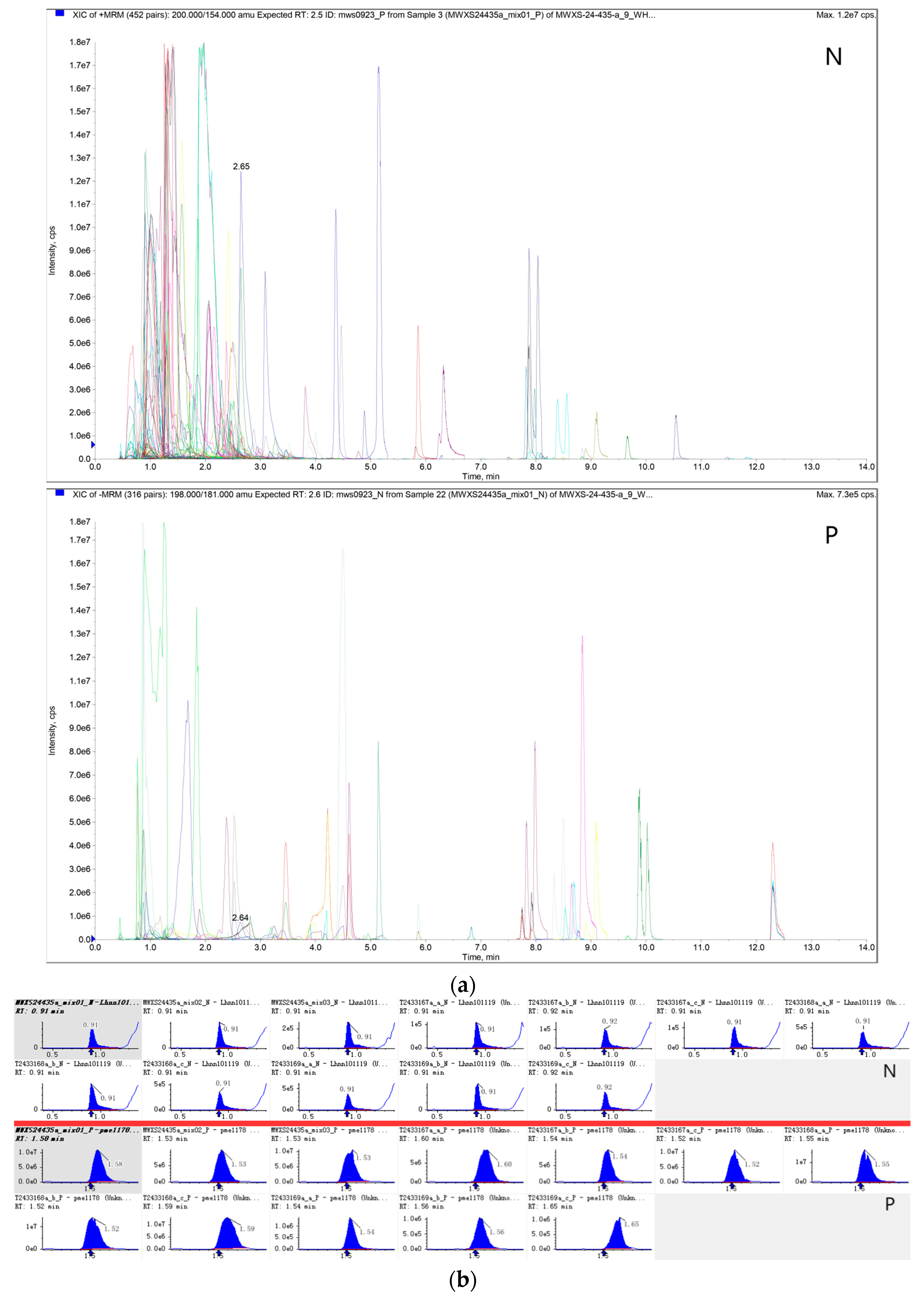
2.4. Identification of GSL Using UPLC-MS/MS

Three experimental groups (50 °C, 80 °C, and the control group) were selected based on sulforaphane quantification results (see Section 3.1 for data interpretation) and designated as Boes-A, Boes-B, and Boes-CK, respectively, to investigate the thermal effects on GSL profiles in broccoli stems.
Three groups of samples were placed into the Scientz-100F lyophilizer (Ningbo, China) using vacuum freeze-drying for 72 h under a condenser temperature of −55 °C and vacuum pressure below 10 Pa and were then ground into powder form using a Retsch MM 400 grinder (Haan, Germany). Subsequently, 50 mg of the sample powder was weighed with an MS105DM electronic balance (Zurich, Switzerland). A 1200 µL aliquot of −20 °C pre-cooled 70% methanol aqueous internal standard (sinigrin) extract was added to the weighed sample. The samples were vortexed for 30 s at 30 min intervals (six cycles total). After vortexing, the samples were centrifuged at 12,000 rpm for three minutes. The supernatant was aspirated and filtered through a 0.22 µm microporous membrane. Finally, the filtered samples were stored in injection vials for UPLC-MS/MS analysis.
The sample extracts were analyzed using an ExionLC™ UPLC-ESI-MS/MS system (SCIEX, Shanghai, China). Chromatographic separation was achieved on an Agilent SB-C18 column (100 × 2.1 mm, 1.8 µm particle size) with a binary mobile phase composed of 0.1% (v/v) formic acid in water (solvent A) and 0.1% (v/v) formic acid in acetonitrile (solvent B). The gradient elution program was as follows: initial conditions of 95% A/5% B (0–0.1 min); a linear gradient to 5% A/95% B over 9.0 min (0.1–9.0 min); an isocratic hold for 1.0 min (9.0–10.0 min); rapid equilibration back to initial conditions within 1.1 min (10.0–11.1 min); and a 2.9 min column re-equilibration period (11.1–14.0 min). The flow rate was maintained at 0.35 mL/min, the column temperature at 40 °C, and the injection volume at 2 μL. The chromatographic effluent was then directed into a QTRAP® 6500+ hybrid triple quadrupole-linear ion trap mass spectrometer for tandem mass spectrometric detection. (SCIEX, Shanghai, China).
The electrospray ionization (ESI) source parameters were configured as follows: source temperature, 550 °C; ion spray voltage (IS), 5500 V (positive ion mode) and −4500 V (negative ion mode); ion source gas I (GSI), gas II (GSII), and curtain gas (CUR), 50 psi, 60 psi, and 25 psi, respectively; collision-activated dissociation (CAD) intensity, high. QQQ scans were performed in MRM mode with nitrogen collision gas pressure set to medium. Declustering potential (DP) and collision energy (CE) for individual MRM transitions were optimized iteratively. A metabolite-specific set of MRM transitions was monitored during each chromatographic period, aligned with the elution window of the corresponding metabolites.

2.5. Data Analysis

Substance characterization was conducted using secondary spectral data (MS/MS). During data preprocessing, isotopic adducts, alkali metal adducts (K+, Na+, NH4+), and fragment ions associated with higher molecular weight compounds were systematically excluded based on the MetWare Database (MetWare Biotechnology Co., Ltd., Wuhan, China). Raw mass spectrometry data were processed using Analyst 1.6.3 for metabolite identification and quantification via database matching. Triple quadrupole-selected characteristic ions were detected, with signal intensities recorded as counts per second (CPS). Chromatographic peak integration and baseline correction were performed in MultiQuant 3.0.3, where peak areas were used as proxies for relative compound abundance. Post-processing included retention time alignment and peak shape normalization across samples using metabolite-specific elution profiles to ensure cross-sample comparability of relative abundances. Final curated datasets containing integrated peak areas were exported for downstream analysis, with all procedures validated using quality control (QC) samples to guarantee analytical accuracy.
Multivariate statistical analysis was conducted using SIMCA-P 14.1 (Umetrics, Malmo, Sweden) with Pareto scaling applied during data preprocessing. Principal component analysis (PCA) provided unsupervised pattern recognition, whereas orthogonal partial least squares-discriminant analysis (OPLS-DA) was employed for supervised classification by jointly modeling the X-matrix (predictor variables) and Y-matrix (response variables). Unlike PCA’s variance-maximization approach, OPLS-DA improves intergroup discrimination through orthogonal signal correction (OSC), which decomposes X-matrix information into Y-correlated variations (predictive component) and Y-orthogonal noise (systematic variation). This OSC-filtered PLS-DA framework facilitates targeted identification of group-discriminant metabolites via variable importance in projection (VIP) scoring, with non-predictive variations systematically excluded [24].
Statistical significance testing and data analyses were conducted using SPSS 22.0 (IBM Corp., Armonk, NY, USA), while graphical representations were generated with Origin 2018 (OriginLab, Northampton, MA, USA).

3. Results and Discussion

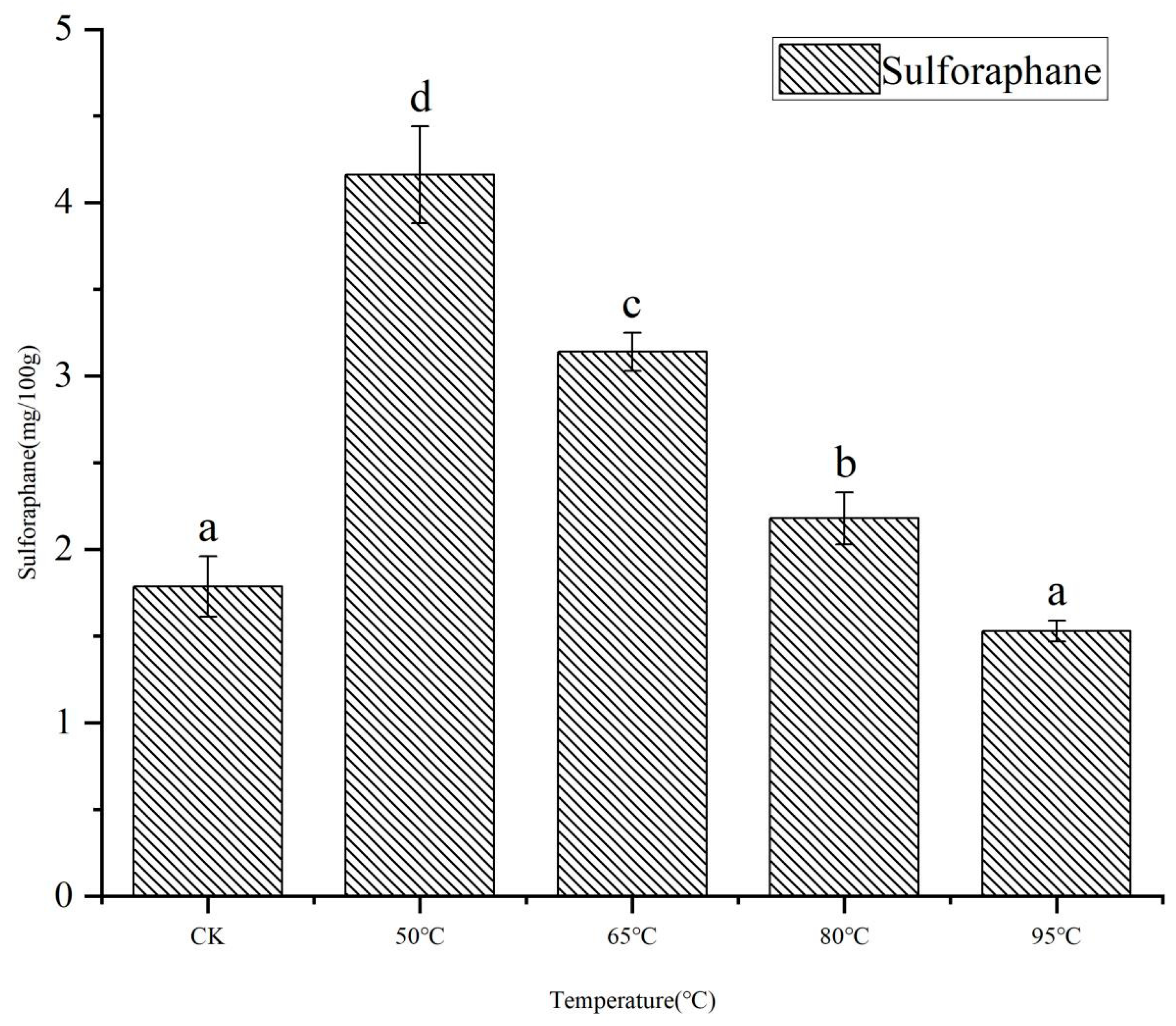
3.1. Content of Sulforaphane

Sulforaphane, the primary hydrolysis product of GSL GRA in broccoli, can serve as a key indicator for monitoring GSL changes in broccoli stems. Figure 1 illustrates the effects of sulforaphane production from GSL degradation in broccoli stems heated at different temperatures. Among the samples, the highest sulforaphane content was observed in the 50 °C group (4.16 ± 0.28 mg/100 g), representing a 132% increase compared to the control group (25 °C, 1.79 ± 0.17 mg/100 g), while the lowest value was recorded in the 95 °C group (1.53 ± 0.06 mg/100 g), slightly lower than the control. Heating at 50 °C and 65 °C enhanced sulforaphane content in broccoli stems, consistent with findings in broccoli sprouts and florets [25,26]. Moderate heat treatment promotes sulforaphane accumulation through two mechanisms: (1) enhancing myrosinase activity to accelerate GSL hydrolysis [27] and (2) suppressing ESP expression, thereby reducing the formation of sulforaphane nitrile (an alternative GSL derivative) and directing more GRA toward sulforaphane production [22]. However, higher temperatures reduced sulforaphane yields. Broccoli stems treated at 80 °C exhibited a sulforaphane content of 2.18 ± 0.15 mg/100 g, higher than the control group but lower than the 50 °C and 65 °C groups. This decline correlates with progressive myrosinase inactivation at elevated temperatures, as further evidenced by the slightly reduced sulforaphane content in the 95 °C group compared to the control. The optimal temperature for maximizing sulforaphane content in broccoli stems was 50 °C, aligning with previously reported processing conditions [28]. Consequently, three sample groups of CK (25 °C), 50 °C, and 80 °C were selected for subsequent metabolomics analysis.

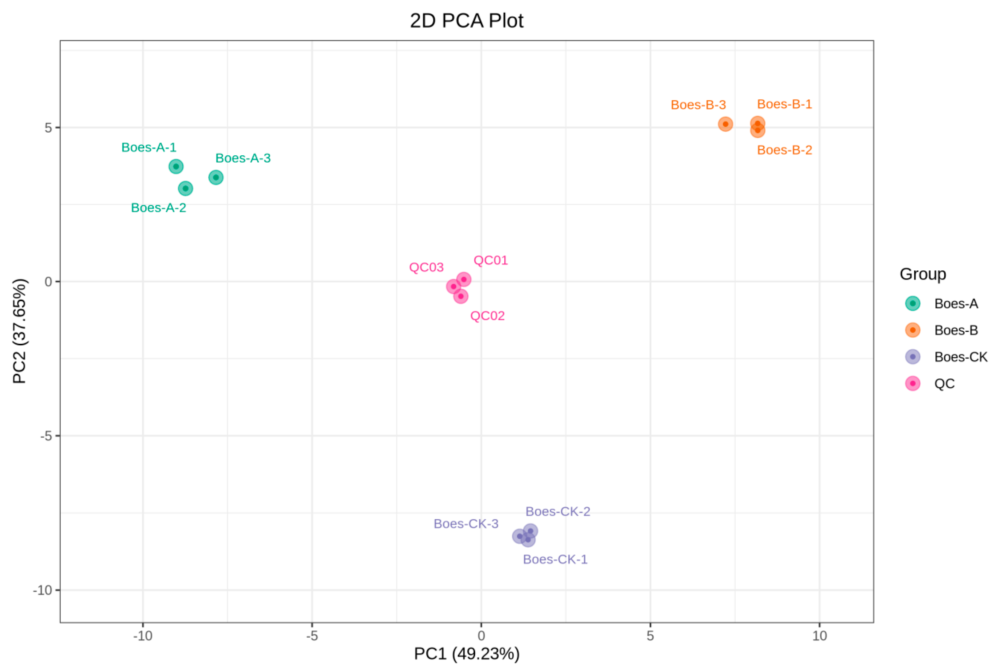
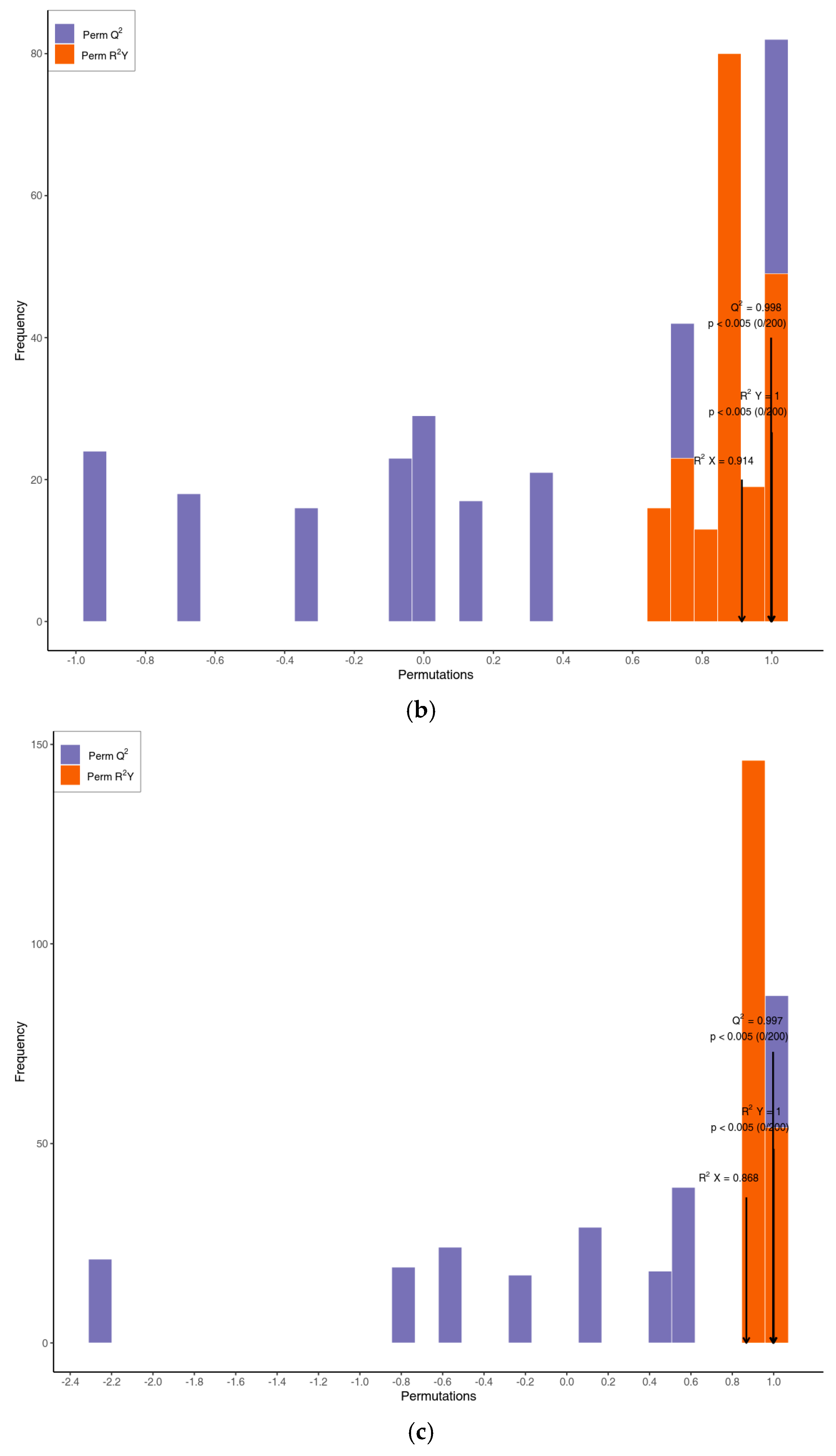
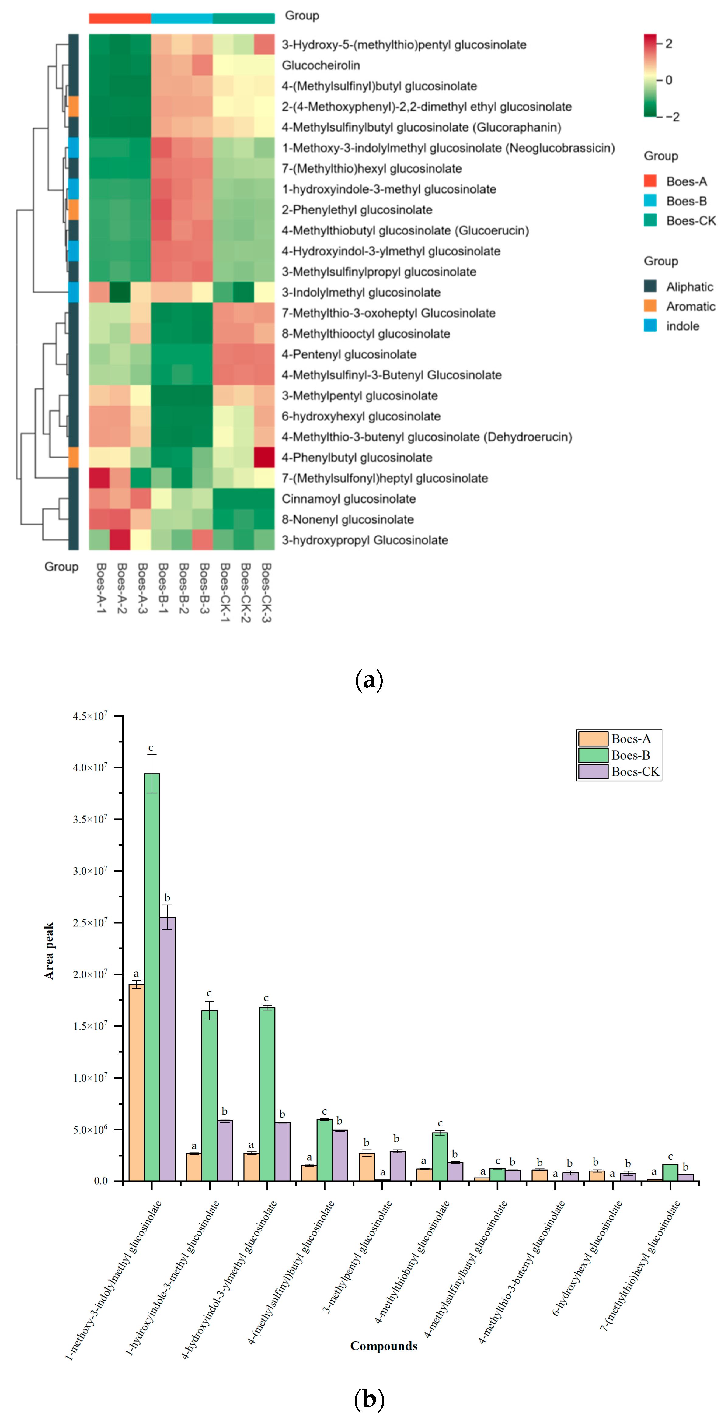
3.2. Multivariate Statistical Analysis

Three experimental groups were selected for metabolomic profiling: a control group (CK, 25 °C) and thermally treated groups at 50 °C and 80 °C, designated as Boes-CK, Boes-A, and Boes-B, respectively. During the process of instrumental analysis, one quality control (QC) sample was inserted every ten test samples. The repeatability of metabolite extraction and detection was assessed by overlapping the total ion current (TIC) chromatograms from mass spectrometric analyses of different QC samples. Figure S1 (Supplementary Material) shows the overlaid TIC chromatograms of QC samples. The results demonstrate that the total ion current curves of metabolite detection exhibited a high degree of overlap, with consistent retention times and peak intensities, indicating satisfactory signal stability of the mass spectrometer when analyzing the same sample at different time points. Figure 2a displays the multi-peak chromatogram of metabolites detected under multiple reaction monitoring (MRM) mode, representing the chemical species identified in the samples. Each color-coded chromatographic peak corresponds to a distinct metabolite. To compare the relative abundance of each metabolite across samples, chromatographic peaks were aligned and corrected based on retention time and peak morphology, ensuring accurate qualitative and quantitative analysis. Figure 2b illustrates the integration-corrected results of randomly selected metabolites, demonstrating the consistency of this alignment process in cross-sample comparisons. Metabolomic analyses identified 76 distinct metabolites: 25 GSLs, one ITC, 28 nucleotide derivatives, 19 amino acid derivatives, and three indole alkaloids (Table S1, Supplementary Material).
Principal component analysis (PCA) of the metabolomic profiles across the three treatment groups (Boes-CK, Boes-A, and Boes-B) revealed significant intergroup variation and intragroup consistency, with the first two principal components explaining 86.88% of the cumulative variance (Figure 3). Distinct separation along PC1 (49.23% variance) corresponded to temperature-dependent metabolic divergence, while PC2 (37.65% variance) discriminated heat treatment effects. To isolate temperature-specific metabolic signatures, orthogonal partial least squares-discriminant analysis (OPLS-DA)—a supervised multivariate approach that filters orthogonal noise through directed pattern recognition—was implemented. The three OPLS-DA models were rigorously validated through permutation testing, with each model undergoing 200 randomized permutations (Figure 4). All models exhibited exceptional predictive validity (Q2 > 0.99) and statistically significant explanatory power for the response matrix Y (R2Y, p < 0.05), confirming their robust capability in capturing metabolite–temperature relationships.

3.3. GSLs in Broccoli Stems

In the three groups of samples, a total of 25 GSLs were identified, including 18 aliphatic, four indole, and three aromatic GSLs, with specific compound names and contents detailed in Table S2 (Supplementary Materials). The indole GSLs showed the highest relative abundance, accounting for 72.64%, 81.91%, and 69.09% of total GSLs in Boes-A, Boes-B, and Boes-CK, respectively, followed by aliphatic GSLs constituting less than one-quarter of total GSLs, while aromatic GSLs represented less than 10%. This distribution contrasts with edible broccoli florets, where aliphatic GSLs predominate [29]. Figure 5a illustrates the distribution patterns of these 25 GSLs across the three experimental groups.
As shown in Table 1 and Figure 5a, the GSL content in Boes-A decreased compared to Boes-CK, with 10 GSLs, including GRA, showing downregulation. This observation aligns with previous findings on sulforaphane production, as moderate temperatures activate myrosinase to promote GSLs hydrolysis while thermal degradation of GSLs simultaneously occurs. The more pronounced reduction observed in aliphatic GSLs compared to indole and aromatic GSLs demonstrates their enhanced degradation susceptibility in broccoli stems under optimal thermal conditions. In contrast, Boes-B displayed elevated GSL levels relative to Boes-CK. Ciska [30] proposed that this phenomenon might result from the heat-induced breakdown of plant tissues, releasing cell wall-bound GSLs. This interpretation concurs with Verkerk’s report of 60% higher GSL concentrations in microwave-treated red cabbage compared to untreated samples [31]. Concurrently, Boes-B demonstrated over twofold increases in indole GSL relative content, potentially indicating superior thermal stability of indole GSLs in broccoli stems. However, contradictory findings by Oerlemans in cabbage studies showed greater heat sensitivity of indole GSLs compared to other classes [32], highlighting species-specific thermal degradation patterns among Brassicaceae vegetables that necessitate customized processing strategies. Comparative analysis between Boes-A and Boes-B revealed upregulation of three indole GSLs in Boes-B: 1-Methoxy-3-indolylmethyl GSL, 1-hydroxyindole-3-methyl GSL, and 4-Hydroxyindol-3-ylmethyl GSL, suggesting potential heat-mediated release of cell wall-associated indole GSLs in broccoli stems.
Figure 5b presents relative content variations of the ten predominant GSLs (seven aliphatic and three indole) across experimental groups. Aliphatic GSLs comprised 4-(methylsulfinyl)butyl GSL, 4-methylthiobutyl GSL, 3-methylpentyl GSL, GRA, 7-(methylthio)hexyl GSL, 4-methylthio-3-butenyl GSL, and 6-hydroxyhexyl GSL. Indole GSLs included 1-methoxy-3-indolylmethyl GSL, 4-hydroxyindol-3-ylmethyl GSL, and 1-hydroxyindole-3-methyl GSL. The three most abundant GSLs in broccoli stems were all indole derivatives: 1-Methoxy-3-indolylmethyl GSL, 1-hydroxyindole-3-methyl GSL, and 4-Hydroxyindol-3-ylmethyl GSL. This indicates distinct GSL profiles compared to florets and sprouts. These indole GSLs showed reduced relative content at 50 °C but increased at 80 °C, suggesting enzyme-mediated rather than thermal degradation predominance in broccoli stems. Figure 5b further identified 3-methylpentyl GSL, 4-methylthio-3-butenyl GSL, and 6-hydroxyhexyl GSL as the most heat-sensitive aliphatic GSLs. This thermal degradation pattern contrasts with broccoli sprouts. Indole GSLs were more heat-intolerant than aliphatic GSLs in broccoli sprouts [33], whereas it was mainly aliphatic GSLs that were affected by thermal degradation in broccoli stems.

4. Conclusions

The results demonstrated temperature-dependent divergence in sulforaphane content and GSL levels within broccoli stems. Optimal sulforaphane biosynthesis was achieved at 50 °C through enhanced myrosinase-mediated conversion of precursor GSLs, whereas 80 °C treatments favored GSL preservation via thermal enzyme inactivation. Given sulforaphane’s superior bioactivity profile, 50 °C is proposed as the optimal processing temperature for functional broccoli stem utilization. Furthermore, indole GSLs exhibited enhanced thermal stability compared to aliphatic homologs in stem tissues.

Supplementary Materials

The following supporting information can be downloaded at https://www.mdpi.com/article/10.3390/horticulturae11040383/s1, Figure S1: Quality control samples in both positive and negative ion detection modes; Table S1: All metabolites qualitative and quantitative information; Table S2: Glucosinolate content in three groups of samples.

Author Contributions

Conceptualization, Y.-H.Z. and G.-Q.Z.; methodology, Y.-F.G. and Z.-H.L.; software, X.-Y.L.; validation, X.-Y.L. and M.-X.M.; formal analysis, X.-Y.L.; investigation, X.-Y.L.; resources, Z.-H.L. and Y.-F.G.; data curation, X.-Y.L.; writing—original draft preparation, X.-Y.L.; writing—review and editing, G.-Q.Z.; visualization, X.-Y.L.; supervision, G.-Q.Z.; project administration, Y.-H.Z. and G.-Q.Z. All authors have read and agreed to the published version of the manuscript.

Funding

This research was funded by Key Research and Development Projects in Anhui Province, grant number 2023n06020045; Major Science and Technology Projects in Xizang Province, grant number XZ202201ZD0001N; Major Project of Universities in Anhui Province, grant number 2022AH04136; Wuhu Science and Technology Plan Project, grant number 2022cg19 and University-level Scientific Research Project, grant number Xjky2022091.

Data Availability Statement

The data that support the findings of this study are available from the corresponding author upon reasonable request.

Conflicts of Interest

The authors declare no conflicts of interest.

References

  1. Chen, Y.-J. Dietary Broccoli Impedes Western Diet-Enhanced Fatty Liver and Hepatocellular Carcinoma Development; University of Illinois at Urbana-Champaign: Champaign-Urbana, IL, USA, 2015. [Google Scholar]
  2. Beaver, L.M.; Löhr, C.V.; Clarke, J.D.; Glasser, S.T.; Watson, G.W.; Wong, C.P.; Zhang, Z.; Williams, D.E.; Dashwood, R.H.; Shannon, J.; et al. Broccoli Sprouts Delay Prostate Cancer Formation and Decrease Prostate Cancer Severity with a Concurrent Decrease in HDAC3 Protein Expression in Transgenic Adenocarcinoma of the Mouse Prostate (TRAMP) Mice. Curr. Dev. Nutr. 2018, 2, nzy002. [Google Scholar] [CrossRef] [PubMed]
  3. Ballance, S.; Knutsen, S.H.; Fosvold, O.W.; Wickham, M.; Trenado, C.D.-T.; Monro, J. Glyceamic and insulinaemic response to mashed potato alone, or with broccoli, broccoli fibre or cellulose in healthy adults. Eur. J. Nutr. 2018, 57, 199–207. [Google Scholar] [CrossRef] [PubMed]
  4. Zhang, T.; Holman, J.; McKinstry, D.; Trindade, B.C.; Eaton, K.A.; Mendoza-Castrejon, J.; Ho, S.; Wells, E.; Yuan, H.; Wen, B.; et al. A steamed broccoli sprout diet preparation that reduces colitis via the gut microbiota. J. Nutr. Biochem. 2023, 112, 109215. [Google Scholar] [CrossRef]
  5. Blazevic, I.; Montaut, S.; Burcul, F.; Olsen, C.E.; Burow, M.; Rollin, P.; Agerbirk, N. Glucosinolate structural diversity, identification, chemical synthesis and metabolism in plants. Phytochemistry 2020, 169, 112100. [Google Scholar] [CrossRef]
  6. Ding, Y.H.; He, H.J.; Song, S.H.; Jian, Y.C.; Zhao, X.Z.; Wang, X.Q. Glucosinolate Component and Content Analysis of Different Broccoli Varieties. J. Change Veg. 2015, 20, 70–74. [Google Scholar]
  7. Yan, M.; Song, C.; Su, S.; Li, J.; Hu, Z.; Lin, S.; Zou, H.; Tang, Z.; Yan, X. Quantification and Diversity Analyses of Glucosinolates in 191 Broccoli Genotypes Highlight Valuable Genetic Resources for Molecular Breeding. Agron.-Basel 2023, 13, 2928. [Google Scholar] [CrossRef]
  8. Wang, J.; Jiang, H.; Chen, S.; Li, Y.; Hamouda, H.I.; Balah, M.A.; Xue, C.; Mao, X. Strategy for preparing of glucosinolate derivatives with outstanding functional activities based on myrosinase. Food Chem. 2025, 479, 143778. [Google Scholar] [CrossRef]
  9. Williams, D.E. Indoles Derived From Glucobrassicin: Cancer Chemoprevention by Indole-3-Carbinol and 3,3′-Diindolylmethane. Front. Nutr. 2021, 8, 734334. [Google Scholar] [CrossRef]
  10. Vo, Q.V.; Nam, P.C.; Dinh, T.N.; Mechler, A.; Tran, T.T.V. Anti-inflammatory activity of synthetic and natural glucoraphanin. J. Serbian Chem. Soc. 2019, 84, 445–453. [Google Scholar] [CrossRef]
  11. Xu, X.; Dai, M.; Lao, F.; Chen, F.; Hu, X.; Liu, Y.; Wu, J. Effect of glucoraphanin from broccoli seeds on lipid levels and gut microbiota in high-fat diet-fed mice. J. Funct. Foods 2020, 68, 103858. [Google Scholar] [CrossRef]
  12. Sun, W.; Sun, J.; Hu, W.; Luo, C.; Lu, Z.; He, F.; Zhao, H.; Zeng, X.; Cao, D.; Li, J.; et al. Sulforaphane inhibits multiple myeloma cell-induced osteoclast differentiation and macrophage proliferation by elevating ferroportin1. Cancer Chemother. Pharmacol. 2025, 95, 3. [Google Scholar] [CrossRef] [PubMed]
  13. Huang, Y.; Wang, B.; Ma, Z.; Chen, T.; Zou, H.; Chen, Y.; Dong, Z.; Chen, J.; Zhang, H.; Ding, Y.; et al. Sulforaphane promotes diabetic wound healing by regulating macrophage efferocytosis and polarization. Int. Immunopharmacol. 2025, 150, 114243. [Google Scholar] [CrossRef] [PubMed]
  14. Ruhee, R.T.; Ma, S.; Suzuki, K. Effects of Sulforaphane Treatment on Skeletal Muscle from Exhaustive Exercise-Induced Inflammation and Oxidative Stress Through the Nrf2/HO-1 Signaling Pathway. Antioxidants 2025, 14, 210. [Google Scholar] [CrossRef]
  15. Wei, R.; Pan, X.; Cai, D.; Pan, L. Synergistic Inhibition of Breast Carcinoma Cell Proliferation by Quercetin and Sulforaphane via Activation of the ERK/MAPK Pathway. Cell Biochem. Biophys. 2025. [Google Scholar] [CrossRef]
  16. Moreira-Rodriguez, M.; Nair, V.; Benavides, J.; Cisneros-Zevallos, L.; Jacobo-Velazquez, D.A. UVA, UVB Light Doses and Harvesting Time Differentially Tailor Glucosinolate and Phenolic Profiles in Broccoli Sprouts. Molecules 2017, 22, 1065. [Google Scholar] [CrossRef]
  17. Sun, J.; Kou, L.; Geng, P.; Huang, H.; Yang, T.; Luo, Y.; Chen, P. Metabolomic Assessment Reveals an Elevated Level of Glucosinolate Content in CaCl2 Treated Broccoli Microgreens. J. Agric. Food Chem. 2015, 63, 1863–1868. [Google Scholar] [CrossRef]
  18. Tian, M.; Yang, Y.; Avila, F.W.; Fish, T.; Yuan, H.; Hui, M.; Pan, S.; Thannhauser, T.W.; Li, L. Effects of Selenium Supplementation on Glucosinolate Biosynthesis in Broccoli. J. Agric. Food Chem. 2018, 66, 8036–8044. [Google Scholar] [CrossRef]
  19. Guo, L.; Yang, R.; Zhou, Y.; Gu, Z. Heat and hypoxia stresses enhance the accumulation of aliphatic glucosinolates and sulforaphane in broccoli sprouts. Eur. Food Res. Technol. 2016, 242, 107–116. [Google Scholar] [CrossRef]
  20. Eun-Sun, H.; Lee, S. Comparative study on the bioactive compound contents and antioxidant activity of broccoli cooked with different methods. Food Sci. Preserv. 2024, 31, 579–589. [Google Scholar]
  21. Lu, Y.; Pang, X.; Yang, T. Microwave cooking increases sulforaphane level in broccoli. Food Sci. Nutr. 2020, 8, 2052–2058. [Google Scholar] [CrossRef]
  22. Matusheski, N.V.; Juvik, J.A.; Jeffery, E.H. Heating decreases epithiospecifier protein activity and increases sulforaphane formation in broccoli. Phytochemistry 2004, 65, 1273–1281. [Google Scholar] [CrossRef] [PubMed]
  23. Kong, F.-h.; Yang, C.-x.; Fang, C.-r.; Qiu, N.-n.; Xu, J.-j.; Bai, S.-s.; Li, D.; Cui, Y.-j. Determination of sulforaphane in cruciferous vegetables by HPLC. Food Ferment. Ind. 2021, 47, 218–223. [Google Scholar]
  24. Forsgren, E.; Bjorkblom, B.; Trygg, J.; Jonsson, P. OPLS-Based Multiclass Classification and Data-Driven Interclass Relationship Discovery. J. Chem. Inf. Model. 2025, 65, 1762–1770. [Google Scholar] [CrossRef] [PubMed]
  25. Hanschen, F.S.; Platz, S.; Mewis, I.; Schreiner, M.; Rohn, S.; Kroh, L.W. Thermally Induced Degradation of Sulfur-Containing Aliphatic Glucosinolates in Broccoli Sprouts (Brassica oleracea var. italica) and Model Systems. J. Agric. Food Chem. 2012, 60, 2231–2241. [Google Scholar] [CrossRef]
  26. Cai, Y.X.; Augustin, M.A.; Jegasothy, H.; Wang, J.H.; Terefe, N.S. Mild heat combined with lactic acid fermentation: A novel approach for enhancing sulforaphane yield in broccoli puree. Food Funct. 2020, 11, 779–786. [Google Scholar] [CrossRef]
  27. Triska, J.; Balik, J.; Houska, M.; Novotna, P.; Magner, M.; Vrchotova, N.; Hic, P.; Jilek, L.; Thorova, K.; Snurkovic, P.; et al. Factors Influencing Sulforaphane Content in Broccoli Sprouts and Subsequent Sulforaphane Extraction. Foods 2021, 10, 1927. [Google Scholar] [CrossRef]
  28. Wu, Y.; Lv, C.; Zou, L.; Sun, J.; Song, X.; Zhang, Y.; Mao, J. Approaches for enhancing the stability and formation of sulforaphane. Food Chem. 2021, 345, 128771. [Google Scholar] [CrossRef]
  29. Kushad, M.M.; Brown, A.F.; Kurilich, A.C.; Juvik, J.A.; Klein, B.P.; Wallig, M.A.; Jeffery, E.H. Variation of glucosinolates in vegetable crops of Brassica oleracea. J. Agric. Food Chem. 1999, 47, 1541–1548. [Google Scholar] [CrossRef]
  30. Ciska, E.; Verkerk, R.; Honke, J. Effect of boiling on the content of ascorbigen, indole-3-carbinol, indole-3-acetonitrile, and 3,3′-diindolylmethane in fermented cabbage. J. Agric. Food Chem. 2009, 57, 2334–2338. [Google Scholar] [CrossRef]
  31. Verkerk, R.; Dekker, M. Glucosinolates and myrosinase activity in red cabbage (Brassica oleracea L. var. Capitata f. rubra DC.) after various microwave treatments. J. Agric. Food Chem. 2004, 52, 7318–7323. [Google Scholar] [CrossRef]
  32. Oerlemans, K.; Barrett, D.M.; Suades, C.B.; Verkerk, R.; Dekker, M. Thermal degradation of glucosinolates in red cabbage. Food Chem. 2006, 95, 19–29. [Google Scholar]
  33. Hanschen, F.S.; Rohn, S.; Mewis, I.; Schreiner, M.; Kroh, L.W. Influence of the chemical structure on the thermal degradation of the glucosinolates in broccoli sprouts. Food Chem. 2012, 130, 1–8. [Google Scholar] [CrossRef]
Figure 1. Sulforaphane content in broccoli stems following 1 h heat treatment at different temperatures. Different lowercase letters above bars indicate significant differences (p < 0.05) in sulforaphane content among broccoli stems subjected to different heating temperatures.
Horticulturae 11 00383 g001
Figure 2. (a) MRM metabolite multi-peak profiles; (b) quantitative analysis with integrated peak correction for randomly selected metabolites across samples. X-axis: retention time (min); Y-axis: ion intensity (cps) of detected metabolite ions; peak area reflects relative abundance in samples. N: negative ion mode, P: positive ion mode.
Horticulturae 11 00383 g002
Figure 3. Metabolomic PCA of broccoli stems across temperature treatments. QC, quality control sample; Boes-A, 50 °C; Boes-B, 80 °C; Boes-CK, 25 °C. Numbers (1, 2, 3) denote triplicate biological replicates for each group.
Horticulturae 11 00383 g003
Figure 4. OPLS-DA score plots between the following pairs of groups: (a) Boes-B vs. Boes-A; (b) Boes-A vs. Boes-CK; (c) Boes-B vs. Boes-CK. X-axis: R2Y and Q2 values of models; Y-axis: frequency of classification efficacy observed across 200 permutation tests. Orange: R2Y of permutated models; Purple: Q2 of permutated models. Black arrows denote original model parameters (R2X, R2Y, Q2).
Horticulturae 11 00383 g004aHorticulturae 11 00383 g004b
Figure 5. Clustering analysis of GSLs in three different pretreatment samples of broccoli stems: (a) heat map of UPLC-MS/MS analysis of GSLs by 3 different pretreatments; (b) the difference in concentration in ten major GSLs. Different lowercase letters above bars indicate significant differences (p < 0.05) in GSL content among broccoli stems subjected to different heating temperatures.
Horticulturae 11 00383 g005
Table 1. Relative content of GSLs in three sample groups.
Boes-ABoes-BBoes-CK
Total GLSs content3.37 × 1078.88 × 1075.20 × 107
Aliphatic6.40 × 1061.58 × 1071.23 × 107
Indole2.45 × 1077.27 × 1073.59 × 107
Aromatic2.94 × 1063.07 × 1053.36 × 106
Percentage of aliphatic18.972%17.746%23.600%
Percentage of indole72.645%81.908%69.095%
Percentage of aromatic8.734%0.345%6.455%
All data are expressed as relative content represented by peak areas.
Disclaimer/Publisher’s Note: The statements, opinions and data contained in all publications are solely those of the individual author(s) and contributor(s) and not of MDPI and/or the editor(s). MDPI and/or the editor(s) disclaim responsibility for any injury to people or property resulting from any ideas, methods, instructions or products referred to in the content.

