Article

Droplet-Vitrification Protocol for Cryopreservation of Ginger (Zingiber officinale) Shoot Tips

1
School of Life Science and Engineering, Southwest University of Science and Technology, Mianyang 621010, China
2
The Agricultural and Rural Bureau of Ming Mountain District, Ya’an 625199, China
3
Hubei Key Laboratory of Spices and Horticultural Plant Germplasm Innovation and Utilization, College of Horticulture and Gardening, Yangtze University, Jingzhou 434025, China
*
Author to whom correspondence should be addressed.
These authors contributed equally to the present study.
Horticulturae 2025, 11(3), 283; https://doi.org/10.3390/horticulturae11030283
Submission received: 23 January 2025 / Revised: 25 February 2025 / Accepted: 3 March 2025 / Published: 5 March 2025

Abstract

:
Ginger (Zingiber officinale), a globally grown and economically valuable plant, has inadequate research on germplasm cryopreservation, and droplet-vitrification is yet to be applied. The present study established an efficient droplet-vitrification protocol for Z. officinale ‘Yunnan Xiaohuangjiang’. The droplet-vitrification procedure was as follows: excise 1.5–2.0 mm shoot tips with 3–4 leaf primordia from five-week-old cultures, preculture on MS medium with 0.25 M sucrose for 1 d, treat with MS liquid medium with 2 M glycerol and 0.4 M sucrose for 20 min, dehydrate with PVS2 plus 0.1 M ascorbic acid at 0 °C for 20 min, plunge into LN for 1 h, thaw in MS liquid medium with 1.2 M sucrose for 20 min, post-culture on shoot recovery medium (MS with 0.1 g/L GA3) in the dark for 3 d. Histological and ultrastructural analyses revealed that PVS + ascorbic acid-treated shoot tips exhibited numerous living cells with small vacuoles in the apical dome, leaf primordia, and basal parts. Genetic stability results showed that the plantlets regenerated from cryopreserved shoot tips had no genetic variation. This is the first report on ginger cryopreservation via droplet-vitrification, providing technical support for ginger germplasm cryopreservation and virus elimination cryotherapy in ginger.

1. Introduction

Ginger (Zingiber officinale) is a major rhizome spice that belongs to the Zingiberaceae family. Due to its distinctive flavor, ginger is widely utilized as a key constituent in many spice blends. It is also used in food, such as cakes, biscuits, wine, and industrial applications. Of the presence of a plethora of bioactive natural compounds, ginger occupies a pivotal position as a fundamental ingredient in the traditional medical practices of Chinese, Indian, and Japanese [1]. Ginger has a long cultivation history in both India and China, where it has been grown in subtropical regions for centuries. Today, it is used on a larger scale worldwide [2].
The conservation of plant genetic resources is essential for the future efforts of breeding programs. As a result, numerous genebanks have been established around the world to conserve and utilize germplasm resources [3]. Cryopreservation involves storing living cells, organs, and tissues in liquid nitrogen (LN) at a temperature of −196 °C or in LN vapor at −160 °C [4,5,6]. Once cryopreserved, the sample maintains its viability upon thawing, allowing for indefinite storage. At extremely low temperatures, the metabolic and biochemical reactions within the cells stop [6]. Cryopreservation is regarded as a safe and cost-efficient method for the long-term conservation of plant germplasm, without the risk of genetic modifications [4,7,8,9].
To date, studies on the cryopreservation of ginger shoot tips are relatively scarce. Yamuna et al. [10] reported that the establishment of cryopreservation procedures for ginger in vitro shoots using encapsulation–dehydration, vitrification, and encapsulation–vitrification techniques. Du et al. [11] further elaborated on the establishment of a vitrification procedure for ginger. Studies have identified that discernible disparities exist in the efficacy of cryogenic techniques when applied to distinct genotypes of the same species [12,13]. Developing and establishing a diverse array of methodologies plays a pivotal role in facilitating the conservation and safeguarding of ginger germplasm resources, which are of significant genetic and agricultural value. Droplet-vitrification originated with the droplet freezing technique that was devised by Kartha et al. [14]. In the droplet-vitrification protocol, loaded shoot tips are pretreated with the plant vitrification solution (PVS). Subsequently, individual shoot tips are placed into 5–10 µL droplets of PVS, which are deposited on an aluminum foil sheet. The foil, bearing the PVS droplets and shoot tips, is immediately immersed in LN. The aluminum foils are rapidly submerged in a fluid medium containing 1.2 M sucrose for warming. After a 20 min unloading phase, the shoot tips are meticulously retrieved from the foils and transferred onto the recovery medium for subsequent cultivation [15]. Droplet-vitrification characterized by its rapid cooling rate and straightforward operation, has demonstrated higher recovery rates than droplet freezing or traditional vitrification methods and has been extensively utilized across various plant species [15,16,17]. Among the drawbacks of droplet-vitrification is its stringent requirement for sophisticated operational techniques and meticulous time management. In scenarios where a substantial volume of samples needs to be processed, this method likely augments labor expenses and elongates the operational timeline [15]. To date, droplet-vitrification is confirmed as the most widely applicable method for cryopreservation of plant germplasm [15,18]. However, to the best of our knowledge, droplet-vitrification has not yet been applied to the cryopreservation of ginger germplasm. Therefore, efforts to develop a droplet-vitrification protocol for this plant species would further increase the cryopreservability of ginger genetic resources. Meanwhile, the droplet-vitrification protocol of ginger has the potential to be utilized as a cryotherapy method for virus elimination [19,20].
The aim of this study was to establish an efficient droplet-vitrification protocol for in vitro cryopreservation of shoot tips from a Chinese ginger genotype Z. officinale ‘Yunnan Xiaohuangjiang’. Histological and ultrastructural observations, as well as evaluation of genetic stability by inter-simple sequence repeat (ISSR) analysis and flow cytometry (FCM), were performed on the regenerants post-cryopreservation.

2. Materials and Methods

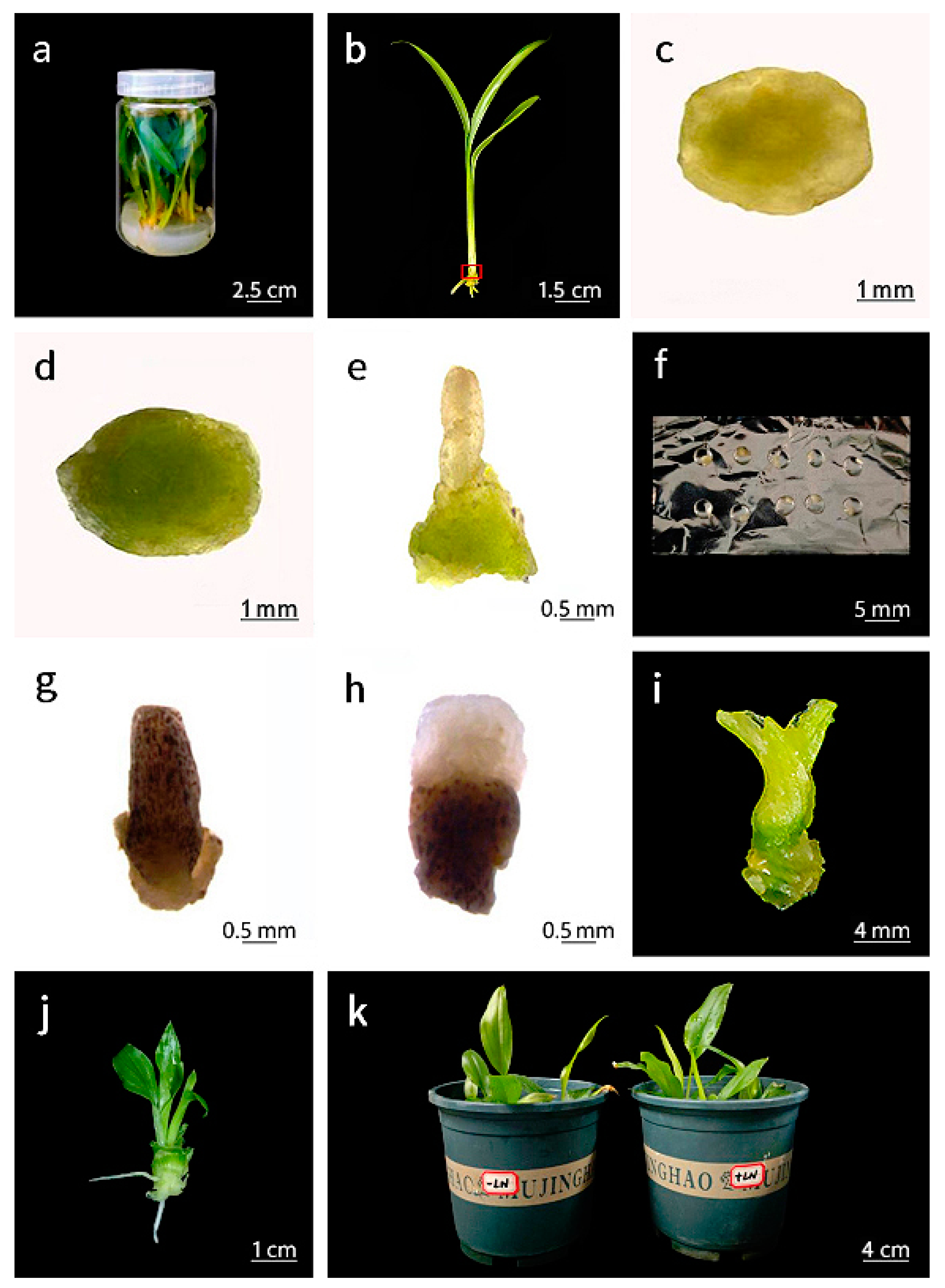
2.1. Plant Materials

Chinese ginger Z. officinale ‘Yunnan Xiaohuangjiang’, a local variety that plays a significant role in the agricultural economy of Yunnan Province, is wildly cultivated across multiple regions within Yunnan province, located in southwest China. This genotype has become a crucial export commodity, contributing substantially to the province’s international trade and economic development [21]. Z. officinale ‘Yunnan Xiaohuangjiang’, in vitro stock shoots were maintained on Murashige and Skoog (MS) [22] medium supplemented with 3 mg·L−1 6-benzyl-aminopurine (6BAP), 0.2 mg·L−1 α-naphthaleneacetic acid (NAA), 30 g·L−1 sucrose, and 7 g·L−1 agar [23]. Before autoclaving at 121 °C for 20 min, the pH of the medium was adjusted to 5.8. The plantlets were kept at a temperature of 25 ± 2 °C. They were exposed to a photoperiod of 16 h per day, provided by cool-white fluorescent tubes (45 µmol m−2s−1) (Topu yunnong, Zhejiang, China). Subculture was conducted on the medium described above every five weeks (Figure 1a). Nodal segments (5–6 mm) with 1–2 nodes, obtained from five-week-old stock cultures, were cultured on an axillary induction medium to promote shoot elongation from axillary buds (Figure 1b–d). The medium was composed of MS supplementing with 1 mg·L−1 thidiazuron (TDZ), 0.5 mg·L−1 NAA, 30 g·L−1 sucrose, and 7 g·L−1 agar [23].

2.2. Cryopreservation Procedure

Shoot tips (Figure 1e), with a length of 1.5–2.0 mm and 3–4 LPs, were excised from the five-week-old induced shoots cultured on an axillary induction medium for 5 d. The shoot tips were precultured on MS medium with 0.25, 0.5, or 0.75 M sucrose and 7 g·L−1 agar for 1, 2, 3, or 4 days at 4 °C in the dark. Before autoclaving at 121 °C for 20 min, the pH of the medium was adjusted to 5.8. Following this, the precultured shoot tips were treated with a loading solution of MS liquid medium supplemented with 2 M glycerol and 0.4 M sucrose and placed in sterile 9 cm diameter dishes for 20 min at room temperature. Before autoclaving at 121 °C for 20 min, the pH of the loading solution was adjusted to 5.8. Subsequently, the loaded shoot tips were exposed to plant vitrification solution 2 (PVS2) [24] at 0 °C for 0, 5, 10, 15, 20, or 25 min. PVS2 consists of MS, which is augmented with 30% (w/v) glycerol (Sinopharm, Beijing, China), 15% (w/v) dimethylsulfoxide (DMSO) (Sinopharm, Beijing, China), 15% (w/v) ethylene glycol (Sinopharm, Beijing, China), and 0.4 M sucrose. Before autoclaving at 121 °C for 20 min, the pH of PVS2 was adjusted to 5.8. Each dehydrated shoot tip was then transferred onto a droplet of 2.5 µL PVS2 on a piece of aluminum foil measuring 2 cm in length and 0.8 cm in width (Figure 1f) and subjected to direct immersion in LN for several minutes. Following that, the aluminum foils that had the shoot tips frozen on them were then moved into a 2-milliliter cryotube filled with LN, and they were kept there for 30 min. Following the removal of the frozen foils from LN, the samples were immediately immersed in an unloading solution composed of MS that is supplemented with 1.2 M sucrose in sterile 9 cm diameter dishes for 20 min at room temperature. Before autoclaving at 121 °C for 20 min, the pH of the unloading solution was adjusted to 5.8. Cryopreserved shoot tips were immediately cultured on four different shoot recovery media: (1) shoot recovery medium (SRM)1, which included MS containing 0.5 mg·L−1 NAA, 1.0 mg·L−1 TDZ, 30 g·L−1 sucrose, and 7 g·L−1 agar [23]; (2) SRM2, which included MS containing 0.1 mg·L−1 NAA, 3.0 mg·L−1 6BA, 30 g·L−1 sucrose, and 7 g·L−1 agar [25]; (3) SRM3, which consisted of MS containing 0.5 mg·L−1 6-furfurylaminopurine (KT), 1.0 mg·L−1 6BA, 30 g·L−1 sucrose, and 7 g·L−1 agar [26]; and (4) SRM4, which included MS containing 0.1 mg·L−1 GA3 (Sinopharm, Beijing, China), 30 g·L−1 sucrose, and 7 g·L−1 agar. The pH of all shoot recovery media was adjusted to 5.8 before autoclaving at 121 °C for 20 min. The shoot tips were individually cultivated on the four types of shoot regeneration media without light for 3 days. Subsequently, they were removed from the identical medium and cultured under standard culture conditions. NAA, TDZ, and 6BA were added to the medium before autoclaving, while KT and GA3 were filter-sterilized and incorporated post-autoclaving. After a post-culture period of two weeks, dead shoot tips exhibited blackening (Figure 1g). Survival was measured by calculating the proportion of shoot tips that exhibited any white tissue (Figure 1h) from the total samples subjected to cryopreservation after two weeks of post-culture. Regrowth was quantitatively evaluated as the proportion, expressed as a percentage, determined by calculating the number of shoot tips that successfully developed into shoots with a length of at least 0.5 cm. This calculation was made on the total number of samples, and the assessment was conducted eight weeks after the completion of the post-culture stage.

2.3. Addition of Exogenous Ascorbic Acid (AsA) and Glutathione (GSH)

Varying concentrations of AsA (Sinopharm, Beijing, China) at 0.1, 0.2, and 0.4 mM, and GSH (Sinopharm, Beijing, China) at 0.4, 0.6, and 0.8 mM were added into the PVS2, respectively, during the dehydration step. This approach aimed to identify the optimal exogenous substances that enhance the efficacy of cryopreservation.

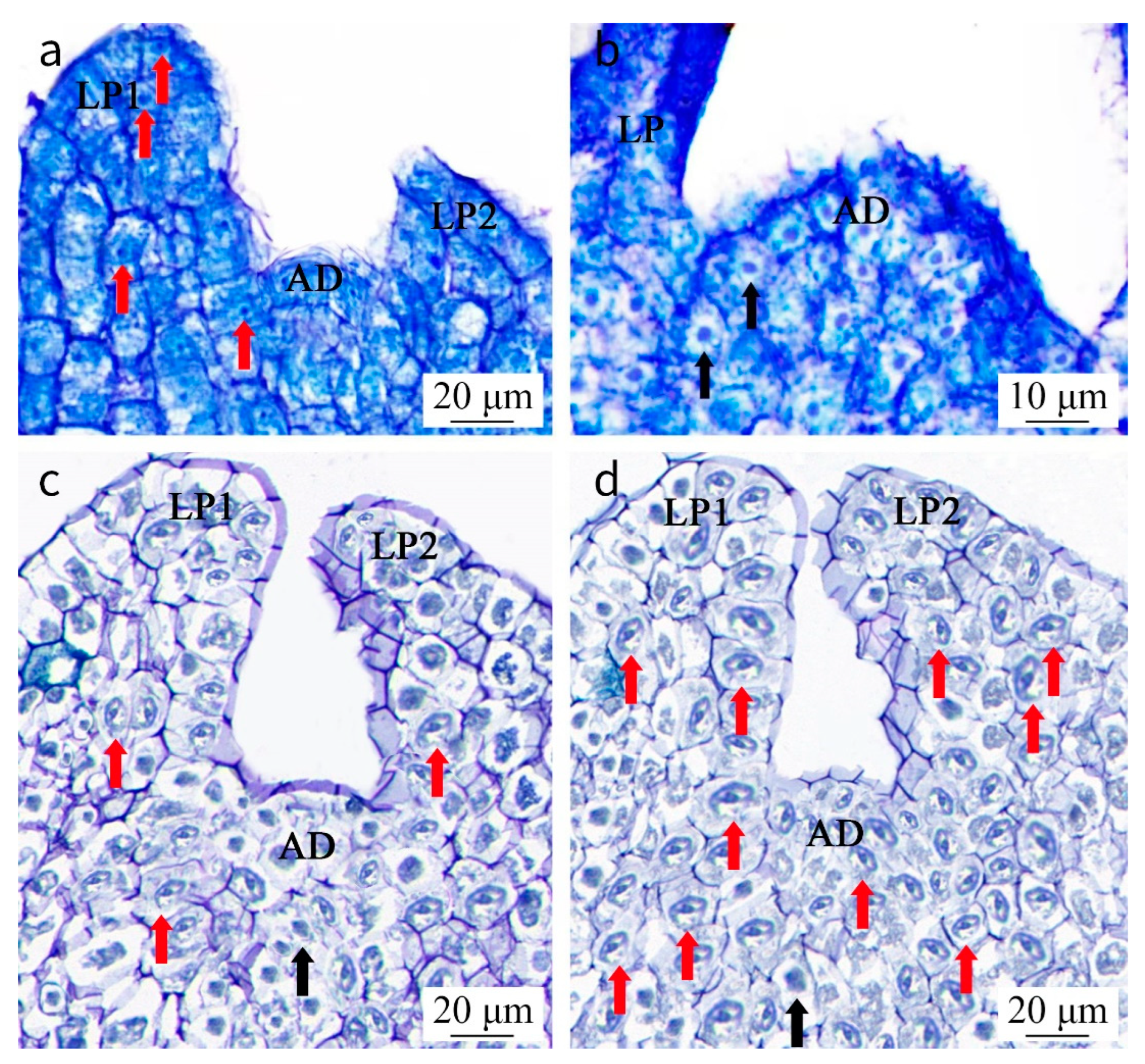
2.4. Analyses from a Histological and Ultrastructural Perspective of Shoot Tips

For histological studies, shoot tips of ‘Yunnan Xiaohuangjiang’ were sampled from the following treatments: (1) in the positive control, shoot tips were freshly excised; (2) in the negative control, shoot tips were freshly cut, submerged in LN, and post-cultured for 1 day; (3) shoot tips underwent treatment with PVS2 − AsA for 20 min at a temperature of 0 °C, after which they were immersed in LN, warmed with an unloading solution at room temperature for 20 min, and incubated in SRM4 for 1 day; and (4) shoot tips treated with PVS2 + AsA for 20 min at a temperature of 0 °C, after which they were immersed in LN, warmed with an unloading solution at room temperature for 20 min, and incubated in SRM4 for 1 day. Thirty samples from each treatment were fixed, dehydrated, and embedded according to the methods described by Wang and Valkonen [27]. Sections, with a thickness of 50 µm, were sliced by a microtome (Leica, RM 1016, Wetzlar, Germany), then positioned onto glass slides and dyed with 0.01% toluidine blue (TB) [28]. After that, a light microscope (Olympus Corporation BH-2, Tokyo, Japan) was used to observe the sections. For ultrastructural studies, thirty shoot tips were excised for each treatment (treatment 1, 3, and 4, as described above) and processed according to the methodology outlined by Wang and Valkonen [27]. Using an ultramicrotome (Leica, EM UC7, Wetzlar, Germany), ultrathin sections, each with a thickness of 50 nm, were made and then gathered onto formvar-coated copper grids. Following this, the sections underwent contrast treatment using alcoholic uranyl acetate and lead citrate, as described by Reynolds [29]. With the preparation finished, a transmission electron microscope (TEM) (JEM-1400, JEOL, Tokyo, Japan) was employed to conduct an examination of the sections and take their photographs.

2.5. ISSR Analysis

In the 12-month period after shoot regeneration, the recovered plantlets and controls were, respectively, propagated to a population of three hundred. Samples of ‘Yunnan Xiaohuangjiang’ were examined for genetic stability compared to non-cryopreserved samples using ISSR analysis. Genomic deoxyribonucleic acid (DNA) was separated from the samples using the cetyltrimethylammonium bromide (CTAB) method. In the DNA extraction procedure, first, 0.5 g of leaves were initially ground with LN in a mortar to a fine powder and quickly transferred to a 15 mL centrifuge tube. Then, 5 mL of 60 °C extraction buffer and 50 mg PVP (Sinopharm, Beijing, China)/0.5 g leaf tissues were added, gently inverted to mix, and incubated at 60 °C with shaking in an oven (Thermo Fisher Scientific, Waltham, MA, USA) for 25–60 min. After cooling to room temperature for 4–6 min, 6 mL of chloroform-octanol (24:1) (Sinopharm, Beijing, China) was added and mixed by inversion. Once homogenized, it was centrifugated at 3000 revolutions per minute (rpm) for 20 min in a room-temperature benchtop centrifuge (Eppendorf, Hamburg, Germany). The top aqueous layer was transferred to a new tube, and the chloroform–octanol extraction was repeated to clarify the solution. Specifically, 1/2 volume of 5 M NaCl (Sinopharm, Beijing, China) was added to the recovered aqueous phase, followed by two volumes of cold (−20 °C) 95% ethanol (Sinopharm, Beijing, China). After mixing and placing at −20 °C for 10 min to promote precipitation, the samples were centrifuged at 3000 rpm for 6 min. The supernatant was discarded, and the pellet was washed with cold (0–4 °C) 70% v/v ethanol, dried at 37 °C, dissolved in 300 μL Tris-EDTA (Sinopharm, Beijing, China) (TE) buffer overnight at 4 °C, and transferred to 1.5 mL Eppendorf tubes. A total of 3 μL of RNase A (Sinopharm, Beijing, China) (10 mg/mL) was added and incubated at 37 °C for 1 h, followed by 3 μL of proteinase K (Sinopharm, Beijing, China) (1 mg/mL) for 15–30 min. A total of 150 μL each of phenol (Sinopharm, Beijing, China) and chloroform (Sinopharm, Beijing, China) were added, vortexed briefly, and centrifuged at 14,000 rpm for 10–15 min. The upper layer was collected, and after adding 50 μL of TE to the phenol phase, vortexing, and centrifuging again, the upper layer was combined with the sample. Additionally, 1/10 volume of 2 M Na acetate (Sinopharm, Beijing, China) and 2 volumes of absolute ethanol were added and mixed, and the tubes were stored at −80 °C overnight. After centrifuging at 14,000 rpm for 10–20 min, the tubes were drained, washed with 70% v/v EtOH (Sinopharm, Beijing, China), vacuum-dried, and resuspended in 100–200 μL TE. Finally, the tubes were incubated at 37°C for 30 min, and the DNA concentration and purity were measured using an ultraviolet spectrophotometer (Thermo Fisher Scientific, Waltham, MA, USA), as described by Porebski et al. [30]. The DNA was stored at −20°C for further analyses. A screening of forty ISSR primers was carried out to determine suitable candidates for assessing the genetic stability in the regenerants. Seven ISSR primers, ISSR06: 5′-(GA)8C-3′, ISSR14: 5′-(AGC)4GT-3′ [31], SPS05: 5′-(GA)9T-3′, SPS06: 5′-T(GA)9T-3′ [32], UBC816: 5′-CACACACACACACACAT-3′, UBC855: 5′-ACACACACACACACACYT-3′, UBC851: 5′-GTGTGTGTGTGTGTGTYG-3′, UBC 859: 5′-TGTGTGTGTGTGTGTGRC-3′ [33], were selected for PCR amplification. For the polymerase chain reaction (PCR), a 25 µL reaction mixture was prepared. This mixture included 1 X PCR buffer (Thermo Fisher Scientific, Waltham, MA, USA), 1.75 mM MgCl2 (Thermo Fisher Scientific, Waltham, MA, USA), 5 mM of each deoxy-ribonucleoside triphosphate (Thermo Fisher Scientific, Waltham, USA) (dNTP), 40 µM of each oligonucleotide primer, 25 ng of genomic DNA, and 1 U of Taq DNA polymerase (Thermo Fisher Scientific, Waltham, MA, USA). In the optimized PCR setup for ISSR amplification, an initial denaturation step was included at 95 °C for 3 min, followed by 35 cycles of 10 s at 95 °C for denaturation, 10 s at 55 °C for annealing, and 30 s at 72 °C for an extension. A final extension step was performed at a temperature of 72 °C for 7 min. Electrophoresis was carried out to separate the PCR products. The medium was 2% agarose gel (Thermo Fisher Scientific, Waltham, MA, USA), which had ethidium bromide (Thermo Fisher Scientific, Waltham, MA, USA) (0.5 µg·mL−1) added to it. The electrophoresis took place in 1× Tris-borate-EDTA (Sinopharm, Beijing, China) (TBE) buffer under a voltage of 90 volts. Under ultraviolet (UV) light, the gel was photographed, and molecular weights were estimated using a 50 bp DNA ladder.

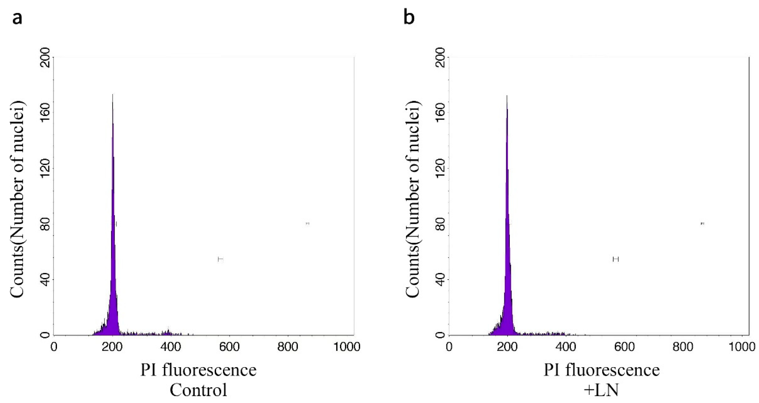
2.6. Flow Cytometry (FCM)

After nine subcultures, the ploidy levels of thirty plantlets regenerated from cryopreserved ‘Yunnan Xiaohuangjiang’ shoot tips were analyzed by FCM, with thirty in vitro stock shoots as controls. Nuclei suspensions were prepared from 50 mg of leaves, according to the method used by Huang et al. [34]. The nuclei were obtained from the cells by slicing the samples using a razor blade within Marie’s isolation buffer [35]. To remove cell fragments and large tissue debris, the nuclei suspension was filtered using a 50 µm nylon filter. Subsequently, DNA staining was carried out by adding 50 µg·mL−1 of propidium iodide (Sinopharm, Beijing, China) (PI) and 20 µg·mL−1 RNAse (Sinopharm, Beijing, China) to the samples. The samples were evaluated using a flow cytometer within a period of 15 min. The fluorescence of the samples was measured through the use of a BD FacsClibur flow cytometer (San Jose, CA, USA). After analyzing no less than 5000 nuclei using Modifit 3.0 software (VSH, Washington, DC, USA), histograms were generated.

2.7. Acclimatization of Plants to Soil

Proliferated shoots (3–4 cm in length, with 3–4 fully expanded leaves and well-developed roots) were transplanted into 12 cm diameter plastic pots filled with a sterile mixture of soil, sand, coir dust, and cow dung at a ratio of 3:1:1:1 and cultivated in a greenhouse [23]. The pots were covered with plastic bags to keep high humidity, gradually opening within a week to decrease the moisture. After two weeks of growth in the greenhouse, the plants were re-established in the field soil.

2.8. Statistical Analysis

During cryopreservation experiments, the shoot tips that had undergone all the experimental treatments, except for the cooling step in LN, were designated as the treated control group (−LN). Conversely, the shoot tips subjected to cryopreservation procedures, including the cooling process in LN, were labeled as the +LN group. Each treatment included three replicates, with a minimum of ten samples per replicate, and all experiments were performed twice. Results are shown as means accompanied by their corresponding standard errors. Statistical analyses were conducted by using one-way ANOVA and Student’s t-test. The least significant differences (LSD) were computed with a significant level set at p < 0.05. In each of the two replicates, twenty samples were utilized for histological and ultrastructural observation. In the assessment of genetic stability through ISSR and FCM, thirty plantlets regenerated from cryopreserved shoot tips and thirty plantlets from in vitro stock cultures were randomly picked from a collective population consisting of three hundred cryo-derived plants and three hundred in vitro culture-derived plants. In the ISSR analysis, every band was meticulously evaluated and manually assigned a score. A score of 1 was designated to indicate the presence of a band, whereas a score of 0 was assigned to denote the absence of a band. Bands that exhibited the same molecular weights and electrophoretic mobilities and were amplified by the same primer were considered to correspond to the identical genetic locus. Both distinctly observable monomorphic bands and polymorphic bands were documented. To guarantee reproducibility, the experiments were carried out two times. Only those bands that were consistent across the replicates and could be reliably reproduced were considered.

3. Results

3.1. Effect of Sucrose Preculture

The survival and regrowth rates were significantly influenced by sucrose concentration and preculture duration. When cultured on the sucrose preculture medium for 1 d, as sucrose concentration increased, both the survival and regrowth rates of the cryopreserved shoot tips decreased. The maximum survival rate and regrowth rate were observed at 0.25 M sucrose, reaching 73.33% and 33.33%, respectively. A similar trend was noted for the treated control shoot tips (Table 1). When precultured in 0.25 M sucrose, both the cryopreserved and treated control shoot tips exhibited significantly different survival and regrowth rates depending on the duration of preculture. Following the treatment with 0.25 M sucrose for 1 d, the highest survival and regrowth rates for both groups were recorded. However, as the preculture duration increased, the survival and regrowth rates for both the cryopreserved and treated control shoot tips declined (Table 2).

3.2. Effect of the Exposure Time Duration to PVS2

The survival rate was significantly influenced by the exposure time duration to PVS2 for cryopreserved shoot tips, whereas it did not affect the treated control shoot tips. The highest survival rate observed was 73.33% at 20 min, which was significantly higher than the rates at 0 min (0%) and 5 min (40%). There were no remarkably significant differences in the survival rates at 10 min (56.67%), 15 min (66.67%), and 25 min (63.33%). On the contrary, the survival rate of the treated control shoot tips varied from 83.33% to 100% during the 0 to 25 min exposure period. Regarding shoot regrowth rate, both the treated control shoot tips and cryopreserved shoot tips were significantly affected by the exposure time duration to PVS2. The regrowth rate increased from 0% to 6.67% for cryopreserved shoot tips as exposure time extended from 0 to 5 min. As the exposure time increased, the regrowth rate continued to improve, reaching a maximum of 33.33% at 20 min. Conversely, the regrowth rate of the treated control shoot tips decreased from 100% without PVS2 exposure to 36.67% when the PVS2 exposure time was 25 min (Figure 2).

3.3. Effect of SRM

The SRM did not have a significant impact on the survival rate of both cryopreserved and treated control shoot tips. Nevertheless, it significantly impacted the regrowth rate of these shoot tips. The cryopreserved shoot tips cultured on SRM4 exhibited normal regeneration, characterized by stem elongation and leaf growth, resulting in a regrowth rate of 33.33%. In contrast, the cryopreserved shoot tips that were cultured on SRM1–3 demonstrated abnormal regrowth, with 13.33%, 6.67%, and 3.33%, respectively. The cryopreserved shoot tips cultured on SRM1 exhibited differentiation. However, neither leaf development nor stem elongation was observed. When the cryopreserved shoot tips were cultured on SRM2, leaf development emerged. Nevertheless, stem elongation failed to be discerned. The cryopreserved shoot tips cultured on SRM3 only showed swelling, with no leaf growth or stem elongation (Figure 3).

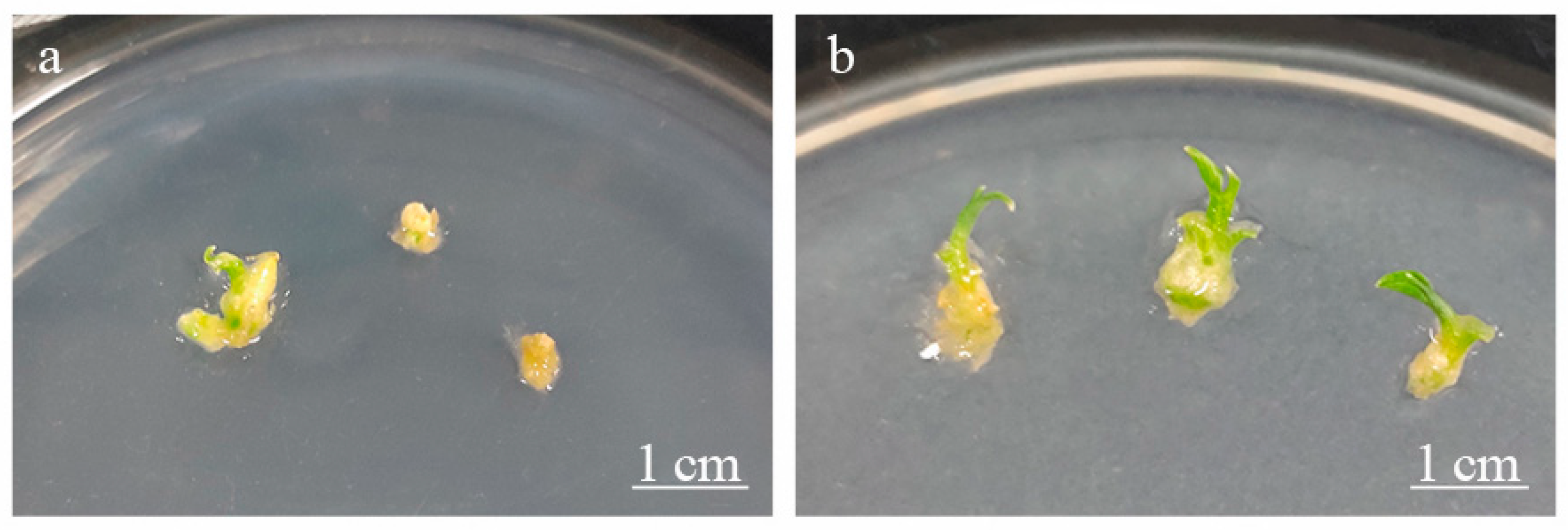
3.4. Effect of AsA and GSH

The treatments involving PVS2 and the addition of 0.1 and 0.2 mM AsA enhanced the survival and regrowth rates of both cryopreserved and treated control shoot tips (Table 3). Notably, the maximum regrowth rate of the cryopreserved shoot tips reached 66.67% with the PVS2 plus 0.1 mM AsA treatment, which was considerably higher than that of the other treatments. The cryopreserved shoot tips treated with PVS2 alone (PVS2 − AsA) exhibited slow and inconsistent growth (Figure 4a). However, those treated with PVS2 and 0.1 mM AsA (PVS2 + AsA) exhibited rapid and uniform growth (Figure 4b). Following the treatment of cryopreserved shoot tips with PVS2 with 0.3 mM AsA, both the survival and regrowth rates were inferior to those associated with the treatment using PVS2 with 0.1 mM AsA. Furthermore, treatments of PVS2 with 0.4 to 0.8 mM GSH significantly diminished the survival and regrowth rates of the shoot tips. The ‘Yunnan Xiaohuangjiang’ plantlet was regenerated from a cryopreserved shoot tip eight weeks after cryopreservation (Figure 1i). After recovery culture, the regenerated plantlets (1 cm in length) were transferred to subculture media for another eight-week period to achieve a vigorous and robust growth state for transplanting. The plantlet, which had developed roots, exhibited healthy growth (Figure 1j). Over 95% of the plantlets were successfully acclimatized in soil within greenhouse conditions. The morphologies of the plants that regenerated from cryopreserved shoot tips were the same as those from in vitro cultures (Figure 1k).

3.5. Histological and Ultrastructural Observations

Shoot tips, which were freshly excised as the positive control, demonstrated that living cells exhibited dense cytoplasm that was stained by TB and nuclei that were well-preserved, with nucleoli enclosed within them (Figure 5a). In contrast, shoot tips, which were freshly cut, submerged in LN, and post-cultured for 1 day as the negative control, revealed that dead cells displayed weaker TB-staining, characterized by cell rupture and heavily condensed nuclei (Figure 5b). Following cryopreservation, shoot tips treated with PVS − AsA showed a small amount of living cells were noticed in the AD (Figure 5c), which were characterized by a large nucleo-cytoplasmic ratio and small vacuoles (Figure 6a). A significant number of dead cells alongside a small proportion of living cells in LPs and the basal parts of the shoot tips which were far from the AD (Figure 5c), exhibiting cell shrinkage, cytoplasmic exudation and a lack of organelles (Figure 6b). In contrast, shoot tips that were treated with PVS + AsA exhibited a significantly greater number of living cells in both the AD, LPs, and the basal parts of the shoot tips (Figure 5d). which contained small vacuoles (Figure 6c).

3.6. Analysis of Genetic Stability

3.6.1. ISSR

Out of the forty ISSR primers employed in the experiment, seven specific primers yielded a total of 23 bands in the plantlets that were regenerated from the cryopreserved shoot tips of ‘Yunnan Xiaohuangjiang’. These bands exhibited clarity, strength, and reproducibility. The number of bands generated by each primer varied from 1 to 5, producing an average of 3.3 bands per primer. Compared to non-cryopreserved samples, the regenerated plantlets originating from cryopreserved shoot tips exhibited no polymorphic bands (Table 4, Figure 7, Figure S1).

3.6.2. FCM

FCM analysis revealed similar patterns of ploidy levels in nuclei extracted from leaves of in vitro cultures and regenerated shoots originating from cryopreserved shoot tips of Z. officinale ‘Yunnan Xiaohuangjiang’ (Figure 8).

4. Discussion

There have been relatively few reports on the cryopreservation of Z. officinale. Techniques such as encapsulation-vitrification, encapsulation-dehydration [10], and vitrification [10,11] have been utilized. Yamuna et al. [10] reported that the vitrification resulted in an 80% regrowth rate, compared to 66% for encapsulation vitrification and 41% for encapsulation-dehydration. Additionally, Du et al. [11] found that the vitrification method yielded a survival rate of 57.7%. Notably, the droplet-vitrification technique, initially documented by Kartha et al. [14], has since undergone further refinement and modification by Leunufna and Keller, contributing to the advancement and optimization of the method within the field of cryopreservation and related biotechnological applications [36,37,38,39], which offers the primary advantage of rapid cooling and warming rates achieved through droplet freezing. This method, combined with the properties of vitrification solutions, results in high success rates for the cryopreservation of shoot tips [40,41]. To date, droplet-vitrification has been extensively employed in the cryogenic preservation of plant germplasm resources. However, the application of droplet-vitrification for the cryopreservation of Z. officinale has not been previously reported. In the present study, we established a droplet-vitrification protocol for the shoot tips of Chinese genotype Z. officinale ‘Yunnan Xiaohuangjiang’. The survival and regrowth rates were 96.67% and 66.67%, respectively.
In the previous studies on cryopreserving Z. officinale, shoot tips with a length of 0.5–1.0 mm and containing 3–4 LPs [10,11] were used for cryopreservation. In the present study, shoot tips excised from shoots induced from nodal segments, as described by Wang et al. [23], served as a source of material for shoot tip isolation. Several studies have explored the cryopreservation of shoot tips that are derived from shoots induced from nodal segments, including research on Solanum tuberosum [42], Indigofera tinctoria [43], Cleome rosea [44], Vitis [45], and Chrysanthemum morifolium [46], among others. The advantages of this method include not only the availability of a large quantity of material but, most importantly, shoot tip uniformity [47].
Successful shoot tip cryopreservation involves several critical steps, including in vitro culture, shoot tip preculture, cryoprotection, dehydration, exposure to LN, and post-culture for plant recovery [48,49]. Preculture typically entails cultivating shoot tips on a medium with elevated concentrations of sugars or sugar alcohols for variable durations to enhance the cells’ ability to withstand dehydration and the subsequent freezing process in LN [50]. When the shoot tips are exposed to high sugar or sugar alcohol levels, the physiological tolerance of shoot tips during dehydration is increased [51]. An excessively low sugar or sugar alcohol concentration may fail to yield the desired survival and regrowth rates. Conversely, an overly high sugar or sugar alcohol concentration has the potential to induce excessive water loss in cells, thereby leading to cellular damage and, in severe cases, even cell death. So, the concentration of sugars or sugar alcohols and the preculture duration must be optimized, which can be genotype-dependent. For instance, Kim et al. [52] reported that the ideal preculture condition for garlic (Allium) cryopreservation comprised MS medium with 0.1–0.3 M sucrose for 2–4 days. Similarly, Wang et al. [53] determined that the optimal preculture condition for blueberry (Vaccinium) using the droplet-vitrification procedure was woody plant medium (WPM) [54], which was supplemented with 0.3 M sucrose and maintained for 1 day. For sweet potato (Ipomoea batatas), the optimal preculture conditions in the droplet-vitrification procedure were reported as liquid MS with 10% sucrose and 17.5% sucrose added for 31 and 17 h, respectively [55]. In the vitrification procedure for Z. officinale, Yamuna et al. [10] identified the optimal preculture condition as liquid MS containing 0.3 M sucrose with a duration of 3 d. Du et al. [11] found that MS containing 0.5 M sucrose with a duration of 2 d was the ideal preculture condition. The present study established the optimal preculture condition as MS containing 0.25 M sucrose with a duration of 1 d. The discrepancies observed may be attributed to differences in methodologies employed and variations in genotypes.
PVS are crucial for cryopreservation techniques based on vitrification as treatment with these solutions enables cells to form a glassy state instead of being damaged by ice crystals when they are immersed in LN [56]. PVS2, a concentrated vitrification solution designed for dehydration [24], is widely employed in plant germplasm cryopreservation [7,18]. However, DMSO, a component of PVS2, is known to be toxic to cells [57]. Therefore, exact control of the exposure duration to PVS2 is essential for the droplet-vitrification procedure. The ideal exposure time to PVS2 depending on plant species. For instance, six genotypes of Chrysanthemum morifolium were dehydrated with PVS2 at the temperature of 0 °C for 30 min [46]. Similarly, six Vitis genotypes were treated with half-strength PVS2 at a temperature of 0 °C for 30 min and subsequently to full-strength PVS2 with a duration of 50 min [58]. Ananas comosus was exposed to PVS2 at 0 °C for 45 min [59], while four genotypes of Helianthus tuberosus were desiccated using PVS2 at 0 °C for 15 min [60]. In the vitrification procedure for Z. officinale, Yamuna et al. [10] reported that the optimal duration of PVS2 was 40 min at 25 °C, resulting in a regrowth rate of 80%. Conversely, Du et al. [11] reported that the optimal PVS2 treatment involved 60% PVS2 for 5 min at room temperature, with 100% PVS2 at the temperature of 0 °C for 30 min. In the present study, Z. officinale ‘Yunnan Xiaohuangjiang’ was desiccated using PVS2 at 0 °C for 20 min, yielding a shoot regrowth rate of 66.67%. This optimal PVS2 duration in the present study was shorter than that reported in the two previous studies.
Studies focusing on establishing cryopreservation protocols make it crucial to screen recovery media for direct shoot regrowth without the formation of callus or abnormal regrowth [61]. Three recovery media were applied to cryopreserved Chrysanthemum morifolium shoot tips. Shoot tips cultured on SRM1 developed callus, resulting in a shoot regrowth rate of only 1.7%. Shoot tips cultured on SRM2 produced leaves exclusively, with no shoot development observed. In contrast, shoot tips cultured on SRM3 successfully regenerated shoots directly without callus formation [46]. The present study identified SRM4, which was composed of MS medium with 0.1 mg·L−1 GA3 added, as the optimal recovery medium, corroborating the findings of Wang et al. [46]. Martín et al. [62] indicated that the cryopreserved shoot tips of mint genotype ‘MEN 186’ cultured on recovery media containing certain plant growth regulators, such as 6BA and NAA, were induced to form calli. The genetic stability of the basal callus and callus was 44% and 30%, respectively. Notably, in the present study, SRM4, containing only GA3, significantly mitigated the risk of genetic variation. Hedden et al. [63] reported that gibberellins promote cell elongation and division, thereby facilitating plant organ growth and development. Recovery media containing GA3 have been proven beneficial for the cryopreserved shoot tips of several plant species, such as Chrysanthemum morifolium [46], Helianthus tuberosus [60], Citrus reticulata [64], among others.
Exogenous antioxidants are defined as molecules or compounds that have the ability to delay, prevent, or get rid of damage caused by oxidative stress [65]. The use of exogenous antioxidants has been shown to improve the success of cryopreservation [66,67,68]. These antioxidants can be categorized into non-enzymatic and enzymatic types [45], both of which play crucial roles in regulating reactive oxygen species (ROS) production [69]. Non-enzymatic antioxidants, including GSH, AsA, abscisic acid (ABA), tocopherol (VE), and melatonin, are frequently utilized in cryopreservation protocols [70,71]. Several studies have documented that incorporating varying concentrations of AsA or GSH into cryopreservation steps, such as preculture, loading, and PVS2, can enhance the efficiency of cryopreservation [45,72,73]. Ren et al. [74] reported that the supplement of 1 mM AsA into PVS2 led to a notable enhancement in the regeneration efficiency of Arabidopsis thaliana seedlings after cryopreservation. In the present study, the treatment of PVS with 0.1 mM AsA was identified as the optimal condition, significantly improving the regrowth rate of cryopreserved shoot tips. This finding was in consonance with Ren’s report, but the concentrations of AsA were different. In the present study, treatment with PVS2 containing 0.3 mM AsA did not improve the survival or regrowth rates of these shoot tips. Jaouad and Torsten [75] noted that high concentrations of antioxidants can disrupt redox balance and lead to cellular dysfunction, a finding that aligns with our results. Furthermore, treatments with PVS2 containing 0.4 to 0.8 mM GSH reduced both the survival and regrowth rates of the shoot tips. These findings suggest that exogenous antioxidants’ effects on cryopreservation are both dose-dependent and specific to plant species.
In the present study, we identified that cryopreserved shoot tips with PVS + AsA exhibited significantly fewer dead cells in the top layers of AD, LPs, and the basal parts of the shoot tips than those treated with PVS − AsA. TEM observations elucidated the reasons for the markedly higher regrowth rate of cryopreserved shoot tips treated with PVS + AsA. Specifically, living cells characterized by small vacuoles in the LPs were observed, and the basal part of the shoot tips of cryopreserved shoot tips treated with PVS + AsA. In contrast, shoot tips treated with PVS − AsA displayed a substantial number of dead cells in the LPs and the basal parts of the shoot tips, indicative of cell shrinkage, cytoplasmic exudation, and a lack of organelles. These findings were consistent with those of the histological observations.
The genetic stability of regenerated individuals is crucial for the clonal cryopreservation of plant germplasm [76]. Several studies have reported that genetic variations observed in shoots regenerated from cryopreserved shoot tips may be linked to factors other than the freezing process in LN [62,77,78]. Therefore, evaluating the genetic stability of regenerants following cryopreservation is an essential step. Molecular techniques including ISSR, amplified fragment length polymorphism (AFLP), random amplified polymorphic DNA (RAPD), simple sequence repeats (SSR), and cell analysis methods like FCM are commonly employed to evaluate the genetic stability of important plant species [79,80,81,82]. Numerous studies have integrated one or two molecular methods with cell analysis to assess the genetic stability of shoots regenerated from cryopreserved shoot tips, thereby enhancing the reliability of the results [46,57,83]. In the present study, we employed ISSR and FCM to evaluate the genetic stability of regenerants following the droplet-vitrification of Z. officinale ‘Yunnan Xiaohuangjiang’, and found no variation.
In the present study, a single genotype of ginger was investigated, thereby lacking the verification of the established droplet-vitrification across multiple genotypes. The applicability of incorporating AsA with the droplet-vitrification protocol for the preservation of Zingiberacease germplasm necessitates further in-depth exploration. In addition, research on applying droplet-vitrification to virus elimination in Zingiberaceae plants remains unconducted. Our results elucidated the alterations in the number of survival cells and the intracellular inclusions from the histological and TEM perspectives for survival and regrowth rates increase. Further studies on the changes in genes and metabolic pathways from the transcriptomic and metabolomic levels would be necessary to analyze the mechanism comprehensively.

5. Conclusions

An efficient droplet-vitrification cryopreservation protocol has been developed for the shoot tips of Z. officinale ‘Yunnan Xiaohuangjiang’. Histological observations and ultrastructural analyses elucidate why cryopreserved shoot tips treated with PVS + AsA exhibited the highest regrowth rate. There were no genetic variations detected in the regenerated shoots from cryopreserved shoot tips, as assessed by both ISSR and FCM techniques. The results presented here supply valuable technical support for founding a cryo-banking system for Zingiber germplasm.

Supplementary Materials

The following supporting information can be downloaded at: https://www.mdpi.com/article/10.3390/horticulturae11030283/s1. Figure S1. Assessment of genetic stability by ISSR in shoots regenerated from cryopreserved shoot tips and in vitro cultures of Z. officinale ‘Yunnan Xiaohuangjiang’. (a) primer SPS05, (b). primer SPS06, (c) primer UBC816, (d) primer UBC851, lines 1–4 in vitro cultures, lanes 5–16 shoots regenerated from cryopreserved shoot tips.

Author Contributions

Conceptualization, R.-R.W., X.L. and J.L.; methodology, R.-R.W., X.L. and R.-F.S.; software, R.-R.W. and R.-F.S.; validation, X.L., R.-F.S., J.-J.H. and Y.Z.; formal analysis, R.-R.W., X.L. and J.-J.H.; investigation, R.-R.W., X.L., R.-F.S. and Y.Z.; resources, R.-R.W. and J.L.; data curation, R.-R.W. and J.L.; writing—original draft preparation, R.-R.W. and J.L.; writing—review and editing, X.-K.S. and X.-D.C.; visualization, X.-K.S.; supervision, J.L.; project administration, R.-R.W. and J.L.; funding acquisition, R.-R.W. and X.-D.C. All authors have read and agreed to the published version of the manuscript.

Funding

This research was funded by Natural Science Foundation of Sichuan Province (2022NSFSC1626) and the Key Research and Development Project of Hubei Province (2022BBA0061).

Data Availability Statement

Data are contained within the article.

Conflicts of Interest

The authors declare no conflicts of interest.

Abbreviations

The following abbreviations are used in this manuscript:
MSMurashige and Skoog
PVSPlant vitrification solutions
PVS2Plant vitrification solution 2
AsAAscorbic acid
LNLiquid nitrogen
SRMShoot recovery medium
GA3Gibberellic acid 3
ADApical dome
LPLeaf primordia
ISSRInter-simple sequence repeat
FCMFlow cytometry
6BAP6-benzyl-aminopurine
NAAα-naphthaleneacetic acid
DMSODimethylsulfoxide
TDZThidiazuron
KT6-furfurylaminopurine
GSHGlutathione
TBToluidine blue
WPMWoody plant medium
TEMTransmission electron microscope
RAPDRandom amplified polymorphic DNA
SSRSimple sequence repeats
AFLPAmplified fragment length polymorphism
rpmRevolutions per minute
TETris-EDTA
DNADeoxyribonucleic acid
PCRPolymerase chain reaction
dNTPDeoxy-ribonucleoside triphosphate
TBETris-borate-EDTA
UVultraviolet

References

  1. Yusuff, O.; Rafii, M.Y.; Fatai, A.; Murugesu, S.; Chukwu, S.; Salisu, M.A.; Fagbohun, I.; Kolawole, T.; Kadar, A.I. Genetic diversity and utilization of ginger (Zingiber officinale) for varietal improvement: A review. AIMS Agric. Food 2024, 9, 183–208. [Google Scholar]
  2. Moghaddasi, M.S. Ginger (Zingiber officinale): A review. J. Med. Plant Res. 2012, 6, 4255–4258. [Google Scholar]
  3. Priyanka, V.; Kumar, R.; Dhailiwal, I.; Kaushik, P. Germplasm conservation: Instrumental in agricultural biodiversity—A review. Sustainability 2021, 13, 6743. [Google Scholar] [CrossRef]
  4. Benelli, C. Plant cryopreservation: A look at the present and the future. Plants 2021, 10, 2744. [Google Scholar] [CrossRef]
  5. Wang, Q.C.; Panis, B.; Engelmann, F.; Lambardi, M.; Valkonen, J.P.T. Cryotherapy of shoot tips: A technique for pathogen eradication to produce healthy planting materials and prepare the healthy plant genetic resources for cryopreservation. Ann. Appl. Biol. 2009, 154, 351–363. [Google Scholar] [CrossRef]
  6. Reed, B.M. Plant Cryopreservation: A Practical Guide; Springer Science +Businuss Medium: New York, NY, USA, 2008; p. 7. [Google Scholar]
  7. Engelmann, F. Plant cryopreservation: Progress and prospects. Vitr. Cell. Dev. Biol.-Plant 2004, 40, 427–433. [Google Scholar] [CrossRef]
  8. Wilkinson, T.; Wetten, A.; Prychid, C.; Fay, M.F. Suitability of cryopreservation of the long-term storage of rare and endangered plant species: A case history for Cosmos atrosanguineus. Ann. Bot. 2003, 91, 65–74. [Google Scholar] [CrossRef] [PubMed]
  9. Villalobos-Olivera, A.; Lorenzo-Feijoo, J.C.; Quintana-Bernabé, N.; Leiva-Mora, M.; Bettoni, J.C.; Maitínez-Montero, M.E. Morpho-anatomical and physiological assessments of cryo-derived pineapple plants (Ananas comosus var. comosus) after acclimatization. Horticulturae 2023, 9, 841. [Google Scholar] [CrossRef]
  10. Yamuna, G.; Sumathi, V.; Geetha, S.P.; Praveen, K.; Swapna, N.; Nirmal Babu, K. Cryopreservation of in vitro grown shoots of ginger (Zingiber officinale Rosc.). CryoLetters 2007, 28, 241–252. [Google Scholar]
  11. Du, L.S.; Feng, J.J.; Li, X.H.; Zhao, F.Z.; Wu, Y.Q.; Yu, H.T. Ginger vitrification cryopreservation technology. North. Horticult. 2014, 13, 123–125, (in Chinese with English abstract). [Google Scholar]
  12. Ozudogru, E.A.; Kirdok, E.; Kaya, E.; Capuana, M.; Benelli, C.; Engelmann, F. Cryopreservation of redwood (Sequoia sumpervirens) in vitro buds using vitrification-based techniques. CryoLetter 2011, 32, 99–110. [Google Scholar]
  13. Wang, B.; Wang, R.R.; Li, J.W.; Ma, Y.L.; Sheng, W.M.; Li, M.F.; Wang, Q.C. Development of three vitrification-based cryopreservation procedures of shoot tips for China’s potato. CryoLetter 2013, 34, 369–380. [Google Scholar]
  14. Kartha, K.K.; Leung, N.L.; Mroginski, L.A. In vitro growth responses and plant regeneration from cryopreserved meristems of cassava (Manihot esculenta Crantz). Z. Pflanzenphysiol. 1982, 107, 133–140. [Google Scholar] [CrossRef]
  15. Sakai, A.; Engelmann, F. Vitrification, encapsulation-vitrification and droplet-vitrification: A review. CryoLetters 2007, 28, 151–172. [Google Scholar]
  16. Kim, H.H.; Lee, Y.G.; Shin, D.J.; Ko, H.C.; Gwag, J.G.; Cho, E.G.; Engelmann, F. Development of alternative plant vitrification solutions in droplet-vitrification procedures. CryoLetters 2009, 30, 320–334. [Google Scholar] [CrossRef] [PubMed]
  17. Cui, M.D.; Zhan, T.J.; Yang, H.Y.; Han, H.X.; Liu, L.F.; Yi, Y. Droplet generation, vitrification, and warming for cell cryopreservation: A review. ACS Biomater. Sci. Eng. 2023, 9, 1151–1163. [Google Scholar] [CrossRef]
  18. Wang, M.R.; Lambardi, M.; Engelmann, F.; Pathirana, R.; Panis, B.; Volk, G.M.; Wang, Q.C. Advances in cryopreservation of in vitro-derived propagules: Technologies and explant sources. Plant Cell Tiss. Org. 2021, 144, 7–20. [Google Scholar] [CrossRef]
  19. Wang, M.R.; Bi, W.L.; Bettoni, J.C.; Zhang, D.; Volk, G.M.; Wang, Q.C. Shoot tip cryotherapy for plant pathogen eradication. Plant Pathol. 2022, 71, 1241–1254. [Google Scholar] [CrossRef]
  20. Bettoni, J.C.; Bonnart, R.; Volk, G.M. Challenges in implementing plant shoot tip cryopreservation technologies. Plant Cell Tiss. Org. 2021, 144, 21–34. [Google Scholar] [CrossRef]
  21. Yang, Q.S.; Wei, X.Y.; Yang, Q.; Wang, Y.M.; Tao, Y.H. Study on rapid propagation technology of ginger landraces in Yunnan. Chin. J. Trop. Agric. 2022, 42, 28–32, (in Chinese with English abstract). [Google Scholar]
  22. Murashige, T.; Skoog, F. A revised medium for rapid growth and bioassays with tobacco cultures. Physiol. Plant 1962, 15, 473–497. [Google Scholar] [CrossRef]
  23. Wang, R.R.; Liu, C.C.; Wang, L.J.; Yuan, M.Y.; Li, J.; Tang, Z.K. An improved micropropagation via nodal segments of Zingiber officinale. Eur. J. Hortic. Sci. 2021, 86, 21–28. [Google Scholar] [CrossRef]
  24. Sakai, A.; Kobayashi, S.; Oiyama, I. Cryopreservation of nucellar cells of navelorange (Citrus sinensis Osb. var. brasiliensis Tanaka) by vitrification. Plant Cell Rep. 1990, 9, 30–33. [Google Scholar] [CrossRef]
  25. Wang, S.M.; Luo, L.S.; Hou, Y.H.; Leng, J.G.; Li, D.W. Establishment of virus-free and rapid propagation system for tissue culture of Guizhou ginger. Guizhou Agr. Sci. 2021, 49, 98–102, (in Chinese with English abstract). [Google Scholar]
  26. Wang, H.; Lin, G.Y.; Li, X.K. Tissue culture and rapid propagation of Curcuma wenyujin Y.H.Chen et C. Ling. Plant Physiol. J. 2007, 3, 509–510, (in Chinese with English abstract). [Google Scholar]
  27. Wang, Q.C.; Valkonen, J.P.T. Efficient elimination of sweetpotato little leaf phytoplasma from sweetpotato by cryotherapy of shoot tips. Plant Pathol. 2008, 57, 338–347. [Google Scholar] [CrossRef]
  28. Sakai, W. Simple method for different staining of paraffin embedded plant material using toluidine blue O. Stain Technol. 1973, 48, 247–249. [Google Scholar] [CrossRef] [PubMed]
  29. Reynolds, E.S. The use of lead at high xylem ray parenchyma cells of Red Osier dogwood (Cornus sericea L.) to freezing stress. Plant Physiol. 1963, 104, 737–746. [Google Scholar]
  30. Porebski, S.; Bailey, L.G.; Baum, B.R. Modification of a CTAB DNA extraction protocol for plant containing high polysaccharides and polyphenol component. Plant Mol. Biol. Rep. 1997, 15, 8–15. [Google Scholar] [CrossRef]
  31. Kizhakkayil, J.; Sasikumar, B. Genetic diversity analysis of ginger (Zingiber officinale Rosc.) germplasm based on RAPD and ISSR markers. Sci. Hortic. 2010, 125, 73–76. [Google Scholar] [CrossRef]
  32. Mohanty, S.; Panda, M.K.; Sahoo, S.; Nayak, S. Micropropagation of Zingiber rubens and assessment of genetic stability through RAPD and ISSR markers. Biol. Plant. 2011, 55, 16–20. [Google Scholar] [CrossRef]
  33. Ray, T.; Roy, S.C. Phylogenetic relationship between members of Amaranthaceae and Chenopodiaceae of lower gangetic plains using RAPD and ISSR markers. Bangl. J. Bot. 2007, 36, 21–28. [Google Scholar] [CrossRef]
  34. Huang, H.; Tong, Y.; Zhang, Q.J.; Gao, L.Z. Genome size variation among and within Camellia species by using flow cytometric analysis. PLoS ONE 2013, 8, e64981. [Google Scholar] [CrossRef] [PubMed]
  35. Galbraith, D.W.; Harkins, K.R.; Maddox, J.M.; Ayres, N.M.; Sharma, D.P.; Firoozabady, E. Rapid flow cytometric analysis of the cell-cycle in intact plant-tissues. Science 1983, 220, 1049–1051. [Google Scholar] [CrossRef] [PubMed]
  36. Leunufna, S.; Keller, E.R.J. Investigating a new cryopreservation protocol for yam (Discorea spp.). Plant Cell Rep. 2003, 21, 1159–1166. [Google Scholar] [CrossRef]
  37. Kim, H. Optimizing the droplet-vitrification procedure by balancing the cryoprotection and cytotoxicity of alternative plant vitrification solutions based on the nature of donor plant vigor. Plants 2023, 12, 4040. [Google Scholar] [CrossRef]
  38. Lee, H.; Choi, B.; Oh, S.; Park, H.; Popova, E.; Paik, M.J.; Kim, H. Dynamics of organic acids during the droplet-vitrification cryopreservation procedure can be a signature of oxidative stress in Pogostemon yatabeanus. Plants 2023, 12, 3489. [Google Scholar] [CrossRef]
  39. Delgado-Aceves, L.; Corona, S.; Martin-Castro, U.R.; Rascó-Díaz, M.P.; Portillo, L.; Gutiérrez-Mora, A.; González-Arnao, M.T. Comparative studies for cryopreservation of Agave shoot tips by droplet-vitrification. Plants 2024, 13, 2609. [Google Scholar] [CrossRef]
  40. Panis, B. Sixty years of plant cryopreservation: From freezing hardy mulberry twigs to establishing reference crop collections for future generations. Acta Hortic. 2019, 1234, 1–7. [Google Scholar] [CrossRef]
  41. Matsumoto, T. Cryopreservation of plant genetic resources: Conventional and new methods. Rev. Agric. Sci. 2017, 5, 13–20. [Google Scholar] [CrossRef]
  42. Hirai, D.; Sakai, A. Cryopreservation of in vitro-grown meristems of potato (Solanum tuberosum L.) by encapsulation-vitrification. Potato Res. 1999, 42, 153–160. [Google Scholar] [CrossRef]
  43. Nair, D.S.; Reghunath, B.R. Cryoconservation and regeneration of axillary shoot meristems of Indigofera tinctoria (L.) by encapsulation-dehydration technique. Vitr. Cell. Dev. Biol.-Plant 2009, 45, 565–573. [Google Scholar] [CrossRef]
  44. Da Silva Cordeiro, L.; Simões-Gurgel, C.; Albarello, N.; Engelmann, F. Cryopreservation of in vitro-grown shoot tips of Cleome rosea Vahl (Cleopmaceae) using the V cryo-plate technique. Vitr. Cell. Dev. Biol.-Plant 2015, 51, 688–695. [Google Scholar] [CrossRef]
  45. Bettoni, J.C.; Bonnart, R.; Shepherd, A.; Kretzschmar, A.A.; Volk, G.M. Cryopreservation of grapevine (Vitis spp.) shoot tips from growth chamber-sourced plants and histological observation. Vitis 2019, 58, 71–78. [Google Scholar]
  46. Wang, R.R.; Gao, X.X.; Chen, L.; Huo, L.Q.; Li, M.F.; Wang, Q.C. Shoot recovery and genetic integrity of Chrysanthemum morifolium shoot tips following cryopreservation by droplet-vitrification. Sci. Hortic. 2014, 176, 330–339. [Google Scholar] [CrossRef]
  47. Bettoni, J.C.; Marković, Z.; Bi, W.L.; Volk, G.M. Grapevine shoot tip cryopreservation and cryotherapy: Secure storage of disease-free plants. Plants 2010, 10, 2190. [Google Scholar] [CrossRef]
  48. Benson, E.E. Cryopreservation of phytodiversity: A critical appraisal of theory & practice. Crit. Rev. Plant Sci. 2008, 27, 141–219. [Google Scholar]
  49. Wang, B.; Wang, R.R.; Cui, Z.H.; Li, J.W.; Bi, W.L.; Wang, Q.C. Potential applications of cryobiotechnology to plant genetic transformation and pathogen eradication. Biotechnol. Adv. 2014, 32, 583–595. [Google Scholar] [CrossRef]
  50. Li, J.W.; Ozudogru, E.A.; Li, J.; Wang, M.R.; Bi, W.L.; Lambardi, M.; Wang, Q.C. Cryobiotechnology of forest trees: Recent advances and future prospects. Biodivers. Conserv. 2018, 27, 795–814. [Google Scholar] [CrossRef]
  51. Leslie, S.B.; Israeli, E.; Lighthart, B.; Crowe, J.H.; Crowe, L.M. Trehalose and sucrose protect both membranes and protein in intact bacteria during drying. Appl. Environ. Microbiol. 1995, 61, 3592–3597. [Google Scholar] [CrossRef]
  52. Kim, H.H.; Lee, J.K.; Yoon, J.W.; Li, J.J.; Engelmann, F. Cryopreservation of garlic bulbil primordia by the droplet-vitrification procedure. CryoLetters 2006, 27, 143–153. [Google Scholar]
  53. Wang, L.Y.; Li, Y.D.; Sun, H.Y.; Liu, H.G.; Tang, X.D.; Wang, Q.C.; Zhang, Z.D. An efficient droplet-vitrification cryopreservation for valuable blueberry germplasm. Sci. Hortic. 2017, 219, 60–69. [Google Scholar] [CrossRef]
  54. Lloyd, G.; McCown, B. Commercially feasible micropropagation of mountain laurel, Kalmia latifolia, by use of shoot-tip culture. Comb. Proc.-Int. Plant Propagators’ Soc. 1980, 30, 421–427. [Google Scholar]
  55. Park, S.U.; Kim, H.H. Cryopreservation of sweet potato shoot tips using a droplet-vitrification procedure. CryoLetter 2015, 36, 344–352. [Google Scholar]
  56. Teixeira, A.S.; Faltus, M.; Zámeámečník, J.; González-Benito, M.E.; Molina-García, A.D. Glass transition and heat capacity behaviors of plant vitrification solutions. Thermochim. Acta 2014, 593, 43–49. [Google Scholar] [CrossRef]
  57. Roque-Borda, C.A.; Kulus, D.; Vacaro de Souza, A.; Kaviani, B.; VaVicente, E.F. Cryopreservation of agronomic plant germplasm using vitrification-based methods: An overview of selected case studies. Int. J. Mol. Sci. 2021, 22, 6157. [Google Scholar] [CrossRef]
  58. Bi, W.L.; Hao, X.Y.; Cui, Z.H.; Volk, Z.H.; Wang, Q.C. Droplet-vitrification cryopreservation of in vitro-grown shoot tips of grapevine (Vitis spp.). Vitr. Cell. Dev. Biol.-Plant 2018, 54, 590–599. [Google Scholar] [CrossRef]
  59. Souza, F.V.D.; Kaya, E.; de Jesus Vieira, L.; de Souza, E.H.; de Oliveira Amorim, V.B.; Skogerboe, D.; Skogerboe, D.; Matsumoto, T.; Alves, A.A.C.; de Silva Ledo, C.A.; et al. Droplet-vitrification and morphohistological studies of cryopreserved shoot tips of cultivated and wild pineapple genotypes. Plant Cell Tiss. Org. 2016, 124, 351–360. [Google Scholar] [CrossRef]
  60. Zhang, J.M.; Han, L.; Lu, X.X.; Volk, G.M.; Xin, X.; Yin, G.K.; He, J.J.; Wang, L.; Chen, X.L. Cryopreservation of Jerusalem artichoke cultivars using an improved droplet-vitrification method. Plant Cell Tiss. Org. 2017, 128, 577–587. [Google Scholar] [CrossRef]
  61. Engelmann, F. Use of biotechnologies for the conservation of plant biodiversity. Vitr. Cell. Dev. Biol.-Plant 2011, 47, 5–16. [Google Scholar] [CrossRef]
  62. Martín, C.; Kremer, C.; González, I.; González-Benito, M.E. Influence of the cryopreservation technique, recovery medium and genotype on genetic stability of mint cryopreserved shoot tips. Plant Cell Tiss. Org. 2015, 122, 185–195. [Google Scholar] [CrossRef]
  63. Hedden, P.; Sponsel, V. A century of gibberellin research. J. Plant Growth Regul. 2015, 34, 740–760. [Google Scholar] [CrossRef] [PubMed]
  64. Avenido, R.A.; Endonela, L.E.; Patena, L.F.; Barba, R.C. Developing plant regeneration systems for in vitro conservation of mandarin (Citrus reticulata) and pummelo (Citrus maxima). Acta Hortic. 2005, 694, 133–136. [Google Scholar] [CrossRef]
  65. Halliwell, B. Reactive species and antioxidants. Plant Physiol. 2006, 141, 312–322. [Google Scholar] [CrossRef]
  66. Ren, L.; Zhang, D.; Shen, X.H.; Reed, B.M. Antioxidants and anti-stress compounds improve the survival of cryopreserved Arabidopsis seedlings. II Int. Symp. Plant Cryopreserv. 2013, 1039, 57–62. [Google Scholar] [CrossRef]
  67. Mathew, L.Y.; Burritt, D.J.; McLachalan, A.; Pathirana, R. Combined pre-treatments enhance antioxidant metabolism and improve survival of cryopreserved kiwifruit shoot tips. Plant Cell Tiss. Org. 2019, 138, 193–205. [Google Scholar] [CrossRef]
  68. Ren, R.F.; Li, Z.D.; Zhang, L.L.; Zhou, H.; Jiang, X.R.; Liu, Y. Enzymatic and nonenzymatic antioxidant systems impact the viability of cryopreserved Paeonia suffruticosa pollen. Plant Cell Tiss. Org. 2021, 144, 233–246. [Google Scholar] [CrossRef]
  69. Halder, T.; Upadhyaya, G.; Basak, C.; Das, A.; Chakraborty, C.; Ray, S. Dehydrins impact protection against oxidative stress in transgenic tobacco plants. Front. Plant Sci. 2018, 9, 136. [Google Scholar] [CrossRef]
  70. Gill, S.S.; Tuteja, N. Reactive oxygen species and antioxidant machinery in abiotic stress tolerance in crop plants. Plant Physiol. Bioch. 2010, 48, 909–930. [Google Scholar] [CrossRef]
  71. Nawaz, M.A.; Huang, Y.; Bie, Z.; Ahmed, W.; Reiter, R.J.; Niu, M.; Hameed, S. Melatonin: Current status and future perspectives in plant science. Front. Plant Sci. 2016, 6, 1230. [Google Scholar] [CrossRef]
  72. Xing, X.D.; Liu, M.; Zhou, R.; Jiang, F.; Bai, Y.; Wei, H.Y.; Zhang, D.; Wei, J.J.; Wu, Z. Ascorbic acid addition during dehydration improves garlic shoot tips cryopreservation but does not affect viral load. Cryobiology 2022, 107, 64–73. [Google Scholar] [CrossRef]
  73. Zhang, Y.Y.; Deng, S.; Lin, H.F.; Chu, Y.X.; Huang, J.Y.; Li, S.G.; Lin, F.Z.; Zhang, S.M.; Jiang, W.L.; Ren, L.; et al. The development of a procedure for the cryopreservation of the callus of Anthurium andraeanum by vitrification. Plants 2024, 13, 3106. [Google Scholar] [CrossRef] [PubMed]
  74. Ren, L.; Zhang, D.; Chen, G.Q.; Reed, B.M.; Shen, X.H.; Chen, H.Y. Transcriptomic profiling revealed the regulatory mechanism of Arabidopsis seedlings response to oxidative stress from cryopreservation. Plant Cell Tiss. Org. 2015, 34, 2161–2178. [Google Scholar] [CrossRef]
  75. Jaouad, B.; Torsten, B. Exogenous antioxidants-double-edged swords in cellular redox state: Health beneficial effects at physiologic doses versus deleterious effects at high doses. Oxidative Med. Cell. Longev. 2010, 3, 228–237. [Google Scholar]
  76. Ozudogru, E.A.; Kaya, E.; Kirdok, E.; Issever-Ozturk, S. In vitro propagation from young and mature explants of thyme (Thymus vulgaris and T. loongicaulis) resulting in genetically stable shoots. Vitr. Cell. Dev. Biol.-Plant 2011, 47, 309–320. [Google Scholar] [CrossRef]
  77. Martín, C.; González-Benito, M.E. Survival and genetic stability of Dendranthema grandiflora Tzvelev shoot apices after cryopreservation by vitrification and encapsulation-dehydration. Cryobiology 2005, 51, 281–289. [Google Scholar] [CrossRef]
  78. Ai, P.F.; Lu, L.P.; Song, J.J. Cryopreservation of in vitro-grown shoot tips of Rabdosia rubescens by encapsulation-dehydration and evaluation of their genetic stability. Plant Cell Tiss. Org. 2012, 108, 381–387. [Google Scholar] [CrossRef]
  79. Vijayan, K. Inter simple sequence repeat (ISSR) polymorphism and its application in mulberry genome analysis. Int. J. Ind. Entomol. 2005, 10, 79–86. [Google Scholar]
  80. Kaya, E. ISSR analysis for determination of genetic diversity and relationship in some Turkish olive (Olea eruopaea L) cultivars. Not. Bot. Horti Agrobot. Cluj-Napoca 2015, 43, 96–99. [Google Scholar] [CrossRef]
  81. Wang, R.R.; Mou, H.Q.; Gao, X.X.; Chen, L.; Li, M.F.; Wang, Q.C. Cryopreservation for eradication of Jujube witches’ broom phytoplasma from Chinese jujube (Ziziphus jujuba). Ann. Appl. Biol. 2015, 166, 128–288. [Google Scholar] [CrossRef]
  82. Krajňáková, J.; Sutela, S.; Aronen, T.; Gömöry, D.; Vianello, A.; Häggman, H.M. Long-term cryopreservation of Greek fir embryogenic cell line: Recovery, maturation and genetic fidelity. Cryobiology 2011, 63, 17–25. [Google Scholar] [CrossRef] [PubMed]
  83. Li, J.W.; Li, H.H.; Wang, R.R.; Gao, X.X.; Wang, Q.C. Cryopreservation for retaining morphology, genetic integrity, and foreign genes in transgenic plants of Torenia fournieri. Acta Physiol. Plant. 2016, 38, 8. [Google Scholar] [CrossRef]
Figure 1. A protocol of droplet-vitrification used for the in vitro shoot tips of Z. officinale ‘Yunnan Xiaohuangjiang’. (a) In vitro five-week-old stock shoots; (b) a five-week-old stock shoot used for excision of nodal segments (the red rectangle) for shoot induction; (c) a nodal segment that was initially cultured on SRM4 for 0 day; (d) a nodal segment that was cultured on SRM4 for 5 days, a small shoot elongation from an axillary bud on the surface; (e) a shoot tip, measuring 1.5–2.0 mm in length and containing 3–4 LPs, excised for nodal segments grown for 5 days was used for cryopreservation; (f) an aluminum foil with droplets of 2.5 µL PVS2, each containing a shoot tip; (g) a dead shoot tip after two weeks of post-cryopreservation culture; (h) a surviving shoot tip after a post-cryopreservation culture of two weeks; (i) a plantlet regenerated from a cryopreserved shoot tip after a post-cryopreservation culture of eight weeks; (j) a plantlet regenerated from a cryopreserved shoot tip after a post-cryopreservation culture of sixteen weeks; (k) five-week-old plants regenerated from cryopreserved shoot tips and in vitro cultures established in soil.
Horticulturae 11 00283 g001
Figure 2. Effect of time duration of exposure to PVS2 on shoot survival rate (a) and shoot regrowth rate (b) of the treated control (−LN) and cryopreserved shoot tips (+LN) of Z. officinale ‘Yunnan Xiaohuangjiang’. Shoot tips, measuring 1.5–2.0 mm in length and containing 3–4 LPs, were excised from the shoots induced from nodal segments of five-week-old stock cultures and precultured with 0.25 M sucrose for 1 day. Precultured and loaded shoot tips were subjected to PVS2 at a temperature of 0 °C for 0–25 min. This was carried out before they were directly immersed in LN. After thawed, the shoot tips were placed in a post-culture stage on an SRM4. They were maintained in a dark environment for 3 days. Subsequently, these shoot tips were transferred to the same medium and cultured under the standard culture conditions. Results are presented as means ± SE. When the data entries within the same column are labeled with distinct letters, this denotes that statistically significant differences exist among them at a significance threshold of p < 0.05, as rigorously ascertained through the application of Student’s t-test.
Horticulturae 11 00283 g002
Figure 3. Effect of SRM on cryopreserved shoot tips of Z. officinale ‘Yunnan Xiaohuangjiang’. (a) Different regrowth of cryopreserved shoot tips. Effect of SRM on shoot survival rate (b) and shoot regrowth rate (c) of the treated control (−LN) and cryopreserved shoot tips (+LN). Shoot tips, measuring 1.5–2.0 mm in length and containing 3–4 LPs, were excised from the shoots induced from nodal segments of five-week-old stock cultures and precultured with 0.25 M sucrose for 1 day. Precultured and loaded shoot tips were subjected to PVS2 at a temperature of 0 °C for 20 min. This was carried out before they were directly immersed in LN. After thawed, the shoot tips were placed in a post-culture stage on an SRM1–4. They were maintained in a dark environment for 3 days. Subsequently, these shoot tips were transferred to the same medium and cultured under the standard culture conditions. Results are presented as means ± SE. When the data entries within the same column are labeled with distinct letters, this denotes that statistically significant differences exist among them at a significance threshold of p < 0.05, as rigorously ascertained through the application of Student’s t-test.
Horticulturae 11 00283 g003
Figure 4. Different growth states of cryopreserved shoot tips treated with PVS2 alone (PVS2 − AsA) (a) and PVS2 and 0.1 mM AsA (PVS2 + AsA) (b) of Z. officinale ‘Yunnan Xiaohuangjiang’. Shoot tips, measuring 1.5–2.0 mm in length and containing 3–4 LPs, were excised from the shoots induced from nodal segments of five-week-old stock cultures and precultured with 0.25 M sucrose for 1 day. Precultured and loaded shoot tips were exposure to PVS2 − AsA or PVS2 + AsA for 20 min prior to a direct immersion in LN. Thawed shoot tips were post-cultured in SRM4 in the dark for 3 days before being transferred to the same medium under standard culture conditions.
Horticulturae 11 00283 g004
Figure 5. Histological observation in transverse sections of shoot tips of Z. officinale ‘Yunnan Xiaohuangjiang’. Red arrows were used to mark the living cells, whereas black arrows denote the dead cells. (a) A freshly excised shoot tip as a positive control. (b) A freshly cut shoot tip, submerged in LN and post-cultured for 1 day, as a negative control. (c) A cryopreserved shoot tip treated with PVS − AsA. (d) A cryopreserved shoot tip treated with PVS + AsA. AD: apical dome, LP: leaf primordium.
Horticulturae 11 00283 g005
Figure 6. Ultrastructural observation in shoot tips following cryopreservation of Z. officinale ‘Yunnan Xiaohuangjiang’. (a) A living cell found in the AD of a cryopreserved shoot tip that was treated with PVS2 − AsA, (b) a dead cell found in the LP of a cryopreserved shoot tip that was treated with PVS2 − AsA, (c) a living cell located in the LP of a cryopreserved shoot tip that was treated with PVS2 + AsA. CW: cell wall, N: nucleus, Nu: nucleolus, NW: nucleus wall, V: vacuole.
Horticulturae 11 00283 g006
Figure 7. Assessment of genetic stability by ISSR in shoots regenerated from cryopreserved shoot tips and in vitro cultures of Z. officinale ‘Yunnan Xiaohuangjiang’. (a) Primer ISSR06, (b) primer ISSR14, (c) primer UBC859, (d) primer UBC855, lines 1–4 in vitro cultures, lanes 5–16 shoots regenerated from cryopreserved shoot tips.
Horticulturae 11 00283 g007
Figure 8. Ploidy levels of in vitro cultures (a) and shoots regenerated from cryopreserved shoot tips (b) of Z. officinale ‘Yunnan Xiaohuangjiang’ by FCM. Control = in vitro cultures, +LN = shoots regenerated from cryopreserved shoot tips.
Horticulturae 11 00283 g008
Table 1. Effects of sucrose concentration in preculture medium on survival and regrowth rates of control shoot tips (−LN) and cryopreserved shoot tips (+LN) of Z. officinale ‘Yunnan Xiaohuangjiang’.
Sucrose Concentration (M)Survival Rate (%)Regrowth Rate (%)
−LN+LN−LN+LN
0.2590.00 ± 0.31 x73.33 ± 0.45 a63.33 ± 0.49 x33.33 ± 0.48 a
0.5083.33 ± 0.38 xy63.33 ± 0.49 a43.33 ± 0.50 xy20.00 ± 0.41 ab
0.7560.00 ± 0.50 y40.00 ± 0.50 b23.33 ± 0.43 y3.33 ± 0.18 b
Shoot tips, measuring 1.5–2.0 mm in length and containing 3–4 LPs, were excised from the shoots induced from nodal segments of five-week-old stock cultures and precultured with 0.25–0.75 M sucrose for 1 day. Precultured and loaded shoot tips were subjected to PVS2 at a temperature of 0 °C for 20 min. This was carried out before they were directly immersed in LN. After thawed, the shoot tips were placed in a post-culture stage on an SRM4. They were maintained in a dark environment for 3 days. Subsequently, these shoot tips were transferred to the same medium and cultured under the standard culture conditions. Results are presented as means ± SE. When the data entries within the same column are labeled with distinct letters, this denotes that statistically significant differences exist among them at a significance threshold of p < 0.05, as rigorously ascertained through the application of Student’s t-test.
Table 2. Effects of preculture duration on survival rate and regrowth rate of control shoot tips (−LN) and cryopreserved shoot tips (+LN) of Z. officinale ‘Yunnan Xiaohuangjiang’.
Preculture Duration (d)Survival Rate (%)Regrowth Rate (%)
−LN+LN−LN+LN
190.00 ± 0.31 x73.33 ± 0.45 a63.33 ± 0.49 x33.33 ± 0.48 a
280.00 ± 0.41 xy66.67 ± 0.48 ab40.00 ± 0.50 xy16.67 ± 0.38 ab
366.67 ± 0.48 xy53.33 ± 0.51 ab23.33 ± 0.43 y6.67 ± 0.25 b
456.67 ± 0.50 y40.00 ± 0.50 b16.67 ± 0.38 y6.67 ± 0.25 b
Shoot tips, measuring 1.5–2.0 mm in length and containing 3–4 LPs, were excised from the shoots induced from nodal segments of five-week-old stock cultures and precultured with 0.25 M sucrose for 1–4 day. Precultured and loaded shoot tips were subjected to PVS2 at a temperature of 0 °C for 20 min. This was carried out before they were directly immersed in LN. After thawed, the shoot tips were placed in a post-culture stage on an SRM4. They were maintained in a dark environment for 3 days. Subsequently, these shoot tips were transferred to the same medium and cultured under the standard culture conditions. Results are presented as means ± SE. When the data entries within the same column are labeled with distinct letters, this denotes that statistically significant differences exist among them at a significance threshold of p < 0.05, as rigorously ascertained through the application of Student’s t-test.
Table 3. Effects of AsA and GSH on survival rate and regrowth rate of control shoot tips (−LN) and cryopreserved shoot tips (+LN) of Z. officinale ‘Yunnan Xiaohuangjiang’.
Exogenous
Substances
Survival Rate (%)Regrowth Rate (%)
−LN+LN−LN+LN
PVS290.00 ± 0.31 xy73.33 ± 0.45 bc63.33 ± 0.49 xy33.33 ± 0.48 bc
PVS2 + 0.1 mM AsA100.00 ± 0.00 x100.00 ± 0.00 a80.00 ± 0.41 x66.67 ± 0.48 a
PVS2 + 0.2 mM AsA100.00 ± 0.00 x96.67 ± 0.18 ab70.00 ± 0.47 y36.67 ± 0.49 b
PVS2 + 0.4 mM AsA93.33 ± 0.25 xy83.33 ± 0.38 abc63.33 ± 0.49 xy36.67 ± 0.49 b
PVS2 + 0.4 mM GSH80.00 ± 0.41 xy73.33 ± 0.45 bc33.33 ± 0.48 yz16.67 ± 6.92 b
PVS2 + 0.6 mM GSH76.67 ± 0.43 y63.33 ± 0.49 c30.00 ± 0.47 z6.67 ± 0.25 c
PVS2 + 0.8 mM GSH76.67 ± 0.43 y63.33 ± 0.49 c20.00 ± 0.41 z6.67 ± 0.25 c
Shoot tips, measuring 1.5–2.0 mm in length and containing 3–4 LPs, were excised from the shoots induced from nodal segments of five-week-old stock cultures and precultured with 0.25 M sucrose for 1 day. Precultured and loaded shoot tips were subjected to PVS2 alone or PVS2 plus different exogenous substances at a temperature of 0 °C for 20 min. This was carried out before they were directly immersed in LN. After thawed, the shoot tips were placed in a post-culture stage on an SRM4. They were maintained in a dark environment for 3 days. Subsequently, these shoot tips were transferred to the same medium and cultured under the standard culture conditions. Results are presented as means ± SE. When the data entries within the same column are labeled with distinct letters, this denotes that statistically significant differences exist among them at a significance threshold of p < 0.05, as rigorously ascertained through the application of Student’s t-test.
Table 4. ISSR primer names, primer sequences, and number of amplified bands in shoots regenerated from cryopreserved shoot tips of Z. officinale ‘Yunnan Xiaohuangjiang’.
Primer NamePrimer SequenceNumber of
Amplified Bands
Number of
Variation Bands
References
ISSR06(GA)8C20[31]
ISSR14(AGC)4GT50
SPS05(GA)9T50[32]
SPS 06T(GA)9T60
UBC816CACACACACACACACAT50[33]
UBC855ACACACACACACACACYT30
UBC851GTGTGTGTGTGTGTGTYG10
UBC859TGTGTGTGTGTGTGTGRC20
Total 290
Disclaimer/Publisher’s Note: The statements, opinions and data contained in all publications are solely those of the individual author(s) and contributor(s) and not of MDPI and/or the editor(s). MDPI and/or the editor(s) disclaim responsibility for any injury to people or property resulting from any ideas, methods, instructions or products referred to in the content.

