Article

Transcription Factor RhCUC3 Regulates Petal Numbers in Rose Flowers

Key Laboratory of Quality and Safety Control for Subtropical Fruit and Vegetable, Ministry of Agriculture and Rural Affairs, Collaborative Innovation Center for Efficient and Green Production of Agriculture in Mountainous Areas of Zhejiang Province, College of Horticulture Science, Zhejiang A&F University, Hangzhou 311300, China
*
Author to whom correspondence should be addressed.
These authors contributed equally to this work.
Horticulturae 2025, 11(2), 170; https://doi.org/10.3390/horticulturae11020170
Submission received: 14 December 2024 / Revised: 31 January 2025 / Accepted: 3 February 2025 / Published: 5 February 2025

Abstract

:
Rose is one of the most popular ornamental plants worldwide. The double-flower trait, referring to flowers with extra petals, has been a key focus in rose breeding history. However, the genetic mechanisms regulating petal number in roses are still not fully understood. Here, we identified the CUP-SHAPED COTYLEDON 3 (RhCUC3) gene in the miniature rose (Rosa hybrida ‘Eclair’). The expression of RhCUC3 was high during the petal and stamen primordium differentiation stages but declined sharply during pistil primordium development. RhCUC3 belongs to the NAM/CUC3 subgroup of NAC transcription factors and is localized in the nucleus. The transcript level of RhCUC3 increased significantly with ABA and GA treatments and was inversely down-regulated with MeJA and 6-BA treatments. Silencing RhCUC3 using virus-induced gene silencing (VIGS) in rose ‘Eclair’ significantly decreased the number of petaloid stamens and normal petals while slightly increasing the number of stamens. Additionally, the expression of RhAG and RhAGL, two MADS-box genes associated with floral organ identity, was significantly higher in TRV-RhCUC3 compared to the TRV control. These findings suggest that RhCUC3 enhances stamen petaloidy and petal number, potentially by modulating the expression of RhAG and RhAGL, providing new insights into the function of NAC transcription factors in plants.

1. Introduction

Rosa hybrida is one of the most significant ornamental plants globally, producing flowers with diverse patterns, notably the double flower. A double flower, a trait first recognized 2000 years ago and documented as the earliest form of floral abnormality, refers to flowers with additional petals [1]. The double-flower characteristic generally holds greater ornamental value compared to normal flowers, and has been a key target in rose breeding throughout history [2]. Research on double-flower formation is of significant biological importance and industrial value for rose. Previous studies of a rose hybrid population have demonstrated that the double-flower trait is a dominant qualitative trait determined by a single gene [3], while the degree of double flowering, or number of petals, is influenced by secondary-effect genes, many of which remain unidentified [4,5].
The molecular networks that regulate floral organ development have been extensively studied in the model plant Arabidopsis thaliana and are increasingly being described in some non-model plants, such as rose (Rosa hybrida) [6], peach (Prunus persica) [7], peony (Paeonia suffruticosa) [8], camellia (Camellia japonica) [9] and crape myrtle (Lagerstroemia indica) [10]. Developed from the classic ABC model, the well-known ABCE model has gradually become accepted [11]. According to this model, the identity of each floral whorl organ is determined through the dependent or cooperative interactions of four classes of homeotic genes, referred to as A-, B-, C-, and E-class genes [12,13,14,15]. Specifically, sepal identity is controlled by the combination of A- and E-class genes; petal formation is specified by the combination of A-, B-, and E-class genes; stamen identity is regulated by the combination of B-, C-, and E-class proteins, and carpel formation is determined by the combination of C- and E-class proteins. All A, B, C, and E proteins, except for APETALA2, belong to the MIKC-type MADS-box transcription factor group [16]. Furthermore, an antagonistic effect exists between the products of A- and C-class genes, and the tissues where the A- and C-class genes are expressed might be variable [2,17].
Flower primordia are delineated by slower-growing zones termed boundaries, which appear between and within whorls of organs during early developmental stages [18]. Increasing evidence suggests that genes associated with floral organ boundaries, including PETAL LOSS (PTL), HAN (HANABA TARANU), SUP (SUPERMAN), and CUP-SHAPED COTYLEDON (CUC), are involved in flower development [18,19,20,21,22]. Among them, CUCs belong to a plant-specific transcription factor NAC family with an NAC domain in the N-terminal region and a highly variable domain at the C-terminus [23]. CUCs play a crucial role in determining petal margin and regulating the separation and number of floral organs. In Arabidopsis, cuc1 cuc2 double mutants exhibit a high degree of fusion between adjacent sepals and stamens [24]. In the tomato plant, a loss-of-function mutation in the CUC2 gene, known as GOBLET, leads to the production of flowers with elongated and fused organs, while a gain-of-function mutation results in an increased number of floral organs in each whorl [25]. Likewise, in Medicago truncatula, mutations in MtNAM cause a significant reduction in the number of floral whorls and organs [26]. Furthermore, the expression of CUCs mRNA is modulated by miR164s in various plants; in Arabidopsis, CUC1 and CUC2, but not CUC3, are degraded by miR164a-c. A loss of function in the miR164c gene leads to the formation of additional petals, owing to inadequate suppression of CUC1 and CUC2 in the second whorl [27,28]. In the woodland strawberry, the miR164-CUC2 regulatory module controls the morphology of the petal, stamen, and carpel [23]. Further research indicates that miR164 and CUC2 operate in a linear fashion during the development of leaf and carpel, but synergistically affect the development of other floral organs and the architecture of the inflorescence. In addition, CUC genes consolidate demarcations between developing organ primordia through various mechanisms, which include interactions with phytohormones, such as auxin and cytokinin [24,29,30]. In Arabidopsis, CUC2 and CUC3 transcript levels were significantly elevated in the AP1::AtIPT4 transgenic plants, wherein cytokinin concentrations were higher within floral tissue [31]. Moreover, CUC2 mutations diminish the phenotypes induced by the AP1AtIPT4 transgene, including the augmented number of floral organs per whorl, suggesting that cytokinin’s influence on floral organ development is predominantly conveyed through CUC2 [31]. Numerous studies have indicated that auxin activity maxima, generated by PIN1, can suppress CUC2 expression [32]. In pin1 mutants, CUC2 expression expands, encircling the inflorescence shoot apical meristem (SAM) [33]. Conversely, CUC genes can also modulate auxin signaling. For instance, in cruciferous plants, ChCUC1 modulates leaf morphology by promoting the transcription of kinase genes that alter the distribution of auxin transporters, thereby shaping leaf development through an auxin feedback mechanism [34]. Despite the significant focus on CUC genes, particularly CUC1 and CUC2, in floral organ development, the potential regulatory supremacy of CUC3 in flower development remains obscure.
A preponderance of species within the Rosaceae family exhibit pentamerous flowers, typified by valvate sepals; five clawed, discrete petals; and a multitude of stamens organized into whorls on a hypanthium [17,35]. Distinctively, the floricultural diversity inherent in the family’s androecium, notably distinguished by the exterior stamens’ petaloid appearance and the fluctuating quantity of inner whorls, underscores the Rosaceae’s unique evolutionary feature within angiosperms [36]. Among the Rosaceae, modern roses are distinguished by their double flower morphology, which incorporates an excess of ten petals, contrasting with wild-type roses that maintain the archetypal simple pentapetalous configuration [37]. Research investigating rose floral organ differentiation has prominently concentrated on stamen and petal morphogenesis, attributes intrinsically linked to a flower’s ornamental worth, primarily through petal abundance. Recent studies have elucidated the homeotic conversion of stamens into petals as a principal mechanism engendering double-flower phenotypes in rose [38,39]. Concurrently, pivotal transcription factors orchestrating the stamen–petal organ identity in rose have been successfully delineated. The downregulation of C-class gene expression has been shown to cause a double-flower phenotype due to stamen petaloidy [40,41,42]. The expression level of the C-class gene AGAMOUS (RhAG) is lower in double flowers compared to normal ones [2]. Further research in roses suggests that low temperatures can alter the methylation level of a specific region of the RhAG promoter, inhibiting its expression and causing stamen petaloidy [42]. Recently, another MADS-box transcription factor, AGAMOUS-LIKE 24 (AGL24), has been demonstrated to play a role in stamen petaloidy in roses [38]. RhAGL24 can affect the transcription of RhAG by binding to the promoter of the RhAG regulator, specifically RhARF18 (AUXIN RESPONSE FACTORS 18). An A-class gene, APETALA2 (RhAP2), plays a positive role in the development of supernumerary petals in roses [43]. Additionally, an MYB transcription factor, RhMYB123, regulates the development of petaloid stamens in roses by influencing the expression of some MADS-box family members and auxin signaling pathway elements [44]. Considering the significant role of boundary genes in flower development, here, we identified a CUC3 gene, RhCUC3, in miniature roses (Rosa hybrida ‘Eclair’) that exhibited high expression levels during the development of petal and stamen primordia. We characterized the phenotypes of RhCUC3-silenced flowers using VIGS technology and proposed that RhCUC3 is crucial for regulating petal number and stamen petaloidy, partly by influencing the expression of C-class genes. This work illustrates the function of a boundary gene in rose, enhancing our understanding of the molecular mechanisms underlying petal number regulation.

2. Materials and Methods

2.1. Plant Materials and Growth Conditions

Cultivars of Rosa hybrida ‘Eclair’ were propagated in the greenhouse at Zhejiang A & F University. Floral bud diameters were monitored until they ranged from 0.2 to 0.8 mm, at which point the primordia of the floral organs were examined using a stereomicroscope; this allowed for the determination of the developmental stages of the buds. To ascertain the correlation between bud diameter and developmental stages, measurements were taken of the flower diameters at varying stages. Floral buds corresponding to these stages were collected based on their diameters and stored at −80 °C in preparation for subsequent quantitative reverse transcription PCR (qRT-PCR) analysis.
The ‘Eclair’ cuttings were planted in akadama soil and kept at a stable temperature of 21 ± 1 °C, under a photoperiod of 16 h light and 8 h dark, at roughly 60% relative humidity. After rooting, which typically took just over one month, the cuttings were transferred to pots containing a 1:1 mixture of peat moss and vermiculite. Two months post-cultivation, uniform cuttings were selected for Virus-Induced Gene Silencing (VIGS) experiments. Similarly, Nicotiana benthamiana plants were cultivated in identical environmental conditions.

2.2. Phytohormone Treatments

Rose petal discs were immersed in 20 mL solutions containing 100 µM of abscisic acid (ABA), methyl jasmonate (MeJA), gibberellin (GA3), cytokinin (6-BA), or 1 mM auxin (IAA) at a consistent temperature of 21 ± 1 °C and a 16/8 h light/dark photoperiod for 12 h. Corresponding mock treatments for ABA, MeJA, GA3, 6-BA, and IAA consisted of 0.1% ethanol, 0.1% ethanol, 1% NaOH, 1% HCl, and pure water, respectively, serving as controls without phytohormones. Twelve hours post-treatment, ten petal discs from each treatment group were randomly selected to create a composite sample. Three composite samples were collected per treatment for subsequent gene expression analysis.

2.3. Sequence Analysis of RhCUC3

ProtParam (https://www.expasy.org/ (accessed on 2 December 2024)) was used to investigate the molecular weight (MW) and isoelectric point (pI) of the proteins. Cis-acting elements of RhCUC3 were analyzed using Plantcare (http://bioinformatics.psb.ugent.be/webtools/plantcare/html/ (27 October 2024)). Multiple sequence alignment was performed with MEGA 7.0 using the ClustalW algorithm. The phylogenetic relationships between RhCUC3 and its homologues from Arabidopsis thaliana were determined using the neighbor-joining algorithm in MEGA 7.0, with bootstrap values derived from 1000 iterations. The resulting tree was further enhanced using ChiPlot (https://www.chiplot.online/#Sankey-plot (21 November 2024)).

2.4. RNA Extraction and Quantitative Real-Time PCR (qRT-PCR)

Total RNA was isolated from rose buds or leaves using an RNA rapid extraction kit (BOLAZ, Nanjing, China). For the reverse transcription experiment, 1 µg of RNA was reverse-transcribed into first-strand complementary DNA (cDNA) using the PrimeScript™ II 1st Strand cDNA Synthesis Kit (Takara, Shiga, Japan) following the manufacturer’s instructions. qRT-PCR was conducted with the StepOnePlus™ Real-Time PCR System (Applied Biosystems, Carlsbad, CA, USA) using the TaKaRa™ SYBR® FAST qPCR Kit (Takara, Shiga, Japan). Relative gene expression was calculated using the 2−∆∆CT method, with the RhUBI2 gene serving as an internal control [45,46]. The primers employed in this study are listed in Table S1.

2.5. Subcellular Localization

The open reading frame (ORF) of RhCUC3 was inserted into the pCAMBIA2300 vector, which contains the 35S promoter and a GFP label, using the ClonExpress II One Step Cloning Kit (Vazyme, Nanjing, China). The resulting construct, pCAMBIA2300-GFP-RhCUC3, along with the negative control p35S::GFP, was transformed into the Agrobacterium strain GV3101. To suppress gene silencing during transient expression in tobacco leaves, the tomato bushy stunt virus p19 protein was employed. NF-YA4-mCherry was used as a nuclear marker. Agrobacterium cultures containing the pCAMBIA2300-GFP-RhCUC3 construct (or negative control), the nuclear marker, and the p19 plasmid were mixed at an OD600 ratio of 0.5:0.5:0.3. After a 2 h incubation, the suspension was infiltrated into the leaves of 1-month-old tobacco (Nicotiana benthamiana) plants. Two days post-infiltration, fluorescence signals were detected using a laser scanning confocal microscope (FV1000; Olympus, Tokyo, Japan).

2.6. Virus-Induced Gene Silencing

Virus-Induced Gene Silencing (VIGS) was conducted in accordance with the protocol established by Dai et al. (2012) [47]. We inserted a fragment of the RhCUC3 gene, comprising 258 base pairs from the 5′ untranslated region and 33 base pairs from the open reading frame (ORF), into the pTRV2 vector. Separate transformations of Agrobacterium tumefaciens strain GV3101 with pTRV1, pTRV2, and pTRV2-RhCUC3 were executed. The cultured Agrobacterium cells were harvested and resuspended in infiltration buffer to reach an optical density at 600 nm (OD600) of approximately 1.5. For the control treatment, pTRV1 and pTRV2 were combined, whilst for the experimental treatment, pTRV1 and pTRV2-RhCUC3 were admixed and incubated in darkness at room temperature for 4 h. Two-month-old rose cuttings were then submerged in the Agrobacterium suspension and subjected twice to a vacuum pressure of −25 kPa. Subsequently, the plants were rinsed with deionized water and kept in darkness at 8 °C for three days before being potted in a mixture of peat moss and vermiculite (1:1 ratio). The potted plants were then placed in a growth chamber with conditions maintained at a constant 21 ± 1 °C, 60% relative humidity, and a photoperiod of 16 h of light and 8 h of darkness.

2.7. Statistical Analysis

Each experiment was conducted independently a minimum of three times. A minimum of three samples were used for each independent experiment. Statistical analyses were conducted using SPSS software (Version 17, SPSS Inc., Chicago, IL, USA). The statistical significance of RhCUC3 expression across various developmental stages and organs was determined via one-way ANOVA and subsequently analyzed with Duncan’s multiple range test. Comparative assessments of gene expression and phenotypic data between TRV controls and RhCUC3-silenced samples were performed using Student’s t-test. Significance levels were indicated as follows: * p < 0.05; ** p < 0.01; *** p < 0.001.

3. Results

3.1. Isolation and Sequence of RhCUC3

To elucidate the molecular mechanisms underlying the development of floral organs, we conducted an analysis of genes differentially expressed during the early stages of flower development in Rosa hybrida ‘Eclair’ using our in-house transcriptome data. We observed that the gene RchiOBHmChr6g0287861, which encodes the CUP-SHAPED COTYLEDON 3 (CUC3) protein, exhibited markedly higher expression levels during the differentiation of petal and stamen primordia, but its expression declined sharply during pistil primordia development. Consequently, we cloned the full-length cDNA of RchiOBHmChr6g0287861 from rose and designated it RhCUC3. The RhCUC3 protein is predicted to be 285 amino acids in length with an approximate isoelectric point of 5.60 and a theoretical molecular weight of 32.81 kDa. Phylogenetic analysis demonstrated that both rose and Arabidopsis NAC proteins can be categorized into 15 established subfamilies, including ANAC063, NAC1, ONAC003, ANAC001, ONAC22, TERN, OSNAC8, TIP, ANAC011, NAC2, SEUN5, NAP, ATAF, OSNAC7, and NAM/CUC3. RhCUC3 was found to be closely related to members of the Arabidopsis NAM/CUC3 subgroup, suggesting its inclusion in this subgroup (Figure 1A). Furthermore, the alignment of RhCUC3 with Arabidopsis NAC proteins from the NAM/CUC3 subgroup revealed a high sequence similarity within the N-terminal region, encompassing five conserved DNA-binding domains (Figure 1B). In contrast, the C-terminal regions are highly diverse, reflecting the functional diversity of NAC proteins in regulating downstream gene transcription.

3.2. Expression Analysis of RhCUC3 in Different Developmental Stages and Organs

Subsequently, we confirmed the expression changes in RhCUC3 during the early stages of rose floral development. The expression level of RhCUC3 was highest during the petal primordium differentiation stage and remained high at the stamen primordium differentiation stage, whereas it decreased significantly during the pistil primordium differentiation stage, indicating that RhCUC3 may play a regulatory role in determining the identity and quantity of petal and stamen in rose flowers (Figure 2A). Additionally, we examined RhCUC3 expression in various mature rose organs. As depicted in Figure 2B, RhCUC3 showed the highest expression in the petal, followed by the pistil, stem, sepal, and stamen, which suggested that RhCUC3 may be involved in the subsequent developmental processes of these organs. Given that RhCUC3 is highly expressed in the petal and stamen during both early and later stages of floral organ development, we hypothesized that RhCUC3 may play a regulatory role in petal and stamen numbers in rose flowers.

3.3. Expression of RhCUC3 Under Different Phytohormones

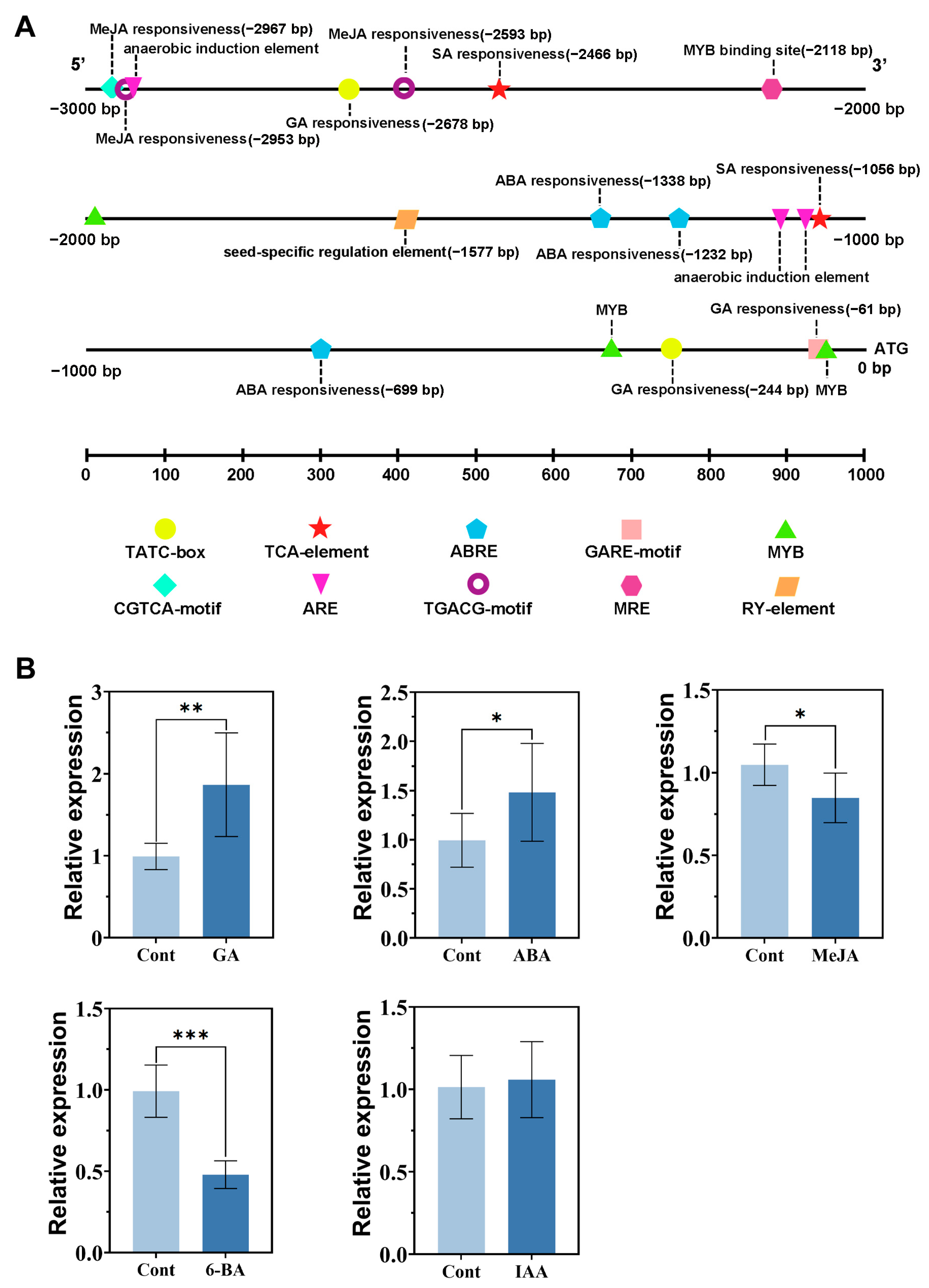
We subsequently obtained a promoter sequence of nearly 3 kb for RhCUC3 and analyzed its cis-acting regulatory elements (CREs) using the PlantCARE database. The sequence revealed typical CREs found in plant gene promoters, including those related to plant hormone responses, growth and development, and biotic and abiotic stress responses (Figure 3A). Notably, it contained three ABA-responsive elements (ABREs), three MeJA-responsive elements, three gibberellin (GA)-responsive elements (TATC-box and GARE motif), and two SA-responsive elements (TCA element). Furthermore, abiotic stress-related CREs were significantly more abundant than other CREs, including one MYB binding site, one light-responsive MYB binding site (MRE), and three anaerobic stress-responsive elements (ARE). These results suggest that RhCUC3 may play a role in regulating plant growth and abiotic stress responses mediated by these phytohormones.
We further conducted a quantitative RT-PCR analysis to investigate the response of RhCUC3 to plant hormones, focusing on ABA, GA, and MeJA. These phytohormones were selected based on their identification during promoter CRE analysis of RhCUC3 and known associations with plant development and the abiotic stress response. Additionally, we examined the effects of 6-BA and IAA on RhCUC3 expression due to the established roles of cytokinin and auxin in floral development [6,19]. Results indicated that treatments with GA and ABA led to significant increases in RhCUC3 expression, by approximately 1.9-fold and 1.5-fold in rose petals, respectively, after 12 h of exposure (Figure 3B). In contrast, MeJA and 6-BA suppressed RhCUC3 expression in petals by approximately 19.1% and 52.1%, respectively, at 12 h post-treatment compared to controls. IAA treatment for 12 h did not notably alter RhCUC3 expression levels. These findings suggest RhCUC3’s potential role in the regulation of development and abiotic stress response in roses modulated by ABA, GA, MeJA, and 6-BA.

3.4. RhCUC3 Was Localized in the Nucleus

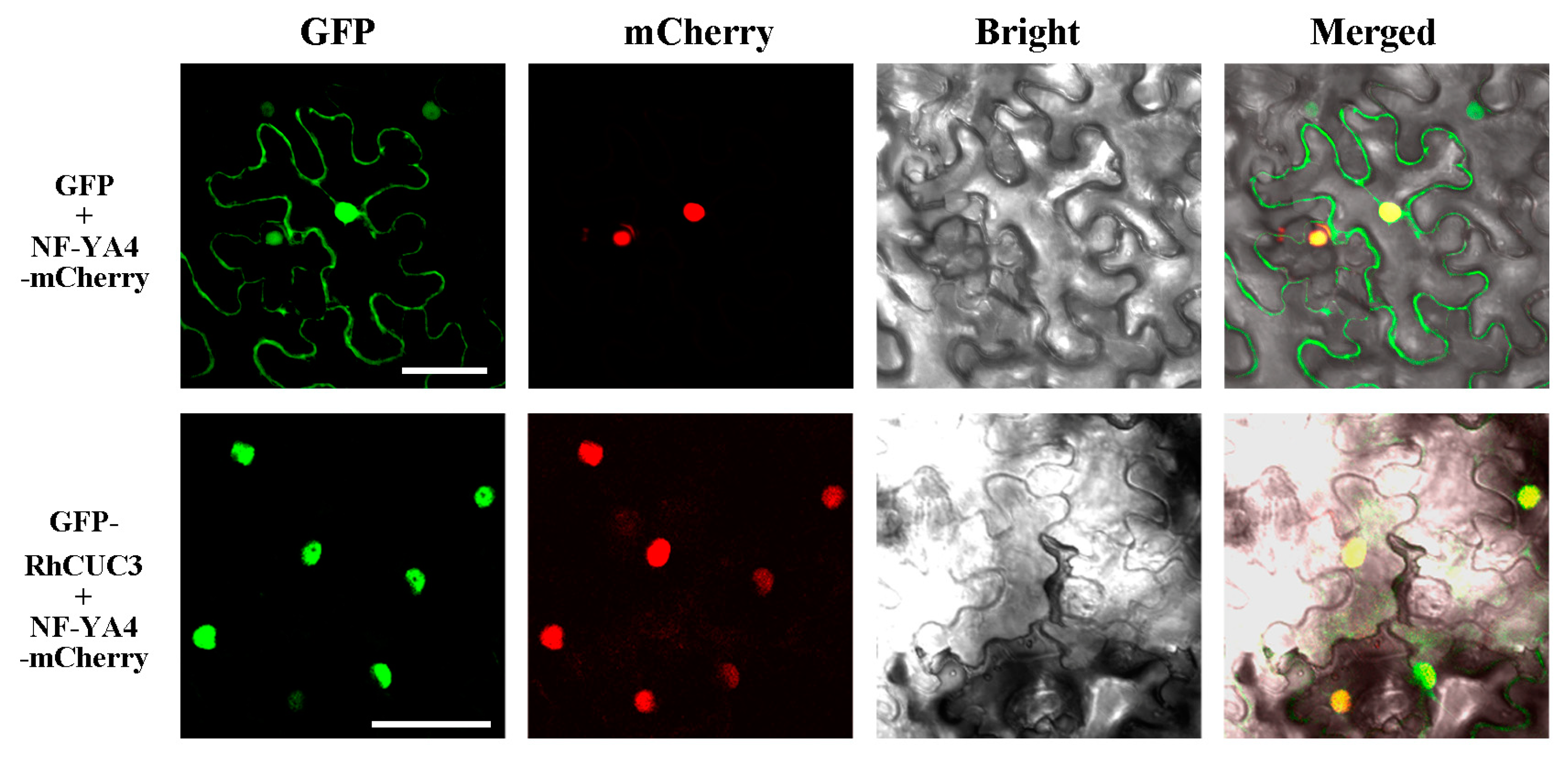
To trace the subcellular localization of RhCUC3, the full-length coding region of RhCUC3 was fused in-frame with GFP. The resulting 35S::GFP-RhCUC3 construct, along with the control 35S::GFP construct, was transiently co-expressed with an mCherry-labeled nuclear marker in Nicotiana benthamiana leaves. Microscopic analysis revealed that the green fluorescence signals of RhCUC3 closely coincided with the red signals from the nuclear markers (Figure 4). This observation confirms that RhCUC3 is localized in the nucleus, consistent with its anticipated role as a transcription factor.

3.5. Silencing of RhCUC3 Decreased the Petal Numbers of Rose Flowers

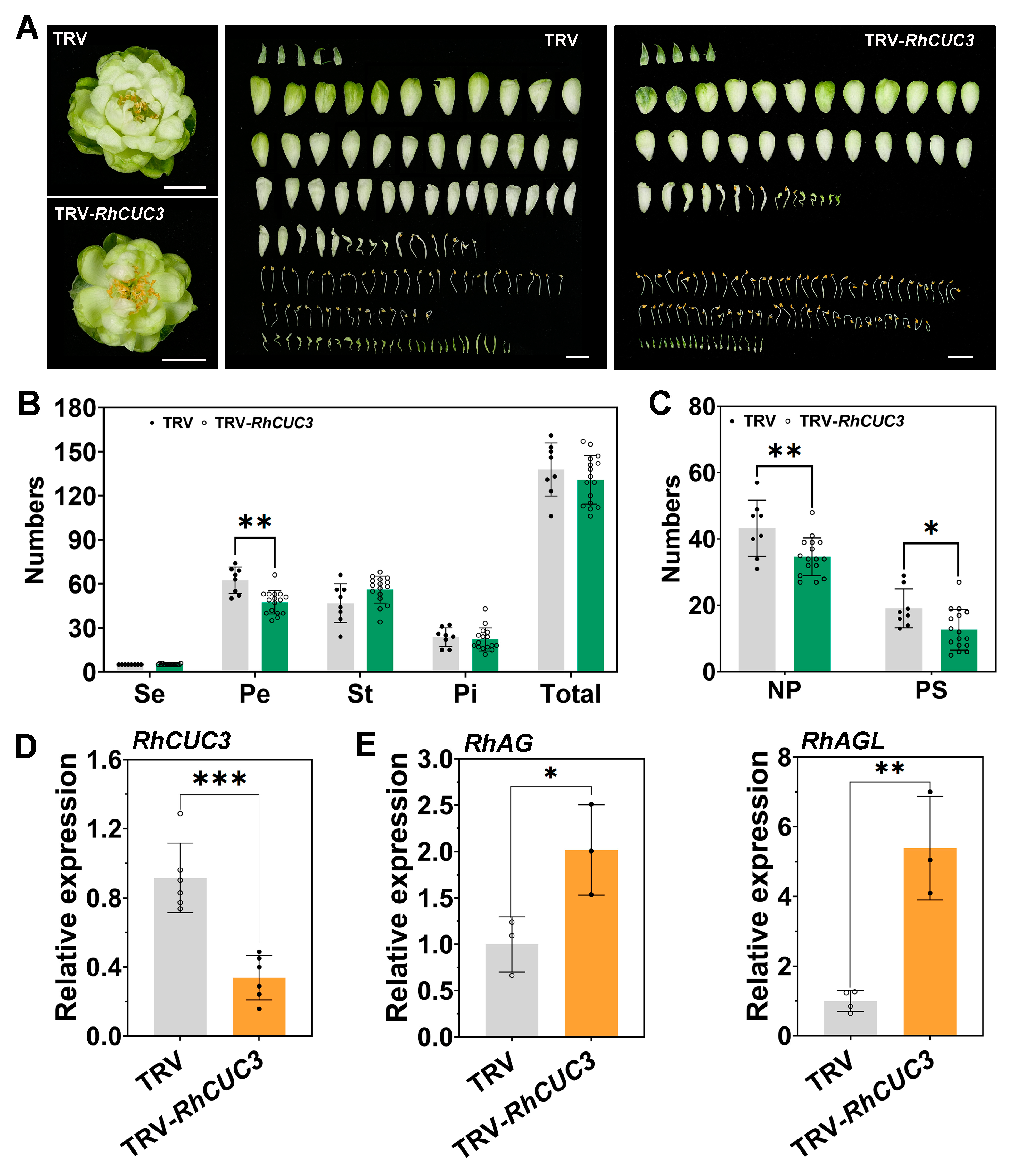
To investigate the function of RhCUC3 in the development of floral organs, we employed VIGS technology on RhCUC3 in the rose cultivar ‘Eclair’. Quantitative RT-PCR analysis demonstrated a substantial reduction in RhCUC3 expression in TRV-RhCUC3 plants, with a decrease of 60.2% relative to TRV controls (Figure 5D). Examination of the floral organs in both TRV and TRV-RhCUC3 flowers revealed that the number of sepals and pistils and the aggregated sum of the floral organs remained relatively unchanged (Figure 5A). However, we observed a considerable decline in petal count in the TRV-RhCUC3 group compared to TRV controls, alongside a slight increase in stamen number (Figure 5B). Considering that rose petals comprise both regular and malformed petals, referred to as petaloid stamens [30], we further quantified both categories of petals. We noted a reduction of 19.8% in normal petals and 33.7% in petaloid stamens when RhCUC3 was silenced (Figure 5A,C). The elevation of stamens and the reduction in petaloid stamens imply an integral role for RhCUC3 in modulating the homology transformation between petal and stamen.
Studies have suggested that the down-regulation of the expression of the C-class gene RhAG or another MADS-box transcription factor, AGL24, can lead to stamen petaloidy and an increase in petal count [38,40,41,42]. To investigate whether RhCUC3 regulation in floral organ identity is linked to the expression levels of RhAG and RhAGL, we analyzed their transcript levels in RhCUC3-silenced flowers. In line with our predictions, RhAG and RhAGL expression levels were significantly up-regulated in the TRV-RhCUC3 plants, by approximately 2.0 and 5.4 times, respectively, in comparison to the controls, and this up-regulation was inversely correlated with petal number. These findings lend further support to the hypothesis that RhCUC3 may modulate stamen petaloidy in roses, at least partially by affecting the expression of RhAG and RhAGL (Figure 5E).

4. Discussion

Floral organogenesis is a critical process governed by various transcription factors, among which numerous members of the MADS-box family have been identified as regulators of floral organs in plants [48]. Conversely, there is scant information regarding other transcription factors, such as those from NAC proteins. Prior studies have indicated that members of the NAM/CUC3 subfamily of NAC transcription factor, particularly CUCs, play a crucial role in several plant development processes, including organ boundary separation, shoot apical meristem formation, leaf morphology, senescence, floral organ development, and responses to abiotic stress [23,49,50]. In terms of plant development, the miR164a-CUC2 module dictates the degree of leaf serration in Arabidopsis [51]. A loss of the CUC2 homolog, GOBLET, in tomato results in simpler compound leaves and changes in fruit shape [25]. Specifically concerning flower development, Arabidopsis CUC1 and CUC2 are essential for accurate organ number and boundary establishment [29,30,52]. In Arabidopsis, the double mutant cuc1 cuc2 presents extensive fusion between adjacent sepals and stamens [18,24]. Moreover, a loss of miR164c function increases petal number by degrading its targets, CUC1 and CUC2 [27,28]. The MiR164-CUC2 regulatory module has also been shown to define petal, stamen, and carpel morphology in strawberry [23]. In this study, we identified the NAM/CUC3 subfamily transcription factor RhCUC3. A disruption in RhCUC3 led to a reduction in normal petals and the formation of malformed petaloid stamens in roses (Figure 5), thus augmenting our understanding of the NAC transcription factor family’s role and providing novel insights into the molecular mechanisms underlying floral organ development.
A previous study identified RhNAC31, an NAM/CUC3 subgroup NAC protein from the rose cultivar ‘Samantha’, as a promoter of tolerance to cold, salt, and drought stresses in Arabidopsis [50]. Our sequence alignment analysis revealed that RhCUC3 from the rose ‘Eclair’ shares 99.3% identity with RhNAC31, implying a potential functional equivalence in rose. Notably, both RhCUC3 and RhNAC31 expression were upregulated by the phytohormone ABA in rose, and several ABA-responsive CREs were identified in the RhCUC3 promoter (Figure 3). Then, contrasts in tissue-specific expression patterns were observed. RhNAC31 transcript levels were significantly higher in sepals than in leaves, stems, or petals, as reported by Ding et al. (2019) [50], while RhCUC3 was most abundantly expressed in petals, as indicated by the present study, with decreasing expression observed in pistils, stems, sepals, stamens, roots, and leaves (Figure 2B). This variation could be attributed to differences between the cultivars tested. ‘Samantha’ is a cut rose variety, whereas ‘Eclair’ is a miniature rose variety, suggesting inherent variances in genetic makeup and metabolic pathways, potentially influencing gene expression and function. Furthermore, the age and condition of the sampled plants can also significantly influence gene expression. However, our findings propose that RhCUC3 is involved in regulating the double-flower phenotype in roses and may also function as a regulator in stress resistance, akin to RhNAC31. This positions RhCUC3 as a promising candidate gene for future molecular breeding in rose.
In addition to ABA, our study has also revealed that the expression of RhCUC3 is modulated by various phytohormones, including GA, 6-BA, and MeJA, as illustrated in Figure 3. We observed that the transcription level of RhCUC3 was inhibited by 6-BA, yet remained unaffected by IAA in roses. Previous research has established that both cytokinin and IAA are instrumental in governing flower development in plants. Cytokinins, in particular, are crucial in plant growth, predominantly through regulating cell division [53]. In Arabidopsis, the ckx3 ckx5 double mutant demonstrated increased cytokinin levels, resulting in enlarged flowers and a modified count of floral organs attributable to postponed cellular differentiation initiation and a protracted period of developmental cell division [54]. Similarly, the rice double hk5 hk6 mutant exhibited a stark reduction in cytokinin response, which severely compromised the flower’s structure and resulted in decreased floral organ numbers [55]. These studies intimate that cytokinin’s influence on the number of floral organs might be complex and species-specific. Further investigation is necessary to ascertain whether cytokinins impact floral organ identity in rose. Moreover, its association with CUCs expression has seldom been discussed. In Arabidopsis, elevated CUC3 transcript levels were discernable in AP1::AtIPT4 transgenic plants, where increased cytokinins in flowers were present [31], a finding opposite to our own (Figure 3). However, mutants of cuc3 in the AP1::AtIPT4 background showed restoration in flower count, although floral organ numbers remained unchanged. Importantly, the floral organ number in cuc3 mutants paralleled that of wild-type plants [31]. Given that RhCUC3 has been implicated in the positive regulation of petal number in our rose study, it is conceivable that differences exist between AtCUC3 and RhCUC3 with respect to floral organ development and hormonal responses. Regarding IAA, it is well established that local auxin maxima and gradients within plant tissue are meticulously controlled by interrelated processes, including biosynthesis, polar transport, storage, and the inactivation of auxin [56,57]. In Arabidopsis, auxin activity peaks generated by PIN1 can suppress CUC2 expression [32]. Here, no auxin-responsive CREs were detected in the promoter of RhCUC3, and its expression was unaffected by IAA treatment, suggesting a lack of transcriptional response to IAA. Nonetheless, it is documented that CUC genes can modulate auxin transport by inducing the expression of kinase genes that influence the polarity of auxin transporters in crucifers [34]. Consequently, it is plausible that RhCUC3 may alter petal numbers by modifying auxin transport. To further elucidate the interplay between auxin and RhCUC3 during floral organogenesis, we plan to perform qRT-PCR analyses on auxin transport-related genes and immunolocalization of IAA in RhCUC3-silenced floral buds in forthcoming research.
In roses, petaloidy of the stamens is a prevalent mechanism contributing to the formation of the double-flower trait, and increases the esthetic value of the rose blossom [39]. Several MADS-box transcription factors have been implicated in the homeotic transformation of petals and stamens [6,38,42,58]. For instance, the down-regulation of RhAG and RhAGL has been shown to increase stamen petaloidy, thereby influencing the petal count in rose flowers [38,42]. Additional transcription factors, including RhMYB123 and RhARF18, have been demonstrated to control the differentiation between petals and stamens by modulating the expression of certain MADS-box genes and elements of the auxin signaling pathway [6,44]. In our research, we noted a marked reduction in malformed petaloid stamen count and a minor increase in stamen count in the TRV-RhCUC3 group compared to the TRV control group (Figure 5), which contrasted with the phenotypes of RhAG-silenced flowers [42]. Additionally, silencing RhCUC3 significantly upregulated the expression of RhAG and RhAGL (Figure 5E), aligning with earlier findings that AG and AG-like genes, such as RhAGL24 and RhAGL15, are inversely associated with petal count [38,44]. Consequently, our findings suggest that RhCUC3 plays a pivotal role in determining floral organ identity and regulating petal number in rose by modulating the expression of RhAG and RhAGL.
It was noticed that TRV-RhCUC3 plants exhibited a significant decrease in normal petal number compared to the TRV controls (Figure 5A,C). Earlier research indicates that CUC genes suppress cell division at the boundaries of developing organ primordia, which is necessary for the separation of the shoot apical meristem (SAM) and individual organs, subsequently influencing flower number and development [24,29,30,52]. In Arabidopsis, for instance, the overexpression of CUC1 and CUC2 in mir164 mutants leads to a rise in petal count [27,28]. In contrast, Arabidopsis plants bearing miR164-resistant CUC2 exhibit dense inflorescences with abbreviated internodes and an increased number of floral organs in the first blooms [51,59]. We thus hypothesize that RhCUC3’s role in petal number augmentation in our study might not only hinge upon its regulatory impact on floral organ identity, but also upon its function in boundary delineation and plant organ segregation. To elucidate how RhCUC3 dictates the boundaries between floral organs like petals and stamens, in situ hybridization could be utilized to compare the expression regions and intensities between CUC3-silenced floral buds and TRV controls during the early phase of rose flower morphogenesis. Additionally, CUC2 and CUC3 have been implicated in cytokinin-mediated floral development in Arabidopsis [31]. In our study, we observed that the expression level of RhCUC3 was suppressed following 6-BA treatment (Figure 3B). However, it remains unclear whether the phenotype of reduced petals in RhCUC3-silenced flowers is related to cytokinin content or the cytokinin signaling pathway. Therefore, further investigation is needed to determine the involvement of cytokinin in floral organ separation in roses and to elucidate the role of RhCUC3 in this process.
Interestingly, some phylloid pistils were observed in both TRV and TRV-RhCUC3 flowers, potentially contributing to the double-flower phenotype due to the increase in the area of floral organs. This homeotic malformation, known as phyllody, has also been reported in other rose varieties, such as Rosa hybrida cv. Motrea [60]. Previous research indicates that phylloid pistils arise from a disruption in normal pistil development, characterized by the absence of marginal cell fusion in style-tube primordia (Chmelnitsky et al., 2002). These undeveloped style-tubes transform into green, leaf-like structures containing chlorophyll and stomata, resembling cauline leaves rather than sepals, as revealed by a scanning electron microscopy (SEM) analysis of transverse sections. Although the number of pistils remained unchanged when RhCUC3 was silenced in our study, further investigation is required to determine whether RhCUC3 or its homologous genes in the NAC family play a role in phylloid pistil formation using VIGS technology and SEM observation during pistil development [60,61].
In addition, several studies have indicated that CUC3 expression in plants is subject to regulation by a variety of transcription factors, including Class I KNOTTED-like homeobox (KNOXI) genes, BRASSINAZOLE RESISTANT 1 (BZR1), and WUSCHEL-RELATED HOMEOBOX1 (WOX1) [62]. The homeodomain transcription factor KNOXI is critical for shoot apical meristem (SAM) initiation and maintenance. Mutations in KNOXI result in SAM deficiency and a concurrent decrease in CUC3 expression [29,63]. It has been observed that CUC3 expression remains unaltered in knat6 mutants, yet it is completely absent in knat6-1-stm-2 double mutants, suggesting that KNAT6 and STM contribute to CUC3 activation in a redundant manner [64]. BZR1, a master regulator of brassinosteroid (BR) signaling, has been shown to downregulate CUC3 by binding to its promoter in Arabidopsis [62]. A recent study suggests that WOX1, a member of the homeodomain transcription factor family, inhibits the development of the leaf margin tooth by blocking BZR1 and CUC3 activity during the late stages of tooth development and restricts CUC3 expression at the early stages in a BZR1-dependent fashion [65]. Our research suggests that RhCUC3 may act upstream of the transcription factors RhAG and RhAGL. However, it is still unknown which transcription factors interact with RhCUC3 in rose, or the nature of the transcription factor regulatory network within which RhCUC3 operates to modulate rose flower morphologies.

5. Conclusions

The double-flower trait significantly enhances the ornamental value of roses. Therefore, understanding the regulatory mechanisms underlying floral organ identity has significant theoretical and practical implications. In this study, we identified the NAM/CUC3 subgroup NAC protein RhCUC3 in miniature roses, which exhibited high expression during the development of petal and stamen primordia. Roses silenced for RhCUC3 demonstrated a significant reduction in the number of petaloid stamens and normal petals, but there was a slight increase in the number of stamens. Furthermore, the expression levels of RhAG and RhAGL were significantly higher in RhCUC3-silenced flowers compared to TRV controls. In summary, we demonstrated that RhCUC3 regulates stamen petaloidy and petal number, at least partially, by modulating the expression of RhAG and RhAGL. Since the double petal trait is a breeding target for various ornamental plants such as roses, peonies, camellias, and wisteria, RhCUC3 may become a potential gene to accelerate the double petalization process through gene-stable overexpression technology in different varieties of these ornamental plants.

Supplementary Materials

The following supporting information can be downloaded at: www.mdpi.com/article/10.3390/horticulturae11020170/s1, Table S1: Primers used in qRT-PCR and vector construction.

Author Contributions

Conceptualization, W.C. and Y.C.; methodology, Y.S. and Y.F.; formal analysis, Y.S.; investigation, Y.S., Y.F. and Z.Z.; data curation, Z.D.; writing—original draft preparation, W.C. and Y.F.; writing—review and editing, W.C.; funding acquisition, W.C. All authors have read and agreed to the published version of the manuscript.

Funding

This work was funded by the Natural Science Foundation of Zhejiang Province (grant number LMS25C150005) and the National Natural Science Foundation of China (grant number 32072612).

Data Availability Statement

All data generated or analyzed during this study are included in this article and its Supplementary Materials.

Acknowledgments

We sincerely thank Xinqiang Jiang (Qingdao Agricultural University) for his generous guidance and suggestions on this manuscript.

Conflicts of Interest

The authors declare no conflicts of interest.

References

  1. Meyerowitz, E.M.; Smyth, D.R.; Bowman, J.L. Abnormal flowers and pattern formation in floral development. Development 1989, 106, 209–217. [Google Scholar] [CrossRef]
  2. Dubois, A.; Raymond, O.; Maene, M.; Baudino, S.; Langlade, N.B.; Boltz, V.; Vergne, P.; Bendahmane, M. Tinkering with the C-Function: A molecular frame for the selection of double flowers in cultivated roses. PLoS ONE 2010, 5, e9288. [Google Scholar] [CrossRef] [PubMed]
  3. Debener, T. Genetic analysis of horticulturally important morphological and physiological characters in diploid roses. Gartenbauwissenschaft 1999, 64, 14–20. [Google Scholar]
  4. Morey, D. Observations on the genetics of doubleness in roses. Am. Rose Annu. 1959, 44, 113–116. [Google Scholar]
  5. Hibrand-Saint Oyant, L.; Crespel, L.; Rajapakse, S.; Zhang, L.; Foucher, F. Genetic linkage maps of rose constructed with new microsatellite markers and locating QTL controlling flowering traits. Tree Genet. Genomes 2007, 4, 11–23. [Google Scholar] [CrossRef]
  6. Chen, J.; Li, Y.; Li, Y.; Li, Y.; Wang, Y.; Jiang, C.; Choisy, P.; Xu, T.; Cai, Y.; Pei, D.; et al. AUXIN RESPONSE FACTOR 18–HISTONE DEACETYLASE 6 module regulates floral organ identity in rose (Rosa hybrida). Plant Physiol. 2021, 186, 1074–1087. [Google Scholar] [CrossRef]
  7. Cai, Y.; Wang, L.; Ogutu, C.O.; Yang, Q.; Luo, B.; Liao, L.; Zheng, B.; Zhang, R.; Han, Y. The MADS-box gene PpPI is a key regulator of the double-flower trait in peach. Physiol. Plant. 2021, 173, 2119–2129. [Google Scholar] [CrossRef]
  8. Peng, L.; Li, Y.; Tan, W.; Wu, S.; Hao, Q.; Tong, N.; Wang, Z.; Liu, Z.; Shu, Q. Combined genome-wide association studies and expression quantitative trait locus analysis uncovers a genetic regulatory network of floral organ number in a tree peony (Paeonia suffruticosa Andrews) breeding population. Hortic. Res. 2023, 10, uhad110. [Google Scholar] [CrossRef] [PubMed]
  9. Li, X.; Li, J.; Fan, Z.; Liu, Z.; Tanaka, T.; Yin, H. Global gene expression defines faded whorl specification of double flower domestication in Camellia. Sci. Rep. 2017, 7, 3197. [Google Scholar] [CrossRef]
  10. Hu, L.; Zheng, T.; Cai, M.; Pan, H.; Wang, J.; Zhang, Q. Transcriptome analysis during floral organ development provides insights into stamen petaloidy in Lagerstroemia speciosa. Plant Physiol. Biochem. 2019, 142, 510–518. [Google Scholar] [CrossRef]
  11. Soltis, P.S.; Soltis, D.E.; Kim, S.; Chanderbali, A.; Buzgo, M. Expression of floral regulators in basal angiosperms and the origin and evolution of ABC-Function. Adv. Bot. Res. 2006, 44, 483–506. [Google Scholar]
  12. Bowman, J.L.; Smyth, D.R.; Meyerowitz, E.M. Genetic interactions among floral homeotic genes of Arabidopsis. Development 1991, 112, 1–20. [Google Scholar] [CrossRef]
  13. Coen, E.S.; Meyerowitz, E.M. The war of the whorls: Genetic interactions controlling flower development. Nature 1991, 353, 31–37. [Google Scholar] [CrossRef]
  14. Theißen, G. Development of floral organ identity: Stories from the MADS house. Curr. Opin. Plant Biol. 2001, 4, 75–85. [Google Scholar] [CrossRef]
  15. Litt, A.; Kramer, E.M. The ABC model and the diversification of floral organ identity. Semin. Cell Dev. Biol. 2010, 21, 129–137. [Google Scholar] [CrossRef]
  16. Irish, V.F. The flowering of Arabidopsis flower development. Plant J. 2010, 61, 1014–1028. [Google Scholar] [CrossRef]
  17. Ronse De Craene, L.P. The evolutionary significance of homeosis in flowers: A morphological perspective. Int. J. Plant Sci. 2003, 164, 225–235. [Google Scholar] [CrossRef]
  18. Lampugnani, E.R.; Kilinc, A.; Smyth, D.R. PETAL LOSS is a boundary gene that inhibits growth between developing sepals in Arabidopsis thaliana. Plant J. 2012, 71, 724–735. [Google Scholar] [CrossRef]
  19. Ding, L.; Yan, S.; Jiang, L.; Zhao, W.; Ning, K.; Zhao, J.; Liu, X.; Zhang, J.; Wang, Q.; Zhang, X. HANABA TARANU (HAN) bridges meristem and organ primordia boundaries through PINHEAD, JAGGED, BLADE-ON-PETIOLE2 and CYTOKININ OXIDASE 3 during flower development in Arabidopsis. PLoS Genet. 2015, 11, e1005479. [Google Scholar] [CrossRef]
  20. Thomson, B.; Zheng, B.; Wellmer, F. Floral organogenesis: When knowing your ABCs is not enough. Plant Physiol. 2017, 173, 56–64. [Google Scholar] [CrossRef] [PubMed]
  21. Quon, T.; Lampugnani, E.R.; Smyth, D.R. PETAL LOSS and ROXY1 interact to limit growth within and between sepals but to promote petal initiation in Arabidopsis thaliana. Front. Plant Sci. 2017, 8, 152. [Google Scholar] [CrossRef]
  22. Xu, Y.; Prunet, N.; Gan, E.; Wang, Y.; Stewart, D.; Wellmer, F.; Huang, J.; Yamaguchi, N.; Tatsumi, Y.; Kojima, M.; et al. SUPERMAN regulates floral whorl boundaries through control of auxin biosynthesis. EMBO J. 2018, 37, e97499. [Google Scholar] [CrossRef] [PubMed]
  23. Zheng, G.; Wei, W.; Li, Y.; Kan, L.; Wang, F.; Zhang, X.; Li, F.; Liu, Z.; Kang, C. Conserved and novel roles of miR164-CUC 2 regulatory module in specifying leaf and floral organ morphology in strawberry. New Phytol. 2019, 224, 480–492. [Google Scholar] [CrossRef] [PubMed]
  24. Aida, M.; Ishida, T.; Fukaki, H.; Fujisawa, H.; Tasaka, M. Genes involved in organ separation in Arabidopsis: An analysis of the cup-shaped cotyledon mutant. Plant Cell 1997, 9, 841–857. [Google Scholar] [CrossRef] [PubMed]
  25. Berger, Y.; Harpaz-Saad, S.; Brand, A.; Melnik, H.; Sirding, N.; Alvarez, J.P.; Zinder, M.; Samach, A.; Eshed, Y.; Ori, N. The NAC-domain transcription factor GOBLET specifies leaflet boundaries in compound tomato leaves. Development 2009, 136, 823–832. [Google Scholar] [CrossRef]
  26. Cheng, X.; Peng, J.; Ma, J.; Tang, Y.; Chen, R.; Mysore, K.S.; Wen, J. NO APICAL MERISTEM (MtNAM) regulates floral organ identity and lateral organ separation in Medicago truncatula. New Phytol. 2012, 195, 71–84. [Google Scholar] [CrossRef]
  27. Baker, C.C.; Sieber, P.; Wellmer, F.; Meyerowitz, E.M. The early extra petals1 mutant uncovers a role for microRNA miR164c in regulating petal number in Arabidopsis. Curr. Biol. 2005, 15, 303–315. [Google Scholar] [CrossRef] [PubMed]
  28. Huang, T.; López-Giráldez, F.; Townsend, J.P.; Irish, V.F. RBE controls microRNA164 expression to effect floral organogenesis. Development 2012, 139, 2161–2169. [Google Scholar] [CrossRef] [PubMed]
  29. Takada, S.; Hibara, K.; Ishida, T.; Tasaka, M. The CUP-SHAPED COTYLEDON1 gene of Arabidopsis regulates shoot apical meristem formation. Development 2001, 128, 1127–1135. [Google Scholar] [CrossRef]
  30. Daimon, Y.; Takabe, K.; Tasaka, M. The CUP-SHAPED COTYLEDON genes promote adventitious shoot formation on calli. Plant Cell Physiol. 2003, 44, 113–121. [Google Scholar] [CrossRef]
  31. Li, X.G.; Su, Y.H.; Zhao, X.Y.; Li, W.; Gao, X.Q.; Zhang, X.S. Cytokinin overproduction-caused alteration of flower development is partially mediated by CUC2 and CUC3 in Arabidopsis. Gene 2010, 450, 109–120. [Google Scholar] [CrossRef] [PubMed]
  32. Maugarny, A.; Gonçalves, B.; Arnaud, N.; Laufs, P. CUC transcription factors: To the meristem and beyond. In Plant Transcription Factors; Gonzalez, D.H., Ed.; Academic Press: London, UK, 2016; pp. 229–247. [Google Scholar]
  33. Vernoux, T.; Kronenberger, J.; Grandjean, O.; Laufs, P.; Traas, J. PIN-FORMED 1 regulates cell fate at the periphery of the shoot apical meristem. Development 2000, 127, 5157–5165. [Google Scholar] [CrossRef] [PubMed]
  34. Hu, Z.L.; Wilson-Sánchez, D.; Bhatia, N.; Rast-Somssich, M.I.; Wu, A.; Vlad, D.; McGuire, L.; Nikolov, L.A.; Laufs, P.; Gan, X.; et al. A CUC1/auxin genetic module links cell polarity to patterned tissue growth and leaf shape diversity in crucifer plants. Proc. Natl. Acad. Sci. USA 2024, 121, e2321877121. [Google Scholar] [CrossRef]
  35. Wang, J.-R.; Wang, X.; Su, N.; Li, Q.-J.; Zhang, X.-H.; Ma, Y.-P.; Zhao, L.; Ginefra Toni, J.F.; De Craene, L.R. Floral morphology and morphogenesis in Sanguisorba (Rosaceae): Flower diversification despite petal reduction and spatial constraints. Bot. J. Linn. Soc. 2020, 193, 47–63. [Google Scholar] [CrossRef]
  36. Wang, X.; Gong, J.; Li, Q.; Wang, J.; Ma, Y.; Zhang, X.; Chang, Z.; Wen, J.; Zhao, L. Floral organogenesis of Prunus Laurocerasus and P. Serotina and its significance for the systematics of the genus and androecium diversity in Rosaceae. Botany 2019, 97, 71–84. [Google Scholar] [CrossRef]
  37. Bendahmane, M.; Dubois, A.; Raymond, O.; Bris, M.L. Genetics and genomics of flower initiation and development in roses. J. Exp. Bot. 2013, 64, 847–857. [Google Scholar] [CrossRef] [PubMed]
  38. Liu, L.; Guo, Y.; Wu, Z.; Ren, H.; Jiang, Y.; Ma, N.; Gao, J.; Sun, X. RhAGL24 Regulating Auxin-related gene RhARF18 affects stamen petaloidy in rose. Horticulturae 2022, 8, 407. [Google Scholar] [CrossRef]
  39. Yang, T.; Wang, Y.; Li, Y.; Liang, S.; Yang, Y.; Huang, Z.; Li, Y.; Gao, J.; Ma, N.; Zhou, X. The transcription factor RhMYB17 regulates the homeotic transformation of floral organs in Rose (Rosa hybrida) under cold stress. J. Exp. Bot. 2024, 75, 2965–2981. [Google Scholar] [CrossRef]
  40. Noor, S.H.; Ushijima, K.; Murata, A.; Yoshida, K.; Tanabe, M.; Tanigawa, T.; Kubo, Y.; Nakano, R. Double flower formation induced by silencing of C-class MADS-box genes and its variation among petunia cultivars. Sci. Hortic. 2014, 178, 1–7. [Google Scholar] [CrossRef]
  41. Nakatsuka, T.; Saito, M.; Yamada, E.; Fujita, K.; Yamagishi, N.; Yoshikawa, N.; Nishihara, M. Isolation and characterization of the C-class MADS-box gene involved in the formation of double flowers in Japanese gentian. BMC Plant Biol. 2015, 15, 182. [Google Scholar] [CrossRef] [PubMed]
  42. Ma, N.; Chen, W.; Fan, T.; Tian, Y.; Zhang, S.; Zeng, D.; Li, Y. Low temperature-induced DNA hypermethylation attenuates expression of RhAG, an AGAMOUS homolog, and increases petal number in rose (Rosa hybrida). BMC Plant Biol 2015, 15, 237. [Google Scholar] [CrossRef] [PubMed]
  43. Han, Y.; Tang, A.; Wan, H.; Zhang, T.; Cheng, T.; Wang, J.; Yang, W.; Pan, H.; Zhang, Q. An APETALA2 homolog, RcAP2, regulates the number of rose petals derived from stamens and response to temperature fluctuations. Front. Plant Sci. 2018, 9, 481. [Google Scholar] [CrossRef] [PubMed]
  44. Li, K.; Li, Y.; Wang, Y.; Li, Y.; He, J.; Li, Y.; Du, L.; Gao, Y.; Ma, N.; Gao, J.; et al. Disruption of transcription factor RhMYB123 causes the transformation of stamen to malformed petal in rose (Rosa hybrida). Plant Cell Rep. 2022, 41, 2293–2303. [Google Scholar] [CrossRef] [PubMed]
  45. Livak, K.J.; Schmittgen, T.D. Analysis of relative gene expression data using real-time quantitative PCR and the 2−ΔΔCT method. Methods 2001, 25, 402–408. [Google Scholar] [CrossRef]
  46. Meng, Y.; Li, N.; Tian, J.; Gao, J.; Zhang, C. Identification and validation of reference genes for gene expression studies in postharvest rose flower (Rosa hybrida). Sci. Hortic. 2013, 158, 16–21. [Google Scholar] [CrossRef]
  47. Dai, F.; Zhang, C.; Jiang, X.; Kang, M.; Yin, X.; Lü, P.; Zhang, X.; Zheng, Y.; Gao, J. RhNAC2 and RhEXPA4 are involved in the regulation of dehydration tolerance during the expansion of rose petals. Plant Physiol. 2012, 160, 2064–2082. [Google Scholar] [CrossRef] [PubMed]
  48. Thomson, B.; Wellmer, F. Molecular regulation of flower development. Curr. Top. Dev. Biol. 2019, 131, 185–210. [Google Scholar]
  49. Hibara, K.; Karim, M.R.; Takada, S.; Taoka, K.; Furutani, M.; Aida, M.; Tasaka, M. Arabidopsis CUP-SHAPED COTYLEDON3 regulates postembryonic shoot meristem and organ boundary formation. Plant Cell 2006, 18, 2946–2957. [Google Scholar] [CrossRef]
  50. Ding, A.; Li, S.; Li, W.; Hao, Q.; Wan, X.; Wang, K.; Liu, Q.; Liu, Q.; Jiang, X. RhNAC31, a novel rose NAC transcription factor, enhances tolerance to multiple abiotic stresses in Arabidopsis. Acta Physiol. Plant. 2019, 41, 75. [Google Scholar] [CrossRef]
  51. Nikovics, K.; Blein, T.; Peaucelle, A.; Ishida, T.; Morin, H.; Aida, M.; Laufs, P. The balance between the MIR164A and CUC2 genes controls leaf margin serration in Arabidopsis. Plant Cell 2006, 18, 2929–2945. [Google Scholar] [CrossRef]
  52. Lee, B.H.; Jeon, J.O.; Lee, M.M.; Kim, J.H. Genetic interaction between GROWTH-REGULATING FACTOR and CUP-SHAPED COTYLEDON in organ separation. Plant Signal. Behav. 2015, 10, e988071. [Google Scholar] [CrossRef] [PubMed]
  53. Jing, W.; Gong, F.; Liu, G.; Deng, Y.; Liu, J.; Yang, W.; Sun, X.; Li, Y.; Gao, J.; Zhou, X.; et al. Petal size is controlled by the MYB73/TPL/HDA19-miR159-CKX6 module regulating cytokinin catabolism in Rosa hybrida. Nat. Commun. 2023, 14, 7106. [Google Scholar] [CrossRef]
  54. Bartrina, I.; Otto, E.; Strnad, M.; Werner, T.; Schmülling, T. Cytokinin regulates the activity of reproductive meristems, flower organ size, ovule formation, and thus seed yield in Arabidopsis Thaliana. Plant Cell 2011, 23, 69–80. [Google Scholar] [CrossRef]
  55. Burr, C.A.; Sun, J.; Yamburenko, M.V.; Willoughby, A.; Hodgens, C.; Boeshore, S.L.; Elmore, A.; Atkinson, J.; Nimchuk, Z.L.; Bishopp, A.; et al. The HK5 and HK6 cytokinin receptors mediate diverse developmental pathways in rice. Development 2020, 147, dev191734. [Google Scholar] [CrossRef]
  56. Sauer, M.; Kleine-Vehn, J. PIN-FORMED and PIN-LIKES auxin transport facilitators. Development 2019, 146, dev168088. [Google Scholar] [CrossRef]
  57. Hayashi, K.; Nakamura, S.; Fukunaga, S.; Nishimura, T.; Jenness, M.K.; Murphy, A.S.; Motose, H.; Nozaki, H.; Furutani, M.; Aoyama, T. Auxin transport sites are visualized in planta using fluorescent auxin analogs. Proc. Natl. Acad. Sci. USA 2014, 111, 11557–11562. [Google Scholar] [CrossRef] [PubMed]
  58. Gattolin, S.; Cirilli, M.; Pacheco, I.; Ciacciulli, A.; Da Silva Linge, C.; Mauroux, J.; Lambert, P.; Cammarata, E.; Bassi, D.; Pascal, T.; et al. Deletion of the miR172 target site in a TOE -type gene is a strong candidate variant for dominant double-flower trait in Rosaceae. Plant J. 2018, 96, 358–371. [Google Scholar] [CrossRef]
  59. Peaucelle, A.; Morin, H.; Traas, J.; Laufs, P. Plants expressing a miR164-resistant CUC2 gene reveal the importance of post-meristematic maintenance of phyllotaxy in Arabidopsis. Development 2007, 134, 1045–1050. [Google Scholar] [CrossRef] [PubMed]
  60. Chmelnitsky, I.; Azizbekova, N.; Khayat, E.; Zieslin, N. Morphological development of normal and phyllody expressing Rosa hybrida cv. Motrea flowers. Plant Growth Regul. 2002, 37, 215–221. [Google Scholar] [CrossRef]
  61. Maas, F.M.; Hofman-Eijer, L.B.; Hulsteijn, K. Flower morphogenesis in Rosa hybrida ’Mercedes’ as studied by cryo-scanning electron and light microscopy. Effects on light and shoot position on a branch. Ann. Bot. 1995, 75, 199–205. [Google Scholar] [CrossRef]
  62. Gendron, J.M.; Liu, J.-S.; Fan, M.; Bai, M.-Y.; Wenkel, S.; Springer, P.S.; Barton, M.K.; Wang, Z.-Y. Brassinosteroids regulate organ boundary formation in the shoot apical meristem of Arabidopsis. Proc. Natl. Acad. Sci. USA 2012, 109, 21152–21157. [Google Scholar] [CrossRef]
  63. Vroemen, C.W.; Mordhorst, A.P.; Albrecht, C.; Kwaaitaal, M.A.C.J.; De Vries, S.C. The CUP-SHAPED COTYLEDON3 gene is required for boundary and shoot meristem formation in Arabidopsis. Plant Cell 2003, 15, 1563–1577. [Google Scholar] [CrossRef] [PubMed]
  64. Belles-Boix, E.; Hamant, O.; Witiak, S.M.; Morin, H.; Traas, J.; Pautot, V. KNAT6: An Arabidopsis homeobox gene involved in meristem activity and organ separation. Plant Cell 2006, 18, 1900–1907. [Google Scholar] [CrossRef] [PubMed]
  65. Xu, L.; Fang, N.; Lu, T.; Tameshige, T.; Nakata, M.T.; Jiang, Y.; Tan, L.; He, H.; Zhang, X.; Huang, Y.; et al. WOX1 controls leaf serration development via temporally restricting BRASSINAZOLE RESISTANT 1 and CUP SHAPED COTYLEDON 3 expression in Arabidopsis. J. Exp. Bot. 2025, 76, 478–492. [Google Scholar] [CrossRef] [PubMed]
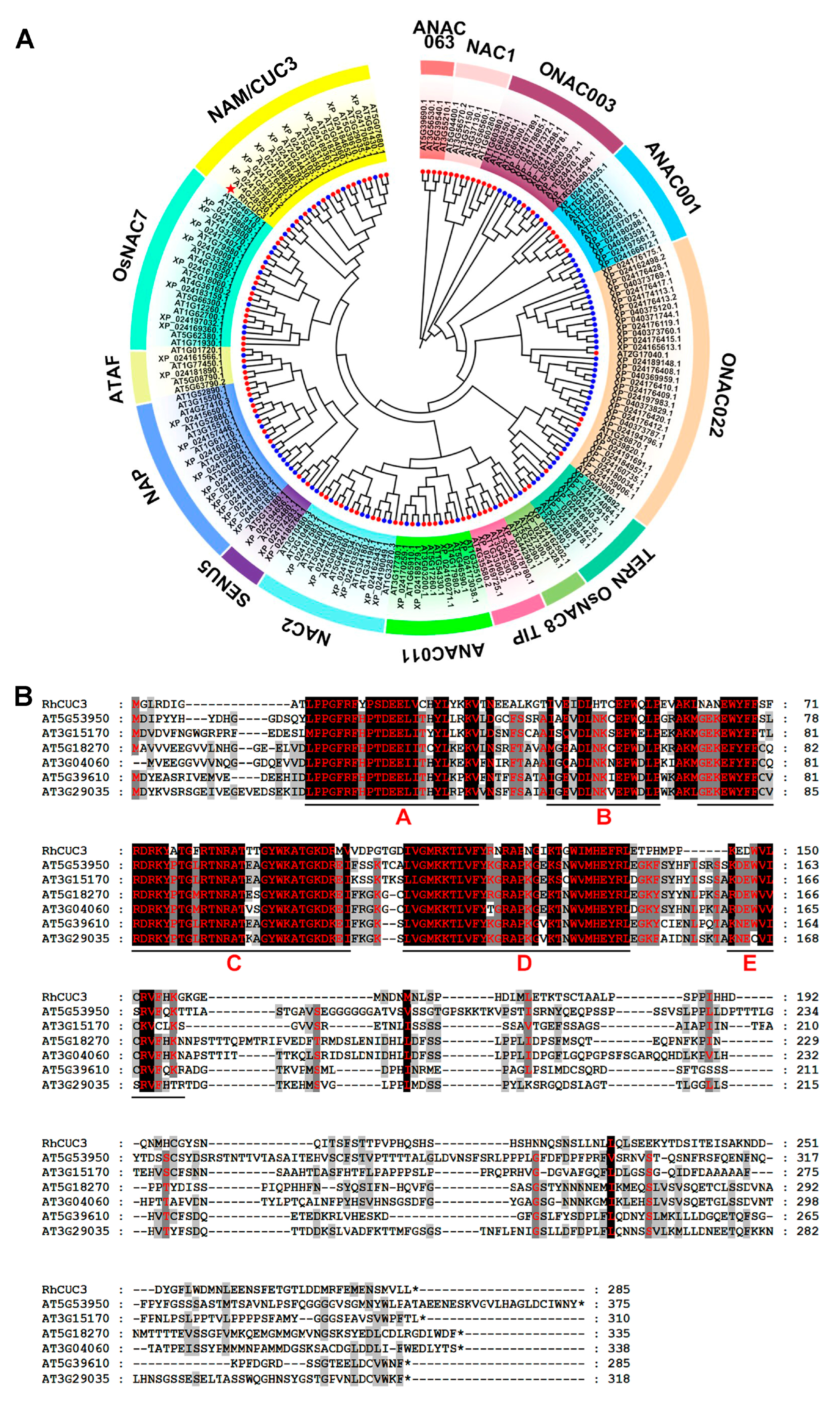
Figure 1. Evolutionary and sequence analysis of RhCUC3. (A) Phylogenetic relationships of RhCUC3 amino acid sequences with NACs in Arabidopsis. The phylogenetic tree was generated using MEGA7, with NAC subfamily names annotated between the outer rings. RhCUC3 is highlighted with a red star. (B) Multiple sequence alignment of RhCUC3 with homologs from the NAM/CUC3 subfamily in Arabidopsis. Red letters A–E denote the subdomains of the NAC binding domain consensus sequence.
Horticulturae 11 00170 g001
Figure 2. Expression analysis of RhCUC3. (A) Relative expression levels of RhCUC3 at early floral developmental stages. Values are means ± SD (n = 3). Pe, petal primordia differentiation stage; St, stamen primordia differentiation stage; Pi, pistil primordia differentiation stage. (B) Relative expression levels of RhCUC3 in different plant organs. Values are means ± SD (n ≥ 3). Statistically significant differences were analyzed using Duncan’s multiple range test and are indicated by lowercase letters (p < 0.05).
Horticulturae 11 00170 g002
Figure 3. Response of RhCUC3 to different plant hormones. (A) Potential cis-acting regulatory elements (CREs) identified in the promoter region of RhCUC3. The upstream sequences preceding the ATG start codon are denoted by negative numbers. ABRE, abscisic acid-responsive element; GARE, gibberellin-responsive element; Myb, recognition site of transcription factor MYB; ARE, anaerobic induced element; MRE, MYB binding site involved in light responsiveness; RY, seed-specific regulation-related element. (B) Expression of RhCUC3 in rose petals in response to exogenous applications of plant hormones. The expression level of RhCUC3 relative to the mock treatment was set as 1, using RhUBI2 as an internal control. Data are presented as means ± SD (n = 3), with asterisks indicating statistically significant differences (two-sided Student’s t-test; * p< 0.05; ** p < 0.01; *** p < 0.001).
Horticulturae 11 00170 g003
Figure 4. Subcellular localization of RhCUC3 in the epidermis cells of Nicotiana benthamiana leaves. GFP-RhCUC3 was co-expressed with an mCherry-labeled nuclear marker (NF-YA4-mCherry). GFP co-expressed with the nuclear marker served as the control. Scale bars, 50 μm.
Horticulturae 11 00170 g004
Figure 5. Silencing of RhCUC3 decreased petal numbers of rose flowers. (A) Phenotype of the TRV control and RhCUC3-silenced flowers. Scale bars = 1 cm. (B,C) The number of floral organs of TRV control and RhCUC3-silenced flowers. Se, sepal; Pe, petal; St, stamen; Pi, pistil; NP, normal petal; PS, petaloid stamens. Data are the mean ± SD (n > 8). Gray column, TRV control flower; green column, TRV-RhCUC3 flower. (D,E) Relative expression of RhCUC3, RhAG, and RhAGL in TRV control and RhCUC3-silenced petals as detected through qRT-PCR. RhUBI2 served as the internal control. Gray column, TRV control petals; orange column, TRV-RhCUC3 petals. Data are presented as means ± SD (n ≥ 3), with asterisks indicating statistically significant differences (two-sided Student’s t-test; * p< 0.05; ** p < 0.01; *** p < 0.001).
Horticulturae 11 00170 g005
Disclaimer/Publisher’s Note: The statements, opinions and data contained in all publications are solely those of the individual author(s) and contributor(s) and not of MDPI and/or the editor(s). MDPI and/or the editor(s) disclaim responsibility for any injury to people or property resulting from any ideas, methods, instructions or products referred to in the content.