Article

Genome-Wide Identification and Expression Pattern Analysis of CrLBD Family Reveal Their Involvement in Floral Development in Chionanthus retusus

1
College of Forestry, Shandong Agricultural University, Taian 271018, China
2
State Forestry and Grassland Administration Key Laboratory of Silviculture in Downstream Areas of the Yellow River, Taian 271018, China
3
Heze Forestry Technology Service Center, Heze 274000, China
*
Author to whom correspondence should be addressed.
Horticulturae 2025, 11(12), 1429; https://doi.org/10.3390/horticulturae11121429
Submission received: 7 October 2025 / Revised: 14 November 2025 / Accepted: 19 November 2025 / Published: 26 November 2025
(This article belongs to the Section Genetics, Genomics, Breeding, and Biotechnology (G2B2))

Abstract

The growth and development of plants are modulated by multiple genes, among which the LBD (Lateral Organ Boundaries Domain) family—a group of plant-specific transcription factors—plays pivotal roles. In this study, we utilized the latest reference genome to identify and characterize LBD genes in Chionanthus retusus (Oleaceae, 2n = 2x = 46) and further explored their expression profiles across the different floral development, as well as their potential functions in floral morphology development. Our analysis identified a total of 76 LBD gene family members in C. retusus, which were categorized into two major families: Class I and Class II. Class I was further subdivided into five subfamilies, while Class II comprised two subfamilies. Chromosomal mapping revealed that LBD genes are distributed across all 23 chromosomes of C. retusus. Additional analyses of gene structure, conserved domains, motifs, and synteny highlighted their structural and evolutionary conservation. Subsequent expression profiling of CrLBD genes across various floral morphologies identified three members—CrLBD3, CrLBD34, and CrLBD72—that are potentially involved in regulating floral morphology in C. retusus.

1. Introduction

The ornamental value and biodiversity of garden plants are gaining increasing recognition in global landscaping practices [1,2]. Species within the Oleaceae family, in particular, enhance landscape esthetics through their remarkable diversity in floral morphology, coloration, and fragrance [3]. Chionanthus retusus, a prized member of the Oleaceae family, is distinguished by its elegant growth habit, dense canopy, and broad utility in landscape architecture [4]. Its floral characteristics further elevate its ornamental appeal: the flowers are snow-white, delicately structured, and emit a subtle, fresh scent. Typically small in size, they aggregate into dense clusters that form spectacular, snow-like displays when the tree reaches full bloom. This striking visual effect is accentuated by the purity of their color and the graceful arrangement of the inflorescences [5]. Morphologically, each flower features a linear corolla, and the inflorescences hang pendulously from branches like decorative fringes—a trait that lends the plant its common name [6].
Chionanthus retusus exhibits considerable intraspecific diversity in floral morphology, particularly in the size and shape of petals [7,8]. These morphological variations are not only ecologically significant for adaptation but also provide valuable genetic resources for ornamental breeding [7,8]. Previous studies have indicated that the LBD gene family may play an important role in regulating floral morphological diversity in plants. The LBD gene family, first identified and characterized in Arabidopsis thaliana, consists of plant-specific transcription factors that typically contain a highly conserved LOB domain. This domain facilitates DNA binding and enables participation in transcriptional regulation [9]. For instance, in rice, the AtLOB ortholog OsRA2 influences panicle architecture by modulating pedicel length—overexpression leads to shorter pedicels, whereas suppression results in elongation [10]. LBD genes are widely distributed across the plant kingdom, though the number of members varies considerably among species [11]—for example, 43 in A. thaliana [12], 35 in rice [13], 58 in Malus domestica [14], and 55 in Populus trichocarpa [15]. Functional studies have demonstrated that LBD genes are key regulators of morphological development in roots [16,17], leaves [18,19], and floral organs [20]. In A. thaliana, the LBD family is divided into five major subfamilies [21]. Class IA proteins primarily regulate the development of aerial organs such as leaves, stems, and flowers [22,23]. Class IB members are mainly involved in the development of underground organs, including lateral, crown, and adventitious roots [24,25,26]. Class IC proteins influence processes such as plant growth, leaf abaxial identity, apical dominance, and secondary growth through the cytokinin signaling pathway [27,28]. In contrast, the transcriptional regulatory mechanisms of Class IE LBD genes remain unclear [21]. Meanwhile, Class II LBD proteins are primarily associated with nitrogen utilization and anthocyanin biosynthesis [29,30,31]. Notable examples illustrate the functional conservation and diversity of LBD genes across species. In A. thaliana, AtLBD6 (also known as AS2), a Class IA member, is expressed in the adaxial domain of floral organs and regulates the symmetry of petals and stamens; its mutation leads to organ fusion or malformation [32]. In Physalis floridana, suppression of the LBD family member Physalis organ size 3 (PfPOS3) through knockdown or knockout approaches significantly reduces floral organ size, consequently leading to abnormal floral phenotypes [33]. In maize, ramosa2 (ra2) controls inflorescence meristem boundary formation and regulates ear branching; mutants exhibit excessive branching and severely disrupted ear structure [34]. Additionally, certain Class IC members also contribute to floral development—for example, AtLBD3 overexpression in transgenic lines often leads to aberrant inflorescence and floral development, resulting in a range of abnormal floral phenotypes [26].
Given the crucial roles of LBD gene family members in floral organogenesis, this study employed the latest C. retusus reference genome to systematically identify and characterize its LBD genes, thereby addressing the current lack of genomic information on this important transcription factor family in this species. To explore the evolution and function of the LBD gene family in C. retusus, this study combined phylogenetic and genomic analyses with transcriptomic profiling. We analyzed gene structure, conserved motifs, and cis-elements to infer functional divergence. Furthermore, we performed transcriptome sequencing across key floral development stages in three varieties with distinct morphologies to identify LBD genes potentially regulating flower architecture. This integrated approach enabled us to characterize the expression profiles of LBD family members during floral development and identify candidate genes potentially associated with flower morphology formation. Our findings provide valuable insights into the molecular mechanisms underlying LBD-mediated regulation in C. retusus and establish a solid theoretical foundation for applying this knowledge to directional breeding programs in ornamental plants.

2. Materials and Methods

2.1. Plant Materials and Treatments

Plant materials were collected from three distinct Chionanthus retusus varieties (‘Xuezaohua’ (XZH), ‘Xuedenglong’ (XDL), and ‘Xuexuan’ (XX)), each characterized by unique floral morphotypes. For each variety, inflorescence samples were harvested from multiple individual plants at four developmental stages, with 10 inflorescences collected per stage. This sampling strategy ensured that 30 individual florets were obtained for each developmental time point across the varieties. These samples were obtained from the Plant Protection Experimental Station at the Panhe Campus of Shandong Agricultural University in Shandong Province, China (36° N, 117° W). The developmental stages were defined as follows: bud stage (approximately 30% of flower buds visibly open, with the remainder closed), initial flowering stage (flowers partially opened but predominantly semi-closed), full flowering stage (over 50% of flowers fully open), and late flowering stage (70% or nearly all flowers fully open). All collected samples were used for both transcriptome sequencing and subsequent analysis of LBD gene expression. Immediately upon collection, samples were flash-frozen in liquid nitrogen and stored at −80 °C to preserve RNA integrity for downstream applications, including total RNA extraction and quantitative gene expression assays.

2.2. Identification of CrLBDs Transcription Factor Family Members in C. retusus

The genomic data of C. retusus were derived from our research group’s whole-genome sequencing project. The genome assembly integrated multiple technologies including Hi-C (for chromosome conformation capture and scaffolding) and Oxford Nanopore (for ultralong reads), yielding a gapless telomere-to-telomere (T2T) genome with an N50 length of 31.57 Mb. The final assembly generated two complete haplotypes: Haplotype 1 (~687 Mb) and Haplotype 2 (~683 Mb), which were successfully anchored onto 23 chromosomes. A total of 42,864 protein-coding genes were predicted [35]. While the reference genome of Arabidopsis thaliana (TAIR10) was acquired from the JGI Phytozome database (https://phytozome-next.jgi.doe.gov/ accessed on 15 May 2025). Coding sequences (CDS) of all A. thaliana LBD (AtLBDs) family members were downloaded from TAIR (https://www.arabidopsis.org/ accessed on 15 May 2025) and used as queries for BLAST analysis in TBtools (V2.376) software to retrieve corresponding protein sequences. This approach enabled the identification of all putative LBD family members in C. retusus (CrLBDs). Subsequently, all identified LBD proteins were examined for the presence of conserved domains using the NCBI Conserved Domain Database (CDD; https://www.ncbi.nlm.nih.gov/Structure/cdd/wrpsb.cgi accessed on 15 May 2025) to validate their classification.

2.3. Phylogenetic Relationship Analysis

A multiple sequence alignment was performed using Molecular Evolutionary Genetics Analysis (MEGAX) (V12.0.11) software with LBD protein sequences from Chionanthus retusus and their homologs from A. thaliana. A phylogenetic tree was then constructed with IQ-TREE (V2.3.6) software using the maximum likelihood (ML) method, with branch support assessed through 1000 bootstrap replicates and other parameters maintained at default settings unless otherwise specified [36]. All A thaliana LBD protein sequences used in this analysis were obtained from The A. thaliana Information Resource (TAIR; https://www.arabidopsis.org/ accessed on 15 May 2025).

2.4. Analysis of CrLBDs Structure, Conserved Motifs and Cis Acting Elements

The genomic positions of CrLBD genes were determined using the genome annotation file (GFF3 format), and their exon-intron structures were visualized through the “Visualize Gene Structure” module in TBtools [37]. Conserved domains within all CrLBD protein sequences were identified using the conserved domain search (CD-search) tool available through NCBI [38]. Concurrently, conserved motifs were detected with MEME (https://meme-suite.org/meme/ accessed on 15 May 2025) under the following parameters: classic mode, Zero or One Occurrence Per Sequence (ZOPS), with the motif count set to 10 and remaining parameters maintained as defaults [39].
To investigate potential cis-regulatory elements, we extracted the 2000 bp genomic sequences upstream of the transcription start sites for all CrLBD genes using TBtools. These promoter sequences were then analyzed in silico through the PlantCARE database (Plant Cis-Acting Regulatory Element; (http://bioinformatics.psb.ugent.be/webtools/plantcare/html/ accessed on 15 May 2025), facilitating the identification of key cis-acting elements implicated in transcriptional regulation [40].

2.5. Collinearity and Chromosome Location Analysis

The chromosomal locations of the LBD genes were determined by parsing the C. retusus genome assembly (GFF3 file) and utilizing the gene mapping visualization function in TBtools [37]. Furthermore, genomic collinearity among C. retusus, Syringa oblata [41], and Olea europaea [42] was analyzed and visualized using the same software.

2.6. Analysis About CrLBD Genes Expression of qRT-PCR During Flower Development

Total RNA was extracted from various samples using the FastPure Universal Plant Total RNA Isolation Kit (RC411, Nanjing, China) from Vazyme. Complementary DNA (cDNA) was then synthesized from the RNA samples using the Evo M-MLV Reverse Transcription Premix (for qPCR) (AG11706, Changsha, China) from AccuraBio. Quantitative real-time PCR (qRT-PCR) reactions were prepared with the 2× qPCR Premix (SYBR Green) (CM0139, Changsha, China) from AccuraBio and performed on a BIO-RAD CFX96 real-time PCR system (785BR10687, Hercules, CA, USA) using 96-well plates sealed with optical film. The CrUBC2 gene was used as an internal reference for normalization, with gene-specific primers listed in Table S1. Each sample was analyzed with four technical replicates and three biological replicates. Relative expression levels of target CrLBD genes were calculated using the 2−∆∆CT method [43].

2.7. Gene Expression Profiling and Co-Expression Network Analysis to Investigate CrLBD Genes Regulation

RNA sequencing was commercially performed by Novogene Bioinformatics Technology Co., Ltd. (Beijing, China). Plant materials included three varieties sampled at four developmental stages: bud, initial flowering, full flowering, and late flowering. Total RNA was isolated using the FastPure Universal Plant Total RNA Isolation Kit (RC411, Nanjing, China) from Vazyme, and RNA integrity was assessed on a Bioanalyzer 2100 system with the RNA Nano 6000 Assay Kit (Agilent, Santa Clara, California, USA). Sequencing libraries were constructed following the standard Illumina protocol and sequenced on the NovaSeq 6000 platform. A total of 36 RNA-seq libraries were aligned to the chromosome-level reference genome of C. retusus using HISAT2 [44], and read counts were generated with HTSeq. Differential expression analysis was performed using the DESeq2 R package, with significantly differentially expressed genes defined as those exhibiting |log2(fold change)| > 1 and an adjusted p-value < 0.05.

3. Results

3.1. Genome-Wide Identification and Phylogenetic Analysis of the LBD Gene Family in C. retusus

To identify LBD genes in Chionanthus retusus, a Hidden Markov Model (HMM) was constructed based on the LBD protein sequences of Arabidopsis thaliana [45]. This model was subsequently employed to screen the C. retusus genome, and candidate genes were retained only if they contained a conserved LOB domain. A total of 76 CrLBD genes were identified (Table S2) and systematically designated CrLBD1 to CrLBD76 according to their physical positions on the chromosomes. The encoded CrLBD proteins range from 129 to 349 amino acids in length, with predicted molecular weights (MW) between 14.66 and 39.03 kDa and theoretical isoelectric points (pI) ranging from 4.91 to 9.22 (Table S2). Based on the instability index and grand average of hydropathicity (GRAVY), all CrLBD proteins are predicted to be hydrophilic, suggesting their solubility and potential localization in cellular aqueous environments. Phylogenetic analysis revealed that the 76 CrLBD members cluster into two major classes (Class I and Class II), with Class I further subdivided into five subgroups (I-1, I-2, I-3, I-4, I-5) and Class II into two subgroups (II-1 and II-2) (Figure 1). Compared with A. thaliana, certain subfamilies in C. retusus have undergone notable expansion, indicating possible lineage-specific diversification of LBD genes (Table S3).

3.2. Analysis of Chromosomal Location, Collinearity and Replication of LBD Gene Family in C. retusus

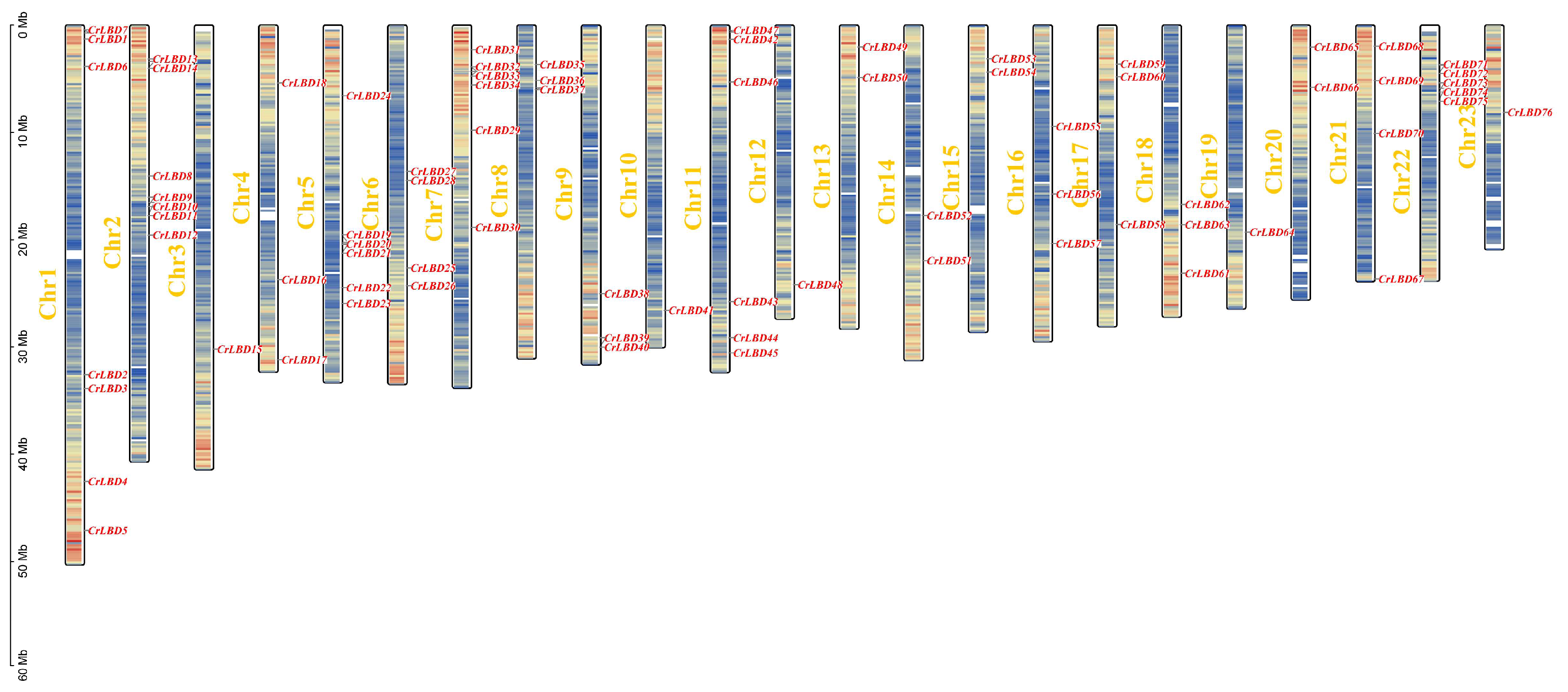
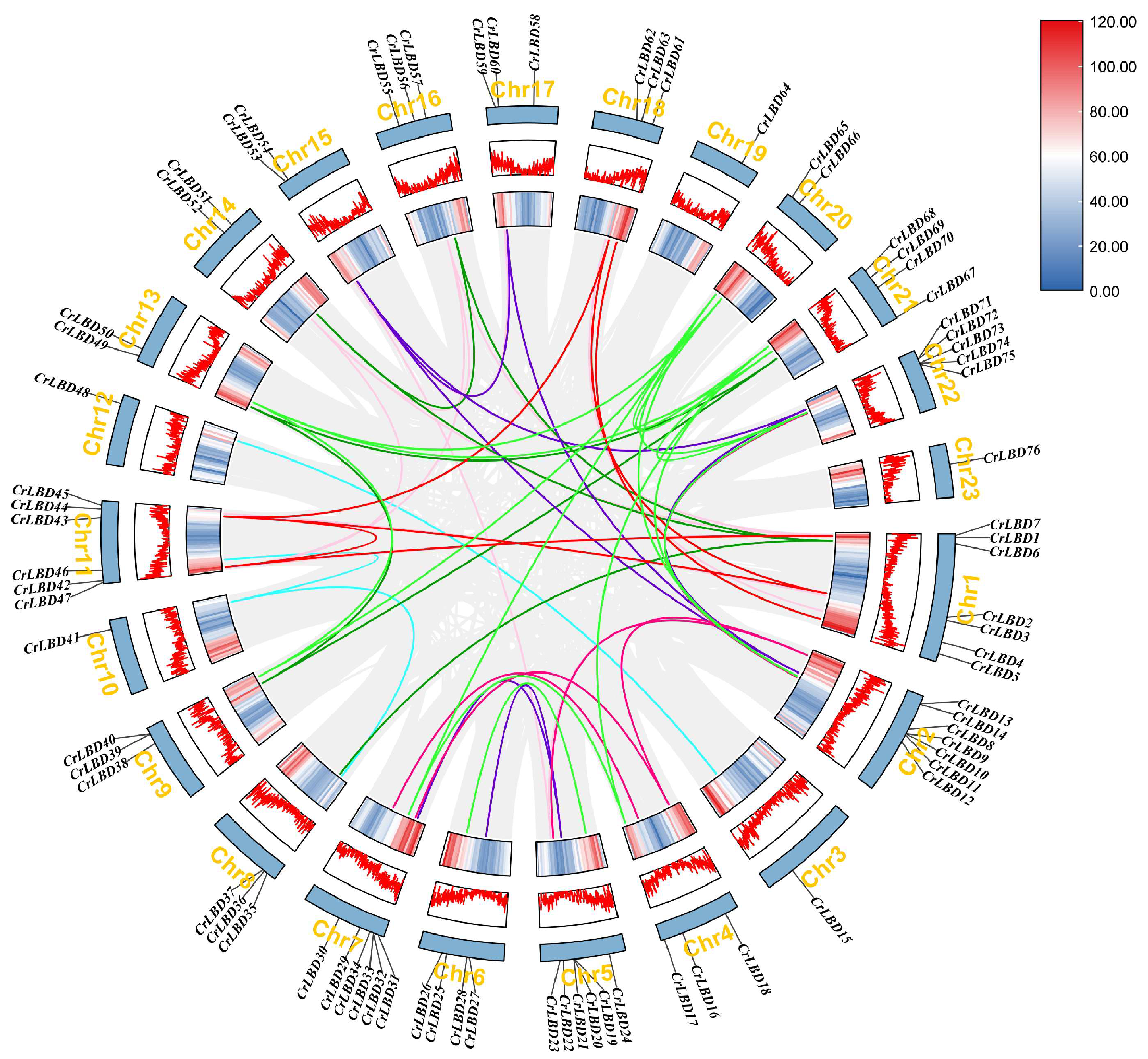
The karyotype of C. retusus comprises 23 chromosomes, with all 76 identified CrLBD genes distributed across these chromosomes (Figure 2). Chromosomal mapping confirmed their positions, revealing that chromosomes 1 and 2 contain the highest number of CrLBD genes (7 each), while chromosomes 3, 10, 12, 19, and 23 contain only one gene each. The remaining chromosomes carry between 2 and 5 CrLBD genes. Collinearity analysis using MCScanX in TBtools revealed extensive duplication events within the CrLBD gene family [46]. We identified 56 pairs of segmentally duplicated genes, with 12 pairs predominantly located on chromosomes 1 and 11 (Figure 3). Synteny analysis confirmed that these 56 segmental duplication events involved 60 CrLBD genes, as some genes participated in multiple duplication pairs (Figure 3), indicating their retention and subsequent evolution after whole-genome duplication events. Additionally, we detected seven tandem duplication clusters: CrLBD9/CrLBD10, CrLBD13/CrLBD14, CrLBD19/CrLBD20/CrLBD21, CrLBD32/CrLBD33, CrLBD39/CrLBD40, CrLBD71/CrLBD72, and CrLBD73/CrLBD74 (Figure 3). Both segmental and tandem duplications have therefore significantly contributed to the expansion and diversification of the CrLBD gene family in C. retusus, explaining its larger size compared to Arabidopsis thaliana. For instance, CrLBD7 (chromosome 7) and CrLBD52 (chromosome 14) in Class I-5 are located in syntenic regions and likely represent retained duplicates from a genome duplication event. Similarly, CrLBD13/CrLBD23 (Class I-4), CrLBD15/CrLBD48 (Class II-1), and CrLBD6/CrLBD36 (Class II-2) were identified as segmentally duplicated pairs.
To elucidate the evolutionary conservation of the LBD gene family within the Oleaceae, we performed a comparative genomic analysis involving Chionanthus retusus and two other diploid species—Syringa oblata and Olea europaea—each possessing 23 chromosomes. Synteny analysis indicated strong macro-synteny among the three genomes, despite the presence of localized structural variations. Within conserved syntenic regions, we identified 205 orthologous LBD gene pairs between C. retusus and O. europaea, and 150 between C. retusus and S. oblata (Figure S1, Table S4). The Ka/Ks analysis of these collinear LBD gene pairs revealed that all calculated ratios were below 1 (Table S4), consistent with purifying selection acting on these genes following speciation. Overall, these findings collectively suggest that the LBD gene family is highly conserved in both gene structure and genomic organization across the Oleaceae lineage.

3.3. Analysis of Gene Structure, Conserved Motifs and Conserved Domains of CrLBD Gene Family in C. retusus

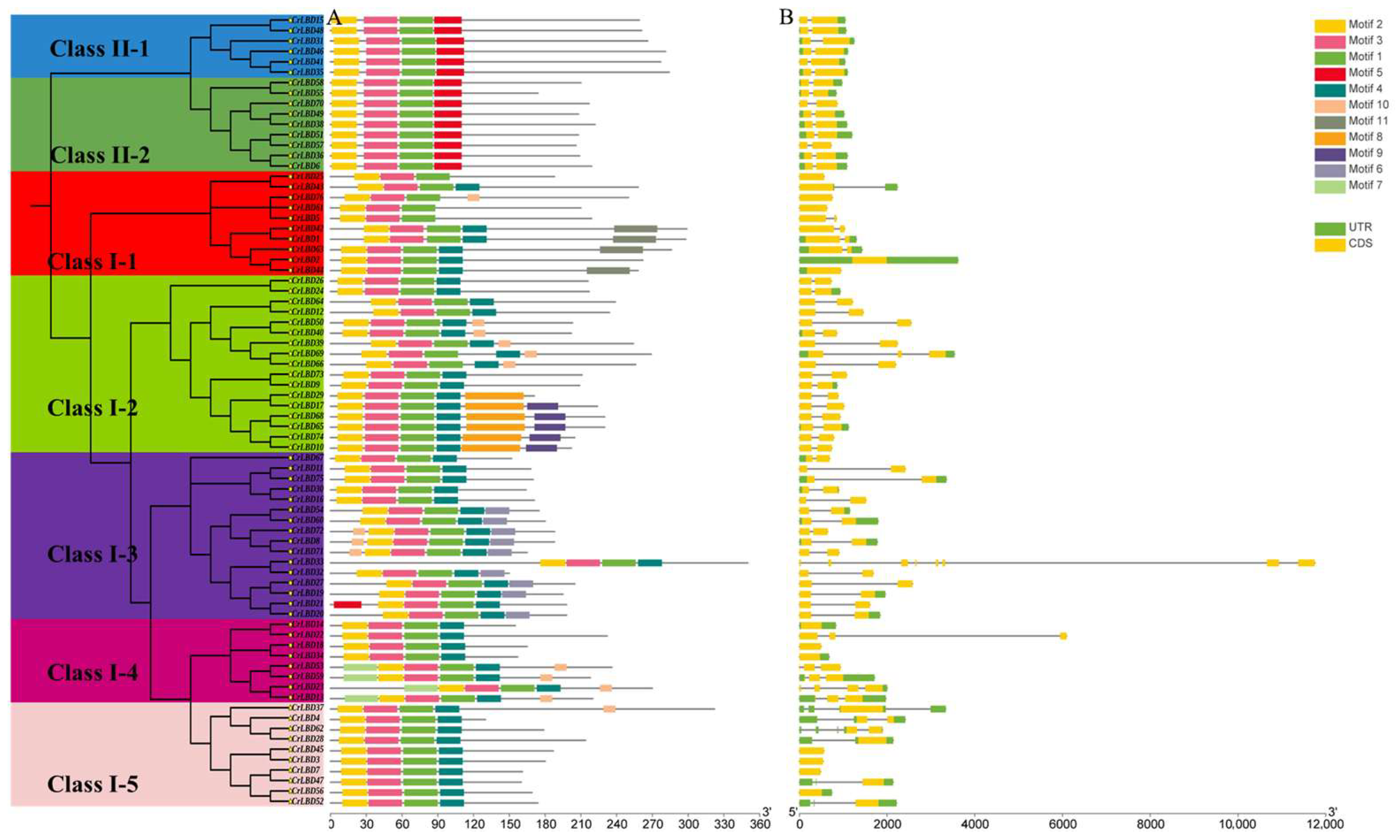
CrLBD genes generally display conserved exon-intron organization across the family (Figure 4B). Exon numbers range from 1 to 8, with the majority (54 of 76 genes) containing two exons. CrLBD39 contains the highest number of exons (8), whereas CrLBD48, CrLBD5, CrLBD22, CrLBD13, and CrLBD20 each possess only a single exon. The average exon count per gene is 1.9. While gene structure varies considerably between subfamilies, it remains well conserved within each subfamily. For instance, all Class I-3 members contain two exons, as do all Class II-2 members. However, certain genes exhibit distinctive structural features: CrLBD39 contains eight exons—significantly more than other Class I-2 members—while CrLBD72 features an unusually long N-terminal intron, distinguishing it from other Class I-3 members. These unique structural characteristics may reflect functional divergence among LBD family members. We next analyzed conserved motifs in the 76 CrLBD protein sequences (Figure 4A). All CrLBD proteins contain the characteristic LOB domain, which is consistently composed of Motif1, Motif2, and Motif3. Notably, within the Class I-3 subfamily, only CrLBD71 possesses the unique Motif5, while in Class II, Motif4 is absent in CrLBD46, CrLBD41, and CrLBD36. Further examination reveals that these subfamily-specific motifs are predominantly located in the C-terminal regions of CrLBD proteins, highlighting considerable structural heterogeneity in this protein domain.

3.4. Identification and Enrichment Analysis of Cis-Acting Elements in the CrLBD Promoter of C. retusus

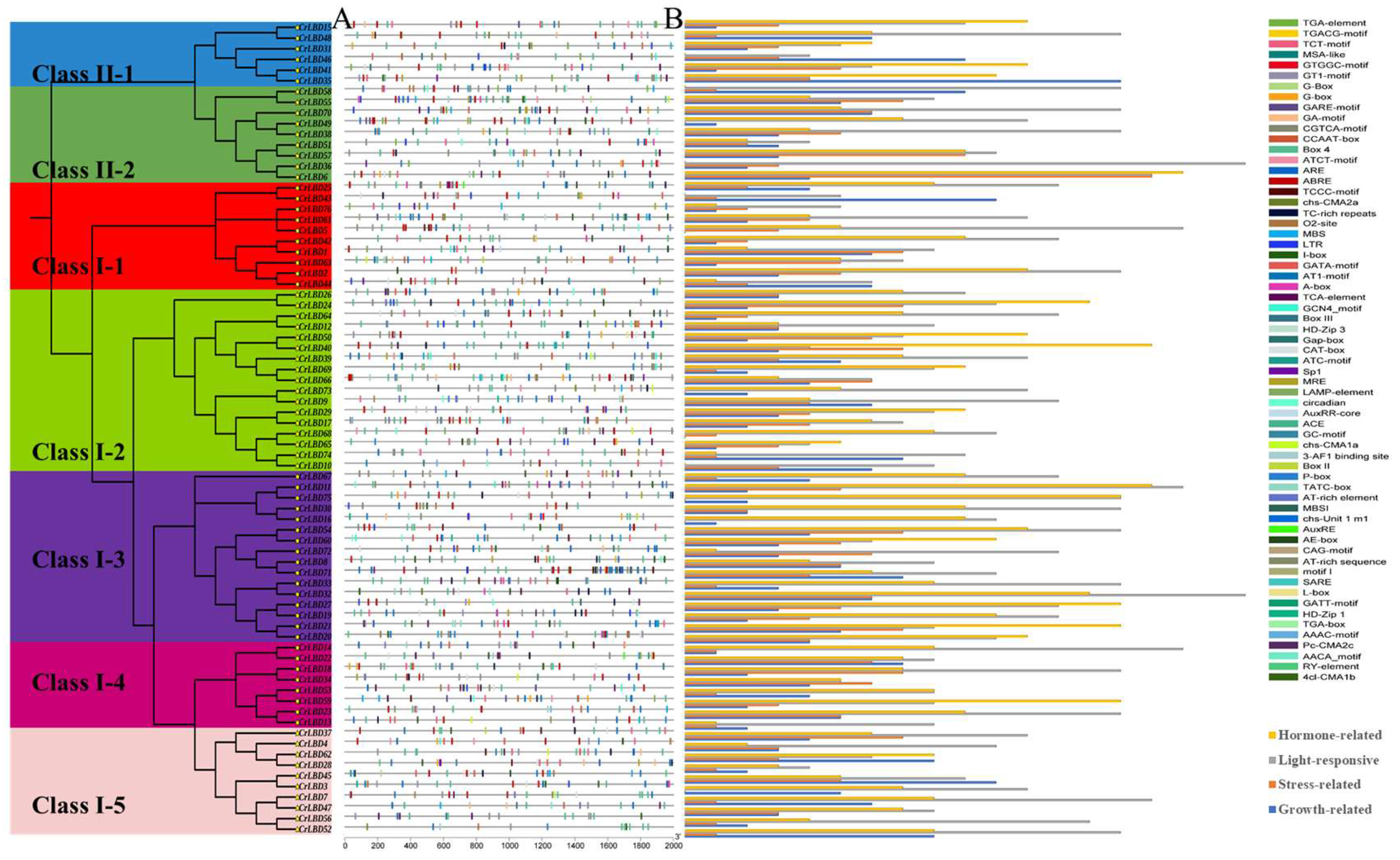
Precise regulation of gene expression depends on cis-acting elements within promoter regions. To investigate the transcriptional regulation of CrLBD genes, we systematically analyzed the 2000 bp promoter sequences of all 76 CrLBD genes using PlantCARE (Figure 5B) [40]. Our analysis identified 7684 functionally annotated cis-elements, which were classified into four categories based on their predicted functions: growth-related, stress-responsive, hormone-responsive, and light-responsive elements. Light-responsive elements were the most abundant (762), followed by hormone-responsive (525), growth-related (282), and stress-responsive elements (275) (Figure 5A). The predominance of light- and hormone-related elements suggests that CrLBD genes are likely involved in plant responses to light and hormonal signals, potentially through interactions with specific transcription factors. Notably, the promoter of CrLBD18 contains 43 cis-elements, which is more than 1.7 times the family average (24.2). This pronounced enrichment implies that CrLBD18 may play an important regulatory role, possibly in floral organ development.
Within the hormone-responsive category, we identified 47 auxin-responsive elements (AuxREs), 98 gibberellin-responsive elements, and 217 abscisic acid-responsive elements across the CrLBD family. Specifically, three AuxREs were detected in two genes (CrLBD60 and CrLBD75). Among the nine Class II-2 members, seven contained AuxREs, representing the highest proportion (77.8%), followed by Class I-3 (68.8%) and Class I-2 (52.9%). Lower proportions were observed in Class I-1 (40%) and Class II-1 (33.33%), while only one member each in Class I-4 and I-5 contained AuxREs. Six gibberellin-responsive elements were identified in CrLBD45. In Class II-1 (6 members) and Class I-5 (10 members), only one member lacked gibberellin-responsive elements, accounting for 83.3% and 80%, respectively. Seven gibberellin-responsive members were detected in both Class I-1 (70%) and Class II-2 (77.8%), while five (62.5%) and nine (56.3%) such members were identified in Class I-4 and Class I-3, respectively. Among Class I-2 members, eight (47.1%) contained gibberellin-responsive elements. These findings suggest that CrLBD genes harboring these hormone-responsive elements may participate in hormone signal transduction pathways.

3.5. The Regulatory Network of the CrLBD Gene Family in C. retusus

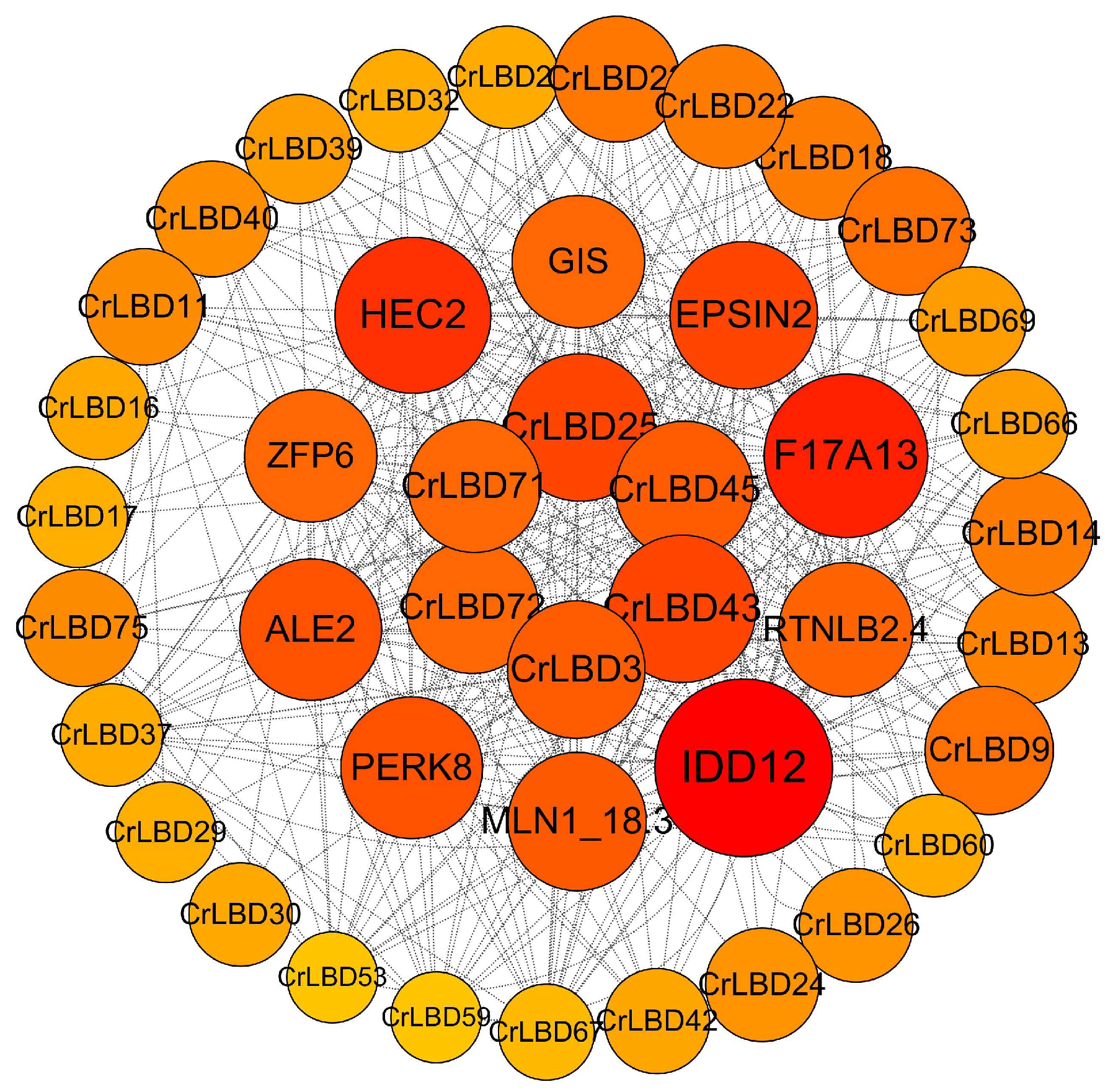
Potential functional interactions among CrLBD proteins were predicted using the STRING database (Figure 6). The resulting protein–protein interaction network contains 16 nodes, with CrLBD3, CrLBD43, CrLBD45, CrLBD25, CrLBD71, and CrLBD72 identified as central hub proteins. While certain proteins such as CrLBD72, CrLBD3, and CrLBD45 engage in direct pairwise interactions, others participate in more complex multi-protein transcriptional complexes. Furthermore, we extended our analysis to predict interactions between CrLBD proteins and other protein families (Figure 6). The results indicate that CrLBD proteins interact with several ancient plant transcription factor families, such as IDD, HEC, GIS, ZFP, PERK, and RTNLB, among which many have been experimentally demonstrated to participate in the regulation of floral organ development (including IDD, HEC, GIS, etc.) [47,48,49].

3.6. Analysis of Differential Expression Patterns of LBD Genes Related to Flower Morphological Diversity of C. retusus

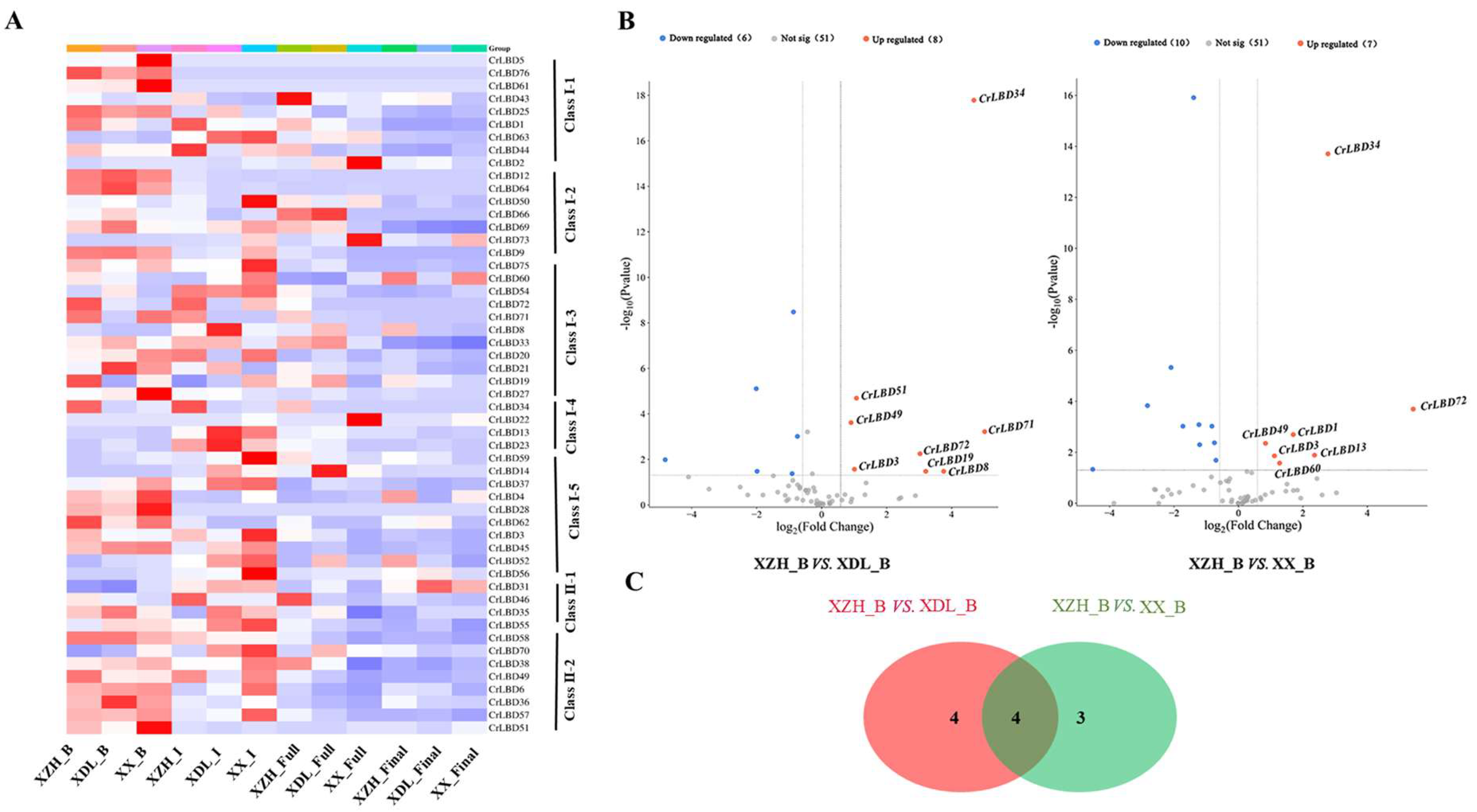
As an ornamental garden species, C. retusus displays significant variation in floral development, a key esthetic value. To investigate how CrLBD gene expression regulates floral organ development in C. retusus, we performed transcriptome sequencing and analysis of three varieties across four key developmental stages. Expression profiling revealed that the 76 CrLBD genes display diverse spatio-temporal expression patterns during floral development (Figure 7A). Notably, members of the same subfamily often showed similar expression profiles. For example, in Class I-5, CrLBD3, CrLBD45, and CrLBD52 exhibited correlated expression, while in Class I-3, CrLBD8, CrLBD71, and CrLBD72 shared comparable patterns (Figure 7A).
Comparative analysis indicated that several CrLBD genes are upregulated in XZH during the bud, initial, and full flowering stages relative to XX and XDL. For instance, CrLBD34 and CrLBD71 show higher expression in XZH, a cultivar characterized by flat petals, unlike the curved petals of XX and XDL. To identify candidate regulators of floral development, we focused on CrLBD genes consistently upregulated in XZH buds compared to the other two cultivars. Four genes were highlighted: CrLBD72 (Class I-3), CrLBD34 (Class I-4), CrLBD3 (Class I-5), and CrLBD49 (Class II-2) (Figure 7B).
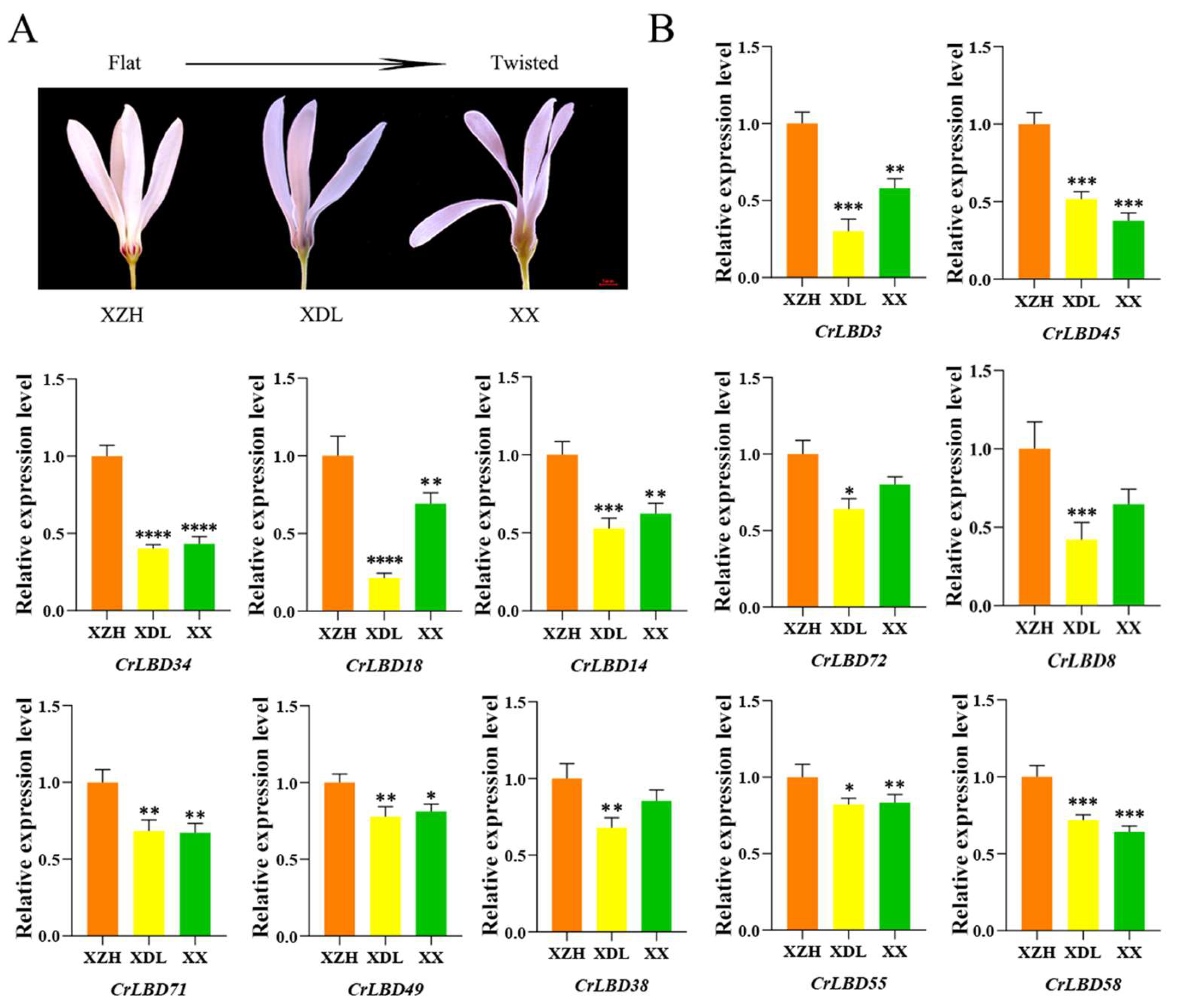
We further validated the expression of these genes using qRT-PCR across the three varieties. Among them, only CrLBD3 and CrLBD34 showed statistically significant and variety-specific expression divergence (Figure 8B). Specifically, CrLBD3 expression in the flat-petaled variety (XZH) was 1.72-to 3.32-fold higher than in other cultivars, while CrLBD34 expression varied 2.31-to 2.49-fold between varieties.
Beyond CrLBD3 and CrLBD34, we identified CrLBD45, CrLBD18, and CrLBD14 as additional homologs of AtLOB and AtLBD21 in C. retusus. To assess whether these homologs influence floral morphology, we compared their expression profiles across the three cultivars. Expression analysis revealed that CrLBD45, CrLBD18, and CrLBD14 exhibit conserved expression patterns with CrLBD3 and CrLBD34 (Figure 8B).

4. Discussion

Floral morphological diversity directly influences plant ornamental value, making the elucidation of intrinsic mechanisms regulating flower shape crucial for breeding new high-quality ornamental varieties [6]. As a commonly used ornamental landscape plant, the flower forms of the C. retusus exhibit remarkable diversity [35]. The LBD gene family, encoding key transcription factors involved in plant development, plays vital roles in lateral organ formation, leaf development, inflorescence architecture, and floral organ morphogenesis [10]. Our study identified 76 LBDs genes in C. retusus, substantially more than in other dicots such as Arabidopsis thaliana (43) and Populus trichocarpa (55), though fewer than in monocots like wheat (90) [50]. The evolutionary history of LBD genes involves complex patterns of duplication and loss across species; yet we found most homologous genes maintained similar expression patterns, suggesting functional conservation despite sequence divergence. For instance, AtLOB orthologs CrLBD3 and CrLBD45 show high expression in flower buds and initial flowering stages, while AtLBD1 orthologs CrLBD72 and CrLBD71 exhibit conserved expression profiles, indicating retention of ancestral expression characteristics. However, some homologous genes display divergent expression patterns, such as AtLBD16 orthologs CrLBD9 (peaking at initial flowering) and CrLBD73 (peaking at full flowering). Comparative analysis with A. thaliana indicates that the C. retusus LBD family has expanded through whole-genome duplication events, with new copies exhibiting spatiotemporally specific expression changes.
Previous studies classified LBD genes into five major subfamilies (IA, IB, IC, IE, and II) based on functional attributes and sequence homology [21]. Here, we identified 76 LBD genes in C. retusus and classified them into seven subfamilies: Class I (I-1 to I-5) and Class II (II-1 and II-2). Compared to A. thaliana, certain subfamilies show notable expansion in C. retusus: Class I-2 and I-4 increased approximately 3-fold and 2.6-fold, respectively, while Class I-5 and I-3 expanded 2.5-fold and 2.29-fold. Given that Class I-3, I-5, and II-2 participate in floral organ development and anthocyanin synthesis, while Class I-2 primarily regulates underground organ development [21], the differential expansion of LBD subfamilies in C. retusus may represent adaptive evolution fine-tuning gene regulatory networks for specific developmental processes.
As an ornamental species with natural floral morphological diversity, understanding the mechanisms underlying floral morphogenesis is essential for breeding new C. retusus varieties [7]. Through transcriptome analysis of cultivars with distinct floral morphologies, we identified three genes differentially expressed at the bud stage: CrLBD3 (Class I-5), CrLBD34 (Class I-4), and CrLBD71 (Class I-3). While Class I-5 primarily functions in floral development [21], Class I-3 members regulate plant growth and development through cytokinin pathways [26]. Subsequent qRT-PCR validation confirmed significant differential expression of CrLBD3 and CrLBD34 and their homologs (CrLBD45, CrLBD18 and CrLBD14). Therefore, the proteins encoded by CrLBD45 and CrLBD18, CrLBD14, which are homologous to AtLOB or AtLBD21, have a crucial impact on the morphology of C. retusus flowers [51]. Notably, these genes belong to Class I-5 and I-4 subfamilies, which are associated with aerial organ development. The CrLBD3 homolog AtLOB interacts with brassinosteroid (BR) signaling during organ boundary formation to modulate local BR accumulation, influencing organ morphology [52,53,54]. Thus, this subfamily in C. retusus may regulate floral traits through similarly complex mechanisms. Although promoter cis-element and conserved motif analyses provided limited explanatory power for subfunctionalization within Class I-5, future population-level studies may yield further insights.
In summary, this study identified 76 LBD gene family members in C. retusus and performed phylogenetic classification of these genes into two distinct subfamilies, revealing their evolutionary relationships. Genomic collinearity analysis revealed both tandem and segmental duplications of CrLBD genes, with evidence of purifying selection acting on these genes during Oleaceae diversification. The evolutionary conservation of CrLBD genes was further supported by their conserved structural features, including characteristic domains and motifs. Expression profiling identified significant upregulation of CrLBD3 and CrLBD34 in flat-petaled cultivars, suggesting their potential role in regulating petal flatness during floral morphogenesis. Collectively, our findings not only advance the current understanding of the LBD gene family in C. retusus but also establish a foundation for further investigation into evolutionary dynamics and molecular mechanisms underlying floral development in this species.

Supplementary Materials

The following supporting information can be downloaded at: https://www.mdpi.com/article/10.3390/horticulturae11121429/s1, Table S1. Primers used in qRT-PCR. Table S2. Physicochemical properties of the CrLBDS gene. Table S3. Differential expansion of LBD gene subfamilies in C. retusus compared to A. thaliana. Table S4. The Ka, Ks, and Ka/Ks values between collinear CrLBDS gene pairs. Figure S1. Collinearity analysis of LBD gene family in C. retusus, Syringa oblata and Olea europaea.

Author Contributions

Conception, J.W., J.L. and M.W.; writing—original draft preparation, M.W.; bioinformatics analysis, L.G. and H.W.; transcriptome analyses, Y.W.; RNA isolation and qRT-PCR experiment, S.Z. and M.W.; samples, W.S.; photos, M.W.; writing—review and editing, J.W.; funding acquisition, J.L. All authors have read and agreed to the published version of the manuscript.

Funding

This work was supported by the Subject of Key R & D Plan of Shandong Province (Major Scientific and Technological Innovation Project) Mining and Accurate Identification of Forest Tree Germplasm Resources (No. 2021LZGC023); and Agricultural science and Technology Fund Project of Shandong province (No. 2019LY001-4).Both funding sources are from the Department of Science & Technology of Shandong Province.

Data Availability Statement

The genomic information of the Chionanthus retusus mentioned in the article is available from the corresponding author upon reasonable request. This assembly used HiC and PacBio methods, with a scaffold quantity of 134. The Hi-C and ONT data, as well was the assemblies have been deposited to National Genomics Data Center with Bioproject ID of CRA011999.

Conflicts of Interest

The authors declare that they have no competing interests.

References

  1. Bretzel, F.; Vannucchi, F.; Romano, D.; Malorgio, F.; Benvenuti, S.; Pezzarossa, B. Wildflowers: From conserving biodiversity to urban greening—A review. Urban For. Urban Green. 2016, 20, 428–436. [Google Scholar] [CrossRef]
  2. Ren, Y.; Ge, Y.; Ma, D.; Song, X.; Shi, Y.; Pan, K.; Chang, J. Enhancing plant diversity and mitigating BVOC emissions of urban green spaces through the introduction of ornamental tree species. Urban For. Urban Green. 2017, 27, 305–313. [Google Scholar] [CrossRef]
  3. De-kui, Z.; Qi-bai, X.; Ri-ming, H.A.O. Study on distribution and utilization of plants in genus osmanthus. J. Southwest For. Univ. 2004, 24, 23–26. [Google Scholar] [CrossRef]
  4. Atchudan, R.; Edison, T.N.J.I.; Chakradhar, D.; Perumal, S.; Shim, J.J.; Lee, Y.R. Facile green synthesis of nitrogen-doped carbon dots using Chionanthus retusus fruit extract and investigation of their suitability for metal ion sensing and biological applications. Sens. Actuators B Chem. 2017, 246, 497–509. [Google Scholar] [CrossRef]
  5. He, Y.; Liu, L.; Yang, S.; Dong, M.; Yuan, W.; Shang, F. Characterization of the complete chloroplast genome of Chinese fringetree (Chionanthus retusus). Conserv. Genet. Resour. 2017, 9, 431–434. [Google Scholar] [CrossRef]
  6. Yang, Y.; He, S. A Review on the Application of Chionanthus retusus in Greening Urban Gardens. Kunming Univ. 2022, 44, 110–115. [Google Scholar]
  7. Song, J.; Oak, M.; Hong, S. Morphological traits in an androdioecious species, Chionanthus retusus (Oleaceae). Flora 2016, 223, 129–137. [Google Scholar] [CrossRef]
  8. Song, J.; Hong, S. Identity and localization of floral scent components in an androdioecious species, Chionanthus retusus (Oleaceae). Asia-Pac. Biodivers. 2020, 13, 288–294. [Google Scholar] [CrossRef]
  9. Shuai, B.; Reynaga-Pena, C.; Springer, P. The Lateral Organ Boundaries Gene Defines a Novel, Plant-Specific Gene Family. Plant Physiol. 2002, 129, 747–761. [Google Scholar] [CrossRef]
  10. Lu, H.; Dai, Z.; Li, L.; Wang, J.; Miao, X.; Shi, Z. OsRAMOSA2 shapes panicle architecture through regulating pedicel length. Front. Plant Sci. 2017, 8, 1538. [Google Scholar] [CrossRef]
  11. Majer, C.; Hochholdinger, F. Defining the boundaries: Structure and function of LOB domain proteins. Trends Plant Sci. 2011, 16, 47–52. [Google Scholar] [CrossRef] [PubMed]
  12. Li, A.; Zhang, Y.; Dai, Z. Progress of LBD gene family in higher plants. Mol. Plant Breed. 2006, 3, 301–308. [Google Scholar]
  13. Yang, Y.; Yu, X.; Wu, P. Comparison and evolution analysis of two rice subspecies LATERAL ORGAN BOUNDARIES domain gene family and their evolutionary characterization from Arabidopsis. Mol. Phylogenet. Evol. 2006, 39, 248–262. [Google Scholar] [CrossRef] [PubMed]
  14. Wang, X.; Zhang, S.; Su, L.; Liu, X.; Hao, Y. A genome-wide analysis of the LBD (LATERAL ORGAN BOUNDARIES domain) gene family in Malus domestica with a functional characterization of MdLBD11. PLoS ONE 2013, 8, e57044. [Google Scholar] [CrossRef]
  15. Dang, H.; Yu, C.; Nan, S.; Li, Y.; Du, S.; Zhao, K.; Wang, S. Genome-wide identification and gene expression networks of LBD transcription factors in Populus trichocarpa. BMC Genom. 2024, 25, 920. [Google Scholar] [CrossRef]
  16. Goh, T.; Toyokura, K.; Yamaguchi, N.; Okamoto, Y.; Uehara, T.; Kaneko, S.; Takebayashi, Y.; Kasahara, H.; Ikeyama, Y.; Okushima, Y.; et al. Lateral root initiation requires the sequential induction of transcription factors LBD16 and PUCHI in Arabidopsis thaliana. New Phytol. 2019, 224, 749–760. [Google Scholar] [CrossRef]
  17. Wang, Y.; Wang, P.; Di, P.; Wang, Y. Genome-wide analysis of LBD genes in the medicinal plant Panax ginseng reveals the roles and molecular mechanisms of PgLBD18 and PgLBD49 in regulating lateral root development. Ind. Crops Prod. 2025, 232, 121232. [Google Scholar] [CrossRef]
  18. Semiarti, E.; Ueno, Y.; Tsukaya, H.; Iwakawa, H.; Machida, C.; Machida, Y. The ASYMMETRIC LEAVES2 gene of Arabidopsis thaliana regulates formation of a symmetric lamina, establishment of venation and repression of meristem-related homeobox genes in leaves. Development 2001, 128, 1771–1783. [Google Scholar] [CrossRef]
  19. Wang, L.; Wang, W.; Xu, D.; Chang, Z.; Cui, X. Overexpression of Arabidopsis ASL25/LBD28 gene affects leaf morphogenesis. Acta Bot. Boreali Occident. 2010, 30, 888–893. [Google Scholar]
  20. Kim, M.J.; Kim, M.; Kim, J. Combinatorial interactions between LBD10 and LBD27 are essential for male gametophyte development in Arabidopsis. Plant Signal. Behav. 2015, 10, e1044193. [Google Scholar] [CrossRef]
  21. Zhang, Y.; Li, Z.; Ma, B.; Hou, Q.; Wan, X. Phylogeny and functions of LOB domain proteins in plants. Mol. Sci. 2020, 21, 2278. [Google Scholar] [CrossRef]
  22. Koppolu, R.; Anwar, N.; Sakuma, S.; Tagiri, A.; Lundqvist, U.; Pourkheirandish, M.; Rutten, T.; Seiler, C.; Himmelbach, A.; Ariyadasa, R.; et al. Six-rowed spike4 (Vrs4) controls spikelet determinacy and row-type in barley. Proc. Natl. Acad. Sci. USA 2013, 110, 13198–13203. [Google Scholar] [CrossRef] [PubMed]
  23. Cao, H.; Liu, C.; Liu, C.; Zhao, Y.; Xu, R. Genomewide analysis of the lateral organ boundaries domain gene family in Vitis vinifera. J. Genet. 2016, 95, 515–526. [Google Scholar] [CrossRef] [PubMed]
  24. Goh, T.; Joi, S.; Mimura, T.; Fukaki, H. The establishment of asymmetry in Arabidopsis lateral root founder cells is regulated by LBD16/ASL18 and related LBD/ASL proteins. Development 2012, 139, 883–893. [Google Scholar] [CrossRef] [PubMed]
  25. Cho, C.; Jeon, E.; Pandey, S.; Ha, S.; Kim, J. LBD13 positively regulates lateral root formation in Arabidopsis. Planta 2019, 249, 1251–1258. [Google Scholar] [CrossRef]
  26. Jeon, E.; Young Kang, N.; Cho, C.; Joon Seo, P.; Chung Suh, M.; Kim, J. LBD14/ASL17 positively regulates lateral root formation and is involved in ABA response for root architecture in Arabidopsis. Plant Cell Physiol. 2017, 58, 2190–2201. [Google Scholar] [CrossRef]
  27. Naito, T.; Yamashino, T.; Kiba, T.; Koizumi, N.; Kojima, M.; Sakakibara, H.; Mizuno, T. A link between cytokinin and ASL9 (ASYMMETRIC LEAVES 2 LIKE 9) that belongs to the AS2/LOB (LATERAL ORGAN BOUNDARIES) family genes in Arabidopsis thaliana. Biosci. Biotechnol. Biochem. 2007, 71, 1269–1278. [Google Scholar] [CrossRef]
  28. Li, C.; Zou, X.; Zhang, C.; Shao, Q.; Liu, J.; Liu, B.; Li, H.; Zhao, T. OsLBD3-7 Overexpression Induced Adaxially Rolled Leaves in Rice. PLoS ONE 2016, 11, e0156413. [Google Scholar] [CrossRef]
  29. Li, C.; Zhu, S.; Zhang, H.; Chen, L.; Cai, M.; Wang, J.; Chai, J.; Wu, F.; Cheng, Z.; Guo, X.; et al. OsLBD37 and OsLBD38, two class II type LBD proteins, are involved in the regulation of heading date by controlling the expression of Ehd1 in rice. Biochem. Biophys. Res. Commun. 2017, 486, 720–725. [Google Scholar] [CrossRef]
  30. Wang, Y.; Song, J.; Wang, Z. ASYMMETRIC LEAVES2-LIKE38, one member of AS2/LOB gene family, involves in regulating ab-adaxial patterning in Arabidopsis lateral organs. Acta Physiol. Plant. 2015, 37, 185. [Google Scholar] [CrossRef]
  31. Causier, B.; Ashworth, M.; Guo, W.; Davies, B. The TOPLESS Interactome: A Framework for Gene Repression in Arabidopsis. Plant Physiol. 2012, 158, 423–438. [Google Scholar] [CrossRef] [PubMed]
  32. Liu, R.; Wang, Z.; He, X.; Zhou, H.; Xu, Y.; Hong, L. PRESSED FLOWER works downstream of ASYMMETRIC LEAVES 2 to affect sepal flatness in Arabidopsis. Exp. Bot. 2025, 76, 4101–4113. [Google Scholar] [CrossRef] [PubMed]
  33. Li, Q.; Wu, L.; Li, J.; Cao, Q.; Gong, P.; Wang, L.; He, C. Heterometric expression of an LBD gene via LBD-TCP assembly regulates floral organ size and fruit weight in Physalis. Hortic. Res. 2025, 12, uhaf211. [Google Scholar] [CrossRef] [PubMed]
  34. Bortiri, E.; Chuck, G.; Vollbrecht, E.; Rocheford, T.; Martienssen, R.; Hake, S. ramosa2 Encodes a LATERAL ORGAN BOUNDARY Domain Protein That Determines the Fate of Stem Cells in Branch Meristems of Maize. Plant Cell 2006, 18, 574–585. [Google Scholar] [CrossRef]
  35. Wang, J.; Xu, D.; Sang, Y.L.; Sun, M.; Liu, C.; Niu, M.; Li, J. A telomere-to-telomere gap-free reference genome of Chionanthus retusus provides insights into the molecular mechanism underlying petal shape changes. Hortic. Res. 2024, 11, uhae249. [Google Scholar] [CrossRef]
  36. Kumar, S.; Stecher, G.; Li, M.; Knyaz, C.; Tamura, K. MEGA X: Molecular Evolutionary Genetics Analysis across Computing Platforms. Mol. Biol. Evol. 2018, 35, 1547–1549. [Google Scholar] [CrossRef]
  37. Chen, C.; Chen, H.; Zhang, Y.; Thomas, H.; Frank, M.; He, Y.; Xia, R. TBtools: An Integrative Toolkit Developed for Interactive Analyses of Big Biological Data. Mol. Plant 2020, 13, 1194–1202. [Google Scholar] [CrossRef]
  38. Marchler-Bauer, A.; Bryant, S. CD-Search: Protein domain annotations on the fly. Nucleic Acids Res. 2004, 32, W327–W331. [Google Scholar] [CrossRef]
  39. Bailey, T.; Boden, M.; Buske, F.; Frith, M.; Grant, C.; Clementi, L.; Ren, J.; Li, W.; Noble, W. MEME SUITE: Tools for motif discovery and searching. Nucleic Acids Res. 2009, 37, W202–W208. [Google Scholar] [CrossRef]
  40. Lescot, M.; Dehais, P.; Thijs, G.; Marchal, K.; Moreau, Y.; Peer, Y.; Rouze, P.; Rombauts, S. PlantCARE, a database of plant cis-acting regulatory elements and a portal to tools for in silico analysis of promoter sequences. Nucleic Acids Res. 2002, 30, 325–327. [Google Scholar] [CrossRef]
  41. Ma, B.; Wu, J.; Shi, L.; Yang, Y.; Wang, B.; Zheng, Y.; Su, S.; Yao, Y.; Xue, W.; Porth, I.; et al. Lilac (Syringa oblata) genome provides insights into its evolution and molecular mechanism of petal color change. Commun. Biol. 2022, 5, 686. [Google Scholar] [CrossRef] [PubMed]
  42. Rao, G.; Zhang, J.; Liu, X.; Lin, C.; Xin, H.; Xue, L.; Wang, C. De novo assembly of a new Olea europaea genome accession using nanopore sequencing. Hortic. Res. 2021, 8, 64. [Google Scholar] [CrossRef] [PubMed]
  43. Livak, K.; Schmittgen, T. Analysis of relative gene expression data using real-time quantitative PCR and the 2−ΔΔCT Method. Methods 2001, 25, 402–408. [Google Scholar] [CrossRef] [PubMed]
  44. Kim, D.; Langmead, B.; Salzberg, S. HISAT: A fast spliced aligner with low memory requirements. Nat. Methods 2015, 12, 357–360. [Google Scholar] [CrossRef]
  45. Finn, R.; Clements, J.; Eddy, S. HMMER web server: Interactive sequence similarity searching. Nucleic Acids Res. 2011, 39, W29–W37. [Google Scholar] [CrossRef]
  46. Wang, Y.; Tang, H.; DeBarry, J.; Tan, X.; Li, J.; Wang, X.; Lee, T.; Jin, H.; Marler, B.; Guo, H.; et al. MCScanX: A toolkit for detection and evolutionary analysis of gene synteny and collinearity. Nucleic Acids Res. 2012, 40, e49. [Google Scholar] [CrossRef]
  47. Fan, S.; Zhang, D.; Xing, L.; Qi, S.; Du, L.; Wu, H.; Han, M. Phylogenetic analysis of IDD gene family and characterization of its expression in response to flower induction in Malus. Mol. Genet. Genom. 2017, 292, 755–771. [Google Scholar] [CrossRef]
  48. Ballester, P.; Martínez-Godoy, M.; Ezquerro, M.; Navarrete-Gómez, M.; Trigueros, M.; Rodríguez-Concepción, M.; Ferrándiz, C. A transcriptional complex of NGATHA and bHLH transcription factors directs stigma development in Arabidopsis. Plant Cell 2021, 33, 3645–3657. [Google Scholar] [CrossRef]
  49. Yang, C.; Sun, L.; Yasin, M.; Zulqarnain, H.; Ali, B.; Liu, Y.; Gan, Y. GIS mediates GA signaling to directly target the expression of SPL15 to regulate trichome development in Arabidopsis thaliana. Plant Physiol. Biochem. 2025, 225, 109975. [Google Scholar] [CrossRef]
  50. Wang, Z.; Zhang, R.; Cheng, Y.; Lei, P.; Song, W.; Zheng, W.; Nie, X. Genome-wide identification, evolution, and expression analysis of LBD transcription factor family in bread wheat (Triticum aestivum L.). Front. Plant Sci. 2021, 12, 721253. [Google Scholar] [CrossRef]
  51. Chalfun-Junior, A.; Franken, J.; Mes, J.; Marsch-Martinez, N.; Pereira, A.; Angenent, G. ASYMMETRIC LEAVES2-LIKE1 gene, a member of the AS2/LOB family, controls proximal–distal patterning in Arabidopsis petals. Plant Mol. Biol. 2005, 57, 559–575. [Google Scholar] [CrossRef]
  52. Gendron, J.; Liu, J.; Fan, M.; Bai, M.; Wenkel, S.; Springer, P.; Barton, M.; Wang, Z. Brassinosteroids regulate organ boundary formation in the shoot apical meristem of Arabidopsis. Proc. Natl. Acad. Sci. USA 2012, 109, 21152–21157. [Google Scholar] [CrossRef]
  53. Sun, Y.; Fan, X.; Cao, D.; Tang, W.; He, K.; Zhu, J.; He, J.; Bai, M.; Zhu, S.; Oh, E.; et al. Integration of brassinosteroid signal transduction with the transcription network for plant growth regulation in Arabidopsis. Dev. Cell 2010, 19, 765–777. [Google Scholar] [CrossRef]
  54. Bell, E.; Lin, W.; Husbands, A.; Yu, L.; Jaganatha, V.; Jablonska, B.; Mangeon, A.; Neff, M.; Girke, T.; Springer, P. Arabidopsis LATERAL ORGAN BOUNDARIES negatively regulates brassinosteroid accumulation to limit growth in organ boundaries. Proc. Natl. Acad. Sci. USA 2012, 109, 21146–21151. [Google Scholar] [CrossRef]
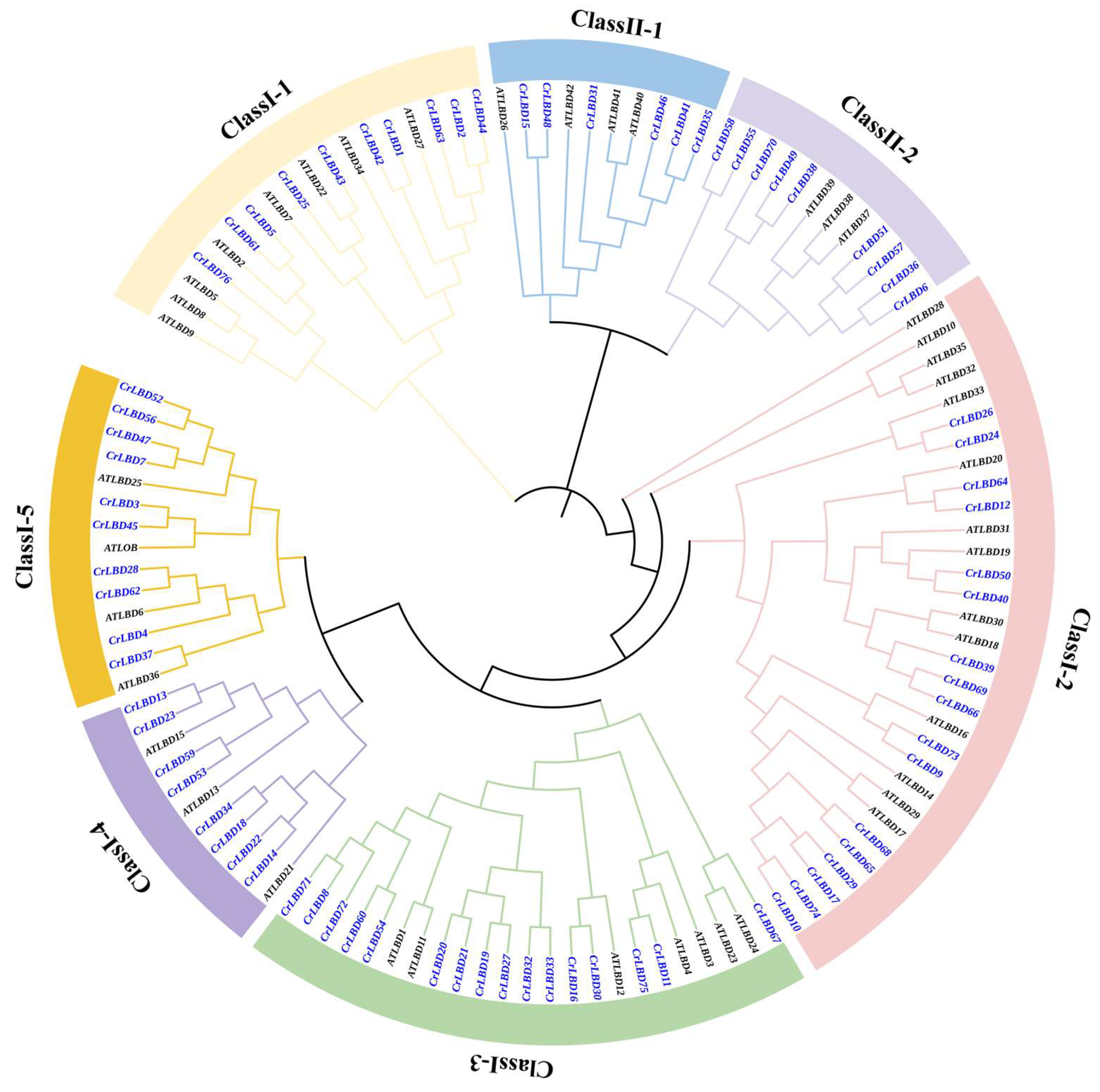
Figure 1. Phylogenetic Tree of LBD genes in C. retusus and Arabidopsis. The blue font represents the LBD genes of C. retusus, and different colored backgrounds represent different subfamilies.
Horticulturae 11 01429 g001
Figure 2. Chromosome distribution analysis of CrLBDs. Chromosome numbers are indicated in yellow, gene identifiers in red, and short gray tick marks denote specific gene positions along the chromosomes. The color gradient within chromosomes (blue to red) represents gene density, with increasing red intensity indicating higher gene concentration.
Horticulturae 11 01429 g002
Figure 3. Chromosome distribution and duplication event analysis of CrLBDs. Lines of the same color connect collinear gene pairs. The gray shaded areas represent collinear gene pairs of other non-CrLBD gene family members. The red lines in the frame indicate the chromosomal density at corresponding positions on the chromosome.
Horticulturae 11 01429 g003
Figure 4. Phylogenetic relationships, domains, and motifs composition of CrLBDs. (A) Conserved domain analysis of LBD gene family in C. retusus, different colors represent different conserved domains; (B) Gene structure analysis of LBD gene family in C. retusus, green represents UTR region, yellow represents CDS encoding region.
Horticulturae 11 01429 g004
Figure 5. Enrichment and distribution of cis-acting elements in the promoter of CrLBDs. (A) Enrichment of each Cis-acting element. The size and color of the dots represent the number of Cis-acting elements. (B) Distribution of Cis-acting elements in CrLBD promoter region.
Horticulturae 11 01429 g005
Figure 6. Protein–protein interaction (PPI) network of significant genes in C. retusus. Nodes represent proteins, central nodes are indicated in red, and black lines indicate interactions between nodes. The darker the color and bigger the circle size, the more important the protein in the interaction network.
Horticulturae 11 01429 g006
Figure 7. Expression and differential analysis of CrLBDs. A Heatmap of the expression of 76 CrLBDs in flower organs at four stages of three varieties. (A) The expression heatmap of 76 LBD in flower organs of three varieties at four stages. B represents the Bud stage, I represents the Initial flowering stage, Full represents the Full flowering stage, and Final represents the Final flowering stage, the distinct color groups above the heatmap represent different stages; (B) Genes differentially expressed in the bud stage between ‘XZH’ and two other varieties. (C) Wayne diagram of differentially expressed genes.
Horticulturae 11 01429 g007
Figure 8. The floral organs of eight varieties and the expression patterns of CrLBDs in floral organs. (A) Vertical sectional views of floral organs during the bud stage. Among them, ‘XZH’ is flat type; ‘XDL’ is semi closed type, ‘XX’ are spiral twisted type. (B) The relative expression levels of differentially expressed CrLBDs in flower organs of 3 varieties. UBC2 was used as an internal control. The expression of each gene in the ‘XZH’ was set as 1. Data are presented as means ± SD (n = 3). Asterisks indicate significant differences (t-test) compared to the ‘XZH’(flat). * p < 0.05, ** p < 0.01, *** p < 0.001, **** p < 0.0001.
Horticulturae 11 01429 g008
Disclaimer/Publisher’s Note: The statements, opinions and data contained in all publications are solely those of the individual author(s) and contributor(s) and not of MDPI and/or the editor(s). MDPI and/or the editor(s) disclaim responsibility for any injury to people or property resulting from any ideas, methods, instructions or products referred to in the content.