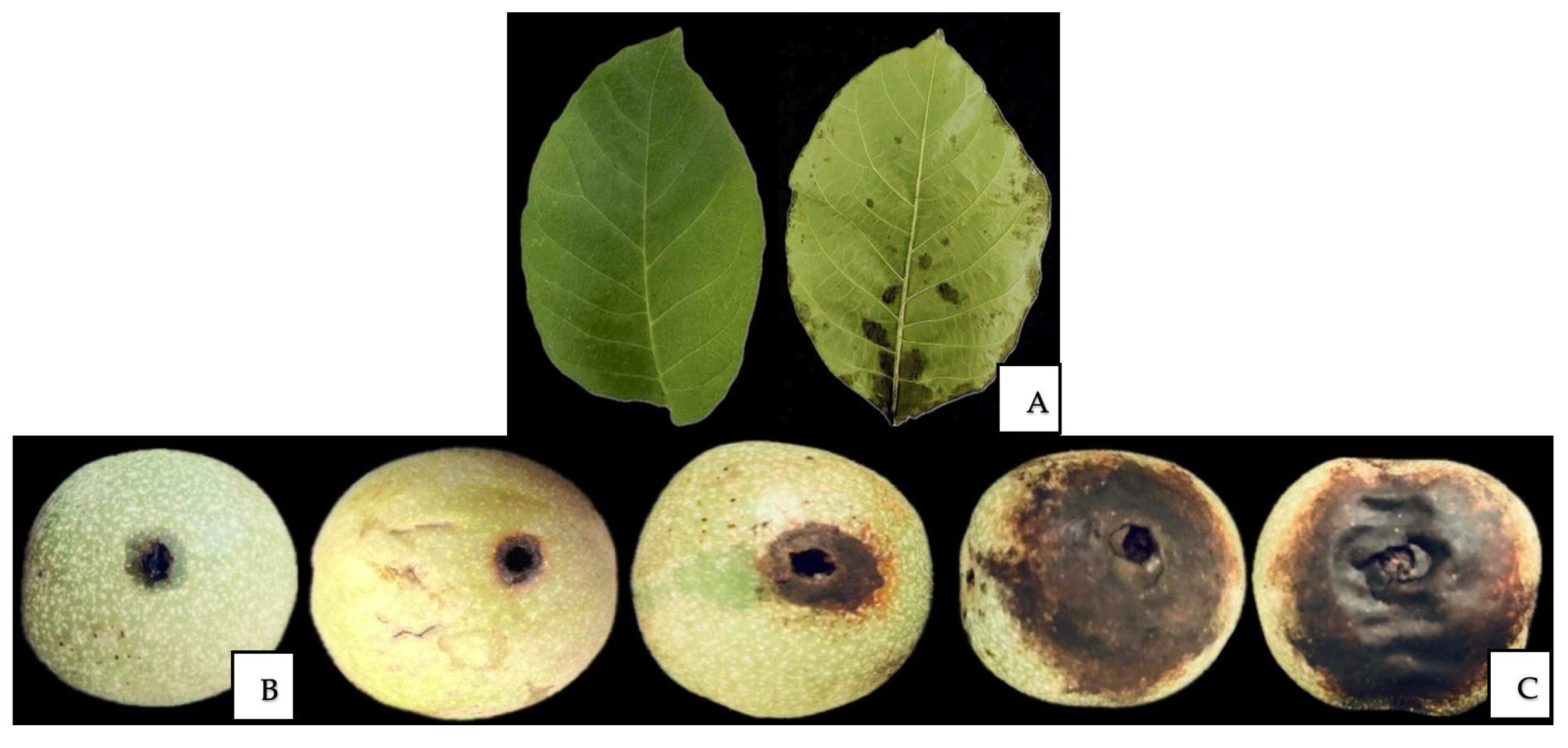
Article

Identification, Characterization, and Pathogenicity of Fungal and Bacterial Pathogens of Walnut (Juglans regia L.) in Kazakhstan

by
Elmira Ismagulova
1,
Sergey Oleichenko
1,
Moldir Sarshayeva
1,2,
Saule Korabayeva
1,3,
Gulnaz Nizamdinova
4,
Dilyara Gritsenko
4,
Gulnur Suleimanova
1,
Zagipa Sapakhova
5,
Huseyin Basim
6 and
Gulshariya Kairova
1,*
1
Department of Horticulture, Plant Protection and Quarantine, Kazakh National Agrarian Research University, Almaty 050010, Kazakhstan
2
Laboratory of Biotechnology of Horticultural Crops, Kazakh Research Institute of Fruit and Vegetable Growing, Almaty 050060, Kazakhstan
3
Department of Plant Protection, Kazakh Research Institute of Fruit and Vegetable Growing, Almaty 050060, Kazakhstan
4
Laboratory of Molecular Biology, Institute of Plant Biology and Biotechnology, Almaty 050040, Kazakhstan
5
Breeding and Biotechnology Laboratory, Institute of Plant Biology and Biotechnology, Almaty 050040, Kazakhstan
6
Department of Plant Protection, Akdeniz University, Antalya 07070, Türkiye
*
Author to whom correspondence should be addressed.
Horticulturae 2025, 11(10), 1217; https://doi.org/10.3390/horticulturae11101217
Submission received: 8 September 2025 / Revised: 1 October 2025 / Accepted: 8 October 2025 / Published: 10 October 2025
(This article belongs to the Section Plant Pathology and Disease Management (PPDM))

Abstract

Walnut (Juglans regia L.) is a significant nut crop in the southern regions of Kazakhstan; however, its productivity is substantially limited by fungal and bacterial diseases. Therefore, a phytopathological investigation was conducted in 2023–2024 in the Almaty and Turkestan regions, including field monitoring, pathogen isolation, molecular identification, and pathogenicity testing. Field monitoring revealed that symptoms of brown spot, walnut canker, walnut blight, bacterial blight, and crown gall were widespread. The overall disease incidence ranged from 8% to 30%, while the disease severity index varied from 15% to 70% across the surveyed sites. Pure cultures of pathogens were isolated from 69 samples, and their morphology was characterized. Molecular identification through sequencing of universal genetic loci (the internal transcribed spacer for fungi and 16S ribosomal RNA for bacteria) revealed the presence of the fungal species Alternaria alternata and Fusarium incarnatum, as well as the bacterial species Pantoea agglomerans and Xanthomonas arboricola pv. juglandis. Pathogenicity testing confirmed the virulence of the identified pathogens, which induced characteristic symptoms of brown spot, walnut canker, and walnut blight, consistent with those observed in the field. These findings have considerable practical significance for improving phytosanitary monitoring and protection systems in walnut plantations, thereby facilitating disease outbreak prediction and the development of effective quarantine measures.

1. Introduction

Juglans regia, commonly known as the common walnut, English walnut, or Persian walnut, is a widely distributed walnut species [1,2]. It is native to Eurasia, particularly Southwest and Central Asia, as well as Southeastern Europe [3]. Walnuts are among the most widely consumed tree nuts worldwide, valued for their nutritional, economic, and ecological significance. They are a rich source of healthy fats, proteins, dietary fiber, vitamins, and minerals, making them an important component of a balanced diet [2]. Economically, walnut cultivation is of considerable importance in many countries, including Kazakhstan—particularly in the Almaty region—where the species is both cultivated and consumed [1,4].
In Kazakhstan, walnut cultivation has grown rapidly in recent years. According to the Bureau of National Statistics, the plantation area increased from only 0.5 hectares in 2013 to nearly 900 hectares in 2021, with an annual production volume exceeding 3000 t. Approximately 65% of plantations are located in the Turkestan region, 30% in Almaty, and smaller areas in Zhambyl and Shymkent [5]. These figures highlight the increasing economic importance of walnut production in the country.
The favorable climatic conditions in southern Kazakhstan—moderate humidity, mild winters, and stable summer temperatures—support walnut cultivation but also create favorable conditions for the spread of fungal and bacterial diseases [1]. In a separate study conducted in China, 64 endophytic fungal strains belonging to 17 genera were isolated from walnut tissues (leaves, branches, fruits, and roots). The most abundant genus was Alternaria sp. In addition, Talaromyces sp., Curvularia sp., and Eurotium sp. were first isolated from walnut plant tissues [6]. Later, differences in fungal diversity (Chao1 index, Shannon index, and Simpson index) and community structure were revealed by sequencing and analyzing the microbiota of diseased and healthy leaves using Illumina HiSeq technology. The main fungal phyla associated with walnut leaves were the Ascomycota, Basidiomycota, and Glomeromycota. The diversity indices (Shannon and Chao1 index values) in healthy leaves differed significantly at the late stages of disease development. The results showed that the composition of fungal species significantly differed between healthy and infected leaves and changed as the disease progressed [7]. In China, the serious walnut diseases include walnut anthracnose, walnut blight, walnut canker, walnut shoot dieback, and walnut brown apical necrosis. Walnut anthracnose is caused by Colletotrichum gloeosporioides; a complex infection of Xanthomonas arboricola pv. juglandis (X. arboricola pv. juglandis), Pantoea agglomerans, and Alternaria spp. causes walnut blight and brown apical necrosis (BAN); and walnut canker is caused by Botryosphaeria dothidea. B. dothidea and Phomopsis sp. can be isolated from walnut dieback [8].
The fungus Alternaria alternata causes brown spot, affecting leaves, branches, and fruits. This disease manifests as brown lesions on the leaves, which can coalesce and lead to leaf blight and premature defoliation. The first cases were reported in Italy in 1998 [9], followed by outbreaks in China (Xinjiang and Sichuan) [10], Serbia [11], India [12], and Chile [13]. This infection results in significant yield reduction and requires continuous monitoring [12].
Fusarium incarnatum (syn. F. semitectum) has also been reported as a causal agent of walnut diseases in several countries. In Argentina, it was identified as the cause of walnut canker, producing fruit necrosis and stem cankers [14]. In India, the pathogen was found in walnut nurseries, where it induced severe stem cankers and dieback [15]. In Italy, F. incarnatum was described as one of the main causal agents of twig cankers and as part of the BAN complex affecting walnut fruits [16].
One of the most aggressive bacterial pathogens affecting walnuts is P. agglomerans [17]. This bacterium was first officially reported as the causative agent of BAN in Shandong Province (China) in 2011. The disease was characterized by premature fruit drop, affecting up to 60% of trees [8]. In 2023, the complete genome sequence of the P. agglomerans strain CHTF15, isolated from infected walnut leaves, was published. Genomic analysis using Tn-seq technology identified 105 key genes required for the bacterium’s survival under nutrient stress, highlighting its strong adaptive capacity and resistance to unfavorable conditions. These characteristics complicate phytosanitary control [18].
The bacterium Xanthomonas campestris pv. juglandis is recognized as the causal agent of bacterial blight. The disease was first described in California in 1901 by Pierce, who observed severe necrosis of shoots and fruits. Mulrean and Schroth [19] further demonstrated the role of overwintering inoculum in buds and catkins, showing that up to 95% of buds of the ‘Payne’ cultivar and more than 40% of catkins could harbor the pathogen, confirming their epidemiological importance. Historically, the pathogen was designated as X. campestris pv. juglandis. However, based on comprehensive DNA–DNA hybridization and polyphasic taxonomic analyses, Vauterin et al. [20] reclassified it as the novel species X. arboricola pv. juglandis. In Lithuania, Burokiene and Pulawska [21] reported bacterial blight for the first time, isolating 59 strains and confirming several isolates as X. arboricola pv. juglandis through pathogenicity assays, biochemical tests, and PCR. Subsequently, Bandi et al. [22] characterized X. arboricola pv. juglandis isolates from Hungary and Romania, confirmed their pathogenicity, and demonstrated high phenotypic similarity among them. In South Korea, Kim et al. [23] documented X. arboricola pv. juglandis in the northern Gyeongbuk Province, confirming the pathogenicity of isolates resistant to copper but sensitive to antibiotics. In northern Italy, Pardatscher et al. [24] reported bacterial blight outbreaks caused by X. arboricola pv. juglandis, affecting leaves, fruits, and young shoots; pathogenicity was confirmed, whereas copper-based treatments showed reduced efficacy. In Poland, Kałużna et al. [25] confirmed the presence and pathogenicity of X. arboricola pv. juglandis, representing the first report of bacterial blight in the country. In Serbia, Iličić et al. [26] associated X. arboricola pv. juglandis with the vertical oozing canker symptoms, and pathogenicity was demonstrated under laboratory and inoculation conditions. More recently, Moragrega and Llorente [27] in Spain showed that infection development by X. arboricola pv. juglandis is strongly influenced by temperature, leaf wetness duration, and host phenology, while young tissues of Chandler and Vina cultivars exhibited high susceptibility. In Argentina, Chorolque et al. [28] reported bacterial blight in major walnut-growing valleys, showing that Chandler was significantly more affected than Franquette. Finally, in Montenegro, Latinović et al. [29] provided the first evidence of bacterial blight caused by X. arboricola pv. juglandis, isolating and confirming pathogenic strains from symptomatic trees.
Agrobacterium tumefaciens was isolated in Iran from root tumors of walnuts. In 2009–2010, crown gall appeared on one-year-old walnut trees grown in the province of Golestan in northern Iran. Galls that were soft, brownish, and spongy, lacking annual growth rings, and measuring 3–8 cm were clearly visible on the crowns of the plants beneath the superficial layer of soil [30]. In the United States, infection with this pathogen has been observed in the hybrid rootstock ‘Paradox’ (Juglans hindsii × J. regia), which is highly susceptible to crown gall. Research has shown that A. tumefaciens can infect walnut seedlings both through infested seeds and soil, establishing systemic populations in root and stem tissues. These systemic populations may remain asymptomatic for some time but can later cause gall formation, especially at wound sites [31]. Studies in Uzbekistan (2022) confirmed the presence of A. tumefaciens in walnut nurseries of the Zarafshan valley, where eight isolates were identified and proven pathogenic using morphological, biochemical, and pathogenicity tests [32]. Similarly, in California, genomic analysis of A. tumefaciens strain 186 from walnut revealed virulence-associated genes on the Ti plasmid, including iaaM, iaaH, ipt, 6b, and 5, confirming its pathogenic potential [33]. While global studies have extensively documented a broad spectrum of walnut pathogens—including bacterial (X. arboricola pv. juglandis, A. tumefaciens), fungal (Marssonina juglandis, Gnomonia leptostyla), viral, and phytoplasma infections—comprehensive phytopathological surveys covering the full pathogen spectrum have not yet been conducted in Kazakhstan. Local studies primarily investigated the genetic diversity, biological traits, and adaptation of J. regia populations [4,34], and some works considered resistance to anthracnose and bacterial blight [35]. This gap between global and local findings underscores the limited understanding of the walnut pathogen complex in Kazakhstan and provides a clear rationale for the present study.
In summary, the most widespread diseases are brown spot (A. alternata) [9,10], walnut canker (F. incarnatum) [14,15], walnut blight (P. agglomerans) [17,18], bacterial blight (X. arboricola pv. juglandis) [21,22,23,24,25,26,27,28,29], and crown gall (A. tumefaciens) [30,31].
Despite extensive international studies, there is little systematic information on the occurrence and severity of walnut pathogens in Kazakhstan. This knowledge gap significantly limits the ability to develop effective phytosanitary monitoring and disease management strategies. Therefore, the rationale for this study was to provide the first comprehensive phytopathological assessment of walnut diseases in Kazakhstan.
The aims of this study were (i) to monitor the incidence and severity of walnut diseases in cultivated and wild stands in the Almaty and Turkestan regions, (ii) to isolate and characterize the main fungal and bacterial pathogens using morphological and molecular approaches, (iii) to compare sequence data with the National Center for Biotechnology Information (NCBI) database, and (iv) to evaluate the pathogenicity of the identified microorganisms.

2. Materials and Methods

2.1. Location and Plant Materials

This research was conducted during 2023–2024 in the Almaty and Turkestan regions of Kazakhstan (Figure 1). The survey focused on nut plantations at the following sites: the rural farm “Manshuk”, “Issyk State Dendrological Park” RSE, “Fazenda UM” LLP (Almaty region), “Saryagash Zher Syyi” LLP, and “Silk alley” LLP (Turkestan region). Additionally, a nut orchard located in the Alma-Arasan gorge area was included in the survey. The objects of the study included both introduced walnut varieties and local forms, as well as wild specimens. Regular disease evaluations were carried out throughout the growing season in southern and southeastern Kazakhstan to ensure the timely detection of fungal and bacterial infections. The sampling of target trees revealed characteristic symptoms, including leaf spotting, chlorosis, wilting, fruit necrosis, stem and branch damage, and bark lesions.
In total, 240 walnut leaves were collected and analyzed, along with samples of fruits, young shoots, branches, and bark. Particular attention was paid to the leaves, as they are the most informative organs for identifying phytopathogens. Plant material was collected at the boundary between affected and visually healthy tissues, to ensure high reliability in subsequent diagnostics [9].

2.2. Disease Evaluation

Pathogenetic symptoms, disease type, onset period, number of cases, and severity grade were recorded, and both disease incidence (DI) and disease severity index (DSI) were calculated. The severity of the disease was assessed using DI and DSI. The DSI was determined based on the percentage of diseased leaves and evaluated on a scale from 0 to 9, as described by Xie et al. [36] and Jiang et al. [37]. Each plant was assigned a disease rating as follows: 0, no diseased leaves; 1, <5% of leaves with disease symptoms; 3, 6–10% of leaves with disease symptoms; 5, 11–25% of leaves with disease symptoms; 7, 26–50% of leaves with disease symptoms; 9, >50% of leaves with disease symptoms.
The DI and DSI were calculated using the following formulas:
Disease incidence (DI, %) = (Σ number of diseased samples /total number of samples surveyed) × 100,
Disease severity index (DSI, %) = [Σ (number of leaves at each scale rating × the scale rating/total number of leaves × highest scale rating)] × 100.

2.3. Isolation of Pathogens from Infected Walnut Trees

Fungal pathogens were analyzed using the wet chamber method to promote mycelial growth and fungal sporulation. Affected plant fragments were pre-washed in running water to remove surface contaminants, then surface-sterilized in 70% ethanol for 30–60 s and dried on sterile filter paper. Sterile plant fragments were transferred to Petri dishes (90 mm, SPL Life Sciences, Pocheon-si, Republic of Korea) containing moistened filter paper disks and incubated in a growth chamber (Binder BD 56, Tuttlingen, Germany) at 20–22 °C for 2–3 days until visible mycelium and/or sporulation developed. Each isolation was performed in three replicates per sample. For the microscopic analysis of the isolated fungi, preparations were made using the “crushed drop” method. Mycelial fragments were mounted in a glycerol–water solution (1:4) on microscope slides (Menzel-Gläser, Thermo Fisher Scientific, Dreieich, Germany) and examined under a compound microscope (Micros MC 300, Vienna, Austria) at magnifications of ×100–400. Measurements were calibrated using a stage micrometer (10 μm divisions, Edmund Optics, Barrington, NJ, USA) [11].
For bacteria, surface-sterilized tissues were homogenized in 1 ml of phosphate-buffered saline (Sigma-Aldrich, St. Louis, MO, USA). Serial dilutions were plated on nutrient agar (NA; Himedia Laboratories, Mumbai, India) using a Drigalski spatula (Brand GmbH, Wertheim, Germany). Plates were incubated for 3–10 days at 28 °C in a calibrated incubator (Memmert IN55, Schwabach, Germany). Colonies were selected based on standard morphological features (shape, pigmentation, texture, consistency), and at least three representative isolates per site were retained for further analysis. All isolations were performed in triplicate [38].

2.4. Isolation of Pure Cultures of Fungi and Bacteria

Pure cultures of the isolated microorganisms were obtained through additional purification to ensure the reliability of subsequent taxonomic and phytopathological analyses. Pure cultures of fungi were established by single-spore isolation on potato dextrose agar (PDA; HiMedia, India). The fungi were incubated at 28–30 °C under high humidity to induce sporulation, and culture purity was verified microscopically [11].
Bacterial isolates were purified by streaking on NA with a sterile loop under aseptic conditions (in a laminar-flow cabinet or near a burner) and incubated at 28–30 °C for 2–5 days. Purity was assessed microscopically based on cell shape, pigmentation, and colony arrangement. The resulting pure isolates were used for subsequent microbiological and phytopathological studies [38].

2.5. Genomic DNA Extraction from Walnut Samples

Genomic deoxyribonucleic acid (DNA) was isolated using a cetyltrimethylammonium bromide (CTAB) buffer containing 100 mM Tris-HCl, 1.4 M NaCl, 20 mM ethylenediaminetetraacetic acid, 4% CTAB, 2% polyvinylpyrrolidone, and 0.2% β-mercaptoethanol [39]. Samples were homogenized in 1 ml of the buffer and incubated at 65 °C for 1 h [40]. One milliliter (mL) of chloroform was added, and the mixture was centrifuged at 13,000 rpm for 12 min. The supernatant was transferred to new tubes, and two-thirds of the volume of isopropanol were added. The mixture was then incubated at −20 °C for 1 h. DNA was precipitated via centrifugation (13,000 rpm, 15 min) and washed twice with 70% ethanol. The precipitate was dissolved in 100 μL of water. Samples were incubated with ribonuclease A at 37 °C for 1 h. DNA quality was assessed by electrophoresis in a 1.5% agarose gel in Tris-acetate-EDTA buffer at 70 V (400 mA) for 30 min [40] and visualized using an ultraviolet transilluminator. The DNA purity and concentration were determined using a NanoDrop One spectrophotometer (Thermo Scientific, Waltham, MA, USA).

2.6. Sequencing of ITS and 16S rRNA

DNA extraction from pure fungal and bacterial cultures was performed using the PROBA-NK reagent kit (AgroDiagnostics, Moscow, Russia). A NanoDrop One spectrophotometer was used to assess DNA concentration and purity. The following universal primers were used for the polymerase chain reaction (PCR) and subsequent sequencing of the 16S rRNA gene: 27F (5′-AGAGTTTGATCCTGGCTCAG-3′), 1492R (5′-TACGGCTACCTTGTTACGACTT-3′) [41], Uni340F (5′-CCTACGGGRBGCASCAG-3′), and Uni806R (5′-GGACTACNNGGGTATCTAAT-3′) [42]. The following primers of the ITS region were used to identify fungi: ITS4 (5′-TCCTCCGCTTATTGATATGC-3′) and ITS5 (5′-GGAAGTAAAAGTCGTAACAAGG-3′) [43]. The amplified products were sequenced using a MinION Mk1B device (Oxford Nanopore Technologies, Oxford, UK). Library preparation was performed according to the manufacturer’s protocol using the Native Barcoding Kit SQK-NBD114.24 (Oxford Nanopore Technologies, Oxford, UK). Prepared libraries were then loaded into a FLO-MIN114-type flow cell for sequencing. Sequencing was conducted using MinKNOW software v24.06.5, with data acquisition and device control carried out according to default parameters. Raw signal data were subsequently base-called using Dorado v7.4.12 (Oxford Nanopore Technologies, Oxford, UK). Raw sequencing reads were filtered with NanoFilt, applying a quality threshold of Q15 to remove low-quality sequences and improve downstream analysis. Reads passing quality filtering were aligned using Minimap2 v2.28 against a reference database of 16S rRNA and ITS gene segments obtained from the NCBI repository.
The phylogenetic tree was constructed using the neighbor-joining method implemented in MEGA (version 12). Evolutionary distances were calculated with the Kimura 2-parameter model, and the robustness of the tree topology was tested with 1000 bootstrap replications. Multiple sequence alignment was performed using MUSCLE, and poorly aligned or ambiguous regions were excluded prior to tree inference. For each pathogen, the outgroup sequence was selected from a species within the same genus to ensure proper rooting. For comparative purposes, the top 10 most genetically related isolates/strains from GenBank (based on BLASTn identity) were included alongside the local isolates.

2.7. Pathogenicity Tests

For pathogenicity testing, different walnut organs (leaves, branches, fruits, and seedlings) were used as hosts. Pure cultures of bacterial and fungal pathogens were obtained from diseased walnut tissues. Fungi were cultured on PDA, and bacteria were isolated on NA. The identity of all isolates was confirmed using both morphological and molecular criteria.
Four isolates of A. alternata were cultured on PDA for two weeks. Leaves (five per replicate; three independent experiments) were sterilized and inoculated with four drops of conidial suspension (1 × 10⁵ conidia/mL). Incubation was carried out at 25 °C in a moist chamber [9]. Branches (five per replicate) were surface-sterilized with 70% ethanol, punctured with a sterile needle (~1 mm), and inoculated with 300 μL of F. incarnatum suspension (1 × 10⁶ colony-forming units (CFU)/mL). Branches were placed in containers lined with moistened filter paper [44]. A suspension of P. agglomerans (1.0 × 10⁶ CFU/mL) was prepared and applied to surface-sterilized leaves (five per replicate, 500 μL each), which were incubated at 28 °C in a moist chamber [18].
The number of conidia per square centimeter of the colony was counted using a hemocytometer and calculated using the following formula:
Conidia production (conidia/cm2) = X × N × 5 × 10⁶/n × π × r2,
where X is the dilution factor, N is the number of conidia counted in five squares of the hemocytometer, n is the number of perforated agar–mycelium plugs, and r is the inner diameter of the perforator [45].
Healthy fruits (ten per replicate) were inoculated with X. arboricola pv. juglandis. Fruits were surface-sterilized in 5% sodium hypochlorite (NaOCl), an approximately 2 mm wound was made on the surface, and then 1 ml of bacterial suspension (1 × 10⁸ CFU/mL) was applied. Incubation was carried out at 28 °C [23]. Walnut seedlings (five per replicate) were inoculated with A. tumefaciens. Stem segments were sterilized with 70% ethanol, and transverse cuts were made with a knife dipped in the bacterial suspension (1 × 10⁹ CFU/mL). A second inoculation was performed three weeks later to confirm the consistency of infection [31]. Sterile water and PDA plugs were included as negative controls in all assays. For each experiment, three independent biological replicates were performed.
Symptom evaluation was performed according to standardized scales, depending on the inoculated organ. For leaves, disease severity was scored on a 0–5 modified Fang scale based on the percentage of necrotic area (0 = no symptoms; 5 = >50% necrosis), and a DSI was calculated [46]. For fruits, husk rot was rated on a 0–5 scale (0 = healthy; 5 = entirely rotten) [47]. For branches and seedlings, the length of necrotic lesions (mm) was measured with a digital caliper [48]. Symptom observations were carried out 6–14 days post-inoculation (dpi) for detached leaves, 3 weeks for fruits, 4–6 weeks for branches, and up to 12 months for seedlings. After symptom development, pathogens were re-isolated and compared with the original cultures to confirm Koch’s postulates [49].

3. Results

3.1. Disease Identification

In the surveyed plantations, the overall DI varied from 8% to 30%, while the DSI ranged from 15% to 70%, reflecting a high phytosanitary burden and highlighting the necessity to implement systematic plant protection measures (Table 1). The results represent the maximum recorded values of DI and DSI for each disease and study location during the 2023–2024 monitoring period.
The occurrence and prevalence of walnut pathogens varied considerably depending on geographical location and climatic conditions during 2023–2024 (Table S1). In the Almaty region, on the rural farm “Manshuk”, walnut blight (DI—13%; DSI—36%) and brown spot (DI—11%; DSI—33%) predominated in introduced cultivars, a pattern associated with elevated spring humidity. At the “Issyk State Dendrological Park” RSE, disease prevalence was higher than at other sites: walnut blight on the Liaohe variety reached DI—14% and DSI—50%, while brown spot was more severe (DI—26%; DSI—51%) under conditions of moderate humidity. On the “Fazenda UM” LLP farm, walnut blight occurred in local forms (DI—14%; DSI—41%) under moderate humidity conditions.
In the Turkestan region, despite a predominantly dry summer, pathogen diversity and incidence remained high. At “Saryagash Zher Syyi” LLP, five diseases were identified: walnut blight (DI—22%; DSI—50%), bacterial blight (DI—13%; DSI—42%), brown spot (DI—19%; DSI—39%), crown gall (DI—9%; DSI—20%), and walnut canker (DI—14%; DSI—29%). The highest incidence was recorded at “Silk alley” LLP, where brown spot prevailed in local forms (DI—30%; DSI—70%) (Table 1).
In wild walnut stands in the Alma-Arasan gorge, walnut blight reached a DI of 26% and a DSI of 57%.
In 2023, dry conditions and low humidity limited disease development, whereas in 2024, warm and humid weather favored pathogen outbreaks. This aligns with broader climate trends: rising temperatures and more frequent extreme weather events increase epidemic risks. A 70-year climate analysis in Kazakhstan showed an average annual temperature increase of 0.28 °C per decade, with more frequent warm and wet periods [50]. These factors prolong the growing season and create additional infection windows, explaining the higher disease severity observed in 2024.
During the monitoring of the phytopathological state of walnut plantations, it was established that in the Almaty region, the most widespread diseases of walnut were bacterial blight, which was detected in all surveyed locations, and brown spot, which was recorded in particular on the rural farm “Manshuk” and the “Issyk State Dendrological Park” RSE. In the Turkestan region, at “Saryagash Zher Syyi” LLP, both fungal and bacterial infections were identified, with bacterial blight, walnut blight, and brown spot being the most frequently observed. In the plantations of “Silk alley” LLP, brown spot caused severe damage to walnut trees (Table 1).
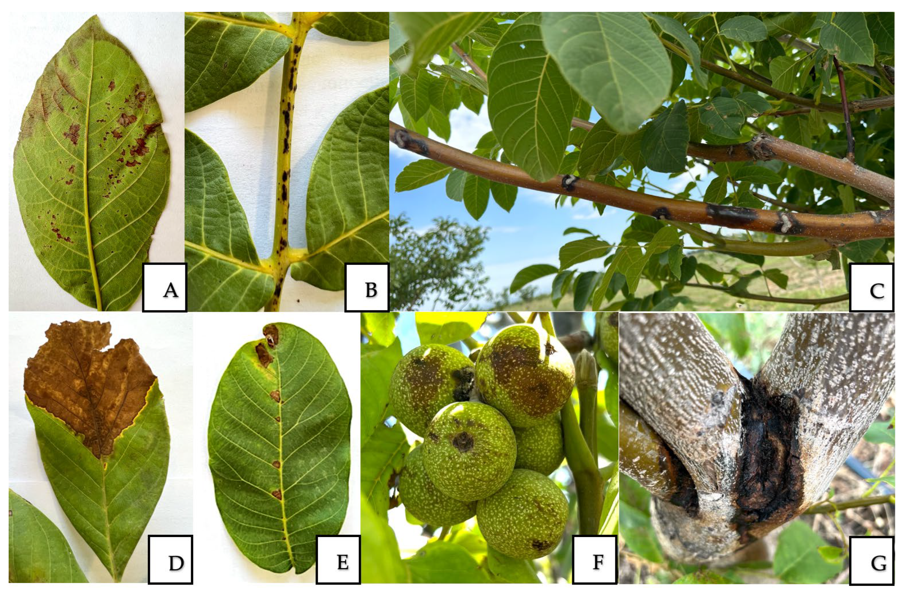
Symptoms of walnut leaf damage caused by brown spot manifested as dark-brown to black-brown lesions of round or irregular form. The size of the spots varied from small to large, reaching 20 mm or more, and in some cases extended over half of the leaf blade. Frequently, concentric zones of darker pigmentation were developed, surrounded by necrotic tissue (Figure 2A,B). In the case of walnut canker, black necrotic lesions of the bark spread along the branches, causing progressive tissue dieback (Figure 2C). Walnut blight, observed at most of the surveyed farms, was characterized by the necrosis of branch tissues and the appearance of leaf spotting (Figure 2D,E). Bacterial blight manifested as dark-brown lesions on leaves and fruits, as well as necrosis in young shoots. Subsequently, the leaves curled along the central vein into a boat-like shape, darkened, dried out, and remained attached to the tree in this condition for an extended period (Figure 2F).
The manifestation of crown gall disease was observed as darkening and necrosis on the damaged bark of the trunk, often without the formation of pronounced gall structures (Figure 2G).
In addition to the occurrence of individual diseases, mixed infections were frequently recorded. In particular, A. alternata was most often detected in association with bacterial pathogens (P. agglomerans, X. arboricola pv. juglandis), while as a single pathogen it was rare. In the “Issyk State Dendrological Park” RSE, “Manshuk” rural farm, and “Saryagash Zher Syyi” LLP, 100% of the cases of A. alternata occurred together with bacterial pathogens, indicating obligatory co-infection patterns at these sites. In contrast, at Alma-Arasan and “Fazenda UM”, P. agglomerans acted as an independent pathogen, whereas at “Silk alley” LLP, A. alternata predominated independently. Mixed infections were consistently associated with more severe disease development compared to single-pathogen cases. Symptoms progressed more rapidly, with expanded necrotic lesions, premature leaf and shoot death, and accelerated spread throughout plant tissues. These effects were particularly evident at the “Manshuk” rural farm, “Saryagash Zher Syyi” LLP, and the “Issyk State Dendrological Park” RSE, where co-infection rates were highest.
At the “Saryagash Zher Syyi” LLP, the most characteristic manifestations were bark damage on tree trunks and scaffold branches. These sites harbored a broad complex of pathogens—both bacterial and fungal. On walnut fruits and leaves, bacterial diseases were more frequently observed, whereas on stems and damaged wood, F. incarnatum and A. alternata were actively developing, with the latter being detected almost everywhere. Altogether, these observations indicate that bark cracks and wound injuries served as entry points for the establishment of mixed infections and the persistence of a diverse pathogen complex.

3.2. Morphological and Cultural Characteristics of Fungal and Bacterial Pathogens

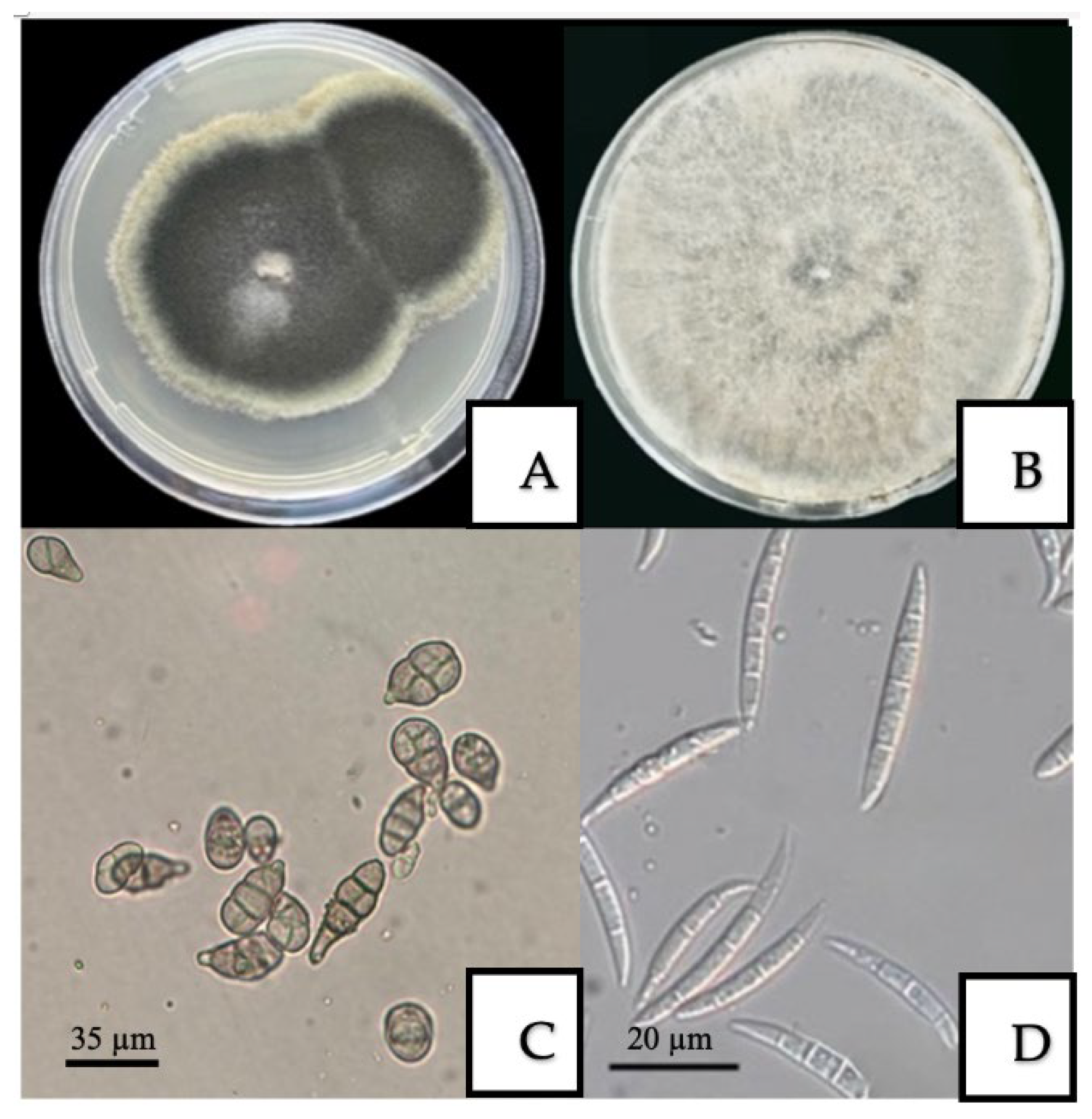
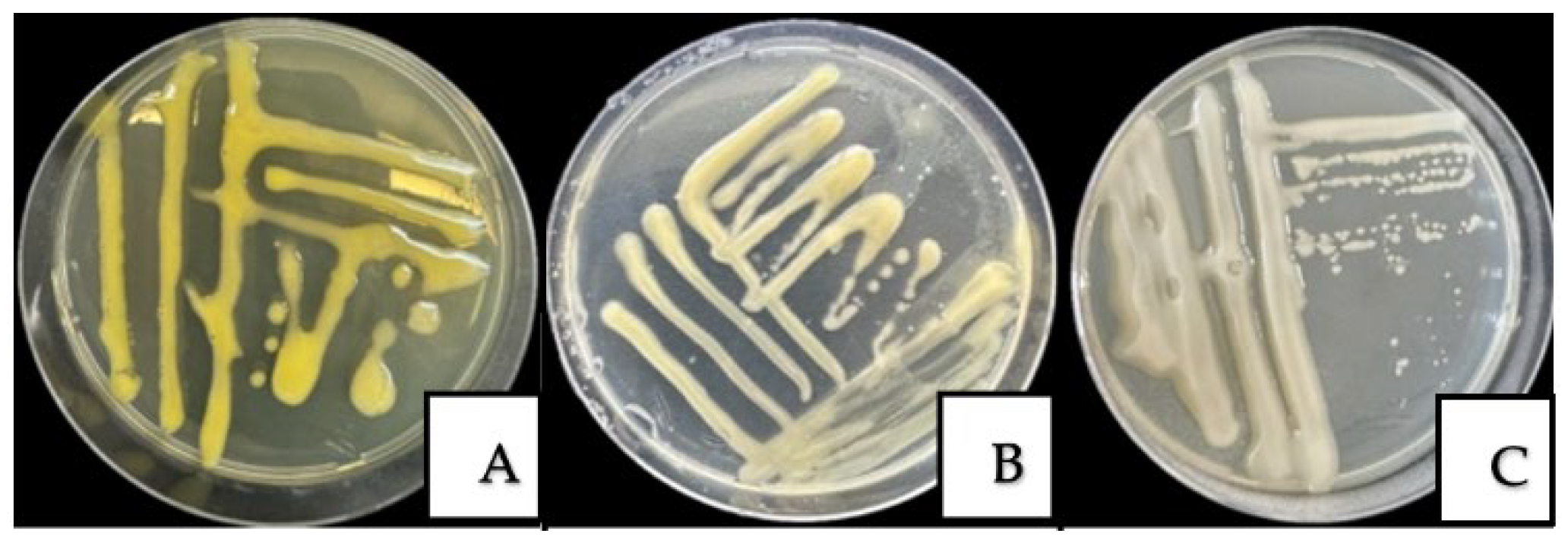
Based on the characteristics of colonies formed on PDA Petri dishes 5 days after inoculation, a total of 69 fungal and bacterial isolates were obtained from infected leaves, branches, and fruits. Among them were 21 strains of A. alternata (Figure 3A,C), 7 strains of F. incarnatum (Figure 3B,D), and bacterial isolates including 30 strains of P. agglomerans (Figure 4A), 8 strains of X. arboricola pv. juglandis (Figure 4B), and 3 strains of A. tumefaciens (Figure 4C, Table S2). The isolation rates were 30%, 10%, 44%, 12%, and 4% for A. alternata, F. incarnatum, P. agglomerans, X. arboricola pv. juglandis, and A. tumefaciens, respectively. The morphological and cultural characteristics of the fungal and bacterial strains are presented in Figure 3 and Figure 4. Detailed descriptions of colony morphology and conidial and bacterial structures are presented in Table 2.

3.3. Molecular Identification of Pathogens

The amplified ITS and 16S rRNA for fungi and bacteria, respectively, were multiplexed across the range of types in order to perform sequencing using the Native barcoding kit. We filtered the reads to retain only those with Phred scores ≥12. After filtering, 58,738 bacterial reads and 43,625 fungal reads remained. Of these, 17,827 bacterial reads and 14,971 fungal reads aligned to the best matching strains or isolates of pathogens (Table 3, Files S1 and S2).
Within the fungal set, the highest identity alignment, with at least 99% similarity, was with the isolate YICASTK5 for A. alternata, and with the isolate ISPaVe 1946 for F. incarnatum; the median read lengths were 367 bp and 355 bp, respectively. Within the bacterial set, the mapped reads with the highest alignment identity (more than 99% similarity) were assigned to strain RB04 for A. tumefaciens; to isolate CTN3 for P. agglomerans; and to isolate USS-ML18 for X. arboricola pv. juglandis. Median read lengths across the bacterial taxa ranged from 288 to 321 bp. The pathogen in the database (NCBI accession PQ460288.1) is listed as X. campestris; however, modern taxonomy assigns it to X. arboricola pv. juglandis, the causal agent of bacterial blight [51].
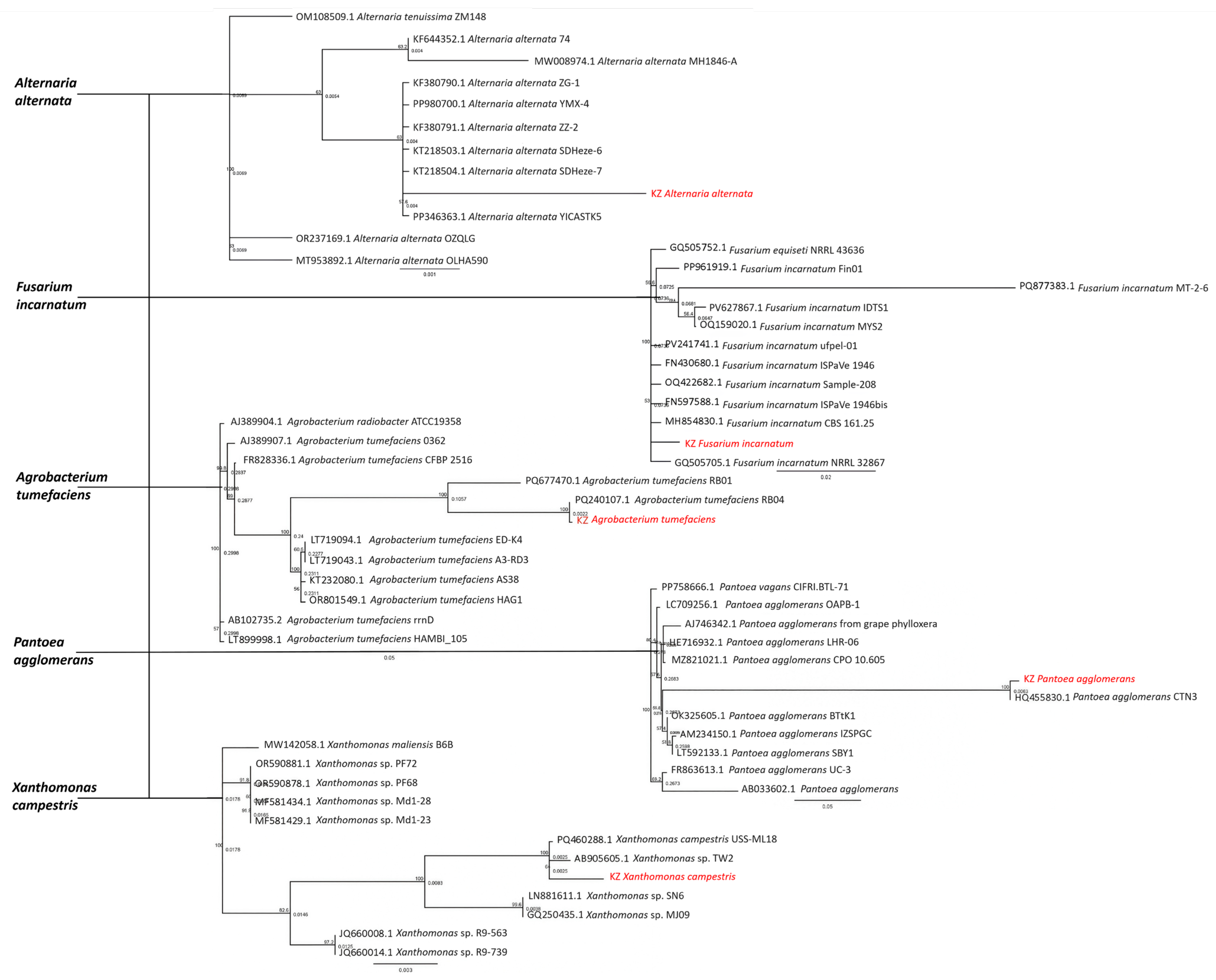
According to the phylogenetic analysis, the isolates of A. alternata collected in Kazakhstan clustered together with isolates originating from China and Turkey (Figure 5). These related isolates have been reported to cause infections in a wide range of host plants. Nevertheless, the genetic distance separating the Kazakhstani isolates from the nearest group members remains relatively large, suggesting a certain degree of divergence. For F. incarnatum, the Kazakhstani isolate showed phylogenetic proximity to isolates from Europe, Australia, and China. However, no predominant closeness to any single geographical region was observed, indicating a more widespread genetic relatedness across continents. The A. tumefaciens isolate from Kazakhstan grouped together with an isolate originating from Iraq, highlighting a possible regional link within this lineage.
Similarly, the Kazakhstani isolate of P. agglomerans clustered with isolates from Spain, while the X. campestris isolate formed a group with those from Chile and Thailand. These findings collectively indicate that Kazakhstani isolates share phylogenetic relationships with geographically distant strains, reflecting the complex evolutionary history and potential global distribution of these pathogens.

3.4. Pathogenicity Tests for Fungal and Bacterial Isolates

3.4.1. Pathogenicity Tests for Fungal Isolates

Pathogenicity was confirmed through artificial inoculation in all strains tested, with the exception of a single isolate (Figure 6). The artificial inoculation of walnut leaves demonstrated the pathogenicity of all fungal isolates tested. The first symptoms appeared within 5–7 days after inoculation, and by day 10, necrotic lesions were well developed. All A. alternata, the causal agent of walnut brown spot, induced necrotic spots on leaves (Figure 6A). Inoculation with F. incarnatum, responsible for walnut canker, localized tissue darkening and a slight depression at the inoculation sites. In contrast, no visible symptoms developed in control plants inoculated with sterile medium (Figure 6B).
Koch’s postulates were fulfilled by re-isolating the microorganisms from symptomatic tissues and confirming their identity using morphological analyses, thereby confirming their etiological role in walnut diseases.

3.4.2. Pathogenicity Tests of Bacterial Isolates

Pathogenicity assays demonstrated that multiple bacterial isolates induced distinct disease symptoms in walnuts (Figure 7). Inoculation with P. agglomerans, the causal agent of walnut blight, led to the formation of multiple necrotic spots with tissue maceration on leaves within 5 days under greenhouse conditions (Figure 7A). Fruits infected with X. arboricola pv. juglandis, responsible for bacterial blight, showed initial necrotic lesions at wound sites by the 7th day after inoculation, which became clearly evident by the 11th day and continued to expand progressively (Figure 7B,C). A. tumefaciens was isolated from cracked bark, but pathogenicity assays did not induce tumor formation within 120 days.
Sterile water was used as a negative control in all assays, with no symptoms observed in control plants. In all cases where symptoms developed, the pathogens were re-isolated from diseased tissues and confirmed to be identical to the original strains, thereby fulfilling Koch’s postulates and validating the pathogenicity tests (Figure 6 and Figure 7). However, under field conditions, disease symptoms frequently resulted from complex interactions between multiple pathogens.

4. Discussion

In Kazakhstan, there is a growing interest in the industrial cultivation of walnut, particularly in the southern regions, where climatic conditions favor high yields. However, walnut productivity is significantly constrained by fungal and bacterial diseases, and effective disease management is therefore essential. This study documents, for the first time in Kazakhstan, the occurrence of brown spot (A. alternata), walnut canker (F. incarnatum), walnut blight (P. agglomerans), bacterial blight (X. arboricola pv. juglandis), and crown gall (A. tumefaciens). Previously, only marssonina leaf blotch and bacterial blight had been reported on walnut plantations in the Turkestan region [4]. Resistance assessments against anthracnose and bacterial blight were conducted by Aksa et al. [35], who found that 2.1% and 36.2% of genotypes were resistant to anthracnose and bacterial blight, while 97.9% and 63.8% exhibited low susceptibility, respectively. Our findings therefore considerably broaden the known spectrum of pathogens affecting walnut in Kazakhstan and highlight the complexity of the pathogen challenge faced by walnut growers.
In our study, various methods were employed to characterize isolates. A total of 69 fungal and bacterial isolates (Table S2) were obtained from the infected leaves, branches, fruits, and bark of walnut trees.
The morphological characteristics of the A. alternata isolates were consistent with descriptions by Simmons [52]. Brown spot was detected on four farms in both Almaty and Turkestan regions, with the DI and DSI reaching 30% and 70%, respectively (Table 1), and this was especially widespread in 2024. Similar findings have been reported from China [10] and Serbia [11], where A. alternata was considered one of the most dangerous walnut pathogens.
Walnut canker, caused by F. incarnatum, was identified only at “Saryagash Zher Syyi” LLP in the Turkestan region, and compared to other diseases showed relatively low aggressiveness. However, F. incarnatum was successfully re-isolated from all inoculated plants, fulfilling Koch’s postulates [14]. This fungus has previously been reported as a walnut pathogen in Argentina [14] and India [15].
Walnut blight, caused by P. agglomerans, proved to be the most common disease affecting walnut trees. In 2023 and 2024, outbreaks were detected on all five surveyed farms in the Almaty and Turkestan regions. The highest values for DI and DSI were recorded in Alma—Arasan (Almaty region), at 26% and 57%, respectively. International studies confirm its damaging potential: In China, walnut blight caused by P. agglomerans (often in combination with X. arboricola pv. juglandis and Alternaria spp.) causes serious yield losses [8]. Genomic studies of P. agglomerans isolates from walnut have revealed a wide array of virulence and resistance genes, confirming its high adaptability to stress conditions and its ability to induce severe necrotic symptoms [18]. Susceptibility to walnut blight depends on multiple factors, including climatic conditions, cultivar characteristics, soil properties, and disease history [53]. Radix et al. [54] showed that soil composition may influence polyphenol levels in plant tissues, thereby altering susceptibility to P. agglomerans.
Bacterial blight, caused by X. arboricola pv. juglandis, was confirmed in “Saryagash Zher Syyi” LLP (Turkestan region) with DI and DSI values of 10–13% and 38–42%, respectively. This pathogen is especially damaging in early leafing cultivars during wet years [55]. Our findings are consistent with international reports of severe outbreaks in California [20], Korea [23], New Zealand [56], and France [57], as well as more recent cases across Europe and South America [21,22,23,24,25,26]. Some isolates form distinct genetic clusters associated with canker symptoms while retaining the ability to cause classic blight. Pathogenicity of such strains has been demonstrated in previous studies [57].
Although A. tumefaciens was isolated from bark showing cracking and localized swelling (Figure 2G), pathogenicity tests did not induce tumor formation in seedlings under experimental conditions. This suggests that the isolates may be non-pathogenic or require additional predisposing factors. Similar observations were reported by Yakabe et al. [31], who noted seasonal variation in A. tumefaciens distribution and gall formation in only 17% of seedlings infected via seeds. This highlights the complexity of interactions between A. tumefaciens and walnut.
Molecular sequencing (ITS and 16S rRNA) identified A. alternata, F. incarnatum, P. agglomerans, X. arboricola pv. juglandis, and A. tumefaciens, with 99–100% similarity to reference strains in GenBank. These results agree with findings from previous studies in other walnut-growing regions [10,12,18,30,58,59,60,61], thereby supporting our morphological and pathogenicity data and strengthening the accuracy of pathogen identification.
The pathogenicity tests in our study demonstrated the virulence of the majority of fungal and bacterial isolates, underscoring their potential to cause significant economic damage to walnut plantations. A. alternata and F. incarnatum proved highly virulent in walnut leaves, stems, and fruits, while X. arboricola pv. juglandis and P. agglomerans produced severe symptoms on leaves, and stems. These results align with previous pathogenicity reports from other walnut-growing regions [9,18,23].
The obtained data emphasize certain limitations of the present study. These include the limited number of isolates tested (only three A. tumefaciens strains from a single location, Table S1), the absence of seasonal repetitions, and potential mismatches between laboratory and natural conditions. Moreover, sampling was restricted to two regions over two years, which may not capture the full diversity of walnut pathogens across Kazakhstan. Environmental and agronomic factors such as irrigation practices, soil characteristics, and cultivar susceptibility were also not systematically evaluated. The unexpected isolation of A. tumefaciens from affected bark without tumor formation in seedlings further illustrates the complexity of host–pathogen interactions and the need for extended research.
Future work should broaden geographic and temporal coverage, increase the diversity of isolates, and incorporate multi-year and multi-seasonal sampling. Comparative studies using different inoculation methods, coupled with advanced genomic approaches, would improve the reproducibility and reliability of results, strengthen pathogen identification, and support the development of effective diagnostic and disease management strategies for walnut cultivation in Kazakhstan.

5. Conclusions

In 2023–2024, studies were conducted to systematically characterize the complex phytopathogens affecting walnut plantations. Using modern microbiological and molecular genetic methods, including PCR analysis and sequencing, key representatives of fungal (A. alternata and F. incarnatum) and bacterial (P. agglomerans, X. arboricola pv. juglandis and A. tumefaciens) groups were identified. Pathogenicity tests confirmed the virulence of most isolates, except for A. tumefaciens, which failed to induce tumor formation under experimental conditions, possibly due to non-pathogenic strains or the absence of required environmental factors. The obtained data on the taxonomic composition of pathogens and their morphological and pathogenic characteristics serve as a scientific basis for the formation of a system for protecting walnut plantations. This work represents a significant contribution to the development of agricultural science in Kazakhstan and lays the foundation for enhancing the resilience and productivity of walnut crops through improved phytosanitary monitoring and targeted disease management strategies.

Supplementary Materials

The following supporting information can be downloaded at https://www.mdpi.com/article/10.3390/horticulturae11101217/s1. Table S1: The weather conditions of Almaty and Turkestan regions in 2023 and 2024; Table S2: The list of collected fungal and bacterial isolates/strains; File S1: The data of sequencing for ITS; File S2: The data of sequencing for 16S rRNA.

Author Contributions

Conceptualization, G.K. and Z.S.; methodology, E.I., G.N. and S.K.; software, E.I.; validation, D.G. and M.S.; formal analysis, S.O.; investigation, E.I., G.S. and G.N.; resources, S.O.; data curation, E.I. and D.G.; writing—original draft preparation, E.I. and G.K.; writing—review and editing, Z.S. and H.B.; visualization, G.K. and H.B.; supervision, project administration and funding acquisition, G.K. All authors have read and agreed to the published version of the manuscript.

Funding

This research was funded by the Science Committee of the Ministry of Science and Higher Education of the Republic of Kazakhstan, Grant Number AP19677936, “Investigation of the main walnut diseases and molecular genetic basis of the resistance of promising varieties to economically important pathogens”.

Data Availability Statement

The original contributions presented in the study are included in the manuscript. Further inquiries can be directed to the first author (Elmira Ismagulova) and corresponding author (Gulshariya Kairova).

Acknowledgments

We are immensely grateful to the farmers (rural farm “Manshuk”, “Issyk State Dendrological Park” RSE, “Fazenda UM” LLP (Almaty region), “Saryagash Zher Syyi” LLP and “Silk alley” LLP (Turkestan region), where walnut diseases were studied, as their involvement was crucial to the success of this research. The authors have reviewed and edited the output and take full responsibility for the content of this publication.

Conflicts of Interest

The authors declare no conflicts of interest. The funders and farmers had no role in the design of the study; in the collection, analyses, or interpretation of data; in the writing of the manuscript; or in the decision to publish the results.

Abbreviations

The following abbreviations are used in this manuscript:
ITSInternal Transcribed Spacer
DIDisease incidence
DSIDisease severity index

References

  1. Kairova, G.; Taskuzhina, A.; Yanin, K.; Ismagulova, E.; Oleichenko, S.; Sarshayeva, M.; Sapakhova, Z.; Gritsenko, D. First Evaluation of Genetic Diversity and Population Structure of Wild and Cultivated Juglans regia in Kazakhstan. Genet. Resour. Crop Evol. 2025, 72, 8281–8292. [Google Scholar] [CrossRef]
  2. Sharma, M.; Sharma, M.; Sharma, M. A Comprehensive Review on Ethnobotanical, Medicinal and Nutritional Potential of Walnut (Juglans regia L.). Proc. Indian Natl. Sci. Acad. USA 2022, 88, 601–616. [Google Scholar] [CrossRef]
  3. Pollegioni, P.; Woeste, K.; Chiocchini, F.; Del Lungo, S.; Ciolfi, M.; Olimpieri, I.; Tortolano, V.; Clark, J.; Hemery, G.E.; Mapelli, S.; et al. Rethinking the History of Common Walnut (Juglans regia L.) in Europe: Its Origins and Human Interactions. PLoS ONE 2017, 12, e0172541. [Google Scholar] [CrossRef]
  4. Nurzhuma, M.; Kokhmetova, A.; Kumarbayeva, M.; Keishilov, Z.; Bakhytuly, K.; Bolatbekova, A.; Mukhametzhanov, K.; Akan, K. Assessment of Genetic Diversity in Walnut (Juglans regia L.) Genotypes from Southern and Southeastern Kazakhstan Using Microsatellite Markers. Horticulturae 2025, 11, 810. [Google Scholar] [CrossRef]
  5. Sapakhova, Z.; Kanat, R.; Choi, K.; Daurov, D.; Daurova, A.; Zhambakin, K.; Shamekova, M. CRISPR-Cas Gene Editing Technology in Potato. Int. J. Mol. Sci. 2025, 26, 7496. [Google Scholar] [CrossRef]
  6. Wang, X.; Li, K.; Han, M.; Zhang, W.; Li, X.; Ma, D.; Wang, F.; Pang, M.; Qi, J. Isolation and Identification of Endophytic Fungi in Walnut. IOP Conf. Ser. Earth Environ. Sci. 2020, 508, 012138. [Google Scholar] [CrossRef]
  7. Wang, S.; Tan, Y.; Li, S.; Zhu, T. Structural and Dynamic Analysis of Leaf-Associated Fungal Community of Walnut Leaves Infected by Leaf Spot Disease Based Illumina High-Throughput Sequencing Technology. Pol. J. Microbiol. 2022, 71, 429–441. [Google Scholar] [CrossRef]
  8. Zhu, Y.F.; Yin, Y.F.; Qu, W.W.; Yang, K.Q. Occurrence and Spread of the Pathogens on Walnut (Juglans regia) In Shandong Province, China. Acta Hortic. 2014, 1050, 347–351. [Google Scholar] [CrossRef]
  9. Belisario, A.; Forti, E.; Corazza, L.; van Kesteren, H.A. First Report of Alternaria alternata Causing Leaf Spot on English Walnut. Plant Dis. 1999, 83, 696. [Google Scholar] [CrossRef] [PubMed]
  10. Liu, Z.; Zhang, R.; Sun, D.; Tuerdi, M.; Zhu, T.; Chen, X. Alternaria alternata Causing Brown Spot Disease on Walnut in Xinjiang, China. Plant Dis. 2023, 107, 2224. [Google Scholar] [CrossRef]
  11. Vasić, T.; Jevremović, D.; Živković, S.; Leposavić, A.; Paunović, S. Morphological and Pathogenic Characteristic of Alternaria alternata Isolates on Walnut. J. Mt. Agric. Balk. 2021, 24, 224–238. [Google Scholar]
  12. Raina, A.; Kaul, S.; Sharma, I.; Khataia, K.; Dhar, M.K. First Report of Alternaria Alternata Causing Leaf Spot on Juglans regia in India. Plant Dis. 2025, 109, 1179. [Google Scholar] [CrossRef]
  13. Moya-Elizondo, E.A.; Lagos, M.J.; San Martin, J.; Ruiz, B. First Report of Alternaria alternata and Fusarium spp. Causing Brown Apical Necrosis in Walnut Fruit in Southern Chile. Plant Health Prog. 2021, 22, 573–574. [Google Scholar] [CrossRef]
  14. Seta, S.; Gonzalez, M.; Lori, G. First Report of Walnut Canker Caused by Fusarium incarnatum in Argentina. Plant Pathol. 2004, 53, 248. [Google Scholar] [CrossRef]
  15. Singh, B.; Kalha, C.S.; Razdan, V.K. First Report of Walnut Canker Caused by Fusarium incarnatum in India. Plant Dis. 2011, 95, 1587. [Google Scholar] [CrossRef]
  16. Belisario, A.; Luongo, L.; Vitale, S.; Santori, A. First report of Fusarium semitectum as the agent of twig cankers on Persian (English) walnut in Italy. Plant Diseas. 2010, 94, 791. [Google Scholar] [CrossRef]
  17. An, X.-H.; Wang, N.; Wang, H.; Li, Y.; Si, X.-Y.; Zhao, S.; Tian, Y. Physiological and Transcriptomic Analyses of Response of Walnuts (Juglans regia) to Pantoea agglomerans Infection. Front. Plant Sci. 2023, 14, 1294643. [Google Scholar] [CrossRef] [PubMed]
  18. Xiao, J.; Liu, Z.; Sun, S.; Fan, C.; Wang, D.; Zhang, D. Complete Genome Sequence of Pantoea agglomerans CHTF15, a Walnut Pathogen. Mol. Plant-Microbe Interact. 2023, 36, 134–137. [Google Scholar] [CrossRef] [PubMed]
  19. Mulrean, E.N. Brown Apical Necrosis of Persian (English) Walnut Fruit. Phytopathology 1982, 72, 434. [Google Scholar] [CrossRef]
  20. Vauterin, L.; Hoste, B.; Kersters, K.; Swings, J. Reclassification of Xanthomonas. Int. J. Syst. Bacteriol. 1995, 45, 472–489. [Google Scholar] [CrossRef]
  21. Burokiene, D.; Pulawska, J. Bacterial blight of walnut (Juglans regia) caused by Xanthomonas arboricola pv. juglandis in Lithuania. J. Plant Pathol. 2012, 94, S1.43–S1.48. [Google Scholar]
  22. Bandi, A.; Kámán-Tóth, E.; Kolozsváriné Nagy, J.; Hevesi, M. Characterisation of Xanthomonas arboricola pv. juglandis strains from Hungary and Romania. Acta Microbiol. Immunol. Hung. 2014, 61, 121–135. [Google Scholar] [CrossRef]
  23. Kim, H.S.; Cheon, W.; Lee, Y.; Kwon, H.-T.; Seo, S.-T.; Balaraju, K.; Jeon, Y. Identification and characterization of Xanthomonas arboricola pv. juglandis causing bacterial blight of walnuts in Korea. Plant Pathol. J. 2021, 37, 137–151. [Google Scholar] [CrossRef]
  24. Pardatscher, R.; Gualandri, V.; Rizza, C.; Martini, M. First report of Xanthomonas arboricola pv. juglandis causing bacterial blight on walnut in Northern Italy. J. Plant Pathol. 2019, 101, 447. [Google Scholar]
  25. Kałużna, M.; Puławska, J.; Janse, J.D.; Young, J.M.; Sobiczewski, P. Genetic diversity and pathogenicity of Xanthomonas arboricola pv. juglandis strains isolated from walnut trees in Poland. Plant Pathol. 2021, 70, 842–857. [Google Scholar] [CrossRef]
  26. Iličić, R.; Popović, T.; Lalošević, V.; Jelušić, A. Association of Xanthomonas arboricola pv. juglandis with vertical oozing canker of walnut in Serbia. Plants 2021, 10, 2109. [Google Scholar] [CrossRef]
  27. Moragrega, C.; Llorente, I. Effects of leaf wetness duration, temperature, and host phenological stage on infection of walnut by Xanthomonas arboricola pv. juglandis. Plants 2023, 12, 2800. [Google Scholar] [CrossRef]
  28. Chorolque, F.; Orellana, R.; Alvarado, I.; Vignolo, G.; Stefani, E. Susceptibility of Chandler and Franquette walnut cultivars to Xanthomonas arboricola pv. juglandis under different climatic conditions in Argentina. J. Plant Pathol. 2024, 106, 45–56. [Google Scholar]
  29. Latinović, J.; Latinović, N.; Kandić, B.; Jelušić, A.; Popović Milovanović, T. Evidence and characterisation of Xanthomonas arboricola pv. juglandis causing bacterial blight of walnut in Montenegro. Plant Prot. Sci. 2025, 61, 255–261. [Google Scholar] [CrossRef]
  30. Rouhrazi, K.; Rahimian, H. Biochemical and genetic characterisation of Agrobacterium tumefaciens the causal agent of walnut crown gall disease in Iran. Arch. Phytopathol. Plant Prot. 2014, 47, 2493–2500. [Google Scholar] [CrossRef]
  31. Yakabe, L.E.; Parker, S.R.; Kluepfel, D.A. Role of systemic Agrobacterium tumefaciens populations in crown gall incidence on the walnut hybrid rootstock ‘Paradox’. Plant Dis. 2012, 96, 1415–1421. [Google Scholar] [CrossRef]
  32. Eshbekova, G.; Ismailov, Z. Isolation and characterization of Agrobacterium tumefaciens isolated from walnut (Juglans regia L.) Crown gall in selected region of Uzbekistan. In Innovations in Sustainable Agricultural Systems, Agriculture 4.0 and Precision Agriculture, Volume 2; Springer: Cham, Switzerland, 2025. [Google Scholar]
  33. Poret-Peterson, A.T.; Bhatnagar, S.; McClean, A.E.; Kluepfel, D.A. Draft genome sequence of Agrobacterium tumefaciens biovar 1 strain 186, isolated from walnut. Genome Announc. 2017, 5, e01232-17. [Google Scholar] [CrossRef]
  34. Kairova, M.; Yegizbayeva, T.K.; Yausheva, T.V.; Oleichenko, S.N.; Apushev, A.K. Biological properties of walnut genotypes in Kazakhstan. Syst. Rev. Pharm. 2020, 11, 932–940. [Google Scholar]
  35. Akça, Y.; Yuldaşulu, Y.B.; Murad, E.; Vahdati, K. Exploring of Walnut Genetic Resources in Kazakhstan and Evaluation of Promising Selections. Int. J. Hortic. Sci. Technol. 2020, 7, 93–102. [Google Scholar] [CrossRef]
  36. Xie, Z.; Li, M.; Wang, D.; Wang, F.; Shen, H.; Sun, G.; Feng, C.; Wang, X.; Chen, D.; Sun, X. Biocontrol Efficacy of Bacillus Siamensis LZ88 against Brown Spot Disease of Tobacco Caused by Alternaria alternata. Biol. Control 2021, 154, 104508. [Google Scholar] [CrossRef]
  37. Jiang, C.-H.; Wu, F.; Yu, Z.-Y.; Xie, P.; Ke, H.-J.; Li, H.-W.; Yu, Y.-Y.; Guo, J.-H. Study on Screening and Antagonistic Mechanisms of Bacillus amyloliquefaciens 54 against Bacterial Fruit Blotch (BFB) Caused by Acidovorax avenae subsp. citrulli. Microbiol. Res. 2015, 170, 95–104. [Google Scholar] [CrossRef]
  38. Khan, M.A. Laboratory Guide for Bacterial Plant Pathology; Pakistan Phytopathological Society: Faisalabad, Pakistan, 2020. [Google Scholar] [CrossRef]
  39. Murray, M.G.; Thompson, W.F. Rapid isolation of high molecular weight plant DNA. Nucleic Acids Res. 1980, 8, 4321–4326. [Google Scholar] [CrossRef] [PubMed]
  40. Sambrook, J.; Russell, D.W. Molecular Cloning: A Laboratory Manual, 3rd ed.; Cold Spring Harbor Laboratory Press: New York, NY, USA, 2001. [Google Scholar]
  41. Weisburg, W.G.; Barns, S.M.; Pelletier, D.A.; Lane, D.J. 16S Ribosomal DNA Amplification for Phylogenetic Study. J. Bacteriol. 1991, 173, 697–703. [Google Scholar] [CrossRef]
  42. Muyzer, G.; De Waal, E.C.; Uitterlinden, A.G. Profiling of Complex Microbial Populations by Denaturing Gradient Gel Electrophoresis Analysis of Polymerase Chain Reaction-Amplified Genes Coding for 16S rRNA. Appl. Environ. Microbiol. 1993, 59, 695–700. [Google Scholar] [CrossRef]
  43. White, T.J.; Bruns, T.; Lee, S.; Taylor, J. Amplification and Direct Sequencing of Fungal Ribosomal RNA Genes for Phylogenetics. In PCR Protocols; Elsevier: Amsterdam, The Netherlands, 1990; pp. 315–322. ISBN 978-0-12-372180-8. [Google Scholar]
  44. Ma, T.; Yang, C.; Cai, F.; Osei, R. Molecular Identification and Characterization of Fusarium Associated with Walnut Branch Blight Disease in China. Pathogens 2023, 12, 970. [Google Scholar] [CrossRef] [PubMed]
  45. Deng, Y.Z.; Qu, Z.; He, Y.; Naqvi, N.I. Sorting Nexin Snx41 Is Essential for Conidiation and Mediates Glutathione-Based Antioxidant Defense during Invasive Growth in Magnaporthe Oryzae. Autophagy 2012, 8, 1058–1070. [Google Scholar] [CrossRef]
  46. Yang, H.; Cao, G.; Jiang, S.; Han, S.; Yang, C.; Wan, X.; Zhang, F.; Chen, L.; Xiao, J.; Zhu, P.; et al. Identification of the anthracnose fungus of walnut (Juglans spp.) and resistance evaluation through physiological responses of resistant vs. susceptible hosts. Plant Pathol. 2021, 70, 1219–1229. [Google Scholar] [CrossRef]
  47. Zabiák, A.; Kovács, C.; Takács, F.; Pál, K.; Peles, F.; Fekete, E.; Karaffa, L.; Mihály, K.; Flipphi, M.; Sándor, E. Diaporthe and Diplodia species associated with walnut (Juglans regia L.) in Hungarian orchards. Horticulturae 2023, 9, 205. [Google Scholar] [CrossRef]
  48. Antony, S.; Billones-Baaijens, R.; Steel, C.; Stodart, J.; Savocchia, S. Pathogenicity and progression of Botryosphaeriaceae associated with dieback in walnut orchards in Australia. Eur. J. Plant Pathol. 2024, 168, 723–742. [Google Scholar] [CrossRef]
  49. Agrios, G.N. Plant Pathology, 5th ed.; Elsevier Academic Press: Amsterdam, The Netherlands, 2008; ISBN 978-0-12-044565-3. [Google Scholar]
  50. Wang, D.; Li, R.; Gao, G.; Jiakula, N.; Toktarbek, S.; Li, S.; Ma, P.; Feng, Y. Impact of climate change on food security in Kazakhstan. Agriculture 2022, 12, 1087. [Google Scholar] [CrossRef]
  51. Noh, Y.-H.; Cha, J.S. Xanthomonas arboricola pv. juglandis str. NCPPB 1447 Genome Sequencing. 2012. Available online: https://www.ncbi.nlm.nih.gov/datasets/genome/GCF_000306055.1/ (accessed on 7 August 2025).
  52. Simmons, E.G. Alternaria: An Identification Manual; CBS Fungal Biodiversity Centre: Utrecht, The Netherlands, 2007. [Google Scholar]
  53. Olson, W.H.; Buchner, R.P.; Adaskaveg, J.E.; Lindow, S.E. Walnut Blight Control in California. Acta Hortic. 1997, 442, 361–366. [Google Scholar] [CrossRef]
  54. Radix, P.; Bastien, C.; Jay-Allemand, C.; Charlot, G.; Seigle-Murandi, F. The Influence of Soil Nature on Polyphenols in Walnut Tissues. A Possible Explanation of Differences in the Expression of Walnut Blight. Agronomie 1998, 18, 627–637. [Google Scholar] [CrossRef]
  55. Mikulic-Petkovsek, M.; Slatnar, A.; Veberic, R.; Stampar, F.; Solar, A. Phenolic Response in Green Walnut Husk after the Infection with Bacteria Xanthomonas arboricola pv. juglandis. Physiol. Mol. Plant Pathol. 2011, 76, 159–165. [Google Scholar] [CrossRef]
  56. McNeil, D.L.; Romero, S.; Kandula, J.; Stark, C.; Stewart, A.; Larsen, S. Bacteriophages a Potential Biocontrol Agent against Walnut Blight (Xanthomonas campestris pv. juglandis). N. Z. Plant Prot. 2001, 54, 220–224. [Google Scholar] [CrossRef]
  57. Hajri, A.; Meyer, D.; Delort, F.; Guillaumès, J.; Brin, C.; Manceau, C. Identification of a Genetic Lineage within Xanthomonas arboricola pv. juglandis as the Causal Agent of Vertical Oozing Canker of Persian (English) Walnut in France. Plant Pathol. 2010, 59, 1014–1022. [Google Scholar] [CrossRef]
  58. Chen, X.; Liu, H.; Dong, L.; Shi, J.; Ma, Z.; Yan, L.; Yin, Y. Identification and Characterization of Fusarium incarnatum Causing Leaf Spot and Fruit Rot on Luffa in China. Plants 2025, 14, 845. [Google Scholar] [CrossRef] [PubMed]
  59. Moisseyev, R.; Pozharskiy, A.; Taskuzhina, A.; Khusnitdinova, M.; Svanbayev, U.; Sapakhova, Z.; Gritsenko, D. Evaluation of Rz2 Gene Expression in Sugar Beet Hybrids Infected with Beet Necrotic Yellow Vein Virus. Curr. Issues Mol. Biol. 2024, 46, 11326–11335. [Google Scholar] [CrossRef] [PubMed]
  60. Gritsenko, D.; Daurova, A.; Pozharskiy, A.; Nizamdinova, G.; Khusnitdinova, M.; Sapakhova, Z.; Daurov, D.; Zhapar, K.; Shamekova, M.; Kalendar, R.; et al. Investigation of Mutation Load and Rate in Androgenic Mutant Lines of Rapeseed in Early Generations Evaluated by High-Density SNP Genotyping. Heliyon 2023, 9, e14065. [Google Scholar] [CrossRef]
  61. Kałużna, M.; Saux, M.F.-L.; Pothier, J.F.; Jacques, M.-A.; Obradović, A.; Tavares, F.; Stefani, E. Xanthomonas arboricola pv. juglandis and pv. Corylina: Brothers or Distant Relatives? Genetic Clues, Epidemiology, and Insights for Disease Management. Mol. Plant Pathol. 2021, 22, 858–869. [Google Scholar] [CrossRef]
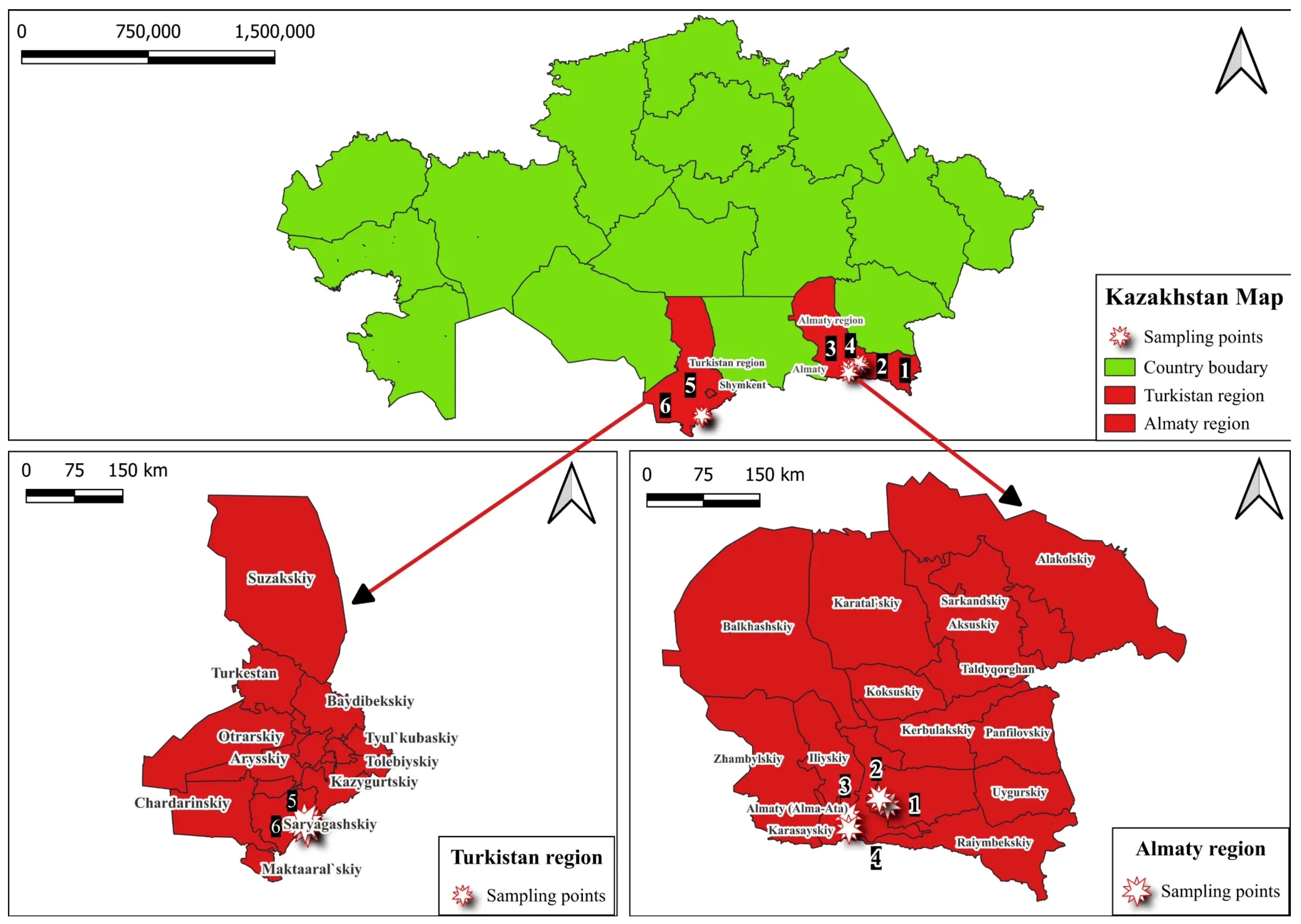
Figure 1. Geographic locations of the sampling area of walnut fungal and bacterial isolates/strains.
Horticulturae 11 01217 g001
Figure 2. Representative symptoms of walnut diseases caused by fungal and bacterial pathogens: (A,B) Brown spot; (C) Walnut canker; (D,E) Walnut blight; (F) Bacterial blight; (G) Bark necrosis associated with Crown gall disease (non-pathogenic in pathogenicity tests).
Horticulturae 11 01217 g002
Figure 3. Colony morphologies and microscopic structures of fungal isolates on PDA; (A) Colony morphology of A. alternata; (B) Colony morphology of F. incarnatum; (C) Conidia of A. alternata, scale bar = 35 µm; (D) Macroconidia of F. incarnatum, scale bar = 20 µm.
Horticulturae 11 01217 g003
Figure 4. Morphological characteristics of bacterial isolates have grown on NA: (A) P. agglomerans; (B) X. arboricola pv. juglandis; (C) A. tumefaciens.
Horticulturae 11 01217 g004
Figure 5. Neighbor-joining phylogenetic tree of fungal and bacterial pathogens (A. alternata, F. incarnatum, A. tumefaciens, P. agglomerans, and X. arboricola pv. juglandis (listed as X. campestris in NCBI)). Bootstrap values (1000 replications) are shown at branch nodes. Local isolates are highlighted in red.
Horticulturae 11 01217 g005
Figure 6. Pathogenicity tests of fungal isolates on walnut after artificial inoculation; (A) Necrotic leaf spots caused by A. alternata (isolate WP003); (B) Localized lesions on walnut branches caused by F. incarnatum (isolate WP013). Control plants inoculated with sterile medium showed no symptoms.
Horticulturae 11 01217 g006
Figure 7. Pathogenicity tests of bacterial isolates on walnut leaves and fruits after artificial inoculation; (A) Walnut leaves showing necrotic spots caused by P. agglomerans; (B) Control walnut fruits (non-inoculated, 5 days post inoculation); (C) Walnut fruits inoculated with X. arboricola pv. juglandis showing progressive necrotic lesions at 5, 7, 11, and 13 days post-inoculation.
Horticulturae 11 01217 g007
Table 1. Disease incidence and disease severity index of walnut trees in 2023 and 2024.
LocationCoordinatesYearDiseaseDI *, %DSI **, %
Rural farm “Manshuk”, Almaty region43.257927° N 77.757149 ° E2023Walnut blight1234
20241336
2023Brown spot928
20241133
“Issyk State Dendrological Park” RSE, Almaty region43.455192° N
77.454092° E
2023Walnut blight1349
20241450
2023Brown spot2346
20242651
“Fazenda UM” LLP, Almaty region43.296643° N 77.383693° E2023Walnut blight1336
20241441
“Saryagash zher syyi” LLP, Turkestan region41.492639° N 69.315194° E2023Walnut blight1837
20242250
2023Bacterial blight1038
20241342
2023Brown spot1739
20241939
2023Crown gall920
2024815
2023Walnut canker1020
20241429
“Silk alley” LLP, Turkestan region41.431411° N 69.112285° E2023Brown spot2864
20243070
“Alma Arasan”, Almaty region43.102843° N 76.906788° E2023Walnut blight2654
20242657
Note: * DI—disease incidence; ** DSI—disease severity index.
Table 2. Morphological characterization of fungal and bacterial pathogens.
The Name of the PathogenMorphological Characterization
A. alternataIsolates formed dark, grayish-black colonies with an olive tint and a characteristic white marginal ring on PDA. After 6 days, colony diameters ranged from 43.5 to 81.5 mm. Light microscopy revealed obclavate to obpyriform conidia arranged in chains of 6–14 elements, with 1–6 transverse and 0–3 longitudinal septa. Conidial dimensions were 16–40 × 4–13 µm.
F. incarnatumIsolates formed colonies on PDA ranging in color from white to pale brown, characterized by abundant, floccose mycelium. Microscopic examination revealed characteristic macroconidia: slightly curved, with 3–5 septa, the apical cell pointed and slightly hooked, and the basal cell exhibiting a “foot-shaped” morphology. Mesoconidia of fusiform shape with 1–5 septa were also observed, as well as intercalary chlamydospores.
P. agglomeransThe isolated colonies of P. agglomerans grown on NA exhibited a yellow to cream pigmentation, were circular in shape, with a smooth, glossy surface and regular margins. Under light microscopy, the bacterial cells appeared as small Gram-negative rod-shaped structures.
X. arboricola pv. juglandisThe isolated colonies of X. arboricola pv. juglandis grown on NA were round, light yellow, with a smooth, glistening, and mucous surface, forming characteristic mucoid colonies. Under light microscopy, the bacterial cells appeared as rod-shaped structures.
A. tumefaciensColonies are round and milky-white to creamy, with smooth edges; the surface is smooth, slightly shiny, and moderately mucous. Under light microscopy, the bacterial cells appeared as Gram-negative, rod-shaped structures.
Table 3. Top matching strains/isolates identified for fungal and bacterial pathogens.
SpeciesIsolate/StrainAccession NumberMedian Read Length, bpMapped Reads
A. alternataYICASTK5PP346363.13678063
F. incarnatumISPaVe 1946FN430680.13556908
P. agglomeransCTN3HQ455830.12886907
X. arboricola pv. juglandis (listed as X. campestris in the NCBI)USS-ML18PQ460288.13212682
A. tumefaciensRB04PQ240107.13188238
Disclaimer/Publisher’s Note: The statements, opinions and data contained in all publications are solely those of the individual author(s) and contributor(s) and not of MDPI and/or the editor(s). MDPI and/or the editor(s) disclaim responsibility for any injury to people or property resulting from any ideas, methods, instructions or products referred to in the content.