Article

Pathogen Survey in Agrocybe chaxingu and Characterization of the Dominant Pathogen Fuligo gyrosa

1
Jiangxi Provincial Key Laboratory of Agricultural Non-Point Source Pollution Control and Waste Comprehensive Utilization, Institute of Agricultural Applied Microbiology, Jiangxi Academy of Agricultural Sciences, Nanchang 330200, China
2
State Key Laboratory of Mycology, Institute of Microbiology, Chinese Academy of Sciences, Beijing 100101, China
3
Jiangxi Key Laboratory for Excavation and Utilization of Agricultural Microorganisms, Jiangxi Agricultural University, Nanchang 330045, China
*
Authors to whom correspondence should be addressed.
Horticulturae 2025, 11(10), 1190; https://doi.org/10.3390/horticulturae11101190
Submission received: 28 August 2025 / Revised: 22 September 2025 / Accepted: 30 September 2025 / Published: 2 October 2025
(This article belongs to the Special Issue Advances in Propagation and Cultivation of Mushroom)

Abstract

Agrocybe chaxingu is a commercially important edible mushroom in China, valued for its rich bioactive compounds and distinctive umami flavor. In recent years, frequent disease outbreaks have severely limited production, as many pathogens spread rapidly and are difficult to control, posing a significant threat to the sustainable development of the industry. In this study, a systematic disease survey across major A. chaxingu cultivation areas in Jiangxi Province led to the isolation and identification of 17 potential fungal pathogens and 2 potential myxomycete pathogens using combined morphological characterization and multilocus phylogenetic analyses including the internal transcribed spacer (ITS) region, 28S large subunit ribosomal RNA (LSU), translation elongation factor (tef1), RNA polymerase largest subunit (rpb1), RNA polymerase second largest subunit (rpb2), Histone (H3), Beta tubulin (tub2), and 18S ribosomal RNA (18S rRNA). Among the identified diseases, white slime disease showed the highest incidence (17.3%) and was attributed to the slime mold Fuligo gyrosa, with pathogenicity confirmed according to Koch’s postulates. F. gyrosa proved highly virulent to both fruiting bodies and mycelia, enveloping host mycelium via plasmodial expansion, inhibiting growth, inducing structural rupture, and causing progressive degradation. Infection was accompanied by the deposition of characteristic stress-related pigments in the mycelium. This study provides the first detailed characterization of F. gyrosa infection dynamics in A. chaxingu mycelium. These findings provide new insights into the myxomycete pathogenesis in edible fungi and provide a foundation for the accurate diagnosis, targeted prevention, and sustainable management of diseases in A. chaxingu cultivation.

1. Introduction

Agrocybe chaxingu (syn. Cyclocybe chaxingu) is a commercially important edible and medicinal mushroom with both nutritional and medicinal values [1,2]. It is rich in active components such as proteins [3], polysaccharides [4], and free amino acids [5], and is popularly known as the “Divine Mushroom of China” in folk tradition. According to the 2023 statistics from the China Edible Fungi Association, the annual production of A chaxingu was 826,000 tons (https://www.cefa.org.cn/web/index.html, accessed on 20 December 2024), accounting for 5.3% of the total edible fungi production in China, and ranked eighth among all edible fungi varieties in terms of production volume. Jiangxi Province remains the leading production area, contributing 448,000 tons (≈54.2%) in 2023 due to its favorable climate, advanced cultivation techniques, and large-scale production systems.
Artificial cultivation of A. chaxingu began in the 1950s with log-based domestication techniques [6]. Despite nearly 40 years of development, large-scale industrial cultivation remains limited, relying primarily on seasonal production in simple mushroom houses due to multiple flushes, long growth cycles, and uneven fruiting [1,7]. As a result, disease problems have become a major bottleneck in industrial development. Disease issues affecting A. chaxingu can be categorized into two stages: the bag culture stage and the fruiting stage. The former is characterized by contamination from competing fungi and can be effectively controlled through the strict management of raw material selection, bag preparation, sterilization processes, and environmental hygiene. In contrast, disease outbreaks during the fruiting stage, caused by environmental pathogens, are harder to prevent and represent the primary source of economic losses [8].
During the fruiting stage, A. chaxingu is susceptible to fungal, bacterial, physiological, and myxomycete disease, all of which can cause substantial yield losses and reduce commercial quality. Previous studies have reported Trichoderma spp. as dominant pathogens, followed by Aspergillus spp., Mucor spp., and Penicillium spp. [9]. In addition, various diseases, including cobweb disease, rust spot disease, and mold rot disease, and the proposed corresponding integrated control strategies, have been reported, with primary bacterial pathogens such as Bacillus spp., Pseudomonas spp., and Erwinia spp. having been documented [10,11,12,13]. Notably, slime mold disease of A. chaxingu has been frequently reported in multiple regions, showing increasing severity and posing a serious threat to the sustainable development of its industry. According to the literature, Liu et al. [14] reported that the incidence of slime mold disease in A. chaxingu reached 20% in the Sanming production area of Fujian Province. Chen [15] observed a comparable incidence in the Guangxi production area, also around 20%, and noted that the disease could cause devastating losses during peak outbreaks. The slime mold Stemonitis herbatica was previously identified as a causal agent through molecular identification [16]. These findings provided a critical pathological basis for subsequent research on the prevention and control of A. chaxingu diseases.
Myxomycete-related diseases are not only widespread across A. chaxingu cultivation, but also affect a broad spectrum of edible fungi, including Pleurotus ostreatus [17], Auricularia heimuer [18], Lentinula edodes [19], Hericium erinaceus [20], Pleurotus pulmonarius [21], Grifola frondose [22], and Dictyophora indusiata [23], highlighting the urgency of understanding these pathogens in edible mushroom production. Most research to date has focused on symptom description and control strategies, while the systematic identification of pathogens remains limited in A. chaxingu. This gap has hindered the development of effective disease prevention systems. To address this, a systematic survey was conducted in two major Jiangxi production areas—Guangchang County and Ganxian District—from May 2021 to June 2022. Pathogens were isolated and purified from diseased fruiting bodies and contaminated substrates, and characterized using morphological and multilocus phylogenetic analyses. The predominant pathogen was further validated through Koch’s postulates and detailed observation of the infection process. These findings provide a solid theoretical foundation and practical guidance for environmentally friendly disease management and the sustainable development of A. chaxingu cultivation.

2. Materials and Methods

2.1. A. chaxingu Strain and Culture Conditions

The A. chaxingu strain used in this study was AS-5, the predominant commercial cultivar in Jiangxi Province. It was preserved in the China General Microbiological Culture Collection Center (CGMCC) under accession number CGMCC No. 40980.

2.2. Disease Survey and Statistical Analysis in A. chaxingu

A disease survey was conducted across six A. chaxingu cultivation enterprises in Guangchang County and Ganxian District, Jiangxi Province, each with over a decade of production history. At each site, 2000 cultivation bags were randomly sampled using a five-point sampling method (one from the center and four from the corners). Cultivation bags exhibiting similar disease symptoms were collected, counted, and subjected to pathogen isolation.

2.3. Isolation and Purification of Potential Pathogens from A. chaxingu

For pathogenic fungi, diseased materials exhibiting consistent symptoms were randomly selected. Using sterile forceps, tissue from the margin of infected mycelia was excised and inoculated at the center of potato dextrose agar (PDA) plates (prepared with 200 g potato, 20 g agar, 20 g glucose, and 1000 mL distilled water), with three replicates per sample. Hyphal tips from colony margins were transferred to fresh PDA for at least two successive subcultures until pure isolates were obtained [24].
For slime mold, diseased materials with abundant plasmodial networks were selected. Segments containing active plasmodia were excised with sterile forceps and placed on water agar medium (prepared with 20 g agar and 1000 mL distilled water), with three replicates per sample. Once the plasmodia had spread across the medium, a 5 mm diameter plug from the actively growing margin was transferred to oat water agar medium (prepared with 20 g oat flakes, 30 g agar, and 1000 mL distilled water) and sub-cultured three times to obtain pure cultures. All isolations were performed in the dark at 25 °C [22].

2.4. Morphological Observation of Potential Pathogenic Microorganisms

For fungal pathogens, a 5 mm mycelial plug of the putative pathogen was inoculated onto PDA plates. A sterile coverslip was inserted into the medium at a 45° angle, approximately 2 cm from the inoculation site. When hyphae had grown to cover two-thirds of the coverslip, it was carefully removed and examined under a light microscope (Eclipse 80i, Nikon, Tokyo, Japan) at 600× magnification to characterize the hyphal and spore morphology [25].
For slime molds, a 5 mm water agar block containing plasmodia was transferred onto fresh water agar medium. A sterile oat flake, moistened with 20 μL sterile distilled water, was placed directly on the plasmodium to stimulate expansion. Plasmodium growth and morphology were examined using a stereomicroscope (SMZ18, Nikon, Tokyo, Japan). Upon nutrient depletion, plasmodia developed into young and then mature fruiting bodies. Mature fruiting bodies were harvested, suspended in sterile distilled water, and gently agitated to release spores. The spore suspension was then collected and examined under a light microscope at 600 × magnification [26].

2.5. Molecular Identification of Potential Pathogens

Genomic DNA was extracted from fresh fungal mycelia or plasmodia grown on PDA or water agar medium using the cetyltrimethylammonium bromide (CTAB) method [27] and stored at −20 °C for subsequent polymerase chain reaction (PCR) analysis. PCR was performed in 25 μL reactions containing 12.5 μL of 2 × Rapid Taq Master Mix (P222, Vazyme, Nanjing, China), 1 μL each of 10 μM forward and reverse primers, 2 μL of genomic DNA template (200 ng/μL), and 8.5 μL of nuclease-free water.
For fungal pathogens, molecular identification was initially carried out through PCR amplification and sequencing the internal transcribed spacer (ITS) region [28]. To achieve higher taxonomic resolution, additional gene loci, including translation elongation factor (tef1) [29], RNA polymerase largest subunit (rpb1) [30], RNA polymerase second largest subunit (rpb2) [31], histone (H3) [32], beta tubulin (tub2) [33], and 28S large subunit ribosomal RNA (LSU) [34], were amplified and sequenced as appropriate for the taxon. For the myxomycetes, identification was based on the amplification and sequencing of the 18S ribosomal RNA (18S rRNA) gene [28,35].
Primer sequences and PCR conditions are listed in Table 1. Amplification products were visualized by electrophoresis on 1% agarose gels. Primer synthesis and sequencing services were provided by Shenggong Bioengineering (Shanghai) Co. Ltd.

2.6. Construction of Phylogenetic Trees

Sequence alignments (Tables S1 and S2) were performed using MAFFT version 7 [36] with default settings. Conserved regions were identified using Gblocks 0.91 [37] to remove ambiguously aligned positions, and the resulting conserved sites from each gene were concatenated to generate a combined dataset (i.e., partial sequences were used). The optimal nucleotide substitution model was selected via ModelFinder 1.0 [38]. Phylogenetic trees were then inferred employing the maximum likelihood approach implemented in IQ-TREE 1.0 [39], with branch support assessed by ultrafast bootstrap approximation [40] and approximate likelihood ratio tests akin to the Shimodaira–Hasegawa method [41].

2.7. Pathogenicity Assessment and Infection Process Observation of Predominant Potential Pathogens

Pathogenicity of the principal potential pathogens of A. chaxingu was evaluated following Koch’s postulates. Five healthy, vigorous fruiting bodies at four days post-primordium formation were selected. The treatment group was inoculated with 8 mm diameter agar blocks containing plasmodial stages of the myxomycete, while the control group received sterile oat agar blocks of the same size. All samples were incubated at 25 °C and a relative humidity of 85–90%. Disease symptoms were assessed and recorded four days post-inoculation. Pathogen were re-isolated from symptomatic fruiting bodies and identified through combined morphological examination and PCR amplification of the 18S rRNA gene.
To investigate interactions between predominant pathogens and A. chaxingu mycelium, dual-culture assays were performed under aseptic conditions. Sterile Petri dishes (with a diameter of 9 cm) were prepared with a bipartite medium consisting of PDA and water agar. A 5 mm mycelial plug of A. chaxingu was inoculated at the center of the PDA side and incubated for five days. Subsequently, the pathogen was inoculated at the center of the water agar side, with a moistened sterile oat flake provided to support its growth. Interaction dynamics were monitored throughout incubation using macroscopic examination, stereomicroscopy, and light microscopy.

3. Results

3.1. Major Diseases and Putative Pathogens of A. chaxingu

A total of 22 potential pathogenic microorganisms were isolated from 19 distinct sample types, comprising 20 fungal strains and 2 myxomycete strains. Based on morphological characteristics and multilocus phylogenetic analyses (including ITS, LSU, tef1, and others) (Table 2; Figures S1–S8), 17 fungal and 2 myxomycete species were reliably identified as potential pathogens. Disease incidence assessments revealed that white slime mold disease, caused by myxomycete strain IAAM-W0002, exhibited the highest infection rate at 17.3% (1730 affected bags out of 10,000), followed by yellow slime mold disease, induced by strain IAAM-W0001,with an incidence of 4.6% (460 bags out of 10,000). In contrast, diseases or contaminations caused by the remaining fungal pathogens each showed incidence rates below 3% (<300 bags out of 10,000). These findings indicate that myxomycetes may represent the predominant pathogenic group responsible for major disease outbreaks during the cultivation of A. chaxingu.
(1)
Identification of Putative Pathogenic Fungi IAAM-A and IAAM-F
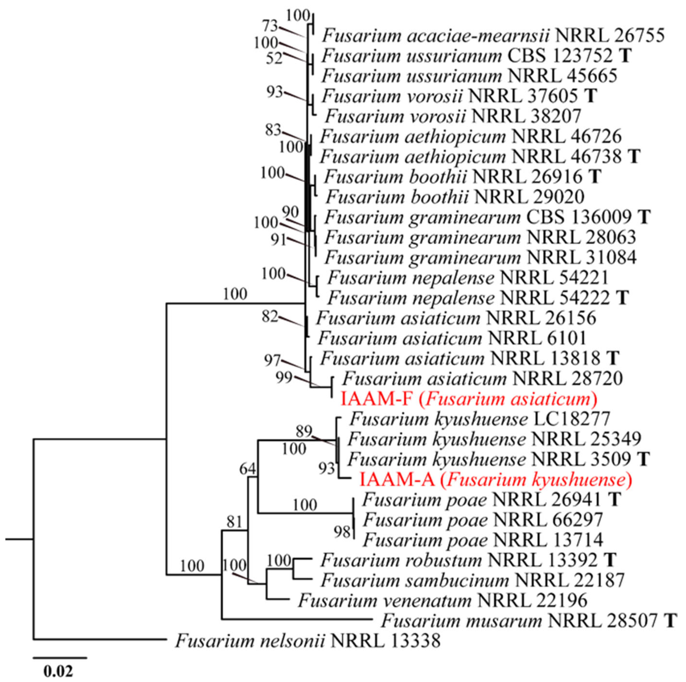
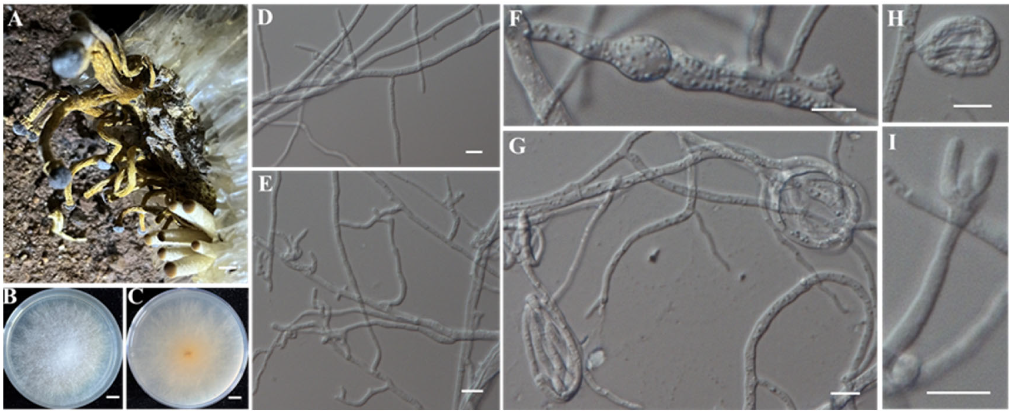
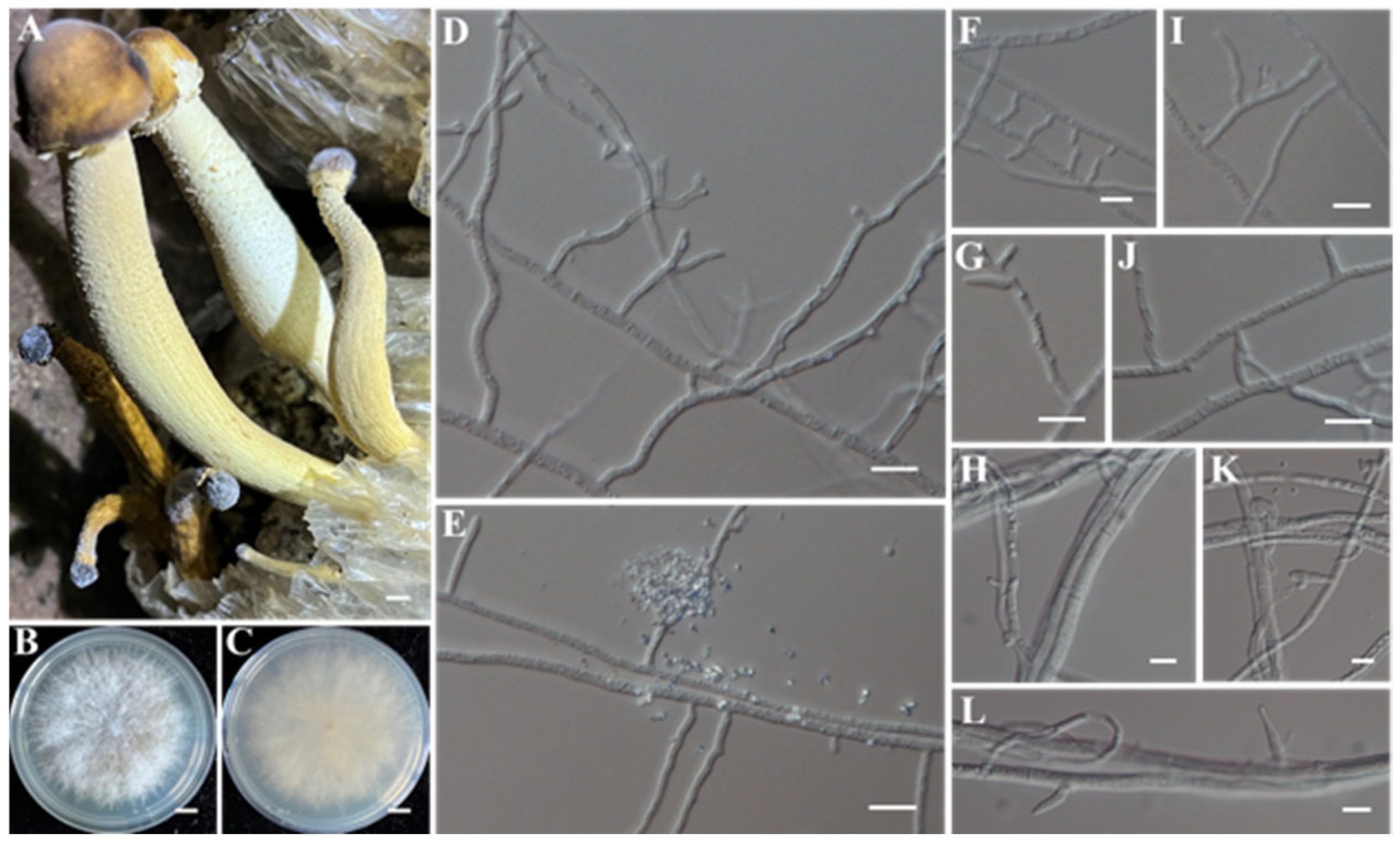
Under dark incubation at 25 °C on PDA medium, the putative pathogenic fungus IAAM-A exhibited a rapid growth rate, with an average radial extension of 7–8 mm/d. The colony developed dense, floccose aerial mycelia that were initially white to pink, later becoming pale yellow. The reverse side of the colony displayed a characteristic reddish-brown pigmentation. Microscopic examination revealed distinct morphological features. The macroconidia were falcate, curved, and tapering at both ends, typically with 3 to 5 septa, and measured 25–47 μm in length and 3.3–5 μm in width (Figure 1I). Microconidia were abundant, mostly straight or slightly curved, with 0 to 3 septa, and measured 7.5–15 μm × 2.5–5 μm (Figure 1I). No chlamydospores were detected (Figure 1). Multilocus sequence analysis based on ITS, tef1, rpb1, rpb2, and H3 revealed that IAAM-A shared 98%, 94%, 100%, 100%, and 100% coverage, with sequence identities of 99.35%, 100%, 100%, 100%, and 99.54%, respectively, with Fusarium kyushuense [42]. Phylogenetic analysis using the maximum likelihood (ML) method based on the concatenated tef1, rpb1, rpb2, and H3 sequences (~4300 bp) further supported the placement of IAAM-A within the F. kyushuense clade (Figure 2). Integrating both morphological characteristics [53] and multilocus phylogenetic evidence [42], IAAM-A was conclusively identified as F. kyushuense.
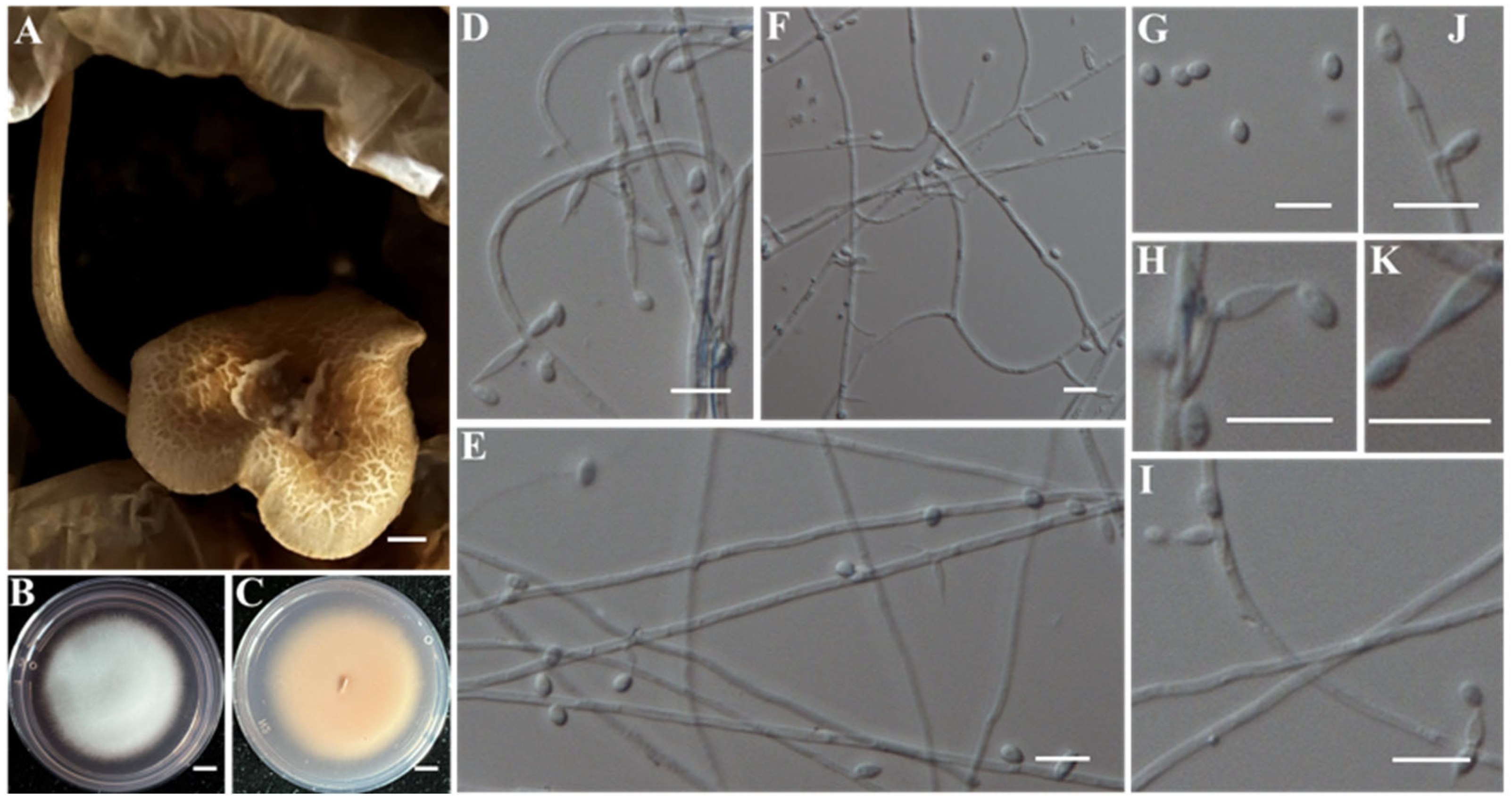
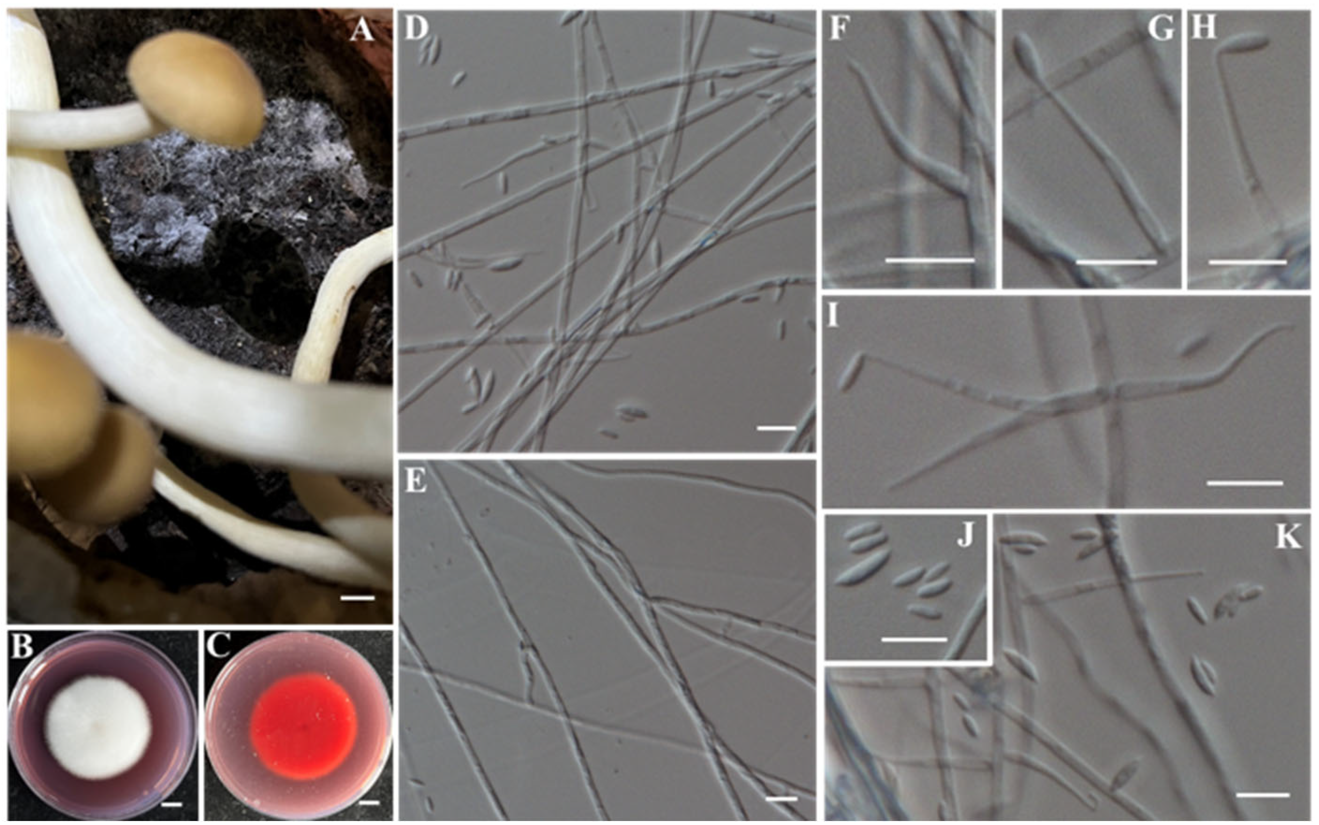
Under dark conditions at 25 °C on PDA medium, the putative pathogenic fungus IAAM-F exhibited a relatively rapid growth rate, with an average daily radial extension of 6.5–7.0 mm. The colony developed dense, white aerial mycelium, with the central region gradually acquiring a pale purplish-red pigmentation. After six days of cultivation, abundant microconidia were observed (Figure 3I,J). These were predominantly straight or slightly curved, containing 0 to 3 septa, and measuring 7.5–15 μm in length and 2.5–5 μm in width. No chlamydospores were detected (Figure 3). Multilocus sequence analysis based on ITS, tef1, rpb1, rpb2, and H3 [42] revealed that IAAM-F sequences shared 100% query coverage with those of Fusarium asiaticum, with sequence identities of 100%, 100%, 99.88%, 100%, and 100%, respectively. Furthermore, an ML phylogenetic tree constructed using the concatenated tef1, rpb1, rpb2, and H3 sequences (~4300 bp) placed IAAM-F within the F. asiaticum clade (Figure 2). Based on the combined morphological characteristics [54] and multilocus phylogenetic evidence [42], IAAM-F was conclusively identified as F. asiaticum.
(2)
Identification of Putative Pathogenic Fungi IAAM-B, IAAM-C, and IAAM-O
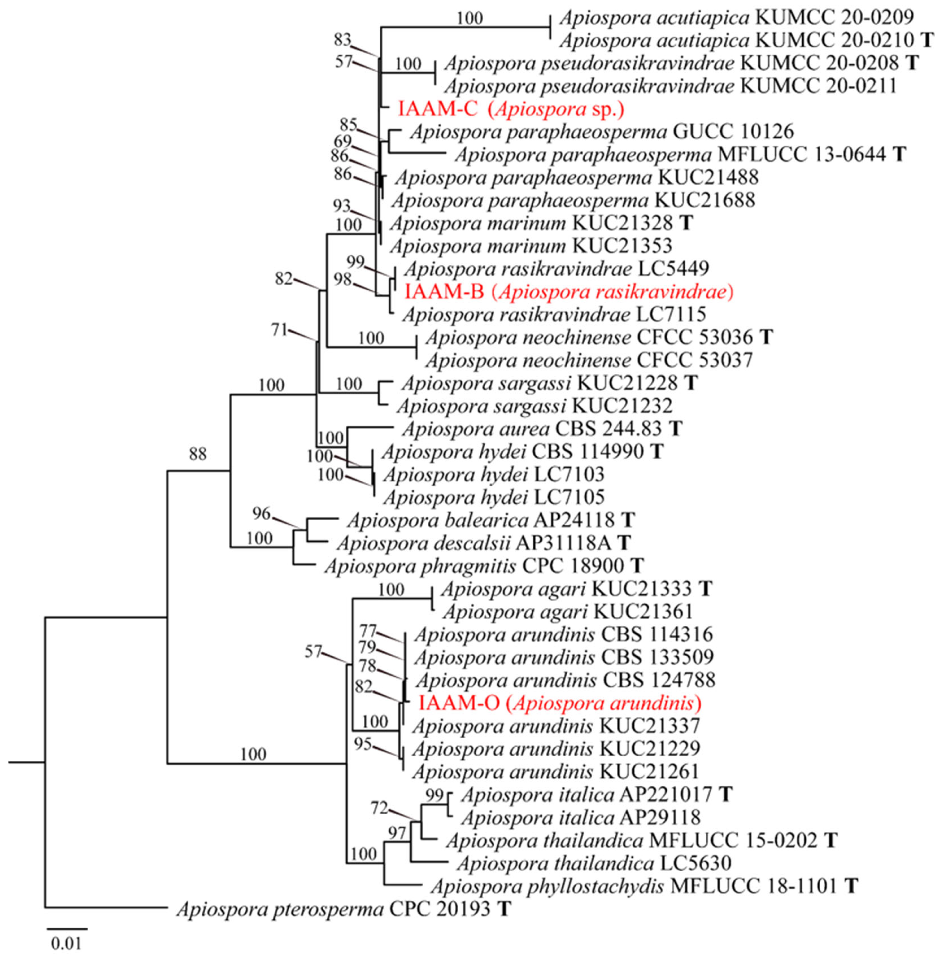
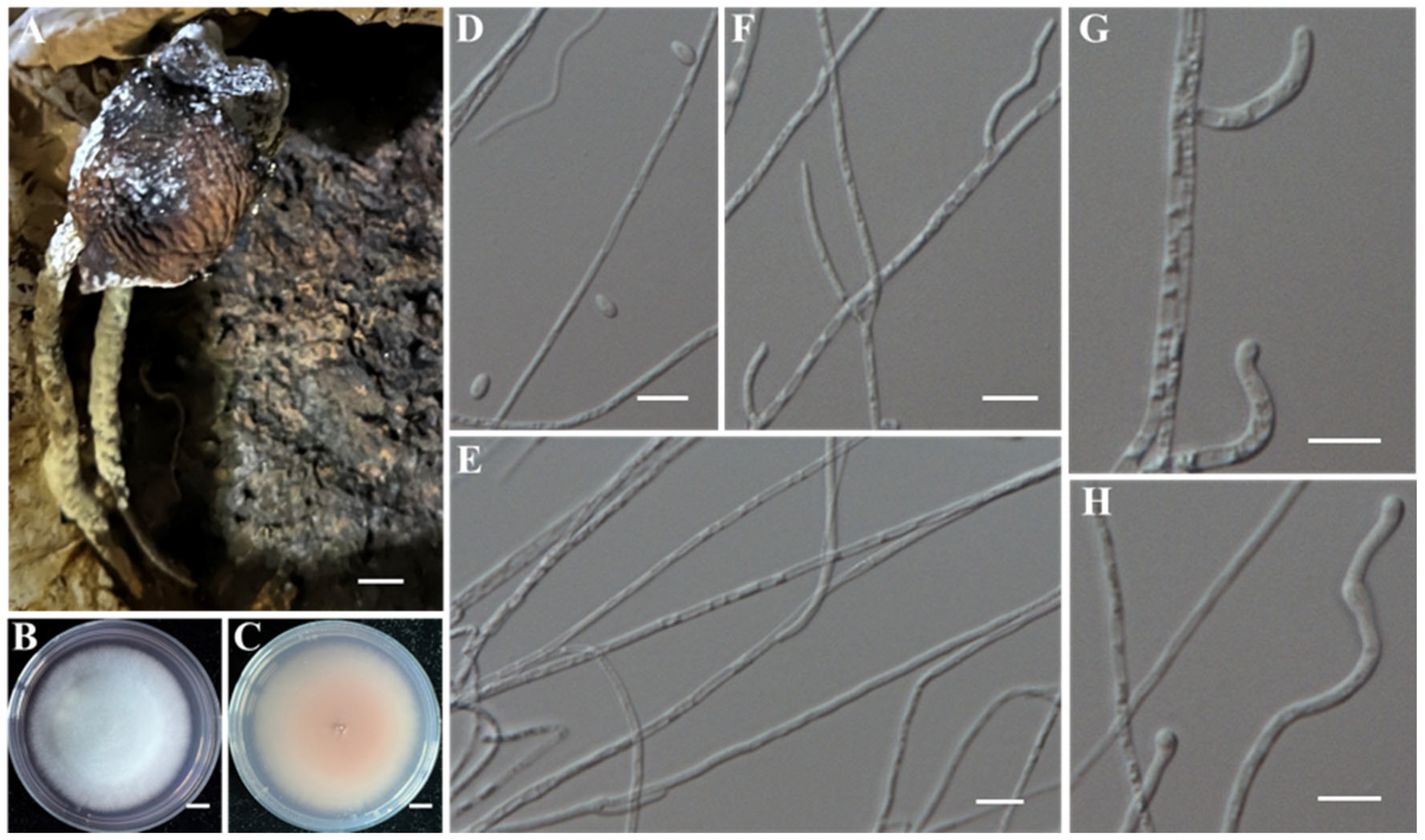
When cultured on PDA medium at 25 °C under dark conditions, the putative pathogenic fungus IAAM-B exhibited a radial growth rate of 3.0–3.3 mm/d. Colonies displayed dense aerial mycelia with well-defined margins, showing milky-white on the surface and pale-yellow on the reverse. Yellow pigments diffused centrifugally from the colony center, and by day five, the mycelium had fully colonized the 6-cm-diameter Petri dish. No conidia were observed microscopically (Figure 4). For molecular identification, the ITS, LSU, tef1, and tub2 regions were amplified using PCR [43]. BLASTn searches against the NCBI nucleotide database showed sequence identities of 100%, 99.67%, 100%, and 98.18%, with query coverages of 100%, 100%, 100%, and 99%, respectively, to Apiospora rasikravindrae. An ML phylogenetic analysis based on the concatenated sequences (~2200 bp) further confirmed that IAAM-B clustered closely with A. rasikravindrae (Figure 5). Collectively, the morphological characteristics and multilocus phylogenetic evidence support the identification of IAAM-B as A. rasikravindrae [43].
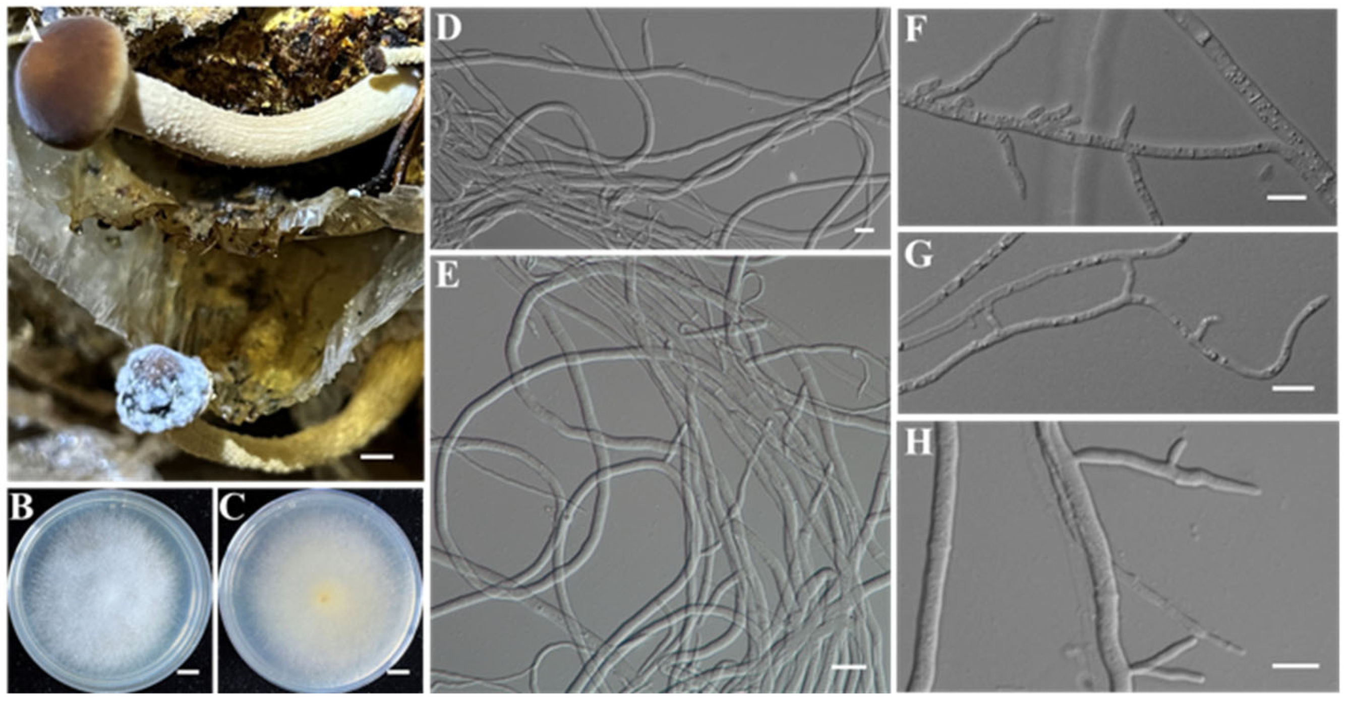
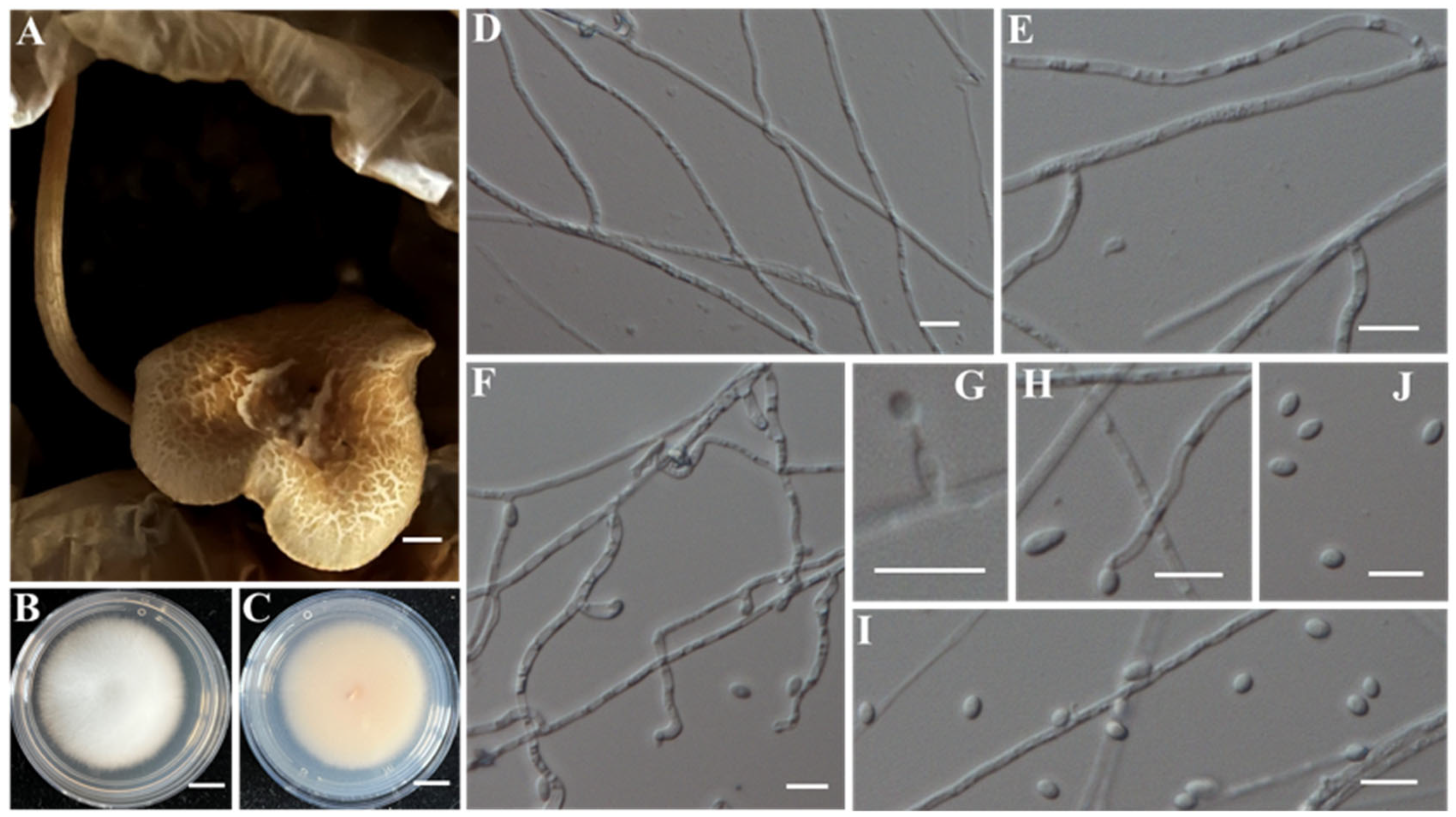
When cultured on PDA medium at 25 °C in darkness, the putative pathogenic fungus IAAM-C exhibited a mean radial growth rate of 5.8 mm/d. Colonies developed dense aerial mycelia with a snow-white surface and a pale-yellow reverse, with a more intense pigmentation concentrated at the center. The colony margins were irregular and exhibited a rough texture. No conidial structures were detected after 5 days of incubation (Figure 6). Molecular identification using the previously described Apiospora-specific protocol [43] was inconclusive, as BLASTn analyses of ITS, LSU, tef1, and tub2 gene sequences failed to resolve the isolate to a definitive species level. To clarify its taxonomic placement, an ML phylogenetic tree was constructed based on the concatenated ITS, LSU, tef1, and tub2 sequences (~2200 bp) of IAAM-C and closely related taxa retrieved from NCBI. The analysis showed that IAAM-C formed a distinct, well-supported clade closely related to A. pseudorasikravindrae and A. acutiapica (Figure 5). Taken together, the morphological features and phylogenetic evidence supported the assignment of IAAM-C to the genus Apiospora, and the isolate was therefore designated as Apiospora sp.
Under dark incubation at 25 °C on PDA medium, the putative pathogenic fungus IAAM-O exhibited a radial growth rate of 4.8 mm/d. Colonies developed dense, white aerial mycelia with a rough margin surface, and the reverse side of the culture appeared pale yellow without evident pigment deposition. After 5 days of cultivation, no conidia were observed. Microscopically, the hyphae were septate, with interseptal distances ranging from 25 to 35 μm (Figure 7). Molecular identification was conducted using sequences from the ITS, LSU, tef1, and tub2 genes [43]. BLASTn analysis revealed that all sequences of IAAM-O shared 100% coverage and identity with those of Apiospora arundinis. An ML phylogenetic tree constructed from the concatenated dataset (~2200 bp) further confirmed that IAAM-O clustered within the A. arundinis clade with strong statistical support (Figure 5). Based on the combined morphological characteristics and multilocus phylogenetic analyses [43], IAAM-O was conclusively identified as A. arundinis.
(3)
Identification of Putative Pathogenic Fungi IAAM-D, IAAM-H, IAAM-I, and IAAM-T
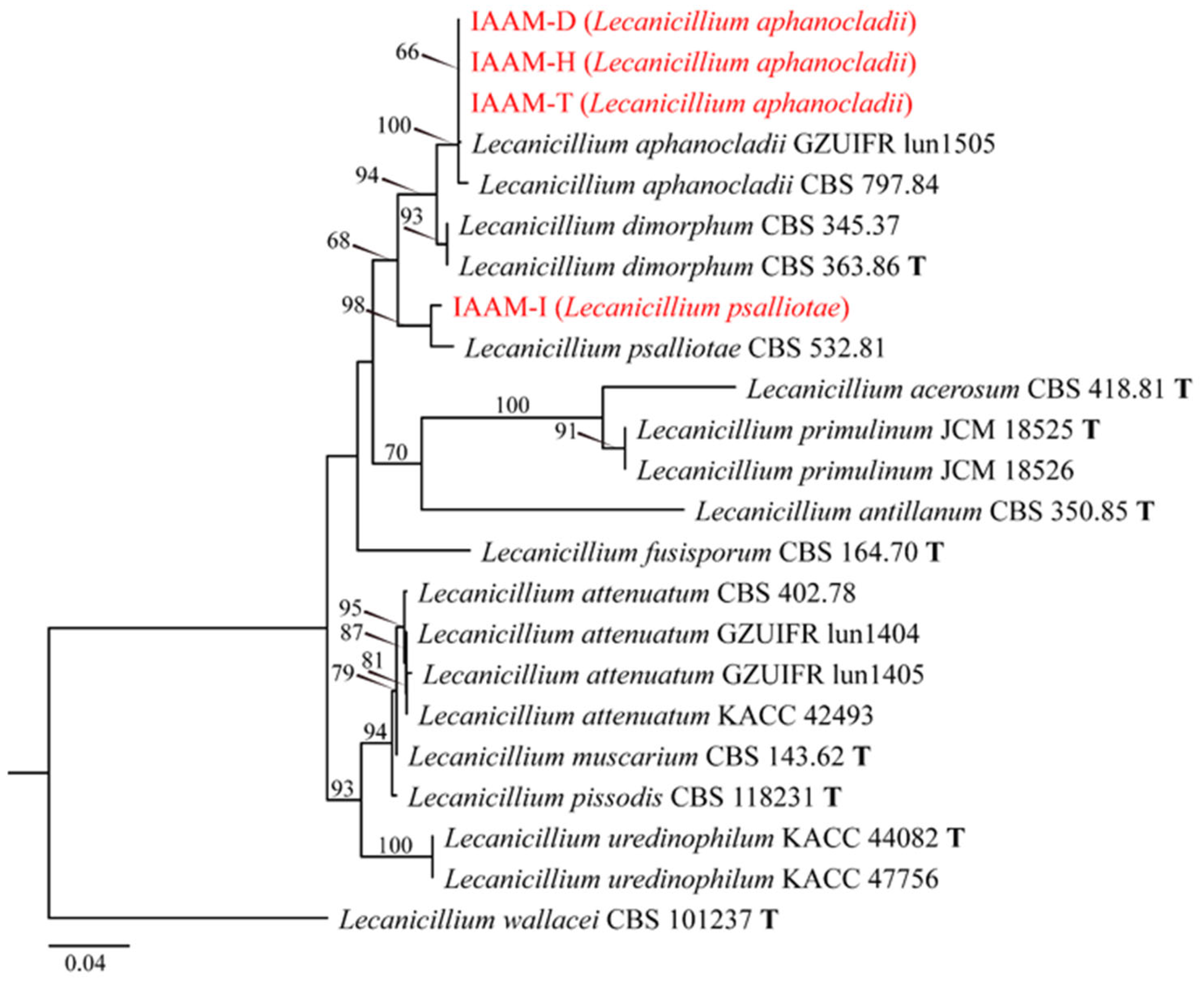
The putative pathogenic fungal isolates IAAM-D, IAAM-H, and IAAM-T, obtained from distinct tissue sources, were purified and cultured on PDA at 25 °C for five days. IAAM-D exhibited dense mycelial growth with a radial extension rate ranging from 1.6–2.6 mm/d. Colonies displayed a pure white surface, while the reverse and adjacent medium displayed a pale reddish pigmentation. Microscopic examination revealed elliptical to subglobose conidia measuring 3.5–5.0 μm × 2.2–2.6 μm (Figure 8). IAAM-H displayed colony morphology comparable to IAAM-D but demonstrated host specificity infecting the living fruiting bodies of A. chaxingu. Microscopically, solitary or paired conidiogenous cells were observed, whereas other microscopic features were consistent with IAAM-D (Figure 9). IAAM-T differed by exhibiting a lighter reverse (nearly white) and producing conidiogenous cells with distinctly elongated necks, while other colony characteristics resembled those of IAAM-D and IAAM-H. (Figure 10). Molecular identification using Lecanicillium-specific DNA barcode markers (ITS and rpb1) [44] showed that the ITS and rpb1 sequences of all three isolates shared 100% and 99.24% sequence identity with 100% and 99% query coverage, respectively, to Lecanicillium aphanocladii. Phylogenetic analyses based on concatenated ITS and rpb1 sequences (~1100 bp) further confirmed that all three isolates clustered within the L. aphanocladii clade (Figure 11). Collectively, the morphological characteristics [55] and multilocus phylogenetic evidence [44] unequivocally supported the identification of IAAM-D, IAAM-H, and IAAM-T as L. aphanocladii.
When incubated in darkness at 25 °C on PDA, the putative pathogenic fungus IAAM-I exhibited a radial growth rate of 1.5–2.5 mm/d, forming dense mycelia with a well-defined, regular colony margin. The colony surface was pure white, while the reverse showed deep red pigmentation that diffused into the surrounding medium. Microscopic examination revealed falcate macroconidia measuring 7.5–11 μm × 1.5–2.0 μm, and rod-shaped to elliptical microconidia measuring 2.5–3.5 μm × 1–1.5 μm (Figure 12J). Conidiogenous cells appeared either singly or in whorls of three to four (Figure 12). For molecular identification, the ITS and rpb1 genes were amplified and sequenced [44]. The resulting sequences of IAAM-I exhibited sequence identities of 99.43% for ITS and 97.99% for rpb1 with 100% query coverage, relative to the reference sequences of Lecanicillium psalliotae. Phylogenetic analysis based on concatenated ITS and rpb1 sequences (~1100 bp) further placed IAAM-I within the L. psalliotae clade (Figure 11). Collectively, both the morphological characteristics [55] and multilocus phylogenetic evidence [44] strongly supported the identification of IAAM-I as L. psalliotae.
(4)
Identification of Putative Pathogenic Fungus IAAM-P
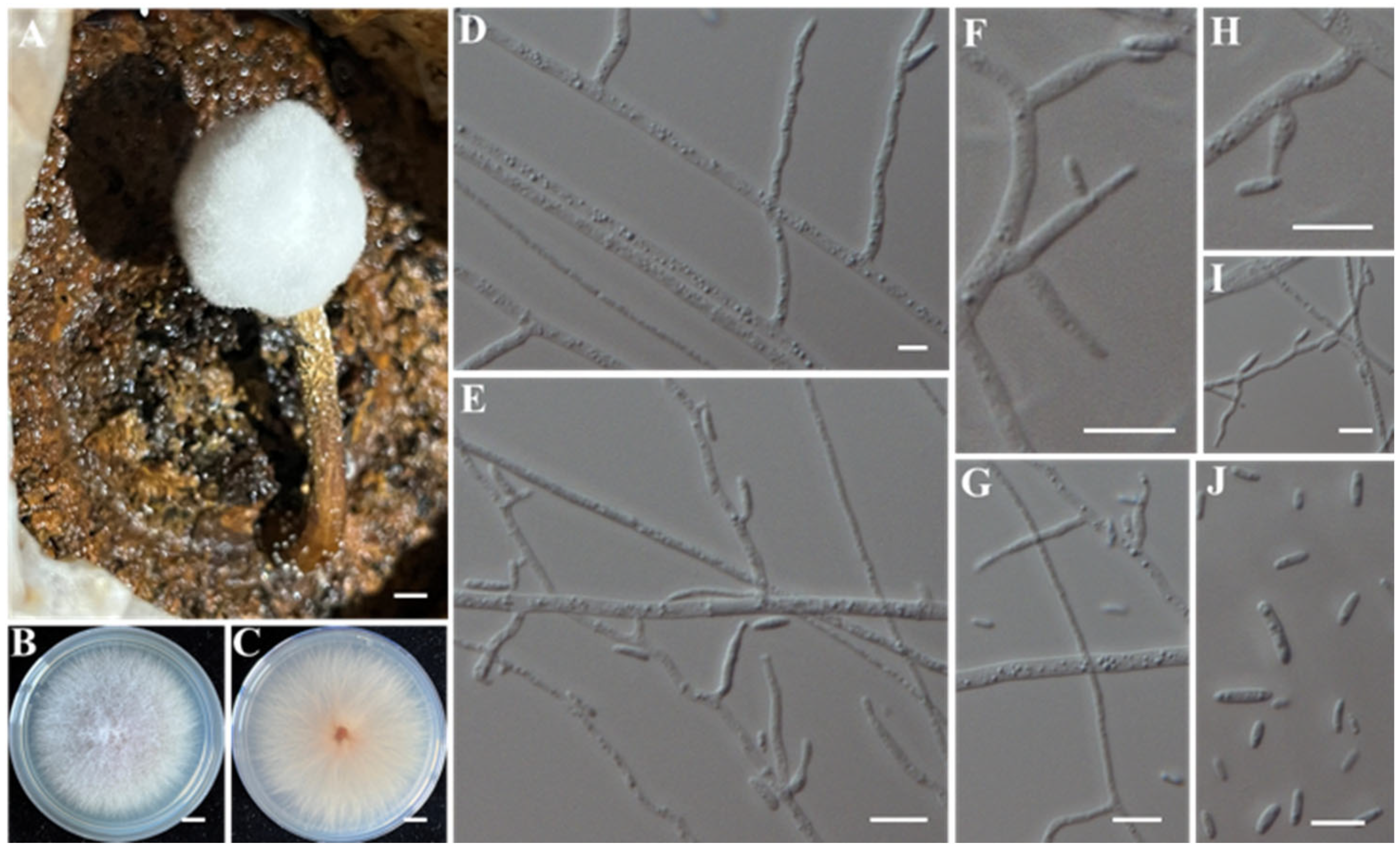
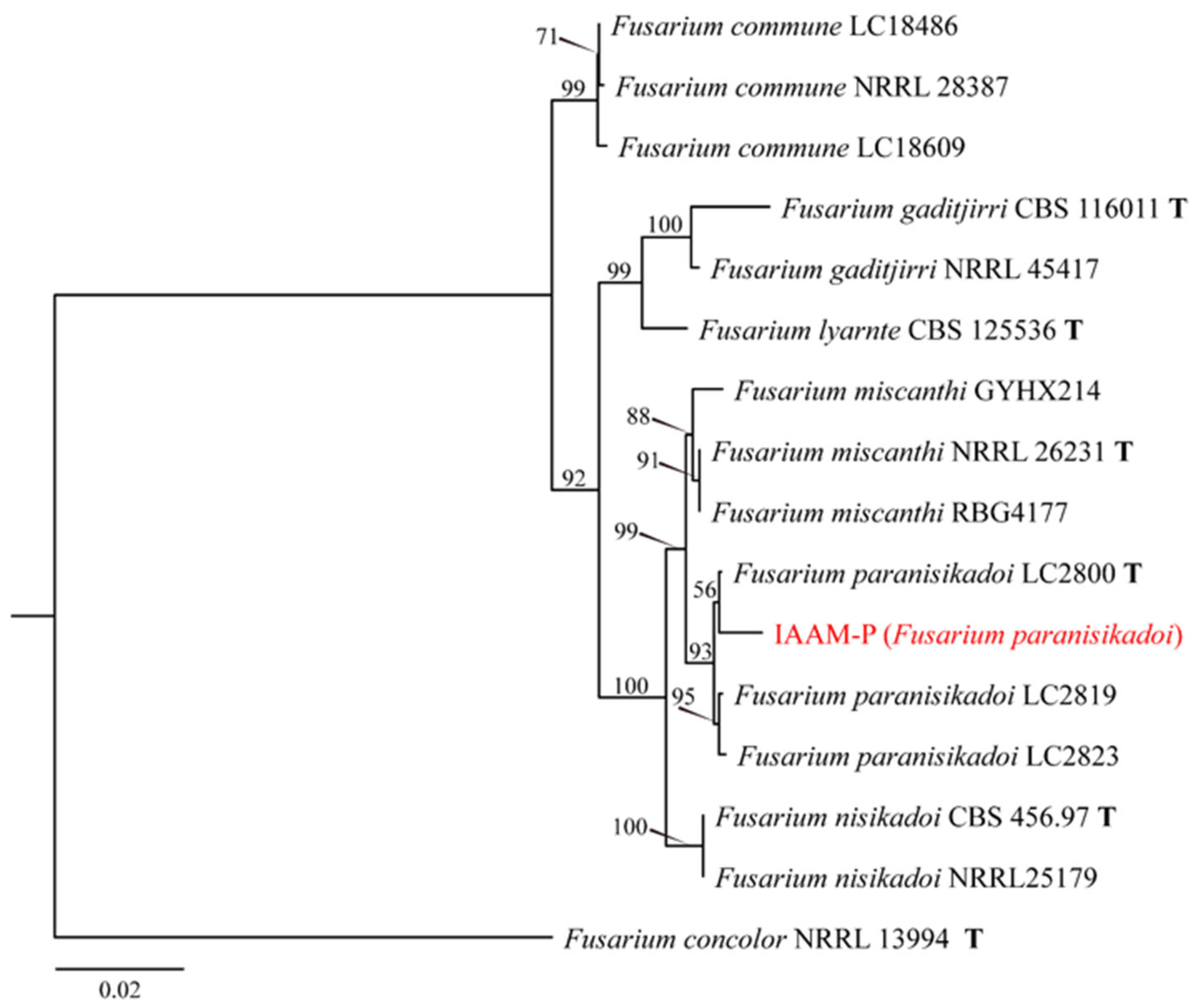
The putative pathogenic fungus IAAM-P was incubated on PDA at 25 °C under dark conditions, exhibiting a relatively rapid radial growth rate, averaging 4.3 mm/d. Colonies developed dense aerial mycelia with a rough margin; the central surface appeared grayish-orange and slightly elevated, while the margin was white. The colony reverse displayed pale orange pigmentation concentrated at the center. Microscopic examination revealed abundant conidial structures: macroconidia were slightly curved or cylindrical, measuring 10–15 μm × 2.5–3.5 μm, whereas microconidia were predominantly oval to rod-shaped, measuring 3–10 μm × 1.5–2.5 μm (Figure 13J). Conidiogenous cells were observed in both monophialidic or polyphialidic forms (Figure 13). For molecular identification, multilocus PCR amplification of tef1, rpb1, and rpb2 was conducted [42]. BLASTn analysis against the NCBI database revealed sequence identity values of 99.84%, 99.75%, and 99.74% with 100%, 100%, and 98% coverage, respectively, to reference sequences of Fusarium paranisikadoi. Phylogenetic analysis using the ML method based on concatenated sequences (~3900 bp) of tef1, rpb1, and rpb2 further supported the clustering of IAAM-P with F. paranisikadoi (Figure 14). Taken together, the morphological characteristics [56] in conjunction with multilocus sequence data [42] unequivocally identified IAAM-P as F. paranisikadoi.
(5)
Identification of Putative Pathogenic Fungi IAAM-K and IAAM-N
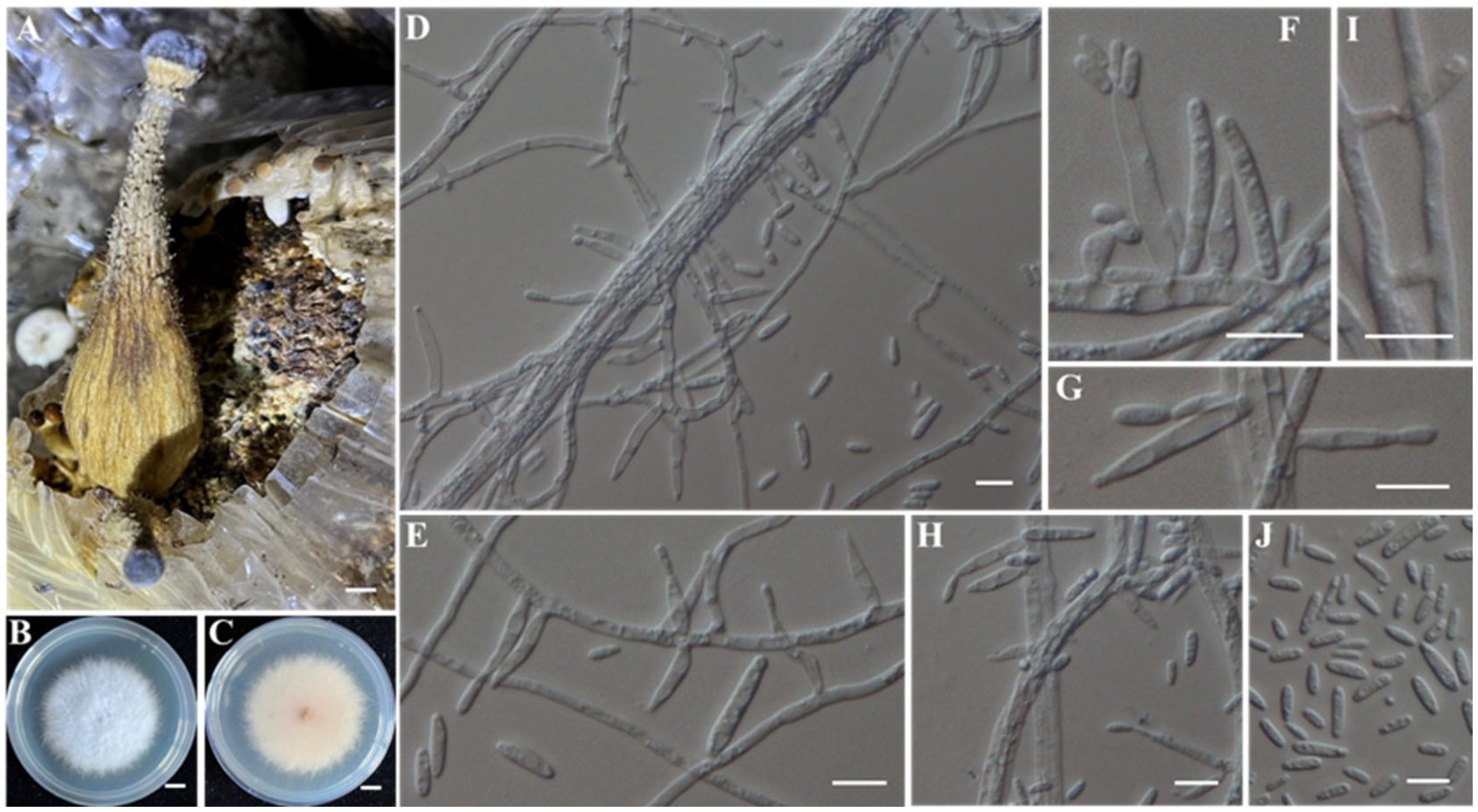
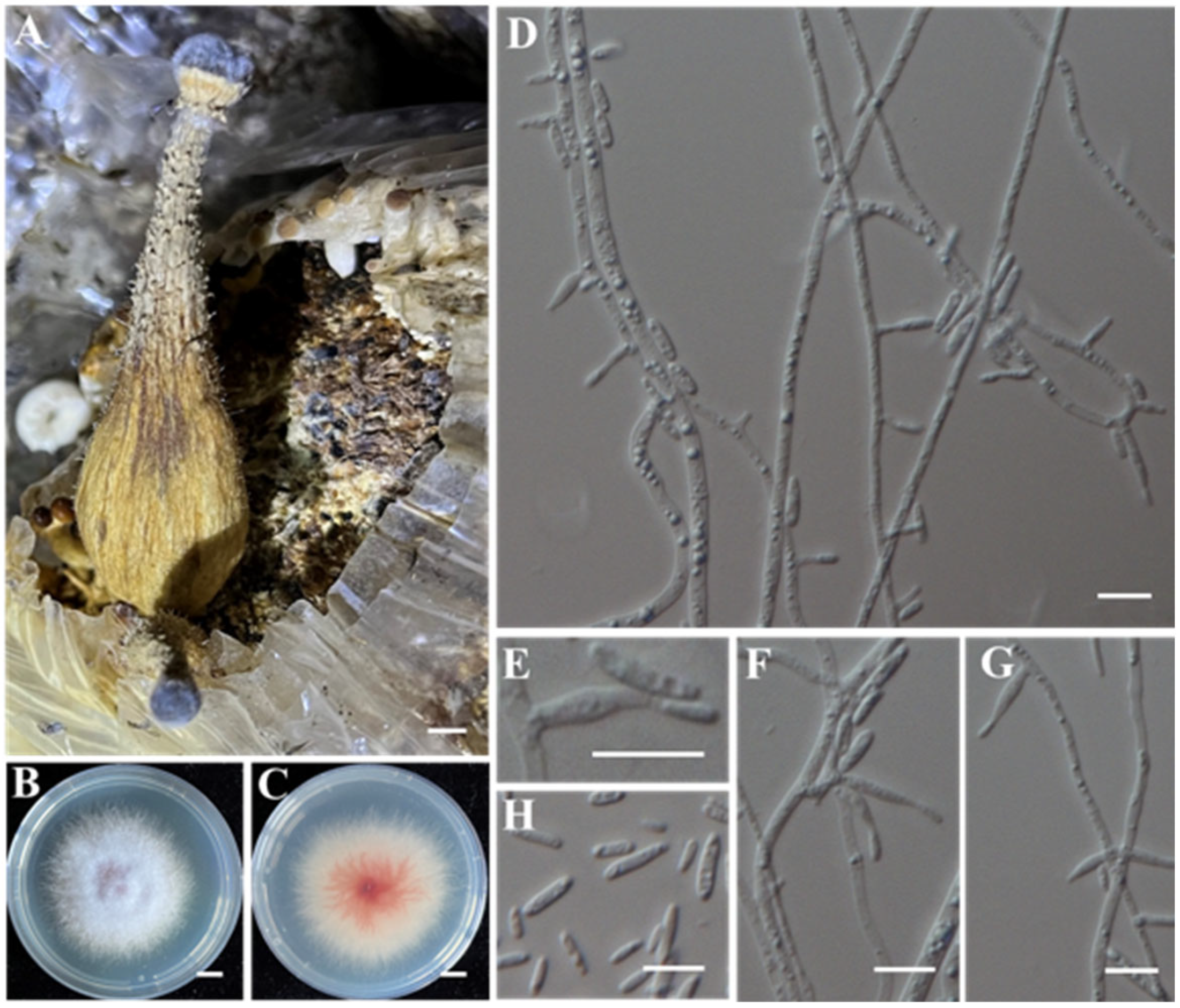
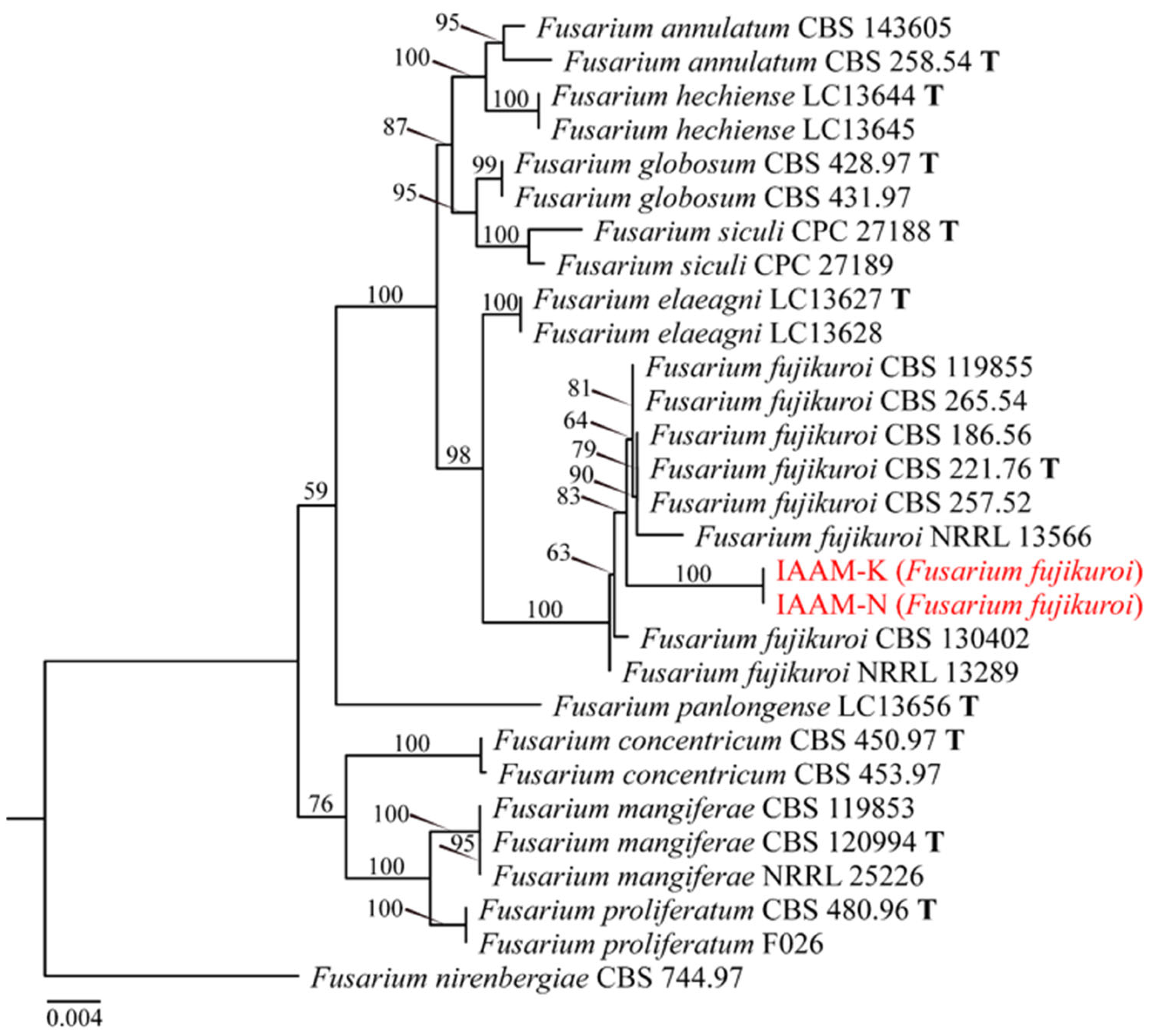
The putative pathogenic fungus IAAM-K exhibited rapid radial growth on PDA, attaining 10–12 mm/d under dark incubation at 25 °C. Colonies developed a circular morphology with filamentous, radially spreading margins, and a white to violet-red surface with a slightly raised center. Dense aerial mycelia were present, with pale red pigmentation in the colony center on the reverse. Microscopic analysis revealed prolific conidial production (Figure 15J): macroconidia were cylindrical or slightly curved (15–25 μm × 3–5 μm), while microconidia were short-rod-shaped (6–12 μm × 2–3 μm). Conidiogenous cells were observed in both monophialidic or polyphialidic forms (Figure 15). IAAM-N, isolated from the same A. chaxingu fruiting body, shared similar morphological traits with IAAM-K but differed in producing more intense violet-red pigmentation on the reverse side (Figure 16). Multilocus sequence analysis targeting the tef1, tub2, and rpb2 genes was performed [42]. For IAAM-K, the sequences showed 100% coverage with Fusarium fujikuroi, with nucleotide identities of 99.68% (tef1), 100% (tub2), and 100% (rpb2). IAAM-N exhibited 100% coverage and identity across all three loci. Phylogenetic reconstruction (ML method) based on concatenated tef1tub2rpb2 sequences (~2900 bp) confirmed that both isolates formed a well-supported monophyletic group with the F. fujikuroi reference strains (Figure 17). Combining morphological [57] and molecular phylogenetic evidence [42,57], both IAAM-K and IAAM-N were unequivocally identified as F. fujikuroi.
(6)
Identification of Putative Pathogenic Fungus IAAM-J
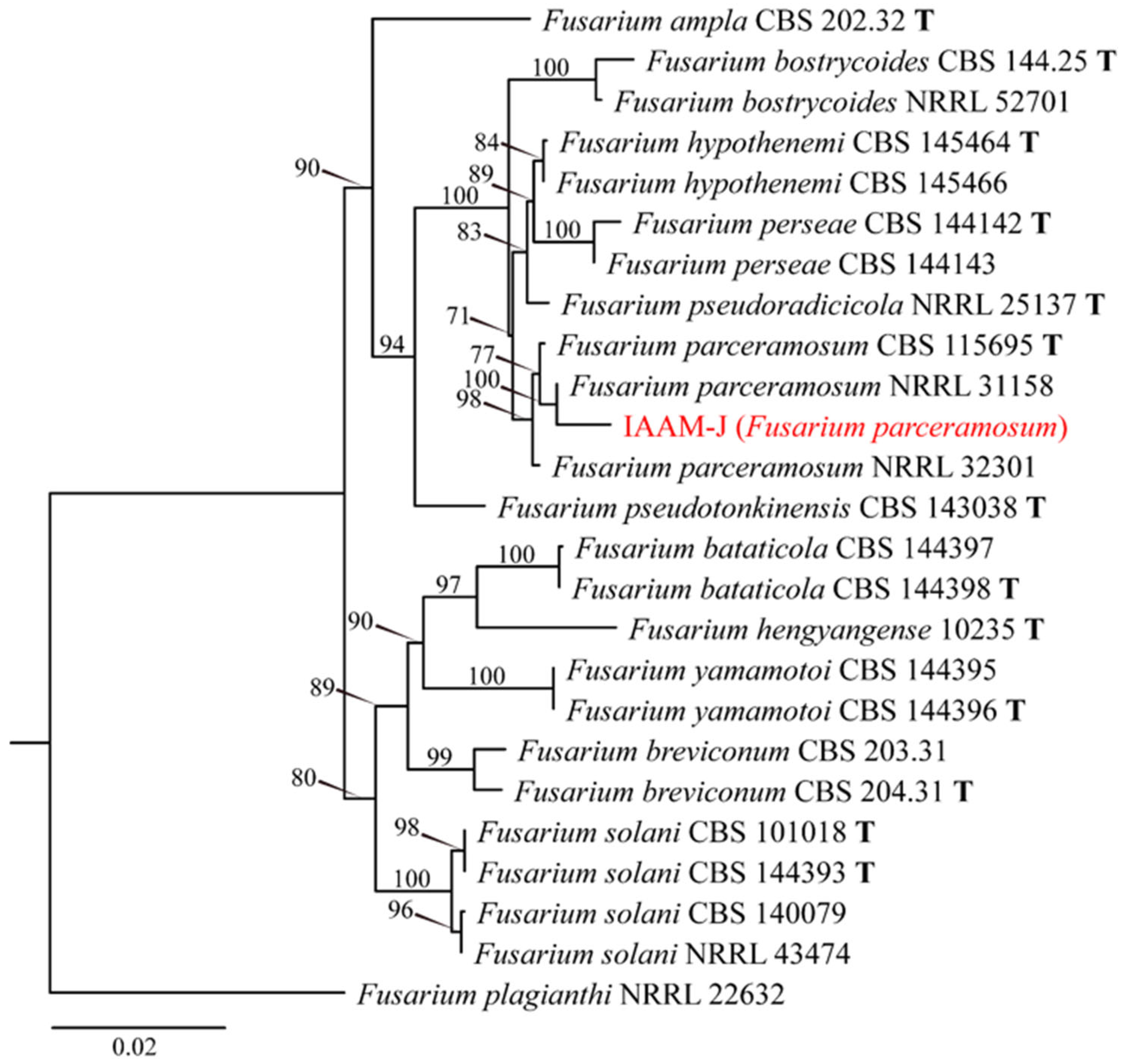
Isolate IAAM-J exhibited an average radial growth rate of 3.5 mm/d on PDA when incubated at 25 °C in the dark. Colonies were floccose, characterized by abundant white aerial mycelia, a well-defined margin, and pale yellow pigmentation on the reverse. Microscopic examination revealed numerous conidia that were straight to curved, clavate, measuring 5.5–10 μm × 2–4 μm (Figure 18I). Conidiogenous cells occurred predominantly solitary or in clusters (Figure 18). Multilocus PCR amplification targeting the ITS region, tef1, and rpb2 was performed [47]. BLASTn analysis in the NCBI database showed that the ITS, tef1, and rpb2 sequences of isolate IAAM-J had query coverages of 100%, 98%, and 100%, respectively, and each shared 100% sequence identity with the corresponding sequences of Fusarium parceramosum. An ML phylogenetic tree based on concatenated ITS, tef1, and rpb2 sequences (~2800 bp) placed isolate IAAM-J within the F. parceramosum clade (Figure 19). Taken together, the morphological and molecular evidence [47] supports the identification of IAAM-J as F. parceramosum.
(7)
Identification of Putative Pathogenic Fungus IAAM-E
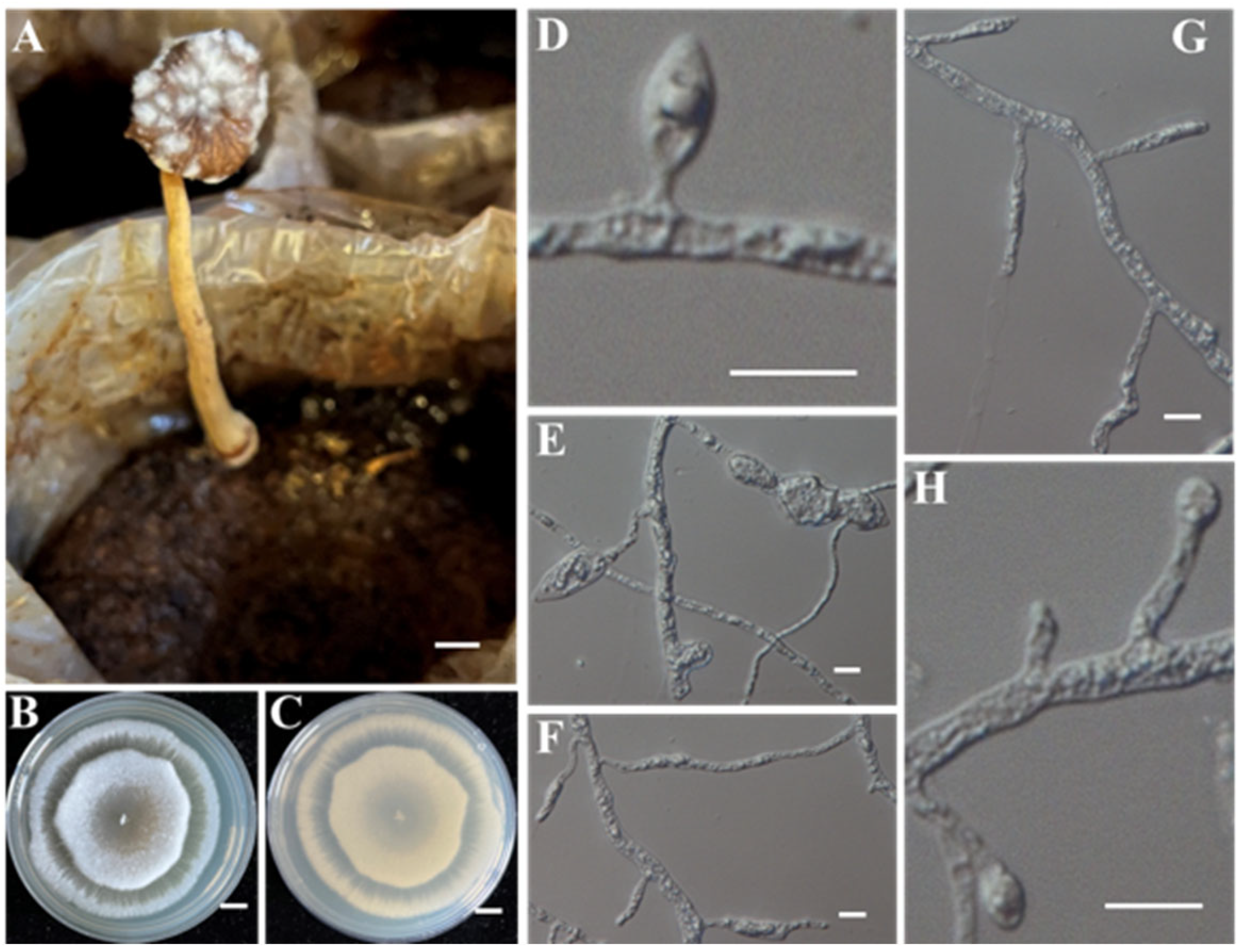
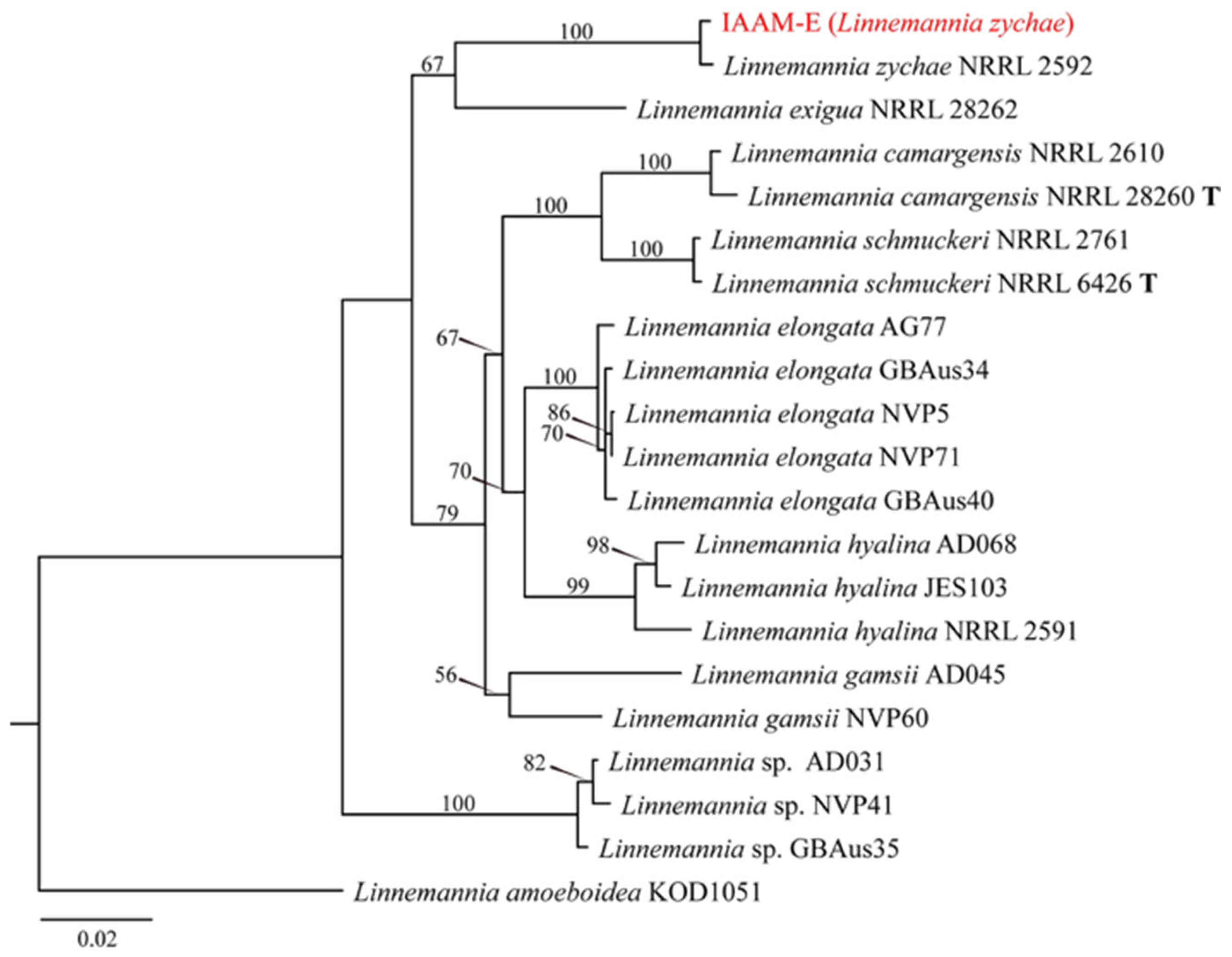
When incubated in the dark at 25 °C, isolate IAAM-E showed slow growth on PDA. The colony displayed distinct zonation, with coloration ranging from white to light gray. Aerial mycelia were sparse, whereas abundant submerged mycelia were present, characterized by localized hyphal swellings or clustered enlargements. After 5 days of incubation, no conidia were observed on aerial hyphae (Figure 20). Multilocus PCR amplification targeting the tef1 and rpb1 genes was performed [45]. BLASTn analysis in the NCBI database showed that the tef1 and rpb1 sequences of IAAM-E exhibited 100% query coverage and sequence identities of 99.71% and 99.74%, respectively, with the corresponding sequences of Linnemannia zychae. An ML phylogenetic tree based on concatenated tef1 and rpb1 sequences (~2600 bp) placed isolate IAAM-E within the L. zychae clade (Figure 21). Taken together, morphological observations [58] and multilocus molecular data [45] confirmed that IAAM-E is L. zychae.
(8)
Identification of Putative Pathogenic Fungus IAAM-G
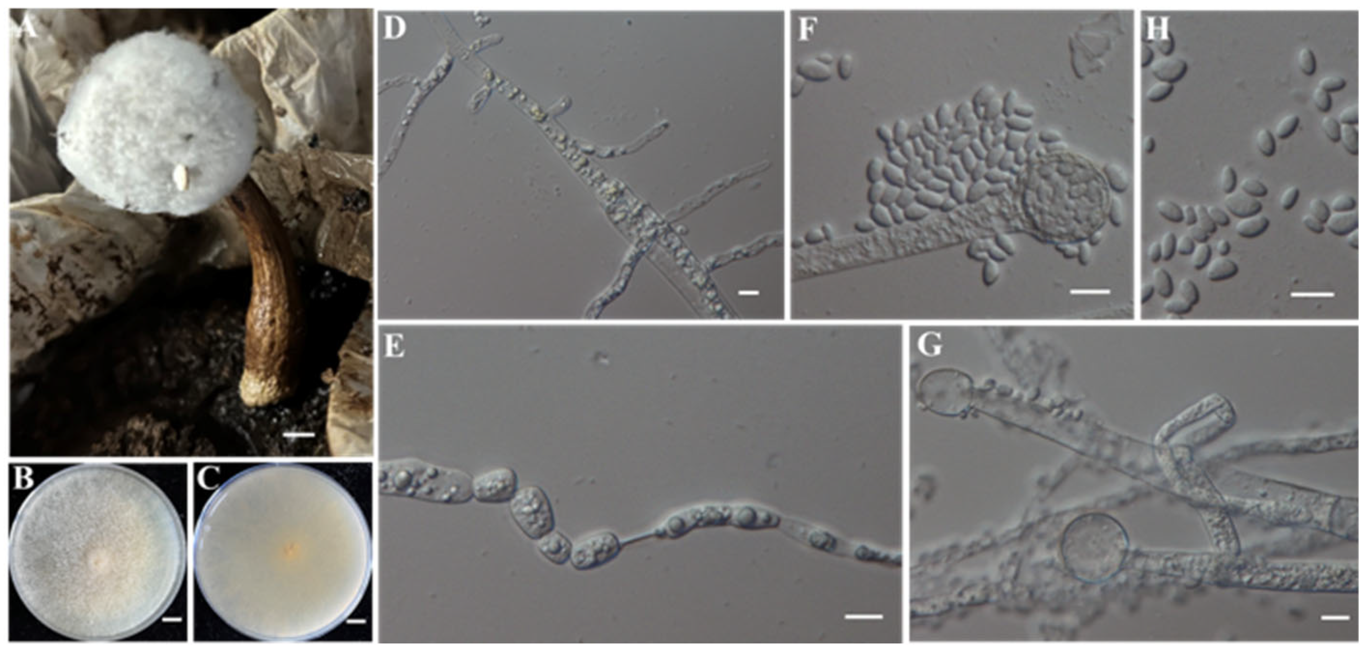
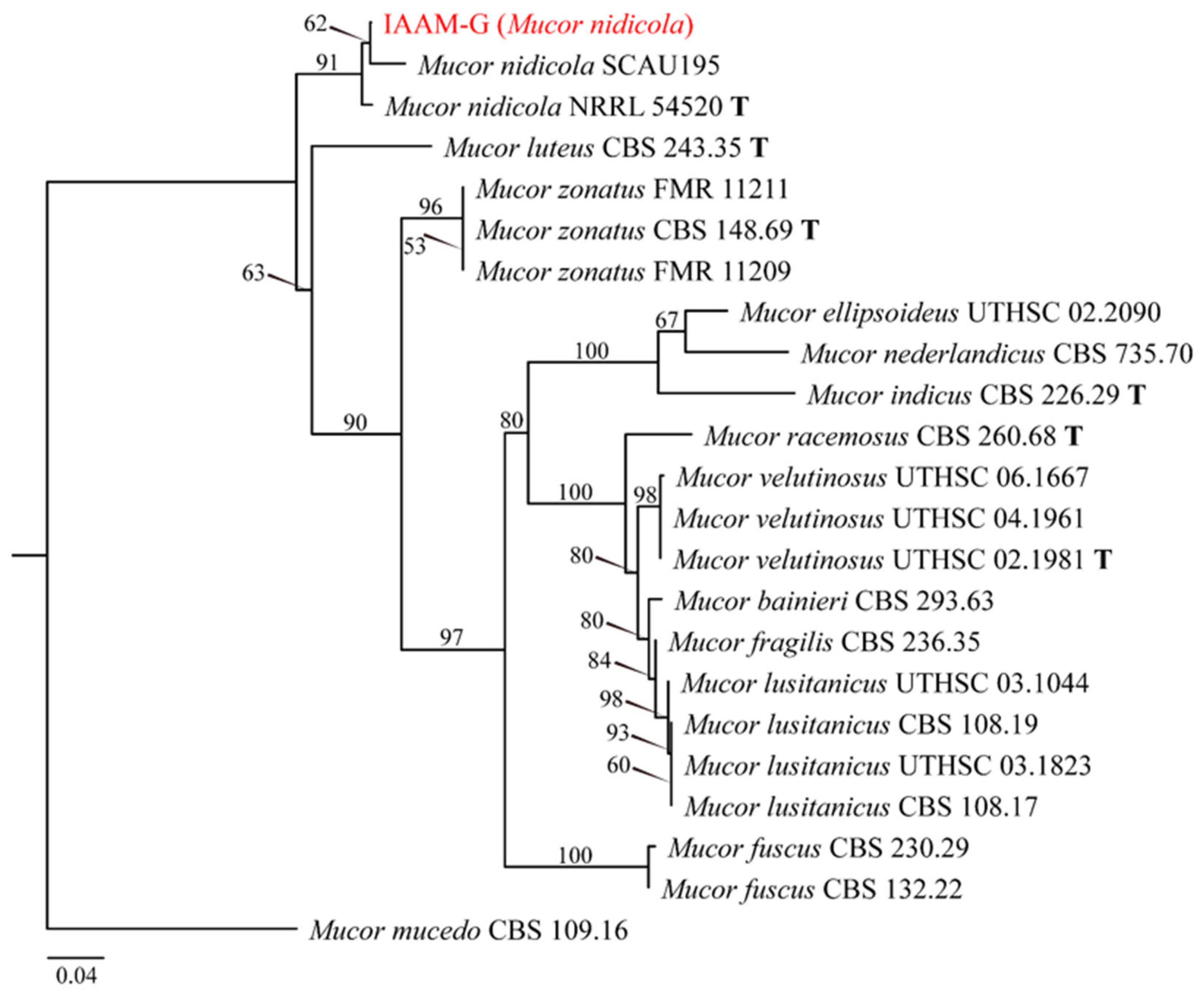
When incubated in the dark at 25 °C, isolate IAAM-G showed rapid growth on PDA, with an average radial growth rate of 10 mm/d. The colony displayed grayish-yellow pigmentation with dense, hairy aerial hyphae. Microscopic examination revealed that young hyphae were aseptate with extensive branching, whereas mature hyphae became septate (Figure 22E). Abundant conidia were observed (Figure 22H), which were elliptical to irregular in shape, measuring 3–10 μm × 2–6 μm. Sporangiophores were erect, terminating in spherical sporangia occurring singly or with one to two branches (Figure 22). PCR amplification and sequencing of the ITS region [46] showed that the ITS sequence of isolate IAAM-G had 100% query coverage and 98.84% sequence identity with the reference sequences of Mucor nidicola in the NCBI database. An ML phylogenetic tree based on ITS sequences (~550 bp) placed IAAM-G within the M. nidicola clade (Figure 23). Combined morphological and molecular evidence [46] confirmed IAAM-G as M. nidicola.
(9)
Identification of Putative Pathogenic Fungus IAAM-L
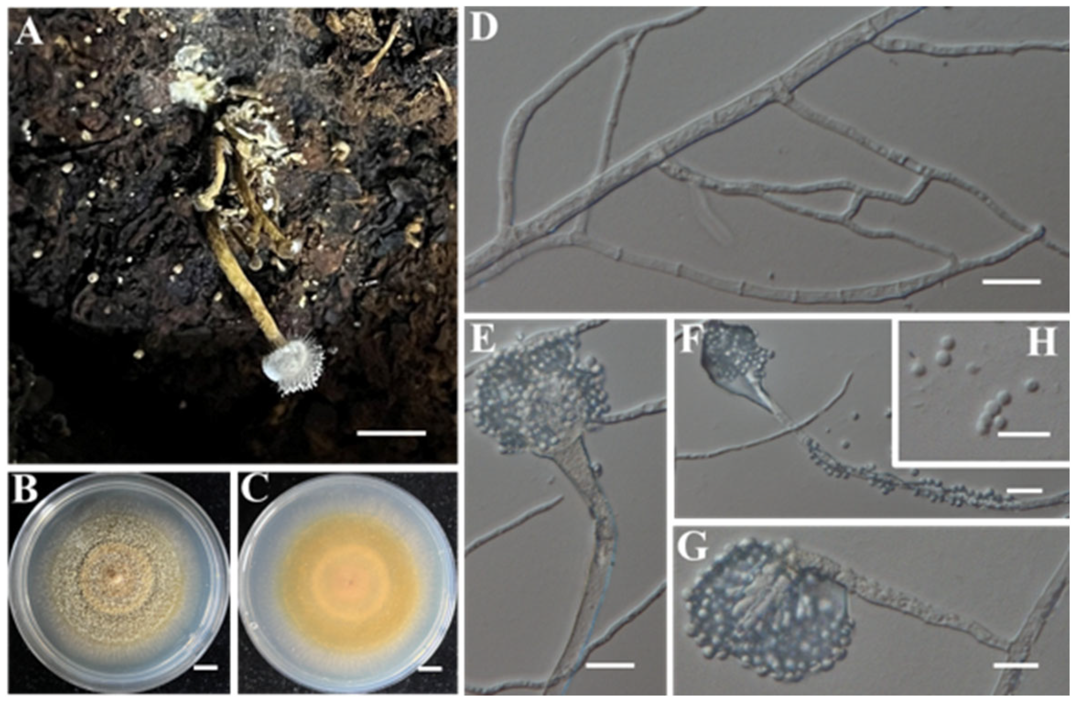
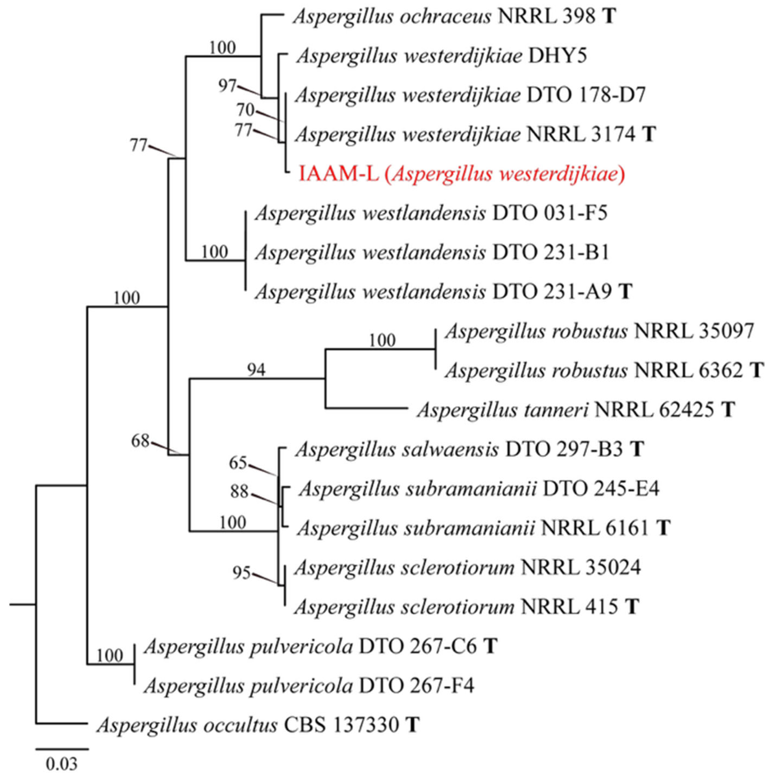
When incubated in the dark at 25 °C, isolate IAAM-L exhibited moderate growth on PDA, with an average radial growth rate of 3.2–3.5 mm/d. The colony exhibited white to pale yellow mycelia with sparse aerial hyphae, and the colony reverse showed brownish-yellow pigmentation. Pale yellow sclerotia were observed (Figure 24B). Abundant spherical conidia measuring 2.3–3 μm × 2.3–2.8 μm were produced (Figure 24). Multilocus PCR amplification targeting the ITS region and tub2 gene [48] showed that the sequences of isolate IAAM-L had 100% query coverage and sequence identity with reference sequences of Aspergillus westerdijkiae in the NCBI database. An ML phylogenetic tree based on concatenated ITS-tub2 sequences (~1000 bp) placed IAAM-L within the A. westerdijkiae clade (Figure 25). Taken together, morphological [59] and molecular evidence [48] confirmed IAAM-L as A. westerdijkiae.
(10)
Identification of Putative Pathogenic Fungus IAAM-M
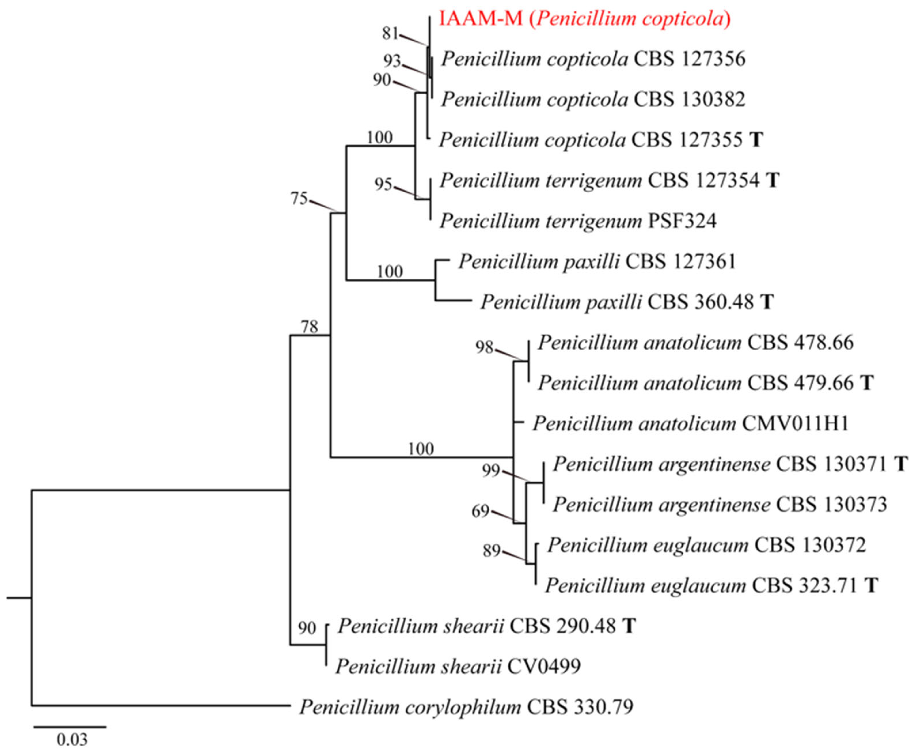
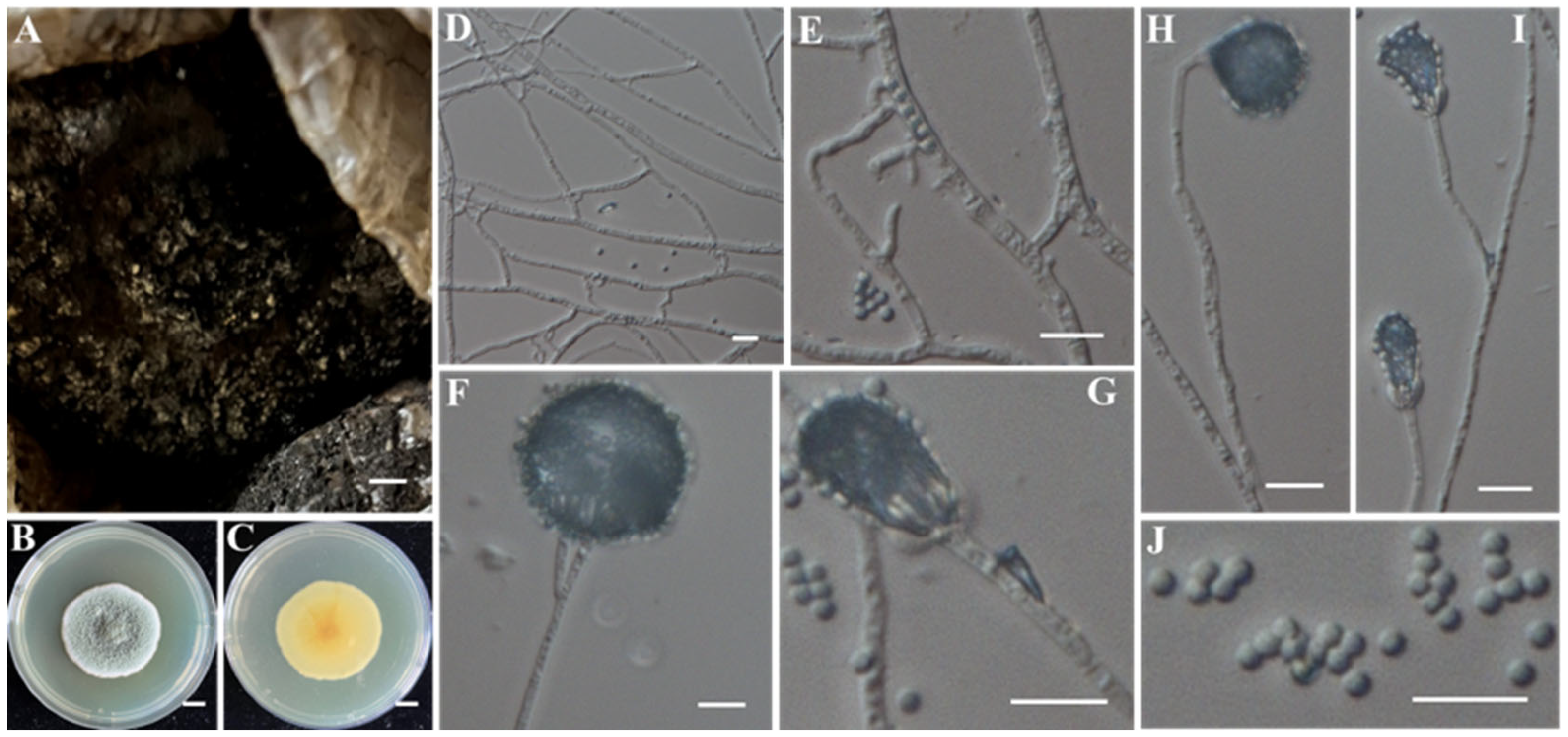
When incubated in the dark at 25 °C, fungal isolate IAAM-M exhibited robust growth on PDA, with an average radial growth rate of 2.1–2.5 mm/d. The colony exhibited dense white marginal hyphae and dark green conidial masses in the central region. Elliptical conidia measured 2.5–3.0 μm × 2.0–2.5 μm (Figure 26H,I). Conidiophores were monoverticillate to symmetrically biverticillate, ranging from several tens to several hundreds of micrometers in length (Figure 26). Multilocus sequence analysis of the ITS region and tub2 [49] showed query coverages of 100% and 90%, with sequence identities of 100% and 99.75%, respectively, to type strains of Penicillium copticola. An ML phylogenetic tree based on concatenated ITS-tub2 sequences (~900 bp) placed IAAM-M within the P. copticola clade (Figure 27). Taken together, morphological characteristics and multilocus molecular data [49] confirmed the identification of IAAM-M as P. copticola.
(11)
Identification of Putative Pathogenic Fungus IAAM-S
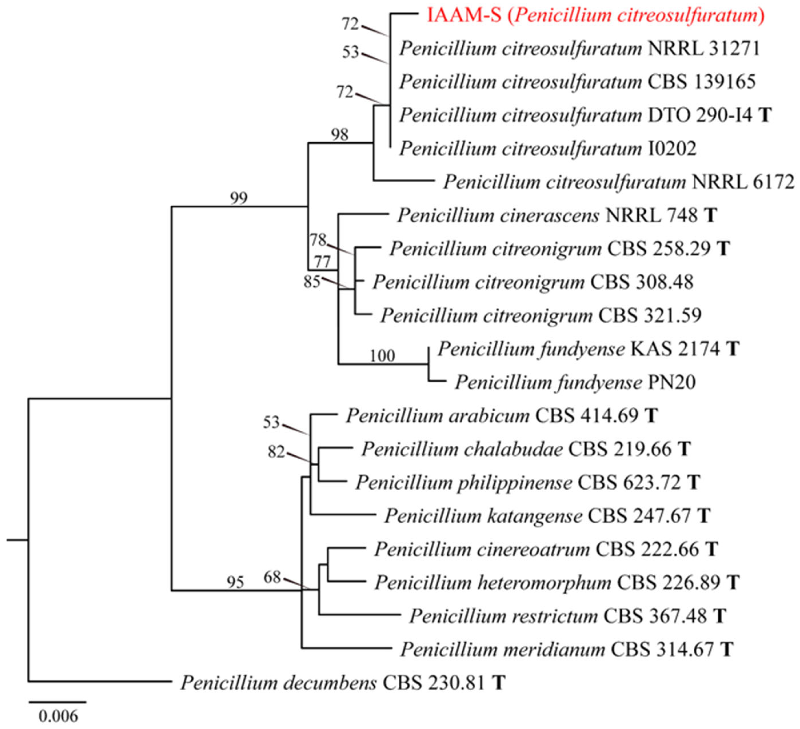
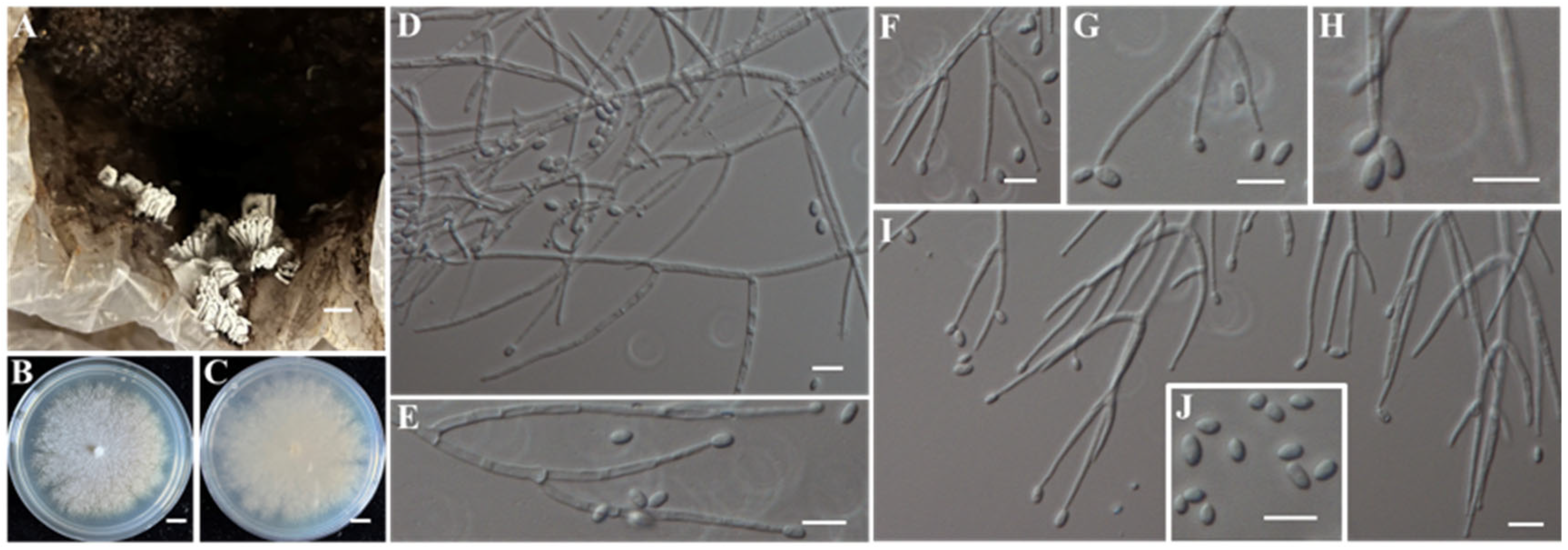
When incubated on PDA at 25 °C in the dark, the putative pathogenic fungus IAAM-S exhibited an average radial growth rate of 1.5–2.0 mm/d. The colony exhibited white marginal hyphae and a velutinous texture, with its surface densely covered by grayish-green conidia. The reverse side showed a chromatic gradient, ranging from dark yellow at the center to pale yellow at the periphery. The strain also produced a diffusible yellow-green pigment that diffused into the surrounding medium. Microscopic examination revealed abundant conidiation, producing spherical conidia measuring 1.5–2.0 μm in diameter (Figure 28J). Conidiophores were predominantly monoverticillate, occasionally biverticillate, and ranged from several tens to several hundreds of micrometers in length (Figure 28). BLASTn analysis of the ITS region and tub2 [52] showed that IAAM-S exhibited 100% sequence identity and 100% query coverage with reference strains of Penicillium citreosulfuratum for the ITS region, and 99.27% identity with 99% coverage for tub2. An ML phylogenetic tree based on concatenated ITS-tub2 sequences (~1100 bp) placed IAAM-S within the P. citreosulfuratum clade (Figure 29). Collectively, morphological and multilocus phylogenetic evidence [52] confirmed IAAM-S as P. citreosulfuratum.
(12)
Identification of Putative Pathogenic Fungus IAAM-Q
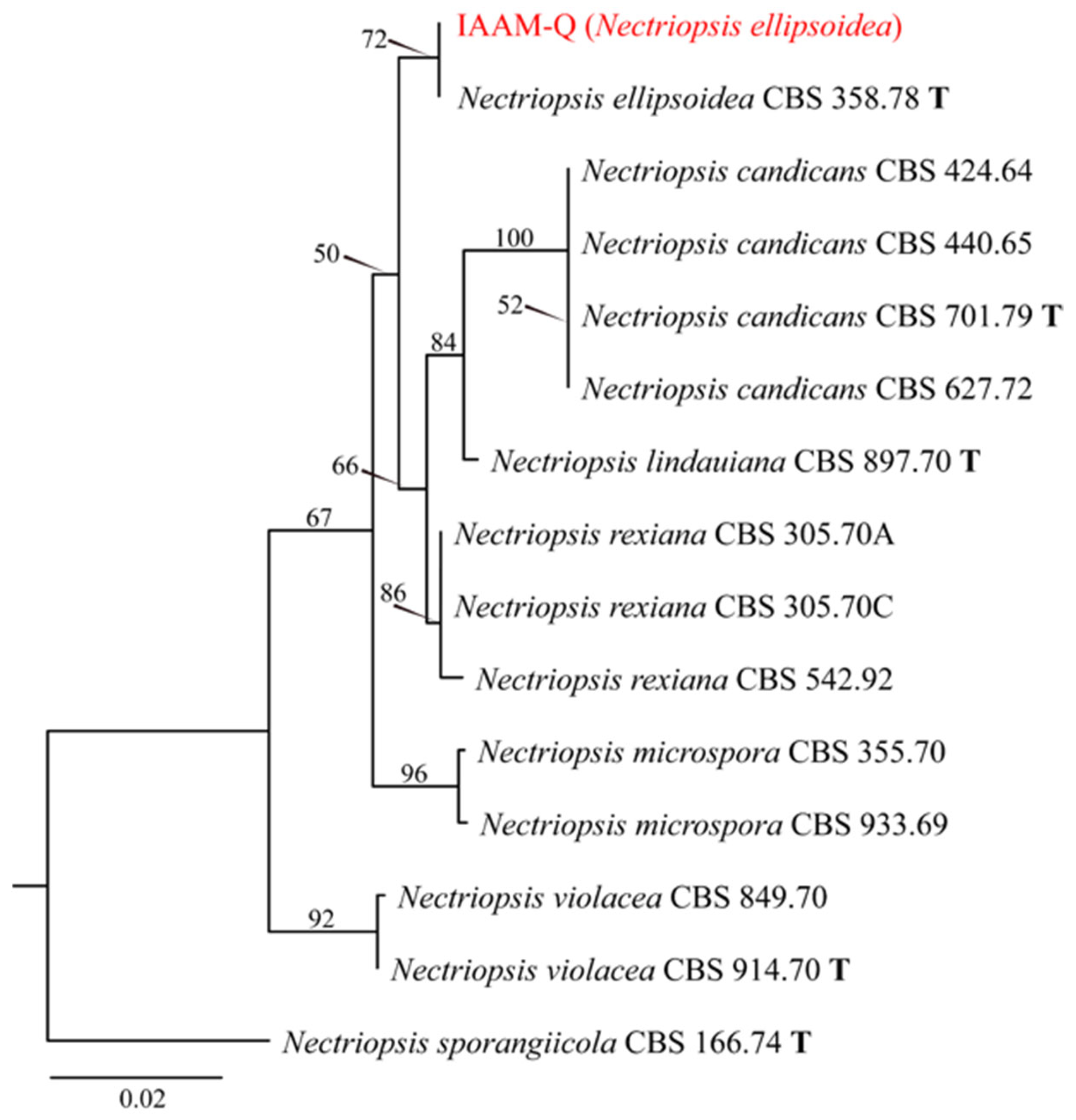
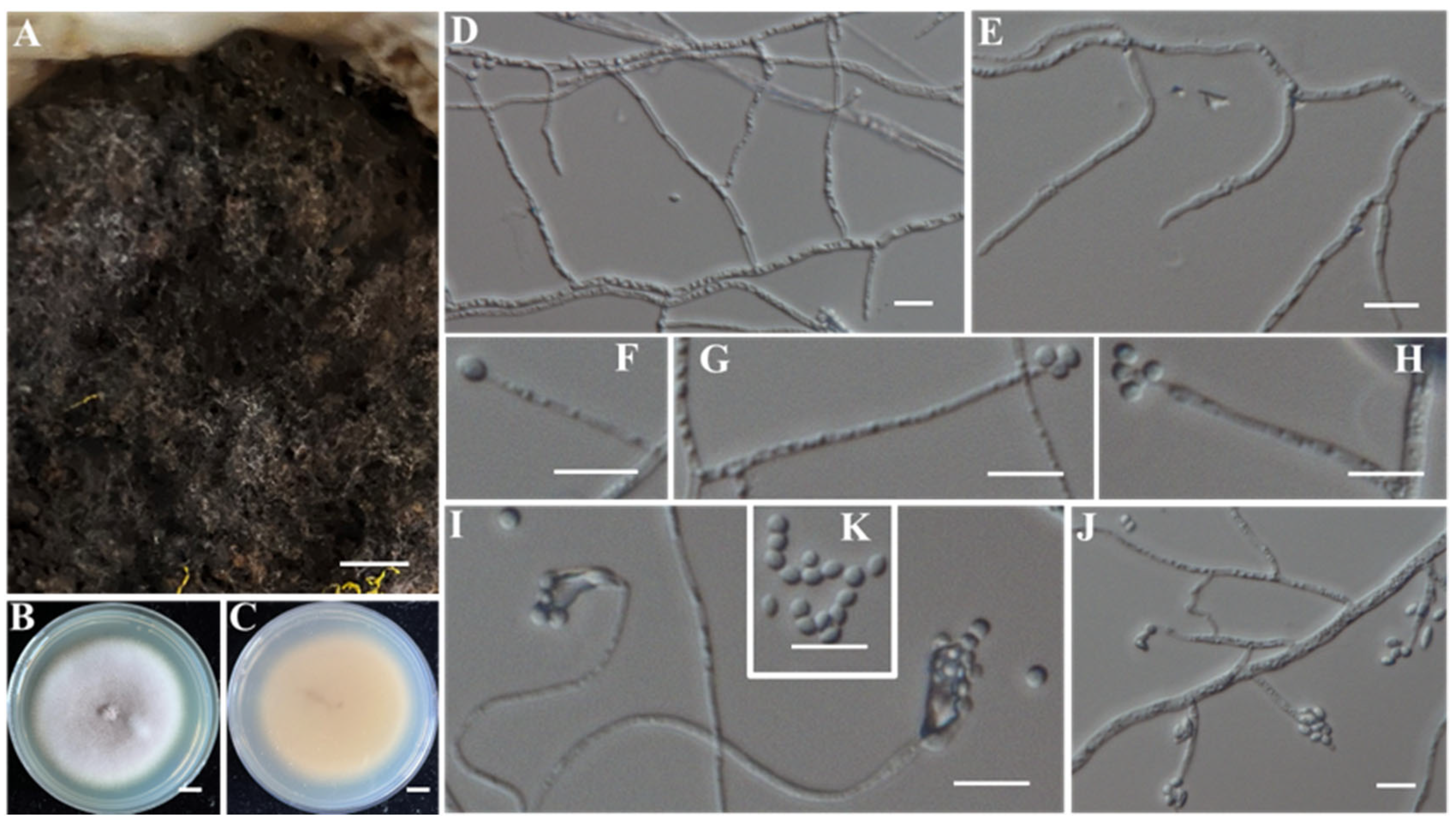
When cultured on PDA at 25 °C in darkness, the putative pathogenic fungus IAAM-Q showed an average radial growth rate of 2.8–3.0 mm/d. The colony consisted of white mycelium growing predominantly prostrate, with sparse aerial hyphae and irregular margins. Microscopic observations revealed septate hyphae with frequent branching (Figure 30I). The fungus produced abundant ellipsoidal, aseptate conidia measuring 3.0–5.0 μm × 2.0–3.0 μm. Conidiophores were erect, oppositely arranged, and ranged from several tens to several hundreds of micrometers in length (Figure 30). BLASTn analysis of the ITS and LSU sequences [50] showed 100% sequence identity to Nectriopsis ellipsoidea, with query coverages of 97% and 99%, respectively. An ML phylogenetic tree based on concatenated ITS-LSU sequences (~1200 bp) placed IAAM-Q within the N. ellipsoidea clade (Figure 31). Combined morphological and molecular phylogenetic evidence [50] confirmed IAAM-Q as N. ellipsoidea.
(13)
Identification of Putative Pathogenic Fungus IAAM-R
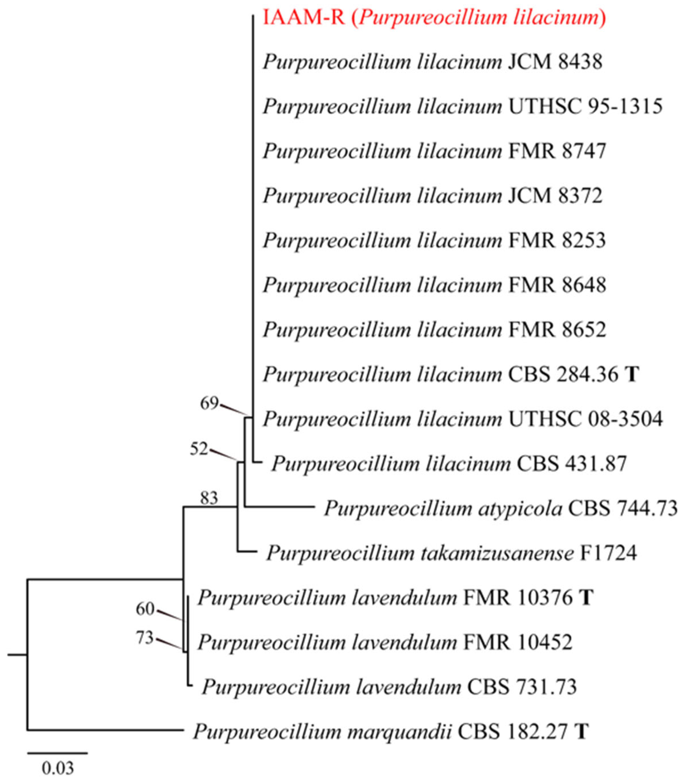
When cultured on PDA at 25 °C in darkness, the putative pathogenic fungus IAAM-R showed an average radial growth rate of 2.4–2.8 mm/d. The colony initially appeared white but gradually turned purplish-red with prolonged incubation. The colony surface was covered with floccose aerial hyphae and had smooth, well-defined margins. The reverse side displayed uniform purplish-gray pigmentation. Microscopic examination revealed abundant ellipsoidal, aseptate conidia measuring 2.0–3.0 μm × 1.5–2.5 μm (Figure 32K). Conidiophores occurred both singly and in clusters, ranging from several tens to over one hundred micrometers in length (Figure 32). BLASTn analysis of the ITS and tef1 sequences [51] showed 100% sequence identity and full query coverage with Purpureocillium lilacinum. An ML phylogenetic tree based on ITS sequences (~500 bp) placed IAAM-R within the P. lilacinum clade (Figure 33). Taken together, morphological [60] and molecular phylogenetic evidence [51] confirmed IAAM-R as P. lilacinum.
(14)
Identification of Putative Pathogen IAAM-W0001
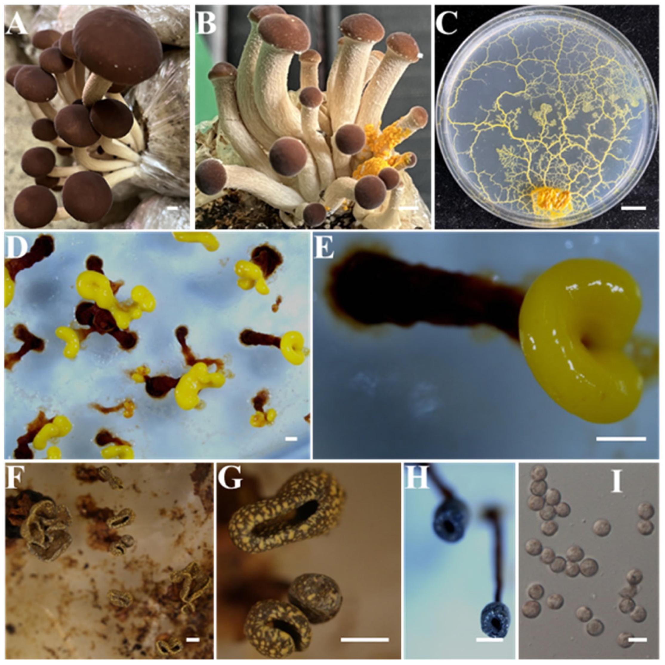
Two randomly selected IAAM-W0001 isolates from infected substrates were cultured on water agar in 90 mm Petri dishes at 25 °C under dark conditions. After inoculation with 1–2 moistened sterile oat flakes, yellow plasmodia completely colonized the medium surface of within 5–7 days (Figure 34C). Subsequent incubation without oats for an additional 5–7 days induced the formation of immature sporocarps, characterized by bright yellow sporangia borne on dark red stalks 2–3 mm in length (Figure 34D,E). Within 4–8 h of maturation, the sporangia turned dark brown to grayish green, while the stalks became black, initiating sporulation (Figure 34F–H). The mature spores were subspherical, measuring 6–10 μm × 5.5–9.5 μm (Figure 34I). Sequence (~1100 bp) analysis of the 18S rRNA gene [28,35] revealed 98.26% identity with 99% query coverage to Physarella oblonga. Based on the diagnostic morphological features (Figure 34) [26] in conjunction with molecular phylogenetic evidence [28,35], IAAM-W0001 was conclusively identified as P. oblonga.
(15)
Identification of Putative Pathogen IAAM-W0002
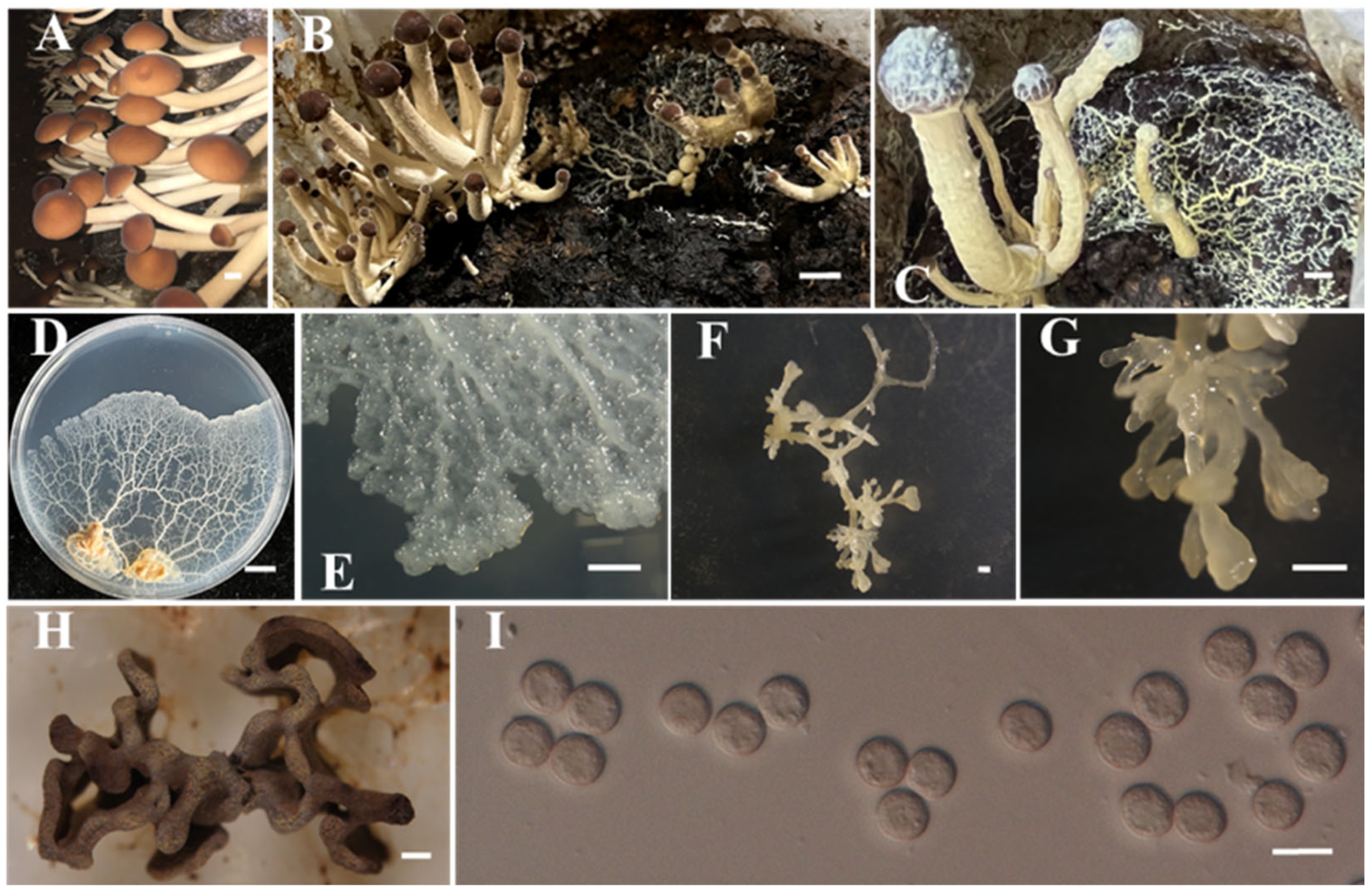
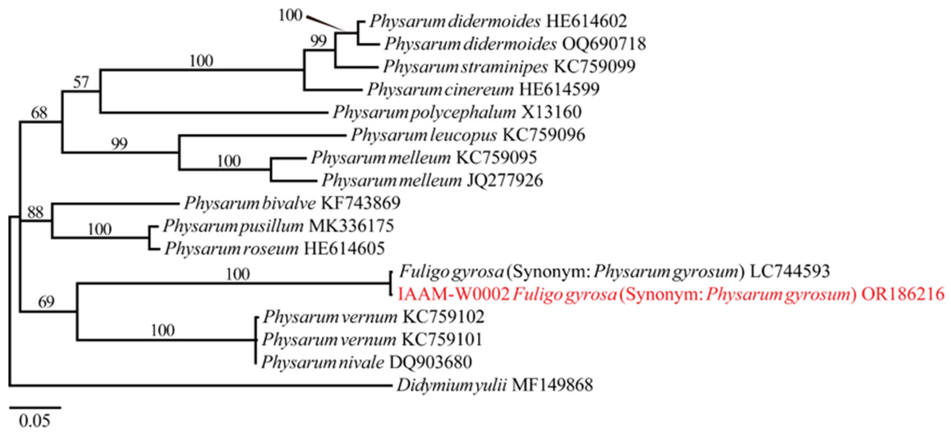
In the natural cultivation environments of A. chaxingu, the putative pathogen IAAM-W0002 extensively colonized the substrate surface and progressively invaded the basal region, stipe, and pileus of the fruiting bodies. This colonization produced characteristic moist, adhesive, white reticulate structures. At advanced stages of infection, fruiting bodies of A. chaxingu became entirely enveloped in a mucilaginous white-to-pale-yellow network, ultimately resulting in wilting and death. The organism was successfully isolated on water agar supplemented with 1–2 sterile oat flakes. After 4–5 days of incubation at 25 °C in darkness, white to pale yellow plasmodia developed, expanding in a fan-shaped manner (Figure 35D,E). Continued incubation for an additional 5–7 days led to the formation of irregular, sessile, pale-yellow sporocarps (Figure 35F,G), which matured within 5–8 h into dark gray structures producing grayish-brown (Figure 35H), spherical spores measuring 7–9 μm in diameter (Figure 35I). Molecular identification based on 18S rRNA gene sequencing (~1050 bp) [28,35] showed 98.26% sequence identity with 99% query coverage with Fuligo gyrosa (synonym: Physarum gyrosum). Taken together with the observed morphological characteristics (Figure 35) [61] and ML phylogenetic analysis constructed from the 18S rRNA sequences (Figure 36), the isolate was conclusively identified as F. gyrosa [62].
Based on a comprehensive disease survey and subsequent pathogen identification in A. chaxingu, white mucilaginous disease was determined as the most prevalent and economically significant disease affecting its cultivation. Integrating morphological characterization with molecular evidence, the putative pathogen was identified as F. gyrosa. Accordingly, F. gyrosa was preliminarily designated as the primary causal agent of white mucilaginous disease in A. chaxingu.

3.2. Pathogenicity Assessment of the Primary Putative Pathogen of A. chaxingu

3.2.1. Pathogenicity of F. gyrosa on the Fruiting Bodies of A. chaxingu

Fruiting bodies of A. chaxingu in the control group developed and matured normally following inoculation with sterile water agar medium (Figure 37A,B). In contrast, fruiting bodies inoculated with F. gyrosa were completely colonized by the slime mold within 4 days, ultimately leading to wilting and death (Figure 37C,D). Subsequent 18S rRNA gene sequence analysis of the organism re-isolated from symptomatic fruiting bodies (Figure S8) confirmed its identity as F. gyrosa, thereby fulfilling Koch’s postulates.

3.2.2. Pathogenicity of F. gyrosa on the Mycelium of A. chaxingu

Previous studies have demonstrated that F. gyrosa exhibits markedly distinct growth characteristics depending on the culture medium. As shown in Figure 38, this slime mold strain exhibited limited growth, accelerated senescence, and reduced plasmodial biomass on PDA (Figure 38A) whereas it displayed vigorous proliferation, sustained metabolic activity, and abundant plasmodial biomass on water agar (Figure 38B). Given that A. chaxingu mycelia grow optimally on PDA, a novel bipartite medium was designed with PDA on the left half and water agar on the right half to accommodate the growth requirements of both organisms. This optimized medium was subsequently employed to assess the pathogenic potential of F. gyrosa on A. chaxingu mycelia under co-culture conditions.
A. chaxingu mycelium incubated for five days was co-cultivated with oat flakes colonized by F. gyrosa plasmodium, placed on the water agar side of the bipartite medium. Observations revealed that F. gyrosa established direct contact with the host mycelia within 1 d. As the interaction progressed, the slime mold markedly suppressed mycelial growth and exhibited pronounced invasive behavior. Temporal analysis indicated that by day 3, characteristic brown pigmentation appeared on the reverse side of the A. chaxingu colonies. By day 4, extensive hyphal lysis occurred, accompanied by intensified pigment accumulation (Figure 38C,D). At day 5, most of the mycelial network was degraded, with distinct browning evident on both the upper and lower surfaces of the colony. In contrast, A. chaxingu mycelia in the control group (uninoculated with F. gyrosa) exhibited normal growth without pigment production (Figure 38E,F). These findings provide compelling evidence that F. gyrosa exerts a significant pathogenic effect on A. chaxingu mycelia under co-culture conditions.

3.3. Infection Process of F. gyrosa on A. chaxingu Mycelia

3.3.1. Stereomicroscopic Examination of F. gyrosa Infection on A. chaxingu Mycelia

Dynamic stereomicroscopic observations of F. gyrosa infecting A. chaxingu mycelia revealed a distinct temporal progression. At 2 d post-inoculation (Figure 39A1-A3), pronounced biomass accumulation of F. gyrosa was observed at the interaction interface, accompanied by apparent moisture accumulation. By day 4 (Figure 39B1-B3), the infection advanced to extensive degradation of the A. chaxingu aerial hyphae, with concurrent depositions of characteristic brown pigments in the colonized regions. These observations demonstrate that F. gyrosa established a progressive infection in A. chaxingu mycelia, characterized by interfacial biomass accumulation and subsequently by systematic host hyphal degradation.

3.3.2. Light Microscopic Observation of F. gyrosa Infection in A. chaxingu Mycelia

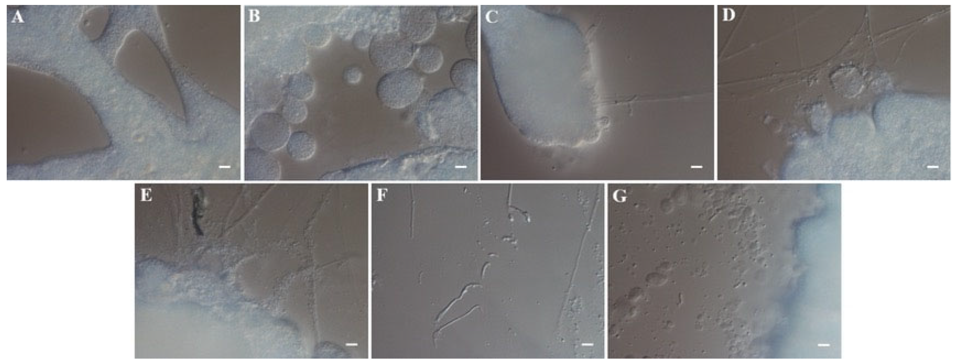
Light microscopy revealed that at 3 d post-inoculation with the myxomycete F. gyrosa, mature plasmodia (Figure 40A) divided into multiple small, spherical plasmodial bodies (Figure 40B). Live-cell imaging showed motile, granular inclusions within the plasmodia. Upon encountering A. chaxingu mycelia (Figure 40C), the plasmodia further fragmented into numerous smaller bodies (Figure 40D), followed by the active release of granular inclusions that subsequently enveloped the host hyphae (Figure 40E). The encased hyphae exhibited progressive fragmentation (Figure 40F), ultimately culminating in complete lysis (Figure 40G). Following host disintegration, various structures, including mature and small plasmodial bodies, granular inclusions, A. chaxingu conidia, and putative protoplast-like remnants, were observed within the same microscopic field. These observations provide direct evidence that F. gyrosa mediates hyphal degradation by releasing granular inclusions that encapsulate and disrupt host structures, ultimately leading to the fragmentation and dissolution of A. chaxingu hyphae.
The combined findings demonstrated that the predominant pathogenic myxomycete F. gyrosa, identified during the disease survey of A. chaxingu, exhibited pronounced pathogenicity toward both fruiting bodies and mycelial structures. These results collectively confirm that white slime disease in A. chaxingu is an infectious disease.

4. Discussion

Disease management remains one of the most formidable challenges in mushroom cultivation due to the diversity of pathogens, their efficient spore-mediated dispersal, and the difficulty of achieving durable control. A. chaxingu is particularly susceptible to disease because it is cultivated on protein-rich substrates, possesses limited lignin-degrading capacity, and requires elevated temperatures for both mycelial growth and fruiting. These factors heighten its vulnerability to pathogen infection, often resulting in severe outbreaks that reduce both yield and economic viability [15]. Despite its economic and nutritional value, the etiology and infection mechanisms of its major diseases remain poorly understood. This study provides the first systematic characterization of white slime disease in A. chaxingu, identifying F. gyrosa as its predominant causal agent. These findings fill a critical knowledge gap in mushroom pathology and establish a foundation for developing effective, sustainable strategies to mitigate disease outbreaks in A. chaxingu cultivation.
Previous studies on A. chaxingu diseases have primarily concentrated on symptom descriptions and partial management strategies [13], with reported disorders including soft rot, Cladobotryum infection, yellow mold, black root mold, and infections by Trichoderma, Penicillium, Aspergillus, Mucor, Rhizopus, and Fusarium species. However, the comprehensive identification of causative agents has remained limited; for example, Choi et al. [9] identified 22 potential pathogens from A. aegerita in Korea, mainly belonging to the genera Trichoderma, Aspergillus, Mucor, and Penicillium. In the present study, a systematic and long-term survey conducted across major production areas in China resulted in the isolation of 20 isolates from 19 categories of symptomatic A. chaxingu samples. Integrative analyses of morphology, multilocus sequencing, and phylogenetics identified 17 species spanning nine genera including Fusarium, Apiospora, Lecanicillium, Linnemannia, Mucor, Aspergillus, Penicillium, Nectriopsis, and Purpureocillium. Notably, isolate IAAM-C (Apiospora sp.) likely represents a novel species. This research constitutes the most thorough investigation to date of fungal pathogens associated with A. chaxingu, expanding the known pathogenic spectrum and providing essential data for accurate diagnosis, disease prevention, and sustainable cultivation.
Among the diseases impacting A. chaxingu, myxomycete infections have emerged as particularly destructive, frequently leading to substantial yield losses, and in severe cases, complete crop failure, which has drawn substantial research interest [14]. Recent studies have characterized disease symptoms, explored etiological factors, clarified transmission pathways, and investigated potential control strategies [15,16]. Despite these efforts, critical gaps remain in pathogen identification, biological characterization, and pathogenicity evaluation. Myxomycete-related diseases not only affect a wide variety of agricultural crops [63], but also exhibit wide host adaptability among various edible fungi [17,18,19,20,21,22,23]. These observations underscore myxomycete as a widespread and urgent threat to sustainable mushroom cultivation.
In species identification studies, multilocus sequence-based molecular methods have become essential tools. Compared with single-locus techniques, this approach provides significant benefits [64], especially in distinguishing closely related species with greater precision [65]. Advances in molecular biology have shown that DNA-based identification is more efficient and accurate than traditional morphology-based methods [65,66]. For example, in this study, although isolates IAAM-K and IAAM-N displayed noticeable color differences on PDA plates when observed visually, multilocus molecular analyses clearly confirmed that both isolates were the same species, F. fujikuroi. Fungal genomes differ from bacterial ones by having many non-coding and repetitive sequences, which makes precise species identification more challenging [67]. The ITS region is widely used as the standard DNA barcode for fungi [64,68]. Nevertheless, ITS alone often does not provide enough resolution to distinguish closely related fungal species in some groups. To address this, multilocus phylogenetic methods that include additional markers like tef1, rpb1, rpb2, LSU, and SSU have been developed to improve taxonomic clarity [69,70]. In this regard, creating molecular identification techniques that are both precise and practical has become essential. For example, Zhou et al. [44] introduced a barcode combining ITS and rpb1 that could differentiate all species within Lecanicillium, eliminating the need to analyze six loci simultaneously (ITS, tef1, rpb1, rpb2, LSU, and SSU). This streamlined approach maintained phylogenetic accuracy while significantly enhancing efficiency and reducing the complexity of analysis. However, several ongoing challenges affect the reliability of DNA-based species identification. These challenges include the lack and incompleteness of reference sequences in public databases—for example, isolate IAAM-C, which, despite being analyzed with commonly used multilocus markers for the genus Apiospora, could not be definitively assigned to a species, underscoring gaps in available reference data. Additional issues arise from inconsistencies caused by non-standardized primer use [71], errors during PCR amplification [72], and variations in naming conventions when submitting sequences. Together, these factors lead to discrepancies between molecular sequence data and morphological identifications, limiting the ability to compare results across studies. Therefore, integrative methods that combine morphological analysis with DNA-based molecular techniques are crucial for achieving fast and accurate species identification across a wide range of taxa.
In this study, a systematic disease survey across major A. chaxingu cultivation areas led to the isolation, purification, and identification of two potential myxomycete pathogens: P. oblonga, associated with yellow slime disease (with a 4.6% incidence), and F. gyrosa, associated with white slime disease (with a 17.3% incidence). These discoveries broaden the known range of myxomycete pathogens affecting edible fungi and fill previously unaddressed gaps in the literature. Pathogenicity assays demonstrated that F. gyrosa is highly infectious to both the fruiting bodies and mycelia of A. chaxingu, ultimately resulting in host mortality and confirming the infectious nature of white slime disease. The preliminary phenotypic characteristics of F. gyrosa on A. chaxingu mycelium were observed through macroscopic observations, stereomicroscopy, and light microscopy. Mature F. gyrosa plasmodia broke down into numerous small, spherical units containing streaming granular inclusions. Upon encountering host mycelia, these units released intracellular contents, enveloping hyphae and inducing progressive fragmentation and degradation. This study provides the first comprehensive microscopic characterization of myxomycete infection in edible mushroom mycelium, providing important insights into the pathogenic strategies employed by myxomycetes.

5. Conclusions

This study systematically identified the major pathogens of A. chaxingu, including 17 fungal and 2 myxomycete species, establishing a comprehensive morphological and molecular reference. F. gyrosa was confirmed as the primary pathogen, exhibiting high virulence toward fruiting bodies and mycelia, ultimately causing host mortality. For the first time, the preliminary phenotypic characteristics of F. gyrosa in A. chaxingu mycelium was documented, revealing plasmodial suppression of hyphal growth, direct tissue invasion, and degradation mediated by granular inclusions. These findings provide critical insights into myxomycete-edible fungus interactions and lay the groundwork for future studies on their molecular pathogenic mechanisms.

Supplementary Materials

The following supporting information can be downloaded at https://www.mdpi.com/article/10.3390/horticulturae11101190/s1, Table S1: Strain names, numbers, and NCBI GenBank accession numbers used in the phylogenetic analysis. Table S2: Names and sequence information of potential pathogens identified in Agrocybe chaxingu. Figure S1: Gel electrophoresis of ITS amplicons from potential pathogenic fungi. Figure S2: Gel electrophoresis of tef1 amplicons from potential pathogenic fungi. Figure S3: Gel electrophoresis of tub2 amplicons from potential pathogenic fungi. Figure S4: Gel electrophoresis of LSU amplicons from potential pathogenic fungi. Figure S5: Gel electrophoresis of rpb1 amplicons from potential pathogenic fungi. Figure S6: Gel electrophoresis of rpb2 amplicons from potential pathogenic fungi. Figure S7: Gel electrophoresis of H3 amplicons from potential pathogenic fungi. Figure S8: Gel electrophoresis of 18S rRNA amplicons from potential pathogenic slime mold.

Author Contributions

X.C., G.H., C.D., and Y.W. conceived and designed the research. X.C., G.M., M.L., and J.D. performed the experiments. X.C., and G.M. performed the data visualization. X.C., and C.D. wrote the manuscript, which was reviewed by all authors. All authors have read and agreed to the published version of the manuscript.

Funding

This work was supported by the Collaborative Innovation Project of Modern Agricultural Research of Jiangxi Province (JXXTCX202409); the Jiangxi Provincial Natural Science Foundation (20252BAC200405); the Project of Science and Technology Department of Jiangxi Province (20212BDH80009); the Crop Seeds Joint Research of Jiangxi Province; and the China Agriculture Research System (CARS20).

Data Availability Statement

Data are contained within the article and Supplementary Materials.

Conflicts of Interest

The authors declare no conflicts of interest.

References

  1. Hu, Q.X.; Liao, C.Z.; Zou, Y.J. Current development status and prospects of rare edible mushrooms in China. Agric. Compr. Dev. China 2022, 4, 50–54. (In Chinese) [Google Scholar]
  2. Wang, M.; Gu, B.L.; Huang, J.; Jiang, S.; Chen, Y.J.; Yin, Y.L.; Pan, Y.F.; Yu, G.J.; Li, Y.M.; Wong, B.H.C.; et al. Transcriptome and proteome exploration to provide a resource for the study of Agrocybe aegerita. PLoS ONE 2013, 8, e56686. [Google Scholar] [CrossRef]
  3. Zhang, M.; Xing, S.H.; Qian, C.L.; Liu, J.; Kan, J.; Jin, C.H. Analysis of the nutritional components and antioxidant properties of seven species of edible mushrooms. Food Sci. Technol. 2022, 47, 120–126. (In Chinese) [Google Scholar]
  4. Wang, Z.W.; Chen, K.Y.; Zhang, K.; He, K.H.; Zhang, D.D.; Guo, X.H.; Huang, T.W.; Hu, J.L.; Zhou, X.T.; Nie, S.P. Agrocybe cylindracea fucoglucogalactan induced lysosome-mediated apoptosis of colorectal cancer cell through H3K27ac-regulated cathepsin D. Carbohyd. Polym. 2023, 319, 121208. [Google Scholar] [CrossRef] [PubMed]
  5. Yang, F.; Chen, E.B.; Fu, A.Z.; Liu, Y.; Bi, S. Formation of key aroma compounds in Agrocybe aegerita during hot air drying: Amino acids and reducing sugars identified as flavor precursors. Food Chem. 2025, 465, 141975. [Google Scholar] [CrossRef] [PubMed]
  6. Zadrazil, F. Cultivation of Agrocybe aegerita (Brig.) Sing. on Lignocellulose Containing Wastes 1. Factors Affecting Saprophytic Colonization and Decomposition of Substrate. Int. Soc. Mushroom Sci. 1989, 12, 357–368. Available online: https://www.pubhort.org/isms/12/2/v12_p2_a38.htm (accessed on 28 August 2025).
  7. Huang, N.L.; Lin, Z.B.; Chen, G.L. Medicinal and Edible Fungi; Shanghai Scientific and Technical Literature Press: Shanghai, China, 2010; pp. 607–608. (In Chinese) [Google Scholar]
  8. Bian, Y.B.; Xiao, Y.; Guo, M.P. Research progress on disease control of edible fungi. Acta Edulis Fungi 2021, 28, 121–131. (In Chinese) [Google Scholar]
  9. Choi, I.Y.; Choi, J.N.; Sharma, P.K.; Lee, W.H. Isolation and identification of mushroom pathogens from Agrocybe aegerita. Mycobiology 2010, 38, 310–315. [Google Scholar] [CrossRef]
  10. Li, W.Z. Several major diseases and pests of Agrocybe aegerita. Edible Fungi 2002, 2, 40. (In Chinese) [Google Scholar]
  11. Ding, H.G. Infectious diseases of Agrocybe chaxingu and pollution-free control techniques. Yunnan Agric. Sci. Technol. 2006, 3, 53–54. (In Chinese) [Google Scholar]
  12. Lu, D.X. Occurrence and prevention and control of common diseases of Agrocybe chaxingu (cylindracea). J. Fungal Res. 2008, 2, 114–118. (In Chinese) [Google Scholar]
  13. Ma, Y.; Yang, Y.H.; Lyu, Y.F.; Cui, S.Q.; Xu, J.Y.; Li, M.L.; Shen, J.D.; Xie, H.T.; Fang, X.H. Common diseases and insect pests in Agrocybe aegerita production and non-pollution comprehensive control technology. Edible Fungi China 2021, 40, 113–116. (In Chinese) [Google Scholar]
  14. Liu, Y.G.; Lin, R.K. Symptoms and control techniques of slime mold disease in Agrocybe aegerita. Edible Fungi 2003, 3, 40. (In Chinese) [Google Scholar]
  15. Chen, S.Z. Reserach on comprehensive prevention and control for edible fungus Ⅱ: Occurrence and control of Polyma disease in Agrocybe chaxingu. J. South. Agric. 2008, 3, 317–319. (In Chinese) [Google Scholar]
  16. Guan, S.G. Control experiment of slime mold disease in Agrocybe aegerita. Edible Fungi 2004, 4, 41–42. (In Chinese) [Google Scholar]
  17. He, X.S. Slime molds on the fruiting bodies of Auricularia cornea and Pleurotus ostreatus. Edible Fungi 1991, 1, 39. (In Chinese) [Google Scholar]
  18. Li, Y.C.; Li, Z.X. Separation and identification and biological characteristics of myxomycetes lyzing the fruitbodies of Auricularia auricula and A. polytricha. J. Guangxi Agric. Coll. 1992, 11, 8–15. (In Chinese) [Google Scholar]
  19. Li, S.S.; Qian, X.C.; Xu, J.Z. Common contaminant fungi and their control in Auricularia heimuer and Lentinula edodes cultivation in Qinba mountain area. Edible Fungi China 1992, 11, 25–26. (In Chinese) [Google Scholar]
  20. Lin, X.M. Slime mold contamination in edible fungus cultivation in Henan province. Edible Fungi 1993, 1, 38. (In Chinese) [Google Scholar]
  21. Hu, J.X.; Xu, M.; Gao, B.; Wang, F.; Jiang, Y.J.; Zhang, J.X. Identification and preliminary control of a slime mold on Pleurotus pulmonarius. Edible Fungi 2023, 45, 62–65. (In Chinese) [Google Scholar]
  22. Dai, D.; Sun, P.; Wu, X.Y.; Hu, J.; Zhang, B.; Wei, Y.H.; Li, Y. First report of yellow rot caused by Physarum galbeum in Grifola frondosa in China. Plant Dis. 2023, 10, 1094. [Google Scholar] [CrossRef]
  23. Zhao, J.; Fu, J.H. Pest and disease control in Dictyophora indusiata cultivation. Hunan Agric. 2024, 11, 14. (In Chinese) [Google Scholar]
  24. Raza, M.; Zhang, Z.F.; Hyde, K.D.; Diao, Y.Z.; Cai, L. Culturable plant pathogenic fungi associated with sugarcane in southern China. Fungal Divers. 2019, 99, 1–104. [Google Scholar] [CrossRef]
  25. Crous, P.W.; Lombard, L.; Sandoval-Denis, M.; Seifert, K.A.; Schroers, H.J.; Chaverri, P.; Gené, J.; Guarro, J.; Hirooka, Y.; Bensch, K.; et al. Fusarium: More than a node or a foot-shaped basal cell. Stud. Mycol. 2021, 98, 100116. [Google Scholar] [CrossRef] [PubMed]
  26. Shi, L.P.; Li, Y. Life cycle of Physarella oblonga. Mycosystema 2004, 23, 381–387. (In Chinese) [Google Scholar]
  27. Hossen, R.; Courtney, M.; Sim, A.; Khan, M.A.A.K.; Verbruggen, H.; Bringloe, T. An optimized CTAB method for genomic DNA extraction from green seaweeds (Ulvophyceae). Appl. Plant Sci. 2024, 13, e11625. [Google Scholar] [CrossRef] [PubMed]
  28. White, T.J.; Bruns, T.; Lee, S.J.W.T.; Taylor, J. Amplification and direct sequencing of fungal ribosomal RNA genes for phylogenetics. In Pcr Protocols: A Guide to Methods and Applications; Innis, M.A., Gelfand, D.H., Sninsky, J.J., White, T.J., Eds.; Academic Press: San Diego, CA, USA, 1990; pp. 315–322. [Google Scholar]
  29. O’Donnell, K.; Kistler, H.C.; Cigelnik, E.; Ploetz, R.C. Multiple evolutionary origins of the fungus causing panama disease of banana: Concordant evidence from nuclear and mitochondrial gene genealogies. Proc. Natl. Acad. Sci. USA 1998, 95, 2044–2049. [Google Scholar] [CrossRef]
  30. O’Donnell, K.; Sutton, D.A.; Rinaldi, M.G.; Sarver, B.A.; Balajee, S.A.; Schroers, H.J.; Summerbell, R.C.; Robert, V.A.; Crous, P.W.; Zhang, N.; et al. Internet-accessible DNA sequence database for identifying fusaria from human and animal infections. J. Clin. Microbiol. 2010, 48, 3708–3718. [Google Scholar] [CrossRef]
  31. Reeb, V.; Lutzoni, F.; Roux, C. Contribution of RPB2 to multilocus phylogenetic studies of the Euascomycetes (Pezizomycotina, Fungi) with special emphasis on the lichen-forming Acarosporaceae and evolution of polyspory. Mol. Phylogenet. Evol. 2004, 32, 1036–1060. [Google Scholar] [CrossRef]
  32. Roux, J.; Steenkamp, E.T.; Marasas, W.F.; Wingfield, M.J.; Wingfield, B.D. Characterization of Fusarium graminearum from Acacia and Eucalyptus using β-tubulin and histone gene sequences. Mycologia 2001, 93, 704–711. [Google Scholar] [CrossRef]
  33. O’Donnell, K.; Cigelnik, E. Two divergent intragenomic rDNA ITS2 types within a monophyletic lineage of the fungus Fusarium are nonorthologous. Mol. Phylogenet. Evol. 1997, 7, 103–116. [Google Scholar] [CrossRef]
  34. Cubeta, M.A.; Echandi, E.; Abernethy, T.; Vilgalys, R. Characterization of anastomosis groups of binucleate Rhizoctonia species using restriction analysis of an amplified ribosomal RNA gene. Phytopathology 1991, 81, 1395–1400. [Google Scholar] [CrossRef]
  35. Rusk, S.A.; Spiegel, F.W.; Lee, S.B. Design of polymerase chain reaction primers for amplifying nuclear ribosomal DNA from slime molds. Mycologia 1995, 87, 140–143. [Google Scholar] [CrossRef]
  36. Katoh, K.; Standley, D.M. MAFFT multiple sequence alignment software version 7: Improvements in performance and usability. Mol. Biol. Evol. 2013, 30, 772–780. [Google Scholar] [CrossRef]
  37. Talavera, G.; Castresana, J. Improvement of phylogenies after removing divergent and ambiguously aligned blocks from protein sequence alignments. Syst. Biol. 2007, 56, 564–577. [Google Scholar] [CrossRef] [PubMed]
  38. Kalyaanamoorthy, S.; Minh, B.Q.; Wong, T.K.F.; Von Haeseler, A.; Jermiin, L.S. ModelFinder: Fast model selection for accurate phylogenetic estimates. Nat. Methods 2017, 14, 587–589. [Google Scholar] [CrossRef] [PubMed]
  39. Nguyen, L.T.; Schmidt, H.A.; Von Haeseler, A.; Minh, B.Q. IQ-TREE: A fast and effective stochastic algorithm for estimating maximum-likelihood phylogenies. Mol. Biol. Evol. 2015, 32, 268–274. [Google Scholar] [CrossRef]
  40. Minh, B.Q.; Nguyen, M.A.T.; Von Haeseler, A. Ultrafast approximation for phylogenetic bootstrap. Mol. Biol. Evol. 2013, 30, 1188–1195. [Google Scholar] [CrossRef] [PubMed]
  41. Guindon, S.; Dufayard, J.F.; Lefort, V.; Anisimova, M.; Hordijk, W.; Gascuel, O. New algorithms and methods to estimate maximum-likelihood phylogenies: Assessing the performance of PhyML 3.0. Syst. Biol. 2010, 59, 307–321. [Google Scholar] [CrossRef]
  42. Han, S.L.; Wang, M.M.; Ma, Z.Y.; Raza, M.; Zhao, P.; Liang, J.M.; Gao, M.; Li, Y.J.; Wang, J.W.; Hu, D.M.; et al. Fusarium diversity associated with diseased cereals in China, with an updated phylogenomic assessment of the genus. Stud. Mycol. 2023, 104, 87–148. [Google Scholar] [CrossRef]
  43. Kwon, S.L.; Cho, M.; Lee, Y.M.; Kim, C.; Lee, S.M.; Ahn, B.J.; Lee, H.; Kim, J.J. Two unrecorded Apiospora species isolated from marine substrates in Korea with eight new combinations (A. piptatheri and A. rasikravindrae). Mycobiology 2022, 50, 46–54. [Google Scholar] [CrossRef]
  44. Zhou, Y.M.; Zou, X.; Zhi, J.R.; Xie, J.Q.; Jiang, T. Fast recognition of Lecanicillium spp., and its virulence against Frankliniella occidentalis. Front. Microbiol. 2020, 11, 561381. [Google Scholar] [CrossRef] [PubMed]
  45. Vandepol, N.; Liber, J.; Desirò, A.; Na, H.; Kennedy, M.; Barry, K.; Grigoriev, I.V.; Miller, A.N.; O’Donnell, K.; Stajich, J.E.; et al. Resolving the Mortierellaceae phylogeny through synthesis of multi-gene phylogenetics and phylogenomics. Fungal Divers. 2020, 104, 267–289. [Google Scholar] [CrossRef] [PubMed]
  46. Madden, A.A.; Stchigel, A.M.; Guarro, J.; Sutton, D.; Starks, P.T. Mucor nidicola sp. nov., a fungal species isolated from an invasive paper wasp nest. Int. J. Syst. Evol. Microbiol. 2012, 62, 1710–1714. [Google Scholar] [CrossRef] [PubMed]
  47. Sandoval-Denis, M.; Lombard, L.; Crous, P.W. Back to the roots: A reappraisal of Neocosmospora. Persoonia 2019, 43, 90–185. [Google Scholar] [CrossRef]
  48. Yang, J.; Li, S.J.; Li, X.M.; Qin, Y.H.; Gao, S.X.; Wen, Y.; Lu, S.H.; Lu, C.T.; Wang, F. First report of Aspergillus westerdijkiae causing leaf blight on Rehmannia glutinosa in China. Plant Dis. 2024, 10, 1094. [Google Scholar] [CrossRef]
  49. Houbraken, J.; Frisvad, J.C.; Samson, R.A. Taxonomy of Penicillium section Citrina. Stud. Mycol. 2011, 70, 53–138. [Google Scholar] [CrossRef]
  50. Hou, L.W.; Giraldo, A.; Groenewald, J.Z.; Rämä, T.; Summerbell, R.C.; Huang, G.Z.; Cai, L.; Crous, P.W. Redisposition of acremonium-like fungi in Hypocreales. Stud. Mycol. 2023, 105, 23–203. [Google Scholar] [CrossRef]
  51. Perdomo, H.; Cano, J.; Gené, J.; García, D.; Hernández, M.; Guarro, J. Polyphasic analysis of Purpureocillium lilacinum isolates from different origins and proposal of the new species Purpureocillium lavendulum. Mycologia 2013, 105, 151–161. [Google Scholar] [CrossRef]
  52. Visagie, C.M.; Seifert, K.A.; Houbraken, J.; Samson, R.A.; Jacobs, K. A phylogenetic revision of Penicillium sect. Exilicaulis, including nine new species from fynbos in South Africa. IMA Fungus 2016, 7, 75–117. [Google Scholar]
  53. Zhao, Z.H.; Lu, G.Z. Fusarium kyushuense, a newly recorded species for China. Mycotaxon 2007, 102, 119–126. [Google Scholar]
  54. O’Donnell, K.; Ward, T.J.; Geiser, D.M.; Corby Kistler, H.; Aoki, T. Genealogical concordance between the mating type locus and seven other nuclear genes supports formal recognition of nine phylogenetically distinct species within the Fusarium graminearum clade. Fungal Genet. Biol. 2004, 41, 600–623. [Google Scholar] [CrossRef] [PubMed]
  55. Zare, R.; Gams, W. The genera Lecanicillium and Simplicillium gen. nov. Nova Hedwigia 2001, 73, 1–50. [Google Scholar] [CrossRef]
  56. Wang, M.M.; Crous, P.W.; Sandoval-Denis, M.; Han, S.L.; Liu, F.; Liang, J.M.; Duan, W.J.; Cai, L. Fusarium and allied genera from China: Species diversity and distribution. Persoonia 2022, 48, 1–53. [Google Scholar] [CrossRef]
  57. He, J.; Li, D.W.; Cui, W.L.; Zhu, L.H.; Huang, L. Morphological and phylogenetic analyses reveal three new species of Fusarium (Hypocreales, Nectriaceae) associated with leaf blight on Cunninghamialanceolata in China. MycoKeys 2024, 101, 45–80. [Google Scholar] [CrossRef]
  58. Wagner, L.; Stielow, B.; Hoffmann, K.; Petkovits, T.; Papp, T.; Vágvölgyi, C.; de Hoog, G.S.; Verkley, G.; Voigt, K. A comprehensive molecular phylogeny of the Mortierellales (Mortierellomycotina) based on nuclear ribosomal DNA. Persoonia 2013, 30, 77–93. [Google Scholar] [CrossRef]
  59. Frisvad, J.C.; Frank, J.M.; Houbraken, J.A.M.P.; Kuijpers, A.F.; Samson, R.A. New ochratoxin a producing species of Aspergillus section Circumdati. Stud. Mycol. 2004, 50, 23–43. [Google Scholar]
  60. Luangsa-Ard, J.; Houbraken, J.; van Doorn, T.; Hong, S.B.; Borman, A.M.; Hywel-Jones, N.L.; Samson, R.A. Purpureocillium, a new genus for the medically important Paecilomyces lilacinus. FEMS Microbiol. Lett. 2011, 321, 141–149. [Google Scholar] [CrossRef]
  61. Shi, L.P.; Li, Y. Life cycle of Physarum gyrosum. Mycosystema 2005, 24, 292–296. (In Chinese) [Google Scholar]
  62. Chen, X.T.; Liu, Q.; Meng, G.L.; Peng, X.H.; Dong, C.H.; Wei, Y.H.; Huo, G.H. First report of white mucus disease caused by Fuligo gyrosa on Agrocybe chaxingu in China. Plant Dis. 2024, 108, 1884. [Google Scholar] [CrossRef]
  63. Agra, L.A.N.N.; Seixas, C.D.S.; Dianese, J.C. False bean smut caused by slime mold. Plant Dis. 2018, 102, 507–510. [Google Scholar] [CrossRef]
  64. Vu, D.; Groenewald, M.; Szöke, S.; Cardinali, G.; Eberhardt, U.; Stielow, B.; de Vries, M.; Verkleij, G.J.; Crous, P.W.; Boekhout, T.; et al. DNA barcoding analysis of more than 9 000 yeast isolates contributes to quantitative thresholds for yeast species and genera delimitation. Stud. Mycol. 2016, 85, 91–105. [Google Scholar] [CrossRef]
  65. Vu, D.; Groenewald, M.; de Vries, M.; Gehrmann, T.; Stielow, B.; Eberhardt, U.; Al-Hatmi, A.; Groenewald, J.Z.; Cardinali, G.; Houbraken, J.; et al. Large-scale generation and analysis of filamentous fungal DNA barcodes boosts coverage for kingdom fungi and reveals thresholds for fungal species and higher taxon delimitation. Stud. Mycol. 2019, 92, 135–154. [Google Scholar] [CrossRef] [PubMed]
  66. Verkley, G.J.M.; Quaedvlieg, W.; Shin, H.D.; Crous, P.W. A new approach to species delimitation in Septoria. Stud. Mycol. 2013, 75, 213–305. [Google Scholar] [CrossRef]
  67. Mohanta, T.K.; Bae, H. The diversity of fungal genome. Biol. Proced. Online 2015, 17, 8. [Google Scholar] [CrossRef] [PubMed]
  68. Irinyi, L.; Serena, C.; Garcia-Hermoso, D.; Arabatzis, M.; Desnos-Ollivier, M.; Vu, D.; Cardinali, G.; Arthur, I.; Normand, A.C.; Giraldo, A.; et al. International society of human and animal mycology (ISHAM)-ITS reference DNA barcoding database--the quality controlled standard tool for routine identification of human and animal pathogenic fungi. Med. Mycol. 2015, 53, 313–337. [Google Scholar] [CrossRef]
  69. Sung, G.H.; Hywel-Jones, N.L.; Sung, J.M.; Luangsa-Ard, J.J.; Shrestha, B.; Spatafora, J.W. Phylogenetic classification of Cordyceps and the clavicipitaceous fungi. Stud. Mycol. 2007, 57, 5–59. [Google Scholar] [CrossRef]
  70. Kepler, R.M.; Luangsa-Ard, J.J.; Hywel-Jones, N.L.; Quandt, C.A.; Sung, G.H.; Rehner, S.A.; Aime, M.C.; Henkel, T.W.; Sanjuan, T.; Zare, R.; et al. A phylogenetically-based nomenclature for Cordycipitaceae (Hypocreales). IMA Fungus 2017, 8, 335–353. [Google Scholar] [CrossRef]
  71. Rodríguez-Ezpeleta, N.; Zinger, L.; Kinziger, A.; Bik, H.M.; Bonin, A.; Coissac, E.; Emerson, B.C.; Lopes, C.M.; Pelletier, T.A.; Taberlet, P.; et al. Biodiversity monitoring using environmental DNA. Mol. Ecol. Resour. 2021, 21, 1405–1409. [Google Scholar] [CrossRef]
  72. Steele, M.P.; Neaves, L.E.; Klump, B.C.; St Clair, J.J.H.; Fernandes, J.R.S.M.; Hequet, V.; Shaw, P.; Hollingsworth, P.M.; Rutz, C. DNA barcoding identifies cryptic animal tool materials. Proc. Natl. Acad. Sci. USA 2021, 118, e2020699118. [Google Scholar] [CrossRef]
Figure 1. Morphological features of IAAM-A cultured on PDA. (A) Fruiting body of Agrocybe chaxingu exhibiting symptoms of IAAM-A infection. (B,C) Colony morphology of IAAM-A on PDA. (D) Hyphae of IAAM-A. (EH) Conidiophores of IAAM-A. (I) Conidia of IAAM-A. Scale bars: 1 cm (AC); 10 μm (DI).
Horticulturae 11 01190 g001
Figure 2. Maximum likelihood phylogenetic tree of isolates IAAM-A and IAAM-F inferred from concatenated sequences of the tef1, rpb1, rpb2, and H3 genes. Fusarium nelsonii NRRL 13338 was designated as the outgroup. Type strains are denoted by “T”, and the two putative pathogenic isolates are marked in red.
Horticulturae 11 01190 g002
Figure 3. Morphological features of IAAM-F cultured on PDA. (A) Fruiting body of Agrocybe chaxingu exhibiting symptoms of IAAM-F infection. (B,C) Colony morphology of IAAM-F on PDA. (D,E) Hyphae of IAAM-F. (FH) Conidiophores of IAAM-F. (I,J) Conidia of IAAM-F. Scale bars: 1 cm (AC); 10 μm (DJ).
Horticulturae 11 01190 g003
Figure 4. Morphological features of IAAM-B cultured on PDA. (A) Fruiting body of Agrocybe chaxingu exhibiting symptoms of IAAM-B infection. (B,C) Colony morphology of IAAM-B on PDA. (DI) Hyphae of IAAM-B. Scale bars: 1 cm (AC); 10 μm (DI).
Horticulturae 11 01190 g004
Figure 5. Maximum likelihood phylogenetic tree of isolates IAAM-B, IAAM-C, and IAAM-O inferred from concatenated sequences of ITS, LSU, tef1, and tub2. Apiospora pterosperma CPC 20193 was designated as the outgroup. Type strains are denoted by “T”, and the three putative pathogenic isolates are marked in red.
Horticulturae 11 01190 g005
Figure 6. Morphological features of IAAM-C cultured on PDA. (A) Fruiting body of Agrocybe chaxingu exhibiting symptoms of IAAM-C infection. (B,C) Colony morphology of IAAM-C on PDA. (DH) Hyphae of IAAM-C. Scale bars: 1 cm (AC); 10 μm (DH).
Horticulturae 11 01190 g006
Figure 7. Morphological features of IAAM-O cultured on PDA. (A) Fruiting body of Agrocybe chaxingu exhibiting symptoms of IAAM-O infection. (B,C) Colony morphology of IAAM-O on PDA. (DL) Hyphae of IAAM-O. Scale bars: 1 cm (AC); 10 μm (DL).
Horticulturae 11 01190 g007
Figure 8. Morphological features of IAAM-D cultured on PDA. (A) Fruiting body of Agrocybe chaxingu exhibiting symptoms of IAAM-D infection. (B,C) Colony morphology of IAAM-D on PDA. (D) Conidia of IAAM-D. (EH) Conidiophores and conidia of IAAM-D. Scale bars: 1 cm (AC); 10 μm (DH).
Horticulturae 11 01190 g008
Figure 9. Morphological features of IAAM-H cultured on PDA. (A) Fruiting body of Agrocybe chaxingu exhibiting symptoms of IAAM-H infection. (B,C) Colony morphology of IAAM-H on PDA. (DF) Hyphae of IAAM-H. (G) Conidia of IAAM-H. (HK) Conidiophores of IAAM-H. Scale bars: 1 cm (AC); 10 μm (DK).
Horticulturae 11 01190 g009
Figure 10. Morphological features of IAAM-T cultured on PDA. (A) Fruiting body of Agrocybe chaxingu exhibiting symptoms of IAAM-T infection. (B,C) Colony morphology of IAAM-T on PDA. (D–F) Hyphae of IAAM-T. (G,H) Conidiophores of IAAM-T. (I,J) Conidia of IAAM-T. Scale bars: 1 cm (AC); 10 μm (DJ).
Horticulturae 11 01190 g010
Figure 11. Maximum likelihood phylogenetic tree of isolates IAAM-D, IAAM-H, IAAM-I, and IAAM-T inferred from the concatenated sequences of ITS and rpb1. Lecanicillium wallacei CBS 101237 was designated as the outgroup. Type strains are denoted by “T”, and the four putative pathogenic isolates are marked in red.
Horticulturae 11 01190 g011
Figure 12. Morphological features of IAAM-I cultured on PDA. (A) Infection of Agrocybe chaxingu cultivation substrate by IAAM-I. (B,C) Colony morphology of IAAM-I on PDA. (D,E) Hyphae of IAAM-I. (FI) Conidiophores of IAAM-I. (J,K) Conidia of IAAM-I. Scale bars: 1 cm (AC); 10 μm (DK).
Horticulturae 11 01190 g012
Figure 13. Morphological features of IAAM-P cultured on PDA. (A) Fruiting body of Agrocybe chaxingu exhibiting symptoms of IAAM-P infection. (B,C) Colony morphology of IAAM-P on PDA. (D,E) Hyphae of IAAM-P. (FI) Conidiophores of IAAM-P. (J) Conidia of IAAM-P. Scale bars: 1 cm (AC); 10 μm (DJ).
Horticulturae 11 01190 g013
Figure 14. Maximum likelihood phylogenetic tree of isolate IAAM-P inferred from concatenated sequences of tef1, rpb1, and rpb2. Fusarium concolor NRRL 13994 was designated as the outgroup. Type strains are denoted by “T”, and the putative pathogenic isolate is marked in red.
Horticulturae 11 01190 g014
Figure 15. Morphological features of IAAM-K cultured on PDA. (A) Fruiting body of Agrocybe chaxingu exhibiting symptoms of IAAM-K infection. (B,C) Colony morphology of IAAM-K on PDA. (D,I) Hyphae of IAAM-K. (EH) Conidiophores of IAAM-K. (J) Conidia of IAAM-K. Scale bars: 1 cm (AC); 10 μm (DJ).
Horticulturae 11 01190 g015
Figure 16. Morphological features of IAAM-N cultured on PDA. (A) Fruiting body of Agrocybe chaxingu exhibiting symptoms of IAAM-N infection. (B,C) Colony morphology of IAAM-N on PDA. (D) Hyphae of IAAM-N. (EG) Conidiophores of IAAM-N. (H) Conidia of IAAM-N. Scale bars: 1 cm (AC); 10 μm (DH).
Horticulturae 11 01190 g016
Figure 17. Maximum likelihood phylogenetic tree of isolates IAAM-K and IAAM-N inferred from the concatenated sequences of tef1, tub2, and rpb2. Fusarium nirenbergiae CBS 744.97 was designated as the outgroup. Type strains are denoted by “T”, and the putative pathogenic isolates are marked in red.
Horticulturae 11 01190 g017
Figure 18. Morphological features of IAAM-J cultured on PDA. (A) Fruiting body of Agrocybe chaxingu exhibiting symptoms of IAAM-J infection. (B,C) Colony morphology of IAAM-J on PDA. (D,E) Hyphae of IAAM-J. (FH) Conidiophores of IAAM-J. (I) Conidia of IAAM-J. Scale bars: 1 cm (AC); 10 μm (DI).
Horticulturae 11 01190 g018
Figure 19. Maximum likelihood phylogenetic tree of isolate IAAM-J inferred from the concatenated sequences of ITS, tef1, and rpb2. Fusarium plagianthi NRRL 22632 was designated as the outgroup. Type strains are denoted by “T”, and the putative pathogenic isolate is marked in red.
Horticulturae 11 01190 g019
Figure 20. Morphological features of IAAM-E cultured on PDA. (A) Fruiting body of Agrocybe chaxingu exhibiting symptoms of IAAM-E infection. (B,C) Colony morphology of IAAM-E on PDA. (DH) Hyphae of IAAM-E. Scale bars: 1 cm (AC); 10 μm (DH).
Horticulturae 11 01190 g020
Figure 21. Maximum likelihood phylogenetic tree of isolate IAAM-E inferred from concatenated sequences of tef1 and rpb1. Linnemannia amoeboidea KOD1051 was designated as the outgroup. Type strains are denoted by “T”, and the putative pathogenic isolate is marked in red.
Horticulturae 11 01190 g021
Figure 22. Morphological features of IAAM-G cultured on PDA. (A) Fruiting body of Agrocybe chaxingu exhibiting symptoms of IAAM-G infection. (B,C) Colony morphology of IAAM-G on PDA. (D,E) Hyphae of IAAM-G. (F,G): Conidiophores of IAAM-G. (H) Conidia of IAAM-G. Scale bars: 1 cm (AC); 10 μm (DH).
Horticulturae 11 01190 g022
Figure 23. Maximum likelihood phylogenetic tree of isolate IAAM-G inferred from the ITS sequence. Mucor mucedo CBS 109.16 was designated as the outgroup. Type strains are denoted by “T”, and the putative pathogenic isolate is marked in red.
Horticulturae 11 01190 g023
Figure 24. Morphological features of IAAM-L cultured on PDA. (A) Fruiting body of Agrocybe chaxingu exhibiting symptoms of IAAM-L infection. (B,C) Colony morphology of IAAM-L on PDA. (D) Hyphae of IAAM-L. (EG) Conidiophores of IAAM-L. (H) Conidia of IAAM-L. Scale bars: 1 cm (AC); 10 μm (DH).
Horticulturae 11 01190 g024
Figure 25. Maximum likelihood phylogenetic tree of isolate IAAM-L inferred from the concatenated sequences of ITS and tub2. Aspergillus occultus CBS 137330 was designated as the outgroup. Type strains are denoted by “T”, and the putative pathogenic isolate is marked in red.
Horticulturae 11 01190 g025
Figure 26. Morphological features of IAAM-M cultured on PDA. (A) Fruiting body of Agrocybe chaxingu exhibiting symptoms of IAAM-M infection. (B,C) Colony morphology of IAAM-M on PDA. (D) Hyphae of IAAM-M. (EG) Conidiophores of IAAM-M. (H,I) Conidia of IAAM-M. Scale bars: 1 cm (AC); 10 μm (DI).
Horticulturae 11 01190 g026
Figure 27. Maximum likelihood phylogenetic tree of isolate IAAM-M inferred from the concatenated sequences of ITS and tub2. Penicillium corylophilum CBS 330.79 was designated as the outgroup. Type strains are denoted by “T”, and the putative pathogenic isolate is marked in red.
Horticulturae 11 01190 g027
Figure 28. Morphological features of IAAM-S cultured on PDA. (A) Infection of Agrocybe chaxingu cultivation substrate by IAAM-S. (B,C) Colony morphology of IAAM-S on PDA. (D,E) Hyphae of IAAM-S. (FI) Conidiophores of IAAM-S. (J) Conidia of IAAM-S. Scale bars: 1 cm (AC); 10 μm (DJ).
Horticulturae 11 01190 g028
Figure 29. Maximum likelihood phylogenetic tree of isolate IAAM-S inferred from the concatenated sequences of ITS and tub2. Penicillium decumbens CBS 230.81 was designated as the outgroup. Type strains are denoted by “T”, and the putative pathogenic isolate is marked in red.
Horticulturae 11 01190 g029
Figure 30. Morphological features of IAAM-Q cultured on PDA. (A) Infection of Agrocybe chaxingu cultivation substrate by IAAM-Q. (B,C) Colony morphology of IAAM-Q on PDA. (D) Hyphae of IAAM-Q. (EI) Conidiophores of IAAM-Q. (J) Conidia of IAAM-Q. Scale bars: 1 cm (AC); 10 μm (DJ).
Horticulturae 11 01190 g030
Figure 31. Maximum likelihood phylogenetic tree of isolate IAAM-Q inferred from the concatenated sequences of ITS and LSU. Nectriopsis sporangiicola CBS 166.74 was designated as the outgroup. Type strains are denoted by “T”, and the putative pathogenic isolate is marked in red.
Horticulturae 11 01190 g031
Figure 32. Morphological features of IAAM-R cultured on PDA. (A) Infection of Agrocybe chaxingu cultivation substrate by IAAM-R. (B,C) Colony morphology of IAAM-R on PDA. (D,E) Hyphae of IAAM-R. (FJ) Conidiophores of IAAM-R. (K) Conidia of IAAM-R. Scale bars: 1 cm (AC); 10 μm (DK).
Horticulturae 11 01190 g032
Figure 33. Maximum likelihood phylogenetic tree of isolate IAAM-R inferred from the ITS sequence. Purpureocillium marquandii CBS 182.27 was designated as the outgroup. Type strains are denoted by “T”, and the putative pathogenic isolate is marked in red.
Horticulturae 11 01190 g033
Figure 34. Morphological characteristics of the putative pathogen IAAM-W0001 on the fruiting body of Agrocybe chaxingu and water agar medium. (A) Healthy fruiting body of A chaxingu. (B) Fruiting body of A. chaxingu at the early infection stage by IAAM-W0001. (C) Plasmodium of IAAM-W0001. (D,E) Immature sporocarps or fruiting bodies of IAAM-W0001. (FH) Mature sporocarps or fruiting bodies of IAAM-W0001. (I) Spores of IAAM-W0001. Scale bars: 1 cm (AC), 500 μm (DH), 10 μm (I).
Horticulturae 11 01190 g034
Figure 35. Morphological characteristics of the putative pathogen IAAM-W0002 on Agrocybe chaxingu and water agar medium. (A) Healthy fruiting body of A. chaxingu. (B) Young fruiting body of A. chaxingu infected by IAAM-W0002. (C) A. chaxingu fruiting body completely infected by IAAM-W0002; (D,E). Plasmodia of IAAM-W0002 on water agar medium. (F,G) Immature sporocarp (fruiting body) of IAAM-W0002. (H) Mature sporocarp of IAAM-W0002. (I) Spores of IAAM-W0002. Scale bars: 1 cm (AD), 500 μm (EH), 10 μm (I).
Horticulturae 11 01190 g035
Figure 36. Maximum likelihood phylogenetic tree of isolate IAAM-W0002 inferred from the 18S rRNA sequence. Didymium yulii MF 149868 was designated as the outgroup. The putative pathogenic isolate is marked in red.
Horticulturae 11 01190 g036
Figure 37. Infection of Agrocybe chaxingu fruiting bodies by the slime mold Fuligo gyrosa. (AC) Before inoculation. (B) Fruiting bodies after inoculation with sterile water. (D) Fruiting bodies after inoculation with F. gyrosa. Scale bar: 1 cm.
Horticulturae 11 01190 g037
Figure 38. Progressive infection of Agrocybe chaxingu mycelia caused by Fuligo gyrosa. (A) Growth of the slime mold Fuligo gyrosa on PDA. (B) Growth of the slime mold Fuligo gyrosa on water agar media. The time points from 2 h to 120 h represent the incubation durations after inoculation of F. gyrosa. (C,D) Temporal progression of Fuligo gyrosa infection on Agrocybe chaxingu mycelia cultured on a bipartite medium plate; C and D indicate the front and reverse surfaces of the culture plate, respectively. The plate’s left side is composed of PDA, while the right side consists of water agar. The time points from days 1 to 7 denote the duration following F. gyrosa inoculation, corresponding to the A. chaxingu mycelial growth stages spanning from day 6 to day 12. (E,F) Growth of Agrocybe chaxingu mycelia without the inoculation of Fuligo gyrosa. E and F indicate the front and reverse sides of the plate, respectively. The left half of the plate contains PDA medium, and the right half contains water agar. The time points from day 6 to day 12 represent the mycelial growth duration. Scale bars: 1 cm.
Horticulturae 11 01190 g038
Figure 39. Stereomicroscopic observation of Fuligo gyrosa infecting Agrocybe chaxingu mycelia. (A1A3) 2 d after inoculation with F. gyrosa; (B1B3) 4 d after inoculation with F. gyrosa. Scale bars: 1 mm.
Horticulturae 11 01190 g039
Figure 40. Light microscopic observation of Fuligo gyrosa infecting Agrocybe chaxingu mycelia. (A) Mature plasmodium. (B) Small plasmodial body. (C) Contact between F. gyrosa and host hyphae. (D) Reactive differentiation of F. gyrosa. (E) Encapsulation of hyphae by small granular plasmodial bodies. (F) Fragmentation of hyphae. (G) Lysis of hyphae. Scale bar: 10 μm.
Horticulturae 11 01190 g040
Table 1. Primer sequences and PCR amplification protocols employed for the molecular identification of potential pathogenic fungi.
Genes/DNA RegionsPrimersPCR Amplification ProceduresAmplicon Length
(bp)
NamePrimer NamePrimer Sequence (5′→3′)
18S ribosomal RNA (18S rRNA)SMNUR101CTGGTTGATCCTGCCAGTAG95 °C 5 min; (95 °C 30 s, 55 °C 30 s, 72 °C 10 s) × 35 cycles; 72 °C 5 min; 10 °C1000–1500
NS4CTTCCGTCAATTCCTTTAAG
Internal transcribed spacer region of the rDNA (ITS)ITS4TCCTCCGCTTATTGATATGC95 °C 5 min; (95 °C 30 s, 52 °C 30 s, 72 °C 10 s) × 35 cycles; 72 °C 5 min; 10 °C500–800
ITS5GGAAGTAAAAGTCGTAACAAGG
Translation elongation factor 1-alpha (tef1)EF1-728FCATCGAGAAGTTCGAGAAGG94 °C 90 s; (94 °C 45 s, 55 °C 45 s, 72 °C 15 s) × 35 cycles; 72 °C 10 min; 10 °C600–800
EF-1αFCTTGCCACCCTTGCCATCG
EF-1αRAACGTCGTCGTTATCGGACAC
EF-1ATGGGTAAGGARGACAAGAC
EF-2GGARGTACCAGTSATCATG
RNA polymerase second largest subunit (rpb2)5f2GGGGWGAYCAGAAGAAGGC94 °C 90 s; (94 °C 45 s, 57 °C 45 s, 72 °C 20 s) × 35 cycles; 72 °C 5 min; 10 °C800–1100
7crCCCATRGCTTGYTTRCCCAT
RNA polymerase largest subunit (rpb1)F7CRACACAGAAGAGTTTGAAGG95 °C 5 min; (95 °C 2 min, 58 °C 45 s, 72 °C 20 s) × 5 cycles; (95 °C 2 min, 57 °C 45 s, 72 °C 20 s) × 5 cycles; (95 °C 2 min, 56 °C 45 s, 72 °C 20 s) × 35 cycles; 72 °C 10 min; 10 °C1000–1200
G2RGTCATYTGDGTDGCDGGYTCDCC
RPB1-EFTCACGWCCTCCCATGGCGT
RPB1-ERAAGGAGGGTCGTCTTCGTGG
RPB1-FCAYCCWGGYTTYATCAAGAA
RPB1-RCCNGCDATNTCRTTRTCCATRTA
Histone (H3)H3-laACTAAGCAGACCGCCCGCAGG96 °C 2 min; (92 °C 1 min, 60 °C 1 min, 72 °C 10 s) × 30 cycles;
72 °C 5 min; 10 °C
400–600
H3-lbGCGGGCGAGCTGGATGTCCTT
Beta tubulin (tub2)T1AACATGCGTGAGATTGTAAGT95 °C 3 min; (94 °C 30 s, 54 °C 45 s, 72 °C 15 s) × 35 cycles; 72 °C 10 min; 10 °C300–600
T2TAGTGACCCTTGGCCCAGTTG
Bt-2aGGTAACCAAATCGGTGCTGCTTTC
Bt-2bACCCTCAGTGTAGTGACCCTTGGC
TUB2FdGTBCACCTYCARACCGGYCARTG
TUB4RdCCRGAYTGRCCRAARACRAAGTTGTC
28S large subunit
ribosomal RNA gene (LSU)
LR0RACCCGCTGAACTTAAGC95 °C 3 min; (94 °C 30 s, 48 °C 50 s, 72 °C 90 s) × 35 cycles; 72 °C 10 min; 10 °C800–1100
LR5ATCCTGAGGGAAACTTC
Table 2. Potential pathogenic microorganisms isolated and identified from Agrocybe chaxingu.
Sample IDStrain IDSpecies NameIncidenceLocationMolecular IdentificationReferences
Ac-01IAAM-AFusarium kyushuense2.6%Guangchang, Fuzhou;
Ganxian, Ganzhou
ITS, tef1, rpb1, rpb2, H3[42]
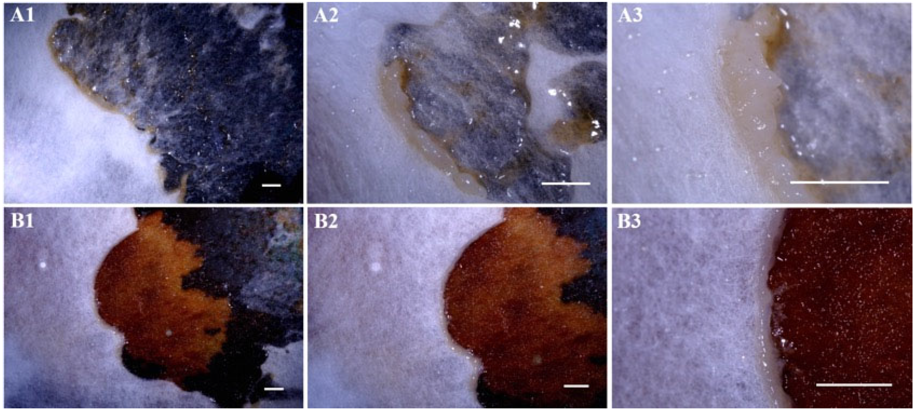
Ac-02IAAM-BApiospora rasikravindrae1.7%ITS, LSU, tef1, tub2[43]
Ac-03IAAM-CApiospora sp.1.2%ITS, LSU, tef1, tub2[43]
Ac-04IAAM-DLecanicillium aphanocladii2.2%ITS, rpb1[44]
Ac-05IAAM-ELinnemannia zychae1.5%ITS, tef1, rpb1[45]
Ac-06IAAM-FFusarium asiaticum0.8%ITS, tef1, rpb1, rpb2, H3[42]
IAAM-PFusarium paranisikadoiITS, tef1, rpb1, rpb2[42]
Ac-07IAAM-GMucor nidicola0.9%ITS[46]
Ac-08IAAM-HLecanicillium aphanocladii1.9%ITS, rpb1[44]
IAAM-TLecanicillium aphanocladiiITS, rpb1[44]
Ac-09IAAM-ILecanicillium psalliotae2.7%ITS, rpb1[44]
Ac-10IAAM-JFusarium parceramosum1.3%ITS, tef1, rpb2[47]
Ac-11IAAM-KFusarium fujikuroi2.9%ITS, tef1, tub2, rpb2[42]
IAAM-NFusarium fujikuroiITS, tef1, tub2, rpb2[42]
Ac-12IAAM-LAspergillus westerdijkiae1.8%ITS, tub2[48]
Ac-13IAAM-MPenicillium copticola2.2%ITS, tub2[49]
Ac-14IAAM-OApiospora arundinis0.6%ITS, LSU, tef1, tub2[43]
Ac-15IAAM-QNectriopsis ellipsoidea1.4%ITS, LSU[50]
Ac-16IAAM-RPurpureocillium lilacinum2.8%ITS, tef1[51]
Ac-17IAAM-SPenicillium citreosulfuratum2.1%ITS, tub2[52]
Ac-18IAAM-W0001Physarella oblonga4.6%18S rRNA[28,35]
Ac-19IAAM-W0002Fuligo gyrosa17.3%18S rRNA[28,35]
Disclaimer/Publisher’s Note: The statements, opinions and data contained in all publications are solely those of the individual author(s) and contributor(s) and not of MDPI and/or the editor(s). MDPI and/or the editor(s) disclaim responsibility for any injury to people or property resulting from any ideas, methods, instructions or products referred to in the content.