Article

Optimizing Micropropagation of Tanacetum balsamita L.: A Machine Learning Approach to Compare Semisolid Media and Temporary Immersion System

1
Institute for BioEconomy (IBE), National Research Council (CNR), 50019 Sesto Fiorentino, Florence, Italy
2
Horticulture Department, Agriculture Faculty, Erciyes University, 38030 Kayseri, Türkiye
*
Author to whom correspondence should be addressed.
Horticulturae 2025, 11(10), 1173; https://doi.org/10.3390/horticulturae11101173
Submission received: 13 August 2025 / Revised: 24 September 2025 / Accepted: 26 September 2025 / Published: 1 October 2025
(This article belongs to the Section Propagation and Seeds)

Abstract

Tanacetum balsamita L. is a medicinal and aromatic plant of high economic value, yet its tissue culture and micropropagation protocols remain poorly developed. This study evaluated and compared two in vitro culture systems, semisolid medium (SS) and Temporary Immersion System (TIS), for enhancing biomass production and growth performance, in terms of relative growth rate (RGR), photosynthetic activity, chlorophyll content, antiradical capacity, and anatomical development. The results demonstrated that the TIS significantly improved RGR, photosynthetic performance, and antiradical activity, and promoted the anatomical development that facilitated greenhouse acclimatization. Machine learning (ML) models, including Multilayer Perceptron (MLP) and Random Forest (RF), were employed to predict morphological and biochemical traits. MLP achieved the highest predictive accuracy (R2 > 0.95) and lowest error metrics for complex, nonlinear traits such as chlorophyll content and antiradical activity, whereas RF excelled in predicting morphological traits with more uniform variance, such as leaf number and shoot length. Overall, this study demonstrates that the TIS provides a high-yield, economically crucial strategy for the micropropagation of T. balsamita, and that integrating ML-based predictive modeling can enhance parameter optimization and phenotyping precision. This combined approach offers a valuable framework for advancing tissue culture research in medicinal and aromatic plants through both production efficiency and data-driven decision-making.

1. Introduction

Tanacetum balsamita L., commonly known as costmary, is an herbaceous perennial plant that belongs to the Asteraceae family. It is widespread in southeastern Europe and southwestern Asia since the Middle Ages, and it is cultivated in several countries, such as Iran, Türkiye, Romania, Germany, Italy, Spain, and England [1,2,3]. T. balsamita is a precious plant for therapy, and it has been used as a hepatoprotective, antibacterial, antiallergic, sedative, tonic, and painkiller [4,5,6]. The validated properties are mainly attributed to the presence of multiple phytoconstituents [7]. It contains biologically active substances such as flavonoids and their derivatives, phenolic acids, monoterpenes, sesquiterpenes, diterpenes, fatty acids, aliphatic hydrocarbons, oligoelements (Ca, K, Mg, P, and Fe), and vitamin C (ascorbic acid) [3]. Due to these features, T. balsamita is used as a spice, an herbal tea, and an important source for producing essential oil [8] in the food and pharmaceutical industries.
T. balsamita can be propagated by division or root cuttings, while the seed germination method is not efficient [5,9]; the endeavors of in vitro culture of T. balsamita seem to give good results [10,11,12]. Biotechnological techniques, such as in vitro propagation and genetic transformation, play a crucial role in increasing the production of medicinal plants. Particularly, to develop medicinal plants that are well-adapted to climate change and able to meet market demand, clonal mass multiplication is essential to ensure uniform yield in terms of both quality and quantity [13] using micropropagation based on gelled or liquid medium. The Temporary Immersion System (TIS) is an advanced technique to propagate in vitro plants, based on alternating immersion cycles of the cultured plant tissue into the liquid medium followed by a dry period [13,14,15].
Machine learning (ML) has become increasingly prominent in plant tissue culture research, providing sophisticated modeling tools to handle the nonlinear and multifactorial aspects of in vitro morphogenesis. Techniques such as Random Forest (RF) and Multilayer Perceptron (MLP) have demonstrated strong potential in predicting plant responses to abiotic stresses and optimizing regeneration procedures in various micropropagation systems [16]. In tissue culture, some studies [17,18,19] confirmed that combining in vitro culture techniques with machine learning can validate the success of applied micropropagation protocols. However, few studies have systematically compared ML performance across both semisolid and TIS cultures, including detailed morphological and stomatal traits.
In this study, the in vitro propagation of T. balsamita shoots was carried out using two in vitro systems, the TIS and the traditional system on semisolid medium, to evaluate the biomass production and growth performance, in terms of relative growth rate (RGR), photosynthetic activity, chlorophyll content, antiradical capacity, and anatomical development in a controlled environment. Moreover, the growth under in vitro conditions allows for obtaining uniform plants with the same genetic origin, and ensuring the stability and high yield of bioactive compounds for the food and pharmaceutical sectors. Also, machine learning approaches were integrated to enhance the research scope. Reliable ML models were employed to predict key in vitro morphological and stomatal characteristics of plantlets grown under semisolid and TIS conditions.

2. Materials and Methods

2.1. Plant Material, Media, and Culture Conditions

T. balsamita shoots used in this study were derived from in vitro stock shoot cultures maintained in jars (500 mL) on half-strength MS ([20]; Sigma-Aldrich®, St. Louis, MO, USA) medium supplemented with cysteine (10 mg/L; Duchefa Biochemie, Haarlem, The Netherlands), ascorbic acid (15 mg/L; Carlo Erba, Cornaredo, MI, USA), glutathione (300 mg/L; Duchefa Biochemie, Haarlem, The Netherlands), 6-benzyladenine (1 mg/L; Sigma-Aldrich, St. Louis, MO, USA), and sucrose (20 g/L). The medium was solidified with 0.3% (w/v) Gelrite (Sigma-Aldrich, St. Louis, MO, USA). The pH of the medium was adjusted to 6.0, and autoclaved at 121 °C for 20 min. The cultures were maintained at 23 ± 1 °C under 16 h light photoperiod conditions, with light (60 μmol m−2 s1 photosynthetic photon flux density) provided by cool-white fluorescent lamps (Osram®, Munich, Germany). The explants were subcultured in fresh medium every eight weeks, and the shoot clumps were divided into individual axes at the end of all subcultures.
For the experiments, shoots (about 4 cm in length) were used as explants. They were cultured in two different in vitro culture systems: semisolid (SS) medium and Temporary Immersion System (TIS). For the TIS, PlantformTM bioreactor (https://www.plantform.se/pub/) was used, containing 50 explants and 500 mL of liquid culture medium, while for the SS system, 5 glass jars of 500 mL were used with 10 explants per jar and 100 mL of gelled medium in each. The immersion time in the PlantformTM bioreactor was set to 4 min every 6 h, while the ventilation time was 15 min every 6 h. Both culture systems were maintained under the same incubation conditions as described above. To compare extracts obtained from in vitro and in vivo samples, three plants cultivated in pots (PP; Ø = 20 cm; h = 20 cm) filled with a commercial substrate (VIGORPLANT Lodi, Italy) were used for chlorophyll and total polyphenol analysis.

2.2. Morpho-Physiological Parameters

2.2.1. Plant Growth and Stomatal Characteristics

After 30 days and 60 days of in vitro culture, the weight of shoot cultures was recorded to calculate the RGR as following: [ln FW final − ln FW initial] × 100/days of culture (ln = natural logarithm; FW: fresh weight, [21]). RGR index is based on the initial and final fresh weights of the plant material and the time of culture.
At the end of the experiment (60 days), the parameters analyzed for each culture system included the number of leaves per shoot, leaf dimensions (length and width average), leaf area, and shoot length.
To study the differences in the number and morphology of stomata in leaves from shoots grown in both culture systems, the epidermis of the leaf (abaxial surface) was cleaned and covered with a thin layer of transparent nail polish. After drying, clear tape was used to gently peel off the dry nail polish from the leaf surface, and then it was mounted on a microscopic slide. The observations were carried out using an optical microscope (Leica DM-500, Heerbrugg, Switzerland). ImageJ software version 1.54 g (Rasband, W.S., ImageJ, U.S. National Institutes of Health, Bethesda, MD, USA), version 1.54 g, with Java 1.8.0-172, was used to measure the length and width of stomata, stomata area, length and width of pore, pore area and stomata density, and to calculate the percentage of closed and open stomata [22]. Stomata traits were examined as reported by Xu and Zhou [23].

2.2.2. Determination of Chlorophyll

The content of chlorophyll a, chlorophyll b, and total chlorophyll in plants grown in the TIS, SS medium, and pots was determined. Briefly, 100 mg of fresh shoots were mixed with 10 mL of 90% acetone in a mortar to break the cells and extract the chlorophyll. The procedure was repeated until the acetone fraction became clear. The absorbance was measured using a UV–Vis spectrophotometer (Cary 3500 Multicell, Agilent Technologies, Santa Clara, CA, USA) at the following wavelengths: chlorophyll a at 663 nm and chlorophyll b at 645 nm. The amount of chlorophyll a and chlorophyll b was calculated according to Lichtenthaler [24]:
Chlorophyll a (µg/mL) = 11.64 (Abs 663) − 2.16 (Abs 645) + 0.1 (Abs 630) (in the extracted volume)
Chlorophyll b (µg/mL) = 20.97 (Abs 645) − 3.94 (Abs 663) − 3.66 (Abs 630) (in the extracted volume)

2.2.3. Total Polyphenolic Content and Antiradical Activity

For phenolic compound extraction, 100 mg of fresh samples was mixed with 7 mL of 100% methanol and mixed on a rotary shaker for 24 h. The total phenol assay was performed using the Folin–Ciocalteau reagent as described by Luthria et al. [25]. The absorbance of the colored reaction product was read at 730 nm, using an Agilent UV–Visible spectrophotometer Multicell Peltier. The results were expressed as 1 µg of gallic acid equivalent (1 µg GAE/g). The extractions of the total phenolic compounds were performed in triplicate.
The free radical scavenging capacity of the polyphenolic extracts was determined in triplicate using the 2,2-diphenyl-1-picrylhydrazyl (DPPH) reagent as described by Brand-Williams et al. [26]. An Agilent UV–Visible spectrophotometer Multicell Peltier was used to quantify this colorimetric reaction at 517 nm. For each extract, the I50 was calculated using the following formula:
% inhibition = [(As − Ax)/As] × 100
where As is the initial absorbance of the sample extract in DPPH solution (t = 0), and Ax is the absorbance of the same sample after 20 min.
The total polyphenol content and antiradical activity in T. balsamita samples were evaluated in vitro culture, using both the TIS and SS system, as well as in vivo pot plants (PP). Each group included three biological replicates. The polyphenol content was expressed as mg/g fresh weight (FW).

2.3. Acclimatization

After 60 days of in vitro propagation, the shoots from both systems are transferred to ex vitro conditions, on substrate perlite and peat (1:1 v/v) in a small indoor greenhouse with ventilation flaps and lid (54 × 28 × 25 cm) to estimate the ex vitro survival rate and the presence of roots at the end of the acclimatization stage (45 days).

2.4. Experimental Design and Statistical Analysis

All experiments were conducted in a completely randomized design with three replications. The effects of the two culture systems on morphological parameters of shoots, stomatal parameters (including behavior and density), chlorophyll content, polyphenol content, and antiradical activity (DPPH) in T. balsamita were examined. Data were subjected to ANOVA to detect overall differences between groups, and mean comparisons were performed using Tukey’s test (p ≤ 0.05) with the agricolae package [27] in R software (version 4.3.1; R Core Team, 2023). Percentage data (proportion of open and closed stomata) were arcsine-transformed using the formula Y = arcsine(√(x/100)), where x is the percentage value. The correlation between total polyphenol content and antiradical activity (DPPH) was assessed using Pearson’s correlation analysis in R software.

2.5. Modeling Procedure

The analysis was performed using R version 4.3.1 within R Studio. The models were built using the caret package [28], which facilitated data preprocessing, model training, cross-validation, and performance assessment. Predictor variables encompassed a wide array of physiological and developmental indicators from micropropagated plantlets exposed to different treatments. The traits predicted included leaf length, leaf width, leaf area, number of leaves, shoot length, and stomatal and pore dimensions (length, width, area). Before modeling, missing values were filled with zeros, and numeric variables were normalized via min-max scaling, specifically the “range” method. Each trait was modeled separately through a supervised learning approach. The data were divided into training (80%) and testing (20%) sets using stratified random sampling with the createDataPartition() function to maintain representative class distributions. Two machine learning algorithms—Multilayer Perceptron (MLP), a deep learning method suitable for capturing complex, nonlinear biological relationships [29], and Random Forest (RF), an ensemble tree-based technique known for its robustness and resistance to overfitting [18,30]—were used to develop predictive models. All models were trained with 10-fold cross-validation on the training data, with data centered and scaled as needed before fitting.
Model performance was evaluated using four complementary statistical metrics [19,31]. The coefficient of determination (R2) (Equation (1)) measured the proportion of variance explained, while the root mean square error (RMSE) (Equation (2)) indicated the size of prediction errors. The mean absolute error (MAE) (Equation (3)) offered an understandable view of average prediction deviations, and the concordance correlation coefficient (CCC) (Equation (4)), computed with the CCC function from the DescTools package, evaluated the agreement between observed and predicted values. Visualization tools such as scatter plots of actual versus predicted values and residual plots, generated using ggplot2, were used to diagnose model accuracy and bias.
R 2 = 1 i = 1 n ( Y i Y ^ i ) 2 i = 1 n ( Y i Y ~ ) 2
  R M S E = i = 1 n ( Y i Y ^ i ) 2 n
M A E = 1 n i = 1 n | Y i Y ^ i |
C C C = 2 ρ σ x σ y σ x 2 + σ y 2 + μ x μ y 2

3. Results and Discussion

The effects of the culture system, vessel design, equipment, immersion time and frequency, and substrate composition are all factors influencing the growth and development of plant cultures. In this study, the application of the TIS, as a Plantform bioreactor, with 4 min/6 h of immersion frequency and 15 min/6 h of ventilation, was compared to a conventional semisolid system. In addition, machine learning (ML) algorithms were employed to analyze and predict growth performance, enabling data-driven insights into the comparative efficiency of the two culture systems.

3.1. Plant Growth Parameters

Plant growth parameters provide informative data to understand the form, function, and performance of plants, enabling the investigation of processes that involve the entire plant. RGR is a useful index for monitoring plant growth and has also been applied to in vitro propagation [21,32,33,34].
In this study, the RGR was calculated at 30 and 60 days, showing an improvement in the growth of the TIS compared to SS culture (Table 1). The highest RGR value was recorded after 30 days in the PlantformTM bioreactor (5.5) and SS medium (3.9), indicating a good adaptability and a better growth rate of T. balsamita plantlets in this culture system. After 60 days of continuous culture without subculture, only renewing the liquid medium, the best RGR value was recorded in the TIS (3.2), while in the SS medium it was lower (2.8).
This trend was consistent with the findings of Elazab et al. [35], who reported the highest RGR index in the TIS, with a slight decrease observed after the second and third subcultures. Similarly, in Quercus robur, Gatti et al. [21] found significant differences in the RGR index between Microbox vessels containing semisolid medium and the Plantform™ bioreactor after eight weeks of culture. In that study, the RGR evaluated at the end of the proliferation stage indicated that the Plantform™ bioreactor enhanced biomass production, both in terms of shoot growth and proliferation, compared with the Microbox system. Additionally, RGR data in our study confirmed that the TIS is a highly promising system for biomass production in T. balsamita.
The morphological evaluation of T. balsamita shoots revealed differences between the TIS and SS medium in several key parameters associated with vegetative growth (Table 2).
The length of shoots grown in the TIS was significantly greater (48.00 mm) compared to those cultured in SS conditions (11.87 mm), and this difference was highly significant. The leaf length was increased dramatically in the TIS-grown shoots (8.16 mm), relative to those in the SS (4.95 mm), supported by a highly significant p-value. In contrast, the number of leaves per shoot was higher in the SS culture (3.97) than in the TIS (2.57), with a statistically significant difference. It is also noteworthy that leaf width showed no significant variation between culture systems (TIS: 2.98 mm; SS: 3.01 mm). Similarly, the leaf area did not differ significantly between the TIS (19.64 mm2) and SS medium (14.86 mm2), although a numerical trend indicated a larger leaf area in the TIS.
These findings demonstrated that the TIS induces a development characterized by significantly longer shoots and leaves, optimizing plantlets for vertical growth. In contrast, SS culture conditions promoted a more compact morphology with a greater number of leaves. The good performances recorded in the TIS can be attributed to increased nutrient uptake by the entire plantlet from the liquid medium, a higher supply of dissolved oxygen for aeration in the container, and more space available for growth [33,36]. In the current study, the Plantform bioreactor provided effective gas exchange, avoiding the accumulation of detrimental volatile compounds [37]. Therefore, the combination of these factors can be considered crucial in the TIS for enhancing biomass production and plant growth, supporting the research objective.
Similar results were reported in Myrtus communis [38]. This species cultured in PlantformTM showed better performance than in the solid medium, particularly in terms of plant length, while the number of leaves was higher in the TIS only when a longer immersion frequency was applied (15 min every 8 h). With a lower frequency of 15 min every 4 h, no difference was detected in this parameter between the TIS and the semisolid system. In Turkish myrtle genotypes [29], the PlantformTM system yielded about four times greater plant heights (5.80 cm) than the solid culture (1.40 cm).
In Vanilla planifolia, the greatest shoot height (4.24 cm) was observed using SETISTM, a TIS bioreactor, compared to semisolid medium (2.61 cm); moreover, the TIS yielded the largest number of leaves [39]. The authors considered that the effect on shoot length during cultures of V. planifolia in the TIS is probably attributed to the design of each bioreactor and light availability. The same species grown in other types of bioreactors (RITA and TIB) always showed a high rate of proliferation [40,41].
In accordance with our results, the application of three TIS bioreactors (BIT®, SETIS®, and RITA®) in Stevia rebaudiana, reported a higher level of proliferation and number of shoots than those shoots cultured in semisolid medium. In particular, the BIT bioreactor showed the best results in terms of plant elongation, as this container allowed for greater availability of gases and space for shoot growth [42]. All TIS bioreactors developed vigorous plantlets with limited hyperhydricity. Similarly, in our study, shoots exhibited no hyperhydration symptoms, as the Plantform bioreactor is a large container, more useful for good plant development and provides suitable ventilation for the cultures (Figure 1). An interesting multiplication rate was achieved with the RITA bioreactor in S. rebaudiana, with the highest explant height, a total absence of basal callus, a good explant quality [43], and 2 times more biomass production compared to semisolid medium [44]. When the in vitro shoots of S. rebaudiana were compared in classical solid culture and the Plantform bioreactor, the maximum biomass production was recorded in the last system, with significant differences in shoot length, about 12 cm in the TIS vs. 5.8 cm in SS [45].
On the contrary, a study on S. rebaudiana by Vilariño et al. [46] reported no differences between solid system and the TIS, although a different behavior was found between the tested varieties (Morita and Criolla) depending on the media and culture systems. In Dianthus caryophyllus L., it was found that the TIB Bioreactor showed great potential to enhance the mass propagation of carnation plantlets, obtaining the highest average number and length of new shoots compared to the solid medium; in particular, the TIB exhibited more than 10 times shoot production [47].
Hwang et al. [37] reported good results in three plant species by applying the TIS. Chrysanthemum morifolium plants had a fresh weight 2.9 times higher than the SS culture, strawberry plants four times higher, and Cnidium officinale also showed the best growth in the TIS with 8.1 times higher fresh weight compared to the SS medium. Moreover, the highest shoot length and number of leaves were obtained in the plants grown in the TIS in all three species.
In Gerbera jemesonii “Shy Pink”, the number of shoots regenerated in the semisolid and TIS cultures was 6.93 and 3.03, respectively, but with 3.33% of hyperhydricity observed in the shoots regenerated on the SS. Although the number of shoots regenerated in the TIS was lower, the shoots were healthy [48]. In our study, no abnormal or hyperhydric T. balsamita shoots were observed during TIS culture with a 4 min immersion time every 6 h. This indicated that setting the appropriate immersion time and dry periods can considerably reduce the hyperhydricity of the tissue, establishing optimal conditions for humidity.
Overall, in our study, as indicated by the aforementioned studies, the TIS culture combines the advantages of immersion and dry periods to maximize gas exchange, increase nutrient absorption, and provide more space for growth.

3.2. Stomatal Analysis

The comparative morpho-anatomical assessment of stomatal parameters in T. balsamita shoots demonstrated consistent and statistically significant differences between those cultured under the TIS and those maintained in SS (Table 3). All evaluated traits, stomatal area, stomatal length and width, pore length and width, and pore area, exhibited significantly higher values in TIS-grown explants.
The stomatal area in TIS-cultured shoots reached 215.00 µm2, markedly exceeding the 125.00 µm2 measured in SS conditions. Also, stomatal length (29.00 µm) and width (15.20 µm) were significantly greater in the TIS. This considerable increase may be attributed to the adequate ventilation and good gas exchange provided in the TIS, confirming the different effect of the in vitro culture conditions on stomatal size [21].
The TIS modality similarly favored pore anatomical traits. Pore length and width in the immersion system were 9.10 µm and 4.20 µm, respectively, compared to 6.10 µm and 2.30 µm in semisolid-grown shoots. The calculated pore area, a critical determinant of gas exchange efficiency, revealed significantly larger values in the TIS (34.50 µm2) than in SS (30.10 µm2). These data confirm the significant effect of ventilation in culture vessels on plant anatomy [49] and some morphological and physiological modifications that are frequent in conventional micropropagation [50], such as hyperhydricity, stomatal malfunction, and chlorophyll deficiency caused by ethylene accumulation.
In our study, quantitative analysis of stomatal characteristics revealed significant differences in terms of stomatal density and behavior between T. balsamita shoots grown in the TIS and those cultured on SS medium (Table 4). Microscopic observation showed an improvement in the stomata performance of in vitro plants grown in the TIS. Indeed, the stomatal density was significantly higher in the shoots developed in the TIS, with an average of 250 stomata per mm2, compared to 187 stomata per mm2 in the SS condition, highlighting the impact of both in vitro culture systems.
Our findings are in accordance with Gatti et al. [21], who found that plants grown in Plantform had a major number of stomata per square millimeter, while those cultured on gelled medium had a lower stomatal density. Increasing the number of stomata can be attributed to continuous ventilation in the TIS compared to closed conventional vessels [51]. In our study, the ventilation inside the bioreactor highlighted the role of the TIS in stomatal function of T. balsamita, reducing the relative humidity, transpiration, and CO2 assimilation [41] to overcome the limitations of SS culture.
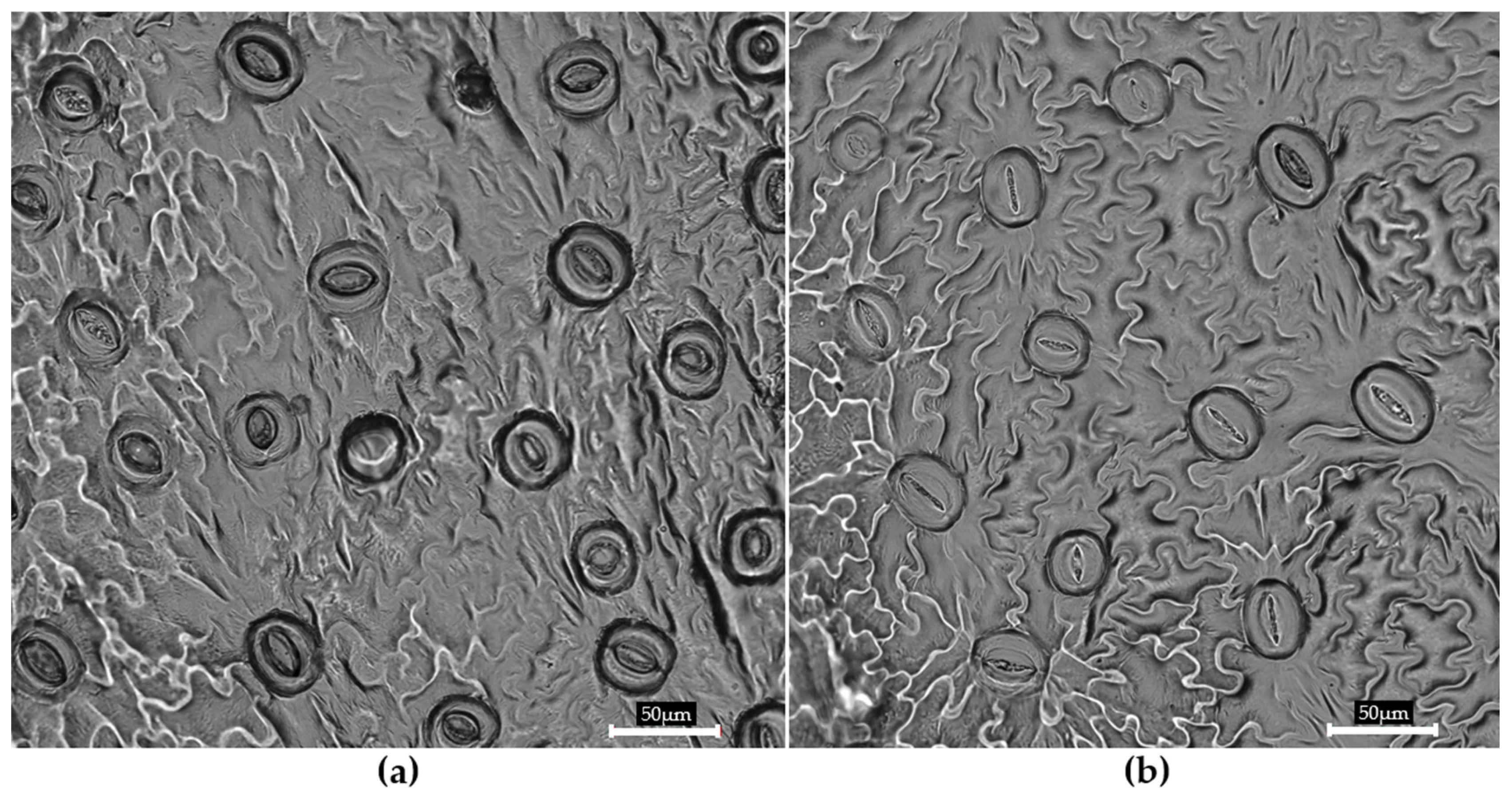
In terms of stomatal behavior (Figure 2), significantly more closed stomata (85%) were observed in TIS-grown shoots (Table 4), while shoots grown on SS medium exhibited 70% closed stomata, indicating that the culture system had a marked influence on stomatal functionality.
Mancilla-Álvarez et al. [22] reported that, in Taro (Colocasia esculenta), the percentage of closed stomata in the temporary immersion ranged from 31 to 35% higher than that in the semisolid system, which had the lowest value of 4.51%. Indeed, no significant difference was noted among the different semi-automated bioreactors, including the TIB, ebb-and-flow bioreactor, and SETIS™. Also, Mancilla-Álvarez et al. [52] stated that shoots of Agave potatorum cultured in the SETIS™ and TIB systems revealed the significantly highest rates of stomatal closure (91.34% and 86.77%, respectively), over the SS system with 10.36% and Monobloc Advance Temporary Immersion System (MATIS®), with approximately 60%. In an earlier study on the use of the TIS for commercial micropropagation of banana (Musa AAA cv. Grand Naine), Bello-Bello et al. [53] reported a significant increase in the percentage of closed stomata from 20% in the semisolid medium to 79% in SETIS™. In anthurium (Anthurium andreanum), shoots grown in an ebb-and-flow bioreactor, regardless of the immersion frequency, presented the highest percentages (between 23 and 29%) compared to 2% obtained in the semisolid medium [54]. The high presence of closed stomata is a physiological indicator of stomatal function [55], promoted by gas exchange and direct contact with the liquid medium [39].
According to Cochard et al. [56], relative humidity, temperature, CO2 concentration, and water potential are environmental factors that determine stomatal function. The stomatal function can be estimated by the open and closed stomata percentage. In our study, the TIS increased the percentage of stomatal closure, suggesting high functionality of stomata that favor more gas exchange, less transpiration rate, and low relative humidity, compared to the SS medium, where the number of closed stomata decreased, indicating poor stomatal functionality [22,52]. Moreover, stomata are responsible for controlling gas exchange between the atmosphere and the plant, and regulating the water potential in the tissues [57,58].
Overall, the results indicated that the TIS culture not only enhanced the structural development of stomata but also significantly affected their physiological behavior. TIS-grown plantlets showed a denser stomatal distribution and a higher level of stomatal closure. This response is likely an adaptation to the microenvironmental conditions characterized by high humidity and intermittent exposure to liquid medium. Therefore, providing regular ventilation in the TIS can improve stomatal function, prevent excessive dehydration, and increase the survival rate of the micropropagated plants after transplanting to the natural environment [51].

3.3. Chlorophyll Content

Significant differences in the analysis of total chlorophyll content for T. balsamita can be attributed to the different cultivation systems, evidencing effects on physiological changes (Figure 3).
Clear statistical differences were observed in both in vitro culture systems and the pots in terms of chlorophyll a content. The TIS yielded the highest mean value (3.47 µg/mg FW), which was significantly greater than the SS medium (2.63 µg/mg FW), and moderately higher than the pot plants (PP) (3.09 µg/mg FW). These findings indicated that the TIS culture environment enhanced the biosynthesis of chlorophyll a; in contrast, the SS culture implied a reduced pigment accumulation even when compared to PP. Chlorophyll b did not exhibit significant variation across groups (Figure 3), suggesting that culture conditions may have less influence on its synthesis or degradation, although the values of chlorophyll b in the TIS were slightly higher than those of SS and PP.
Total chlorophyll, which represents the combined functional capacity of the photosynthetic apparatus, showed a trend similar to that of chlorophyll a. Once again, the TIS recorded the maximum value (4.35 μg/mg FW), significantly higher than PP and SS (3.84 and 3.34 μg/mg FW, respectively). These data further support the hypothesis that the intermittent immersion provided by the TIS facilitated a microenvironment favorable to maximizing chlorophyll synthesis.
The findings on chlorophyll content from the current study were in agreement with those of Thi et al. [59] on Carnation (Dianthus caryophyllus) in RITA type, Arano-Avalos et al. [60] on taro (Colocasia esculenta L. Schott) in TIB, and Hwang et al. [37] on strawberries (Fragaria × ananassa Duch.) in SETIS TM bioreactor. The highest chlorophyll content was recorded in shoots obtained from the TIS when compared to those grown in solid culture. The same was noted in Anthurium andreanum [54] and Bougainvillea glabra [61], where chlorophyll content was significantly higher in the TIS culture compared to conventional closed glass jars. A recent study by Matuszkiewicz et al. [62] using Arabidopsis showed that plants cultured in ventilated culture sealed with permeable tape enhanced the efficiency of photosynthesis and decreased ethylene production by decreasing the stress marker compared to those cultured in air-tight sealed plates. Our results clearly showed greater stress in conventional culture, while the TIS improved photosynthesis performance due to better gas exchange inside the bioreactor. Mediterranean fruit species, such as Rubus fruticosus, Arbutus unedo, and Myrtus communis, showed an increase in chlorophylls after 60 days in TIS culture, with 120 min of immersion per day, compared to those grown in solid and stationary liquid conditions [63]. The increase in photosynthetic pigment content could be due to a partial restoration of the autotrophic activity in the TIS. Indeed, in TIS culture, the explants are immersed in the culture medium only temporarily to provide nutrients for growth, and the immersion period is followed by a drying period. Under these conditions, the plant photosynthetic apparatus (chlorophyll content) becomes active and starts to prepare its carbon complexes, as reported by Ul-Haq and Dahot [64]. Also, the variation in chlorophyll content within the culture systems could be explained by the light availability provided by different types of containers [22].

3.4. Total Polyphenolic Content and Antiradical Activity

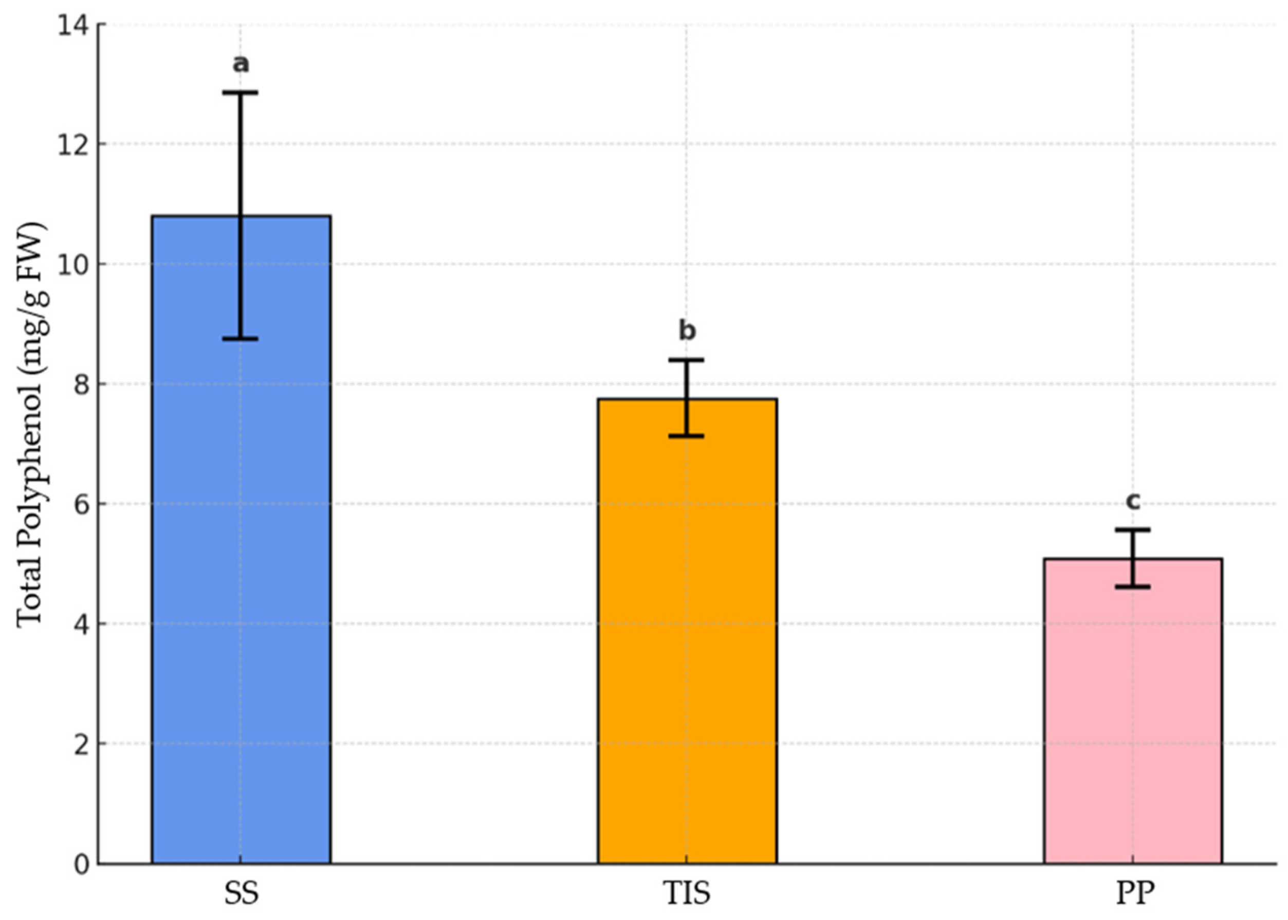
The quantitative evaluation of total polyphenol content in T. balsamita under different culture conditions revealed statistically significant differences (Figure 4). The highest amount of polyphenols (10.87 mg/g FW) was observed in samples grown in the SS system, followed by those in the TIS (7.76 mg/g FW), while the PP samples exhibited the lowest value (5.09 mg/g FW). The results supported the assumption that the in vitro system, both SS and TIS, had the potential to enhance the accumulation of polyphenols, particularly in the SS culture, where the total polyphenols recorded were approximately 2-fold higher than those in the PP samples. On one hand, while the accumulation of secondary metabolites can be increased under stress conditions, the optimal growth conditions can activate the metabolic pathways that induce the production of compounds that are constitutively produced by the plant. SS conditions may be considered a method of culturing with a lower availability of nutrients fundamental for plant growth, resulting in reduced growth rate, and the metabolism may be addressed to the increment of the synthesis of secondary metabolites [65]. On the other hand, with the PP condition, nutrient availability may sustain plant growth at the expense of secondary metabolite accumulation. These assumptions were confirmed in the current study on T. balsamita, as these two culture conditions, SS and PP, induced the highest and lowest accumulation of polyphenolic compounds, respectively. Interestingly, the polyphenol content observed under TIS conditions was intermediate between that of the SS and PP extracts, highlighting the capacity of this culture system to effectively support both biomass production and the accumulation of secondary metabolites in the plant.
The antioxidant potential of T. balsamita was evaluated based on its DPPH radical scavenging capacity, showing statistically significant differences among the group samples (Figure 5).
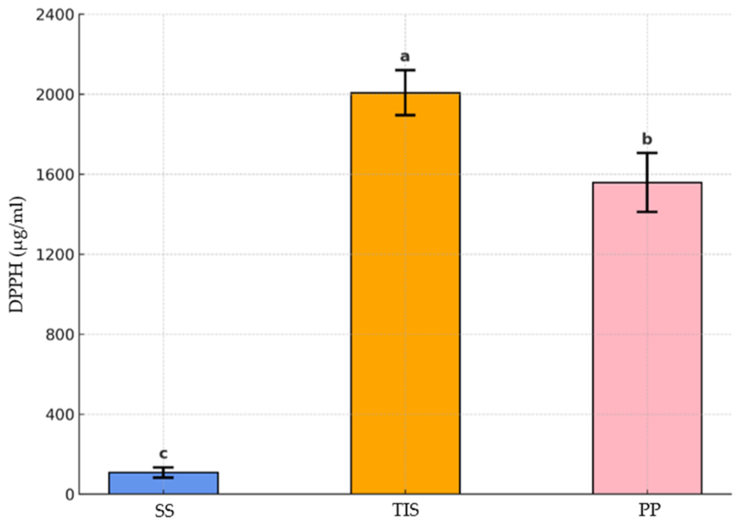
The measurements of the antiradical activity revealed that the extract of SS samples exhibited the lowest I50 (108.4 µg/mL), which increased in PP (1558 µg/mL) and TIS (2006.3 µg/mL) extracts. In particular, the SS I50 value resulted in 14-fold and 18-fold lower values than the ones measured for the PP and TIS samples, respectively, indicating the highest antiradical activity of this extract. Khorasani Esmaeili et al. [66] underlined a significant correlation between the antioxidant activity of extracts and their total phenolic and total flavonoid content in Trifolium pratense L. (Red Clover). Instead, in our study on T. balsamita, a strong negative correlation (r2 = −0.756) was recorded between total polyphenol content and DPPH-based antiradical activity. This finding suggested that higher polyphenol levels may not directly predict stronger antioxidant activity under experimental conditions, but other biochemical compounds might also contribute to the observed antioxidant potential. However, the DPPH assay presents limitations, as it evaluates only one mechanism of antioxidant action and may not reflect the full spectrum of biological antioxidant activity. Further investigations of individual compounds would be necessary to clarify the biochemical basis of the found antioxidant activity.
Based on our results, the extract of SS demonstrated strong antiradical activity, while the extract of aerial parts of in vivo T. balsamita plants and TIS samples showed lower activity. In another study on Exacum bicolor, plant extracts rich in secondary metabolites exhibited high antiradical activity [67], indicating that in vitro-generated plantlet extracts have better activity compared to native plant extracts. These results evidenced that the accumulation of polyphenolic compounds in vitro culture on semisolid medium contributed to the highest antioxidant activity.

3.5. Acclimatization

Since acclimatization is an important step in determining the success of micropropagation protocols [68], the survival rate of plantlets obtained from both in vitro culture systems was evaluated 45 days after transplanting under ex vitro conditions. Our results indicated that the highest survival rate (85%) was recorded for the acclimatized plantlets from TIS culture, while those obtained from the semisolid medium reported a lower percentage of survival (50%). Roots developed spontaneously in the substrate, without hormone treatment, regardless of the in vitro culture system. Figure 6 shows the plants after acclimatization with at least 8–10 roots per plant. Similarly, in Dianthus caryophyllus plantlets from the TIB bioreactor, 90% survival in acclimatization [47], or up to 96% in Agave tequilana were achieved when temporary immersion was applied during the proliferation stage [69].
Moreover, histological and physiological changes occurred in the leaves from TIS plants, including an increase in the percentage of closed stomata can significantly enhance the acclimatization of these plants compared to those from SS medium, especially when the culture container was ventilated during in vitro culture [50]. All morpho-physiological parameters evaluated in this study highlighted the positive response of the acclimatized plants from the TIS, in agreement with other studies [36,59].

3.6. Machine Learning Analysis

A comprehensive comparative analysis of the predictive performance of two machine learning models—Multilayer Perceptron (MLP) and Random Forest (RF)—regarding the estimation of five key morphological parameters of plantlets cultivated under semisolid and TIS conditions is presented in Table 5. The evaluation of these models was conducted utilizing four standard regression metrics: the coefficient of determination (R2), root mean square error (RMSE), mean absolute error (MAE), and concordance correlation coefficient (CCC). This approach offers a thorough perspective on accuracy, precision, and agreement between observed and predicted values.
The R2 scores range from 0 to 1, where 1 indicates perfect predictive performance, and 0 indicates no explanatory power. The RMSE values, which range from zero to positive infinity, reflect the model’s precision, while lower RMSE values indicate better model performance. Similarly, MAE also ranges from zero to positive infinity, with lower values representing higher accuracy. It provides a straightforward measure of the model’s predictive error [19]. The CCC measures the agreement between two sets of observations; values close to 1 indicate strong agreement, while lower values suggest weak agreement or poor correspondence [31].
The comparative evaluation of MLP and RF models for morphological traits demonstrated clear differences in predictive ability across traits and culture systems (Table 5). MLP consistently achieved the highest predictive accuracy for leaf-related parameters, with R2 values above 0.97 and CCC values approaching 0.99 under both SS and TIS conditions. These results indicated that MLP is highly effective in capturing nonlinear growth relationships, particularly for traits such as leaf length, width, and area. By contrast, RF produced lower R2 values (0.80–0.96) for these traits, confirming that ensemble tree-based methods are less fitted to complex, continuous variables in this dataset.
Notably, leaf number predictions revealed a distinct pattern. Under SS conditions, both models showed weak predictive power (R2 ≤ 0.49), suggesting that morphological variability was high and difficult to capture. However, under the TIS, RF reached near-perfect accuracy (R2 = 0.96, CCC = 0.98), whereas MLP showed only moderate performance (R2 = 0.50, CCC = 0.57). This finding highlights a synergy between RF and the morphological uniformity generated by the immersion cycles of the TIS, which favors the rule-based structure of decision tree models. Shoot length predictions further emphasized the complementarity of the two approaches: under SS conditions, both models performed poorly (R2 < 0.50), but under the TIS, MLP markedly outperformed RF (R2 = 0.85 vs. 0.30), confirming its strength in modeling temporal and continuous growth patterns. Together, these results validated the reliability of the models and demonstrated that prediction performance depends on both the trait type and the culture system.
Overall, these findings showed that the MLP model provided better predictive ability for most morphological traits and culture systems, especially for traits affected by leaf structure. Although RF is typically less accurate, it performed well in predicting leaf number in TIS conditions. The differences in model performance across traits and culture systems highlighted the need to select models based on specific contexts in plant morphometric analysis, supporting the use of deep learning methods like MLP for precise phenotyping in plant tissue culture.
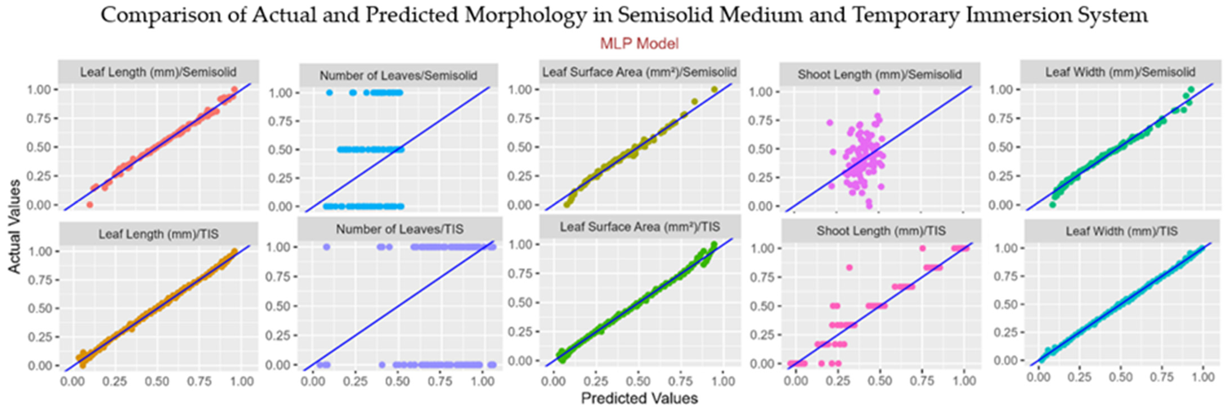
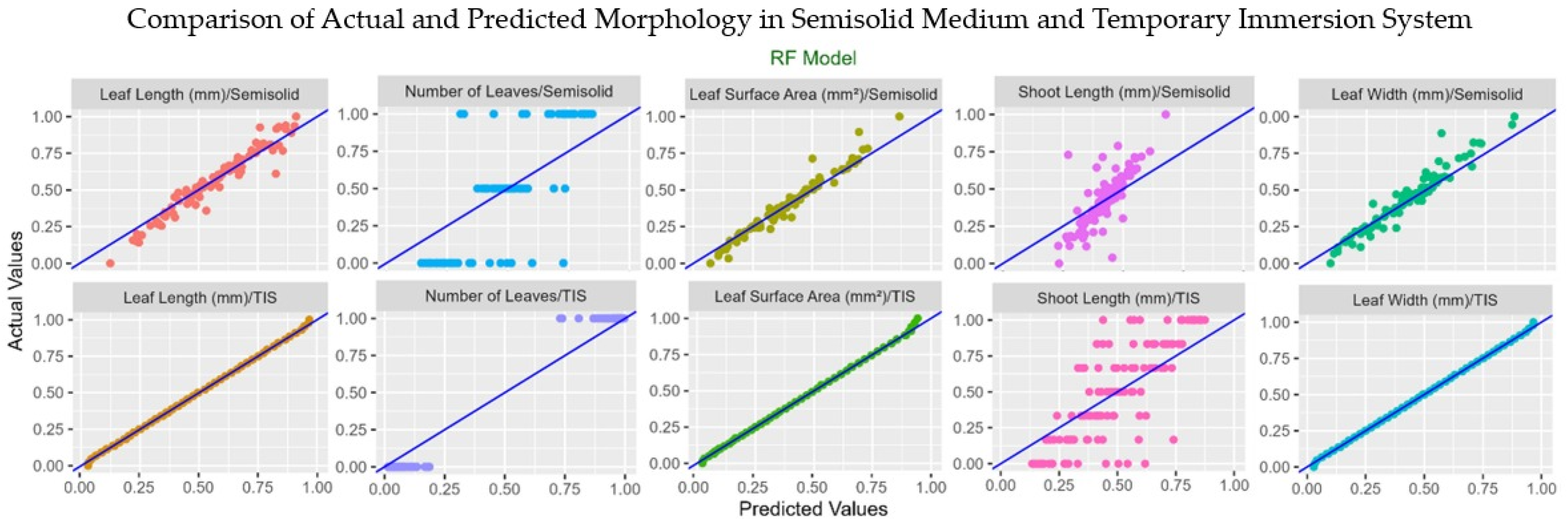
The scatter plots illustrating the relationship between actual and predicted morphological trait values for the MLP and RF models are presented in Figure 7 and Figure 8, respectively.
Table 6 presents a comparative evaluation of MLP and RF models concerning the prediction of various stomatal and pore-related anatomical characteristics in shoots cultivated under SS and TIS conditions. The performance of the models was assessed via essential regression metrics: R2, RMSE, MAE, and CCC, for a better understanding of their predictive capabilities, accuracy, and consistency with empirical observations.
The performance of MLP and RF in predicting stomatal and pore-related anatomical traits also confirmed the robustness of the modeling framework (Table 6). Under SS conditions, both models showed moderate-to-strong predictive ability, with MLP generally outperforming RF. For example, MLP achieved higher R2 values for stomatal area (0.78 vs. 0.65) and pore area (0.84 vs. 0.74), and consistently maintained greater concordance with observed data in CCC (>0.90). These findings indicated that MLP provided more stable predictions for complex stomatal morphology in shoots grown in SS medium.
Under TIS conditions, the predictive performance improved dramatically for both models, with R2 values ranging from 0.89 to 0.97 across all stomatal traits. In this context, RF slightly exceeded MLP for certain dimensional parameters such as stomatal area (R2 = 0.93, CCC = 0.98) and stomatal width (R2 = 0.97, CCC = 0.97), reflecting its ability to capture trait uniformity under controlled immersion cycles. Conversely, MLP provided superior accuracy for pore length (R2 = 0.97, CCC = 0.98), again confirming its strength in modeling continuous and nonlinear variables. Importantly, the consistently high accuracy of both models under the TIS validated their effectiveness in predicting stomatal parameters and confirmed that culture conditions play a central role in model performance. These results demonstrated that MLP and RF offer complementary advantages: MLP excels in complex nonlinear traits, while RF is particularly effective in traits with lower variance and more uniform distributions.
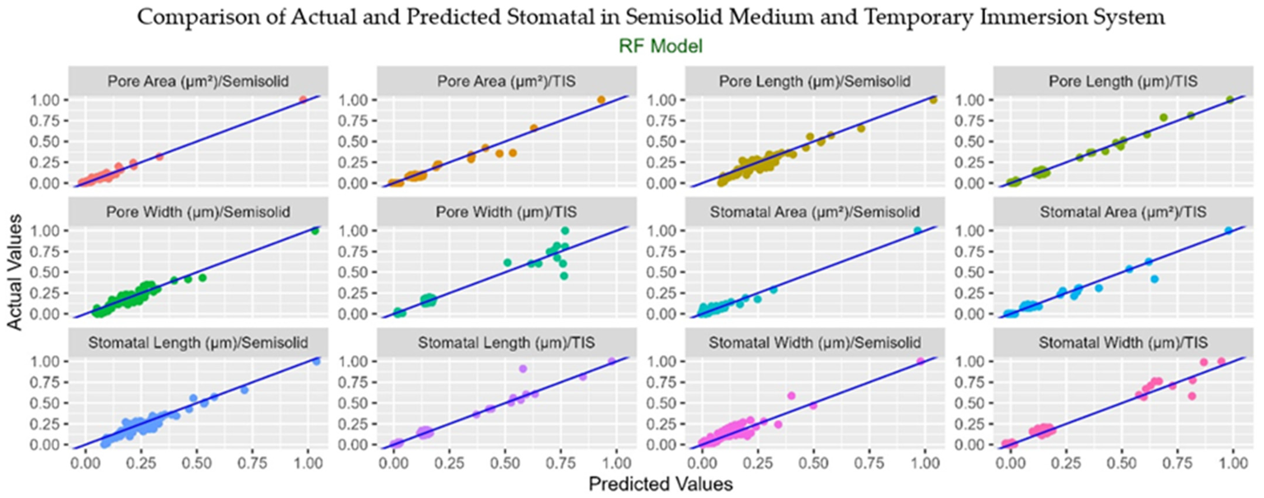
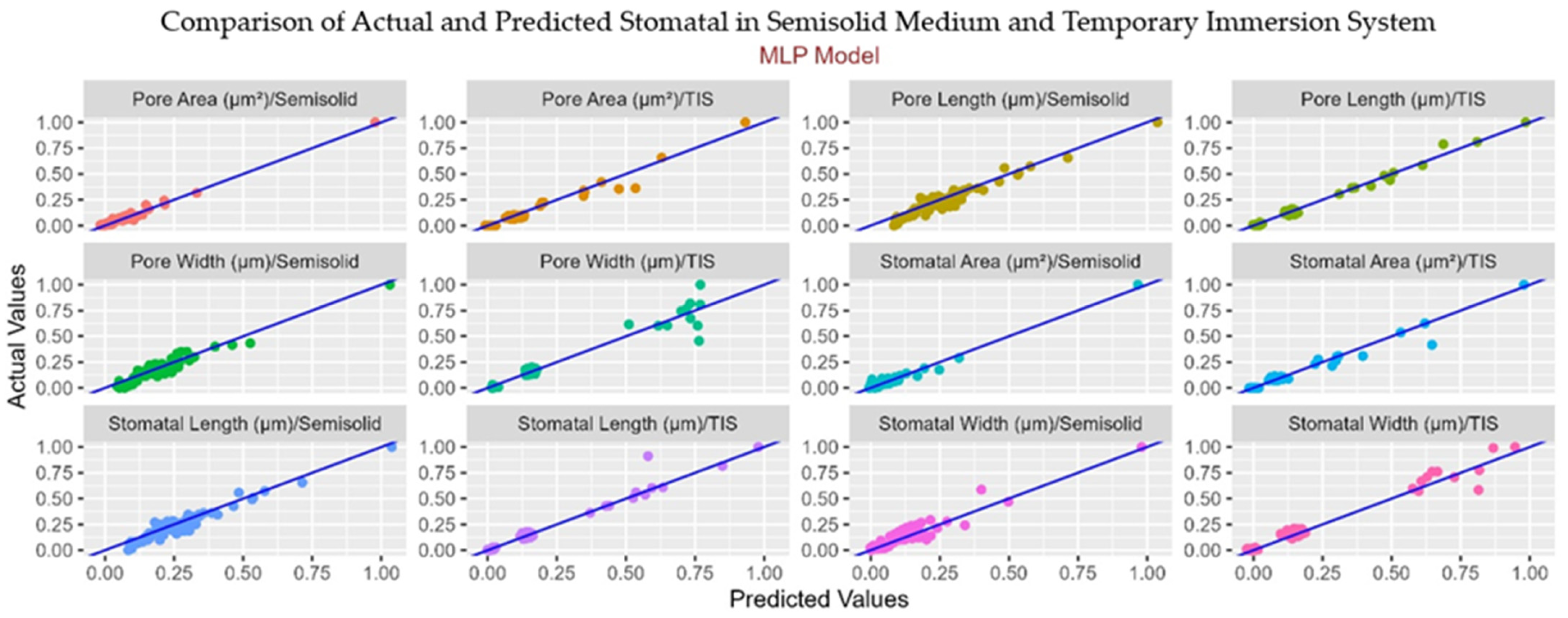
The scatter plots illustrate the relationship between actual and predicted stomatal trait values for the RF and MLP models (Figure 9 and Figure 10, respectively). The integration of ML techniques into plant tissue culture research has demonstrated substantial potential for accurately modeling complex, nonlinear relationships between culture parameters and plant responses. In our study, which compared the predictive performance of MLP and RF models across two distinct culture systems, SS and TIS, for both morphological and stomatal traits, model–trait–system interactions were observed that are consistent with, yet also distinct from, trends reported in the literature.
For morphological traits, MLP consistently outperformed RF across most leaf-related parameters in both the SS and TIS, achieving R2 values exceeding 0.97 for leaf length, width, and area, with correspondingly low RMSE (<0.25) and MAE (<0.15). This high predictive power aligns with findings on Camellia sinensis, where MLP outperformed RF in the prediction of micropropagation parameters [70]. Similarly, in Passiflora caerulea, deep learning models, such as MLP, demonstrated higher predictive accuracy than ensemble methods for rooting traits, with R2 values of up to 0.95 [71]. However, in our study, for leaf number under the TIS, RF achieved near-perfect accuracy (R2 = 0.96, CCC = 0.98), surpassing MLP. The RF advantage in leaf number predictions in the TIS may be explained by the morphological uniformity induced by immersion cycles, which allows tree-based models to exploit structured variance, similar to Vicia spp., where RF improved accuracy for uniform, discrete traits [72]. Shoot length predictions further highlighted system-dependent trends: under SS, both models performed poorly (R2 < 0.50), but under the TIS, MLP accuracy improved markedly (R2 = 0.85, CCC = 0.93), while RF performance decreased. This finding is in agreement with Özcan et al. [18] on Glossostigma elatinoides, where MLP achieved R2 values of 0.957 for rooting percentage and 0.806 for fresh weight, outperforming RF (R2 = 0.952 and 0.799, respectively), indicating the advantage of neural network in modeling temporally dynamic growth patterns
Our stomatal trait predictions further distinguish this study from prior ML applications in tissue culture. Under the TIS, RF slightly outperformed MLP for traits such as stomatal area and width (CCC up to 0.98). This may be due to reduced variance within-treatment in TIS-derived anatomical data, which benefits ensemble models.
In Crataegus monogyna, where RF achieved 97.3% accuracy alongside MLP and XGBoost, high trait uniformity similarly enhanced ensemble model performance [73]. This parallel suggests that trait distribution characteristics, combined with culture system dynamics, critically influence optimal model choice.
In summary, our findings, integrated with comparative results from published studies, reinforce the principle that while MLP generally excels at modeling complex, nonlinear traits, especially those with high variability, RF can outperform in cases with low variance or discrete distributions, particularly under optimized TIS conditions. By simultaneously covering morphological, stomatal, and pore-level traits, employing multi-metric evaluation (R2, RMSE, MAE, CCC), and comparing two culture systems within a single framework, our study expands the methodological and analytical scope of ML applications in plant tissue culture.

4. Conclusions

The TIS allows for greater biomass, minimizes intensive manual handling, reduces production costs related to the workforce, and saves energy respect to conventional techniques [13]. Furthermore, several studies reported the TIS as a suitable technique to overcome the limitations of the semisolid culture for a wide range of fruit, ornamental, and woody species [54,74,75]. The results obtained in the current research demonstrated that the application of the TIS, using the PlantformTM bioreactor, significantly enhanced the growth and physiological performance of T. balsamita plantlets compared to conventional semisolid culture. By providing ventilated culture conditions and liquid medium contact, the TIS promoted key physiological processes, such as photosynthesis, chlorophyll development, and stomatal functionality, resulting in improved ex vitro acclimatization and higher survival rates. Anatomical traits of TIS-derived plantlets closely resembled those of in vivo plants, indicating better physiological adaptation. Therefore, the successful micropropagation is dependent not only on the number of shoots obtained per explant but also on the morphological quality and vitality of the plants produced. Moreover, the integration of machine learning models into the experimental framework enabled accurate prediction of morphological and stomatal traits, revealing trait-specific model superiority, with MLP excelling in complex, nonlinear traits, and RF performing better under uniform morphological patterns. Future research should focus on extending the integrated TIS–ML strategy to other Tanacetum species and medicinal plants, particularly those with high-value secondary metabolites. Additionally, large-scale pilot trials in commercial greenhouse settings are recommended to validate the scalability, economic feasibility, and long-term performance of TIS–ML protocols for plant production.

Author Contributions

Conceptualization, C.B., W.T., and T.İ.; methodology, C.B., W.T., T.İ., C.F., and Ö.Ş.; software, T.İ. and Ö.Ş.; validation, C.B., W.T., T.İ., C.F., and Ö.Ş.; formal analysis, T.İ., C.F., and Ö.Ş.; investigation, C.B., W.T., T.İ., C.F., and Ö.Ş.; resources, C.B. and C.F.; data curation, C.B., W.T., T.İ., C.F., and Ö.Ş.; writing—original draft preparation, C.B. and W.T.; writing—review and editing, C.B., W.T., T.İ., C.F., and Ö.Ş.; visualization,. C.B., W.T., T.İ., C.F., and Ö.Ş.; supervision, C.B.; project administration, C.B.; funding acquisition, C.B. All authors have read and agreed to the published version of the manuscript.

Funding

This research received no external funding.

Data Availability Statement

The original contributions presented in this study are included in the article. Further inquiries can be directed to the corresponding author.

Acknowledgments

The authors thank Monica Anichini for the technical support in stomatal analysis.

Conflicts of Interest

The authors declare no conflicts of interest.

References

  1. Ihtyarova, M.; Angelova, V. Heavy metal accumulation and chemical composition of essential oils of tansy (Tanacetum vulgare L.) cultivated on heavy metal-contaminated soils. Sci. Pap. Ser. E. L. Reclam. Earth Obs. Surv. Environ. Eng. 2023, XII, 320–329. [Google Scholar]
  2. Bylaite, E.; Venskutonis, R.; Roozen, J.P.; Posthumus, M.A. Composition of essential oil of costmary [Balsamita major (L.) Desf.] at different growth phases. J. Agric. Food Chem. 2000, 48, 2409–2414. [Google Scholar] [CrossRef] [PubMed]
  3. Hassanpouraghdam, M.; Tabatabaie, S.; Nazemiyeh, H.; Vojodi, L.; Aazami, M.; Shoja, A.M.; Applied, D. Chrysanthemum balsamita (L.) Baill.: A forgotten medicinal plant. Facta Univ. Ser. Med. Biol. 2008, 15, 119–124. [Google Scholar]
  4. Marculescu, A.; Sand, D.; Barbu, C.H.; Bobit, D.; Hanganu, D. Possibilities of influencing the biosynthesis and accumulation of the active principles in Chrysanthemum balsamita L. species. Rom. Biotechnol. Lett. 2001, 7, 577–584. [Google Scholar]
  5. Hassanpouraghdam, M.B. Flowerhead volatile oil composition of soilless culture-grown Chrysanthemum balsamita L. Nat. Prod. Res. 2009, 23, 672–677. [Google Scholar]
  6. Jaimand, K.; Rezaee, M.B. Chemical constituents of essential oils from Tanacetum balsamita L. ssp. balsamitoides (Schultz-Bip.) Grierson. from Iran. J. Essent. Oil Res. 2005, 17, 565–566. [Google Scholar] [CrossRef]
  7. Khatib, S.; Faraloni, C.; Bouissane, L. Tanacetum balsamita L.: Botany, traditional uses, phytochemical profiling, and biological activities. Drugs Drug Candidates 2025, 4, 10. [Google Scholar] [CrossRef]
  8. Venskutonis, P.R. Costmary (Chrysanthemum balsamita) Oils. In Essential Oils in Food Preservation, Flavor and Safety; Preedy, V.R., Ed.; Elsevier Inc.: Amsterdam, The Netherlands, 2016; pp. 365–375. [Google Scholar]
  9. Marculescu, A.; Hanganu, D.; Kinga, O.N. Qualitative and quantitative determination of the caffeic acid and chlorogenic acid from three chemovarieties of Chrysanthemum balsamita L. Rom. Biotechnol. Lett. 2001, 6, 477–484. [Google Scholar]
  10. Ghiorghita, G.; Ionica, D.E.; Toma, I.; Nicuta, D. Observations on the in vitro and ex vitro behaviour of Chrysanthemum balsamita L. Species. Analele Stiint. Univ. Alexandru Ioan Cuza Iasi Sec. II a Genet. Biol. Mol. 2004, 5, 232–239. [Google Scholar]
  11. Shoja, A.M.; Hassanpouraghdam, M.B.; Khosrowshahli, M.; Movafeghi, A. Callogenesis capability and calli somaclonal variation of costmary (Tanacetum balsamita L.). Rom. Biotechnol. Lett. 2010, 15, 5120–5124. [Google Scholar]
  12. Faraloni, C.; Bonetti, A.; Leva, A.R. Long–term micropropagation of Balsamita major as a promising in vitro strategy for producing phenolic compounds with antioxidant capacity. Plant Biosyst. 2020, 155, 336–343. [Google Scholar] [CrossRef]
  13. De Carlo, A.; Tarraf, W.; Lambardi, M.; Benelli, C. Temporary immersion system for production of biomass and bioactive compounds from medicinal plants. Agronomy 2021, 11, 2414. [Google Scholar] [CrossRef]
  14. Watt, M.P. The status of temporary immersion system (TIS) technology for plant micropropagation. Afr. J. Biotechnol. 2012, 11, 14025–14035. [Google Scholar] [CrossRef]
  15. Georgiev, V.; Schumann, A.; Pavlov, A.; Bley, T. Temporary immersion systems in plant biotechnology. Eng. Life Sci. 2014, 14, 607–621. [Google Scholar] [CrossRef]
  16. Rezaei, H.; Mirzaie-Asl, A.; Abdollahi, M.R.; Tohidfar, M. Enhancing petunia tissue culture efficiency with machine learning: A pathway to improved callogenesis. PLoS ONE 2023, 18, e0293754. [Google Scholar] [CrossRef] [PubMed]
  17. Şimşek, Ö.; Sekerci, A.D.; Isak, M.A.; Bulut, F.; Izgü, T.; Tütüncü, M.; Dönmez, D. Optimizing micropropagation and rooting protocols for diverse lavender genotypes: A synergistic approach integrating machine learning techniques. Horticulturae 2024, 10, 52. [Google Scholar] [CrossRef]
  18. Özcan, E.; Ali, S.A.; Aasim, M.; Atar, H.H. Precision in vitro propagation by integrating response surface methodology and machine learning for Glossostigma elatinoides (Benth) Hook. F. Vitr. Cell. Dev. Biol.-Plant 2025, 61, 827–841. [Google Scholar] [CrossRef]
  19. Tarraf, W.; İzgü, T.; Şimşek, Ö.; Cicco, N.; Benelli, C. An innovative method by temporary immersion system (TIS), Integrated with machine learning analysis. Horticulturae 2024, 10, 454. [Google Scholar] [CrossRef]
  20. Murashige, T.; Skoog, F. A revised medium for rapid growth and bioassays with tobacco tissue cultures. Physiol. Plant 1962, 15, 473–497. [Google Scholar] [CrossRef]
  21. Gatti, E.; Sgarbi, E.; Ozudogru, E.A.; Lambardi, M. The effect of PlantformTM bioreactor on micropropagation of Quercus robur in comparison to a conventional in vitro culture system on gelled medium, and assessment of the microenvironment influence on leaf structure. Plant Biosyst. 2017, 151, 1129–1136. [Google Scholar] [CrossRef]
  22. Mancilla-álvarez, E.; Pérez-Sato, J.A.; Núñez-Pastrana, R.; Spinoso-Castillo, J.L.; Bello-Bello, J.J. Comparison of different semi-automated bioreactors for in vitro propagation of taro (Colocasia esculenta L. schott). Plants 2021, 10, 1010. [Google Scholar] [CrossRef]
  23. Xu, Z.; Zhou, G. Responses of leaf stomatal density to water status and its relationship with photosynthesis in a grass. J. Exp. Bot. 2008, 59, 3317–3325. [Google Scholar] [CrossRef]
  24. Lichtenthaler, H.K. Chlorophylls Carotenoids: Pigments of photosynthetic biomembranes. Methods Enzymol. 1987, 148, 350–382. [Google Scholar]
  25. Luthria, D.L.; Mukhopadhyay, S.; Kwansa, A.L. A systematic approach for extraction of phenolic compounds using parsley (Petroselinum crispum) flakes as a model substrate. J. Sci. Food Agric. 2006, 86, 1350–1358. [Google Scholar] [CrossRef]
  26. Brand-Williams, W.; Cuvelier, M.E.; Berset, C. Use of a free radical method to evaluate antioxidant activity. LWT—Food Sci. Technol. 1995, 28, 25–30. [Google Scholar] [CrossRef]
  27. de Mendiburu, F. Statistical Procedures for Agricultural Research. Available online: https://cran.r-project.org/web/packages/agricolae/agricolae.pdf (accessed on 9 September 2025).
  28. Kuhn, M. Building predictive models in R using the caret package. J. Stat. Softw. 2008, 28, 1–26. [Google Scholar] [CrossRef]
  29. Şimşek, Ö.; Dönmez, D.; Sarıdaş, M.A.; Acar, E.; Kaçar, Y.A.; Kargı, S.P.; İzgü, T. In vitro and ex vitro propagation of Turkish myrtles through conventional and plantform bioreactor systems. PeerJ 2023, 11, e16061. [Google Scholar] [CrossRef]
  30. Isak, M.A.; Bozkurt, T.; Tütüncü, M.; Dönmez, D.; İzgü, T.; Şimşek, Ö. Leveraging machine learning to unravel the impact of cadmium stress on goji berry micropropagation. PLoS ONE 2024, 19, e0305111. [Google Scholar] [CrossRef]
  31. Choi, Y.; Lee, D.H.; Lee, K.E. Concordance correlation coefficients for multivariate measurements. J. Korean Stat. Soc. 2025, 54, 685–717. [Google Scholar] [CrossRef]
  32. Benelli, C.; De Carlo, A. In vitro multiplication and growth improvement of Olea europaea L. cv Canino with temporary immersion system (PlantformTM). 3 Biotech 2018, 8, 317. [Google Scholar] [CrossRef]
  33. Gianguzzi, V.; Inglese, P.; Barone, E.; Sottile, F. In vitro regeneration of Capparis spinosa L. by using a temporary immersion system. Plants 2019, 8, 177. [Google Scholar] [CrossRef]
  34. Gianguzzi, V.; Sottile, F. Temporary immersion system as an innovative approach for in vitro propagation of Sorbus domestica L. Horticulturae 2024, 10, 164. [Google Scholar] [CrossRef]
  35. Elazab, D.; Capuana, M.; Ozudogru, E.A.; Anichini, M.; Lambardi, M. Use of liquid culture with the electis bioreactor for faster recovery of blackberry (Rubus fruticosus L.) shoots from conservation at 4 °C. Horticulturae 2023, 9, 680. [Google Scholar] [CrossRef]
  36. Thanonkeo, S.; Kitwetcharoen, H.; Thanonkeo, P.; Klanrit, P. Temporary immersion bioreactor (TIB) system for large-scale micropropagation of Musa sp. cv Kluai Numwa Pakchong 50. Horticulturae 2024, 10, 1030. [Google Scholar] [CrossRef]
  37. Hwang, H.D.; Kwon, S.H.; Murthy, H.N.; Yun, S.W.; Pyo, S.S.; Park, S.Y. Temporary immersion bioreactor system as an efficient method for mass production of in vitro plants in horticulture and medicinal plants. Agronomy 2022, 12, 364. [Google Scholar] [CrossRef]
  38. Aka Kaçar, Y.; Biçen, B.; Şimşek, Ö.; Dönmez, D.; Erol, M.H. Evaluation and comparison of a new type of temporary immersion system (TIS) bioreactors for myrtle (Myrtus communis L.). Appl. Ecol. Environ. Res. 2020, 18, 1611–1620. [Google Scholar] [CrossRef]
  39. Ramírez-Mosqueda, M.A.; Bello-Bello, J.J. SETISTM bioreactor increases in vitro multiplication and shoot length in vanilla (Vanilla planifolia Jacks. Ex Andrews). Acta Physiol. Plant. 2021, 43, 52. [Google Scholar] [CrossRef]
  40. Ramos-Castellá, A.; Iglesias-Andreu, L.G.; Bello-Bello, J.; Lee-Espinosa, H. Improved propagation of vanilla (Vanilla planifolia Jacks. ex Andrews) using a temporary immersion system. Vitr. Cell. Dev. Biol.-Plant 2014, 50, 576–581. [Google Scholar] [CrossRef]
  41. Ramírez-Mosqueda, M.A.; Iglesias-Andreu, L.G. Evaluation of different temporary immersion systems (BIT®, BIG, and RITA®) in the micropropagation of Vanilla planifolia Jacks. Vitr. Cell. Dev. Biol.-Plant 2016, 52, 154–160. [Google Scholar] [CrossRef]
  42. Rosales, C.; Brenes, J.; Salas, K.; Arce-Solano, S.; Abdelnour-Esquivel, A. Micropropagación de Stevia rebaudiana en sistemas de inmersión temporal para incursionar en la producción hortícola. Rev. Chapingo Ser. Hortic. 2018, 24, 69–84. [Google Scholar] [CrossRef]
  43. Sacco, E.; Mascarello, C.; Pamato, M.; Musso, V.; Ruffoni, B. Evaluation of Temporary Immersion System for in vitro propagation of Stevia rebaudiana Bertoni. Acta Hortic. 2015, 1083, 327–333. [Google Scholar] [CrossRef]
  44. Melviana, A.C.; Esyanti, R.R.; Mel, M.; Setyobudi, R.H. Biomass enhancement of Stevia rebaudiana bertoni shoot culture in temporary immersion system (TIS) RITA® bioreactor optimized in two different immersion periods. E3S Web Conf. 2021, 226, 00007. [Google Scholar] [CrossRef]
  45. Clapa, D.; Radomir, A.M.; Peticila, A.G.; Hârța, M. Evaluation of biomass production of Stevia Rebaudiana Bertoni using classical in vitro culture and temporary immersion bioreactor system. Sci. Pap. Ser. B Hortic. 2023, 67, 558–565. [Google Scholar]
  46. Vilariño, S.; Florido, M.d.C.; García, J.L.; Cantos, M. Effects of culture system and substrate composition on micropropagated plantlets of two varieties of Stevia rebaudiana Bert. Physiologia 2023, 3, 74–85. [Google Scholar] [CrossRef]
  47. Ahmadian, M.; Babaei, A.; Shokri, S.; Hessami, S. Micropropagation of carnation (Dianthus caryophyllus L.) in liquid medium by temporary immersion bioreactor in comparison with solid culture. J. Genet. Eng. Biotechnol. 2017, 15, 309–315. [Google Scholar] [CrossRef] [PubMed]
  48. Lim, M.J.; Han, J.E.; Murthy, H.N.; Song, H.Y.; Lee, S.Y.; Park, S.Y. A comparison of semi-solid, liquid, and temporary immersion bioreactor systems for effective plant regeneration of Gerbera jamesonii “Shy Pink”. Horticulturae 2024, 10, 836. [Google Scholar] [CrossRef]
  49. Regueira, M.; Rial, E.; Blanco, B.; Bogo, B.; Aldrey, A.; Correa, B.; Varas, E.; Sánchez, C.; Vidal, N. Micropropagation of axillary shoots of Salix viminalis using a temporary immersion system. Trees-Struct. Funct. 2018, 32, 61–71. [Google Scholar] [CrossRef]
  50. Zobayeda, S.M.A. Ventilation in micropropagation. In Photoautotrophic (Sugar-Free Medium) Micropropagation as a New Micropropagation and Transplant Production System; Kozai, T., Afreen, F., Zobayed, S.M.A., Eds.; Springer: Dordrecht, The Netherlands, 2005; pp. 147–186. [Google Scholar]
  51. Deccetti, S.F.C.; Soares, A.M.; Paiva, R.; de Castro, E.M. Effect of the culture environment on stomatal features, epidermal cells and water loss of micropropagated Annona glabra L. plants. Sci. Hortic. 2008, 117, 341–344. [Google Scholar] [CrossRef]
  52. Mancilla-Álvarez, E.; Spinoso-Castillo, J.L.; Schettino-Salomón, S.S.; Bello-Bello, J.J. Temporary immersion systems induce photomixotrophism during in vitro propagation of agave Tobalá. 3 Biotech 2024, 14, 74. [Google Scholar] [CrossRef]
  53. Bello-Bello, J.J.; Cruz-Cruz, C.A.; Pérez-Guerra, J.C. A new temporary immersion system for commercial micropropagation of banana (Musa AAA cv. Grand Naine). Vitr. Cell. Dev. Biol.-Plant 2019, 55, 313–320. [Google Scholar] [CrossRef]
  54. Martínez-Estrada, E.; Islas-Luna, B.; Pérez-Sato, J.A.; Bello-Bello, J.J. Temporary immersion improves in vitro multiplication and acclimatization of Anthurium andreanum Lind. Sci. Hortic. 2019, 249, 185–191. [Google Scholar] [CrossRef]
  55. Hazarika, B.N. Morpho-physiological disorders in in vitro culture of plants. Sci. Hortic. 2006, 108, 105–120. [Google Scholar] [CrossRef]
  56. Cochard, H.; Coll, L.; Le Roux, X.; Améglio, T. Unraveling the effects of plant hydraulics on stomatal closure during water stress in walnut. Plant Physiol. 2002, 128, 282–290. [Google Scholar] [CrossRef] [PubMed]
  57. Wang, F.; Chen, Z.H.; Shabala, S. Hypoxia Sensing in Plants: On a Quest for Ion Channels as Putative Oxygen Sensors. Plant Cell Physiol. 2017, 58, 1126–1142. [Google Scholar] [CrossRef]
  58. Schulze, E.D.; Beck, E.; Buchmann, N.; Clemens, S.; Müller-Hohenstein, K.; Scherer-Lorenzen, M. Water deficiency (drought). In Plant Ecology; Schulze, E.-D., Beck, E., Buchmann, N., Clemens, S., Müller-Hohenstein, K., Scherer-Lorenzen, M., Eds.; Springer: Berlin/Heidelberg, Germany, 2018; pp. 165–202. [Google Scholar]
  59. Thi, L.T.; Park, Y.G.; Jeong, B.R. Growth and development of carnation ‘Dreambyul’ plantlets in a temporary immersion system and comparisons with conventional solid culture methods. Vitr. Cell. Dev. Biol.-Plant 2019, 55, 539–548. [Google Scholar] [CrossRef]
  60. Arano-Avalos, S.; Gómez-Merino, F.C.; Mancilla-Álvarez, E.; Sánchez-Páez, R.; Bello-Bello, J.J. An efficient protocol for commercial micropropagation of malanga (Colocasia esculenta L. Schott) using temporary immersion. Sci. Hortic. 2020, 261, 108998. [Google Scholar] [CrossRef]
  61. Mousavi, M. Improve bougainvillea shoot growth in vitro using a modified temporary immersion system (TIS). Int. J. Rev. Life Sci. 2015, 5, 325–330. [Google Scholar]
  62. Matuszkiewicz, M.; Koter, M.D.; Filipecki, M. Limited ventilation causes stress and changes in Arabidopsis morphological, physiological and molecular phenotype during in vitro growth. Plant Physiol. Biochem. 2019, 135, 554–562. [Google Scholar] [CrossRef]
  63. Damiano, C.; Arias, P.; La Starza, S.R.; Frattarelli, A. Temporary immersion system for temperate fruit trees. Acta Hortic. 2007, 748, 87–90. [Google Scholar] [CrossRef]
  64. Haq, I.; Dahot, U.M. Effect of immersion systems on chlorophyll contents in micro-propagating banana. Afr. J. Biotechnol. 2007, 6, 1095–1101. [Google Scholar]
  65. Ramakrishna, A.; Ravishankar, G.A. Influence of abiotic stress signals on secondary metabolites in plants. Plant Signal. Behav. 2011, 6, 1720–1731. [Google Scholar] [CrossRef]
  66. Khorasani Esmaeili, A.; Mat Taha, R.; Mohajer, S.; Banisalam, B. Antioxidant activity and total phenolic and flavonoid content of various solvent extracts from in vivo and in vitro grown Trifolium pratense L. (Red clover). Biomed Res. Int. 2015, 2015, 643285. [Google Scholar] [CrossRef]
  67. Achar, S.S.; Pethakamsetty, L.; Vinjamuri, S. Phytochemical analysis and hypoglycemic potential of Exacum bicolor Roxb. in diet induced obese mice. Indian J. Pharm. Sci. 2025, 87, 44–50. [Google Scholar] [CrossRef]
  68. Gonçalves, S.; Martins, N.; Romano, A. Physiological traits and oxidative stress markers during acclimatization of micropropagated plants from two endangered Plantago species: P. algarbiensis Samp. and P. almogravensis Franco. Vitr. Cell. Dev. Biol.-Plant 2017, 53, 249–255. [Google Scholar] [CrossRef]
  69. Monja-Mio, K.M.; Olvera-Casanova, D.; Herrera-Herrera, G.; Herrera-Alamillo, M.Á.; Sánchez-Teyer, F.L.; Robert, M.L. Improving of rooting and ex vitro acclimatization phase of Agave tequilana by temporary immersion system (BioMINTTM). Vitr. Cell. Dev. Biol.-Plant 2020, 56, 662–669. [Google Scholar] [CrossRef]
  70. Bozkurt, T.; İnan, S.; Dündar, İ.; Isak, M.A.; Şimşek, Ö. Optimizing the In Vitro Propagation of Tea Plants: A Comparative Analysis of Machine Learning Models. Horticulturae 2024, 10, 721. [Google Scholar] [CrossRef]
  71. Jafari, M.; Daneshvar, M.H.; Jafari, S.; Hesami, M. Machine Learning-Assisted In Vitro Rooting Optimization in Passiflora caerulea. Forests 2022, 13, 2020. [Google Scholar] [CrossRef]
  72. Okumuş, O.; Şimşek, Ö.; Isak, M.A.; Şahin, N.K.; Aydin, A.; Eren, B.; Demirel, F.; Kahramanoğulları, C.T.; Uzun, S.; Yaman, M. Integrating Machine Learning and In Vitro Screening to Evaluate Drought and Temperature Stress Responses for Vicia Species. Processes 2025, 13, 1845. [Google Scholar] [CrossRef]
  73. Koçak, M.; Yılmaz, M.C.; Kuzğun, C.; Sirke, S.T.; Yildiz, M. Synthetic seed production in Crataegus monogyna L. and prediction of regeneration of synthetic seeds with machine learning algorithms. Plant Cell. Tissue Organ Cult. 2025, 161, 22. [Google Scholar] [CrossRef]
  74. Meimand, M.J.M.; Ruffoni, B.; Mascarello, C.; Shamshiri, M.H.; Malekzadeh, K. Micropropagation of Pistacia lentiscus L.—Optimization of the surface sterilization protocol and forced ventilation in Temporary Immersion Culture. Fruit Grow. Res. 2020, 36, 75–82. [Google Scholar] [CrossRef]
  75. Carvalho, L.S.O.; Ozudogru, E.A.; Lambardi, M.; Paiva, L.V. Temporary immersion system for micropropagation of tree species: A bibliographic and systematic review. Not. Bot. Horti Agrobot. Cluj-Napoca 2019, 47, 269–277. [Google Scholar] [CrossRef]
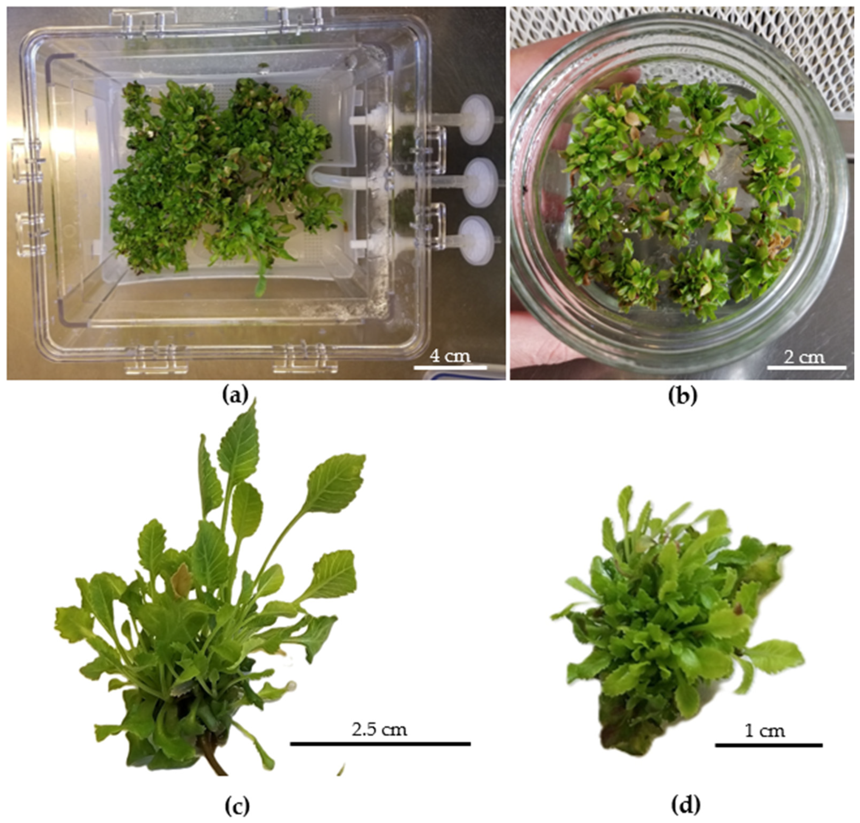
Figure 1. Shoot development of T. balsamita after 30 days. (a) Micropropagation of shoots in Plantform bioreactor. (b) Micropropagation of shoots in semisolid medium. (c,d) Shoot clusters grown in Plantform and semisolid medium.
Horticulturae 11 01173 g001
Figure 2. Stomatal behavior of leaves from T. balsamita shoots cultured in semisolid medium and Temporary Immersion System (TIS): (a) open stomata on leaves from shoots cultured in semisolid medium; (b) closed stomata on leaves from shoots cultured in TIS.
Horticulturae 11 01173 g002
Figure 3. Chlorophyll content in T. balsamita cultured in Temporary Immersion System (TIS, Plantform bioreactor), semisolid medium (SS), and pot plants (PP). Bars with different letters indicate significant differences among culture systems (Tukey’s test), according to the ANOVA.
Horticulturae 11 01173 g003
Figure 4. Total polyphenols in T. balsamita cultured in Temporary Immersion System (TIS, Plantform bioreactor), semisolid medium (SS), and pot plants (PP). Different letters indicate significant differences among culture systems (Tukey’s test), according to the ANOVA.
Horticulturae 11 01173 g004
Figure 5. Antiradical activity in T. balsamita cultured in Temporary Immersion System (TIS, Plantform bioreactor), semisolid medium (SS), and pot plants (PP). Different letters indicate significant differences among culture systems (Tukey’s test), according to the ANOVA.
Horticulturae 11 01173 g005
Figure 6. Acclimatization of T. balsamita plantlets after 45 days (a). Rooted plantlets derived from the Temporary Immersion System (TIS, Plantform bioreactor) (b) and semisolid medium (c).
Horticulturae 11 01173 g006
Figure 7. Actual and predicted values of morphological traits: leaf length, leaf width, leaf area, number of leaves, and shoot length in T. Balsamita plantlets grown in the Temporary Immersion System (TIS) and semisolid medium (SS), as predicted by the Multilayer Perceptron (MLP) model.
Horticulturae 11 01173 g007
Figure 8. Actual and predicted values of morphological traits: leaf length, leaf width, leaf area, number of leaves, and shoot length in T. Balsamita plantlets grown in the Temporary Immersion System (TIS) and semisolid medium (SS), as predicted by the Random Forest (RF) model.
Horticulturae 11 01173 g008
Figure 9. Actual and predicted values of stomatal traits: pore area, pore width, pore length, stomatal area, stomatal length, and stomatal width in T. Balsamita plantlets grown in the Temporary Immersion System (TIS) and semisolid medium (SS), as predicted by the Random Forest (RF) model.
Horticulturae 11 01173 g009
Figure 10. Actual and predicted values of stomatal traits: pore area, pore width, stomatal area, stomatal length, and stomatal width in T. Balsamita plantlets grown in Temporary Immersion System (TIS) and semisolid medium (SS), as predicted by the Multilayer Perceptron (MLP) model.
Horticulturae 11 01173 g010
Table 1. Relative growth rate (RGR) of T. balsamita in PlantformTM bioreactor (Temporary Immersion System, TIS) and semisolid (SS) medium 30 and 60 days after the beginning of in vitro culture.
Culture System30 Days of Culture60 Days of Culture
Initial/Final Weight
(g)
RGRInitial/Final Weight
(g)
RGR
TIS2.82/15.015.515.01/30.033.2
SS medium2.76/7.193.97.19/15.262.8
Table 2. Comparison of morphological parameters of T. balsamita shoots grown under Temporary Immersion System (TIS) and semisolid culture (SS).
ParametersTIS
(Mean ± SE)
Semisolid
(Mean ± SE)
p-Value
Leaf Length (mm)8.16 ± 0.26 a4.95 ± 0.02 b0.0001 ***
Leaf Width (mm)2.98 ± 0.133.01 ± 0.030.9249 ns
Leaf Area (mm2)19.64 ± 1.4714.86 ± 0.170.1235 ns
Number of Leaves2.57 ± 0.00 b3.97 ± 0.06 a0.0000 ***
Shoot Length (mm)48.00 ± 0.00 a11.87 ± 0.12 b0.0001 ***
Values followed by different lowercase letters indicate significant differences between systems based on ANOVA (Tukey’s test). *** p ≤ 0.001; ns: not significant.
Table 3. Stomatal parameters of T. balsamita shoots grown under Temporary Immersion System (TIS) and semisolid culture (SS) conditions.
ParametersTISSemisolidp-Value
Stomatal Area (µm2)215.00 ± 4.00 a125.00 ± 3.50 b0.00001 ***
Stomatal Length (µm)29.00 ± 0.70 a20.00 ± 0.60 b0.00001 ***
Stomatal Width (µm)15.20 ± 0.35 a10.10 ± 0.30 b0.00001 ***
Pore Length (µm)9.10 ± 0.25 a6.10 ± 0.20 b0.00002 ***
Pore Width (µm)4.20 ± 0.15 a2.30 ± 0.10 b0.00002 ***
Pore Area (µm2)34.50 ± 0.38 a30.10 ± 0.35 b0.00003 ***
Means (± SE) followed by different letters are significantly different between systems based on ANOVA (Tukey’s test). *** p ≤ 0.0001 differences.
Table 4. Stomatal behavior and density of T. balsamita shoots grown under Temporary Immersion System (TIS) and semisolid culture (SS) conditions.
TraitTIS (Mean ± SD)SS (Mean ± SD)p-Value
Open Stomata (%)15 ± 2 b30 ± 2 a0.0016 **
Closed Stomata (%)85 ± 2 a70 ± 2 b0.0016 **
Stomatal Density (mm2)250 ± 5 a187 ± 5 b0.0001 ***
Values followed by different lowercase letters are significantly different between systems according to the ANOVA (Tukey’s test). Significance levels: p ≤ 0.01 **, p ≤ 0.001 ***.
Table 5. Comparative performance of machine learning models for predicting morphological traits under semisolid (SS) and Temporary Immersion System (TIS) cultures.
ParametersEvaluation MetricsSSTIS
MLPRFMLPRF
Leaf Length R20.990.850.990.95
RMSE0.020.100.020.06
CCC0.990.860.990.99
MAE0.020.080.010.05
Leaf Width R20.980.800.990.96
RMSE0.030.110.010.07
CCC0.990.960.990.99
MAE0.020.080.010.05
Leaf Area R20.970.920.980.95
RMSE0.040.070.020.07
CCC0.980.900.990.98
MAE0.020.050.010.05
Number of LeavesR20.490.300.500.96
RMSE0.440.370.710.51
CCC0.430.270.570.98
MAE0.390.320.680.36
Shoot Length R20.420.360.850.30
RMSE0.190.170.120.19
CCC0.350.260.930.29
MAE0.150.150.080.17
TIS: Temporary Immersion System; SS: semisolid medium; RF: Random Forest; MLP: Multilayer Perceptron; R2: coefficient of determination; MAE: mean absolute error; RMSE: root mean square error; CCC: concordance correlation coefficient.
Table 6. Performance evaluation of machine learning models for stomatal traits under semisolid (SS) and Temporary Immersion System (TIS) cultures.
ParametersEvaluation MetricsSSTIS
MLPRFMLPRF
Stomatal Area R20.780.650.890.93
RMSE0.050.050.050.04
CCC0.910.760.900.98
MAE0.030.020.030.02
Stomatal Length R20.820.780.960.96
RMSE0.060.060.030.06
CCC0.840.930.920.91
MAE0.050.040.020.03
Stomatal Width R20.610.620.970.97
RMSE0.060.050.030.04
CCC0.860.700.950.97
MAE0.040.040.020.02
Pore Length R20.820.740.970.96
RMSE0.060.060.020.06
CCC0.830.850.980.93
MAE0.050.040.010.03
Pore Width R20.710.650.960.95
RMSE0.080.070.040.05
CCC0.890.860.930.93
MAE0.050.050.020.03
Pore Area (µm2)R20.840.740.920.95
RMSE0.040.050.040.05
CCC0.970.900.920.97
MAE0.020.020.020.01
RF: Random Forest; MLP: Multilayer Perceptron; TIS: Temporary Immersion System; R2: coefficient of determination; MAE: mean absolute error; RMSE: root mean square error; CCC: concordance correlation coefficient.
Disclaimer/Publisher’s Note: The statements, opinions and data contained in all publications are solely those of the individual author(s) and contributor(s) and not of MDPI and/or the editor(s). MDPI and/or the editor(s) disclaim responsibility for any injury to people or property resulting from any ideas, methods, instructions or products referred to in the content.