Article

Diversity and Function Potentials of Seed Endophytic Microbiota in a Chinese Medicinal Herb Panax notoginseng

1
School of Ecology and Environmental Science, Yunnan University, Kunming 650504, China
2
Institute of International Rivers and Ecological Security, Yunnan University, Kunming 650504, China
3
Southwest United Graduate School, Kunming 650092, China
4
Key Laboratory of Chemistry in Ethnic Medicinal Resources, State Ethnic Affairs Commission & Ministry of Education, School of Ethnic Medicine, Yunnan Minzu University, Kunming 650504, China
5
College of Plant Protection, Yunnan Agricultural University, Kunming 650201, China
6
Key Laboratory of Forest Resources Conservation and Utilization in the Southwest Mountains of China Ministry of Education, Southwest Forestry University, Kunming 650224, China
*
Authors to whom correspondence should be addressed.
These authors contributed equally to this work.
Horticulturae 2025, 11(10), 1162; https://doi.org/10.3390/horticulturae11101162
Submission received: 15 August 2025 / Revised: 22 September 2025 / Accepted: 23 September 2025 / Published: 29 September 2025

Abstract

As an important complementation of plant genetic traits, seed endophytes (SEs) have garnered significant attention due to their crucial roles in plant germination and early seedling establishment. In this study, we employed both culture-dependent and amplicon sequencing-based approaches to characterize the endophytic microbiome in seed samples derived from different individual Panax notoginseng plants. Additionally, we evaluated the antagonistic activity of isolated culturable bacterial SEs against the root rot pathogens Fusarium solani and F. oxysporum. Our results demonstrated that a greater sampling quantity substantially increased the species richness (Observed OTUs) and diversity of seed endophytic microbiota, underscoring the importance of seed population size in facilitating the vertical transmission of diverse endophytes to progeny. The endophytic communities (including both fungi and bacteria) exhibited a conserved core microbiota alongside host-specific rare taxa, forming a phylogenetically and functionally diverse endophytic resource pool. Core bacterial genera included Streptococcus, Methylobacterium-Methylorubrum, Sphingomonas, Burkholderia-Caballeronia-Paraburkholderia, Pantoea, Halomonas, Acinetobacter, Pseudomonas, Vibrio, and Luteibacter, while core fungal genera comprised Davidiella, Thermomyces, Botryotinia, Myrothecium, Haematonectria, and Chaetomium. Among 256 isolated endophytic bacterial strains, 11 exhibited strong inhibitory effects on the mycelial growth of F. solani and F. oxysporum. Further evaluation revealed that two antagonistic strains, Bacillus cereus and B. toyonensis, significantly enhanced seed germination and plant growth in P. notoginseng, and effectively suppressed root rot disease in seedlings. These findings highlight the potential use of SEs as biocontrol agents and growth promoters in sustainable agriculture.

1. Introduction

Plant endophytes refer to microorganisms—including bacteria, fungi, and actinomycetes—that colonize plant tissues without causing apparent disease symptoms [1,2]. They are ubiquitously distributed in various plant organs such as roots, stems, leaves, flowers, fruits, and seeds [2]. The symbiotic relationships between endophytes and their host plants range from mutualism to commensalism, contributing significantly to plant growth, development, and adaptation to environmental stresses [3]. Through mutualistic interactions with their hosts, these microbes participate in plant growth promotion, disease suppression, and stress resistance enhancement [4], playing a crucial role in plant–soil ecosystems [5,6].
Among these, seed endophytes (SEs), as the initial source of the plant microbiome, not only exhibit high diversity but also serve as key regulators of seedling establishment and plant health [7,8]. Due to their significant effects and unique transmission mode, seed endophytes (SEs) serve as a critical complement to the host’s genetic traits. However, traditional research has predominantly focused on soil or mature plant endophytic communities, often overlooking the unique ecological characteristics of “individual seeds” as independent microecosystems—particularly the host-specific functions of seed-transmitted endophytes and their long-term effects on seedling health. Consequently, deciphering the structure and function of seed endophytic communities, especially identifying core seed-transmitted endophytes with biocontrol potential, holds significant promise for developing microbial fertilizers and reducing reliance on chemical pesticides [9,10].
Panax notoginseng (Burk.) F.H. Chen, a perennial medicinal herb of the Araliaceae family, is renowned for its hematopoietic, anti-inflammatory, antioxidant, and anti-aging properties [11]. Native to China and predominantly cultivated in Yunnan and Guangxi provinces, its large-scale production faces severe threats from root rot disease caused by Fusarium spp. [12,13,14]. While chemical control remains the primary management strategy, concerns over pesticide residues and ecological risks have spurred interest in endophyte-mediated biocontrol as a sustainable alternative [15]. Although previous studies have characterized endophytic communities in P. notoginseng plants, the structure and function of SEs—particularly inter-individual heterogeneity and their antagonistic potential against common pathogens Fusarium spp.—remain unexplored [16]. A deeper understanding of the antagonistic activities and plant-growth-promoting capacities of seed-originating microbes could provide new insights into integrated disease management strategies for this economically important medicinal species.
In this study, we focused on single-plant-derived seeds of 3-year-old P. notoginseng from Wenshan Prefecture, Yunnan Province. Using high-throughput sequencing, we characterized the seed endophytic microbiome at the individual seed level, capturing intra-species homogeneity and heterogeneity, and overcoming the limitations associated with bulk sample analyses [17,18]. Furthermore, we isolated culturable endophytic bacteria from seeds and evaluated their antagonistic activity against Fusarium spp. pathogens. The most promising isolates were subsequently assessed for their ability to promote plant growth and suppress disease in seedling assays. The key innovations of this work include (i) profiling the endophytic community structure of individual P. notoginseng seeds, (ii) identifying core taxonomical and functional members involved in host–microbe interactions, and (iii) establishing a direct link between seed-derived beneficial bacteria and resistance to root rot disease. Our findings provide valuable endophytic resources for the development of eco-friendly biocontrol agents and contribute to a theoretical framework for leveraging seed-transmitted microbiomes in the sustainable cultivation of medicinal plants.

2. Materials and Methods

2.1. Plant Materials

Mature and healthy fruits of Panax notoginseng (Pg) were collected in November 2022 from an experimental field in Wenshan Autonomous Prefecture, Yunnan Province, China (104°5′40″ E, 24°8′40″ N, altitude 1662 m). The samples were transported to the laboratory under low-temperature preservation for further analysis. Additionally, seeds used for sowing were commercially sourced from a local Pg market within the same region.

2.2. Pathogenic Strains and Microbial Culture Media

The tested pathogens included F. solani and F. oxysporum, 2 fungal strains associated with root rot of Pg [19], which were previously isolated and identified by our laboratory.
The fungal culture medium was potato dextrose agar (PDA), prepared with 200 g potato, 20 g glucose, 15–20 g agar, and 1000 mL distilled water (natural pH). For bacterial culture, beef extract peptone agar medium was used, containing 3 g beef extract, 10 g peptone, 5 g NaCl, 15–25 g agar, and 1000 mL distilled water (pH 7.0 ± 0.2).

2.3. Profiling the Seed Endophytic Microbiome

Under laboratory conditions, mature and healthy Pg fruits were rinsed under running water, and seeds were manually separated from the pericarp by gentle squeezing in sterile sealed bags. After washing and air-drying, the seeds underwent standardized surface sterilization [20] as follows: (1) rinsing with sterile distilled water followed by surface moisture removal using sterile filter paper; (2) immersion in 75% ethanol for 30 s; (3) two subsequent sterile water washes; (4) treatment with 0.1% HgCl2 solution for 10 min [21,22]; (5) five final sterile water rinses. Sterilization efficacy was confirmed by plating 200 µL of the final rinse solution on agar plates, with no microbial growth observed after incubation. Surface-dried seeds were then aliquoted into 2 mL centrifuge tubes (5 individual plants as 5 biological groups, each with 3 biological replicates), flash-frozen in liquid nitrogen, and submitted for high-throughput sequencing (performed by Lianchuan BioTechnologies Company, Hangzhou, China). Identical procedures were used for the profiling and subsequent computational analysis of the microbiota in different Pg seed samples [23]. For each sample replicate, the DNA of the seed were extracted separately with a FastDNA® Spinkit (MP Biomedicals, Solon, OH, USA), according to the manufacturer’s protocol. Genomic DNA of SEs served as the template for PCR amplification of fungal ITS regions (using primers ITS1: 5′-GTGARTCATCGAATCTTTG-3′ and ITS2: 5′-TCCTCCGCTTATTGATATGC-3′ [24]) and bacterial 16S rDNA V3-V4 hypervariable regions (using primers 341F: 5′-CCTACGGGNGGCWGCAG-3′ and 805R: 5′-GACTACHVGGGTATCTAATCC-3′ [25]). The PCR products were separated by electrophoresis on a 1.5% agarose gel, and the target bands were manually excised under UV illumination and purified using an agarose gel DNA purification kit (TaKaRa Bio Inc., Shiga, Japan) according to the manufacturer’s instructions. The amplicon pools were prepared for sequencing, and the size and quantity of the amplicon library were assessed using an Agilent 2100 Bioanalyzer (Agilent, Santa Clara, CA, USA) and a Library Quantification Kit for Illumina (Kapa Biosciences, Woburn, MA, USA), respectively. The libraries were sequenced using an Illumina NovaSeq 6000 Sequencer (Illumina, Inc., San Diego, CA, USA) at LC-BioTechnologies Co., Ltd. (Hangzhou, China). Raw bacterial and fungal sequence data were deposited in the NCBI under accession numbers PRJNA891501 (Endophytic bacteria of Panax notoginseng fruits and seeds) and PRJNA892017 (Endophytic fungi of Panax notoginseng fruits and seeds).

2.4. Culturable Bacteria Isolation and Identification

Surface-sterilized Pg seeds were subjected to tissue patch isolation for endophytic bacteria [26]. Seed tissues were aseptically dissected into small fragments (approximately 2 × 2 mm) and placed on beef extract-peptone agar plates, followed by incubation at 25 °C for 5 days. Bacterial colonies emerging from tissue margins were purified and streaked onto fresh beef extract-peptone agar and incubated at 25 °C for 48 h. Colony morphology was examined and preliminary identification was performed according to Bergey’s Manual of Determinative Bacteriology. Genomic DNA was extracted using the Chelex-100 method. The 16S rDNA gene was amplified with universal bacterial primers 27F (5′-AGAGTTTGATCCTGGCTCAG-3′) and 1492R (5′-GGTTACCTTGTTACGACTT-3′) [27] in a 25 μL PCR system containing: 12.5 μL 2×Taq PCR Mix, 1 μL each forward/reverse primer, 1 μL DNA template, and 9.5 μL ddH2O. Amplification conditions: initial denaturation at 94 °C for 5 min; 32 cycles of 94 °C for 30 s, 50 °C for 45 s, and 72 °C for 45 s; final extension at 72 °C for 10 min. PCR products were verified by 1% agarose gel electrophoresis and sequenced by LC-Bio Technologies (Hangzhou, China). The obtained sequences were submitted to NCBI (https://www.ncbi.nlm.nih.gov/, accessed on 12 October 2024) for nucleotide analysis.

2.5. Screening of Antagonistic Bacteria

The strains to be tested in this section include all the culturable endophytic bacteria isolated, and the test pathogenic bacteria include the 2 tested pathogenic strains (F. solani and F. oxysporum) mentioned in Section 2.2. The antagonistic potential of culturable bacterial SEs against pathogenic fungi was evaluated using the dual-culture technique [28]. After 5 days of pure culturing, mycelial plugs (5 mm diameter) of the test pathogens were inoculated at the center of PDA plates. Subsequently, 20 μL aliquots of each target bacterial strains were spot-inoculated at 4 symmetrical points (20 mm from the pathogen plug), and control plates contained only the fungal pathogen. All treatments were performed in triplicate and incubated at 25 °C for 5 days. The inhibitory effect was quantified by measuring fungal colony diameters and calculating the inhibition rate using the following formula:
I n h i b i t i o n     r a t e = C o n t r o l     c o l o n y     d i a m e t e r T r e a t m e n t     c o l o n y     d i a m e t e r C o n t r o l     c o l o n y     d i a m e t e r × 100 %

2.6. Functional Validation of Antagonistic Bacteria

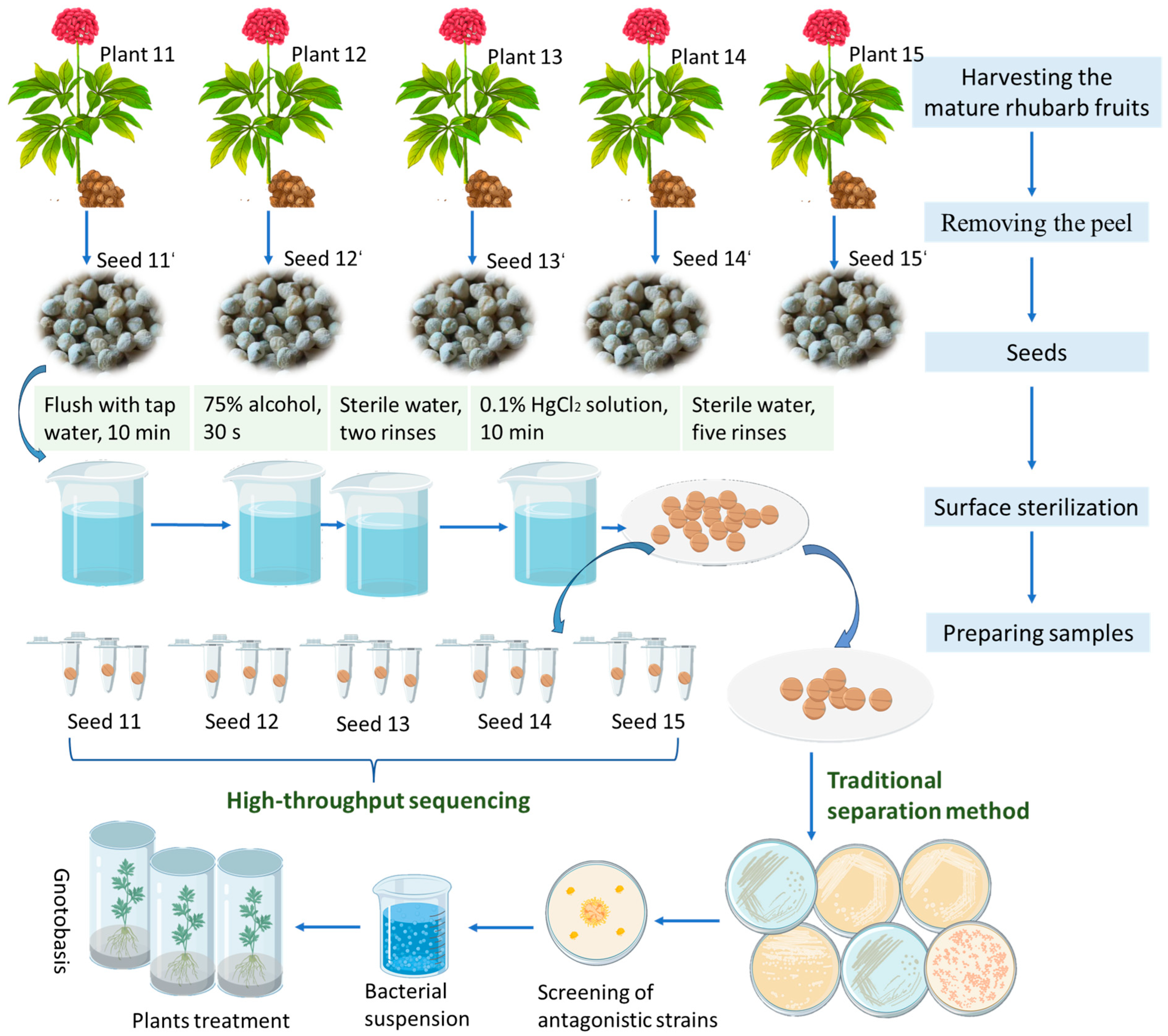
To investigate the effects of microbial inoculants on Pg growth and seedling quality, we conducted a controlled experiment using Pg seeds treated with two bacterial suspensions (1 g/L): B. toyonensis (Bt group) and B. cereus (Bc group). Seeds treated with sterile water served as the control (CK group). We evaluated seed germination and agronomic traits by measuring plant height, shoot fresh weight, root length, and root fresh weight. For disease resistance assessment, one-year-old Pg seedlings were used to examine the preventive and therapeutic effects of B. toyonensis and B. cereus against root rot caused by F. oxysporum. The experimental design included different treatment timings (before or after pathogen inoculation), as detailed in Table 1. Disease severity was assessed using the grading standard established by Fang [29], and the disease index was calculated according to the method described by Zong and Kang [30]. All treatments were performed with 3 biological replicates, each consisting of 36 seedlings. The experimental operation process of this study is shown in Figure 1. The calculation formulas were as follows:
g e r m i n a t i o n     r a t e = N u m b e r     o f     s e e d l i n g s     e m e r g i n g T o t a l     n u m b e r     o f     s e e d s     s o w n × 100 %
i n c i d e n c e = N u m b e r     o f     d i s e a s e d     p l a n t s T o t a l     n u m b e r     o f     p l a n t s × 100 %
d i s e a s e     i n d e x = N u m b e r     o f     d i s e a s e d     p l a n t s × R e p r e s e n t a t i v e     s e r i e s T o t a l     n u m b e r     o f     p l a n t s × T h e     h i g h e s t     r e p r e s e n t a t i v e     l e v e l     v a l u e × 100

2.7. Data Analysis

The raw high-throughput sequencing data were processed using QIIME2 (via the DADA2 plugin) for quality filtering, denoising, and generation of amplicon sequence variants (ASVs) with corresponding abundance tables. Non-microbial sequences (host plant-derived) and ASVs corresponding to chloroplasts and mitochondria were removed based on NCBI database (https://www.ncbi.nlm.nih.gov/, accessed on 12 October 2024) alignments [31]. Alpha diversity metrics including Observed OTUs, Chao1, Goods coverage, Shannon, and Simpson indices were calculated using QIIME2. Rarefaction curves and violin plots were generated using R software (version 3.4.4), with between-group differences assessed by Kruskal–Wallis tests. Beta diversity was evaluated through principal coordinates analysis (PCoA).
Statistical analyses were conducted on the following parameters: the inhibition rate from Section 2.5, as well as germination rate, biomass-related traits (including plant height, shoot fresh weight, root length, and root fresh weight), and disease assessment parameters (including incidence rate and disease index) from Section 2.6. Preliminary data processing was performed using Microsoft Excel (version 2019) Subsequently, one-way analysis of variance (ANOVA) and Tukey’s HSD tests (α = 0.05) were carried out with IBM SPSS Statistics (version 19) to assess significant differences among treatment groups.

3. Results

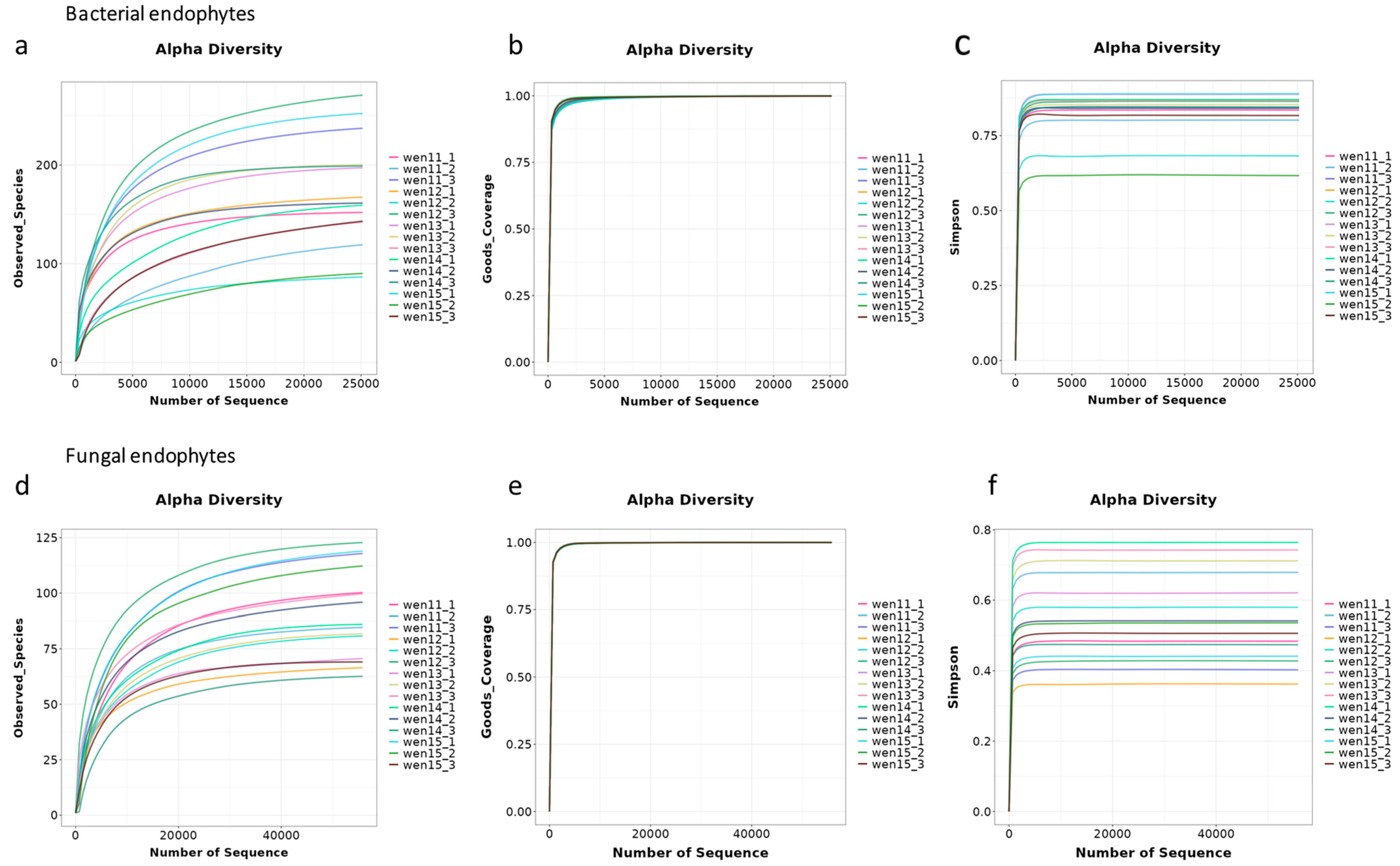
High-throughput sequencing of endophytic bacterial and fungal communities was successfully performed on 15 Pg seed samples (derived from 5 individual plants). After quality filtering, the bacterial dataset comprised generated 701,135 high-quality reads, while the fungal (ITS) dataset yielded 1,192,668 reads. All sequencing results, including detailed taxonomic classifications at multiple levels, are comprehensively summarized in Table A1. The rarefaction curves of all 15 samples demonstrated progressively flattening slopes with increasing sequencing depth, eventually reaching plateaus (Figure A1), indicating that the sequencing effort was sufficient to capture the majority of microbial diversity present in the samples.

3.1. Seeds Are Vectors for Diversity of Endophytes

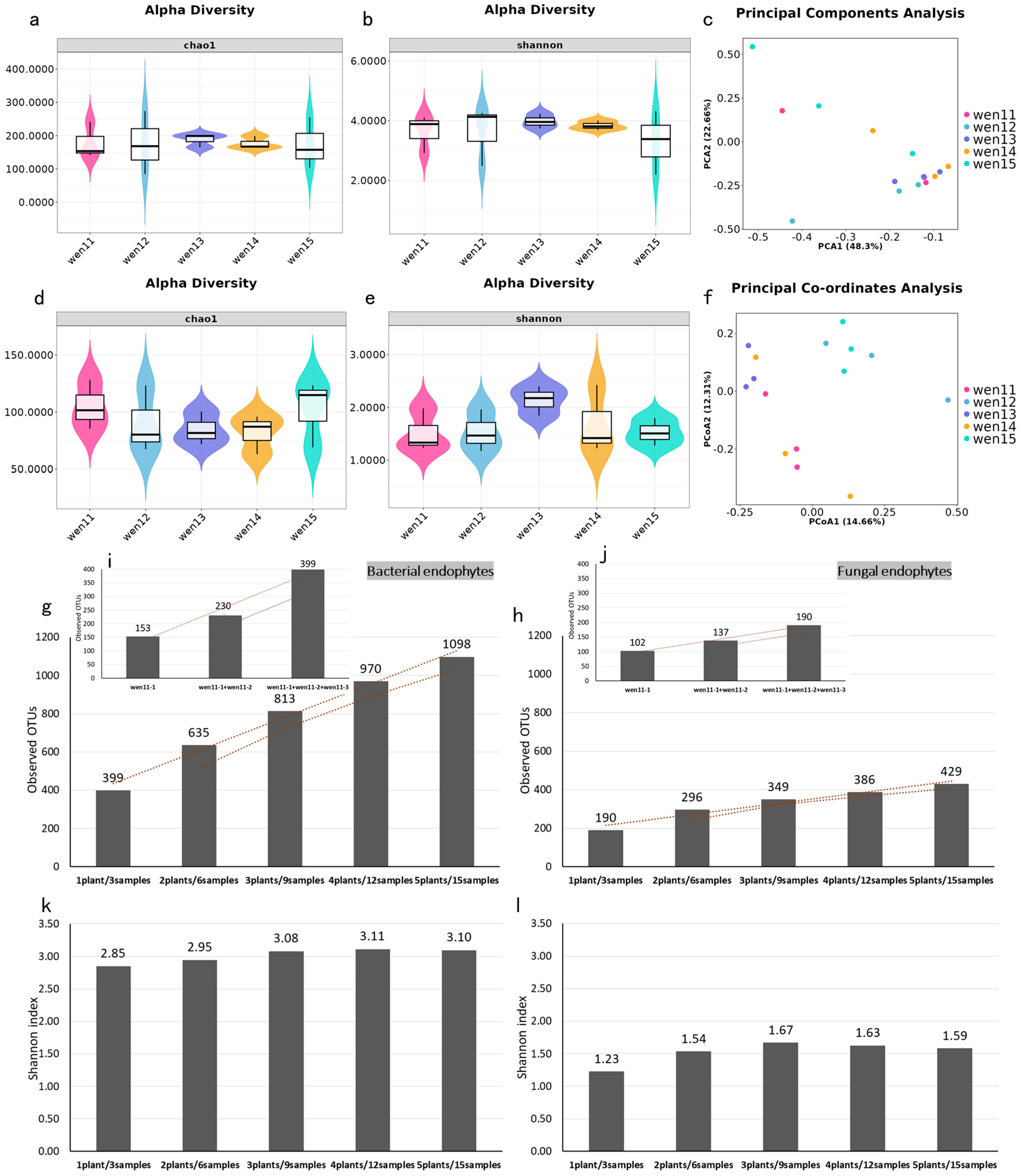
High-throughput sequencing detects subtle inter-individual variations in seed-associated microbiota across different Pg plants. The bacterial Chao1 indices for the 5 Pg seed samples range from 174.79 to 189.57 with an average of 180.57, while the Shannon indices were 3.29 to 3.97 with an average of 3.68 (Table A2). Kruskal–Wallis tests indicated marginal but non-significant differences in alpha diversity among individual plants (p > 0.05) (Figure 2a,b). Principal coordinates analysis (PCoA) of endophytic bacteria revealed that PCoA1 (48.30%) and PCoA2 (22.66%) explained most of the variation, with one plant (wen15) showing slight compositional divergence from other plants, though this was not statistically significant (p > 0.05) (Figure 2c).Fungal communities showed no significant differences in alpha diversity (Chao1: 82.29–103.30; Shannon: 1.52–2.14) across samples (Figure 2d,e; Table A3). PCoA (Jaccard distance) showed moderate separation, and microbial community analysis revealed no significant clustering patterns among individual plants (Figure 2f).
Our results demonstrate a significant positive correlation between seed sample size and endophytic microbial richness and diversity (evaluated by the observed OTUs and Shannon indices). Both bacterial and fungal diversity exhibited a clear increasing trend with expanding sample pools (Figure 2g,h). Specifically, (1) for multi-plant sampling, the bacterial observed ASVs increased progressively from 399 (1 plant/3 samples) to 1098 (5 plants/15 samples); the fungal observed ASVs similarly rose from 190 to 429 across the same sampling gradient as bacteria. (2) For intra-individual sampling (wen11), bacterial ASVs increased from 153 (1 sample) to 399 (3 samples); fungal ASVs expanded from 102 to 190 with equivalent sampling intensification (Figure 2i,j). This consistent pattern—observed across both inter- and intra-individual sampling scales—highlights the crucial role of seed population size in determining the diversity of vertically transmitted endophytes.

3.2. Pg Seeds Contain a Core Endophytic Microbiota

Endophytic bacteria (31 phyla) showed greater phylogenetic diversity than fungi (4 phyla), with higher richness across all samples (Figure 3a,b). Proteobacteria dominated bacterial communities (90.91–95.12%), followed by Firmicutes (4.60–8.51%), while other phyla each represented a relative abundance less than 1% (Figure 3a). Fungal endophytes in Pg were predominantly by the phyla Ascomycota (29.01–74.68%) and Basidiomycota (0.03–2.05%) (Figure 3b).
High variability in species richness was observed among the 15 seed samples, with detectable bacterial genus ranging from 54 to 151 per sample (Figure 3c), with Vibrio, Pantoea, and Burkholderia-Caballeronia-Paraburkholderia representing the 3 most dominant genera (Figure 3e). Strikingly, we identified a core bacterial microbiome consisting of 28 shared ASVs (2.55% of total ASVs), which accounted for 9.18–23.33% of individual sample ASVs (Figure 3g). The core microbiota demonstrated high relative abundance (>83%) in all samples with 2 replicates being the exception (wen13-3, 77.01%; wen15-2, 78.43%) (Figure 3i). Taxonomic analysis revealed these core ASVs belonged to two phyla (Firmicutes and Proteobacteria) and 20 genera (Table 2). In addition to the mostly distributed 3 bacterial genera, 7 other dominant bacterial genera (RA > 1%) (Streptococcus, Methylobacterium-Methylorubrum, Sphingomonas, Halomonas, Acinetobacter, Pseudomonas, and Luteibacter) were detected in this experiment (Table 2).
Analysis of the 97 fungal genus identified considerable inter-sample variability, with genus richness per sample ranging from 25 to 45 (Figure 3d), and Davidiella was the most abundant genus (0.27–42.10%), followed by the genera Botryotinia (0.43–16.40%), Didymella (0–10.86%) and Rhodosporidium (0–1.94%) (Figure 3f). Venn diagram analysis demonstrated the existence of a conserved core microbiome, consisting of 9 ubiquitous fungal ASVs (representing 2.10% of total ASVs) (Figure 3h). These core ASVs accounted for 6.77–12.5% of the total fungal ASVs detected in each individual sample. Remarkably, the core fungal microbiota maintained exceptionally high relative abundance (>90%) in most samples, with the exception of 2 samples (wen12-2, RA = 69.30%; wen14-1, RA = 85.50%) (Figure 3j). Taxonomic classification indicated that this core fungal community was predominantly composed of unclassified fungi and members of the phylum Ascomycota. At the genus level, 6 core taxa were consistently identified: Davidiella (13.66%), Botryotinia (2.17%), Thermomyces, Myrothecium, Haematonectria, and Chaetomium (Table 3).
Phenotype prediction by using Bugbase for bacterial endophytes in Pg seeds revealed predominant phenotypes as aerobic, Gram-negative and containing mobile elements, followed by the facultatively anaerobic, pathogenic potential and stress tolerance, whereas less abundant bacteria had the phenotypes of anaerobic, biofilm-forming, and Gram-positive. Minor variations in the same bacterial trait among individual plant seed samples were observed (Figure 4a). Further phenotypic prediction of the bacterial endophytes across single-seed samples revealed great variations in traits such as biofilm formation, Gram-positive, pathogenic potential, and stress tolerance (Figure 4b). Fungal functional prediction using the FUNGuild algorithm (BioCloud platform) classified endophytic fungi into 3 distinct trophic guilds (Figure 4c) [32]: saprotrophs, dominating the community and likely facilitating the decomposition of plant detritus; pathotrophs, containing known phytopathogens; and symbiotrophs, potential mutualists involved in nutrient exchange. Considerable heterogeneity was observed in the distribution of saprotrophically functional fungi across single-seed samples (Figure 4d). A substantial proportion of fungal functional annotations remained unclassified, suggesting potential novel or undefined ecological roles within the seed microbiome. This trophic structure suggests an ecological equilibrium between decomposition processes (mediated by saprotrophs), pathogenic pressure (from pathotrophs), and mutualistic interactions (with symbiotrophs) within the seed microbiome [33]. The relative abundance of these functional groups may significantly influence seed germination success and early seedling development.

3.3. Diversity of Culturable Bacterial SEs in Pg

Our cultivation-based approach yielded 256 bacterial isolates from Pg seeds. Combined morphological and molecular identification results, these isolates categorized into 47 OTUs, 17 genera, 9 families, and 4 bacterial phyla: Proteobacteria, Firmicutes, Bacteroidota, and Actinobacteriota (Table A4). Analysis of isolation frequency revealed the dominant genera: Pseudomonas, Bacillus, Lactococcus, Stenotrophomonas, and Kluyvera. Pseudomonas and Bacillus collectively represented 58.25% of total isolates, establishing them as the predominant culturable endophytes. All remaining genera were isolated at frequencies below 5% (Table 4).

3.4. Bacterial SEs with Antagonistic Effects on F. solani and F. oxysporum

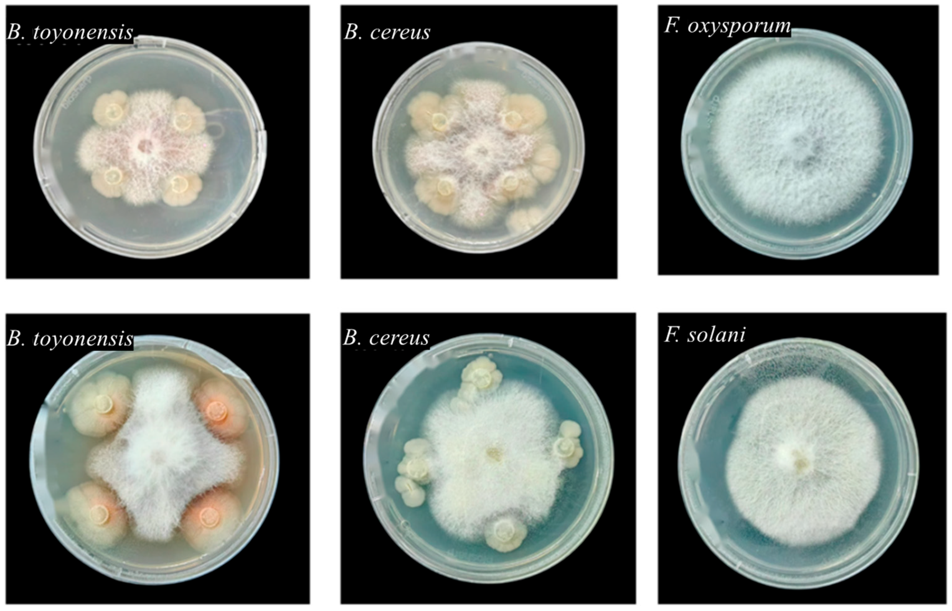
Eleven bacterial OTUs exhibiting significant inhibitory effects on mycelial growth of both F. solani and F. oxysporum were identified from 256 culturable Pg SE strains. These antagonistic strains were taxonomically classified into 8 genera (Bacillus, Erwinia, Pseudomonas, Flavobacterium, Stenotrophomonas, Lactococcus, Klebsiella, and Delftia), 3 phyla (Table 5). At the phylum level, Proteobacteria represented the dominant group, followed by Firmicutes. Among these antagonistic strains, Pseudomonas was the most prevalent and effective genus, followed by the genus Bacillus being the second prominent. Surprisingly, 2 Bacillus strains, including OTU4 (B. cereus) and OTU5 (B. toyonensis), exhibited the highest antagonistic activity among all tested strains, with inhibition rates exceeding 35% against both pathogens (Table 5 and Figure A2).

3.5. Bacterial SEs B. toyonensis and B. cereus Enhance Pg Seed Germination and Plant Growth

Experimental results showed that compared to the control, treatment with B. toyonensis (Bt) and B. cereus (Bc) enhanced the germination rates of Pg seeds by 14.00% and 5.50%, with the differences between Bt treatment and control showing statistical significance (p < 0.05) (Figure 5a). In addition, both the Bt and Bc treatments demonstrated significant (p < 0.05) growth-promoting effects to most of the detected parameters including plant height, root length and fresh weight of above-ground parts (Figure 5b–d).
In conclusion, both B. toyonensis and B. cereus capably promote Pg seed germination and plant growth. B. toyonensis demonstrated superior growth-promoting effects across all experimental parameters, providing crucial experimental evidence for developing specialized biofertilizers from Pg SEs for Pg cultivation.

3.6. Biocontrol Potential of B. toyonensis and B. cereus Against Pg Root Rot Disease

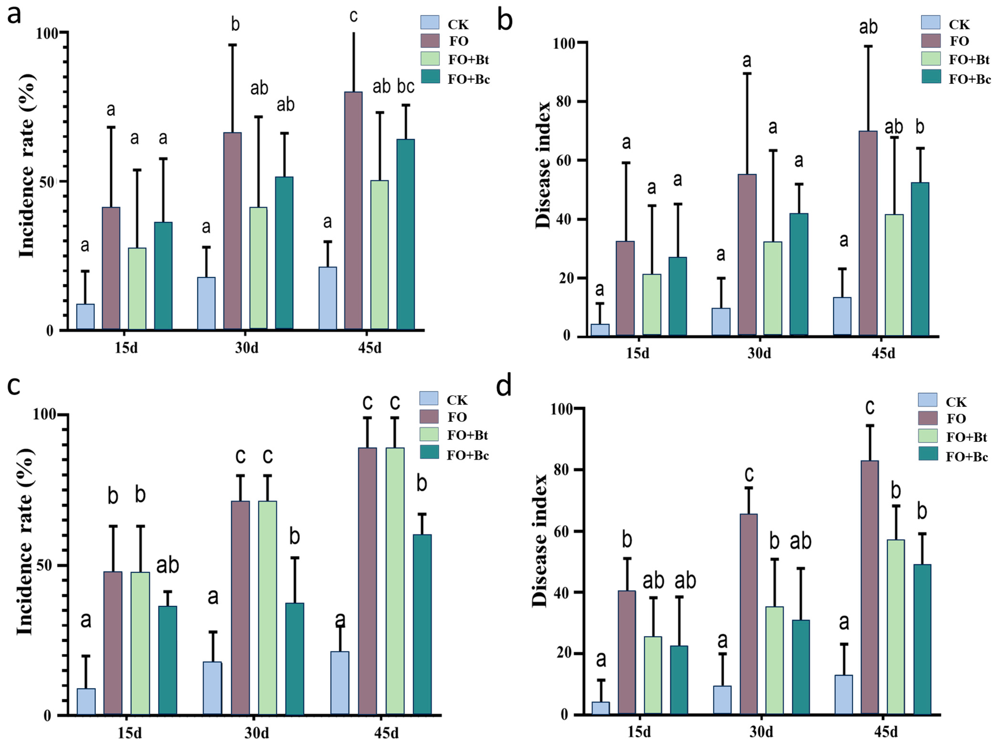
In the protective efficacy assay (pre-inoculation treatment), infection with F. oxysporum (Fo) significantly increased the disease incidence and disease index of Pg seedlings to 80% and 70.00, respectively, at 45 days post-inoculation. Compared with the pathogen-only group (Fo), both Bt + Fo and Bc + Fo treatments exhibited degrees of disease suppression at different time points. Specifically, the disease incidence was reduced by 13.15% and 5% at 15 days, 25% and 15% at 30 days, and 30% and 16.25% at 45 days, respectively. Distinctively, only the Bt + Fo group showed a statistically significant reduction in disease incidence at 45 days (p < 0.05). Regarding the disease index, decreases of 6.11 and 11.25 were observed at 15 days, 23.22 and 13.57 at 30 days, and 28.57 and 17.68 at 45 days, respectively, though these differences were not statistically significant (Figure 6a,b).
In the therapeutic efficacy assay (post-inoculation treatment), the disease incidence and disease index reached 88.75% and 83.04, respectively, at 45 days after pathogen inoculation. Subsequent bacterial treatment revealed that the Fo + Bc group exhibited significant disease control: the disease incidence decreased by 13.15% at 15 days, 33.75% at 30 days, and 28.75% at 45 days, while the disease index was reduced by 17.86, 34.83, and 34.11 at the respective time points. In contrast, the Fo + Bt group only demonstrated significant effects on the disease index, with reductions of 30.18 and 25.72 (p < 0.05) at 30 and 45 days, respectively (Figure 6c,d).
These findings indicate that B. cereus exhibits more comprehensive efficacy in therapeutic applications, whereas B. toyonensis demonstrates greater potential in disease prevention. This study provides critical insights for developing tailored biocontrol strategies against Pg root rot.

4. Discussion

This study demonstrates that P. notoginseng seeds host a rich endophytic microbial community, dominated by the fungal phyla Ascomycota and Basidiomycota and the bacterial phyla Firmicutes and Proteobacteria—a profile consistent with those documented in other plant species [3]. Core microbiota analysis revealed conserved structural patterns across individual seeds, with a highly stable core microbiome constituting over 90% of the sequencing reads, a distribution congruent with prior observations in Plantago lanceolata seeds [34]. And accumulating evidence supports that core microbial taxa maintain compositionally conserved profiles across generational cycles, accounting for more than 90% of total microbial abundance [34,35,36]. These findings indicate that the seed endophytic microbiome possesses strong transgenerational stability, suggesting its potential role as an epigenetic element in host plants [3]. This heritable stability likely stems from a mutually beneficial symbiosis established through prolonged co-evolution between the host and its microbial partners [3]. Moreover, we observed a dose-dependent accumulation of microbial diversity with increasing sample size, implying that larger seed pools serve as more diverse microbial inocula for progeny. This enhanced inoculum diversity may contribute to improved ecological adaptability in offspring through microbiome-mediated phenotypic modulation. Thus, deciphering the mechanisms underlying the heritability of seed endophytes and their co-evolutionary trajectory with hosts is essential for advancing our understanding of plant-microbe symbioses. These insights may ultimately inform strategies for microbiome-driven crop enhancement and sustainable agricultural practices [37].
Core bacterial and fungal taxa exhibit closer associations with the host plant compared to other microbial groups. Notably, Bacillus, Halomonas, and Shewanella were identified as the dominant seed-transmitted bacterial genera, while Aspergillus predominated among seed-associated fungi [38]. A spore-forming bacterial strain (BS3) isolated from maize sprouts demonstrated plant growth-promoting effects and enhanced resistance against Fusarium verticillioides in infested soils [38]. A study on tomato seed endophytes characterized the Bacillus community using 16S rRNA gene PCR-RFLP (restriction fragment length polymorphism) analysis across four commercial cultivars. The B. subtilis strain HYT-12-1 exhibited significant growth-promoting activity in tomato seedlings, highlighting its potential for industrial application as a biofertilizer [39]. Functional studies of endophytic bacteria in Platycodon grandiflorus revealed that Luteibacter sp. JG09 efficiently biotransforms protopanaxadiol-type ginsenosides (Rb1, Rb2, Rc, Rd) into rare ginsenosides F2 and C-K, while converting protopanaxatriol-type ginsenoside Rg1 to Rh1 [40]. The bacterial genus Burkholderia-Caballeronia-Paraburkholderia consistently ranked among the top 5 most abundant taxa (7.82–18.01%) across 5 seed samples. This genus, along with core endophytes Massilia and Dyella, has been implicated in the regulation of continuous cropping obstacles [41]. These bacteria modulate the production of carbamic acid derivatives (N-(2-butyl)-N-octadecyl ethyl ester) and soil metabolites in Gastrodia elata cultivation systems, which may serve as initial triggers for replant disease [41]. Intriguingly, orobanchol levels showed significant correlation with the relative abundance of phosphate-solubilizing Burkholderia-Caballeronia-Paraburkholderia, while 4-deoxyorobanchol was associated with Dyella and Umbelopsis species [42]. Agronomic practices such as maize-peanut rotation significantly enhance crop productivity, with Burkholderia-Caballeronia-Paraburkholderia emerging as both a dominant and indicator genus in the critical 20–40 cm soil layer. This microbial group demonstrates detoxification capabilities against peanut-derived autotoxins and provides protection against soil-borne pathogens [43]. These findings collectively demonstrate that P. notoginseng seeds harbor diverse endophytic bacterial communities containing multiple taxa with beneficial functional traits. Further investigation is warranted to explore the agricultural potential of these probiotic microorganisms in P. notoginseng cultivation systems.
SEs play a crucial yet often overlooked role in the classical “disease triangle” framework (encompassing plant, pathogen, and environmental factors). The exploration of disease-resistant resources within seed endophytes holds significant potential for green prevention strategies and modern agricultural practices. With studies revealing that rice plants of the same cultivar can differentiate into resistant and susceptible phenotypes [44,45,46], and Sphingomonas melonis accumulates in disease-resistant rice seeds and demonstrates transgenerational transmission, conferring resistance through the production of anthranilic acid [46]. In the present study, B. toyonensis and B. cereus isolated from P. notoginseng seeds exhibited measurable biocontrol effects against root rot. These findings align with numerous reports on the biocontrol potential of seed-derived Bacillus spp. For instance, B. velezensis (syn. B. olimyquefaciens) isolated from surface-sterilized Urochloa ramosa seeds provided significant protection against Fusarium infection in seedlings [47]. Similarly, 6 antagonistic bacterial strains (B. subtilis, B. velezensis, Leuconostoc mesenteroides, Lactococcus lactis, and B. amyloliquefaciens) isolated from freshly collected Solanum nigrum seeds demonstrated effective control of bacterial wilt (Ralstonia solanacearum) in pepper seedlings, with B. subtilis showing the highest efficacy. This suggests that seeds from disease-resistant cultivars represent a rich reservoir of antagonistic microorganisms [48]. Furthermore, Trichoderma harzianum spore treatment significantly enhanced germination rates and seedling vigor indices in soybean (Glycine max) and castor (Ricinus communis), while markedly reducing disease incidence. Quantitative assessments revealed reductions in Fusarium root rot of soybean (23.3%) and castor wilt (20%) [49]. These results highlight the potential of such biological formulations as robust, sustainable alternatives to conventional methods, offering scalable solutions for plant disease management while addressing the challenges associated with chemical pesticide use. However, the utilization of beneficial plant endophytes, such as strains of Bacillus spp., as microbial inoculants presents notable ecological benefits but also necessitates careful risk assessment. While these microbes can enhance plant growth and confer resistance against pathogens, their introduction into natural or agricultural ecosystems may lead to unintended consequences [50]. Potential risks include disruption of native microbial communities, horizontal gene transfer—particularly involving antibiotic resistance or metabolic genes—and possible adverse effects on non-target organisms, including soil fauna and beneficial microbiota [51]. Furthermore, the persistence, colonization efficiency, and potential for off-target movement of introduced endophytes require thorough environmental monitoring [52]. It is imperative to employ comprehensive toxicological evaluations and field-based ecological risk assessments to ensure that the application of such bioagents is both effective and environmentally sustainable, prioritizing strains with well-documented biosafety profiles and minimal ecological impact.
This study has several limitations that should be acknowledged. Firstly, the bacterial sampling was conducted exclusively from Pg seeds within a localized geographical region, which may limit the generalizability of the findings to other populations or ecosystems. Secondly, the functional experiments were performed under controlled in vitro conditions using a single growth environment, which does not fully represent the complex interactions occurring in natural soil–plant systems. Furthermore, the study primarily focused on descriptive characterizations of microbial diversity and antagonistic effects without in-depth mechanistic investigations. The molecular basis underlying the observed plant growth-promoting traits and antifungal activities—such as the identification of specific bioactive compounds, gene regulatory mechanisms, or signal transduction pathways—remains unexplored. To address these limitations, future studies could include multi-region sampling to enhance the representativeness of the endophytic community. Incorporating greenhouse or field experiments would better reflect real-world conditions and strengthen ecological relevance. Moreover, applying transcriptomic, metabolomic, or genetic approaches could help elucidate the functional mechanisms behind the beneficial interactions, such as knock-out mutant assays to validate the key genes involved in antifungal compound production or root colonization. Finally, evaluating the ecological safety and long-term persistence of introduced strains would be critical for potential application in sustainable agriculture.
5. Conclusions

The work revealed that P. notoginseng seeds harbor a diverse endophytic microbiome with a diversity of function potentials, and a bigger seed population size is substantial for progeny plants benefiting from diversity of seed endophytes. However, phylogenetically conserved core microbiota in Pg seeds were identified, suggesting stable host–microbe associations through evolutionary time. Function validation experiments further revealed beneficial seed bacteria with disease-suppressive potential, highlighting the seed microbiome as a valuable reservoir of plant-beneficial microbes for sustainable agriculture. This research substantially enhances the current knowledge of seed endophytes and establishes a valuable paradigm for future investigations and applications of endophytic communities in diverse crop species.

Author Contributions

Conceptualization, M.-Z.Y. and X.-H.H.; methodology, Y.W.; validation, S.-C.G., Y.-N.Z. and Y.-B.Z.; formal analysis, H.-Y.H.; investigation, Y.W.; resources, M.-Z.Y. and Y.-Y.Z.; data curation, Y.W. and H.-Y.H.; writing—original draft preparation, H.-Y.H.; writing—review and editing, M.-Z.Y.; visualization, X.-X.P.; supervision, X.-H.H.; project administration, M.-Z.Y.; funding acquisition, M.-Z.Y. All authors have read and agreed to the published version of the manuscript.

Funding

This work was financially supported by the National Natural Science Foundation of China (NSFC: 32360255 and 32471746); Yunnan provincial key S&T special project (202102AE090042).

Data Availability Statement

All data and materials are available in the manuscript; raw data of high throughput sequencing for the seeds of Panax notoginseng were deposited at NCBI under following accession numbers: PRJNA891501 (seed bacterial endophytes) (https://dataview.ncbi.nlm.nih.gov/object/PRJNA891501?reviewer=k7qvq2vann86tn45kn1j9e1dc0, accessed on 12 October 2024); PRJNA892017 (seed fungal endophytes) (https://dataview.ncbi.nlm.nih.gov/object/PRJNA892017?reviewer=klgbfj93d7vfeg3midn93icu95, accessed on 12 October 2024).

Conflicts of Interest

The authors declare no conflicts of interest.

Abbreviations

The following abbreviations are used in this manuscript:
SEsSeed endophytes
PgPanax notoginseng (Burk.) F.H. Chen
Pg-coSBEPg core seed bacterial endophyte
Pg-coSFEPg core seed fungal endophyte
RARelative abundance
OUTOperational Taxonomic Unit
ASVAmplicon Sequence Variant

Appendix A

Figure A1. Rarefaction curve of sequencing data. (a,d): Observed OTUs; (b,e): Goods coverage; (c,f): Simpson index.
Horticulturae 11 01162 g0a1
Figure A2. The in vitro interaction of B. toyonensis and B. cereus vs. F. oxysporum and F. solani in confrontation assays in agar plates.
Horticulturae 11 01162 g0a2
Table A1. Classification unit of high-throughput sequencing results of Pg SEs.
KingdomPhylumClassOrderFamilyGenusSpecies
Bacterium3150108180370240
Fungi41639659498
Table A2. Alpha diversity indices of endophytic fungi in individual Pg seeds.
SampleObserved SpeciesShannonSimpsonChao1Goods CoveragePieloue
wen11101.331.520.52103.301.000.23
wen1290.331.530.4691.971.000.24
wen1384.672.140.6986.331.000.33
wen1481.331.690.5982.291.000.27
wen15100.671.530.49102.211.000.23
Table A3. Alpha diversity indices of endophytic bacteria in individual Pg seeds.
SampleObserved SpeciesShannonSimpsonChao1Goods CoveragePieloue
wen11167.333.640.83174.791.000.50
wen12176.003.640.81184.281.000.49
wen13181.333.970.87189.571.000.53
wen14173.673.840.85178.441.000.52
wen15163.333.290.77175.781.000.45
Table A4. Isolated culturable endophytic bacteria from Pg seeds.
OTU IDSpeciesOTU IDSpecies
OTU1Erwinia gerundensisOTU25Bacillus sp.
OTU2Enterobacter sp.OTU26Enterobacter sp.
OTU3Pseudomonas marginalisOTU27Pantoea vagans
OTU4Bacillus cereusOTU28Bacillus thuringiensis
OTU5Bacillus toyonensisOTU29Pseudomonas gessardii
OTU6Pseudomonas rhodesiaeOTU30Pantoea sp.
OTU7Erwinia sp.OTU31Lelliottia sp.
OTU8Pseudomonas putidaOTU32Lelliottia jeotgali
OTU9Bacillus safensisOTU33Pseudomonas sp.
OTU10Bacillus sp.OTU34Lelliottia aquatilis
OTU11Pseudomonas sp.OTU35Pseudomonas veronii
OTU12Stenotrophomonas sp.OTU36Pantoea endophytica
OTU13Pseudomonas fluorescensOTU37Stenotrophomonas rhizophila
OTU14Microbacterium sp.OTU38Erwinia sp.
OTU15Curtobacterium sp.OTU39Pseudoclavibacter sp.
OTU16Pseudomonas simiaeOTU40Pseudomonas sp.
OTU17Pantoea ananatisOTU41Enterobacter ludwigii
OTU18Kocuria sp.OTU42Enterobacter cloacae
OTU19Bacillus pumilusOTU43Lactococcus lactis
OTU20Pantoea sp.OTU44Enterobacter quasihormaechei
OTU21Kluyvera sp.OTU45Klebsiella oxytoca
OTU22Kluyvera cryocrescensOTU46Sphingobium sp.
OTU23Pseudomonas sp.OTU47Delftia acidovorans
OTU24Flavobacterium sp.

References

  1. Stone, J.K.; Bacon, C.W.; White, J.F. An Overview of Endophytic Microbes: Endophytism Defined. In Microbial Endophytes; Bacon, C.W., White, J.F., Eds.; CRC Press: Boca Raton, FL, USA, 2000; pp. 3–30. [Google Scholar] [CrossRef]
  2. Jørgensen, H. Plant Endophytes. In Encyclopedia of Life Sciences; Wiley Online Library: Hoboken, NJ, USA, 2020; Available online: https://onlinelibrary.wiley.com/doi/10.1002/9780470015902.a0028893 (accessed on 15 September 2025).
  3. Hu, H.; Geng, S.; Zhu, Y.; He, X.; Pan, X.; Yang, M. Seed-Borne Endophytes and Their Host Effects. Microorganisms 2025, 13, 842. [Google Scholar] [CrossRef] [PubMed]
  4. Luo, L.; Wang, L.; Deng, L.; Mei, X.; Liu, Y.; Huang, H.; Du, F.; Zhu, S.; Yang, M. Enrichment of Burkholderia in the Rhizosphere by Autotoxic Ginsenosides to Alleviate Negative Plant-Soil Feedback. Microbiol. Spectr. 2021, 9, e0140021. [Google Scholar] [CrossRef] [PubMed]
  5. Babalola, O.O.; Adedayo, A.A. Endosphere Microbial Communities and Plant Nutrient Acquisition toward Sustainable Agriculture. Emerg. Top. Life Sci. 2023, 7, 207–217. [Google Scholar] [CrossRef]
  6. Verma, S.K.; Kumar, K.; Pal, G.; Verma, A. Chapter 3—The Roles of Endophytes in Modulating Crop Plant Development. In Microbiome Stimulants for Crops; White, J., Kumar, A., Droby, S., Eds.; Woodhead Publishing: Cambridge, UK, 2021; pp. 33–39. ISBN 978-0-12-822122-8. [Google Scholar]
  7. Pal, G.; Kumar, K.; Verma, A.; Verma, S.K. Seed Inhabiting Bacterial Endophytes of Maize Promote Seedling Establishment and Provide Protection against Fungal Disease. Microbiol. Res. 2021, 255, 126926. [Google Scholar] [CrossRef]
  8. Kim, J.; Ahn, S.-H.; Yang, J.S.; Choi, S.; Jung, H.W.; Jeon, J. Plant Protective and Growth Promoting Effects of Seed Endophytes in Soybean Plants. Plant Pathol. J. 2023, 39, 513–521. [Google Scholar] [CrossRef]
  9. Cordovez, V.; Dini-Andreote, F.; Carrión, V.J.; Raaijmakers, J.M. Ecology and Evolution of Plant Microbiomes. Annu. Rev. Microbiol. 2019, 73, 69–88. [Google Scholar] [CrossRef]
  10. Ghosh, S.; Bhagwat, T.; Webster, T.J. Endophytic Microbiomes and Their Plant Growth-Promoting Attributes for Plant Health. In Current Trends in Microbial Biotechnology for Sustainable Agriculture; Springer: Berlin/Heidelberg, Germany, 2020; Available online: https://link.springer.com/chapter/10.1007/978-981-15-6949-4_11 (accessed on 13 June 2025).
  11. People’s Bank of China. Pharmacopoeia of the People’s Republic of China. People’s Med. Publ. House 2005, 43, 67–85. [Google Scholar] [CrossRef]
  12. Feng, Y.; Shuai, X.; Chen, J.; Zhang, Q.; Jia, L.; Sun, L.; Su, Y.; Su, Y.; Dong, G.; Liu, T.; et al. Unveiling the Genomic Features and Biocontrol Potential of Trichoderma hamatum against Root Rot Pathogens. J. Fungi 2025, 11, 126. [Google Scholar] [CrossRef]
  13. Zheng, K.Y.; Su, X.; Zheng, X.; Zhang, L.; Chen, Y.; Wu, K.; Li, T.; Zhang, Z.-K.; Zhao, Z. First Report of Stenotrophomonas maltophilia Causing Root Soft Rot of Sanqi (Panax notoginseng) in China. Plant Dis. 2022, 106, 755. [Google Scholar] [CrossRef]
  14. Wang, T.; Guo, R.; Zhou, G.; Zhou, X.; Kou, Z.; Sui, F.; Li, C.; Tang, L.; Wang, Z. Traditional Uses, Botany, Phytochemistry, Pharmacology and Toxicology of Panax notoginseng (Burk.) F.H. Chen: A Review. J. Ethnopharmacol. 2016, 188, 234–258. [Google Scholar] [CrossRef]
  15. Li, Y.; Liu, Y.; Zhang, Z.; Li, J.; Zhu, S.; Yang, M.; Luo, L. Application of plant survival-promoting and pathogen-suppressing Trichoderma species for crop biofertilization and biocontrol of root rot in Panax notoginseng. J. Plant Pathol. 2022, 104, 1361–1369. [Google Scholar] [CrossRef]
  16. Zheng, Y.-K.; Miao, C.-P.; Chen, H.-H.; Huang, F.-F.; Xia, Y.-M.; Chen, Y.-W.; Zhao, L.-X. Endophytic Fungi Harbored in Panax notoginseng: Diversity and Potential as Biological Control Agents against Host Plant Pathogens of Root-Rot Disease. J. Ginseng Res. 2017, 41, 353–360. [Google Scholar] [CrossRef]
  17. Delseny, M.; Han, B.; Hsing, Y.I. High Throughput DNA Sequencing: The New Sequencing Revolution. Plant Sci. 2010, 179, 407–422. [Google Scholar] [CrossRef]
  18. Mason, J. High-Throughput Sequencing Technology: A New Chapter in Epigenetics and Disease Research. Cancer Genet. Epigenetics 2024, 12, 47–54. [Google Scholar] [CrossRef]
  19. Miao, Z.Q.; Li, S.D.; Liu, X.Z.; Chen, Y.J.; Zhang, K.Q. The Causal Microorganisms of Panax notoginseng Root Rot Disease. Sci. Agric. Sin. 2006, 39, 1371–1378. [Google Scholar]
  20. Sahu, P.K.; Tilgam, J.; Mishra, S.; Hamid, S.; Gupta, A.K.J.; Verma, S.K.; Kharwar, R.N. Surface Sterilization for Isolation of Endophytes: Ensuring What (Not) to Grow. J. Basic Microbiol. 2022, 62, 647–668. [Google Scholar] [CrossRef] [PubMed]
  21. Wang, L.; Du, Y.; Rahman, M.; Tang, B.; Fan, L.-J.; Kilaru, A. Establishment of an efficient in vitro propagation system for Iris sanguinea. Sci. Rep. 2018, 8, 17100. [Google Scholar] [CrossRef]
  22. Darkazanli, M.; Kiseleva, I. New Surface Sterilization Protocols for Isolation of Endophytic Bacteria from Plants (Black Turtle Beans, Peas, and Barley). AIP Conf. Proc. 2021, 2388, 5. [Google Scholar] [CrossRef]
  23. Wen, Y.; Yang, W.-X.; Li, Y.-Q.; Lu, C.-X.; Hu, H.-Y.; Guo, L.-R.; Huang, S.-Y.; Pan, X.-X.; Zhu, S.-S.; Yang, M.-Z. Reconsidering the ecological effect of seed endophytes in building plant microbial environments: Lessons from a Chinese medicinal plant Panax notoginseng. Environ. Microbiome 2025, 20, 87. [Google Scholar] [CrossRef] [PubMed]
  24. Walters, W.; Hyde, E.R.; Berg-Lyons, D.; Ackermann, G.; Humphrey, G.; Parada, A.; Gilbert, J.A.; Jansson, J.K.; Caporaso, J.G.; Fuhrman, J.A.; et al. Improved Bacterial 16S rRNA Gene (V4 and V4-5) and Fungal Internal Transcribed Spacer Marker Gene Primers for Microbial Community Surveys. mSystems 2016, 1, e00009-15. [Google Scholar] [CrossRef]
  25. Logue, J.B.; Stedmon, C.A.; Kellerman, A.M.; Nielsen, N.J.; Andersson, A.F.; Laudon, H.; Lindström, E.S.; Kritzberg, E.S. Experimental Insights into the Importance of Aquatic Bacterial Community Composition to the Degradation of Dissolved Organic Matter. ISME J. 2016, 10, 533–545. [Google Scholar] [CrossRef]
  26. Ruiz, D.; Agaras, B.; de Werra, P.; Wall, L.G.; Valverde, C. Characterization and Screening of Plant Probiotic Traits of Bacteria Isolated from Rice Seeds Cultivated in Argentina. J. Microbiol. 2011, 49, 902–912. [Google Scholar] [CrossRef]
  27. Frank, J.A.; Reich, C.I.; Sharma, S.; Weisbaum, J.S.; Wilson, B.A.; Olsen, G.J. Critical Evaluation of Two Primers Commonly Used for Amplification of Bacterial 16S rRNA Genes. Appl. Environ. Microbiol. 2008, 74, 2461–2470. [Google Scholar] [CrossRef] [PubMed]
  28. Xie, T.; Shen, S.; Hu, R.; Li, W.; Wang, J. Screening, Identification, and Growth Promotion of Antagonistic Endophytes Associated with Chenopodium quinoa Against Quinoa Pathogens. Phytopathology 2023, 113, 1839–1852. [Google Scholar] [CrossRef] [PubMed]
  29. Fang, Z. Plant Disease Research Method; Agricultural Press: Beijing, China, 2001; Available online: https://www.hanspub.org/reference/ReferencePapers?ReferenceID=10740 (accessed on 9 June 2025). (In Chinese)
  30. Zong, Z.; Kang, Z. Principles of Plant Pathology; China Agriculture Press: Beijing, China, 2002; ISBN 978-7-109-07535-1. (In Chinese) [Google Scholar]
  31. Bars-Cortina, D.; Moratalla-Navarro, F.; García-Serrano, A.; Mach, N.; Riobó-Mayo, L.; Vea-Barbany, J.; Rius-Sansalvador, B.; Murcia, S.; Obón-Santacana, M.; Moreno, V. Improving Species Level-Taxonomic Assignment from 16S rRNA Sequencing Technologies. Curr. Protocol. 2023, 3, e930. [Google Scholar] [CrossRef] [PubMed]
  32. Nguyen, N.H.; Song, Z.; Bates, S.T.; Branco, S.; Tedersoo, L.; Menke, J.; Schilling, J.S.; Kennedy, P.G. FUNGuild: An Open Annotation Tool for Parsing Fungal Community Datasets by Ecological Guild. Fungal Ecol. 2016, 20, 241–248. [Google Scholar] [CrossRef]
  33. Gao, L.; Huang, Y.; Ma, J.; Jiang, H.; Li, W.; Li, L. Diversity and Roles of Endophytic Fungi in Two Halophytes. Microbiol. China 2023, 50, 3357–3371. (In Chinese) [Google Scholar] [CrossRef]
  34. de Souza, Y.P.A.; Schloter, M.; Weisser, W.; Huang, Y.; Schulz, S. The Seeds of Plantago lanceolata Comprise a Stable Core Microbiome along a Plant Richness Gradient. Environ. Microbiome 2024, 19, 11. [Google Scholar] [CrossRef]
  35. Sulesky-Grieb, A.; Simonin, M.; Bintarti, A.F.; Marolleau, B.; Barret, M.; Shade, A. Stable, Multigenerational Transmission of the Bean Seed Microbiome despite Abiotic Stress. mSystems 2024, 9, e0095124. [Google Scholar] [CrossRef]
  36. Zhai, K.; Zhang, Y.; Zhao, C.; Wang, Q.; Gao, X. Conservation and Dynamics of Maize Seed Endophytic Bacteria across Progeny Transmission. Microorganisms 2024, 12, 2399. [Google Scholar] [CrossRef]
  37. Crop Health. The Seed Microbiota from an Application Perspective: An Underexplored Frontier in Plant–Microbe Interactions. Available online: https://link.springer.com/article/10.1007/s44297-025-00051-6 (accessed on 4 August 2025).
  38. Wang, W.; Li, Y.; Qin, W.; Sun, C.; Tan, H.; Cao, L. Using Illumina-Based Sequence Analysis to Guide Probiotic Candidate Selection and Isolation. Probiotics Antimicro. Prot. 2018, 10, 478–484. [Google Scholar] [CrossRef]
  39. Xu, M.; Sheng, J.; Chen, L.; Men, Y.; Gan, L.; Guo, S.; Shen, L. Bacterial Community Compositions of Tomato (Lycopersicum Esculentum Mill.) Seeds and Plant Growth Promoting Activity of ACC Deaminase Producing Bacillus Subtilis (HYT-12-1) on Tomato Seedlings. World J. Microbiol. Biotechnol. 2014, 30, 835–845. [Google Scholar] [CrossRef]
  40. Cui, L.; Wu, S.-Q.; Zhao, C.-A.; Yin, C.-R. Microbial Conversion of Major Ginsenosides in Ginseng Total Saponins by Platycodon Grandiflorum Endophytes. J. Ginseng Res. 2016, 40, 366–374. [Google Scholar] [CrossRef]
  41. Duan, M.; Yang, C.; Bao, L.; Han, D.; He, H.; Zhang, Y.; Dong, L.; Yang, S. Unraveling the Key Mechanisms of Gastrodia Elata Continuous Cropping Obstacles: Soil Bacteria Massilia, Burkholderia-Caballeronia-Paraburkholderia, and Dyella along with Soil Metabolites 4-Hydroxy-Benzenemethanol and N-(2-Butyl)-N-Octadecyl-, Ethyl Ester as Crucial Indicators. Front. Microbiol. 2024, 15, 1478330. [Google Scholar] [CrossRef]
  42. Kim, B.; Westerhuis, J.A.; Smilde, A.K.; Floková, K.; Suleiman, A.K.A.; Kuramae, E.E.; Bouwmeester, H.J.; Zancarini, A. Effect of Strigolactones on Recruitment of the Rice Root-Associated Microbiome. FEMS Microbiol. Ecol. 2022, 98, fiac010. [Google Scholar] [CrossRef]
  43. Zou, X.; Liu, Y.; Huang, M.; Li, F.; Si, T.; Wang, Y.; Yu, X.; Zhang, X.; Wang, H.; Shi, P. Rotational Strip Intercropping of Maize and Peanut Enhances Productivity by Improving Crop Photosynthetic Production and Optimizing Soil Nutrients and Bacterial Communities. Field Crops Res. 2023, 291, 108770. [Google Scholar] [CrossRef]
  44. Liu, Z.; Zhu, Y.; Shi, H.; Qiu, J.; Ding, X.; Kou, Y. Recent Progress in Rice Broad-Spectrum Disease Resistance. Int. J. Mol. Sci. 2021, 22, 11658. [Google Scholar] [CrossRef] [PubMed]
  45. Yang, X.; Yan, S.; Li, Y.; Li, G.; Sun, S.; Li, J.; Cui, Z.; Huo, J.; Sun, Y.; Wang, X.; et al. Comparison of Transcriptome between Tolerant and Susceptible Rice Cultivar Reveals Positive and Negative Regulators of Response to Rhizoctonia Solani in Rice. Int. J. Mol. Sci. 2023, 24, 14310. [Google Scholar] [CrossRef]
  46. Matsumoto, H.; Fan, X.; Wang, Y.; Kusstatscher, P.; Duan, J.; Wu, S.; Chen, S.; Qiao, K.; Wang, Y.; Ma, B.; et al. Bacterial Seed Endophyte Shapes Disease Resistance in Rice. Nat. Plants 2021, 7, 60–72. [Google Scholar] [CrossRef] [PubMed]
  47. Verma, S.K.; White, J.F. Indigenous Endophytic Seed Bacteria Promote Seedling Development and Defend against Fungal Disease in Browntop Millet (Urochloa ramosa L.). J. Appl. Microbiol. 2018, 124, 764–778. [Google Scholar] [CrossRef]
  48. Dowarah, B.; Agarwal, H.; Krishnatreya, D.B.; Sharma, P.L.; Kalita, N.; Agarwala, N. Evaluation of Seed Associated Endophytic Bacteria from Tolerant Chilli Cv. Firingi Jolokia for Their Biocontrol Potential against Bacterial Wilt Disease. Microbiol. Res. 2021, 248, 126751. [Google Scholar] [CrossRef]
  49. Malavathu, K.; Godbole, V.; Singh, A.; Chandrika, K.S.V.P.; Prasad, R.D. Porous Biopolymer Matrices: Advanced Seed Delivery Platforms for Beneficial Microbes to Combat Soilborne Diseases. ACS Omega 2025, 10, 20723–20731. [Google Scholar] [CrossRef] [PubMed]
  50. Kumari, M.; Swarupa, P.; Kesari, K.K.; Kumar, A. Microbial Inoculants as Plant Biostimulants: A Review on Risk Status. Life 2022, 13, 12. [Google Scholar] [CrossRef] [PubMed]
  51. Etesami, H. The Dual Nature of Plant Growth-Promoting Bacteria: Benefits, Risks, and Pathways to Sustainable Deployment. Curr. Res. Microb. Sci. 2025, 9, 100421. [Google Scholar] [CrossRef] [PubMed]
  52. Shahwar, D.; Mushtaq, Z.; Mushtaq, H.; Alqarawi, A.A.; Park, Y.; Alshahrani, T.S.; Faizan, S. Role of Microbial Inoculants as Bio Fertilizers for Improving Crop Productivity: A Review. Heliyon 2023, 9, e16134. [Google Scholar] [CrossRef]
Figure 1. Schematic diagram of the experimental operation process (including sampling, surface sterilization, high-throughput sequencing, isolation and identification of culturable endophytes, screening of antagonistic strains, and functional validation of target strains; detailed methods correspond to those described in the main text). In this study, 5 individual seed samples were designated as wen11, wen12, wen13, wen14, and wen15. For each sample, 3 biological replicates were prepared, labeled with the suffix -1, -2, and -3 (e.g., wen11-1, wen11-2, and wen11-3 for sample wen11).
Horticulturae 11 01162 g001
Figure 2. Diversity of the endophytic microbiota associated with Pg seeds (each treatment included 3 biological replicates). (af): Alpha and Beta-diversity indices of bacterial (ac) and fungal (df) communities across individual Pg seeds. (gl): dynamics of microbial diversity metrics with increasing sample size in Pg seeds, including Observed OUTs (gj) and Shannon index (k,l).
Horticulturae 11 01162 g002
Figure 3. Endophytic microbiome composition and core microbiota across different Pg seeds (each treatment included 3 biological replicates). (a,b): Phylum-level composition of endophytic communities across individual Pg sees. (c,d): The bar charts show the total number of endophytic bacteria (c) and endophytic fungi (d) genera detected in different samples. (e,f): show the top 30 genera of endophytic bacterium (e) and endophytic fungi (f) in different samples. (g,h): The petal plots showing the specific and common ASV counts of endophytic bacteria (g) and fungi (h) in different sample replicates. (i,j): The proportion and relative abundance of the core bacterial (i) and fungal (j) ASVs in different sample replicates.
Horticulturae 11 01162 g003aHorticulturae 11 01162 g003b
Figure 4. Functional prediction of microbiota in Pg seeds. (a,b): phenotype prediction (Bugbase) of the bacterial endophytes in samples of individual plants (a) and seeds (b); (c,d): function prediction for the fungal endophytes in samples of individual plants (c) and seeds (d), by using FUNGuild program [32].
Horticulturae 11 01162 g004
Figure 5. Plant growth-promoting effects of B. toyonensis and B. cereus on Pg: from seed germination to seedling development (each treatment included 3 biological replicates, one replicate consisting of 36 seedlings). (a): effects of B. toyonensis and B. cereus on Pg seeds germination rate; (be): effects of B. toyonensis and B. cereus on Pg plant height (b), root length (c), shoot fresh weight (d), and fresh root weight (e). * and ** in the figure indicate significant difference in Tukey’s HSD test (p < 0.05).
Horticulturae 11 01162 g005
Figure 6. Effects of different treatments on root rot of Pg (each treatment included 3 biological replicates, one replicate consisting of 36 seedlings). (a,b): pre-pathogen inoculation treatment with endophytic bacterial inoculants; (c,d): post-pathogen inoculation treatment with endophytic bacterial inoculants. Legends explanation: CK represents sterile water treatment (control group); Fo indicates treatment with a 1 g/L F. oxysporum suspension alone (pathogen-inoculated group); “Bt + Fo”, “Bc + Fo”, “Fo + Bt”, and “Fo + Bc” denote sequential treatments with 2 respective microbial suspensions. Bt refers to B. toyonensis and Bc refers to B. cereus. For detailed information, please refer to Table 1 in Section 2.6 of the Methods. Different lowercase letters in the figure indicate significant difference in Tukey’s HSD test (p < 0.05).
Horticulturae 11 01162 g006
Table 1. Treatments for validating the effects of microbial agents on Pg disease.
NoteGroupsTreatment
Negative controlCKSterile water treatment (control)
Positive controlFo1 g/L F. oxysporum suspension treatment (pathogen challenge)
Preventive effectBt + Fo1 g/L B. toyonensis suspension treatment → 1 g/L F. oxysporum suspension treatment
Bc + Fo1 g/L B. cereus suspension treatment → 1 g/L F. oxysporum suspension treatment
Therapeutic effectFo + Bt1 g/L F. oxysporum suspension treatment → 1 g/L B. toyonensis suspension treatment
Fo + Bc1 g/L F. oxysporum suspension treatment → 1 g/L B. cereus suspension treatment
Table 2. Core endophytic bacterial ASVs in Pg seeds.
PhylumGenusASV IDRA (%)
ProteobacteriaVibrioPg-coSBE1, Pg-coSBE218.07
PantoeaPg-coSBE314.56
PseudomonasPg-coSBE4, Pg-coSBE511.62
Burkholderia-Caballeronia-ParaburkholderiaPg-coSBE6, Pg-coSBE79.72
SphingomonasPg-coSBE86.63
HalomonasPg-coSBE9, Pg-coSBE10, Pg-coSBE116.42
LuteibacterPg-coSBE126.36
Methylobacterium-MethylorubrumPg-coSBE135.22
AcinetobacterPg-coSBE142.57
ParacoccusPg-coSBE150.74
SalinivibrioPg-coSBE160.49
ShewanellaPg-coSBE170.26
AfipiaPg-coSBE180.26
ComamonasPg-coSBE190.12
BradyrhizobiumPg-coSBE200.10
PseudoalteromonasPg-coSBE210.09
UndibacteriumPg-coSBE220.07
FirmicutesStreptococcusPg-coSBE23, Pg-coSBE24,
Pg-coSBE25, Pg-coSBE26
3.78
EnterococcusPg-coSBE270.24
AeribacillusPg-coSBE280.06
Note: An Amplicon Sequence Variant (ASV) is a precisely defined DNA sequence obtained through high-resolution denoising of marker-gene (e.g., 16S rRNA) amplicon data, representing a unique biological taxon without the clustering artifacts associated with OTUs. The ASV identifier (e.g., Pg-coSBE1, indicating Pg core seed bacterial endophyte 1) is assigned sequentially according to the mean relative abundance of the corresponding bacterial genus in the sample. The same nomenclature applies to the following Table 3.
Table 3. Core endophytic fungal ASVs in Pg seeds.
PhylumGenusASV IDRA (%)
AscomycotaAscomycota_unclassifiedPg-coSFE131.71
DavidiellaPg-coSFE213.66
BotryotiniaPg-coSFE42.17
ThermomycesPg-coSFE30.07
HaematonectriaPg-coSFE60.06
ChaetomiumPg-coSFE70.03
MyrotheciumPg-coSFE50.03
Fungi_unclassifiedFungi_unclassifiedPg-coSFE8, Pg-coSFE946.42
Table 4. Isolation rate of cultured endophytic bacteria from Pg seeds.
PhylumGenusStrain NumberIsolation Rates (%)
ProteobacteriaPseudomonasOTU3, OTU11, OTU13, OTU6, OTU16, OTU40, OTU8, OTU33, OTU23, OTU29, OTU3530.96
EnterobacterOTU2, OTU26, OTU42, OTU41, OTU442.67
KluyveraOTU21, OTU225.62
StenotrophomonasOTU12, OTU378.26
ErwiniaOTU1, OTU7, OTU384.43
PantoeaOTU17, OTU20, OTU27, OTU30, OTU362.43
LelliottiaOTU31, OTU32, OTU340.81
DelftiaOTU472.70
SphingobiumOTU461.51
KlebsiellaOTU450.07
FirmicutesBacillusOTU4, OTU5, OTU9, OTU25, OTU10, OTU28, OTU1927.29
LactococcusOTU4311.58
BacteroidotaFlavobacteriumOTU240.11
ActinobacteriotaCurtobacteriumOTU150.91
PseudoclavibacterOTU390.41
KocuriaOTU180.11
MicrobacteriumOTU140.11
Note: OUT, Operational Taxonomic Unit, is a cluster of similar DNA sequences, typically grouped based on a fixed similarity threshold, used to classify and quantify microbial diversity in phylogenetic studies without requiring exact species identification. These abbreviations are used in all following figures and tables.
Table 5. Inhibition effects of the Pg seed endophytic bacteria strains on 2 pathogenic fungi.
NumberInhibitory Effect
F. oxysporum (%)F. solani (%)
OTU439.13 ± 2.1538.66 ± 1.98
OTU538.04 ± 2.0235.10 ± 1.85
OTU726.63 ± 1.5634.57 ± 1.72
OTU1029.77 ± 1.6817.64 ± 1.12
OTU1119.19 ± 1.2510.82 ± 0.85
OTU2425.54 ± 1.4814.66 ± 1.02
OTU2828.26 ± 1.6240.42 ± 2.05
OTU3716.16 ± 1.109.55 ± 0.72
OTU4317.41 ± 1.1526.14 ± 1.52
OTU4520.78 ± 1.3221.56 ± 1.35
OTU4714.67 ± 1.0029.25 ± 1.68
Note: Data are shown as mean ± SD (standard deviation).
Disclaimer/Publisher’s Note: The statements, opinions and data contained in all publications are solely those of the individual author(s) and contributor(s) and not of MDPI and/or the editor(s). MDPI and/or the editor(s) disclaim responsibility for any injury to people or property resulting from any ideas, methods, instructions or products referred to in the content.