Article

Mechanisms of Morphological Development and Physiological Responses Regulated by Light Spectrum in Changchuan No. 3 Pepper Seedlings

1
Putian Institute of Agricultural Sciences, Putian 351100, China
2
College of Agriculture, Fujian Agriculture and Forestry University, Fuzhou 350002, China
3
College of Resources and Environment, Fujian Agriculture and Forestry University, Fuzhou 350001, China
4
College of Life Science, Luoyang Normal University, Luoyang 471934, China
5
College of Horticulture, Fujian Agriculture and Forestry University, Fuzhou 350002, China
*
Authors to whom correspondence should be addressed.
Horticulturae 2025, 11(10), 1161; https://doi.org/10.3390/horticulturae11101161
Submission received: 26 August 2025 / Revised: 19 September 2025 / Accepted: 25 September 2025 / Published: 29 September 2025
(This article belongs to the Special Issue Genomics and Genetic Diversity in Vegetable Crops)

Abstract

This study aimed to evaluate the effects of specific LED light spectra on the growth and physiology of Changchuan No. 3 Capsicum annuum L. seedlings. The experimental design involved exposing pepper seedlings to six different spectral light combinations for 7, 14, and 21 days, with the treatments consisting of 2R1B1Y (red/blue/yellow = 2:1:1), 2R1B1FR (red/blue/far-red = 2:1:1), 2R1B1P (red/blue/purple = 2:1:1), 4R2B1G (red/blue/green = 4:2:1), 2R1B1G (red/blue/green = 2:1:1), and 2R1B (red/blue = 2:1). The results demonstrated distinct spectral regulation of seedling development: compared to the white light (CK), the 2R1B1FR (far-red light supplementation) treatment progressively stimulated stem elongation, increasing plant height and stem diameter by 81.6% and 25.9%, respectively, at day 21, but resulted in a more slender stem architecture. The 2R1B1G (balanced green light) treatment consistently promoted balanced growth, culminating in the highest seedling vigor index at the final stage. The 2R1B1P (purple light supplementation) treatment exhibited a strong promotive effect on root development, which became most pronounced at day 21 (126% increase in root dry weight), while concurrently enhancing soluble sugar content and reducing oxidative stress. Conversely, the 2R1B1Y (yellow light supplementation) treatment increased MDA content by 70% and led to a reduction in chlorophyll accumulation, while 2R1B (basic red–blue) resulted in lower biomass accumulation compared to the superior spectral treatments. The 4R2B1G (low green ratio) treatment showed context-dependent outcomes. This study elucidates how targeted spectral compositions, particularly involving far-red and green light, can optimize pepper seedling quality by modulating photomorphogenesis, carbon allocation, and stress physiology. The findings provide a mechanistic basis for designing efficient LED lighting protocols in controlled-environment agriculture to enhance pepper nursery production.

1. Introduction

Light spectral composition, as a critical environmental cue, governs photomorphogenesis and photosynthetic efficiency through photoreceptor-mediated signaling pathways [1,2]. The development of light-emitting diodes (LEDs) technology now enables precise spectral manipulation to optimize these processes, moving beyond traditional lighting constraints. In controlled environment agriculture, artificial lighting sustains year-round production of high-value horticultural crops, including pepper and tomato [3,4]. Among lighting technologies, LEDs offer unparalleled advantages for this application due to their spectral precision, energy efficiency, and tunability [5,6,7]. Red (R, 600–700 nm) and blue (B, 400–500 nm) lights are considered established drivers of photosynthesis and photomorphogenesis [7,8]. Red light (RL) is established as the primary spectral region governing plant development, wherein R-absorbing phytochromes (phys) mediate critical multiple processes, including leaf morphogenesis, photosynthetic complex assembly, and carbohydrate partitioning [9]. Blue light (BL) perception occurs through photoreceptors such as cryptochromes (crys) and phototropins (phots), which orchestrate chloroplast ultrastructure organization, chlorophyll biogenesis, and stomatal aperture regulation [10]. Consequently, integrated RL-BL illumination is increasingly prevalent in contemporary photobiological research and demonstrates significant applicability for commercial horticultural production systems within controlled-environment agriculture and semi-controlled greenhouse facilities [11,12,13]. However, the physiological roles of non-photosynthetic wavelengths beyond RL and BL, such as far-red light (FRL, 700–800 nm), green light (GL, 500–600 nm), yellow light (YL, 570–600 nm), and purple light (PL, 400–420 nm), remain insufficiently explored, particularly in solanaceous vegetable seedlings. Moreover, polychromatic light combinations often produce synergistic effects that cannot be achieved with single wavelengths, highlighting the importance of optimizing multi-spectral recipes [14,15]. However, research on how to scientifically combine light qualities of different wavelengths—especially non-photosynthetic wavelengths with mixed RL and BL—to better promote plant growth remains limited.
Pepper (Capsicum annuum L.), one of the most high-value greenhouse vegetables worldwide, displays heightened spectral sensitivity during early seedling development [16]. Prior LED studies on peppers have focused on monochromatic light, mixed RL and BL, or limited metrics [17,18,19,20,21]. Previous studies confirm that RL:BL ratios regulate pepper hypocotyl elongation and chlorophyll biosynthesis [22]. Several critical research gaps remain unaddressed. For instance, the effects of FRL enrichment on stem robustness and biomass partitioning in pepper seedlings have not been thoroughly quantified, despite its established role in shade avoidance responses [23,24,25]. Similarly, the function of green light—which has been reported to have species-specific inhibitory or stimulatory effects—has not been systematically investigated in pepper [26,27,28,29]. Furthermore, the interactions within complex spectral combinations (e.g., R:B:G:FR) and their effects on key physiological traits (e.g., soluble protein content) are poorly understood. There is also a lack of integrated studies tracking temporal responses across developmental stages and linking morphological, photosynthetic, and biochemical adaptations. A comprehensive, multi-parameter evaluation is therefore necessary to resolve such trade-offs and develop tailored light recipes for pepper nursery production.
In view of the above, this study investigated the temporal effects (7, 14, and 21 days post-treatment) of six LED spectral regimens on pepper seedling growth and physiological performance. The treatments included: green-, yellow-, or purple-enriched spectra (2R1B1G, 2R1B1Y, 4R2B1G, 2R1B1P); a far-red-augmented spectrum (2R1B1FR); and a conventional red–blue baseline (2R1B).
The aim of this work was to elucidate how specific spectral components regulate growth and physiological adaptations in pepper seedlings, thereby providing mechanistic insights into plant photobiology and supporting the design of optimized LED lighting protocols for commercial nursery production.

2. Results

2.1. Morphological Parameters

To elucidate the effects of specific light spectra on growth of pepper in seedlings periods, varied LED combinations were designed (Figure 1). Pepper seedlings were cultivated under the controlled LED spectra for 7, 14, and 21 days, and their growth status and morphological parameters were recorded. At day 7, seedlings under 2R1B1FR treatment were the tallest, followed by those under 2R1B treatment (Figure 2, upper panel). The other treatments showed comparable plant heights. By day 14, the 2R1B1FR seedlings maintained the most accelerated growth, whereas the 2R1B1Y treatment resulted in visibly reduced growth (Figure 2, middle panel). At day 21, a distinct growth hierarchy was evident: the 2R1B1FR treatment produced the tallest plants. Seedlings under 2R1B1Y, 2R1B1G, and 2R1B treatments exhibited intermediate stature, slightly shorter than the CK. In contrast, the 2R1B1P and 4R2B1G treatments yielded the smallest plants (Figure 2, lower panel). These results underscore that 2R1B1FR treatments most effectively enhance seedling vigor, while 2R1B1P/4R2B1G may impose growth-limiting constraints during late-stage cultivation.
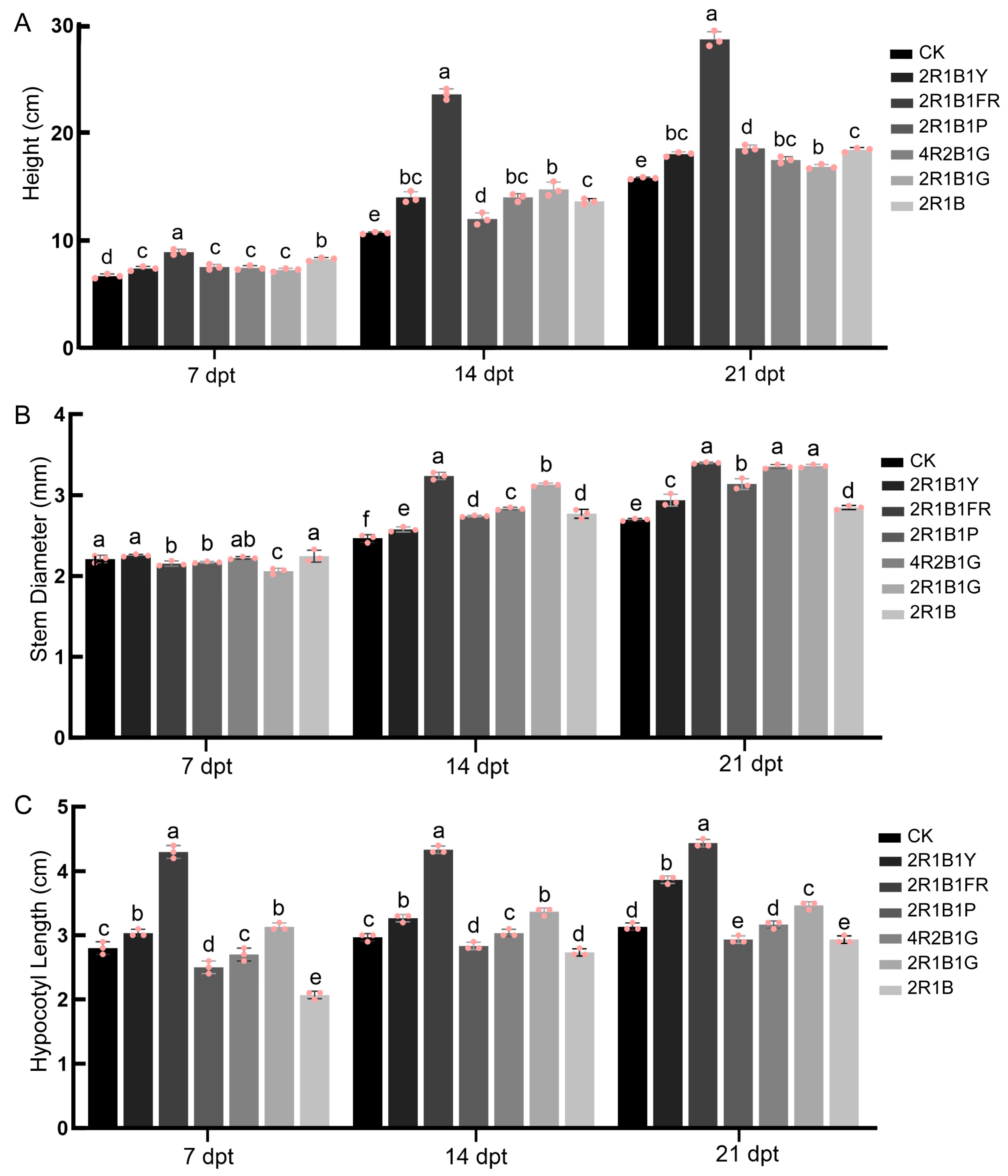
Plant height (PH) exhibited significant variations under different light spectra throughout the seedling stages (7, 14, and 21 days post treatment, dpt) (Figure 3A). The 2R1B1FR spectrum consistently promoted stem elongation most significantly (Figure 3A). At 7 dpt, seedlings of 2R1B1FR treatment were significantly taller (33.33%) than the control (CK, white light) (Figure 3A). This pronounced promotive effect intensified by 14 dpt, with 2R1B1FR plants exceeding CK height by 120.56% (Figure 3A). At 21 dpt, the 2R1B1FR group maintained its dominant position, with plants being 81.65% taller than CK (Figure 3A). This sustained hyper-elongation strongly suggests the induction of FRL-mediated shade-avoidance responses by the 2R1B1FR spectrum. In contrast, treatments containing green light (4R2B1G and 2R1B1G) showed moderate height increases compared to CK at 14 dpt (30.84% and 38.01%) and 21 dpt (10.76% and 6.75%), exceeding the height achieved by the monochromatic 2R1B treatment (Figure 3A). Notably, the higher green ratio in 4R2B1G did not confer additional benefits over 2R1B1G (Figure 3A). Meanwhile, treatments with yellow light (2R1B1Y) or purple (2R1B1P) additives generally had limited or inconsistent effects on stem elongation (Figure 3A). While 2R1B1Y and 2R1B showed slight increases in plant height at 7 dpt (10.45% and 23.38%, respectively), their growth promotion became less pronounced by 21 dpt, with increases in plant height of only 14.14% for 2R1B1Y and 16.67% for 2R1B compared to CK (Figure 3A). In contrast, the 2R1B1P group exhibited a modest acceleration at 21 dpt (+17.51% vs. CK), but its plant height remained substantially shorter than that of 2R1B1FR (Figure 3A). Overall, the results highlight the potent and persistent effect of supplemental FRL (2R1B1FR) in promoting pepper seedling stem elongation. Green light supplementation (4R2B1G and 2R1B1G) provided a moderate, ratio-dependent increase over basic red–blue light (2R1B), while yellow and purple wavelengths (2R1B1Y and 2R1B1P) had minimal impact.
Stem diameter (SD) displayed distinct, stage-dependent responses to light spectra (Figure 3B). The most notable pattern was observed under the 2R1B1FR spectrum, which initially suppressed stem thickness at 7 dpt, but subsequently induced the most substantial thickening (Figure 3B). By 14 dpt, the stems of 2R1B1FR group became significantly thicker than all other treatments (+29–33% vs. CK) (Figure 3B). However, stems of 2R1B1FR, 4R2B1G, and 2R1B1G were significantly thicker than CK and other treatments. Treatments supplemented with green light (4R2B1G; 2R1B1G) consistently promoted stem thickening relative to CK from 14 dpt onwards (Figure 3B). Both 4R2B1G and 2R1B1G achieved robust diameters comparable to or exceeding other treatments (except 2R1B1FR) at late stages (Figure 3B). Notably, the higher green ratio in 4R2B1G provided no consistent advantage over 2R1B1G. In contrast, the monochromatic R:B = 2:1 treatment (2R1B) and the purple light-supplemented treatment (2R1B1P: R:B:P = 2:1:1) generally resulted in stem diameters statistically similar to CK (Figure 3B), showing no persistent stimulatory effect. Similarly, the yellow-supplemented treatment (2R1B1Y: R:B:Y = 2:1:1) exhibited only minimal increment over CK at later stages (Figure 3B). Overall, FRL (2R1B1FR) induced a unique biphasic response culminating in the thickest stems, while green light (4R2B1G, 2R1B1G) provided consistent positive effects on stem robustness. Basic red–blue light (2R1B) and additions of purple (2R1B1P) or yellow (2R1B1Y) wavelengths lacked substantial impact on stem diameter development.
Hypocotyl length varied notably among treatments over time. At 7 days, the 2R1B1FR treatment recorded the longest hypocotyls (4.30 cm), while 2R1B was the shortest (2.07 cm) (Figure 3C). At 14 days, the group of 2R1B1FR again showed the maximum length (4.33 cm), with 2R1B exhibiting the minimum (2.73 cm) (Figure 3C). By 21 days, the 2R1B1FR group sustained the greatest length (4.43 cm), and 2R1B1P and 2R1B had the smallest (2.93 cm) (Figure 3C). The 2R1B1FR group consistently produced the longest hypocotyls, suggesting its light quality may accelerate early seedling development. Additionally, the 2R1B1FR treatment produced the longest hypocotyls at all measured intervals, indicating its light treatment fosters early seedling development effectively.

2.2. Biomass and Vigor Indicators

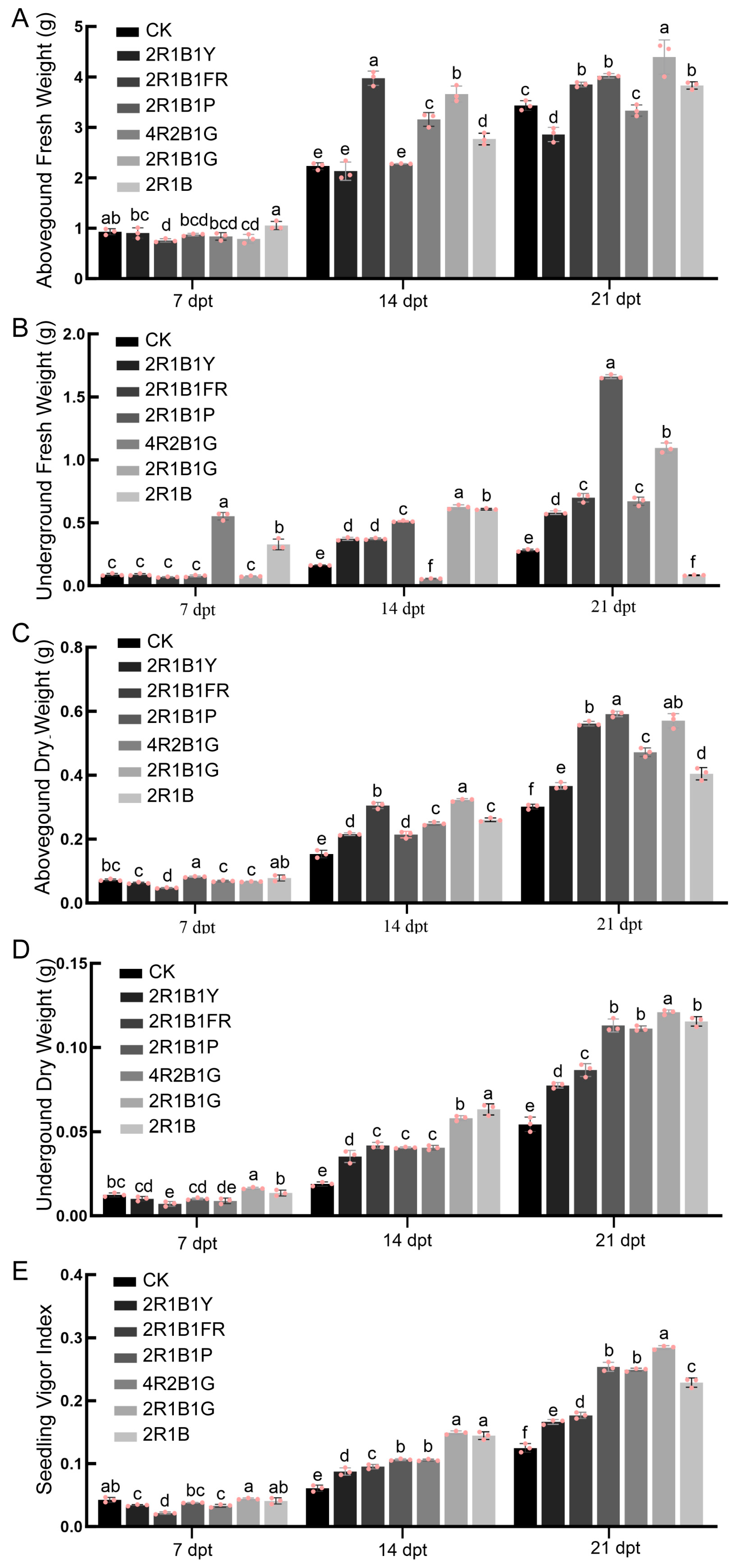
Aboveground fresh weight (AFW) exhibited an increase across all groups from 7 to 21 days. At 7 days, the 2R1B group exhibited the highest AFW (1.053 g), while 2R1B1FR was the lowest (0.758 g) (Figure 4A). By day 14, the treatment of 2R1B1FR reached the highest AFW (3.975 g), while CK showed the lowest (2.230 g) (Figure 4A). At 21 days, 2R1B1G reached the peak weight (4.395 g), and CK remained the lightest (3.433 g) (Figure 4A).
Underground fresh weight (UFW) showed treatment-specific fluctuations. At 7 days, 4R2B1G had the highest weight (0.553 g), while 2R1B1FR was the lowest (0.069 g) (Figure 4B). At 14 days, 2R1B1G peaked (0.626 g), and 4R2B1G dropped to the minimum (0.056 g) (Figure 4B). By 21 days, 2R1B1P exhibited the maximum weight (1.661 g), whereas 2R1B was the minimum (0.084 g) (Figure 4B). This variability indicates that light treatments like 2R1B1FR and 2R1B1P may enhance root growth at specific stages, but no treatment consistently outperformed others. Although 4R2B1G was more effective early, 2R1B1FR showed enhanced root growth by the end of the experiment (Figure 4B), suggesting specific light treatments can positively influence root development at different stages.
Based on the comprehensive analysis of aboveground dry weight (ADW) data, significant treatment-dependent dynamics and temporal progression were observed across developmental stages. At the initial 7-day measurement, ADW ranged from 0.048 g (2R1B1FR) to 0.082 g (2R1B1P), with 2R1B1P exceeding CK (0.072 g) by 13.9% (Figure 4C). By day 14, a pronounced shift occurred: 2R1B1G emerged as dominant (0.322 g), demonstrating a 3.9-fold increase from that of day 7 and significantly exceeding CK (0.153 g) by 110.5% (Figure 4C). Concurrently, 2R1B1FR exhibited a substantial recovery from the lowest initial value to the second highest (0.305 g), surpassing CK by 99.3% (Figure 4C). At day 21, maximal ADW was recorded in 2R1B1P (0.591 g), closely followed by 2R1B1G (0.570 g), both substantially outperforming CK (0.301 g) by 96.3% and 89.4%, respectively (Figure 4C). The 2R1B1FR treatment maintained strong performance (0.561 g, 86.4% above CK), confirming a consistent positive effect. Meanwhile, 2R1B1Y and 2R1B exhibited divergent ADW trajectories: 2R1B1Y progressed from suboptimal initial biomass (7 d: 0.063 g, −13% vs. CK) to sustained acceleration, culminating in a 244% increase by day 14 (0.216 g, +41% vs. CK) and maintaining 23% superiority at day 21 (0.369 g) (Figure 4C). Conversely, 2R1B declined from early advantage (7 d: 0.078 g, +9% vs. CK) to the lowest treatment value by day 21 (0.404 g) (Figure 4C), reflecting constrained late-stage accumulation despite remaining above CK. Notably, 2R1B1G demonstrated sustained accumulation superiority, while 2R1B1P’s early advantage translated into maximal late-stage biomass (Figure 4C), suggesting distinct temporal optimization patterns under specific spectral regimes. These contrasts highlight treatment-specific modulation of photoassimilate partitioning and dry matter accumulation kinetics.
Underground dry weight (UDW) exhibited significant spatiotemporal variations across light treatments. At 7 days, UDW ranged from 0.007 g (2R1B1FR) to 0.016 g (2R1B1G), with 2R1B1G exceeding CK (0.012 g) by 31% (Figure 4D). By 14 days, 2R1B maintained dominance (0.063 g), demonstrating a 4.6-fold increase from initial values and significantly outperforming CK (0.019 g) (Figure 4D). Notably, 2R1B1G (0.058 g) also showed robust growth, surpassing CK by 200% (Figure 4D). At 21 days, maximal UDW was observed in 2R1B1G (0.121 g), followed closely by 2R1B (0.115 g) and 2R1B1P (0.113 g), collectively exceeding CK (0.05 g) by 122–142% (Figure 4D). Interestingly, 2R1B1FR exhibited remarkable recovery: from the lowest at 7 days to the third-highest at 21 days (0.088 g) (Figure 4D), suggesting adaptive plasticity.
The seedling vigor index (SVI), integrating stem robustness, root-shoot partitioning, and total biomass, revealed pronounced treatment effects across developmental stages. At 7 days, 2R1B1G exhibited the highest SVI (0.044), surpassing CK (0.043) by 4.5% (Figure 4E). However, 2R1B1FR showed the lowest SVI (0.021), reflecting compromised early establishment (Figure 4E). By 14 days, 2R1B1G surged significantly to 0.149—a 3.4-fold increase from day 7—significantly exceeding CK (0.061) by 145% (Figure 4E). The 2R1B group also demonstrated strong mid-term SVI (0.145), closely rivaling 2R1B1G (Figure 4E). At 21 days, 2R1B1G peaked at 0.284, followed by 2R1B1P (0.254) and 4R2B1G (0.250), collectively exceeding CK (0.125) by 127–154% (Figure 4E). Notably, 2R1B1P showed the most rapid late-stage acceleration (21d SVI = 2.3× 14 d SVI), indicating sustained vigor. The 2R1B1FR group recovered substantially, from the lowest at 7 days to the fourth-highest at 21 days (0.177). The 2R1B treatment declined to 0.229 (still 83% above CK), revealing treatment-specific constraints on long-term vigor.

2.3. Metabolites and Stress Indicators

2.3.1. Soluble Protein Content

Soluble protein content, a key indicator of metabolic activity and nitrogen assimilation, exhibited notable fluctuations across time points and treatments. At day 7, the protein content ranged from 3.000 mg g−1 in 2R1B to 11.226 mg g−1 in 4R2B1G, indicating that 4R2B1G stimulated early protein accumulation, while 2R1B suppressed it compared to the control (CK: 6.734 mg g−1) (Figure 5A). By day 14, a general increase was observed in most treatments, with 2R1B1P showing the highest (19.598 mg g−1), significantly exceeding CK (11.751 mg g−1) (Figure 5A). This suggests that treatments like 2R1B1P may enhance protein synthesis during mid-phase growth. However, by day 21, a decline occurred across all groups, particularly in 2R1B1Y (3.215 mg g−1) and 4R2B1G (4.986 mg g−1), which fell below CK (7.062 mg g−1) (Figure 5A). In contrast, 2R1B1G maintained relatively high levels (7.959 mg g−1) (Figure 5A), implying potential resilience to degradation. The temporal pattern indicates that treatments 2R1B1P and 4R2B1G induced early protein accumulation but led to later depletion, possibly due to accelerated metabolic turnover or stress-induced proteolysis. Comparatively, CK and 2R1B1G demonstrated more stability, highlighting their role in sustaining protein homeostasis over time.

2.3.2. Soluble Sugar Content

Soluble sugar content, reflecting carbohydrate metabolism and energy status, varied considerably with treatment and duration (Figure 5). At day 7, soluble sugar contents were lowest in 4R2B1G (0.054 mg g−1) and 2R1B1G (0.053 mg g−1), while 2R1B1Y (0.065 mg g−1) and CK (0.062 mg g−1) showed moderate levels (Figure 5B). This early phase suggests that certain treatments (e.g., 4R2B1G and 2R1B1G) may inhibit sugar accumulation. By day 14, a universal increase occurred, with 2R1B1P peaking at 0.169 mg g−1, and 2R1B1G peaking at 0.166 mg g−1, both markedly higher than CK (0.127 mg g−1) (Figure 5B), indicating enhanced sugar synthesis or mobilization under this treatment. Treatment of 2R1B1FR also displayed elevated levels (0.161 mg g−1) (Figure 5B), consistent with a potential role in promoting carbohydrate storage. At day 21, soluble sugar content declined in most groups, but 2R1B1Y (0.080 mg g−1) and 4R2B1G (0.080 mg g−1) remained above CK (0.058 mg g−1) (Figure 5B), implying sustained metabolic activity. Notably, 2R1B1P showed a relative decrease from day 14 to day 21 (0.169 to 0.072 mg g−1) (Figure 5B), which may indicate instability. Overall, treatments like 2R1B1Y and 2R1B1P appear to enhance soluble sugar accumulation during stress phases, while CK and 2R1B (0.065 mg g−1 at day 21) maintained baseline levels, suggesting minimal perturbation to soluble sugar dynamics.

2.3.3. Malondialdehyde (MDA) Content

Malondialdehyde (MDA) content, a biomarker of oxidative stress and lipid peroxidation, demonstrated pronounced treatment-specific and temporal trends (Figure 5C). At day 7, MDA levels were highest in 2R1B1Y (35.914 nmol/g) and lowest in 2R1B1P (13.979 nmol/g), with CK at 20.215 nmol/g (Figure 5C). This indicates that 2R1B1Y induced early oxidative stress, whereas 2R1B1P may have antioxidant properties. By day 14, MDA decreased in most groups, notably in 2R1B1G (7.100 nmol/g), which was substantially below CK (15.269 nmol/g) (Figure 5C), suggesting that 2R1B1G effectively mitigated oxidative damage. In contrast, 2R1B1Y showed the highest elevated levels (17.100 nmol/g) (Figure 5C), implying persistent stress. At day 21, a general increase in MDA occurred, with 2R1B1G reaching the highest (39.570 nmol/g), far exceeding CK (37.960 nmol/g) (Figure 5C), which could indicate late-phase oxidative burst or cumulative damage. Conversely, 2R1B maintained lower levels (27.419 nmol/g), similar to 2R1B1Y (31.935 nmol/g) (Figure 5C), hinting at adaptive stress responses. The data underscore that treatments like 2R1B1G exacerbated oxidative stress over time, while 2R1B1P and 2R1B showed protective effects, particularly in the early and mid-phases.
Overall, the data reveal significant temporal variations and treatment-dependent responses, suggesting that the treatments differentially influenced metabolic and stress-related pathways. Below, each indicator is analyzed in detail.

2.4. Photosynthetic Pigment Indicators

Chlorophyll a content exhibited significant temporal variations under different light quality treatments. At 7 days, the chlorophyll a content in CK was significantly higher (1.378 mg g−1 FW) than in all treatment groups, among which 2R1B showed the most pronounced reduction in chlorophyll a content (0.867 mg g−1 FW). The values for the other treatments were statistically comparable, though numerically ordered as 2R1B1FR > 2R1B1G > 2R1B1P > 4R2B1G > 2R1B1Y (Figure 6A). By day 14, a notable shift occurred: 4R2B1G reached the highest level (1.610 mg g−1 FW), significantly surpassing CK (1.526 mg g−1 FW; p < 0.05), while 2R1B remained the lowest (1.301 mg g−1 FW). Treatments including 2R1B1P and 2R1B1G did not differ significantly from CK. At 21 days, CK again registered the highest chlorophyll a content (1.338 mg g−1 FW), with significant reductions observed in 2R1B1Y (0.781 mg g−1 FW; p < 0.01) and 2R1B (0.987 mg g−1 FW; p < 0.05). In contrast, 2R1B1G showed no significant difference from CK, indicating a relative stability under this treatment.
Chlorophyll b levels demonstrated a higher sensitivity to spectral treatments compared to chlorophyll a (Figure 6B). On day 7, CK yielded the highest chlorophyll b content (0.623 mg g−1 FW), significantly greater than all other groups, with 2R1B again being the most adversely affected (0.349 mg g−1 FW). By day 14, 4R2B1G outperformed all others (0.691 mg g−1 FW), showing a statistically significant increase over CK (0.665 mg g−1 FW; p < 0.05). The treatment 2R1B continued to exhibit significantly depressed values. At the final time point (21 days), CK maintained significantly superior chlorophyll b levels relative to most treatments, except 2R1B1G, which did not differ significantly. The most severe decline was observed in 2R1B1Y (0.363 mg g−1 FW; p < 0.01), underscoring its negative impact over time.
Total chlorophyll content mirrored the composite effects observed in the individual chlorophyll fractions (Figure 6C). At 7 days, total chlorophyll in CK (2.001 mg g−1 FW) was significantly higher than in all light-quality treatments, with 2R1B being the lowest (1.216 mg g−1 FW). A significant recovery was observed at 14 days, where 4R2B1G achieved a total chlorophyll content (2.301 mg g−1 FW) significantly exceeding that of CK (2.190 mg g−1 FW; p < 0.05). By day 21, CK again showed the highest value (1.961 mg g−1 FW), with 2R1B1Y being significantly lower (1.144 mg g−1 FW) and 2R1B also substantially reduced (1.422 mg g−1 FW). The treatment 2R1B1G consistently performed closest to CK across time points, showing no significant difference at 14 and 21 days.
Carotenoid content revealed dynamic and treatment-specific photoprotective adaptation patterns (Figure 6D). Initially (7 days), carotenoids in CK (0.309 mg g−1 FW) were significantly elevated compared to all treatments. By 14 days, 4R2B1G not only recovered but attained a significantly higher level (0.383 mg g−1 FW) than CK (0.348 mg g−1 FW), suggesting an adaptive enhancement under this light regime. At 21 days, CK (0.306 mg g−1 FW) was again significantly higher than six treatments, most notably 2R1B1Y (0.202 mg g−1 FW). The treatment 2R1B1G maintained carotenoid levels closest to CK without significant differences, indicating better functional retention. CK’s early-stage dominance suggests better photoprotection under control conditions, but variability at later stages indicates treatment-specific impacts on carotenoid synthesis. CK’s early dominance in carotenoid content suggests better photoprotection under control conditions, despite later-stage variability indicating treatment-specific impacts on this process.

3. Discussion

This study provides a comprehensive assessment of how specific LED spectral compositions dynamically regulate the morpho-physiological development of Capsicum annuum seedlings. The findings of this study demonstrate that light quality acts as a potent modulator, significantly altering growth architecture, biomass partitioning, photosynthetic apparatus composition, and stress physiology. These mechanistic insights are pivotal for refining LED protocols in controlled environment horticulture to produce high-quality, resilient pepper transplants.

3.1. Spectral Regulation of Morphogenesis and Biomass Partitioning

The divergent morphological responses underscore the critical role of spectral composition in directing photomorphogenesis, further highlighting its potential as a precise regulatory tool for crop architecture optimization in controlled environments. The 2R1B1FR spectrum (R:B:FR = 2:1:1) induced a significant and sustained promotion of stem elongation (e.g., +81.65% vs. CK at 21 dpt, Figure 3A), consistent with the well-established phytochrome-mediated shade-avoidance syndrome (SAS) triggered by low RL:FRL ratios [30,31]. This result aligns with universal phytochrome signaling mechanisms across angiosperms, confirming the reliability of our spectral manipulation approach.
However, the data reveal a more nuanced temporal response in stem robustness: 2R1B1FR initially suppressed stem diameter at 7 dpt (likely due to prioritized photosynthate allocation to stem elongation for rapid “shade escape”) but subsequently induced the most significant thickening by 14 and 21 dpt (+29–33% vs. CK, Figure 3B). This biphasic pattern suggests that FRL signaling not only drives elongation but may also initiate a time-dependent shift in carbon allocation away from the roots and towards secondary growth in the stem, potentially mediated by altered expression of sucrose transporter genes (e.g., SUT1) [32] or secondary cell wall biosynthesis genes (e.g., CesA) [33]. Such a dynamic carbon partitioning strategy likely represents an adaptive mechanism for plants to balance “competitive growth” and “structural support” under shaded conditions.
The observed increase in stem diameter could contribute to improved mechanical properties (e.g., lodging resistance), a possibility that warrants further investigation through biomechanical testing or lignin content analysis. This potential trade-off necessitates the development of management strategies to avoid excessive root growth thinning early on and ensure balanced growth under FR supplementation. For instance, adjusting FR application timing to avoid the seedling root establishment phase, or combining FR treatment with root-promoting nutrients (e.g., phosphorus and potassium), could optimize the agronomic utility of spectral regulation in commercial greenhouse production.
Conversely, the 2R1B1G spectrum fostered compact growth (e.g., hypocotyl length and plant height comparable to or moderately exceeding CK, Figure 3A) while achieving the highest seedling vigor index (SVI) at 21 dpt (0.284 vs. CK 0.125, Figure 4E). This higher vigor, integrating stem robustness, favorable root-shoot partitioning, and high total biomass, highlights the beneficial role of balanced green light supplementation in pepper seedling development. Notably, the addition of green light to the red–blue background appears to have mitigated the typical shade-avoidance responses, promoting a more balanced architecture instead. This finding aligns with the emerging concept that green light can antagonize blue-light-mediated cryptochrome signaling [34,35,36], potentially leading to a more moderated photomorphogenic program. Of note, 2R1B1P treatment also demonstrated exceptional performance, particularly in enhancing soluble protein and sugars (Figure 5A,B). This suggests that 2R1B1G promotes a more balanced allocation of resources, resulting in overall higher seedling quality, whereas 2R1B1P excels more in promoting specific traits associated with carbon metabolism. The distinct advantages of each spectrum suggest their potential for stage-specific application: 2R1B1G for overall vigor and compactness, and 2R1B1P for enhancing metabolic activity. The enhanced compactness aligns with cryptochrome activation by BL [34,35,36], present in the same proportion as in 2R1B, suggesting that the supplemented green light in 2R1B1G did not negate but complemented BL effects, potentially improving light penetration and distribution within the vegetative growth period.
An important finding was the significant enhancement of root development under the 2R1B1P spectrum, evidenced by the high underground dry weight (UDW) at 21 dpt (0.113 g, +126% vs. CK 0.05 g, Figure 4B). This robust root system, coupled with elevated soluble sugar accumulation at 21 dpt (0.072 mg g−1 vs. CK 0.058 mg g−1, Figure 5B), strongly implies a shift in photoassimilate partitioning favoring root sinks. We hypothesize that the specific purple wavelengths (415 ± 5 nm) may act as an environmental cue that modulates auxin-polar transport or sensitivity in root tissues, thereby stimulating meristem activity and root development. This mechanistic possibility warrants further investigation. This effect is distinct from the FR response in 2R1B1FR and represents a valuable trait for improving transplant establishment resilience. The capacity of 2R1B1P to enhance root architecture without triggering excessive shoot elongation, as seen with FRL, makes it a particularly valuable spectral tool for producing pepper seedlings with superior potential for post-transplant survival.

3.2. Remodeling of the Photosynthetic Apparatus and Photoprotection

Light quality significantly influenced the composition and stability of photosynthetic pigments, reflecting strategic adjustments in light harvesting and potential photoprotective mechanisms. While the control (CK, white light) generally maintained high chlorophyll levels throughout the experiment, specific treatments induced dynamic shifts. The 4R2B1G spectrum transiently enhanced total chlorophyll accumulation at 14 dpt (2.301 mg g−1 vs. CK 2.190 mg g−1, Figure 6), but this advantage diminished by 21 dpt (1.492 mg g−1 vs. CK 1.961 mg g−1, Figure 6). This pattern indicates a temporal sensitivity and potential instability in chlorophyll biosynthesis or degradation under a high proportion of green light, which may inefficiently drive photosynthesis or trigger feedback inhibition.
Regarding photoprotection, 2R1B1P maintained carotenoid levels of 0.264 mg g−1 at 21 dpt, which was lower than that in CK (Figure 6D). This suggests that purple light supplementation may help sustain this important photoprotective pigment pool more efficiently possible by reducing the need for its excessive synthesis. Carotenoids are crucial for quenching excess energy and scavenging reactive oxygen species [37,38]. The role of 2R1B1P in mitigating oxidative stress is further confirmed by significantly lower malondialdehyde (MDA) levels—a key indicator of lipid peroxidation—at both 7 dpt (13.98 nmol/g) and 14 dpt (16.02 nmol/g) compared to other treatments (Figure 5C).
However, evaluating the significance of these pigment responses requires considering their relationship with overall seedling growth and performance. While photosynthetic pigments are fundamental components of the photosynthetic machinery and their status provides valuable insights into light acclimation and photoprotection [39,40], the observed pigment dynamics (e.g., transient chlorophyll increase in 4R2B1G, sustained carotenoids in 2R1B1P) did not consistently correlate with key growth and biomass accumulation parameters (e.g., total biomass, SVI, plant height) across all treatments and time points. This apparent dissociation highlights the multifaceted nature of light quality effects on plant development: optimization of light capture and photoprotection does not necessarily translate directly into growth, which may be co-limited by other factors such as carbon allocation, photosynthetic efficiency, or energy partitioning. Thus, while light quality can modulate photosynthetic pigment composition, its ultimate effect on productivity depends on a complex interplay between light harvesting, photoprotection, and downstream physiological processes.

3.3. Modulation of Oxidative Stress and Metabolite Dynamics

The study revealed significant spectral effects on oxidative stress markers and key metabolites, underscoring the role of light quality in modulating physiological homeostasis. The 2R1B1G treatment exhibited significantly lower oxidative damage at 14 dpt (MDA 7.100 nmol/g vs. CK 15.269 nmol/g, Figure 5C), which correlated strongly with its peak SVI and biomass accumulation. This alignment implies that the 2R1B1G spectrum facilitated a high-growth-vigor phenotype with minimal oxidative burden during the active vegetative growth phase, likely due to enhanced photoprotection and balanced energy utilization.
Conversely, 2R1B1Y induced significant oxidative stress, manifested as elevated MDA levels at 7 dpt (35.914 nmol/g vs. CK 20.215 nmol/g, Figure 5C) and progressive chlorophyll degradation (lowest total Chl at 21 dpt: 1.144 mg g−1 vs. CK 1.961 mg g−1, Figure 6). This was further exacerbated by severe soluble protein depletion at 21 dpt (3.215 mg g−1 vs. CK 7.062 mg g−1, Figure 5), indicating substantial metabolic disruption and proteolysis under yellow light supplementation. These findings challenge the conventional notion that yellow light has minimal morphogenic effects and instead reveal its strongly disruptive impact on pepper seedling physiology, highlighting the importance of spectral quality in avoiding phototoxicity and maintaining metabolic integrity.
Metabolite analysis highlighted distinct carbon allocation strategies influenced by light quality. The 2R1B1FR treatment accumulated the highest soluble sugars at 14 dpt (0.161 mg g−1 vs. CK 0.127 mg g−1, Figure 5B), consistent with the known role of RL in promoting carbohydrate synthesis and export [41,42,43]. However, this did not translate into greater biomass efficiency compared to 2R1B1G later on, suggesting possible sink limitations or higher metabolic costs associated with the FR-induced shade-avoidance syndrome phenotype. The 2R1B1P group uniquely combined enhanced root growth with elevated soluble sugars at 21 dpt (0.072 mg g−1 vs. CK 0.058 mg g−1, Figure 4 and Figure 5), implying efficient carbon partitioning to belowground sinks under purple light. Furthermore, 2R1B1G demonstrated superior preservation of soluble proteins at 21 dpt (7.959 mg g−1 vs. CK 7.062 mg g−1, Figure 5). The sustained protein levels under 2R1B1G, potentially linked to BL signaling pathways known to influence nitrogen metabolism and osmoprotectant synthesis [44,45,46], thereby contributing to enhanced cellular stability and overall stress resilience.

3.4. Implications for Optimized Nursery Production

Our multi-parameter assessment identifies specific spectral recipes with distinct advantages for pepper seedling production: (1) 2R1B1G consistently promoted the highest seedling vigor index (SVI) (Figure 4), supporting coordinated dry matter partitioning (Figure 4), a favorable chlorophyll a/b ratio (Figure 6), and low mid-term oxidative stress, making it ideal for producing robust and uniform transplants under intensive nursery conditions. (2) 2R1B1P significantly enhanced root development (Figure 4) and soluble sugar accumulation while maintaining low oxidative stress (Figure 5). This spectrum is particularly promising for improving transplant establishment success in field or stress-prone environments, where root system strength and carbohydrate reserves are critical. (3) 2R1B1FR powerfully stimulated stem elongation and late-stage thickening (Figure 3), which could be strategically employed in the earliest growth phase to accelerate canopy closure or overcome initial lag but requires careful management or subsequent spectral shifts to mitigate early stem thinning and reduced root investment.
Notably, treatments such as 2R1B1Y and the basic 2R1B generally resulted in inferior seedling quality compared to the other spectral treatments, characterized by oxidative damage (2R1B1Y), weak biomass/pigment accumulation (2R1B), and reduced vigor. On the other hand, the 4R2B1G treatment also showed great potential, significantly enhancing root biomass allocation and seedling sturdiness, as evidenced by its high seedling vigor index at harvest (Figure 4E). This high vigor index, which integrates stem robustness, root-shoot ratio, and total biomass, indicates a pronounced ability to produce robust, well-rooted transplants primed for successful establishment.
Therefore, these results offer a mechanistic foundation for designing dynamic, stage-specific, or trait-oriented LED lighting regimens. For example, a multi-phase protocol could begin with 2R1B1FR to stimulate early establishment, shift to 2R1B1G to enhance vegetative vigor and compactness, and finally apply 2R1B1P before transplanting to strengthen root systems and carbohydrate storage. Overall, this research significantly advances the photobiology of solanaceous seedlings and paves the way for more efficient and sustainable nursery production systems in controlled environment agriculture.
It is important to note that these findings are based on a specific pepper cultivar (‘Changchuan No. 3’). Future research should validate these spectral recipes across a wider range of pepper varieties to assess their broader applicability and potential need for variety-specific adjustments.

4. Materials and Methods

4.1. Plant Growth Conditions

Tested pepper variety and seedling management: The pepper cultivar Changchuan No. 3 was used in this study. Plump seeds with uniform size and good luster were selected, soaked in distilled water (ddH2O) for 8 h, and then evenly placed on Petri dishes with moist filter paper. The dishes were subsequently placed in a constant-temperature incubator at 28 °C for germination under dark conditions. Germination progress was observed daily, and water was replenished as needed. After the seeds showed radicle emergence (termed “seed sprouting”), uniformly healthy seeds were selected and individually sown in a soil mix [peat moss:perlite (2/1, v/v)] in plastic pots. Each treatment consisted of three trays, totaling 150 seeds. After covering with substrate and thoroughly watering, the trays were placed under different light quality treatments. A 1/2 Hoagland nutrient solution [47] was applied once every 7 days.
Basic parameters of experimental LED light sources: The experimental light sources were purchased from Kedao Technology Co., Ltd. (Huizhou, China). These LED linear strip lights (L1000 × W80 × H10 mm) could emit six different light qualities: red light (RL: 660 ± 5 nm), blue light (BL: 460 ± 5 nm), green light (GL: 530 ± 5 nm), yellow light (YL: 585 ± 5 nm), far-red light (FRL: 730 ± 5 nm), and purple light (PL: 415 ± 5 nm).
Design of LED composite light quality treatments and control setup: The study employed LED composite light qualities to treat pepper seed cultivation, with LED white light (CK) serving as the control. Six composite light quality treatments were established, as shown in Table 1. The LED strips were mounted 30 cm above plastic pots. The photosynthetic photon flux density of all treatments was uniformly calibrated to 150 μmol m−2 s−1 at the canopy level using a quantum sensor (PG200N; UPRtek, Taiwan Province, China) prior to the experiment. The photoperiod was set to 12 h/12 h (light/dark).

4.2. Morphological Measurements

Physiological and biochemical indices were measured at 7, 14, and 21 days after transplanting (the pepper plants developed four true leaves), corresponding to key developmental phases of early seedling establishment. For each treatment group, three uniformly grown seedlings were randomly selected and rinsed with distilled water.
Plant height and hypocotyl length were measured using a ruler. Stem diameter was measured using a digital vernier caliper. Fresh weight and dry weight of shoots and roots were measured using an electronic balance.
Plant height: the distance from the apex of the main stem to the base where the stem contacts the substrate surface.
Hypocotyl length: the distance from the cotyledonary node (point of cotyledon attachment) to the root-shoot transition zone (~1 mm above the root cap).
Stem diameter: Measured 1 cm below the cotyledonary node or at one-third of the plant height.
Fresh weight measurement: After cutting at the root-shoot transition zone, the shoot portions (including stem and leaves) and root portions were weighed separately to determine the aboveground fresh weight and underground fresh weight, respectively.
Dry weight measurement: Aboveground parts (stems and leaves) and underground parts (root tissues) were separated, thoroughly washed with deionized water to remove adhering soil particles, and carefully blotted dry with paper towels. Samples were then killed at 105 °C for 25 min and subsequently oven-dried at 72 °C to constant weight. Dry weights were recorded separately for aboveground parts and roots.
Seedling vigor index (SVI) was calculated as: SVI = (Stem diameter/Plant height + Root dry weight/shoot dry weight) × Total dry weight.

4.3. Biomass and Biochemical Measurements

Photosynthetic pigment content was determined using a mixed solvent extraction method [48]. To describe briefly, leaf samples (0.2 g) were cut into small pieces (avoiding major veins) and placed in a mortar. 5 mL of 80% acetone was added, and the samples were ground into a homogenate under low light conditions. The homogenate was filtered into a 25 mL volumetric flask, and the mortar and residue were rinsed repeatedly with 80% acetone until the residue was colorless. The volume was made up to 25 mL with 80% acetone. The absorbance of the extract was measured at 663 nm, 645 nm, and 470 nm using a spectrophotometer, with 80% acetone as the blank control.
The contents of chlorophyll a (Chl a), chlorophyll b (Chl b), total chlorophyll (Chl a + b), and carotenoids (Caro) were calculated using the following formulas: Chl a (mg·g−1 FW) = (12.7 × OD663 − 2.69 × OD645) × V/1000 × W; Chl b (mg·g−1 FW) = (22.9 × OD645 − 4.68 × OD663) × V/1000 × W; Total chl (mg·g−1 FW) = (20.21 × OD645 + 8.02 × OD663) × V/1000 × W; Caro (mg·g−1 FW) = (4.7 × OD440 − 0.27 × (20.21 × OD645 + 8.02 × OD663) × V /1000 × W.
Soluble sugar content was measured by the anthrone colorimetric method as described previously with a slight modification [49]. Briefly, accurately weigh 0.1 g fresh plant samples, homogenize them in 1 mL distilled water, extract in a boiling water bath for 10 min, centrifuge the homogenate at 8000× g for 10 min after cooling, re-extract the residue twice with distilled water, combine all supernatants and dilute to 10 mL to obtain the extract; then mix 1 mL diluted extract with 4 mL anthrone reagent (0.2 g anthrone dissolved in 100 mL 80% sulfuric acid), heat in a boiling water bath for 10 min to form a blue-green complex (via reaction of soluble sugars with anthrone under acidic heating conditions), cool immediately in an ice bath, measure absorbance at 620 nm with distilled water as blank control after equilibrating to room temperature, and calculate soluble sugar content (expressed as mg·g−1 FW) using a pre-constructed glucose standard curve (0.125–10 mg/mL).
Soluble protein content was confirmed using Bradford’s method (coomassie brilliant blue G250) with bovine serum albumin as the standard, with slight modification [50]. Briefly, accurately weigh 1 g fresh plant samples, homogenize them in 8 mL pre-cooled phosphate buffer (pH 7.0), centrifuge the homogenate at 10,000× g for 15 min at 4 °C after homogenization, collect the supernatant as the soluble protein extract; then mix 0.1 mL diluted extract with 5 mL Bradford reagent (coomassie brilliant blue G250 dissolved in 95% ethanol and 85% phosphoric acid, diluted with distilled water), incubate at room temperature for 5 min to form a stable blue complex (via binding of coomassie brilliant blue G250 to protein under acidic conditions), measure the absorbance at 595 nm with distilled water instead of extract as the blank control, and calculate the soluble protein content (expressed as mg·g−1 FW). BSA was used as a standard sample.
Malondialdehyde (MDA) content was measured following the method of Muhammad et al., [51]. As the end product of lipid peroxidation, MDA reacts with thiobarbituric acid (TBA) to form a trimethine complex, which has an absorption maximum at 532 nm. For each sample, the nonspecific absorption value at 600 nm was also recorded, and this value was subtracted from the absorbance measured at 532 nm to eliminate interference. The concentration of MDA was calculated using an extinction coefficient of 155 mM−1 cm−1, and the lipid peroxidation level was finally expressed as nanomoles of MDA per gram of fresh weight (nmol MDA·g−1 FW).

4.4. Data Processing

For each treatment, two independent experimental replicates were performed. In each replicate, measurements were conducted on 3 pepper plants, ensuring consistent sampling across replicates.
Tukey’s test and graphical analyses were performed using GraphPad Prism software (version 10.5, GraphPad Prism Software, USA).

5. Conclusions

In conclusion, this study demonstrates that precise manipulation of LED light spectra is a highly effective tool to direct the growth and physiological trajectory of pepper seedlings, moving beyond the conventional red–blue paradigm.
Our results clearly identify a red/blue/green (2R1B1G) spectrum as the optimal recipe for producing high-quality pepper transplants, as it uniquely promotes balanced growth, higher dry matter partitioning, and stress resilience, culminating in the highest seedling vigor. We further elucidate the distinct and specialized roles of other wavelengths: far-red (2R1B1FR) potently stimulates stem elongation and biomass accumulation but necessitates careful management due to initial trade-offs; purple light (2R1B1P) specifically enhances root development and metabolite accumulation, offering value for pre-transplant hardening; and yellow light (2R1B1Y) proves detrimental to seedling physiology. These findings provide mechanistic evidence that treatment with green light in a balanced ratio resolves growth trade-offs often associated with narrow-band spectra and that purple light can specifically modulate carbon partitioning to roots.
Practically, this work provides a scientific basis for the designs of targeted lighting strategies in commercial nursery production. We propose a dynamic protocol utilizing far-red for early establishment, balanced green light for vigor development, and purple light to boost root strength pre-transplanting, thereby enhancing the efficiency and sustainability of controlled environment agriculture.

Author Contributions

Conceptualization, W.Z., B.X. and L.S.; methodology, W.Z., B.X. and L.S.; formal analysis, W.Z., B.X. and L.S.; investigation, W.Z., Z.H., S.Z., Z.C., Q.H., Y.W. (Yuna Wang) and Y.W. (Yu Wu); resources, W.Z., B.X., Y.G., H.C. and L.S.; data curation, W.Z., B.X. and L.S.; writing—original draft, W.Z. and L.S.; writing—review and editing, W.Z., B.X. and L.S.; supervision, L.S.; funding acquisition, W.Z. and L.S. All authors have read and agreed to the published version of the manuscript.

Funding

This research was funded by the Special Project for the Development of Characteristic Modern Agriculture in Fujian Province (2024SN06), the National Natural Science Foundation of China (32172553 and 32302524), and the Natural Science Foundation of Fujian Province (2024J01382).

Data Availability Statement

The original contributions presented in this study are included in the article. Further inquiries can be directed to the corresponding authors.

Conflicts of Interest

The authors declare that the research was conducted in the absence of any commercial or financial relationships that could be construed as a potential conflict of interest.

References

  1. Teixeira, R.T. Distinct Responses to Light in Plants. Plants 2020, 9, 894. [Google Scholar] [CrossRef]
  2. Fankhauser, C.; Staiger, D. Photoreceptors in Arabidopsis thaliana: Light perception, signal transduction and entrainment of the endogenous clock. Planta 2002, 216, 1–16. [Google Scholar] [CrossRef]
  3. Munyanont, M.; Lu, N.; Ruangsangaram, T.; Takagaki, M. Effect of Far-Red Light and Nutrient Solution Formulas on Calendula Production in a Plant Factory. Biology 2025, 14, 716. [Google Scholar] [CrossRef]
  4. Kitazaki, K.; Fukushima, A.; Nakabayashi, R.; Okazaki, Y.; Kobayashi, M.; Mori, T.; Nishizawa, T.; Reyes-Chin-Wo, S.; Michelmore, R.W.; Saito, K.; et al. Metabolic Reprogramming in Leaf Lettuce Grown Under Different Light Quality and Intensity Conditions Using Narrow-Band LEDs. Sci. Rep. 2018, 8, 7914. [Google Scholar] [CrossRef]
  5. Su, P.; Ding, S.; Wang, D.; Kan, W.; Yuan, M.; Chen, X.; Tang, C.; Hou, J.; Wu, L. Plant morphology, secondary metabolites and chlorophyll fluorescence of Artemisia argyi under different LED environments. Photosynth. Res. 2024, 159, 153–164. [Google Scholar] [CrossRef] [PubMed]
  6. Jung, W.S.; Chung, I.M.; Hwang, M.H.; Kim, S.H.; Yu, C.Y.; Ghimire, B.K. Application of Light-Emitting Diodes for Improving the Nutritional Quality and Bioactive Compound Levels of Some Crops and Medicinal Plants. Molecules 2021, 26, 1477. [Google Scholar] [CrossRef]
  7. Goins, G.D.; Yorio, N.C.; Sanwo, M.M.; Brown, C.S. Photomorphogenesis, photosynthesis, and seed yield of wheat plants grown under red light-emitting diodes (LEDs) with and without supplemental blue lighting. J. Exp. Bot. 1997, 48, 1407–1413. [Google Scholar] [CrossRef] [PubMed]
  8. Schiff, J.A. Evolution of the control of pigment and plastid development in photosynthetic organisms. Bio Syst. 1981, 14, 123–147. [Google Scholar] [CrossRef]
  9. Rehman, M.; Ullah, S.; Bao, Y.; Wang, B.; Peng, D.; Liu, L. Light-emitting diodes: Whether an efficient source of light for indoor plants? Environ. Sci. Pollut. Res. Int. 2017, 24, 24743–24752. [Google Scholar] [CrossRef]
  10. Savvides, A.; Fanourakis, D.; van Ieperen, W. Co-ordination of hydraulic and stomatal conductances across light qualities in cucumber leaves. J. Exp. Bot. 2012, 63, 1135–1143. [Google Scholar] [CrossRef] [PubMed]
  11. Li, X.; Yu, H.; Guo, Y.; Liu, J.; Yu, W.; Guo, X. Comparative role of oxidative balance, alkaloid production, and metabolites in exposure to red-to-blue light in Catharanthus roseus. Plant Physiol. Biochem. 2025, 227, 110170. [Google Scholar] [CrossRef]
  12. Jishi, T.; Matsuda, R.; Fujiwara, K. Manipulation of Intraday Durations of Blue- and Red-Light Irradiation to Improve Cos Lettuce Growth. Front. Plant Sci. 2021, 12, 778205. [Google Scholar] [CrossRef]
  13. Samuoliene, G.; Virsile, A.; Miliauskiene, J.; Haimi, P.J.; Lauzike, K.; Brazaityte, A.; Duchovskis, P. The Physiological Response of Lettuce to Red and Blue Light Dynamics Over Different Photoperiods. Front. Plant Sci. 2020, 11, 610174. [Google Scholar] [CrossRef]
  14. Siuda, R.; Balcerowska, G.; Sadowski, C. Comparison of the usability of different spectral ranges within the near ultraviolet, visible and near infrared ranges (UV-VIS-NIR) region for the determination of the content of scab-damaged component in blended samples of ground wheat. Food Addit. Contam. 2006, 23, 1201–1207. [Google Scholar] [CrossRef] [PubMed]
  15. Varga, I.; Kristic, M.; Lisjak, M.; Tkalec Kojic, M.; Iljkic, D.; Jovic, J.; Kristek, S.; Markulj Kulundzic, A.; Antunovic, M. Antioxidative Response and Phenolic Content of Young Industrial Hemp Leaves at Different Light and Mycorrhiza. Plants 2024, 13, 840. [Google Scholar] [CrossRef]
  16. Wang, T.; Wei, H.; Zhou, C.; Gu, Y.; Li, R.; Chen, H.; Ma, W. Estimating cadmium concentration in the edible part of Capsicum annuum using hyperspectral models. Environ. Monit. Assess. 2017, 189, 548. [Google Scholar] [CrossRef]
  17. Gai, S.; Su, L.; Tang, C.; Xia, M.; Zhou, Z. The antagonistic effects of red and blue light radiation on leaf and stem development of pepper (Capsicum annuum L.) seedlings. Plant Sci. 2025, 351, 112338. [Google Scholar] [CrossRef]
  18. Gai, S.; Chen, Y.; Long, Y.; Luo, Y.; Yi, X.; Zhao, Z.; Li, X.; Zhou, Z. Effects of LED polarized and vortex light on growth and photosynthetic characteristics of pepper (Capsicum annuum L.). J. Plant Physiol. 2024, 303, 154360. [Google Scholar] [CrossRef] [PubMed]
  19. Chen, S.; Marcelis, L.F.M.; Offringa, R.; Kohlen, W.; Heuvelink, E. Far-red light-enhanced apical dominance stimulates flower and fruit abortion in sweet pepper. Plant Physiol. 2024, 195, 924–939. [Google Scholar] [CrossRef]
  20. Kim, D.; Son, J.E. Adding Far-Red to Red, Blue Supplemental Light-Emitting Diode Interlighting Improved Sweet Pepper Yield but Attenuated Carotenoid Content. Front. Plant Sci. 2022, 13, 938199. [Google Scholar] [CrossRef] [PubMed]
  21. Naznin, M.T.; Lefsrud, M.; Gravel, V.; Azad, M.O.K. Blue Light added with Red LEDs Enhance Growth Characteristics, Pigments Content, and Antioxidant Capacity in Lettuce, Spinach, Kale, Basil, and Sweet Pepper in a Controlled Environment. Plants 2019, 8, 93. [Google Scholar] [CrossRef] [PubMed]
  22. Li, Y.; Xin, G.; Shi, Q.; Yang, F.; Wei, M. Response of photomorphogenesis and photosynthetic properties of sweet pepper seedlings exposed to mixed red and blue light. Front. Plant Sci. 2022, 13, 984051. [Google Scholar] [CrossRef]
  23. Shin, J.; Runkle, E.S. Plant Morphology and a Phytochrome B Model Reveal That the Effects of Far-Red Light on Shade-Avoidance-Like Responses Persist Under High Light Intensity. Plant Cell Environ. 2025, 48, 5802–5818. [Google Scholar] [CrossRef] [PubMed]
  24. Mizuno, T.; Oka, H.; Yoshimura, F.; Ishida, K.; Yamashino, T. Insight into the mechanism of end-of-day far-red light (EODFR)-induced shade avoidance responses in Arabidopsis thaliana. Biosci. Biotechnol. Biochem. 2015, 79, 1987–1994. [Google Scholar] [CrossRef]
  25. Dubois, P.G.; Brutnell, T.P. Topology of a maize field: Distinguishing the influence of end-of-day far-red light and shade avoidance syndrome on plant height. Plant Signal. Behav. 2011, 6, 467–470. [Google Scholar] [CrossRef]
  26. Lee, H.; Depuydt, S.; Shin, K.; De Saeger, J.; Han, T.; Park, J. Interactive Effects of Blue Light and Water Turbulence on the Growth of the Green Macroalga Ulva australis (Chlorophyta). Plants 2024, 13, 266. [Google Scholar] [CrossRef]
  27. Alrajhi, A.A.; Alsahli, A.S.; Alhelal, I.M.; Rihan, H.Z.; Fuller, M.P.; Alsadon, A.A.; Ibrahim, A.A. The Effect of LED Light Spectra on the Growth, Yield and Nutritional Value of Red and Green Lettuce (Lactuca sativa). Plants 2023, 12, 463. [Google Scholar] [CrossRef]
  28. Trojak, M.; Skowron, E.; Sobala, T.; Kocurek, M.; Palyga, J. Effects of partial replacement of red by green light in the growth spectrum on photomorphogenesis and photosynthesis in tomato plants. Photosynth. Res. 2022, 151, 295–312. [Google Scholar] [CrossRef] [PubMed]
  29. Sorokin, C.; Krauss, R.W. The Effects of Light Intensity on the Growth Rates of Green Algae. Plant Physiol. 1958, 33, 109–113. [Google Scholar] [CrossRef]
  30. Sun, F.; Cheng, H.; Song, Z.; Yan, H.; Liu, H.; Xiao, X.; Zhang, Z.; Luo, M.; Wu, F.; Lu, J.; et al. Phytochrome-interacting factors play shared and distinct roles in regulating shade avoidance responses in Populus trees. Plant Cell Environ. 2024, 47, 2058–2073. [Google Scholar] [CrossRef]
  31. O’Rourke, J.A.; Vincent, S.A.; Williams, I.E.I.; Gascoyne, E.L.; Devlin, P.F. Phytochrome-mediated shade avoidance responses impact the structure and composition of the bacterial phyllosphere microbiome of Arabidopsis. Environ. Microbiome 2025, 20, 20. [Google Scholar] [CrossRef]
  32. Marco, F.; Batailler, B.; Thorpe, M.R.; Razan, F.; Le Hir, R.; Vilaine, F.; Bouchereau, A.; Martin-Magniette, M.L.; Eveillard, S.; Dinant, S. Involvement of SUT1 and SUT2 Sugar Transporters in the Impairment of Sugar Transport and Changes in Phloem Exudate Contents in Phytoplasma-Infected Plants. Int. J. Mol. Sci. 2021, 22, 745. [Google Scholar] [CrossRef] [PubMed]
  33. Unda, F.; Kim, H.; Hefer, C.; Ralph, J.; Mansfield, S.D. Altering carbon allocation in hybrid poplar (Populus alba x grandidentata) impacts cell wall growth and development. Plant Biotechnol. J. 2017, 15, 865–878. [Google Scholar] [CrossRef]
  34. Wang, Q.; Wu, X.; Ren, M.; Zhang, F.; Zhang, Y.; Wang, Y.; Li, W.; Xie, Z.; Qi, K.; Zhang, S.; et al. Cryptochrome-mediated blue light regulates cell lignification via PbbHLH195 activation of the PbNSC in pear fruits. Mol. Hortic. 2025, 5, 27. [Google Scholar] [CrossRef]
  35. Chibani, K.; Gherli, H.; Fan, M. The role of blue light in plant stress responses: Modulation through photoreceptors and antioxidant mechanisms. Front. Plant Sci. 2025, 16, 1554281. [Google Scholar] [CrossRef] [PubMed]
  36. Hao, Y.; Zeng, Z.; Yuan, M.; Li, H.; Guo, S.; Yang, Y.; Jiang, S.; Hawara, E.; Li, J.; Zhang, P.; et al. The blue-light receptor CRY1 serves as a switch to balance photosynthesis and plant defense. Cell Host Microbe 2025, 33, 137–150.e136. [Google Scholar] [CrossRef]
  37. Levin, G.; Yasmin, M.; Liveanu, V.; Burstein, C.; Hanna, R.; Kleifeld, O.; Simanowitz, M.C.; Meir, A.; Tadmor, Y.; Hirschberg, J.; et al. A desert Chlorella sp. that thrives at extreme high-light intensities using a unique photoinhibition protection mechanism. Plant J. 2023, 115, 510–528. [Google Scholar] [CrossRef]
  38. Dall’Osto, L.; Lico, C.; Alric, J.; Giuliano, G.; Havaux, M.; Bassi, R. Lutein is needed for efficient chlorophyll triplet quenching in the major LHCII antenna complex of higher plants and effective photoprotection in vivo under strong light. BMC Plant Biol. 2006, 6, 32. [Google Scholar] [CrossRef]
  39. Bag, P.; Chukhutsina, V.; Zhang, Z.; Paul, S.; Ivanov, A.G.; Shutova, T.; Croce, R.; Holzwarth, A.R.; Jansson, S. Direct energy transfer from photosystem II to photosystem I confers winter sustainability in Scots Pine. Nat. Commun. 2020, 11, 6388. [Google Scholar] [CrossRef]
  40. Biswas, D.K.; Ma, B.L.; Xu, H.; Li, Y.; Jiang, G. Lutein-mediated photoprotection of photosynthetic machinery in Arabidopsis thaliana exposed to chronic low ultraviolet-B radiation. J. Plant Physiol. 2020, 248, 153160. [Google Scholar] [CrossRef] [PubMed]
  41. Wang, S.; Liu, X.; Liu, X.; Xue, J.; Ren, X.; Zhai, Y.; Zhang, X. The red/blue light ratios from light-emitting diodes affect growth and flower quality of Hippeastrum hybridum ‘Red Lion’. Front. Plant Sci. 2022, 13, 1048770. [Google Scholar] [CrossRef] [PubMed]
  42. Li, C.X.; Xu, Z.G.; Dong, R.Q.; Chang, S.X.; Wang, L.Z.; Khalil-Ur-Rehman, M.; Tao, J.M. An RNA-Seq Analysis of Grape Plantlets Grown in vitro Reveals Different Responses to Blue, Green, Red LED Light, and White Fluorescent Light. Front. Plant Sci. 2017, 8, 78. [Google Scholar] [CrossRef]
  43. Tassoni, A.; Durante, L.; Ferri, M. Combined elicitation of methyl-jasmonate and red light on stilbene and anthocyanin biosynthesis. J. Plant Physiol. 2012, 169, 775–781. [Google Scholar] [CrossRef]
  44. Chen, R.; Chen, Y.; Lin, K.; Ding, Y.; Liu, W.; Wang, S. Growth, Quality, and Nitrogen Metabolism of Medicago sativa Under Continuous Light from Red-Blue-Green LEDs Responded Better to High Nitrogen Concentrations than Under Red-Blue LEDs. Int. J. Mol. Sci. 2024, 25, 13116. [Google Scholar] [CrossRef]
  45. Hogewoning, S.W.; Trouwborst, G.; Maljaars, H.; Poorter, H.; van Ieperen, W.; Harbinson, J. Blue light dose-responses of leaf photosynthesis, morphology, and chemical composition of Cucumis sativus grown under different combinations of red and blue light. J. Exp. Bot. 2010, 61, 3107–3117. [Google Scholar] [CrossRef]
  46. Dame, G.; Gloeckner, G.; Beck, C.F. Knock-out of a putative transporter results in altered blue-light signalling in Chlamydomonas. Plant J. 2002, 31, 577–587. [Google Scholar] [CrossRef] [PubMed]
  47. Hoagland, D.R. Optimum Nutrient Solutions for Plants. Science 1920, 52, 562–564. [Google Scholar] [CrossRef]
  48. Lichtenthaler, H.K. Chlorophylls and carotenoids: Pigment of photosynthetic biomembranes. Methods Enzymol. 1987, 148, 350–382. [Google Scholar]
  49. Kaur, A.; Zhang, L.; Maness, N.O.; Ferguson, L.; Graham, C.J.; Sun, Y.; Panta, S.; Pokhrel, N.; Yang, M.; Moss, J.Q. Dormant carbohydrate reserves enhance pecan tree spring freeze tolerance: Controlled environment observations. Front. Plant Sci. 2024, 15, 1393305. [Google Scholar] [CrossRef]
  50. Wang, F.H.; Wang, B.B.; Gao, J.; Yang, X.J.; Jia, Y.B.; Tian, S.Y.; Li, X.; Zhang, N.; Zhang, X.C.; Wei, Y.M.; et al. Determination of cyclic adenosine phosphate and protein content in dormant chlamydospore and nondormant chlamydospore of Arthrobotrys flagrans. J. Basic Microbiol. 2024, 64, e2400008. [Google Scholar] [CrossRef]
  51. Altaf, M.A.; Hao, Y.; He, C.; Mumtaz, M.A.; Shu, H.; Fu, H.; Wang, Z. Physiological and Biochemical Responses of Pepper (Capsicum annuum L.) Seedlings to Nickel Toxicity. Front. Plant Sci. 2022, 13, 950392. [Google Scholar] [CrossRef] [PubMed]
Figure 1. Relative spectral distribution of the lighting treatments. The light recipes are defined as: CK (white light), 2R1B1Y, 2R1B1FR, 2R1B1P, 4R2B1G, 2R1B1G, and 2R1B (red/blue = 2:1).
Horticulturae 11 01161 g001
Figure 2. Growth status of pepper plantlets under different combinations of light spectra at 7, 14, and 21 days post treatment. Bar = 6 cm.
Horticulturae 11 01161 g002
Figure 3. Effects of LED composite light qualities on growth parameters of pepper seedlings. (A) Plant height, (B) Stem diameter, (C) Hypocotyl length. Data are presented as mean ± SD (n = 3). Different lowercase letters indicate significant differences among treatments at each time point (p < 0.05, Tukey’s test). dpt: days post treatment.
Horticulturae 11 01161 g003
Figure 4. Effects of LED composite light qualities on biomass accumulation and seedling vigor index of pepper seedlings. (A) Fresh weight of shoots, (B) Fresh weight of roots, (C) Dry weight of shoots, (D) Dry weight of roots, (E) Seedling vigor index [calculated as (stem diameter/plant height + root dry weight/shoot dry weight) × whole plant dry weight]. Data are presented as mean ± SD (n = 3). Different lowercase letters indicate significant differences among treatments at each time point (p < 0.05, Tukey’s test). dpt: days post treatment.
Horticulturae 11 01161 g004
Figure 5. Effects of LED composite light qualities on physiological metabolites of pepper seedlings. (A) Soluble protein content, (B) Soluble sugar content, (C) Malondialdehyde (MDA) content. Soluble proteins measured by the Coomassie Brilliant Blue G-250 method; Soluble sugars measured by anthrone colorimetry. Data are presented as mean ± SD (n = 3). Different lowercase letters indicate significant differences among treatments at each time point (p < 0.05, Tukey’s test). dpt: days post treatment.
Horticulturae 11 01161 g005
Figure 6. Effects of LED composite light qualities on photosynthetic pigment contents of pepper seedlings. (A) Chlorophyll a, (B) Chlorophyll b, (C) Total chlorophyll, (D) Carotenoid content. Pigments were extracted at 7, 14, and 21 days after treatment. Data are presented as mean ± SD (n = 3). Different lowercase letters indicate significant differences among treatments at each time point (p < 0.05, Tukey’s test). dpt: days post treatment.
Horticulturae 11 01161 g006
Table 1. Six composite light quality treatments used in this study.
TreatmentLED Composite Light Quality RatioLight IntensityPhotoperiod
CKLED-white light150 μmol m−2s−112 h light/12 h dark
2R1B1YRed/blue/yellow = 2:1:1150 μmol m−2s−112 h light/12 h dark
2R1B1FRRed/blue/far-red = 2:1:1150 μmol m−2s−112 h light/12 h dark
2R1B1PRed/blue/purple = 2:1:1150 μmol m−2s−112 h light/12 h dark
4R2B1GRed/blue/green = 4:2:1150 μmol m−2s−112 h light/12 h dark
2R1B1GRed/blue/green = 2:1:1150 μmol m−2s−112 h light/12 h dark
2R1BRed/blue = 2:1150 μmol m−2s−112 h light/12 h dark
Disclaimer/Publisher’s Note: The statements, opinions and data contained in all publications are solely those of the individual author(s) and contributor(s) and not of MDPI and/or the editor(s). MDPI and/or the editor(s) disclaim responsibility for any injury to people or property resulting from any ideas, methods, instructions or products referred to in the content.