Article

Comparative Analysis of Vegetative Development and Leaf Morpho-Anatomy in Three Taxa of Ornamental Alocasia (Araceae)

1
Department of Agronomy and Horticulture, Faculty of Agriculture, IPB University, Bogor 16680, Indonesia
2
Department of Biology, Faculty of Mathematics and Natural Sciences, University of Cenderawasih, Jayapura 99581, Indonesia
3
Research Center of Biosystematics and Evolution, National Research and Innovation Agency (BRIN), Jl. Raya Jakarta—Bogor KM. 46 Cibinong, Bogor 16911, Indonesia
4
Queensland Herbarium and Biodiversity Science (BRI), Department of Environment, Science and Innovation, Brisbane Botanic Gardens Mt Coot-tha, Mt Coot-tha Rd, Toowong, QLD 4066, Australia
*
Author to whom correspondence should be addressed.
Horticulturae 2024, 10(8), 778; https://doi.org/10.3390/horticulturae10080778
Submission received: 31 May 2024 / Revised: 10 July 2024 / Accepted: 17 July 2024 / Published: 23 July 2024
(This article belongs to the Section Floriculture, Nursery and Landscape, and Turf)

Abstract

:
This study examines the growth patterns, leaf morpho-anatomical traits and physiological characteristics of three ornamental taxa of Alocasia: A. melo, A reginae and Alocasia × morfontanensis ‘Bambino’. Using optical and scanning electron microscopy, the research obtained anatomical details of the leaves. Alocasia × morfontanensis ‘Bambino’ exhibited the highest leaf growth rate among the taxa studied, showing significant leaf number increases from the 8th to the 12th week after planting. Alocasia melo produced 2–3 new basal shoots after 20 weeks, whereas the other two taxa did not produce any. Alocasia melo displayed the greatest relative water content and leaf thickness, while A. reginae showed the highest chlorophyll stability. This study also revealed variations in the adaxial and abaxial leaf color, stomatal width, epidermal thickness, and elemental composition among the taxa involved, with A. melo containing rhodium and high levels of calcium on their leaf surface. These findings contribute to the understanding of the Alocasia leaf morpho-anatomy and growth, which is useful for optimizing Alocasia cultivation and production across diverse ecosystems.

1. Introduction

The family Araceae constitutes a critically significant and large group of taxa among the monocotyledonous flowering plants [1]. Within this family of more than 100 genera, Alocasia represents a genus characterized predominantly by their perennial life cycle and presence of rhizomes or tubers. To date, taxonomic consensus recognizes the existence of 89 species within the genus Alocasia, which are native to tropical and subtropical Asia and eastern Australia [2]. Alocasia is vastly cultivated on a global scale, not only for its inherent botanical features but also for its horticultural appeal. This widespread cultivation is attributed to the remarkable diversity manifested in the morphological traits of its species and hybrids—specifically the variation in foliar architecture, dimensions of the lamina, leaf surface texture and coloration [3].
Representatives of the genus Alocasia are characterized by their substantial and expansive vegetative features, most prominently their sizeable leaves, which can be broadly classified into two types: cordate, exhibiting a heart-like shape, and sagittate, resembling an arrowhead. These leaves, notable for their expanse—typically ranging from 20 to 90 centimeters in length—are structurally supported by elongated petioles, which can significantly lift the leaf blades above the ground. In addition to their notable foliage, Alocasia species demonstrate a well-developed vigorous vegetative proliferation. They can form large, dense, extensive stands within the forest understory, independent of their reproductive successes, indicative of well-adapted vegetative propagation mechanisms within the Araceae family [4]. The reproductive structures of Alocasia adhere to the floral architecture commonly associated with the Araceae. Their inflorescences consist of a spadix—a spike where the tiny flowers are concentrated—and this is enclosed in a spathe, which is a typically bract-like structure in varying shades from white to green. Often, this inflorescence arrangement is strategically positioned behind the leaf petioles, possibly as a mode of protective concealment. Furthermore, several species within the genus Alocasia develop specialized subterranean storage organs known as corms. These corms function as reservoirs of nutrients, which facilitate the plant’s survival across seasonal variations and possess potential culinary value. Post-harvest culinary processing can render these corms edible, while ethnobotanical records attribute medicinal qualities to particular taxa within the genus, highlighting their pharmacological relevance alongside their aesthetic and nutritional values [5].
Alocasia is also known for its ecological plasticity, thriving across a diverse range of habitats and exhibiting various vegetative forms or habits. The botanical attention and focus of this study were drawn to three distinct entities within this genus: Alocasia melo and Alocasia reginae, both native to the island of Borneo [6,7], as well as Alocasia × mortfontanensis ‘Bambino’, a cultivar whose taxonomic status remains unconfirmed [8]. Alocasia melo and A. reginae have adapted specifically to the Bornean ecosystem, displaying characteristics typical of their native habitat. Meanwhile, Alocasia × mortfontanensis ‘Bambino’ is proposed to be a horticultural selection, likely developed from Alocasia × mortfontanensis. In the botanical nomenclature, Alocasia × mortfontanensis is synonymous with Alocasia × amazonica, a deliberate horticultural hybrid resulting from the crossbreeding of Alocasia longiloba with Alocasia sanderiana [9].
These three Alocasia taxa are united by their ornamental value, which owes to the distinctive features in leaf morphology, coloration and variegation patterns. This is exemplified by A. reginae, especially those cultivated in the nurseries in Bogor, Indonesia; they showcase a remarkable variation in foliar pigmentations, with the adaxial (upper) surfaces of the leaves exhibiting a gradient from silver to light blue hues and abaxial (lower) surfaces ranging from silver to reddish-purple hues. The accessions of A. reginae observed in this study are noted for their prominent silver-tinted adaxial foliar surfaces.
Taxonomically, the genus Alocasia is extensively described. However, research into their growth dynamics and leaf anatomical details remains limited. Proper understanding of morphological features and vegetative processes is crucial for its horticultural optimization. Leaf morpho-anatomy, influenced by environmental factors, plays a pivotal role in species adaptation, enhancing plant resilience, development, and fecundity across diverse ecosystems [10].
This study focuses on quantifying the vegetative growth, foliar anatomy and physiological attributes of Alocasia melo, Alocasia reginae and the cultivar Alocasia × mortfontanensis ‘Bambino’. Investigations include assessments of the leaf chlorophyll content and stability, water content and rate of moisture losses, along with morphological and anatomical examinations. These insights could supplement advancements in Alocasia cultivation, bolster its horticultural value and provide important data for plant breeding and horticultural innovations.

2. Materials and Methods

2.1. Plant Morphology and Vegetative Growth Measurement

Specimens of Alocasia were obtained from a commercial nursery in Ciapus, West Java, Indonesia. These plants were subsequently cultivated under controlled conditions within the greenhouse facility at the Leuwikopo Experimental Farm, IPB University, Dramaga, Indonesia. The conditions within the greenhouse were maintained within a temperature range of 24–35 °C, complemented by a light intensity of 2600 lux and photosynthetic photon flux density of 47 μmol/s·m2. The growth medium comprised an equal volumetric mix of composted bamboo leaves and rice hulls (50:50 ratio). At planting, each specimen received two grams of slow-release fertilizer Osmocote (N:P:K = 14:14:14).
The morphology was described qualitatively and quantitatively. The qualitative leaf morphology was defined based on the leaf type, lobing, margin, leaf margin types, apex, base, phyllotaxy, leaf lamina color (adaxial and abaxial), leaf lamina and petiole patterns. Quantitative measurement focused on traits including shape, color, pattern, venation and textural surface attributes, conducted on a fully expanding leaf of each plant. Plant height, leaf length, leaf width and the number of leaf primary veins were determined. Plant height and leaf traits were measured once at 20 weeks after planting. Leaf size was determined by measuring each plant’s longest and widest mature, fully expanding leaf.
Leaf growth was recorded every two weeks. The number of new basal shoots was measured every 4 weeks for 20 weeks. The propagation rate was calculated based on how many new shoots were formed after 20 weeks. Findings for this study are presented as the leaf count quantified on a four-weekly basis, along with the monthly rate of leaf increment. Leaf increment rates were calculated using Equation (1) as follows:
M o n t h l y   l e a f   i n c r e m e n t   r a t e ( l e a f / m o n t h ) = t o t a l   l e a f   i n c r e m e n t   o v e r f   o u r   w e e k s n u m b e r   o f   w e e k s

2.2. Leaf Anatomy

Fresh leaves of Alocasia × mortfontanensis ‘Bambino’, A. melo and A. regina were harvested from plants reared in the greenhouse at the Leuwikopo Experimental Farm, IPB University. For leaf anatomy, fresh leaves were transversely sectioned into thin slices, mounted on glass slides and sealed with cover glasses for observation. These sections were examined with an Olympus CX23LEDRFS1 microscope (Olympus Corporation, Singapore) (at 100× magnification. Analytical measurements, comprising adaxial and abaxial epidermal thickness, mesophyll thickness and total leaf thickness, were conducted on three leaf samples per species, with measurements taken at three points and averaged for accuracy.
Stomatal observations were conducted by obtaining thin sections of the leaf’s abaxial epidermis and placing them on glass slides. Stomatal parameters such as density, type, length and width were evaluated. Stomatal density was determined by dividing the number of stomata within a microscopic field of view area of 3.461 mm2 at 100× magnification, with calculations performed across three replicates to generate an average value. Stomatal length and width measurements were recorded for three randomly selected stomata at 400× magnification. The terminology for describing the stomata was based on [11].

2.3. Relative Water Content, Leaf Moisture Loss and Specific Leaf Weight

Relative water content was determined following the method outlined by [12]. Five leaves of each Alocasia species were cut into squares (5 × 5 cm2) using a scalpel and immediately weighed to obtain their fresh weight (FW). These leaves were then immersed in deionized water maintained at a temperature of 25 °C for a duration of four hours to achieve full turgidity and the turgid weight (TW). Subsequently, the samples were dried in a hot air oven at 60 °C for 72 h to obtain the dry weight (DW). Relative water content was calculated using Equation (2) as follows:
Relative   water   content   ( % ) = F W D W T W D W
Leaf moisture loss was determined following the established methodology of [13]. Leaf samples were placed in a laminar airflow cabinet at a controlled ambient temperature of 25 °C. The leaf weight of all samples was measured at 15 min intervals for 240 min using a precision electronic balance (Adam Equipment, Milton Keynes, UK).
Specific leaf weight (SLW) is defined as the ratio of a leaf’s dry mass to its corresponding leaf area [14], while specific leaf area (SLA) is the ratio of leaf area to leaf dry mass [15]. Triplicate biological samples were collected for analysis to calculate SLW.

2.4. Pigment Content and Chlorophyll Stability Index

Leaf chlorophyll and leaf carotene content were determined according to [16]; leaf samples were cut into fine pieces of about 25 mm2 following extraction using 100% acetone. Solutions were stored for 48 h in a −20 °C fridge until the leaves turned white, which indicated that the pigments had been fully dissolved. The analytical determination was performed with a spectrophotometer Genesys 10 s UV-Vis (Thermo Scientific, Madison, WI, USA) at the 662 and 645 wavelengths for chlorophyll a and b, and 470 nm for carotene [16].
The chlorophyll stability index (CSI) of the three Alocasia species was determined based on the methodology established by [17]. Fresh leaf samples were collected and cut into 5 × 5 cm2 pieces. To assess the stability of the photosynthetic pigments, the leaf pieces were immersed in a water bath containing deionized water at 56 °C for 30 min, while control leaf samples were immersed in deionized water at room temperature (25 °C). Quantification of chlorophyll content for both the hot-water-treated and control leaf samples was performed using a SPAD-502 chlorophyll meter (Minolta Corp, Romsey, NJ, USA). Eleven readings were logged for each treatment at intervals of 0, 15, 30, 45, 60, 90, 120, 150, 180, 210 and 240 min for each species. The CSI was according to [17].

2.5. Scanning Electron Microscopy

Scanning electron microscopy (SEM) was performed to analyze the microstructure of the fresh leaves of the three Alocasia species. One leaf sample of each species was cross-sectioned using a scalpel, mounted on holders and coated with Au–Pd (gold–palladium) particles using a Quorum sputter coater. Leaf surface microscopic visualization was performed using a scanning electron microscope (Thermo Scientific Phenom ProX, Eindhoven, The Netherlands) at 1000×, 2000×, 3000×, 5000× and 10,000× magnifications. The elemental composition of the leaf surface was analyzed using SEM with an energy-dispersive X-ray (EDX) system to determine the elemental content on the sample surface. Simultaneously, mapping analysis was conducted to determine the distribution of elements on the leaf sample surface. The weight percentage of an element is the weight of that element measured in the sample divided by the weight of all elements in the sample multiplied by 100.

3. Results

3.1. Plant Morphology and Vegetative Growth

Alocasia vegetative growth was measured in terms of the number of leaves, and the results are shown in Table 1. Alocasia × mortfontanensis ‘Bambino’ exhibited the highest number of leaves throughout the observation period, with a significant increase from 9.3 ± 0.6 in month zero to 15.3 ± 2.5 in month four, followed by a slight decrease in month five. A. melo and A. regina had fewer leaves per plant than Alocasia × mortfontanensis ‘Bambino’. Statistical analysis revealed significant differences between the species across all months, indicating distinct growth patterns.
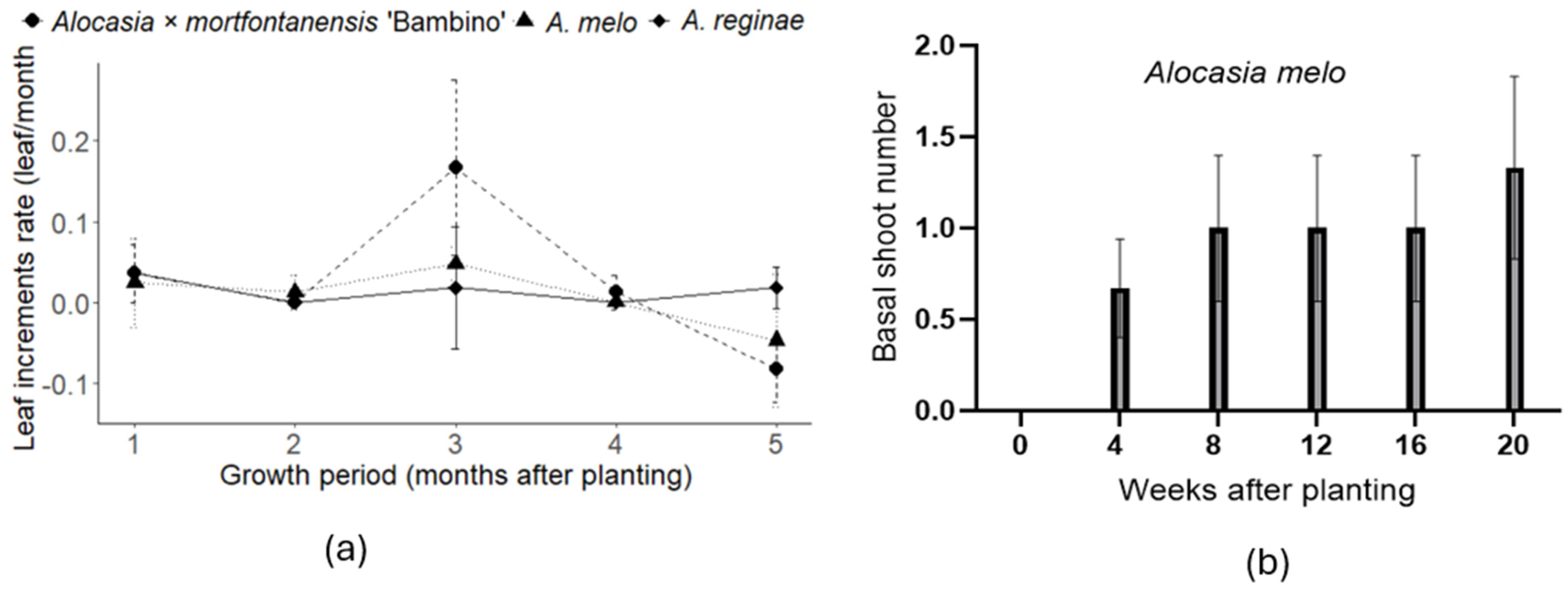
Figure 1 provides leaf increments and basal shoot growth for the three species over five months. Alocasia × mortfontanensis ‘Bambino’ exhibits varied leaf increment patterns: an initial increase in month one, stagnation in month two, resurgence in months three and four and a decline in month five. Alocasia melo displays fluctuations in leaf increments, with growth in the first three months, followed by no growth in months four and five. Alocasia reginae shows consistent positive leaf increments in the first three months, no growth in month four and a slight increase in month five. Only A. melo produced basal shoots; the other two taxa had none (Figure 1b). Each species exhibits distinct temporal leaf increment patterns, reflecting differences in growth dynamics and responses to environmental factors. The most pronounced leaf increment period for all three studied Alocasia taxa was in the third month after planting.
Analysis revealed significant differences in leaf width and length-to-width ratio, with Alocasia × mortfontanensis ‘Bambino’ exhibiting the species’ widest leaves and highest length-to-width ratio (Table 2). Additionally, the number of primary veins differed significantly, with Alocasia × mortfontanensis ‘Bambino’ and A. regina having a lower number than A. melo. However, no significant differences were found in plant height, petiole length or leaf length among the species.
Alocasia × mortfontanensis ‘Bambino’, A. melo and A. reginae have simple leaves with no lobing and varying leaf margins (undulate, slightly undulate, and entire to very slightly undulate) (Table 3). The adaxial leaf color ranges from dark green to silver to silver greenish, while the abaxial leaf color varies from red to light purple. All species have pendulous leaf lamina with different primary venation colors and leaf thicknesses; none have patterns on their petioles (Table 3). None of the Alocasia species produced flowers during this study.

3.2. Leaf Anatomy

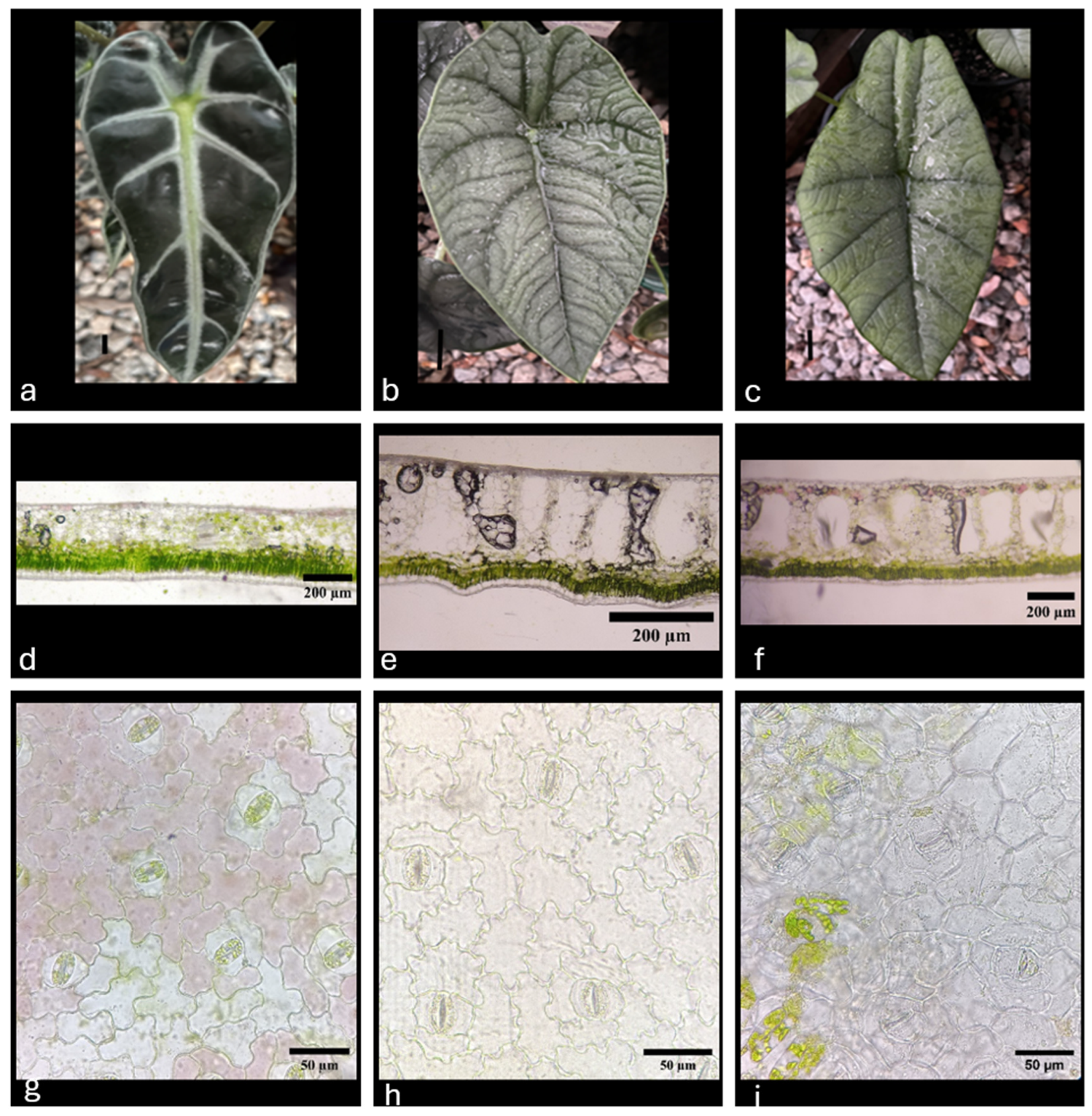
Figure 2 presents micrographs illustrating the leaves’ detailed morphological and anatomical features, including the stomatal complexes, of the three Alocasia taxa examined. The stomatal morphology across all Alocasia taxa investigated is paracytic, defined by the presence of two subsidiary cells positioned parallel to and on the exterior flanks of the guard cells. This specific configuration of the stomatal complex permits considerable flexibility, allowing the guard cells to undergo significant expansion and contraction.
The three Alocasia species show variability in characteristics such as stomatal width, abaxial epidermis thickness and overall leaf thickness (Table 4). Alocasia melo is distinguished by having the narrowest stomata compared to Alocasia × mortfontanensis ‘Bambino’ and A. reginae. However, the leaf tissue of A. melo is significantly thicker, measuring 268.37 µm, compared to the notably thinner leaves of Alocasia × mortfontanensis ‘Bambino’, which measure 119.82 µm. Consequently, Alocasia × mortfontanensis ‘Bambino’ had the highest palisade proportion relative to total leaf thickness. Despite A. melo exhibiting the most substantial leaf thickness overall, its abaxial epidermal layer is the thinnest, recorded at just 9.44 µm. This contrasts sharply with the thicker abaxial epidermises of Alocasia × mortfontanensis ‘Bambino’ and A. reginae, both exceeding 21 µm (Table 4, Figure 2).

3.3. Relative Water Content, Specific Leaf Weight and Leaf Moisture Loss

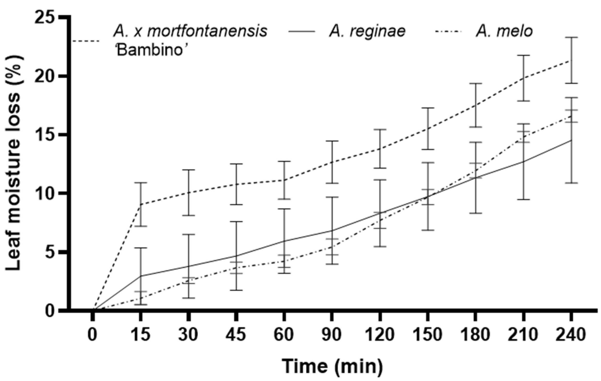
Alocasia melo leaves had the highest RWC (90.43%), followed by Alocasia × mortfontanensis ‘Bambino’ (88.52%) and A. regina (78.26%), as shown in Table 5. However, Alocasia × mortfontanensis ‘Bambino’ demonstrated a rapid initial dehydration, losing approximately 9% of its moisture content within the first 15 min, a rate greater than that of A. melo and A. reginae. Subsequent rates of moisture loss were similar (Figure 3). After four hours, Alocasia × mortfontanensis ‘Bambino’ incurred the highest (22%) moisture loss, while the losses recorded for A. melo and A. reginae were at 16–17% (Figure 3). In terms of specific leaf weight, Alocasia melo exhibited the highest specific leaf weight, significantly differing from Alocasia × mortfontanensis ‘Bambino’ and Alocasia reginae (Table 5). These results may indicate notable interspecies variations in leaf anatomical characteristics related to leaf thickness and density among the studied Alocasia species (Table 5). The Pearson correlation analysis between specific leaf weight and leaf moisture loss results in a coefficient of −0.297, suggesting a moderately negative relationship (Supplementary Figure S1). This implies that as the leaf’s weight increases, there tends to be a decrease in the amount of moisture lost, indicating a potential physiological adaptation where heavier leaves might exhibit a higher water retention capacity.

3.4. Leaf Chlorophyll, Carotene Content and Chlorophyll Stability Index

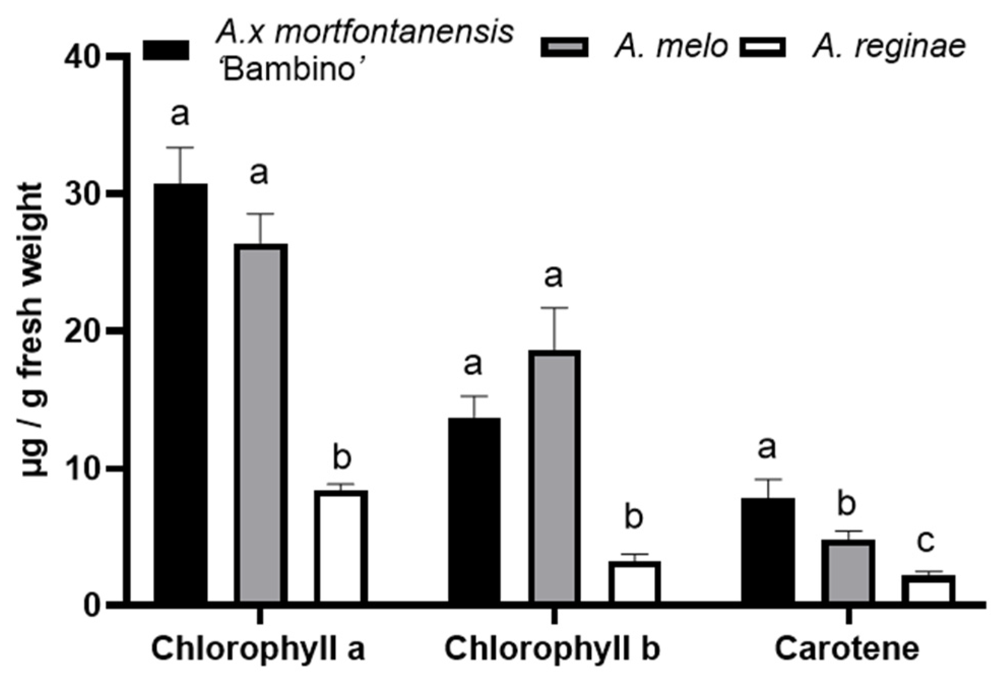
Quantitative profiles of the chlorophyll and carotene content within leaves of Alocasia × mortfontanensis ‘Bambino’, A. melo and A. reginae are graphically shown in Figure 4. The levels of leaf chlorophyll a and b in Alocasia × mortfontanensis ‘Bambino’ and A. melo are similar and higher than those in A. reginae. Leaf carotene content was highest in Alocasia × mortfontanensis ‘Bambino’, followed by A. melo and then A. reginae (Figure 4). Conversely, the ratio of chlorophyll a to chlorophyll b in Alocasia × mortfontanensis ‘Bambino’ is similar to that of A. reginae, both exceeding a ratio of >2, while A. melo has a comparatively lower ratio of 1.5 (Table 6).
Regarding the CSI, which denotes the resilience of pigments at both 25 °C and 56 °C temperatures, A. reginae had the highest stability, maintaining 92% and 89% stability at the respective temperatures. This was followed by A. melo, which showed stability of 87% at 25 °C and 84% at 56 °C. Alocasia × mortfontanensis ‘Bambino’ demonstrated the least chlorophyll stability with a CSI of 25 °C, further reducing to 68% at 56 °C.

3.5. Leaf Ultrastructure and Composition Analysis

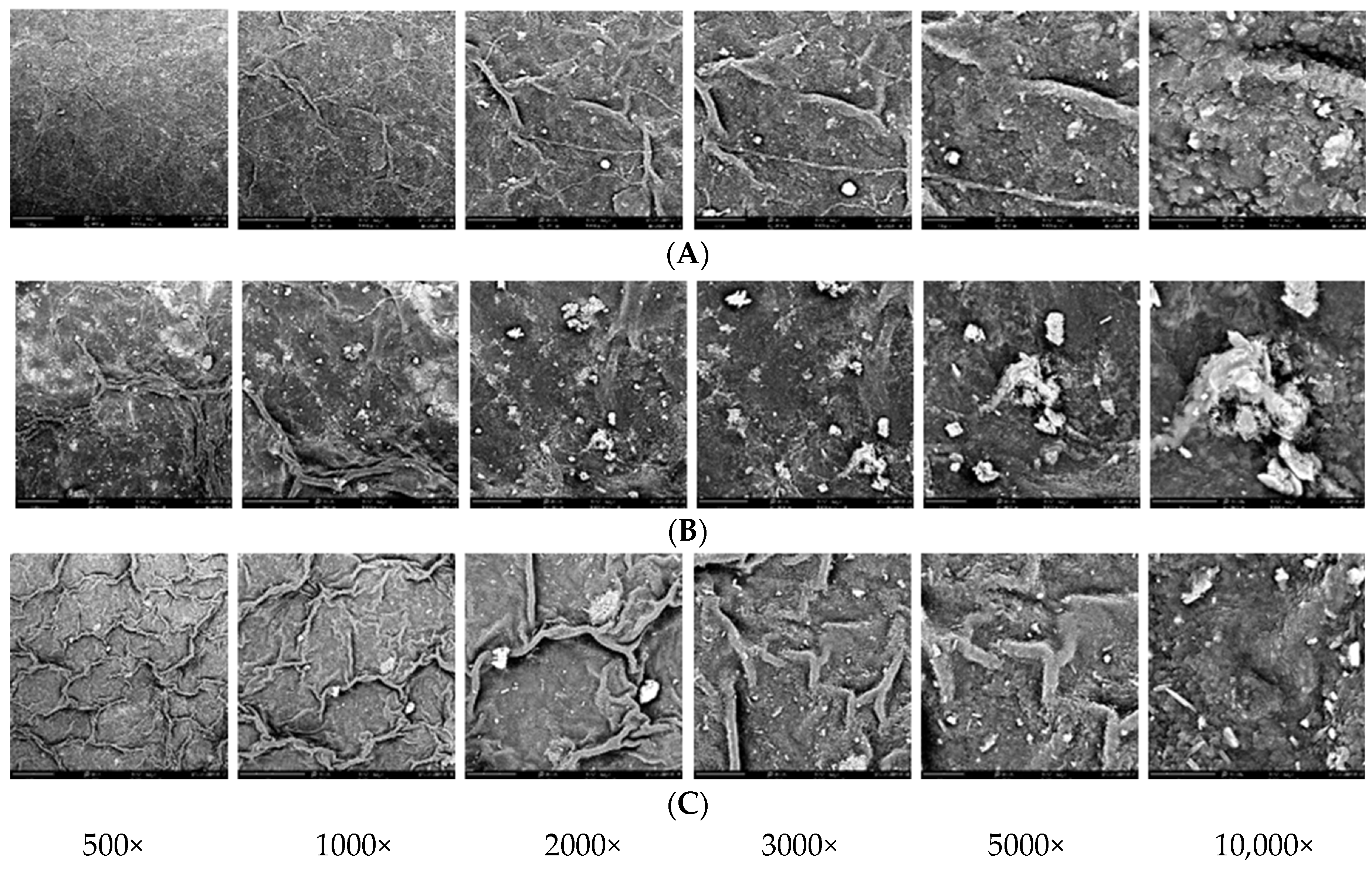
Figure 5 shows the leaf ultrastructure of Alocasia × mortfontanensis ‘Bambino’ (A), A. melo (B) and A. reginae (C) at magnifications of 500×, 1000×, 2000×, 3000×, 5000× and 10,000×. SEM EDX analysis indicated a relatively high percentage of potassium, calcium, chloride and silicon on the A. melo leaf surface compared to A. reginae and Alocasia × mortfontanensis ‘Bambino’ (Table 7). Additionally, A. melo shows the presence of crystals on the leaf surface (Figure 5) and contains rhodium, which is not detectable in A. reginae and Alocasia × mortfontanensis ‘Bambino’. Alocasia melo has about 15% potassium, A. reginae has about 10% and Alocasia × mortfontanensis ‘Bambino’ has about 8% (Table 7).

4. Discussion

Our study demonstrated the leaf morphological characteristics that can be used to identify the three Alocasia species. Alocasia × mortfontanensis ‘Bambino’ exhibits the fastest leaf growth (Figure 1), alongside the greatest ratio of chlorophyll a to chlorophyll b and carotene content (Figure 4). However, this taxon also presented the lowest chlorophyll stability (Table 6) and the fastest leaf moisture loss (Figure 3). A higher stability index indicates better resilience of chlorophyll to stress, while a lower stability index may suggest damage or degradation of chlorophyll molecules. Accordingly, A. melo and A. reginae leaves maintain a higher stability index than Alocasia × mortfontanensis ‘Bambino,’ inferring broader adaptability to temperature variations, as proposed by [18].
It was observed that Alocasia × mortfontanensis ‘Bambino’ possesses the thinnest leaves (Table 4), and this physiognomy potentially facilitates its highest water loss compared to A. reginae and A. melo. The correlation analysis between specific leaf weight and leaf moisture loss suggests a negative relationship (−0.297, Supplementary Figure S1); this is consistent with previous findings [18] that thicker leaves, especially those with a more substantial cuticular layer, can mitigate desiccation.
Alocasia melo and A. reginae display similarities in their leaf growth rates (Figure 1), leaf thickness (Table 4), rate of moisture loss (Figure 3) and chlorophyll stability (Table 6). The chlorophyll stability index is an indicative measure of the endurance of chlorophyll molecules under varying environmental stresses [17].
Leaf quality is important for tropical ornamentals, with moisture loss being a crucial factor influencing this aspect. To the best of our knowledge, apart from A. longiloba and A. mycorrhiza [19], no information is available on the edibility of the tubers and leaves of the three Alocasia involved in this study. Interestingly, A. melo is the only species that produces basal shoots, whereas the other two species only show leaf increments. This information provides insights into the propagation capabilities of the three Alocasia, which will be useful for commercial production practices and postproduction handling of these ornamental plants.
Variations in the ratio of chlorophyll a to chlorophyll b may reflect the presence of additional pigments, such as anthocyanins or carotenoids, which are contributors to the red or purple hues in leaf tissues [20,21], as exemplified by the leaf blade in A. melo (Figure 2). Moreover, variations in pigment composition alongside leaf structure and genetic factors can influence the observed color diversity in both wild and cultivated plants.
Leaf thickness and leaf water content can offer valuable insights into the plant’s water utilization, including its tolerance to drought and salinity conditions [22]. Alocasia × mortfontanensis ‘Bambino’ showed the highest moisture loss compared to the other two species, likely due to its thinner leaf structure and potentially higher stomatal density, facilitating transpiration [23]. This characteristic was not observed in A. melo and A. reginae, which possess relatively thicker leaves. These findings may provide information for optimal cultivation practices, including irrigation strategies and the selection of suitable planting media [24].
All three Alocasia taxa used in this study showed a lower stomatal density (Table 4) compared to other Indonesian species of Alocasia, such as A. alba, which exhibits stomatal densities ranging from 25 to 55 stomata per mm2 on the adaxial surface and 80–106 stomata per mm2 on the abaxial surface [25]. Other leaf characters were consistent with other species of Alocasia. However, A. melo and A. reginae have much thicker leaves than A. alba, likely due to a thick spongy mesophyll layer, as shown in Table 4. Thick layers of spongy mesophyll have also been reported in other species of Alocasia [25]. The contributory role of abaxial gas exchange in photosynthesis, accounting for approximately 50% of the overall activity, emphasizes its importance in overall leaf gas exchange processes [23].
Specific leaf weight fluctuates in response to environmental changes and can vary at different times of the day. Studies on alfalfa have shown that the specific leaf weight is at its lowest in the morning and increases in the afternoon due to the production of photosynthates during the day [26]. As such, specific leaf weight can affect dry matter production and the efficiency with which photosynthates are translocated within the plant, providing valuable information for cultivation and production methods.
Morphological and anatomical investigations are relevant in elucidating diversity levels and provide taxonomic information within the genus Alocasia. SEM EDX analysis demonstrated relatively high concentrations of potassium, calcium, chloride and silicon on the A. melo leaf surface as compared to A. reginae and Alocasia × mortfontanensis ‘Bambino’ (Figure 5). Alocasia melo contains rhodium, which is not detectable in A. reginae and Alocasia × mortfontanensis ‘Bambino’ (Table 7). Potassium (K) is involved in stomatal regulation [27], but its interaction with other measured parameters was not explored within the context of this study.
Araceae, including Alocasia and Colocasia, are characterized by the presence of calcium oxalate (Ca(COO)2) in all parts of the plants, which may lead to skin irritation [28].
Our study demonstrated that Alocasia melo leaves contain about 6% Ca, the highest of the three Alocasia. While not essential for plant development, silicon offers advantages for plants under stressful conditions. It has been recognized for mitigating abiotic and biotic stressors in plants [29,30,31].
Typically, plants contain trace amounts of rhodium, estimated to be in the range of 1–2 ppb [10], yet we found an unusually high (2%) proportion of rhodium on the A. melo leaves. Rhodium is a rare non-radioactive metal and does not play a known biological role. It occurs uncombined in nature and with other platinum metals in river sands [32].
Our study detected the presence of magnesium in A. reginae and Alocasia × mortfontanensis ‘Bambino’ leaves (Table 7). Mg is an essential component of chlorophyll pigments in the light-capturing complex of chloroplasts; therefore, it is involved in photosynthetic CO2 assimilation [33].

5. Conclusions

Through various methods employed, this study shows that growth rates, leaf morphology and anatomy differ among the three taxa of Alocasia studied. This study reveals variations in the adaxial and abaxial leaf color, stomatal width and epidermal thickness. Alocasia × mortfontanensis ‘Bambino’ shows the fastest leaf production, with peak growth occurring from the 8th to the 12th week post-planting across all taxa. Alocasia melo has the capability to produce new basal shoots and exhibits the highest relative water content, while Alocasia × mortfontanensis ‘Bambino’ exhibits rapid moisture loss initially. Alocasia reginae has the most consistent chlorophyll levels at 25 °C and 56 °C. Elemental analysis using SEM EDX showed that the leaf surface of A. melo contains higher concentrations of potassium, calcium, chloride and silicon than the other two species and uniquely possesses rhodium. Information on the Alocasia leaf morpho-anatomy and growth from this study is useful in optimizing Alocasia production across diverse ecosystems.

Supplementary Materials

The following supporting information can be downloaded at https://www.mdpi.com/article/10.3390/horticulturae10080778/s1, Figure S1: Pearson correlation analysis of leaf moisture loss and specific leaf weight of Alocasia × mortfontanensis ‘Bambino’, A. melo and A. reginae.

Author Contributions

Conceptualization, K. and M.F.; methodology, K., M.S.R. and D.D.; software, D.D.M. and Y.S.P.; validation, J.G.K., N.P.S.A. and M.S.R.; formal analysis, K., D. and M.S.R.; data curation, M.F., Y.S.P., D.D.M. and N.P.S.A.; writing—original draft preparation, K.; writing—review and editing, M.F., N.P.S.A. and M.S.R.; visualization, M.F. and D.; supervision, K., J.G.K. and D.D.; project administration, J.G.K.; funding acquisition, K. All authors have read and agreed to the published version of the manuscript.

Funding

This research was funded by Dana Abadi Perguruan Tinggi—Lembaga Pengelola Dana Pendidikan (DAPT-LPDP) through National Research Collaboration Funding Program (Riset Nasional) grant number 508/IT3.D10/PT.01.03/P/B/2023. The APC was funded by the authors and DRI, IPB University.

Data Availability Statement

Data supporting the findings of this work are available in the Supplementary Materials.

Acknowledgments

The authors thank Imzar Pulungan for maintaining the plants in the greenhouse, Joko Mulyono of IPB University for helping with histology sample preparation, Nurwita Sari for her administrative support and Anita Kurnia from Walisongo University for her assistance in the SEM analysis.

Conflicts of Interest

The authors declare no conflicts of interest. The funder had no role in the design of this study, in the collection, analyses or interpretation of data, in the writing of the manuscript or in the decision to publish the results.

References

  1. Calle, J.; Gasparre, N.; Benavent-Gil, Y.; Rosell, C.M. Aroids as Underexplored Tubers with Potential Health Benefits. In Advances in Food and Nutrition Research; Elsevier: Amsterdam, The Netherlands, 2021; Volume 97, pp. 319–359. ISBN 978-0-12-824580-4. [Google Scholar]
  2. The World Flora Online. Alocasia (Schott) G. Don. Available online: https://www.worldfloraonline.org/taxon/wfo-4000001337 (accessed on 27 May 2024).
  3. Nauheimer, L.; Boyce, P.C.; Renner, S.S. Giant Taro and Its Relatives: A Phylogeny of the Large Genus Alocasia (Araceae) Sheds Light on Miocene Floristic Exchange in the Malesian Region. Mol. Phylogenetic Evol. 2012, 63, 43–51. [Google Scholar] [CrossRef]
  4. Croat, T.B.; Ortiz, O.O. Distribution of Araceae and the Diversity of Life Forms. Acta Soc. Bot. Pol. 2020, 89, 8939. [Google Scholar] [CrossRef]
  5. Ongpoy, T. The Medicinal Properties of the Alocasia Genus: A Systematic Review. J. Asian Assoc. Sch. Pharm. 2017, 6, 25–33. [Google Scholar]
  6. Hay, A.; Boyce, P.C.; Wong, K.M. Plate 315. Alocasia melo. Curtis’s Bot. Mag. 1997, 14, 82–86. [Google Scholar] [CrossRef]
  7. Plants of the World Online. Alocasia reginae N.E.Br. Available online: https://powo.science.kew.org/taxon/urn:lsid:ipni.org:names:84234-18 (accessed on 27 May 2024).
  8. Aroidpedia. Alocasia. Available online: https://www.aroidpedia.com/alocasia (accessed on 27 May 2024).
  9. Plants of the World Online. Alocasia × mortfontanensis André. Available online: https://powo.science.kew.org/taxon/urn:lsid:ipni.org:names:84211-1 (accessed on 18 May 2024).
  10. Arogundade, O.O.; Adedeji, O. The Importance of Foliar Anatomy in the Taxonomy of the Genus Alocasia (Schott) G. Don. Jordan J. Biol. Sci. 2019, 12, 67–75. [Google Scholar]
  11. Fahn, A. Plant Anatomy, 2nd ed.; Pergamon Press: Oxford, MI, USA; New York, NY, USA, 1974; ISBN 978-0-08-017241-5. [Google Scholar]
  12. Pérez-Pérez, J.G.; Syvertsen, J.P.; Botía, P.; García-Sánchez, F. Leaf Water Relations and Net Gas Exchange Responses of Salinized Carrizo citrange Seedlings during Drought Stress and Recovery. Ann. Bot. 2007, 100, 335–345. [Google Scholar] [CrossRef]
  13. Bueno, A.; Sancho-Knapik, D.; Gil-Pelegrín, E.; Leide, J.; Peguero-Pina, J.J.; Burghardt, M.; Riederer, M. Cuticular Wax Coverage and its Transpiration Barrier Properties in Quercus coccifera L. Leaves: Does the Environment Matter? Tree Physiol. 2020, 40, 827–840. [Google Scholar] [CrossRef]
  14. Hou, W.; Tränkner, M.; Lu, J.; Yan, J.; Huang, S.; Ren, T.; Cong, R.; Li, X. Diagnosis of Nitrogen Nutrition in Rice Leaves Influenced by Potassium Levels. Front. Plant Sci. 2020, 11, 165. [Google Scholar] [CrossRef] [PubMed]
  15. Pauli, D.; White, J.W.; Andrade-Sanchez, P.; Conley, M.M.; Heun, J.; Thorp, K.R.; French, A.N.; Hunsaker, D.J.; Carmo-Silva, E.; Wang, G.; et al. Investigation of the Influence of Leaf Thickness on Canopy Reflectance and Physiological Traits in Upland and Pima Cotton Populations. Front. Plant Sci. 2017, 8, 1405. [Google Scholar] [CrossRef] [PubMed]
  16. Costache, M.A.; Campeanu, G.; Neata, G. Studies Concerning the Extraction of Chlorophyll and Total Carotenoids from Vegetables. Rom. Biotechnol. Lett. 2012, 17, 7702–7708. [Google Scholar]
  17. Mohan, M. Chlorophyll Stability Index (CSI): Its Impact on Salt Tolerance in Rice. Int. Rice Res. Notes 2000, 25, 38–39. [Google Scholar]
  18. Ndlovu, E.; Van Staden, J.; Maphosa, M. Morpho-Physiological Effects of Moisture, Heat and Combined Stresses on Sorghum bicolor [Moench (L.)] and its Acclimation Mechanisms. Plant Stress 2021, 2, 100018. [Google Scholar] [CrossRef]
  19. Arbain, D.; Maria, L.; Sinaga, R.; Taher, M.; Susanti, D.; Amiruddin, Z.; Khotib, J. Traditional Uses, Phytochemistry, Biological Activities of Alocasia Species: A Systematic Review. Front. Pharmacol. 2022, 13, 849704. [Google Scholar] [CrossRef]
  20. Li, Z.; Tang, M.; Hassan, M.J.; Zhang, Y.; Han, L.; Peng, Y. Adaptability to High Temperature and Stay-Green Genotypes Associated with Variations in Antioxidant, Chlorophyll Metabolism, and γ-Aminobutyric Acid Accumulation in Creeping Bentgrass Species. Front. Plant Sci. 2021, 12, 750728. [Google Scholar] [CrossRef]
  21. Song, B.; Xu, H.; Chen, L.; Fan, X.; Jing, Z.; Chen, S.; Xu, Z. Study of the Relationship between Leaf Color Formation and Anthocyanin Metabolism among Different Purple Pakchoi Lines. Molecules 2020, 25, 4809. [Google Scholar] [CrossRef]
  22. Afzal, A.; Duiker, S.W.; Watson, J.E. Leaf Thickness to Predict Plant Water Status. Biosyst. Eng. 2017, 156, 148–156. [Google Scholar] [CrossRef]
  23. Daningsih, E.; Mardiyyaningsih, A.N.; Da Costa, Y.O.; Primawati, R.; Karlina, S. Changes in Stomatal Distribution and Leaf Thickness in Response to Transpiration Rate in Six Dicot Plant Species. IOP Conf. Ser. Earth Environ. Sci. 2022, 976, 012060. [Google Scholar] [CrossRef]
  24. Asih, N.P.S.; Hendriyani, E.; Tihurua, E.F. Morphological and Anatomical Variations among Alocasia alba Schott Accessions in Bali Botanic Garden. J. Trop. Biodivers. Biotechnol. 2022, 7, 66823. [Google Scholar] [CrossRef]
  25. Wall, S.; Vialet-Chabrand, S.; Davey, P.; Van Rie, J.; Galle, A.; Cockram, J.; Lawson, T. Stomata on the Abaxial and Adaxial Leaf Surfaces Contribute Differently to Leaf Gas Exchange and Photosynthesis in Wheat. New Phytol. 2022, 235, 1743–1756. [Google Scholar] [CrossRef]
  26. Robinson, D.; Dobrenz, A. Specific Leaf Weight: Daily Trends in an Alfalfa Field. Forage Grain Rep. 1986, 67, 5–6. [Google Scholar]
  27. Kumar, P.; Kumar, T.; Singh, S.; Tuteja, N.; Prasad, R.; Singh, J. Potassium: A Key Modulator for Cell Homeostasis. J. Biotechnol. 2020, 324, 198–210. [Google Scholar] [CrossRef]
  28. Du Thanh, H.; Phan Vu, H.; Vu Van, H.; Le Duc, N.; Le Minh, T.; Savage, G. Oxalate Content of Taro Leaves Grown in Central Vietnam. Foods 2017, 6, 2. [Google Scholar] [CrossRef]
  29. Pieniazek, F.; Dasgupta, M.; Messina, V.; Devi, M.P.; Devi, Y.I.; Mohanty, S.; Singh, S.; Sahoo, B.B.; Nongdam, P.; Acharya, G.C.; et al. Differential Occurrence of Cuticular Wax and Its Role in Leaf Physiological Mechanisms of Three Edible Aroids of Northeast India. Agriculture 2022, 12, 724. [Google Scholar] [CrossRef]
  30. Zargar, S.M.; Mahajan, R.; Bhat, J.A.; Nazir, M.; Deshmukh, R. Role of Silicon in Plant Stress Tolerance: Opportunities to Achieve a Sustainable Cropping System. 3 Biotech 2019, 9, 73. [Google Scholar] [CrossRef]
  31. Zellner, W.; Tubaña, B. Silicon’s Role in Plant Stress Reduction and Why This Element is Not Used Routinely for Managing Plant Health. Plant Dis. 2021, 105, 2033–2046. [Google Scholar] [CrossRef]
  32. The Royal Society of Chemistry. Rhodium. Available online: https://www.rsc.org/periodic-table/element/45/rhodium (accessed on 27 May 2024).
  33. Gerendás, J.; Führs, H. The Significance of Magnesium for Crop Quality. Plant Soil 2013, 368, 101–128. [Google Scholar] [CrossRef]
Figure 1. Leaf increment rate (a) and basal shoot growth (b) during five-month growth period of Alocasia × mortfontanensis ‘Bambino’, A. melo and A. reginae.
Horticulturae 10 00778 g001
Figure 2. Alocasia mortfontanensis ‘Bambino’ leaf morphology (a), leaf cross-section (d) and stomata (g); A. melo leaf morphology (b), leaf cross-section (e) and stomata (h); and A. reginae leaf morphology (c), leaf cross-section (f) and stomata (i).
Horticulturae 10 00778 g002
Figure 3. Leaf moisture loss of Alocasia × mortfontanensis ‘Bambino’, A. melo and A. reginae.
Horticulturae 10 00778 g003
Figure 4. Leaf chlorophyll a, chlorophyll b and carotene of Alocasia × mortfontanensis ‘Bambino’, A. melo and A. reginae. Different letters represent significant differences in the pigment content among three Alocasia species according to Tukey’s test at α = 0.05. Bars in each column represent the standard error of means (N = 4).
Horticulturae 10 00778 g004
Figure 5. Leaf ultrastructure of (A) Alocasia × mortfontanensis ‘Bambino’, (B) A. melo and (C) A. reginae.
Horticulturae 10 00778 g005
Table 1. The number of leaves per plant and leaf increase rate among three taxa of Alocasia (A. melo, A. reginae and Alocasia × mortfontanensis ‘Bambino’) observed within a five-month period.
SpeciesNumber of Leaves per Plant (Months after Planting)
012345
Alocasia × mortfontanensis ‘Bambino’9.3 ± 0.6 a10.3 ± 0.6 a10.3 ± 0.6 a15.0 ± 0.6 a15.3 ± 2.5 a13.0 ± 2.6 a
A. melo5.3 ± 0.6 b6.0 ± 0.1 b6.3 ± 0.6 b7.7 ± 1.0 b7.7 ± 0.6 b6.3 ± 2.1 b
A. regina3.5 ± 0.7 c4.5 ± 0.7 b4.5 ± 0.7 c5.0 ± 0.7 b5.0 ± 1.4 b5.5 ± 0.7 b
p-value2.9 × 10−4 **9.4 × 10⁻⁴ **2.9 × 10⁻⁴ **3.6 × 10⁻³ **2.6 × 10⁻³ **0.018 *
Note: * = significant; ** = highly significant. Different letters represent significant differences according to Tukey’s test at α = 0.05.
Table 2. Plant height, petiole length, leaf length and width, leaf length-to-width ratio and number of primary veins among three taxa of Alocasia (Alocasia × mortfontanensis ‘Bambino’, A. melo and A. reginae).
SpeciesPlant Height (cm)Petiole Length (cm)Leaf Length (cm)Leaf Width (mm)Leaf Length-to-Width RatioNumber of Primary Veins
Alocasia × mortfontanensis ‘Bambino’33.3 ± 1.5 a24.1 ± 1.5 a18.8 ± 1.3 a7.0 ± 0.7 b2.7 ± 0.1 a14.0 ± 0.0 b
A. melo28.7 ± 1.5 a17.3 ± 4.6 a18.2 ± 4.5 a14.5 ± 2.8 a1.2 ± 0.1 b20.7 ± 1.1 a
A. regina32.5 ± 3.5 a23.0 ± 1.4 a13.7 ± 1.8 a10.75 ± 1.8 ab1.3 ± 0.0 b13.0 ± 1.4 b
p-value0.087 ns0.098 ns0.258 ns0.016 *2 × 10−5 **4.7 × 10⁻⁴ **
Note: * = significant; ** = highly significant; ns = not significant according to ANOVA at α = 0.05. Different letters represent significant differences according to Tukey’s test at α = 0.05.
Table 3. Leaf morphological features of Alocasia × mortfontanensis ‘Bambino’, A. melo and A. reginae.
Morphological FeatureAlocasia × mortfontanensis ‘Bambino’A. meloA. reginae
Leaf typeSimpleSimpleSimple
Leaf lobingAbsentAbsentAbsent
Leaf marginUndulateSlightly undulateEntire to very slightly undulate
Leaf shapeSagittateOvateElliptic
Leaf apexAcuteAcuteAcute
Leaf baseCordate CordateCordate
Peltate sizeHalf peltateAlmost completely peltateAlmost completely peltate
PhyllotaxyBasalBasalBasal
Adaxial leaf colorDark greenSilverSilver greenish
Abaxial leaf colorRedLight purpleRed
Leaf thicknessLeatheryCoriaceous Thickly leathery
Leaf laminaPendulous, base upPendulous, base upPendulous, base up
Leaf patternAbsentRugoseSlightly rugulose
Main venation colorGreen grayishDark greenDark green
Petiole patternAbsentAbsentAbsent
Table 4. Leaf anatomical characteristics of Alocasia × mortfontanensis ‘Bambino’, A. melo and A. reginae.
SpeciesStomatal Density (per mm2)Stomatal Length (µm)Stomatal Width (µm)Palisade Thickness (µm)Epidermal Thickness (µm)Leaf Thickness (µm)
AdaxialAbaxial
Alocasia × mortfontanensis ‘Bambino’11.17 ± 2.56 a32.99 ± 1.21 a20.14 ± 2.91 a52.73 ± 13.34 a22.45 ± 6.28 a21.95 ± 5.77 a119.82 ± 11.13 b
A. melo11.67 ± 1.53 a33.61 ± 3.87 a11.53 ± 1.74 b44.73 ± 2.51 a21.66 ± 12.61 a9.44 ± 2.99 b268.37 ± 37.54 a
A. reginae11.17 ± 3.87 a36.31 ± 3.69 a20.32 ± 2.39 a43.77 ± 7.13 a24.99 ± 8.01 a22.88 ± 3.66 a224.49 ± 22.09 a
p-value0.969 ns0.155 ns0.001 **0.445 ns0.866 ns0.027 *0.001 **
Note: * = significant; ** = highly significant; ns = non-significant according to ANOVA at α = 0.05. Different letters represent significant differences in leaf anatomical characteristics among the three Alocasia species according to Tukey’s test at α = 0.05, except for leaf thickness, which was assessed using Dunn’s test at α = 0.05.
Table 5. Leaf relative water content and specific leaf weight of Alocasia × mortfontanensis ‘Bambino’, A. melo and A. reginae.
SpeciesRelative Water Content (%)Specific Leaf Weight (mg·cm−2)
Alocasia × mortfontanensis ‘Bambino’88.52 ± 2.17 a28.54 ± 2.04 c
A. melo90.43 ± 2.04 a56.53 ± 1.66 a
A. reginae78.26 ± 2.62 b38.17 ± 0.51 b
p-value4.2 × 10−3 **4.9 × 10⁻⁹ **
Note: ** = highly significant. Different letters represent significant differences in leaf anatomical characteristics among the three Alocasia species according to Tukey’s test at α = 0.05.
Table 6. Chlorophyll a–b ratio and chlorophyll stability index (1) of Alocasia × mortfontanensis ‘Bambino’, A. melo and A. reginae leaves.
SpeciesRatio of Chlorophyll a to Chlorophyll bChlorophyll Stability Index at 25 °C (%)Chlorophyll Stability Index at 56 °C (%)
Alocasia × mortfontanensis ‘Bambino’2.26 a75.8 ± 0.2 a68.3 ± 0.1 b
A. melo1.52 b87.9 ± 0.2 a83.8 ± 0.1 a
A. reginae2.77 a92.4 ± 0.1 a89.2 ± 0.1 a
p-value0.017 *0.307 ns0.011 *
Note: (1) Based on the SPAD values. * = significant; ns = not significant according to ANOVA at α = 0.05. Different letters represent significant differences according to Tukey’s test at α = 0.05.
Table 7. Elemental composition by weight based on SEM-EDX scans of Alocasia leaf surface.
% Dry WeightA. × mortfontanensis ‘Bambino’A. meloA. reginae
C65.3537.160.38
O21.2515.4820.8
K8.0415.3110.6
Cl1.458.913.79
Ca1.295.970.85
S0.860.840.49
Mg0.7301.35
p0.600.68
Si0.431.711.07
Rh02.230
Disclaimer/Publisher’s Note: The statements, opinions and data contained in all publications are solely those of the individual author(s) and contributor(s) and not of MDPI and/or the editor(s). MDPI and/or the editor(s) disclaim responsibility for any injury to people or property resulting from any ideas, methods, instructions or products referred to in the content.