Article

Identification of Resistance QTLs to Black Leaf Streak Disease (Due to Pseudocercospora fijiensis) in Diploid Bananas (Musa acuminata)

1
CIRAD, UMR AGAP Institute, F-34398 Montpellier, France
2
UMR AGAP Institute, University Montpellier, CIRAD, INRAe, Institut Agro, F-34398 Montpellier, France
3
CIRAD, UMR PHIM, F-34398 Montpellier, France
4
UMR PHIM, University Montpellier, INRAe, CIRAD, Institut Agro, F-34398 Montpellier, France
5
CIRAD, UMR AGAP Institute, F-97130 Capesterre-Belle-Eau, France
*
Author to whom correspondence should be addressed.
Horticulturae 2024, 10(6), 608; https://doi.org/10.3390/horticulturae10060608
Submission received: 3 April 2024 / Revised: 16 May 2024 / Accepted: 31 May 2024 / Published: 7 June 2024
(This article belongs to the Special Issue Developments in the Genetics and Breeding of Banana Species)

Abstract

:
Black Leaf Streak Disease (BLSD), caused by the fungus Pseudocercospora fijiensis, is a recent pandemic and the most economically and environmentally important leaf disease of banana. To assist breeding of varieties with durable resistance to the rapidly evolving P. fijiensis, we used a diploid genitor ‘IDN 110’ with partial resistance to BLSD to search for QTLs. We assessed diploid progeny of 73 hybrids between ‘IDN 110’ and the diploid cultivar ‘Khai Nai On’, which is susceptible to BLSD. Hybrids were phenotyped with artificial inoculation under controlled conditions. This method allowed us to focus on resistance in the early stages of the interaction already identified as strongly influencing BLSD epidemiology. Progeny were genotyped by sequencing. As both parents are heterozygous for large reciprocal translocations, the distribution of recombination was assessed and revealed regions with low recombination rates. Fourteen non-overlapping QTLs of resistance to BLSD were identified of which four main QTLs from the ‘IDN110‘ parent, located on chromosomes 06, 07, 08, and 09, were shown to be of interest for marker-assisted selection. Genes that underline those four QTLs are discussed in the light of previous literature.

1. Introduction

Bananas are a major starchy staple food, which is consumed worldwide, and a major cash crop. Banana cultivars were domesticated for their edible fruits from wild species native of South and South East Asia, the main one being M. acuminata (A genome, AAw, 2 n = 22). Cultivars that have parthenocarpic unseeded fruit are mainly diploid or triploid [1,2]. Banana is a giant perennial herbaceous plant that easily reproduces asexually via suckers. More than half (57%) of the world banana production, including local and export banana, are dessert varieties that are somaclonal and belong to the AAA group Cavendish subgroup [3]. This mono-varietal culture has facilitated the expansion of emerging diseases such as the Black Leaf Streak Disease (BLSD), also named black Sigatoka disease. This foliar disease is caused by Pseudocercospora fijiensis (formerly named Mycosphaerella fijiensis), a hemibiotrophic ascomycete fungus with high evolutionary potential [4,5,6,7].
BLSD is considered to be the most economically important leaf disease of banana [8]. P. fijiensis gradually displaced P. musicola, another of the three members of the Sigatoka disease complex. BLSD was first recognized in 1963 in Fiji (South Pacific Ocean) and first appeared on the American continent in Honduras in 1972 and on the African continent in Zambia in 1973. It continued its expansion in the 21st century, which can now be considered as almost complete [6]. BLSD affects a wide range of banana genotypes including the Cavendish subgroup (AAA). On the one hand, BLSD is very detrimental to the banana trade because it reduces the photosynthetic surface resulting in serious yield losses. On the other hand, it accelerates fruit ripening and has a negative impact on the quality and transportability of the fruits, thereby rendering them unsuitable for sale [9,10,11]. BLSD is controlled chemically through the application of fungicides and by preventive leaf removal. When grown for export, banana requires weekly fungicide applications involving up to 50 treatments a year in some environmental contexts. Chemical control is not only costly, but causes major environmental problems [12]. Populations of P. fijiensis are large and, depending on the environmental conditions, can reproduce almost all year round [13,14]. P. fijiensis can disperse over long distance via ascospores resulting from sexual reproduction and over shorter distances via conidia resulting from asexual reproduction [15], giving it rapid adaptation potential, as already observed in the form of fungicide-resistant strains [14].
To alleviate the human, environmental, and financial impact of BLSD, there is a need for further advances in genetic control as well as preventive biocontrol. The development of resistant cultivars is the focus of several banana breeding programmes worldwide through pedigree or reconstructive breeding strategies to obtain triploid cultivars from diploid accessions [16,17].
Field reactions of Musa accessions to BLSD have been evaluated to identify resistant cultivars or genitors (for a review, see Guzman et al. [6]). Studies have shown a graduated response ranging from susceptibility to marked resistance. Three major types of reaction to the disease were first reported by Fouré et al. [18]. The first type is characterised by blocked symptom development at an early stage, and no asexual or sexual sporulation. This reaction, called highly resistant or resistant, has been described in particular in the accession ‘Calcutta 4’ (AAw, M. acuminata ssp. burmannica) and in the cultivar ‘Yangambi KM5’ (AAA, Ibota subgroup), but their resistance was overcome by some strains [19,20]. The second type of reaction is partial resistance (PR), also called moderately resistant reaction, characterised by normal but slow development of symptoms and a low sporulation rate. PR is recommended for breeding programmes aimed at producing varieties with durable disease resistance in particular against pathogens with rapid adaptive potential like P. fijiensis [21,22]. The third type of reaction is susceptibility (S) characterised by rapid development of symptoms from streaks to mature leaf spot and necrosis with a high sporulation rate.
Few resistant hybrids have been released to date. The Honduras Foundation for Agricultural Research breeding programme was the first breeding programme to deliver resistant hybrids [23,24]. Unfortunately, some that were intensively cultivated in Cuba have shown a drop in resistance and locally aggressive strains of P. fijiensis have been identified [25].
Breeding banana varieties is subject to major constraints, namely the reduced fertility of cultivars and the need to manage polyploidy, since the optimum ploidy level for cultivars is triploid. One major aim of breeding is to obtain hybrids with durable resistance to BLSD [17,26,27,28,29,30,31]. Long-lasting resistance is important as varietal change is slow in banana, e.g., Cavendish somaclones are the world main varieties since 1950s. To this end, better knowledge is needed of host resistance, including its genetic inheritance [7]. Ortiz and Vuylsteke [32] and Craenen and Ortiz [33] concluded from genetic analyses that the resistance of the accession ‘Calcutta 4’ was due to a recessive allele in a major locus and at least two minor independent loci with additive effects. However, to the best of our knowledge, no QTL that confers total or partial resistance to BLSD has been published to date. However, today, QTL identification in banana can benefit from banana genome sequencing and from the identification of the location of chromosome translocations that influence segregation and genetic mapping [34,35,36,37].
The aim of the present study is to identify QTLs for resistance to BLSD using the progeny of two diploid parents. Both parents are cultivars (AAcv) to ensure their direct application in breeding with no undesirable characters from wild accessions. The nearest diploid cultivars to the ancestors of the main world dessert cultivars have been identified as belonging to the AAcv Mchare and Khai subgroups [2,38,39]. The Khai subgroups include both parents used in this study, accession ‘Khai Nai On’ (KNO) and accession ‘IDN 110’ (IDN110). KNO, which is used as the female parent, is susceptible to BLSD, whereas IDN110, which is used as male parent, shows partial resistance [16,40,41]. IDN110, also known as ‘AAcv Rose’ or its somaclone ‘Gu Nin Chiao’, is a frequent genitor in world breeding programmes thanks to both its resistance and fruit quality [31]. The progeny of 73 hybrids between the two heterozygous parents were genotyped by sequencing and phenotyped in controlled conditions. The recombinations along chromosomes, the QTLs identified, and the genes in the QTLs from the PR parent are discussed later on.

2. Materials and Methods

2.1. Plant Material

Resistance to BLSD was evaluated on a population derived from crosses between two diploid banana cultivars: the susceptible cultivar KNO (PT-BA-00148) as female, and IDN110 (PT-BA-00131), a cultivar with partial resistance, as male. Both accessions are field grown and belong to the CIRAD collection managed by the CIRAD-INRAe Biological Resource Center for Tropical Plants in Guadeloupe (French West Indies) [42].
To get around the problem of the parents’ low fertility, the population was obtained from seeds belonging to five bunches. The five crosses were performed in the CIRAD breeding station at Neufchâteau in Guadeloupe. To maximise germination of all viable seeds, embryo rescue was performed as described in Bakry [43] and 107 hybrids were obtained. The in vitro plants were sent to CIRAD Montpellier (France) for phenotyping and genotyping. Hybrids were kept in a climate chamber from weaning up to two months old in 0.5 L, and then 1 L pots containing peat and potting soil [respectively, 4 vol.: 1 vol., and 2 vol.:1 vol.]. They were then transplanted in 5 L pots containing a mixture of peat, potting soil, and pozzolan [1 vol.: 2 vol.: 1 vol.] into a greenhouse. Climate chamber and greenhouse were at 70–80% humidity, 26 to 28 °C, and a 12 h light photoperiod. A total of 73 hybrids developed normally up to adulthood, and were then phenotyped. All the plants retained had a similar size and a minimum of 6 green leaves. When the plants were 6.5 months old, the youngest fully mature leaf was harvested, as described in [40].

2.2. Artificial Inoculation and Disease Evaluation

Disease evaluation was carried out in controlled conditions through artificial inoculation of fragments of detached leaves kept on a survival medium in Petri dishes. Plant material, survival medium (0.4% bacto agar and 0.0005% gibberellic acid), and fungal inoculum preparations and inoculation were performed using the methods described in Pérez-Vicente, Carreel et al. [40]. Inoculum was obtained from isolate GLP701 (CIRAD collection), which is considered to be aggressive. Conidial suspension was adjusted to a final concentration of 40,000 conidia/mL in a 0.02% Tween solution [40]. Four leaf fragments were cut out of the youngest fully mature leaf of each parent and each hybrid. Three of them, sprayed with the inoculum, formed our three replicates. The fourth fragment was inoculated with a solution with no isolate as control. The plates were randomly distributed in the incubation chamber and scanned using ‘Epson perfection V10’ with a 300 dpi resolution at 2, 3, 4, 5, 7, and 9 weeks post-inoculation (wpi). Disease incidence was quantified from scans using the machine learning image analysis software IPSDK Explorer v. 3.0.2.2. [44] supplemented by the LeAFtool program (https://gitlab.cirad.fr/phim/LeAFtool, accessed on 23 January 2023). The number of lesions and the percentage of diseased leaf surface were obtained for each inoculated leaf fragment and each wpi time point.

2.3. Analysis of Phenotypic Data

Eleven phenotypic variables were obtained directly from image analysis: the percentage of diseased leaf surface (PD) at wpi 2, 3, 4, 5, 7, and 9 and the number of lesions (NL) at wpi 2, 3, 4, 5, and 7, named, respectively, PD_wpiX and NL_wpiX with X = 2, 3, 4, 5, 7, or 9. As lesions may merge at late stages, the number of lesions at wpi 9 was replaced by the maximum number of lesions observed the 9 weeks (NL_max). Four disease parameters were calculated from these phenotypic variables. The area under the disease progression curve (AUDPC) produced by PD_wpiX values, is a synthetic value that is representative of the level of disease over the entire 9 week monitoring period. AUDPC was obtained with the R ‘epifitter’ package [45], hereafter termed PD_Audpc. The incubation period was estimated as the week with at least 4 lesions and termed NL_Tmin4L. The two last calculated disease parameters were obtained from the growth curve of the percentage of diseased surface that was identified closest to the nonlinear “Weibull” function using Akaike’s information criterion (AIC) [46,47]. These two parameters were obtained using the R ‘nlme’ package, and represent the value of the horizontal asymptote (‘Asym’) and the value of y at the inflection point (‘Drop’), termed, respectively, PD_Wei-A and PD_Wei-D [48].
The best linear unbiased prediction (BLUP) values of these phenotypic data were extracted from analysis of generalised linear mixed models (glmm) using R software v. 4.2.2 [49]. Normality and variance homogeneity of the residuals were tested using, respectively, the Shapiro–Wilk test and Levene’s test. Like many disease data, the 16 phenotypic variables did not fit the assumed Gaussian distributions, and consequently, the glmm models were applied using the glmmTMB package [50]. Models were selected according to the data distribution to minimise the AIC [46] and to confirm the application conditions that had been checked using the DHARMA package [51]. Glmm models with a negative binomial distribution were fitted to the data for NL_wpiX (X = 2, 3, 4, 5, and 7) and NL_max. PD_wpiX (X = 2, 3, 4, 5, 7, and 9) and PD_Audpc were fitted with a zero inflated beta distribution. NL_Tmin4L was fitted with a generalised Poisson distribution. Figure of phenotypic results was performed with the ggplot package of R software v. 4.2.2 [49].

2.4. Genotyping and Linkage Mapping

DNA extraction and genotyping by sequencing (GBS) were performed as described in [52], except that total genomic DNA was extracted from 33 mg of fresh leaf.
Raw GBS sequencing data from the F1 progeny were demultiplexed using GBSX version 1.3 [53], and adaptors were removed with cutadapt [54]. Mapping was performed on the M. acuminata reference sequence assembly, version 4 [37]. Variant calling was performed using the VcfHunter toolbox [55]. The obtained vcf was filtered according to data point coverage, allele frequency, missing data, and biallelic sites, as detailed in Martin, Baurens et al. [36] using the ‘vcfFilter.1.0.py’ script of the VcfHunter toolbox. Segregating markers were selected according to their ratio in the progeny (1:1 for parental markers or 1:2:1 for bridge markers) using the ‘vcf2popNew.1.0.py’ script of the VcfHunter toolbox [36].
The population resulted from a cross between two heterozygous diploid parents so the 97 015 SNPs markers were coded in the same way as in a cross pollinated (CP) population type. Markers were grouped and phased with JoinMap 5.0 [56]. As the hybrid genotypes originated from two independent segregating meiosis events, SNPs were distributed in three sets of markers, markers that segregate in the female KNO parent (coded lm × ll), markers that segregate in the male IDN110 parent (coded nn × np), and bridge markers that segregate in both parents (coded hk × hk). The suitability of the markers’ genetic and physical orders was confirmed before punctual GBS corrections using the ‘GBS_corrector.py’ tool in the ScaffHunter toolbox [57].
In this way, all redundant markers were removed from each set of markers and a total of 2084 informative markers were finally retained (786 for the female parent, 798 for the male parent, and 500 bridge markers). To construct the two parental and the consensus maps, the markers were ordered with the default parameters of Kosambi’s mapping function of the regression-based algorithm of JoinMap 5.0. The maps were illustrated with the Spidemap v1.7.1 free software [58].

2.5. QTL Analysis

The linkage maps and disease data were used for QTL analysis using MapQTL® 6 software [59]. As recommended for a CP population, a two-way pseudo-test cross approach was used. The first analysis was conducted for each parental meiosis. This analysis was performed by combining the two parental datasets (lm × ll and nn × np). Consequently it was possible to use markers of both parents as cofactors in one or the other parent. The second analysis was then conducted using all the markers—including the bridge markers—to identify interaction QTLs, also called genotypic QTLs. Candidate QTLs were first identified through interval mapping (IM) analysis. The neighbouring markers with the highest LOD value were used as cofactors in the final multiple QTL model (MQM) analysis to identify the final location of the QTLs. The LOD thresholds for each QTL were calculated using a permutation test with 1000 iterations at p < 0.05 (5%) and at p < 0.10 (10%). The confidence interval for each QTL was determined by a decrease of 1 LOD unit from the QTL LOD peak. The resulting Figure was obtained using Spidermap v1.7.1 free software [58]. Manhattan plots were performed with the ggplot package of R software v. 4.2.2 [49].
The genes in the four main IDN110 QTLs were identified using the annotated file of the M. acuminata ‘DH-Pahang’ reference sequence assembly version 4 [37] (https://banana-genome-hub.southgreen.fr/ accessed on 17 August 2023). The search for resistance gene analogues (RGA) in these QTLs was made using the Disease Resistance Analysis and Gene Orthology, version 3, tool (DRAGO3, http://www.prgdb.org/prgdb4/) accessed on 17 August 2023) [60]. RGA identified with DRAGO3 were further validated with their Interpro domains. If differences were found, an NCBI BlastP was performed against the NCBI database to conclude. Information on RLK types available in Dievart et al. [61] was also used to refine the DRAGO3 classification.

3. Results and Discussion

3.1. Analysis of Parental Meiosis

The genotyping by sequencing of the 73 hybrids obtained from the cross between KNO and IDN110 resulted in 97,015 polymorphic SNP markers after variant calling and filtering steps. Markers were associated with chromosomes (chr) 01 to 11, on the basis of their location on the Musa acuminata reference sequence.
SNPs were distributed all along the genome except in centromeric or peri-centromeric regions as expected, and in KNO chr 09 between 2.4 and 3.5 Mb, due to total homozygosity of the sequence in this region (Figure 1 and Figure S1). This is the only homozygous zone greater than one Mb in length in both parents, which is in line with the high heterozygosity of most parthenocarpic banana accessions [62]. In centromeric regions, gaps of SNPs greater than 1 Mb each cover less than 8.2 Mb in all, except for the chromosome 06 of IDN110 where the gap covers 31.6 Mb between 9.6 and 41.3 Mb. The total absence of one homozygous genotype between 36.8 and 38.5 Mb implies a strong segregation distortion up to the centromere.
Previous works have shown that both parents are heterozygous for a large reciprocal translocation between reference chr 01 and chr 07, and that IDN110 is also heterozygous for a reciprocal translocation between reference chr 01 and chr 04 [36,63]. A strong impact of the chr 01–chr 07 translocation on the recombination of chr 01 of accession ‘Pisang Madu’ has already been described [64]. Also in our study, these translocations had a strong impact on both parents, with absence of crossing over (CO) in chr 01 of IDN110 and very few CO in chr 01 of KNO (Figure 1 and Figure S1). On translocated chr 04 and chr 07, the absence of CO only affected the surrounding translocated zones, not the entire chromosome.
Across the entire genome, the mean number of recombinations per hybrid and per chromosome were 0.95 for KNO and 1.04 for IDN110 (1.01 and 1.13, respectively, without chr 01, chr 04, and chr 07 implicated in translocations). They correspond to the low range of one to four crossovers per chromosome on average in most of the plants species studied by Brazier and Glémin [65]. The number of single COs per chromosome arm between KNO and IDN110 was almost the same (0.81 and 0.83, respectively). But more double or triple COs per chromosome arm were observed in IDN110 (0.30) than in KNO (0.19) due to the absence of double or triple COs on one arm of KNO chromosomes 07, 08, 09, and 11 (Figure S1). The absence of double or triple COs on some chromosome arms was investigated in correlation with the ancestral origins of the parents (i.e., the contributions of Musa wild gene pools along the cultivar’s chromosomes, Figure S1), but no correlation was found based on current knowledge of the parents’ ancestral origins, as reported by Martin et al. [66]. Also, the partially resistant IDN110 parent recombined easily, except around translocation break points and on chr 01. This information is important for breeding.

3.2. Linkage Maps

A total of 2084 non-redundant markers were kept for the linkage maps, 1584 markers for the parental maps and 500 bridge markers for the consensus map. The genotypes of all the hybrids are listed in Table S1. Linkage groups (LGs) were named according to the reference M. acuminata sequence assembly version 4 [37]. The two parental genetic maps obtained are illustrated in parallel to the physical map in Figure 1.
The linkage map of the KNO parent includes 786 SNP markers in 10 LGs for a total length of 1076.9 cM with an average distance between adjacent markers of 1.40 cM. As expected, the large chromosomal rearrangement between chr 01 and chr 07 led to their grouping in one LG named 01-07 [36]. Thus, 10 LGs were found for 11 chromosomes, as shown in Figure 1. The size of LGs ranged from 70.3 cM (LG02k) to 158.4 cM (LG04k).
The IDN110 parent linkage map included 798 SNP markers in 11 LGs for a total length of 1092.5 cM with an average distance between adjacent markers of 1.39 cM. Eleven LGs were found, but, like the KNO parent, chr 01 and chr 07 were linked in LG01-07. For chromosome 6 of IDN110, two LGs, LG06-1 and LG06-2, were found because of the gap in SNPs between 9.1 and 41.5 Mb that includes the centromeric region, as described above. If LG06-1 and LG06-2 are linked, the size of the IDN110 LGs ranges from 75.5 cM (LG02) to 138.3 cM (LG04), which is in the same range as for KNO.

3.3. Reaction of the Progeny to P. fijiensis Inoculation

Interaction between Musa and P. fijiensis is not easy to phenotype because of the size of the plant and the relatively slow growth of both the fungus and the plant. Field evaluation protocols under natural infection are reviewed in Pérez-Vincente, Carreel et al. [40]. Although field evaluations remain the benchmark in the selection of new varieties, field notations are commonly affected by environmental fluctuations such as foliar emission rates and spore dispersal homogeneity. The interaction between banana and this hemibiotrophic fungus comprises several phases that are difficult to separate and assess in the field. A method of evaluation using artificial inoculation under controlled conditions has been developed to focus on early stages of the interaction which are more difficult to test in vivo [40]. These stages correspond to the biotrophic-like phases of the hemibiotrophic life cycle of the fungus. The most influential epidemiological parameters were found to be linked to these early stages of interaction, and in particular, the authors underlined the impact of both the lesion growth rate and the incubation period [67]. Both these parameters are notable with this test. We used this test in the present study, which also allowed us to study the interaction with an aggressive strain [40].
Using this test, disease incidence in the progeny of the susceptible cultivar KNO and the partially resistant IDN110 was quantified using machine learning image analysis software, as illustrated in Figure 2a,b. Results revealed clear differences between hybrids (Figure 2c).
The symptoms on the leaves of IDN110 were fewer and smaller than on KNO. There was a continuous segregation in their 73 hybrids for all the 16 phenotypic variables obtained from wpi 2 to 9: NL_wpiX with X = 2, 3, 4, 5 and 7, NL_max, NL_Tmin4L, PD_wpiX with X = 2, 3, 4, 5, 7 and 9, PD_Audpc, PD_Wei-A and PD_Wei-D. BLUP values of the phenotypic variables of all hybrids are listed in Table S1. For example, in Figure 2c, the PD_wpi7 ranged from 0.04 to 17.87 among the progeny. Some hybrids were seen to be more susceptible and others more resistant than their parents. The distribution of disease scores across the segregating population was continuous. Such a profile corresponds to quantitative resistance that may be due to different resistant genes and, according to French et al. [68], could be a source of more durable resistance.

3.4. Identified QTLs

We searched for interaction QTLs for the 16 phenotypic variables obtained from the test in controlled conditions. QTL analysis was performed using a two-way pseudo-test cross approach to identify QTLs specific to each parent and QTLs in interaction, also called genotypic QTLs [59]. As illustrated in Figure 3 and detailed on Manhattan plots in Figure S2, fourteen non-overlapping QTLs were detected on chr 02, 03, 04 (with 2 QTLs), 06 (with 2 QTLs), 07, 08 (with 3 QTLs), 09 (with 2 QTLs), 10, and 11. Three of them overlap centromeric regions for some of their phenotypic variables (chr 04, chr 09, chr 11), and one overlaps the centromeric and translocation between chr 01 and chr 07. But none overlap the IDN110 gap of SNPs near the chr 06 centromere.
In total, 4 of the 14 QTLs are only interaction QTLs. They are located on chr 06, 08, 10 and 11. They are due to the association of favorable alleles from both parents and are consequently of interest in this specific cross.
The ten remaining QTLs show a parental effect for at least one phenotypic variable. At least 15–36% of the variance of one phenotypic variable is explained by each of the 10 QTLs (Figure 3).
Of the ten QTLs, seven are linked to IDN110, the parent with partial resistance. These QTLs are located on chr 02, 03, 04, 06, 07, 08, and 09. Among the seven, the three main QTLs (LOD > 5) each explained more than 20% of the variance of one phenotypic variable. These QTLs are located on chr 06, 07, and 08, and cover early and late stages of the interaction, especially the percentage of diseased leaf at wpi 2, 4, and 9 and the asymptote of the Weibull-estimated growth curve. One QTL was identified on chr 06 where a strong segregation distortion zone, caused by the total absence of one genotype was observed, as discussed above. However, this QTL is located at the end of the chromosome where only limited distortion remains (segregation ratio of 1:2). The QTL on chr 09 is also of interest because it is shared by five phenotypic variables, even if it has less effect (11% < explained variance < 17%).
Six QTLs are linked to KNO, the susceptible parent. One QTL on each of chr 03, 04, 07, and 08, and two QTLs on chr 09. The main QTL (LOD > 5) is on chr 07 and corresponds to effects on the early stages of the plant–fungus interaction: estimated incubation period and percentage of lesions at wpi 3 and 5. It colocalized with the IDN110 QTL on chr 07. Two others of the four main QTLs of IDN110 also partially colocalized with KNO QTLs. One is located on chr 08, and the other is one of the two KNO QTLs located on chr 09.
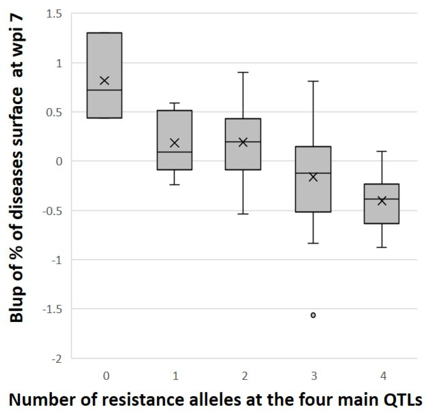
Thus, four main resistance QTLs to BLSD were identified among this KNO x IDN110 progeny. Each one was detected for several phenotypic variables with an explained variance > 20%. Three are shared by IDN110 and KNO and/or their interaction (chr 06, 07, and 09) and one is specific to IDN110 (chr 08). IDN110, also named ‘AAcv rose’, or its clonal variant ‘Gu Nin Chiao’, are genitors of interest for breeding sweet bananas, and in particular for their partial resistance to BLSD. The four main BLSD QTLs identified here on chr 06, 07, 08, and 09, are thus keys for breeding. The phenotypic differences between plants showing zero to four of the resistance alleles at those four main QTLs are illustrated in Figure 4 at the QTL peak for the phenotypic variable with the best LOD/R2. This suggests that combining the IDN110 favourable alleles at these four QTLs, in breeding schemes, should be of interest for durable resistance to BLSD.

3.5. Putative Candidate Genes in the Four Main QTLs Detected on IDN110

We selected the four regions corresponding to the largest QTL intervals detected for IDN110 on chromosomes 06 to 09 to search for potential candidate genes present on the reference genome sequence. The four main QTLs (chr 06 to 09) of the partially resistant IDN110 contain, respectively, 208, 275, 363, and 867 predicted genes according to version 4 of the M. acuminata ‘DH-Pahang’ reference sequence assembly [37]. They have diverse predicted functions and cellular locations and several of them may be involved in plant defence, as shown by Interpro domain and gene ontology information (Table S2).
The few cloned genes underlying QTLs for disease resistance in other plants are diverse, but several belong to major families of plant disease resistance genes [68]. Sekhwal et al. [69] divide resistance gene analogues (RGA) into four main types: the nucleotide binding site (NBS) encoding genes that code for intracellular proteins; receptor-like kinases (RLK) with an extracellular domain, a transmembrane domain (TM), and an intracellular kinase domain; receptor-like proteins (RLP) that differ from RLK in their lack of the intracellular kinase domain; and other resistance genes (Oth-R) with TM receptors, but that lack complete previous domains.
A total of 98 RGA were identified by the DRAGO3 tool and validated using InterPro domains. In total, 8, 17, 15, and 58 RGA were found in the QTLs on chr 06, 07, 08, and 09, respectively (Table 1 and details in Table S2). Among them, nine are NBS-encoding genes that were also identified by Anuradha et al. [70]. Seven out the nine NBS-encoding genes were predicted as complete NBS-LRR by Belser et al. [37]. Thirteen of these RGA are RLPs, a class of R proteins to which the tomato Cladosporium fulvum (Cf, syn. Passalora fulva) resistance proteins belong [71]. Cladosporium and Pseudocercospora species are related pathogenic fungi of the Mycosphaerellaceae family [5,72]. Stergiopoulos et al. [73] showed that P. fijiensis homologues of the C. fulvum effectors Avr4 and Ecp2 are recognised and display a hypersensitive response in tomato plants carrying resistance genes Cf-4 and Cf-Ecp2, respectively.
The QTL region on chr 06 contains one NBS encoding gene annotated as non-regular NBS-LRR by Anuradha et al. [70], one RLK, one RLP, and five Oth-R proteins.
The QTL region on chr 07 contains three NBS-encoding genes, including neighbouring genes named chr07_nlr_14 and chr07_nlr_1 by Belser et al. [37]. It also contains three RLKs, one RLP, and ten Oth-R. The RLP gene was identified by Pinheiro et al. [74] as down-regulated in resistant wild accession M. a. ‘Calcutta 4’ during its transcriptomic interaction with P. musae, a fungus related to P. fijiensis.
The QTL region on chr 08 contains one NBS-encoding gene and ten RLKs, four of which are present in a cluster. The region also bears one RLP and three Oth-R. The NBS-encoding gene named RGC1 by Peraza-Echeverria et al. [75] and named chr08_nlr_2 by Belser et al. [37] was found by Zhang et al. [76] to be threefold higher expressed in accession M. a. ‘Pahang’ which is resistant, than in susceptible Cavendish, in unchallenged conditions. This led the authors to state that it may ‘indicate its role in constitutive defence mechanism’.
The QTL region on chr 09 contains 4 NBS-encoding genes, 25 RLKs, 10 RLPs and 19 Oth-R. The NBS-encoding genes were identified and named chr09_nlr_7, chr09_nlr_10, chr09_nlr_9, and chr09_nlr_8 by Belser et al. [37]. The first two are annotated as putative disease resistance protein RGA3 and RGA4, respectively. The last two that colocalize are both annotated as putative RPP8-like protein 3, and were both identified by Pinheiro et al. [74] as modulated in resistant accession M. a. ‘Calcutta 4’ during the transcriptomic P. musae interaction. It led the authors to conclude that modulation of these genes provide evidence for an effector-triggered immunity (ETI) response in this interaction. Some of the RLKs are present in three clusters of wall-associated receptor-like kinases, L-type lectin domain containing receptor kinases, and putative cysteine-rich receptor-like kinases. These types of genes have been shown to confer resistance to wheat against the hemibiotrophic fungus Zymoseptoria triticii, which is related to Pseudocercopora. For example, wheat Stb6 and Stb16q genes encode, respectively, a wall-associated kinase-like protein and a cysteine-rich receptor-like kinase [77,78]. Another RLK in this QTL region is a receptor protein kinase-like protein ZAR1 proposed by Breit-McNally et al. [79] to be involved in effector recognition. Six out of the ten RLPs are present as a cluster of genes and are annotated as homologues of genes that have been shown in tomato to interact with fungal protein ethylene-inducing xylanase, as reviewed by Monaghan and Zipfel [80].
Other types of defence-related mechanisms involve different types of genes such as pentatricopeptide repeats (PPRs) and peroxidases [69]. A total of 25 PPRs or putative PPRs were identified in QTLs located on chr 06 to 09, with, respectively, 4, 3, 6, and 12 genes. Six peroxidases were identified in QTLs on chr 07 and 09, containing, respectively, five genes and one gene. During the biotrophic phase of P. fijiensis, which corresponds to our in vitro test, Rodriguez et al. [81], in agreement with previous results of Torres et al. [82], identified higher expression of genes encoding peroxidases in the resistant M. a. ‘Calcutta 4’ accession. Other genes annotated as being implicated in defence responses are underlined in Table S2.
Two reviews highlight genes cited as plant defence-related during Musa gene expression analysis in interaction with P. fijiensis [7,16]. The genes or types of genes cited as being present in the four QTLs are indicated in Table S2, along with genes implicated in resistance cited in studies on Musa [34,37,70,74,75,76,81,82,83,84,85,86,87,88,89,90,91,92]. Among them are several GDSL esterase-encoding genes with a cluster in the QTL on chr 08 and several caffeoyl methyltransferase-encoding genes [34,83].
Finally, as this analysis was performed on the DH-Pahang reference genome from the wild M. acuminata subspecies malaccensis, it is not excluded that IDN110 and KNO bear genes that are not included in this reference.

4. Conclusions

QTL identification in banana benefited both from the availability of the reference banana genome sequence assembly and from the identification of the location of chromosome translocations that affect segregation and genetic mapping [34,35,36,37]. Before the first genome sequence assembly in 2012 [34], the only QTL and markers developed to assist Musa selection were obtained against banana streak disease caused by the banana streak virus [93,94,95,96]. Since then, Musa QTLs have been identified either through dedicated populations or GWAS analyses for fruit seedlessness [97], bunch weight [98], fruit organoleptic quality during ripening [64], resistance to the weevils Cosmopolites sordidus [89], resistance to banana Xanthomonas wilt [92], and resistance to Fusarium wilt [99,100,101].
In the present study, we identified QTLs for partial resistance to BLSD using two diploid banana cultivars. One of them, IDN110, is a genitor of interest for breeding dessert bananas. IDN110 (or ‘AAcv Rose’) and its somaclone ‘Gu Nin Chiao’ are appreciated for their fruit quality and are also reported to be resistant to Panama disease either race 1 or tropical race 4 (TR4) and tolerant to nematodes Radopholus similis and Pratylenchus coffeae [102,103,104,105].
We did not detect a major QTL against BLSD, unlike what was found against panama disease [100]. The QTLs identified here conferred partial resistance that does not lead to an absence, but a reduction in disease that must, of course, be compatible with production. With partial resistance, the selection pressure on the pathogen is expected to be reduced. It is particularly important for pathogens with a high evolutionary potential, such as P. fijiensis. Less selection pressure on the pathogen should lead to more durable resistance [21,22,68,106]. Seven QTLs were identified in the partially resistant parent IDN110. Most of these QTLs are not in regions that are influenced by translocations or reduced recombination, which are frequent in banana genomes. Selection of those QTLs in hybrids is thus possible, in particular the four QTLs with the main effects. The gain in resistance that can be cumulated with the four main QTLs identified here should convince breeders of the interest in cumulating favourable partially resistant alleles using molecular-assisted selection (MAS). MAS should also enable evaluation of the best dose of alleles of these QTLs in triploid hybrids. These QTLs could be associated with other resistances, as recommended by Cowger and Brown [106], in particular, those with resistance affecting different stages of the banana–P. fijiensis interaction (i.e., later stages, such as the sporulation stage).

Supplementary Materials

The following supporting information can be downloaded at: https://www.mdpi.com/article/10.3390/horticulturae10060608/s1, Figure S1: Location of the recombinations detected on each parent (KNO and IDN110) on the physical map of DH Pahang reference genome version 4 [37]; Figure S2: Manhattan plots of KNO (a.) and IDN110 (b.) for the 16 phenotypic variables; Table S1: For all hybrids, BLUP values of the phenotypic variables and genotypes for the 2084 non-redundant markers; Table S2: List of the genes or putative genes identified in the four main QTLs of IDN110.

Author Contributions

Conceptualization: F.C. and F.S.; Resources: F.C., G.M., V.R., C.P., R.H., T.C., C.G., J.-M.D., C.H. and P.M.; Data curation: F.C.; Formal analysis: F.C., G.M. and N.Y.; Software: S.R.; Writing—original draft preparation: F.C. and N.Y.; Writing—review and editing: F.C., G.M., A.D., N.Y. and F.S.; Project administration: F.C. and F.S.; Funding acquisition: F.S. All authors have read and agreed to the published version of the manuscript.

Funding

This study was funded by the 2018 ‘DUREBAN’ project financed by the French Biodiversity Office as part of the “Seeds” call for research projects co-sponsored by the special allocation account for agricultural and rural development (CASDAR) and the research and innovation axis of the Écophyto II+ plan managed by the French Ministry of Ecological Transition and Territorial Cohesion (French acronym MTECT), Ministry of Agriculture and Food Sovereignty (French acronym MASA), Ministry of Health and Prevention (French acronym MSP) and the Ministry of Higher Education and Research (French acronym MESR).

Data Availability Statement

Data are contained in this article and in the Supplementary Files. The illumina data of the progenies are available in the SRA database under project PRJNA1101903.

Acknowledgments

The authors thank Gaëtan Droc, Jean-Claude Efile, Sandrine Fabre, Sonia Guillou and Franck Marius for their assistance; and Catherine Abadie, Franc-Christophe Baurens, Jean Carlier, Christophe Jenny, Simon Rio and Didier Tharreau for their advice. This work has been realized with the support of MESO@LR-Platform at the University of Montpellier, the financial support of CIRAD, and the technical support of the bioinformatics group of the UMR AGAP Institute, member of the French Institute of Bioinformatics (IFB)—South Green Bioinformatics Platform.

Conflicts of Interest

The authors have no conflict of interest to declare.

References

  1. Simmonds, N.W.; Shepherd, K. The Taxonomy and Origins of the Cultivated Bananas. J. Linn. Soc. Lond. Bot. 1955, 55, 302–312. [Google Scholar] [CrossRef]
  2. Perrier, X.; de Langhe, E.; Donohue, M.; Lentfer, C.J.; Vrydaghs, L.; Bakry, F.; Carreel, F.; Hippolyte, I.; Horry, J.-P.; Jenny, C.; et al. Multidisciplinary Perspectives on Banana (Musa Spp.) Domestication. Proc. Natl. Acad. Sci. USA 2011, 108, 11311–11318. [Google Scholar] [CrossRef] [PubMed]
  3. Lescot, T. Banana Genetic Diversity. FruiTrop 2020, 269, 98–102. [Google Scholar]
  4. Churchill, A.C.L. Mycosphaerella fijiensis, the Black Leaf Streak Pathogen of Banana: Progress towards Understanding Pathogen Biology and Detection, Disease Development, and the Challenges of Control. Mol. Plant Pathol. 2011, 12, 307–328. [Google Scholar] [CrossRef] [PubMed]
  5. Crous, P.W.; Braun, U.; Hunter, G.C.; Wingfield, M.J.; Verkley, G.J.M.; Shin, H.-D.; Nakashima, C.; Groenewald, J.Z. Phylogenetic Lineages in Pseudocercospora. Stud. Mycol. 2013, 75, 37–114. [Google Scholar] [CrossRef] [PubMed]
  6. Guzman, M.; Pérez Vicente, L.; Carlier, J.; Abadie, C.; Lapeyre de Bellaire, L.; Carreel, F.; Marin, D.H.; Romero, R.A.; Gauhl, F.; Pasberg-Gauhl, C.; et al. Black Leaf Streak. In Handbook of Diseases of Banana, Abacà and Enset; CABI: Boston, MA, USA, 2019; pp. 41–115. ISBN 978-1-78064-719-7. [Google Scholar]
  7. Noar, R.D.; Thomas, E.; Daub, M.E. Genetic Characteristics and Metabolic Interactions between Pseudocercospora fijiensis and Banana: Progress toward Controlling Black Sigatoka. Plants 2022, 11, 948. [Google Scholar] [CrossRef]
  8. Pennisi, E. Armed and Dangerous. Science 2010, 327, 804–805. [Google Scholar] [CrossRef]
  9. Ramsey, M.D.; Daniells, J.W.; Anderson, V.J. Effects of Sigatoka Leaf Spot (Mycospherella musicola Leach) on Fruit Yield, Field Ripening and Greenlife of Bananas in North Queensland. Sci. Hortic. 1990, 41, 305–313. [Google Scholar] [CrossRef]
  10. Chillet, M.; Abadie, C.; Hubert, O.; Chilin-Charles, Y.; de Lapeyre de Bellaire, L. Sigatoka Disease Reduces the Greenlife of Bananas. Crop Prot. 2009, 28, 41–45. [Google Scholar] [CrossRef]
  11. Castelan, F.; Saraiva, L.; Lange, F.; de Lapeyre de Bellaire, L.; Cordenunsi, B.; Chillet, M. Effects of Black Leaf Streak Disease and Sigatoka Disease on Fruit Quality and Maturation Process of Bananas Produced in the Subtropical Conditions of Southern Brazil. Crop Prot. 2011, 35, 127–131. [Google Scholar] [CrossRef]
  12. Lapeyre de Bellaire, L.; Fouré, E.; Abadie, C.; Carlier, J. Black Leaf Streak Disease Is Challenging the Banana Industry. Fruits 2010, 65, 327–342. [Google Scholar] [CrossRef]
  13. Fouré, E.; De Lapeyre De Bellaire, L. Maladies et Ravageurs de La Banane. FruiTrop 2020, 269, 104–112. [Google Scholar]
  14. Chong, P.; Essoh, J.N.; Arango Isaza, R.E.; Keizer, P.; Stergiopoulos, I.; Seidl, M.F.; Guzman, M.; Sandoval, J.; Verweij, P.E.; Scalliet, G.; et al. A World-wide Analysis of Reduced Sensitivity to DMI Fungicides in the Banana Pathogen Pseudocercospora fijiensis. Pest Manag. Sci. 2021, 77, 3273–3288. [Google Scholar] [CrossRef] [PubMed]
  15. Rieux, A.; Soubeyrand, S.; Bonnot, F.; Klein, E.K.; Ngando, J.E.; Mehl, A.; Ravigne, V.; Carlier, J.; Bellaire, L.d.L.d. Long-Distance Wind-Dispersal of Spores in a Fungal Plant Pathogen: Estimation of Anisotropic Dispersal Kernels from an Extensive Field Experiment. PLoS ONE 2014, 9, e103225. [Google Scholar] [CrossRef] [PubMed]
  16. Soares, J.M.S.; Rocha, A.J.; Nascimento, F.S.; Santos, A.S.; Miller, R.N.G.; Ferreira, C.F.; Haddad, F.; Amorim, V.B.O.; Amorim, E.P. Genetic Improvement for Resistance to Black Sigatoka in Bananas: A Systematic Review. Front. Plant Sci. 2021, 12, 657916. [Google Scholar] [CrossRef]
  17. Salmon, F.; Bakry, F.; Efile, J.C.; Ricci, S.; Toniutti, L.; Horry, J.P. Banana Breeding at CIRAD: Creating Resistant New Cultivars to Avoid the Use of Pesticides. Acta Hortic. 2023, 1367, 201–208. [Google Scholar] [CrossRef]
  18. Fouré, E.; Mouliom Pefoura, A.; Mourichon, X. Etude de La Sensibilité Variétale Des Bananiers et Des Plantains à Mycosphaerella fijiensis Morelet Au Cameroun. Caractérisation de La Résistance Au Champ de Bananiers Appartenant à Divers Groupes Génétiques. Fruits 1990, 45, 339–345. [Google Scholar]
  19. Fullerton, R.A.; Olsen, T.L. Pathogenic Variability in Mycosphaerella fijiensis Morelet, Cause of Black Sigatoka in Banana and Plantain. N. Z. J. Crop Hortic. Sci. 1995, 23, 39–48. [Google Scholar] [CrossRef]
  20. Mouliom-Pefoura, A. First Observation of the Breakdown of High Resistance in Yangambi Km 5 (Musa Sp.) to the Black Leaf Streak Disease in Cameroon. Plant Dis. 1999, 83, 78. [Google Scholar] [CrossRef]
  21. Niks, R.E.; Qi, X.; Marcel, T.C. Quantitative Resistance to Biotrophic Filamentous Plant Pathogens: Concepts, Misconceptions, and Mechanisms. Annu. Rev. Phytopathol. 2015, 53, 445–470. [Google Scholar] [CrossRef]
  22. Nelson, R.; Wiesner-Hanks, T.; Wisser, R.; Balint-Kurti, P. Navigating Complexity to Breed Disease-Resistant Crops. Nat. Rev. Genet. 2018, 19, 21–33. [Google Scholar] [CrossRef] [PubMed]
  23. Rowe, P.; Rosales, F. Musa breeding at FHIA. In The Improvement and Testing of Musa: A Global Partnership, Proceedings of the First Global Conference of the IMTP, San Pedro Sula, Honduras, 27–30 April 1994; Bioversity International: Rome, Italy, 1994; pp. 117–129. [Google Scholar]
  24. Bakry, F.; Carreel, F.; Jenny, C.; Horry, J.-P. Genetic Improvement of Banana. In Breeding Plantation Tree Crops: Tropical Species; Mohan Jain, S., Priyadarshan, P.M., Eds.; Springer: New York, NY, USA, 2009; pp. 3–50. ISBN 978-0-387-71199-7. [Google Scholar]
  25. Dumartinet, T.; Abadie, C.; Bonnot, F.; Carreel, F.; Roussel, V.; Habas, R.; Martinez, R.T.; Perez-Vicente, L.; Carlier, J. Pattern of Local Adaptation to Quantitative Host Resistance in a Major Pathogen of a Perennial Crop. Evol. Appl. 2020, 13, 824–836. [Google Scholar] [CrossRef]
  26. Cheesman, E.E. Principles of Banana Breeding. Trop. Agric. 1934, 11, 132–137. [Google Scholar]
  27. Shepherd, K. Banana Breeding in the West Indies. Pest Artic. News Summ. 1968, 14, 370–379. [Google Scholar] [CrossRef]
  28. Rowe, P.R.; Richardson, D.L. Breeding Bananas for Disease Resistance Fruit Quality and Yield; Tropical Agriculture Research Services (SIATSA): La Lima, Honduras, 1975; pp. 1–23. [Google Scholar]
  29. Tézenas du Montcel, H. Genetic Improvement of Bananas for Resistance to Diseases and Pests: Improvement Strategies. Fruits 1993, 48, 11–14. [Google Scholar]
  30. Brown, A.; Carpentier, S.C.; Swennen, R. Breeding climate-resilient bananas. In Genomic Designing of Climate-Smart Fruit Crops; Kole, C., Ed.; Springer International Publishing: Cham, Switzerland, 2020; pp. 91–115. ISBN 978-3-319-97946-5. [Google Scholar]
  31. Bakry, F.; Horry, J.-P.; Jenny, C. Making Banana Breeding More Effective. Available online: https://agritrop.cirad.fr/597567/ (accessed on 1 December 2023).
  32. Ortiz, R.; Vuylsteke, D. Inheritance of Black Sigatoka Disease Resistance in Plantain-Banana (Musa Spp) Hybrids. Theor. Appl. Genet. 1994, 89, 146–152. [Google Scholar] [CrossRef] [PubMed]
  33. Craenen, K.; Ortiz, R. Effect of the Bs1 Gene in Plantain-Banana Hybrids on Response to Black Sigatoka. Theor. Appl. Genet. 1997, 95, 497–505. [Google Scholar] [CrossRef]
  34. D’Hont, A.; Denoeud, F.; Aury, J.-M.; Baurens, F.-C.; Carreel, F.; Garsmeur, O.; Noel, B.; Bocs, S.; Droc, G.; Rouard, M.; et al. The Banana Musa acuminata Genome and the Evolution of Monocotyledonous Plants. Nature 2012, 488, 213–219. [Google Scholar] [CrossRef]
  35. Dupouy, M.; Baurens, F.-C.; Derouault, P.; Hervouet, C.; Cardi, C.; Cruaud, C.; Istace, B.; Labadie, K.; Guiougou, C.; Toubi, L.; et al. Two Large Reciprocal Translocations Characterized in the Disease Resistance-Rich Burmannica Genetic Group of Musa acuminata. Ann. Bot. 2019, 124, 319–329. [Google Scholar] [CrossRef]
  36. Martin, G.; Baurens, F.; Hervouet, C.; Salmon, F.; Delos, J.; Labadie, K.; Perdereau, A.; Mournet, P.; Blois, L.; Dupouy, M.; et al. Chromosome Reciprocal Translocations Have Accompanied Subspecies Evolution in Bananas. Plant J. 2020, 104, 1698–1711. [Google Scholar] [CrossRef]
  37. Belser, C.; Baurens, F.-C.; Noel, B.; Martin, G.; Cruaud, C.; Istace, B.; Yahiaoui, N.; Labadie, K.; Hribova, E.; Dolezel, J.; et al. Telomere-to-Telomere Gapless Chromosomes of Banana Using Nanopore Sequencing. Commun. Biol. 2021, 4, 1047. [Google Scholar]
  38. Raboin, L.M.; Carreel, F.; Noyer, J.-L.; Baurens, F.-C.; Horry, J.P.; Bakry, F.; Tézenas du Montcel, H.; Ganry, J.; Lanaud, C.; Lagoda, P.J.L. Diploid Ancestors of Triploid Export Banana Cultivars: Molecular Identification of 2n Restitution Gamete Donors and n Gamete Donors. Mol. Breed. 2005, 16, 333–341. [Google Scholar] [CrossRef]
  39. Martin, G.; Baurens, F.-C.; Labadie, K.; Hervouet, C.; Salmon, F.; Marius, F.; Paulo-de-la-Reberdiere, N.; Van Den Houwe, I.; Aury, J.-M.; D’Hont, A.; et al. Shared Pedigree Relationships and Transmission of Unreduced Gametes in Cultivated Banana. Ann. Bot. 2023, 131, 1149–1161. [Google Scholar] [CrossRef] [PubMed]
  40. Pérez-Vicente, L.; Carreel, F.; Roussel, V.; Carlier, J.; Abadie, C. Guidelines for the Evaluation of Resistance to Pseudocercospora Leaf Spots of Banana. In Practical Guidelines for Early Screening and Field Evaluation of Banana against Fusarium Wilt, Pseudocercospora Leaf Spots and Drought; Dita, M., Ed.; Bioversity International: Rome, Italy, 2021; pp. 21–63. ISBN 978-92-9255-192-6. [Google Scholar]
  41. Kimunye, J.; Were, E.; Swennen, R.; Viljoen, A.; Mahuku, G. Sources of Resistance to Pseudocercospora fijiensis, the Cause of Black Sigatoka in Banana. Plant Pathol. 2021, 70, 1651–1664. [Google Scholar] [CrossRef] [PubMed]
  42. Roux-Cuvelier, M.; Grisoni, M.; Bellec, A.; Bloquel, E.; Charron, C.; Delalande, M.; Delmas, M.; Didier, A.; Durel, C.-E.; Duval, C.-H.; et al. Conservation of Horticultural Genetic Resources in France. Chron. Hortic. 2021, 61, 21–36. [Google Scholar]
  43. Bakry, F. Zygotic Embryo Rescue in Bananas. Fruits 2008, 63, 111–115. [Google Scholar] [CrossRef]
  44. IPSDK Explorer. Reactiv’IP (Grenoble, France). 2022. Available online: https://www.reactivip.com/ (accessed on 23 January 2023).
  45. Alves, K.d.S.; Del Ponte, E.M. Epifitter, Analysis and Simulation of Plant Disease Progress Curves. R package Version 0.3.0. 2021. Available online: https://cran.r-project.org/web/packages/epifitter/index.html (accessed on 23 January 2023).
  46. Akaike, H. A New Look at the Statistical Model Identification. IEEE Trans. Autom. Control. 1974, 19, 716–723. [Google Scholar] [CrossRef]
  47. Ratkowsky, D.A. Nonlinear Regression Modeling, Self-Starting Nls Weibull Growth Curve Model; Marcel Dekker Inc.: New York, NY, USA, 1983. [Google Scholar]
  48. Pinheiro, J.; Bates, D.; DebRoy, S.; Sarkar, D.; Eispack; Heisterkamp, S.; Van Willigen, B.; Ranke, J.; R Core Team. Nlme, Linear and Nonlinear Mixed Effects Models; R Package Version 3.1-160. 2022. Available online: https://cran.r-project.org/web/packages/nlme/index.html (accessed on 11 November 2022).
  49. Wickham, H.; Chang, W.; Henry, L.; Pedersen, T.L.; Takahashi, K.; Wilke, C.; Woo, K.; Yutani, H.; Dunnington, D.; van den Brand, T.; et al. Ggplot2, Create Elegant Data Visualisations Using the Grammar of Graphics; R Package Version 3.5.0. 2024. Available online: https://cran.r-project.org/web/packages/ggplot2/index.html (accessed on 24 February 2024).
  50. Brooks, M.E.; Kristensen, K.; van Benthem, K.J.; Magnusson, A.; Berg, C.W.; Nielsen, A.; Skaug, H.J.; Mächler, M.; Bolker, B.M. glmmTMB Balances Speed and Flexibility among Packages for Zero-Inflated Generalized Linear Mixed Modeling. R J. 2017, 9, 378–400, R Package Version 1.1.4. 2022. Available online: https://cran.r-project.org/web/packages/glmmTMB/index.html (accessed on 11 November 2022). [CrossRef]
  51. Hartig, F.; Lohse, L. DHARMa: Residual Diagnostics for Hierarchical (Multi-Level/Mixed) Regression Models; R Package Version 0.4.6. 2022. Available online: https://cran.r-project.org/web/packages/DHARMa/index.html (accessed on 23 January 2023).
  52. Cormier, F.; Lawac, F.; Maledon, E.; Gravillon, M.-C.; Nudol, E.; Mournet, P.; Vignes, H.; Chaïr, H.; Arnau, G. A Reference High-Density Genetic Map of Greater Yam (Dioscorea alata L.). Theor. Appl. Genet. 2019, 132, 1733–1744. [Google Scholar] [CrossRef]
  53. Herten, K.; Hestand, M.S.; Vermeesch, J.R.; Van Houdt, J.K. GBSX: A Toolkit for Experimental Design and Demultiplexing Genotyping by Sequencing Experiments. BMC Bioinform. 2015, 16, 73. [Google Scholar] [CrossRef]
  54. Martin, M. Cutadapt Removes Adapter Sequences from High-Throughput Sequencing Reads. EMBnet J. 2011, 17, 10–12. [Google Scholar] [CrossRef]
  55. Garsmeur, O.; Droc, G.; Antonise, R.; Grimwood, J.; Potier, B.; Aitken, K.; Jenkins, J.; Martin, G.; Charron, C.; Hervouet, C.; et al. A Mosaic Monoploid Reference Sequence for the Highly Complex Genome of Sugarcane. Nat. Commun. 2018, 9, 2638. [Google Scholar] [CrossRef] [PubMed]
  56. Van Ooijen, J.W. JoinMap® 5, Software for the Calculation of Genetic Linkage Maps in Experimental Populations of Diploid Species; Kyazma BV: Wageningen, The Netherlands, 2018. [Google Scholar]
  57. Martin, G.; Baurens, F.-C.; Droc, G.; Rouard, M.; Cenci, A.; Kilian, A.; Hastie, A.; Doležel, J.; Aury, J.-M.; Alberti, A.; et al. Improvement of the Banana “Musa acuminata” Reference Sequence Using NGS Data and Semi-Automated Bioinformatics Methods. BMC Genom. 2016, 17, 12. [Google Scholar] [CrossRef] [PubMed]
  58. Rami, J.-F. (CIRAD, Paris, France). Personal communication, 2017. [Google Scholar]
  59. Van Ooijen, J.W. MapQTL® 6, Software for Mapping of Quantitative Trait Loci in Experimental Populations of Diploid Species; Kyazma BV: Wageningen, The Netherlands, 2009. [Google Scholar]
  60. Calle García, J.; Guadagno, A.; Paytuvi-Gallart, A.; Saera-Vila, A.; Amoroso, C.G.; D’Esposito, D.; Andolfo, G.; Aiese Cigliano, R.; Sanseverino, W.; Ercolano, M.R. PRGdb 4.0: An Updated Database Dedicated to Genes Involved in Plant Disease Resistance Process. Nucleic Acids Res. 2022, 50, D1483–D1490. [Google Scholar] [CrossRef] [PubMed]
  61. Dievart, A.; Gottin, C.; Périn, C.; Ranwez, V.; Chantret, N. Origin and Diversity of Plant Receptor-Like Kinases. Annu. Rev. Plant Biol. 2020, 71, 131–156. [Google Scholar] [CrossRef] [PubMed]
  62. Perrier, X.; Bakry, F.; Carreel, F.; Jenny, C.; Horry, J.-P.; Lebot, V.; Hippolyte, I. Combining Biological Approaches to Shed Light on the Evolution of Edible Bananas. Ethnobot. Res. Appl. 2009, 7, 199–216. [Google Scholar] [CrossRef]
  63. Martin, G.; Carreel, F.; Coriton, O.; Hervouet, C.; Cardi, C.; Derouault, P.; Roques, D.; Salmon, F.; Rouard, M.; Sardos, J.; et al. Evolution of the Banana Genome (Musa acuminata) Is Impacted by Large Chromosomal Translocations. Mol. Biol. Evol. 2017, 34, 2140–2152. [Google Scholar] [CrossRef] [PubMed]
  64. Biabiany, S.; Araou, E.; Cormier, F.; Martin, G.; Carreel, F.; Hervouet, C.; Salmon, F.; Efile, J.-C.; Lopez-Lauri, F.; D’Hont, A.; et al. Detection of Dynamic QTLs for Traits Related to Organoleptic Quality during Banana Ripening. Sci. Hortic. 2022, 293, 110690. [Google Scholar] [CrossRef]
  65. Brazier, T.; Glémin, S. Diversity and Determinants of Recombination Landscapes in Flowering Plants. PLoS Genet. 2022, 18, e1010141. [Google Scholar] [CrossRef]
  66. Martin, G.; Cottin, A.; Baurens, F.-C.; Labadie, K.; Hervouet, C.; Salmon, F.; Paulo-de-la-Reberdiere, N.; Van den Houwe, I.; Sardos, J.; Aury, J.-M.; et al. Interspecific Introgression Patterns Reveal the Origins of Worldwide Cultivated Bananas in New Guinea. Plant J. 2023, 113, 802–818. [Google Scholar] [CrossRef]
  67. Landry, C.; Bonnot, F.; Ravigné, V.; Carlier, J.; Rengifo, D.; Vaillant, J.; Abadie, C. A Foliar Disease Simulation Model to Assist the Design of New Control Methods against Black Leaf Streak Disease of Banana. Ecol. Model. 2017, 359, 383–397. [Google Scholar] [CrossRef]
  68. French, E.; Kim, B.-S.; Iyer-Pascuzzi, A.S. Mechanisms of Quantitative Disease Resistance in Plants. Semin. Cell Dev. Biol. 2016, 56, 201–208. [Google Scholar] [CrossRef] [PubMed]
  69. Sekhwal, M.K.; Li, P.; Lam, I.; Wang, X.; Cloutier, S.; You, F.M. Disease Resistance Gene Analogs (RGAs) in Plants. Int. J. Mol. Sci. 2015, 16, 19248–19290. [Google Scholar] [CrossRef] [PubMed]
  70. Anuradha, C.; Chandrasekar, A.; Backiyarani, S.; Uma, S. MusaRgeneDB: An Online Comprehensive Database for Disease Resistance Genes in Musa Spp. 3 Biotech 2022, 12, 222. [Google Scholar] [CrossRef] [PubMed]
  71. Zhao, T.; Pei, T.; Jiang, J.; Yang, H.; Zhang, H.; Li, J.; Xu, X. Understanding the Mechanisms of Resistance to Tomato Leaf Mold: A Review. Hortic. Plant J. 2022, 8, 667–675. [Google Scholar] [CrossRef]
  72. Isaza, R.E.A.; Diaz-Trujillo, C.; Dhillon, B.; Aerts, A.; Carlier, J.; Crane, C.F.; de Jong, T.V.; de Vries, I.; Dietrich, R.; Farmer, A.D.; et al. Combating a Global Threat to a Clonal Crop: Banana Black Sigatoka Pathogen Pseudocercospora fijiensis (Synonym Mycosphaerella fijiensis) Genomes Reveal Clues for Disease Control. PLoS Genet. 2016, 12, e1005876. [Google Scholar] [CrossRef] [PubMed]
  73. Stergiopoulos, I.; van den Burg, H.A.; Okmen, B.; Beenen, H.G.; van Liere, S.; Kema, G.H.J.; de Wit, P.J.G.M. Tomato Cf Resistance Proteins Mediate Recognition of Cognate Homologous Effectors from Fungi Pathogenic on Dicots and Monocots. Proc. Natl. Acad. Sci. USA 2010, 107, 7610–7615. [Google Scholar] [CrossRef] [PubMed]
  74. Pinheiro, T.D.M.; Rego, E.C.S.; Alves, G.S.C.; Fonseca, F.C.D.A.; Cotta, M.G.; Antonino, J.D.; Gomes, T.G.; Amorim, E.P.; Ferreira, C.F.; Costa, M.M.D.C.; et al. Transcriptome Profiling of the Resistance Response of Musa acuminata Subsp. burmannicoides, Var. Calcutta 4 to Pseudocercospora Musae. Int. J. Mol. Sci. 2022, 23, 13589. [Google Scholar] [CrossRef] [PubMed]
  75. Peraza-Echeverria, S.; Dale, J.L.; Harding, R.M.; Smith, M.K.; Collet, C. Characterization of Disease Resistance Gene Candidates of the Nucleotide Binding Site (NBS) Type from Banana and Correlation of a Transcriptional Polymorphism with Resistance to Fusarium oxysporum f.Sp. Cubense Race 4. Mol. Breed. 2008, 22, 565–579. [Google Scholar] [CrossRef]
  76. Zhang, L.; Cenci, A.; Rouard, M.; Zhang, D.; Wang, Y.; Tang, W.; Zheng, S.-J. Transcriptomic Analysis of Resistant and Susceptible Banana Corms in Response to Infection by Fusarium oxysporum f. Sp. Cubense Tropical Race 4. Sci. Rep. 2019, 9, 8199. [Google Scholar] [CrossRef]
  77. Saintenac, C.; Lee, W.-S.; Cambon, F.; Rudd, J.J.; King, R.C.; Marande, W.; Powers, S.J.; Bergès, H.; Phillips, A.L.; Uauy, C.; et al. Wheat Receptor-Kinase-like Protein Stb6 Controls Gene-for-Gene Resistance to Fungal Pathogen Zymoseptoria tritici. Nat. Genet. 2018, 50, 368–374. [Google Scholar] [CrossRef]
  78. Saintenac, C.; Cambon, F.; Aouini, L.; Verstappen, E.; Ghaffary, S.M.T.; Poucet, T.; Marande, W.; Berges, H.; Xu, S.; Jaouannet, M.; et al. A Wheat Cysteine-Rich Receptor-like Kinase Confers Broad-Spectrum Resistance against Septoria Tritici Blotch. Nat. Commun. 2021, 12, 433. [Google Scholar] [CrossRef] [PubMed]
  79. Breit-McNally, C.; Laflamme, B.; Singh, R.A.; Desveaux, D.; Guttman, D.S. ZAR1: Guardian of Plant Kinases. Front. Plant Sci. 2022, 13, 981684. [Google Scholar] [CrossRef]
  80. Monaghan, J.; Zipfel, C. Plant Pattern Recognition Receptor Complexes at the Plasma Membrane. Curr. Opin. Plant Biol. 2012, 15, 349–357. [Google Scholar] [CrossRef] [PubMed]
  81. Rodriguez, H.A.; Rodriguez-Arango, E.; Morales, J.G.; Kema, G.; Arango, R.E. Defense Gene Expression Associated with Biotrophic Phase of Mycosphaerella fijiensis M. Morelet Infection in Banana. Plant Dis. 2016, 100, 1170–1175. [Google Scholar] [CrossRef] [PubMed]
  82. Torres, J.M.; Calderón, H.; Rodríguez-Arango, E.; Morales, J.G.; Arango, R. Differential Induction of Pathogenesis-Related Proteins in Banana in Response to Mycosphaerella fijiensis Infection. Eur. J. Plant Pathol. 2012, 133, 887–898. [Google Scholar] [CrossRef]
  83. Portal, O.; Izquierdo, Y.; De Vleesschauwer, D.; Sánchez-Rodríguez, A.; Mendoza-Rodríguez, M.; Acosta-Suárez, M.; Ocaña, B.; Jiménez, E.; Höfte, M. Analysis of Expressed Sequence Tags Derived from a Compatible Mycosphaerella fijiensis–Banana Interaction. Plant Cell Rep. 2011, 30, 913–928. [Google Scholar] [CrossRef] [PubMed]
  84. Sreedharan, S.; Shekhawat, U.K.S.; Ganapathi, T.R. MusaSAP1, a A20/AN1 Zinc Finger Gene from Banana Functions as a Positive Regulator in Different Stress Responses. Plant Mol. Biol. 2012, 80, 503–517. [Google Scholar] [CrossRef] [PubMed]
  85. Passos, M.A.N.; de Cruz, V.O.; Emediato, F.L.; de Teixeira, C.C.; Azevedo, V.C.R.; Brasileiro, A.C.M.; Amorim, E.P.; Ferreira, C.F.; Martins, N.F.; Togawa, R.C.; et al. Analysis of the Leaf Transcriptome of Musa acuminata during Interaction with Mycosphaerella musicola: Gene Assembly, Annotation and Marker Development. BMC Genom. 2013, 14, 78. [Google Scholar] [CrossRef] [PubMed]
  86. Sánchez Timm, E.; Hidalgo Pardo, L.; Pacheco Coello, R.; Chávez Navarrete, T.; Navarrete Villegas, O.; Santos Ordóñez, E. Identification of Differentially-Expressed Genes in Response to Mycosphaerella fijiensis in the Resistant Musa Accession “Calcutta-4” Using Suppression Subtractive Hybridization. PLoS ONE 2016, 11, e0160083. [Google Scholar] [CrossRef]
  87. Uma, S.; Backiyarani, S.; Saravanakumar, A.S.; Chandrasekar, A.; Thangavelu, R.; Saraswathi, M.S. Identification of Mycosphaerella Eumusae Responsive Unique Genes/Transcripts from a Resistant Banana Cultivar. Acta Hortic. 2016, 1114, 111–118. [Google Scholar] [CrossRef]
  88. Rodriguez, H.A.; Hidalgo, W.F.; Sanchez, J.D.; Menezes, R.C.; Schneider, B.; Arango, R.E.; Morales, J.G. Differential Regulation of Jasmonic Acid Pathways in Resistant (Calcutta 4) and Susceptible (Williams) Banana Genotypes during the Interaction with Pseudocercospora fijiensis. Plant Pathol. 2020, 69, 872–882. [Google Scholar] [CrossRef]
  89. Uwimana, B.; Mwanje, G.; Batte, M.; Akech, V.; Shah, T.; Vuylsteke, M.; Swennen, R. Continuous Mapping Identifies Loci Associated with Weevil Resistance [Cosmopolites Sordidus (Germar)] in a Triploid Banana Population. Front. Plant Sci. 2021, 12, 753241. [Google Scholar] [CrossRef]
  90. Álvarez-López, D.; Herrera-Valencia, V.A.; Góngora-Castillo, E.; García-Laynes, S.; Puch-Hau, C.; López-Ochoa, L.A.; Lizama-Uc, G.; Peraza-Echeverria, S. Genome-Wide Analysis of the LRR-RLP Gene Family in a Wild Banana (Musa acuminata Ssp. malaccensis) Uncovers Multiple Fusarium Wilt Resistance Gene Candidates. Genes 2022, 13, 638. [Google Scholar] [CrossRef] [PubMed]
  91. Tian, D.; Qin, L.; Verma, K.K.; Wei, L.; Li, J.; Li, B.; Zhou, W.; He, Z.; Wei, D.; Huang, S.; et al. Transcriptomic and Metabolomic Differences between Banana Varieties Which Are Resistant or Susceptible to Fusarium Wilt. PeerJ 2023, 11, e16549. [Google Scholar] [CrossRef] [PubMed]
  92. Uwimana, B.; Nakato, G.V.; Kanaabi, R.; Nasuuna, C.; Mwanje, G.; Mahuku, G.S.; Akech, V.; Vuylsteke, M.; Swennen, R.; Shah, T. Identification of the Loci Associated with Resistance to Banana Xanthomonas Wilt (Xanthomonas Vasicola Pv. Musacearum) Using DArTSeq Markers and Continuous Mapping. Horticulturae 2024, 10, 87. [Google Scholar] [CrossRef]
  93. Lheureux, F.; Carreel, F.; Jenny, C.; Lockhart, B.E.L.; Iskra Caruana, M.L. Identification of Genetic Markers Linked to Banana Streak Disease Expression in Inter-Specific Musa Hybrids. Theor. Appl. Genet. 2003, 106, 594–598. [Google Scholar] [CrossRef] [PubMed]
  94. Iskra-Caruana, M.-L.; Baurens, F.-C.; Gayral, P.; Chabannes, M. A Four-Partner Plant–Virus Interaction: Enemies Can Also Come from Within. Mol. Plant Microbe Interact 2010, 23, 1394–1402. [Google Scholar] [CrossRef] [PubMed]
  95. Chabannes, M.; Baurens, F.-C.; Duroy, P.-O.; Bocs, S.; Vernerey, M.-S.; Rodier-Goud, M.; Gayral, P.; Iskra Caruana, M.L. Three Infectious Viral Species Lying in Wait in the Banana Genome. J. Virol. 2013, 87, 8624–8637. [Google Scholar] [CrossRef]
  96. Umber, M.; Pichaut, J.-P.; Farinas, B.; Laboureau, N.; Janzac, B.; Plaisir-Pineau, K.; Pressat, G.; Baurens, F.-C.; Chabannes, M.; Duroy, P.-O.; et al. Marker-Assisted Breeding of Musa balbisiana Genitors Devoid of Infectious Endogenous Banana Streak Virus Sequences. Mol. Breed. 2016, 36, 74. [Google Scholar] [CrossRef]
  97. Sardos, J.; Rouard, M.; Hueber, Y.; Cenci, A.; Hyma, K.E.; van den Houwe, I.; Hribova, E.; Courtois, B.; Roux, N. A Genome-Wide Association Study on the Seedless Phenotype in Banana (Musa Spp.) Reveals the Potential of a Selected Panel to Detect Candidate Genes in a Vegetatively Propagated Crop. PLoS ONE 2016, 11, 25. [Google Scholar] [CrossRef] [PubMed]
  98. Nyine, M.; Uwimana, B.; Akech, V.; Brown, A.; Ortiz, R.; Doležel, J.; Lorenzen, J.; Swennen, R. Association Genetics of Bunch Weight and Its Component Traits in East African Highland Banana (Musa Spp. AAA Group). Theor. Appl. Genet. 2019, 132, 3295–3308. [Google Scholar] [CrossRef] [PubMed]
  99. Ahmad, F.; Martawi, N.M.; Poerba, Y.S.; de Jong, H.; Schouten, H.; Kema, G.H.J. Genetic Mapping of Fusarium Wilt Resistance in a Wild Banana Musa acuminata Ssp. malaccensis Accession. Theor. Appl. Genet. 2020, 133, 3409–3418. [Google Scholar] [CrossRef]
  100. Chen, A.; Sun, J.; Martin, G.; Gray, L.-A.; Hřibová, E.; Christelová, P.; Yahiaoui, N.; Rounsley, S.; Lyons, R.; Batley, J.; et al. Identification of a Major QTL-Controlling Resistance to the Subtropical Race 4 of Fusarium oxysporum f. Sp. Cubense in Musa acuminata Ssp. malaccensis. Pathogens 2023, 12, 289. [Google Scholar] [CrossRef]
  101. Chen, A.; Sun, J.; Viljoen, A.; Mostert, D.; Xie, Y.; Mangila, L.; Bothma, S.; Lyons, R.; Hřibová, E.; Christelová, P.; et al. Genetic Mapping, Candidate Gene Identification and Marker Validation for Host Plant Resistance to the Race 4 of Fusarium oxysporum f. Sp. Cubense Using Musa acuminata Ssp. malaccensis. Pathogens 2023, 12, 820. [Google Scholar] [CrossRef] [PubMed]
  102. Garcia-Bastidas, F. Panama Disease in Banana, Spread, Screens and Genes. Ph.D. Thesis, Wageningen University, Wageningen, The Netherlands, 19 March 2019. [Google Scholar]
  103. Thangavelu, R.; Saraswathi, M.S.; Uma, S.; Loganathan, M.; Backiyarani, S.; Durai, P.; Raj, E.E.; Marimuthu, N.; Kannan, G.; Swennen, R. Identification of Sources Resistant to a Virulent Fusarium Wilt Strain (VCG 0124) Infecting Cavendish Bananas. Sci. Rep. 2021, 11, 3183. [Google Scholar] [CrossRef] [PubMed]
  104. Zaelani, A.; Herlina; Poerba, Y.S. Germplasm Screening of Diploid Musa Spp. Collection for Fusarium Wilt Disease Resistance Using Multiplex-SCAR Markers. In Proceedings of the 9th International Symposium on Innovative Bioproduction Indonesia on Biotechnology and Bioengineering 2022: Strengthening Bioeconomy through Applied Biotechnology, Bioengineering, and Biodiversity, Bogor, Indonesia, 23–24 November 2022; AIP Publishing: Long Island, NY, USA, 2023; p. 020006. [Google Scholar]
  105. Quénéhervé, P.; Valette, C.; Topart, P.; Tezenas du Montcel, H.; Salmon, F. Nematode Resistance in Bananas: Screening Results on Some Wild and Cultivated Accessions of Musa Spp. Euphytica 2008, 165, 123–136. [Google Scholar] [CrossRef]
  106. Cowger, C.; Brown, J.K.M. Durability of Quantitative Resistance in Crops: Greater Than We Know? Annu. Rev. Phytopathol. 2019, 57, 253–277. [Google Scholar] [CrossRef]
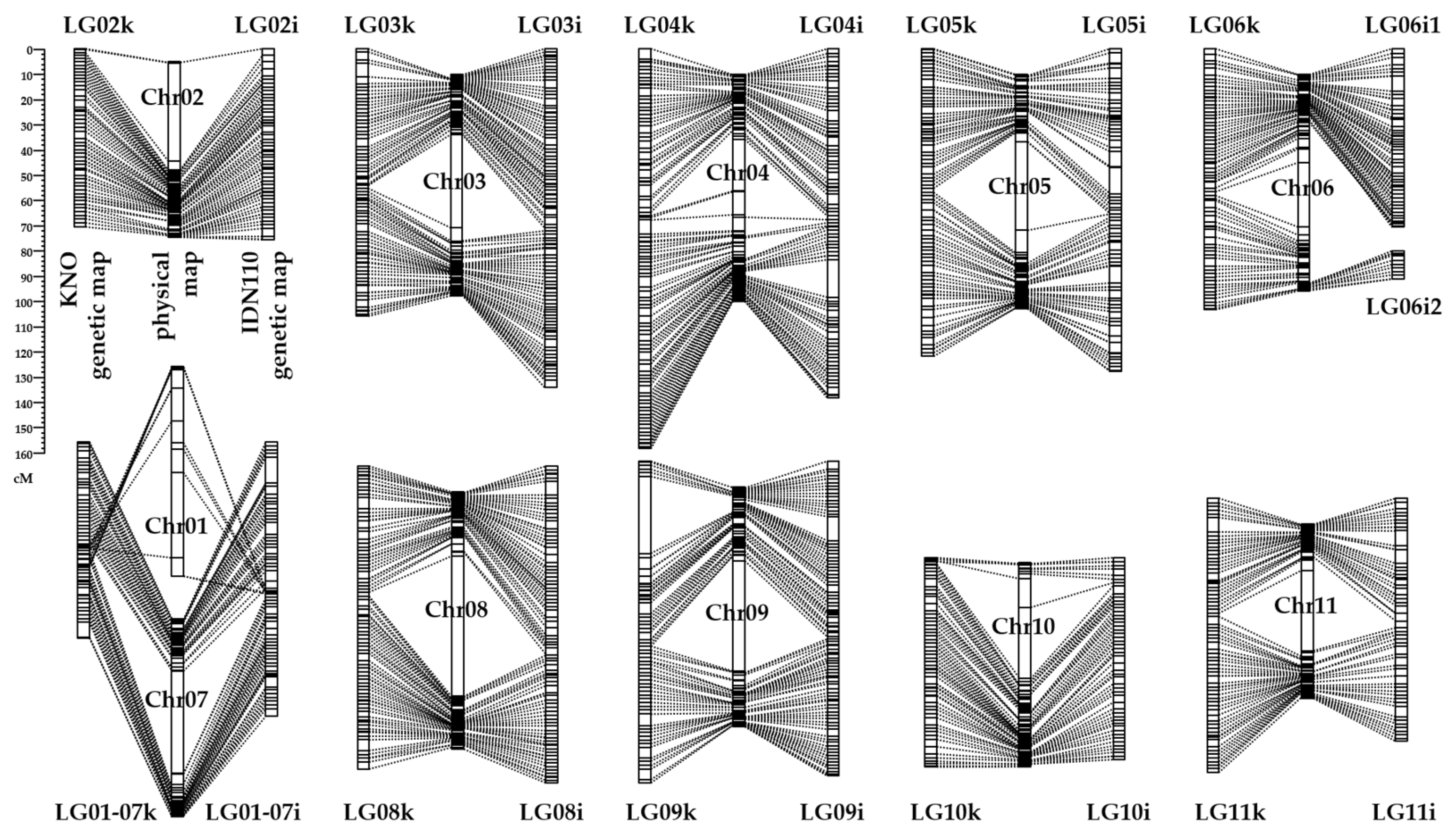
Figure 1. Genetic maps using non-redundant SNP markers of the two parents ‘Khai Nai On’ (KNO, noted as k) and ‘IDN 110’ (noted as i) linked to the reference genome sequence assembly (DH Pahang v4); translocations between chr01 and chr07 for both parents induce their association in the same linkage group.
Horticulturae 10 00608 g001
Figure 2. Reaction of the progeny and parents to P. fijiensis inoculation; (a,b) an example of the detection of symptoms, respectively, before and after analysis using the machine learning software IPSDK Explorer v. 3.0.2.2. (c) Percentage of diseased leaf surface for each of the three replicates (black dots) of the 73 progenies and the two parents KNO and IDN110 at wpi 7. Individuals were ordered according to their median value (white circle). The percentage of diseased leaf surface shows a continuous distribution, from the lower percentage corresponding to partial resistance up to the higher percentage corresponding to susceptibility.
Horticulturae 10 00608 g002
Figure 3. QTLs of resistance to BLSD identified in the KNO x IDN110 progeny projected on the physical map of DH-Pahang reference genome version 4. QTLs linked to IDN110 (names ending in _IDN110) are in green, to KNO (names ending in _KNO) in red, and to their interaction (names ending in _interact) in blue. Phenotypic variables at the origin of the QTL are indicated at the top and the QTL LOD, and the explained variance (R2) at the bottom.
Horticulturae 10 00608 g003
Figure 4. The BLUP values of KNO x IDN110 hybrid phenotypes relative to their percentage of diseased surface at wpi 7 according to the number of favourable resistance alleles at the four main QTLs on chromosomes 06, 07, 08, and 09 of IDN110.
Horticulturae 10 00608 g004
Table 1. Number of resistance gene analogues (RGA) identified in the four main QTLs located on chromosomes 6 to 9 (chr06 to chr09). Four main types of RGA are considered: the nucleotide binding site (NBS); receptor-like kinases (RLK); receptor-like proteins (RLP); and other resistance genes (Oth-R). Each of those four types of RGA include the different protein classes identified by the DRAGO3 tool.
RGA/QTL onchr06chr07chr08chr09TotalInclude Protein Classes of DRAGO3 Tool 1
NBS encoding13149CNL + NL + CN + N
RLK13102539RLK + LecRK
RLP1111013RLP + CL + Lec
Other-R51031937CK + K + L
Total817155898
1 Grouping by DRAGO3 [60] validated with the InterPro domain. Genes are grouped in distinct classes based on the presence of specific domains: C (coiled-coil), N (nucleotide binding site), L (leucine-rich repeat), RLK (receptor-like kinase), Lec (lectin-like), RLP (receptor-like protein), and K (kinase).
Disclaimer/Publisher’s Note: The statements, opinions and data contained in all publications are solely those of the individual author(s) and contributor(s) and not of MDPI and/or the editor(s). MDPI and/or the editor(s) disclaim responsibility for any injury to people or property resulting from any ideas, methods, instructions or products referred to in the content.