Article

Bacterial and Fungal Communities of Table Grape Skins in Shanghai

1
School of Agriculture and Biology, Shanghai Jiao Tong University, Shanghai 200240, China
2
Shanghai Pu Yun Technology Co., Ltd., Shanghai 200240, China
3
Shanghai Agricultural Technology Extension Service Center, Shanghai 201103, China
*
Authors to whom correspondence should be addressed.
Horticulturae 2024, 10(6), 560; https://doi.org/10.3390/horticulturae10060560
Submission received: 22 April 2024 / Revised: 14 May 2024 / Accepted: 24 May 2024 / Published: 27 May 2024
(This article belongs to the Section Viticulture)

Abstract

:
Grape skin serves as a reservoir for many bacteria and fungi, which can affect grape health, quality, and safety. However, grape skin microbiota and mycobiota of table grapes remain largely understudied. This study investigated bacterial and fungal communities residing on different table grapevine cultivars (‘Summer Black’ and ‘Kyoho’) as well as the impact of potential contributors thereby, via culture-dependent and culture-independent (analysis of 16S rRNA gene and internal transcribed spacer sequences) methods with different purposes. Microbiota of both grapevine cultivars were dominated by Cladosporium, Alternaria, Aspergillus, Thauera, and Pantoea. In addition, yeast strains belonging to Hanseniaspora opuntiae, Pichia terricola, Rhodotorula mucilaginosa, Candida stellimalicola, and Kodamaea ohmeri were enriched from the studied grapes, while some strains were considered as health-threatening pathogens. Differences in grapevine cultivars did not significantly affect their mycobiota and microbiota profiles. Nevertheless, their mycobiota exhibited significant variations across different grape-sampling sites in Shanghai, indicating the contribution of the grape-growing environment to grape skin mycobiota. Altogether, the current study demonstrated the contribution of the grape-growing environment to table grape skin mycobiota, and highlighted the importance of microbiota management in the production and consumption of table grapes.

1. Introduction

Grapevines (Vitis spp.) are one of the most widely cultivated fruits worldwide and are consumed fresh, dried, or processed into a variety of products, for instance wine, juice, and vinegar [1]. Table grapes are one type of grapes that are often consumed fresh. Among popular table grapes, ‘Summer Black’ [2] and ‘Kyoho’ [3] are two of the most popular table grape varieties. They are known for their unique flavor, aroma, nutritional value, and health benefits, which, in part, can be attributed to the high phenolic content in their grape skins and their potential use to reduce the risk of cardiovascular disease and inflammation [4].
Grapes harbor a variety of bacteria and fungi that play an important role in grape health, quality, and food safety [5]. Depending, in part, on the presence of beneficial or potentially harmful microbes on the grape, inhabiting bacteria and/or fungi could be beneficial and/or potentially harmful to grapes or consumers [5]. The composition and dynamics of inhabiting bacteria and fungi can be affected by various factors, such as grapevine cultivars, geographic origin, climate, agricultural practices, and postharvest handling [6,7,8]. Understanding these factors and their effects on grape skin bacteria and fungi is essential for developing effective strategies to manage grape quality and safety.
In recent years, advances in high-throughput sequencing technologies have enabled more comprehensive characterization of microbial communities in various environments, including grape skins [9]. These methods, such as 16S rRNA gene sequencing and internal transcribed spacer (ITS) sequencing, allow for the identification and quantification of bacterial and fungal taxa present in complex samples [10,11]. Nevertheless, to date, available studies investigating grape skin bacteria and fungi have mainly focused on wine-making grapes [5,9,12,13]. Relatively little is known about the bacterial and fungal communities of table grapes, which are often harvested at a different stage of ripeness and sweetness compared to wine making grapes.
The current study aimed to characterize the bacterial and fungal communities of table grapes using ‘Summer Black’ and ‘Kyoho’ grapes as representatives. Based on culture-independent methods, we studied grape skins’ microbiota and mycobiota profiles, as well as the effect of grapevine cultivars, grape-sampling districts, and viticultural conditions thereof. Using a cultivation-dependent method, we isolated yeasts that could grow under given conditions. We hypothesized that grapevine cultivars, grape-sampling districts and viticultural conditions could contribute to variation in bacterial and fungal communities.

2. Materials and Methods

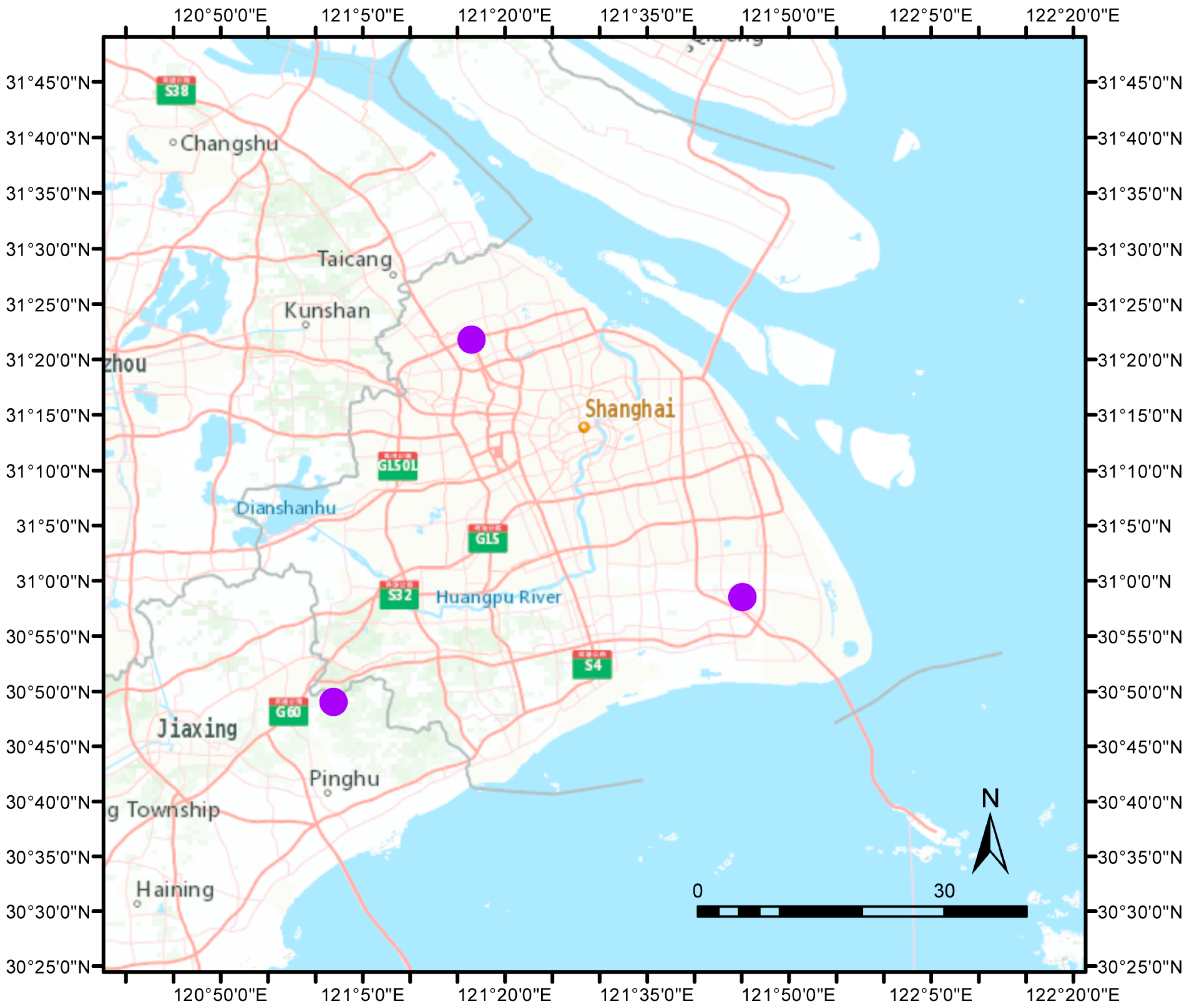
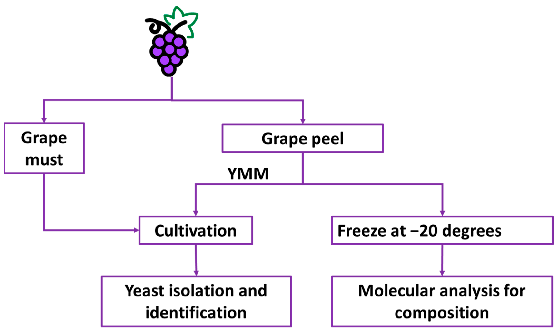
2.1. Study Area and Grape Sampling

In total, sixty bunches of undamaged table grapes were obtained from Jinshan, Pudong, and Jiading, which are key table grape production districts in Shanghai. Within each sampling district, grapes were collected from one open-air vineyard and one greenhouse vineyard. The distance from each sampling site to another is shown based on the projected coordinate system (WGS 1984 UTM zone 51N) in ArcGIS 10.8.2 (Figure 1).
Within each vineyard, two grapevine cultivars, namely ‘Summer Black’ (five bunches) and ‘Kyoho’ (five bunches) were collected. Obtained grapes were subjected to yeast cultivation and sequence analysis to investigate bacterial and fungal composition (Figure 2).

2.2. Yeast Cultivation

Differing from culturomics, in this study the yeast cultivation experiment was set to isolate yeasts that may act as new starters in the fermentation process. One kilogram of each cultivar of grapes from different growing districts and viticulture conditions were crushed into grape must and cultivated at 20 °C at 155 rpm for four consecutive days. Simultaneously, approximately three grams of grape skin were randomly extracted from five bunches of undamaged grapes. Extracted grape skins were then soaked in 80 mL yeast maintenance media (YMM, per liter composed of 3 g yeast extract, 3 g malt extract, 5 g peptone, and 10 g glucose, Sangon Biotech Co., Shanghai, China, pH 5.5) and cultivated at 30 °C for two consecutive days. Afterward, ten μL of each of the above cultures were evenly spread on YMM agar plates and cultivated overnight at 30 °C. Obtained colonies were selected based on colony shapes, colors, and surface features, as well as their morphologies, under light microscopy. Each colony was purified until the colony’s morphology was consistent, and preserved at −80 °C with 30% glycerol.

2.3. Yeast Identification

Yeast genomic DNA was extracted using an Ezup Column Yeast Genomic DNA Purification Kit (Sangon Biotech Co., Shanghai, China) according to the manufacturer’s instructions. Their ITS and 5.8S rDNA genes were amplified with ITS1 (5′-TCCGTAGGTGAACCTGCGG-3′) and ITS4 (5′-TCCTCCGCTTATTGATATGC-3′) [14]. The amplification was conducted using 2× Taq master mix (Vazyme Biotech Co., Nanjing, China), 2 μL forward primer (10 μM), 2 μL reverse primer (10 μM) and 2 μL DNA (20 ng/μL) DNA template in a volume of 50 μL. The PCR program was as follows: denaturing at 95 °C for 5 min, 35 amplification cycles (94 °C, 1 min; 55.5 °C, 2 min; and 72 °C, 2 min) and extension at 72 °C for 10 min. PCR product was purified and sequenced (Shanghai Sunny Biotechnology Co., Ltd., Shanghai, China). Obtained sequences were aligned with the NCBI GenBank database using the local alignment search tool BLASTN.

2.4. DNA Extraction, Amplification, and Sequencing

Approximately ten grams of grape skin was randomly extracted from five bunches of undamaged grapes. Grape skins were washed with 20 mL PBS (Sangon Biotech, Shanghai, China) at 80 rpm/min for 30 min. The washing solution was then transferred into a new tube and centrifuged at 4 °C 10,000× g for 15 min. The obtained pellet was re-suspended with 1 mL PBS and centrifugated again at 4 °C 10,000× g for 5 min. The obtained pellet was stored at −20 °C for bacterial and fungal compositional analysis.
Total DNA was extracted using the Mabio DNA isolation kit (Mabio, Guangzhou, China) following the manufacturer’s instructions. Obtained microbial DNA was used to study bacterial and fungal compositions. Specifically, for bacterial compositional analysis, the V4 region of the 16S rRNA gene was amplified using primers 515F (5′-GTGCCAGC[AC]GCCGCGGTAA-3′) and 806R (5′-GGACTAC[ACT][ACG]GGGT[AT]TCTAAT-3′) and extracted DNA as a template [15,16]. For fungal sequence analysis, ITS genes were amplified using primers ITS1 (5′-CTTGGTCATTTAGAGGAAGTAA-3′) and ITS2 (5′-GCTGCGTTCTTCATCGATGC-3′) and extracted DNA as a template. Amplification was conducted in triplicate using 25 μL 2× premix Taq (Takara Biotechnology, Dalian Co., Ltd., Dalian, China), 1 μL forward primer (10 μM), 1 μL reverse primer (10 μM), and 3 μL DNA template (20 ng/μL) in a volume of 50 μL. The amplification program was 5 min at 94 °C for initialization; 30 cycles of 30 s denaturation at 94 °C, 30 s annealing at 52 °C, and 30 s extension at 72 °C; followed by 10 min final elongation at 72 °C using BioRad S1000 (Bio-Rad Laboratory, Hercules, CA, USA). Following the manufacturer’s instructions, obtained PCR products were used to construct sequencing libraries using the NEBNext® Ultra™ II DNA Library Prep Kit for Illumina® (New England Biolabs, Ipswich, MA, USA), and sequenced by Illumina Nova 6000 (PE250, Illumina, San Diego, USA).

2.5. Sequence Analysis and Bacterial Functional Prediction

Amplicon sequence variants (ASVs) were generated from raw sequence data using DADA2 (v3.19) [17]. Briefly, after removing primer sequences using Cutadapt (v4.3) [18], sequences were filtered and trimmed (maxN = 0, maxEE = c (2,2), truncQ = 2). Taxonomy assignment was performed with the assign taxonomy function (minimal bootstrap value 50) using the SILVA database (version 138) for 16S sequences and the UNITE database (version 8) for ITS region sequences. Prediction of bacterial functions was conducted using PICRUSt2 (v2.5.2) [19] default settings, generated metagenome predictions, and pathway abundances. Raw sequencing data were deposited in the European Nucleotide Archive with accession number PRJEB65061.

2.6. Statistical Analysis

The 16S rRNA gene sequence read counts and ITS read counts were normalized to relative abundance. Microbial diversity indices (Fisher alpha and observed species) were calculated based on ASVs. We used the Shapiro–Wilk test to test data normality, and used the Wilcoxon signed-rank test for comparisons of not-normally distributed data. Variation partitioning was assessed by fitting environmental factors (i.e., sampling district, viticultural conditions, and grapevine cultivars) to the composition of microbial populations using the RDA (Redundancy Analysis, RDA) function in combination with the Monte Carlo permutation in the vegan package. Obtained p values < 0.05 were considered to indicate significant differences. All statistics were conducted in R (R 4-4.2.3).

3. Results

3.1. General Characteristics of Table Grape Microbiota

In total, we identified 252 fungi and 594 bacteria genera from collected grapes. The fungal population was on average dominated by Cladosporium (57.5%), followed by Aspergillus (16.6%), Alternaria (10.9%), and Zygosporium (4.9%). On average, Cladosporium accounted for 70.70% of the total fungal community of Kyoho, and 45.3% of that of Summer Black. Percentages of Cladosporium were 72.9% and 66.8% of the total fungal communities, respectively, of grapes collected from Jinshan and Pudong. The percentage of Cladosporium was only 41.6% in grapes collected from Jiading.
The bacterial population was on average dominated by 43.6% Enterobacteriaceae and 22.8% Rhodocyclaceae (Figure S1). At the genus level, unknown bacterial genera accounted for 32.7% of sequences; Thauera (22.8%) and Pantoea (17.3%) were the other dominant sequence types. Of note, Thauera and Pantoea accounted for more than 50% of the total identified bacteria on average. Nevertheless, this dominance was only observed in grapes collected from Jinshan and Jiading. Specifically, Thauera and Pantoea collectively accounted for 47.12% and 47.73% of the total bacterial communities, respectively, of grapes collected from Jiading and Jinshan. The percentage of Thauera and Pantoea was only 26.94% of the total bacterial community of grapes collected from Pudong, which was instead dominated by unknown bacterial genera. Nevertheless, despite differences in sampling districts, the Kyoho bacterial community was composed of 26.66% Thauera and 5.75% Pantoea, while Summer Black’s was composed of 17.65% and 32.77%, respectively.

3.2. Potential Contributors to Grape Skin Mycobiota and Microbiota

The RDA investigation of potential contributions of differences in grapevine cultivars (‘Summer Black’ and ‘Kyoho’), viticultural conditions (i.e., grapes grown in open air or in a greenhouse), and sampling locations (three different districts in Shanghai) to the variation of grape-skin microbiota and mycobiota found only a significant contribution from sampling locations, which explained 16.94% of the total mycobiota variation.
Permutational multivariate analysis of variance (PERMANOVA) revealed a significant difference only between the mycobiota of Jiading grapes and those of Pudong grapes based on the Bray–Curtis distance matrix (considering the relative abundance of ASVs) (Figure 3A), but no significant difference based on the Jaccard distance matrix (considering only presence or absence of ASVs) (Figure 3B). Regarding specific fungal taxa, their mycobiota were all dominated by Cladosporium, which was highest in Pudong grapes and lowest in Jiading grapes (Figure 3A). In addition, the relative abundance of Aspergillus was higher in Jiading and Jinshan grapes compared to that of Pudong grapes. Moreover, the relative abundance of Alternaria was highest in Jiading grapes and lowest in Pudong grapes. Nevertheless, no significant difference was identified in the relative abundance of specific fungal genera between different districts.

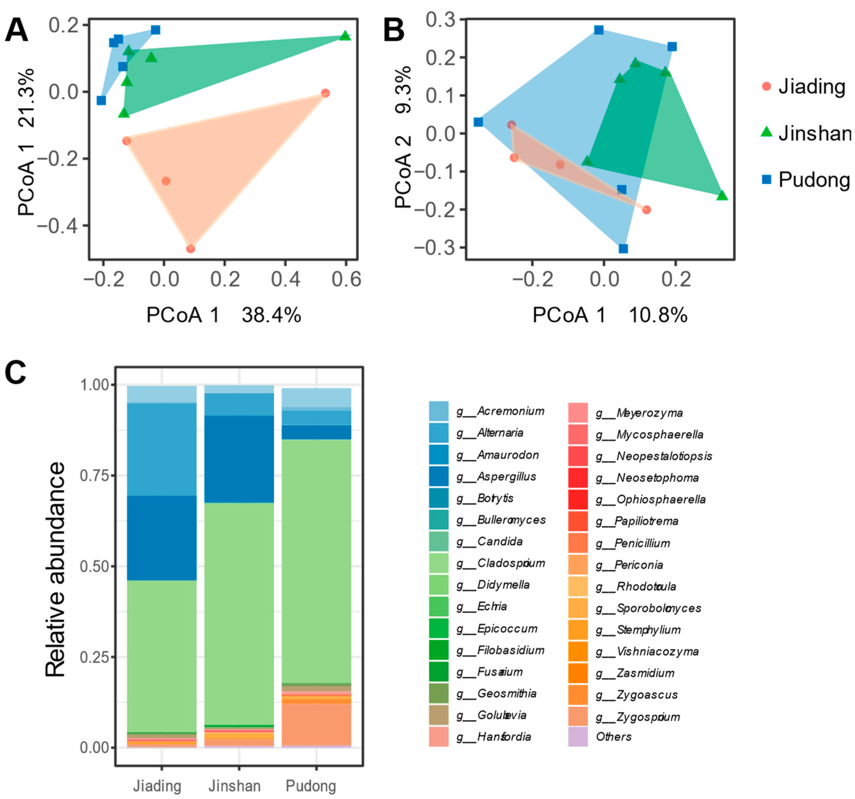
3.3. Mycobiota and Microbiota of Different Table Grapes

Although no significant contribution from grapevine cultivars (‘Summer Black’ and ‘Kyoho’) was identified based on the RDA, it was generally believed that different grapevine cultivars enharbour distinct bacterial and fungal communities. As such, we further investigated the impact of grapevine cultivars on table grape microbiota and mycobiota.
PERMANOVA based on Bray–Curtis and Jaccard distance matrices revealed no significant difference between mycobiotas (Figure 4A,B) of ‘Summer Black’ and ‘Kyoho’ grapes. Nor was a significant difference identified in the fungal richness (observed ASVs) or diversity (inverse Simpson) between these two grape cultivars (Figure 4C,D). Similarly, no significant differences were detected in the bacterial profile (Figure 4E,F), richness, or diversity (Figure 4G,H) between these two grape varieties.
Regarding microbial functionality, predicted bacterial functionality profiles did not differ significantly between ‘Kyoho’ and ‘Summer Black’ grapes (Figure 5A), and redundancy analysis found no significant contribution from sampling districts and viticulture conditions, while studied grape skin microbiota enharboured pathways coding for aerobic respiration, fatty acid oxidation, and pathways for biosynthesis (Figure 5B).

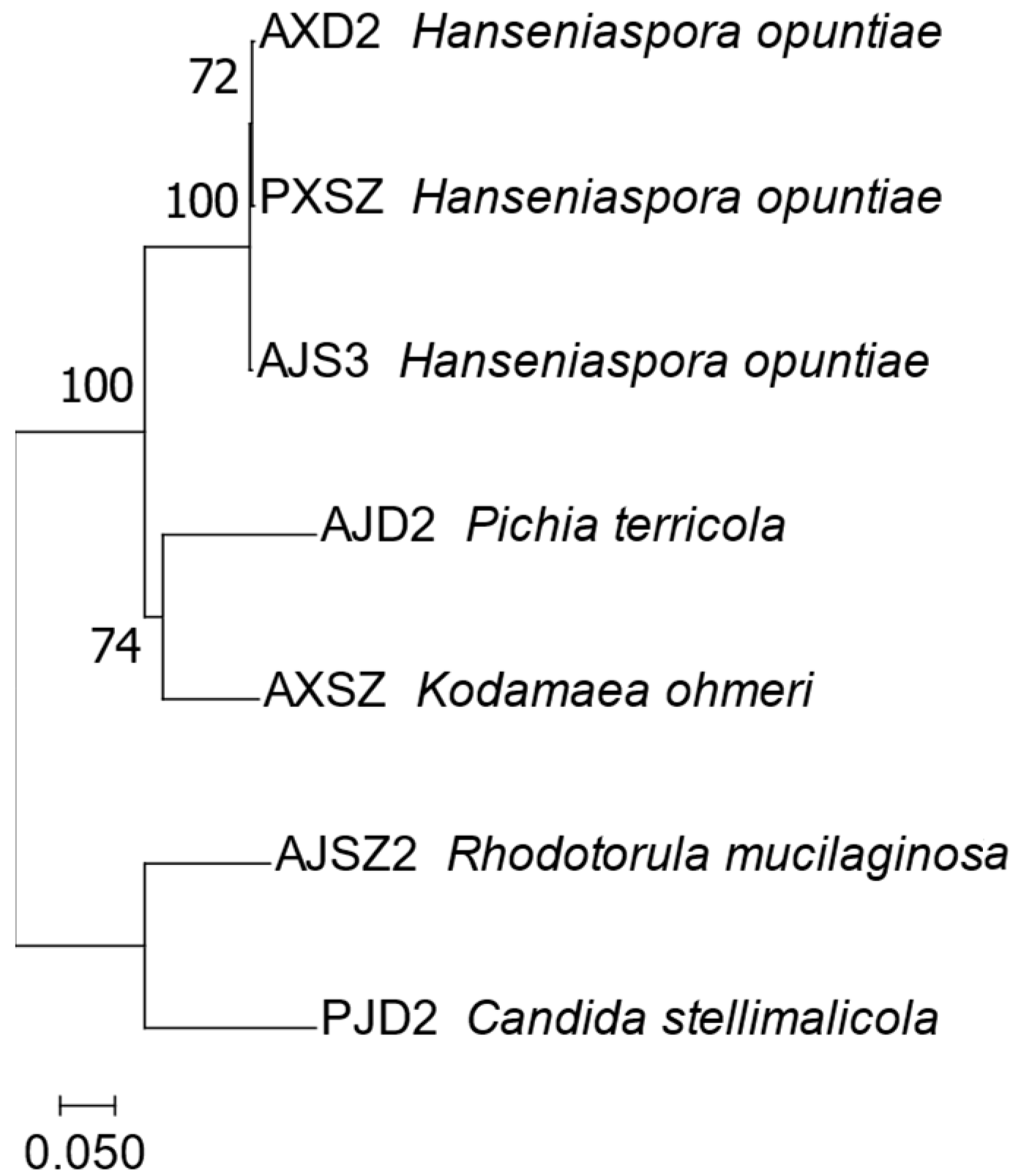
3.4. Cultivated Yeasts

Cultivated yeasts were classified as Hanseniaspora opuntiae (identity 99.86–100%), Pichia terricola (identity 98.81–99.05%), Rhodotorula mucilaginosa (identity 100%), Candida stellimalicola (identity 99.39%), and Kodamaea ohmeri (identity 98.54%). Their representative ITS sequences are provided in Table S1. The phylogenetic tree based on ITS1 and ITS2 regions generated three different groups, which were grouped into three clusters, in line with their classification results (Figure 6). Additionally, among these cultivated yeasts, only Pichia and Kodamaea genera were not identified based on the sequencing approach.

4. Discussion

In the current study, we investigated the bacterial and fungal communities of two table grape types and explored the impact of potential contributors (grape-growing districts, viticultural conditions, and grapevine cultivars) thereof. Our hypothesis was that grapevine cultivars, grape-sampling districts, and viticultural conditions could contribute to variation in the bacterial and fungal communities of table grapes. We found that the microbiota of both grapevine cultivars was dominated by Cladosporium, Alternaria, Aspergillus, Thauera, and Pantoea. Of note, the impact of grapevine cultivars and viticultural conditions on grape skin microbiota and mycobiota variation was not detectable, while grapes’ growing locations/districts significantly contributed to the variation thereof. Moreover, the enrichment and isolation of yeasts successfully identified Hanseniaspora opuntiae, Pichia terricola, Rhodotorula mucilaginosa, Candida stellimalicola, and Kodamaea ohmeri from the skins. Some of these are co-fermenters in fermentation, while others can act as spoilage yeast or even pathogens. Collectively, these findings document the potential contribution of grape-growing environments to table grape skin microbiota and mycobiota, and highlight the importance of microbiota management in the production and consumption of these grapes.
In contrast to the thought that different grapevine cultivars select for distinct bacterial and fungal communities [7], in this study, cultivar differences did not clearly influence grape skin microbiota and mycobiota of ‘Summer Black’ and ‘Kyoho’ grapes. Grape-growing environments, i.e., sampling districts in Shanghai, affected their mycobiota, highlighting a regional effect on grape skin microbiota, as suggested for wine-making grapes [20]. These differences in grape skin mycobiota among districts of Shanghai could, in part, be attribute to the environment or air fungal composition, which might be linked with existing biogeographical patterns and/or the presence of nearby industry sites.
Regarding specifical bacterial and fungal taxa, in line with earlier studies, this study found that grape skin mycobiota were dominated by Cladosporium and Alternaria [21]. Nevertheless, in addition to these two genera, this study also identified that a large portion of table grapes’ skin mycobiota was Aspergillus (16.6%). Aspergillus is present on mature berries and could be considered grape invaders that produce ochratoxin, i.e., a mycotoxin that might be carcinogenic to humans [22]. In terms of grape skin microbiota, this study found that Thauera, Pantoea, and unknown bacterial genera dominate the skin. This finding, however, differed from that of wine grapes, which were dominated by Lactobacillus, Streptococcus, and Tatumella [23]. This difference could therefore assist in the differentiation of table grape skin microbiota from those of wine grapes. Moreover, the presence and dominance of the identified Thauera and Pantoea could possibly affect the flavor of the grapes and their shelf-life post harvesting.
Complementary to high throughput sequence analysis, in the current study, the cultivation-based method successfully isolated a few rare yeasts. In addition to Hanseniaspora, Pichia, and Candida, which are commonly present on the surface of grapes [8], the current study also successfully cultivated and identified Rhodotorula mucilaginosa [24] and Kodamaea ohmeri [25]. Notably, Rhodotorula mucilaginosa and Kodamaea might potentially be pathogenic to humans and lead to safety concerns regarding the studied grapes [26]. Specifically, Rhodotorula mucilaginosa [24] could be an opportunistic infectious fungi leading to oral ulcers in AIDs patients [27], while Kodamaea ohmeri [25,26], also known as Pichia ohmeri or Yamadazyma ohmeri, could be a life-threatening pathogen in humans, especially immunocompromised patients [28,29]. Furthermore, future studies are warranted to investigate characteristics of these isolated yeasts and uncover their role in grape production, as well as their potential application in food/beverage production. In addition to fungi identified after cultivation, the sequencing-based approach identified some plant pathogens, like Alternaria and Botrytis. The presence of these fungi could also have an impact on Vitis vinifera cultivars. Nevertheless, data on Vitis vinifera cultivars were not noted in the current study. Studies investigating the impact of these fungi on grapevines are still warranted. For instance, the presence of fungus Botrytis, as identified in the current study, could have an impact on grape flavors. Future studies could consider and evaluate the flavor profiles of grapes and co-existing bacterial and fungal communities.
In conclusion, instead of the type of grapevine cultivars (‘Summer Black’ and ‘Kyoho’), the grape-growing environment, i.e., sampling districts in Shanghai, affected grapes’ mycobiota significantly, while their mycobiota were dominated by Cladosporium, Alternaria, and Aspergillus. In terms of their microbiota, all were dominated by Thauera, Pantoea, and unknown bacterial genera. Of note, Hanseniaspora opuntiae, Pichia terricola, Rhodotorula mucilaginosa, Candida stellimalicola, and Kodamaea ohmeri were isolated and identified from grape skins. Collectively, the current study demonstrated the impact of the cultivation environment, i.e., sampling location, on grape skin mycobiota, as well as the increased need to monitor/manage table grape skin microbiota. This paper contributes to the growing body of knowledge regarding grape skin microbiota and its potential applications in grape production, food safety, and human health.

Supplementary Materials

The following supporting information can be downloaded at: https://www.mdpi.com/article/10.3390/horticulturae10060560/s1, Table S1: Representative ITS sequences and identities of isolates. Figure S1: Relative abundance of most predominant bacterial genus (top 20, ranked base on the average relative abundance across the entire dataset). Top 20 microbial genera are listed in the legend. Other genera are summarized as “other”. Each column represents a given type of sample from one district.

Author Contributions

Conceptualization, R.A. and S.S. (Shiren Song); methodology, R.A.; software, R.A. and S.S. (Shiren Song); validation, S.S. (Sijie Sun) and H.Z.; formal analysis, R.A.; investigation, R.A., S.S. (Sijie Sun), and H.Z.; resources, S.S. (Shiren Song) and D.W.; data curation, H.Z.; writing—original draft preparation, R.A.; writing—review and editing, Q.M., D.W. and S.S. (Shiren Song); visualization, C.L.; supervision, S.S. (Shiren Song); project administration, S.S. (Shiren Song); funding acquisition, S.S. (Shiren Song) and D.W. All authors have read and agreed to the published version of the manuscript.

Funding

This research was funded by the Key Area Research and Development Program of Guangdong Province, grant number 2022B1111070006, the Shanghai Municipal Agricultural Commission, grant number 2022-02-08-00-12-F01131, and the Shanghai Municipal Commission for Science and Technology, grant number 21ZR1432500.

Data Availability Statement

Raw sequencing data were deposited in the European Nucleotide Archive with accession number PRJEB65061.

Conflicts of Interest

Sijie Sun is an employee of Shanghai Pu Yun Technology Co., Ltd. The remaining authors declare that the research was conducted in the absence of any commercial or financial relationships that could be construed as a potential conflict of interest.

References

  1. Creasy, G.L.; Creasy, L.L. Grapes (Crop Production Science in Horticulture), 2nd ed.; CABI: Wallingford, UK, 2018; Volume 27. [Google Scholar]
  2. Ozawa, T.; Amemiya, T.; Sato, T.; Furuya, J.; Furuya, K.; Miyake, M.; Saito, N.; Hirabayashi, T.; Mochiduki, T.; Kondo, M. Summer Black; A New Seedless Grape; Bulletin of the Yamanashi Fruit Tree Experiment Station; Yamanashi Fruit Tree Experiment Station: Yamanashi, Japan, 2000. [Google Scholar]
  3. Yamada, M.; Sato, A. Advances in table grape breeding in Japan. Breed. Sci. 2016, 66, 34–45. [Google Scholar] [CrossRef] [PubMed]
  4. Sochorova, L.; Prusova, B.; Cebova, M.; Jurikova, T.; Mlcek, J.; Adamkova, A.; Nedomova, S.; Baron, M.; Sochor, J. Health effects of grape seed and skin extracts and their influence on biochemical markers. Molecules 2020, 25, 5311. [Google Scholar] [CrossRef] [PubMed]
  5. Barata, A.; Malfeito-Ferreira, M.; Loureiro, V. The microbial ecology of wine grape berries. Int. J. Food Microbiol. 2012, 153, 243–259. [Google Scholar] [CrossRef] [PubMed]
  6. Vitulo, N.; Lemos, W.J.F., Jr.; Calgaro, M.; Confalone, M.; Felis, G.E.; Zapparoli, G.; Nardi, T. Bark and grape microbiome of Vitis vinifera: Influence of geographic patterns and agronomic management on bacterial diversity. Front. Microbiol. 2019, 9, 3203. [Google Scholar] [CrossRef]
  7. Mezzasalma, V.; Sandionigi, A.; Guzzetti, L.; Galimberti, A.; Grando, M.S.; Tardaguila, J.; Labra, M. Geographical and cultivar features differentiate grape microbiota in Northern Italy and Spain vineyards. Front. Microbiol. 2018, 9, 946. [Google Scholar] [CrossRef] [PubMed]
  8. Bozoudi, D.; Tsaltas, D. Grape microbiome: Potential and opportunities as a source of starter cultures. In Grape and Wine Biotechnology; InTech: Houston, TX, USA, 2016. [Google Scholar]
  9. Morgan, H.H.; Toit, M.D.; Setati, M.E. The grapevine and wine microbiome: Insights from high-throughput amplicon sequencing. Front. Microbiol. 2017, 8, 820. [Google Scholar] [CrossRef] [PubMed]
  10. Begerow, D.; Nilsson, H.; Unterseher, M.; Maier, W. Current state and perspectives of fungal DNA barcoding and rapid identification procedures. Appl. Microbiol. Biotechnol. 2010, 87, 99–108. [Google Scholar] [CrossRef]
  11. Schoch, C.L.; Seifert, K.A.; Huhndorf, S.; Robert, V.; Spouge, J.L.; Levesque, C.A.; Chen, W.; Consortium, F.B.; List, F.B.C.A.; Bolchacova, E. Nuclear ribosomal internal transcribed spacer (ITS) region as a universal DNA barcode marker for Fungi. Proc. Natl. Acad. Sci. USA 2012, 109, 6241–6246. [Google Scholar] [CrossRef] [PubMed]
  12. Ding, Y.; Wei, R.; Wang, L.; Wang, W.; Wang, H.; Li, H. Exploring the ecological characteristics of natural microbial communities along the continuum from grape berries to winemaking. Food Res. Int. 2023, 167, 112718. [Google Scholar] [CrossRef]
  13. Gao, F.; Chen, J.; Xiao, J.; Cheng, W.; Zheng, X.; Wang, B.; Shi, X. Microbial community composition on grape surface controlled by geographical factors of different wine regions in Xinjiang, China. Food Res. Int. 2019, 122, 348–360. [Google Scholar] [CrossRef]
  14. González-Alonso, I.; Walker, M.E.; Vallejo-Pascual, M.-E.; Naharro-Carrasco, G.; Jiranek, V. Capturing yeast associated with grapes and spontaneous fermentations of the Negro Saurí minority variety from an experimental vineyard near León. Sci. Rep. 2021, 11, 3748. [Google Scholar] [CrossRef] [PubMed]
  15. Parada, A.E.; Needham, D.M.; Fuhrman, J.A. Every base matters: Assessing small subunit rRNA primers for marine microbiomes with mock communities, time series and global field samples. Environ. Microbiol. 2016, 18, 1403–1414. [Google Scholar] [CrossRef] [PubMed]
  16. Apprill, A.; McNally, S.; Parsons, R.; Weber, L. Minor revision to V4 region SSU rRNA 806R gene primer greatly increases detection of SAR11 bacterioplankton. Aquat. Microb. Ecol. 2015, 75, 129–137. [Google Scholar] [CrossRef]
  17. Callahan, B.J.; McMurdie, P.J.; Rosen, M.J.; Han, A.W.; Johnson, A.J.A.; Holmes, S.P. DADA2: High-resolution sample inference from Illumina amplicon data. Nat. Methods 2016, 13, 581–583. [Google Scholar] [CrossRef] [PubMed]
  18. Martin, M. Cutadapt removes adapter sequences from high-throughput sequencing reads. EMBnet. J. 2011, 17, 10–12. [Google Scholar] [CrossRef]
  19. Douglas, G.M.; Maffei, V.J.; Zaneveld, J.R.; Yurgel, S.N.; Brown, J.R.; Taylor, C.M.; Huttenhower, C.; Langille, M.G.I. PICRUSt2 for prediction of metagenome functions. Nat. Biotechnol. 2020, 38, 685–688. [Google Scholar] [CrossRef] [PubMed]
  20. Taylor, M.W.; Tsai, P.; Anfang, N.; Ross, H.A.; Goddard, M.R. Pyrosequencing reveals regional differences in fruit-associated fungal communities. Environ. Microbiol. 2014, 16, 2848–2858. [Google Scholar] [CrossRef] [PubMed]
  21. Bettenfeld, P.; Cadena, I.C.J.; Jacquens, L.; Fernandez, O.; Fontaine, F.; van Schaik, E.; Courty, P.E.; Trouvelot, S. The microbiota of the grapevine holobiont: A key component of plant health. J. Adv. Res. 2022, 40, 1–15. [Google Scholar] [CrossRef] [PubMed]
  22. Bellí, N.; Marín, S.; Coronas, I.; Sanchis, V.; Ramos, A.J. Skin damage, high temperature and relative humidity as detrimental factors for Aspergillus carbonarius infection and ochratoxin A production in grapes. Food Control. 2007, 18, 1343–1349. [Google Scholar] [CrossRef]
  23. Kamilari, E.; Mina, M.; Karallis, C.; Tsaltas, D. Metataxonomic analysis of grape microbiota during wine fermentation reveals the distinction of xyprus regional terroirs. Front. Microbiol. 2021, 12, 726483. [Google Scholar] [CrossRef]
  24. Wirth, F.; Goldani, L.Z. Epidemiology of Rhodotorula: An emerging pathogen. Interdiscip. Perspect. Infect. Dis. 2012, 2012, 465717. [Google Scholar] [CrossRef] [PubMed]
  25. Zhou, M.; Li, Y.; Kudinha, T.; Xu, Y.; Liu, Z. Kodamaea ohmeri as an emerging human pathogen: A review and update. Front. Microbiol. 2021, 12, 736582. [Google Scholar] [CrossRef] [PubMed]
  26. Ioannou, P.; Papakitsou, I. Kodamaea ohmeri infections in humans: A systematic review. Mycoses 2020, 63, 636–643. [Google Scholar] [CrossRef] [PubMed]
  27. Kaur, R.; Wadhwa, A.; Agarwal, S.K. Rhodotorula mucilaginosa: An unusual cause of oral ulcers in AIDS patients. AIDS 2007, 21, 1068–1069. [Google Scholar] [CrossRef] [PubMed]
  28. Lee, J.S.; Shin, J.H.; Kim, M.N.; Jung, S.I.; Park, K.H.; Cho, D.; Kee, S.J.; Shin, M.G.; Suh, S.P.; Ryang, D.W. Kodamaea ohmeri isolates from patients in a university hospital: Identification, antifungal susceptibility, and pulsed-field gel electrophoresis analysis. J. Clin. Microbiol. 2007, 45, 1005–1010. [Google Scholar] [CrossRef]
  29. Otag, F.; Kuyucu, N.; Erturan, Z.; Sen, S.; Emekdas, G.; Sugita, T. An outbreak of Pichia ohmeri infection in the paediatric intensive care unit: Case reports and review of the literature. Mycoses 2005, 48, 265–926. [Google Scholar] [CrossRef]
Figure 1. Sampling locations in Shanghai. Purple dots represented specific sampling locations in each district of Shanghai.
Horticulturae 10 00560 g001
Figure 2. Illustration of study design. YMM: yeast maintenance media.
Horticulturae 10 00560 g002
Figure 3. Impact of grape-growing district on overall mycobiota profile. PCoA of mycobiota based on (A) Bray–Curtis or (B) Jaccard distance matrices. (C) Relative abundance of most predominant fungal genus (top 20, ranked based on average relative abundance across entire dataset). Top 20 microbial genera are listed in the legend. Other genera are summarized as “Others”. Each column represents a given type of sample from one district.
Horticulturae 10 00560 g003
Figure 4. Impact of grape cultivars on mycobiota and microbiota of grape skins. PCoA of mycobiota based on (A) Bray–Curtis or (B) Jaccard distance matrices. (C) Mycobiota richness and (D) diversity. PCoA of microbiota based on (E) Bray–Curtis and (F) Jaccard distance matrices. (G) Microbial richness and (H) diversity. Significant differences between samples based on Bray–Curtis and Jaccard distance matrices (considering only the presence or absence of ASVs) at ASV level were tested by PERMANOVA. The percentage of variation in microbial composition explained by the two principal coordinates is shown at the axes. Purple triangles represent Kyoho grapes, and green circles represent ‘Summer Black’ grapes. Pairwise comparisons between different sample types were evaluated by the Wilcoxon signed-rank test. PCoA: principal coordinate analysis. InvSimpson: inverse Simpson. PERMANOVA: permutational multivariate analysis of variance.
Horticulturae 10 00560 g004
Figure 5. (A) PCoA of microbial functional pathways. (B) Relative abundance of most predominant bacterial functional pathways (top 10, ranked base on the averaged relative abundance across the entire dataset). Top 10 microbial functional pathways are listed on the left.
Horticulturae 10 00560 g005
Figure 6. Phylogenetic tree constructed based on the sequence alignment of ITS1 and ITS2 regions. The phylogenetic tree was inferred from the “neighbour-joining method”, and numbers on the branches represent percentages of supports from the bootstrap replication. The length of the branch represents bootstrap values; values smaller than 70 were collapsed.
Horticulturae 10 00560 g006
Disclaimer/Publisher’s Note: The statements, opinions and data contained in all publications are solely those of the individual author(s) and contributor(s) and not of MDPI and/or the editor(s). MDPI and/or the editor(s) disclaim responsibility for any injury to people or property resulting from any ideas, methods, instructions or products referred to in the content.

