Article

Can SPAD Values and CIE L*a*b* Scales Predict Chlorophyll and Carotenoid Concentrations in Leaves and Diagnose the Growth Potential of Trees? An Empirical Study of Four Tree Species

1
College of Life and Sciences, Nanjing Forestry University, Nanjing 210037, China
2
Hongze Lake East Wetland Provincial Nature Reserve Management Office, Huaian 223200, China
3
Jiangsu Academy of Forestry, Nanjing 211153, China
*
Authors to whom correspondence should be addressed.
These authors contributed equally to this work.
Horticulturae 2024, 10(6), 548; https://doi.org/10.3390/horticulturae10060548
Submission received: 13 April 2024 / Revised: 13 May 2024 / Accepted: 17 May 2024 / Published: 24 May 2024
(This article belongs to the Section Plant Nutrition)

Abstract

:
Photosynthetic pigments are fundamental for plant photosynthesis and play an important role in plant growth. Currently, the frequently used method for measuring photosynthetic pigments is spectrophotometry. Additionally, the SPAD-502 chlorophyll meter, with its advantages of easy operation and non-destructive testing, has been widely applied in land agriculture. However, the application prospects of its test results in horticultural plants have not yet been proven. This study examines the reliability of SPAD values for predicting chlorophyll concentrations. Using fresh and senescent leaves from four common horticultural plants, we measured SPAD values, photosynthetic pigment concentrations, and leaf color parameters. A generalized linear mixed model demonstrated that SPAD values are a reliable indicator for predicting chlorophyll concentrations, yet interspecific variations exist. Based on the predictive power of SPAD values for chlorophyll, we first propose an Enrichment Index (CEI) and a Normal Chlorophyll Concentration Threshold (NCCT). The CEI can be used to compare SPAD values among different species, and the NCCT value can serve as a more accurate indicator for assessing the growth potential of old trees. However, due to a limited sample size, further research with larger samples is needed to refine the diagnosis of plant growth potential and enhance the management of ornamental plant cultivation.

1. Introduction

Chlorophyll and carotenoid are color-related pigments [1], and their functions are related to photosynthesis and photoprotection, respectively [2,3]. The visible colors observed in nature occur in the domain of 400–700 nm, and they primarily depend on the reflective properties of specific objects. For plants, the strong absorption of photosynthetic pigments in the visible domain, especially non-green wavelengths, leads to lower leaf reflectance [4]. Plant coloration is related to the pigment types and their mass fractions and distribution within leaf cells. The change of different plants over time and space is the result of variations in pigment proportions and types in leaves.
Both substances are called plastid pigments because of their location in the pigment organelle, the plastid. Chlorophyll is distributed in chloroplasts, whereas carotenoids are enriched in chromoplasts [5]. Different pigments exhibit separate colors: chlorophyll a appears as bluish-green, whereas chlorophyll b is characterized by a yellowish-green color [6]. As tetraterpene pigments, carotenoids exhibit various colors, such as yellow, orange, red, and purple [7]. Plastid pigments are not all the pigments that contribute to the color of leaves, and the red and purple coloration in senescing leaves is most commonly due to anthocyanins [8]. All these pigments have their metabolic pathways, such as chlorophyll biosynthesis [9], xanthophyll cycles [7], and anthocyanin biosynthesis pathways [10].
The appearance of plant leaves is mainly reflected in the relative concentration of different pigments inside. Due to the correlations between chlorophyll and photosynthesis, as well as between carotenoids and photoprotection, the fluctuation in their concentrations in leaves reflects rich eco-physiological information. Nitrate deficiency inhibits the synthesis of the chlorophyll precursor porphobilinogen, thus affecting nitrogen assimilation and chlorophyll synthesis [11]. During the process of senescence, leaves show yellowing due to the loss of chlorophyll [12]. Low temperatures inhibit chlorophyll biosynthesis and lead to a decline in chlorophyll accumulation [13]. Pathogens may induce leaf chlorosis [14]. Correspondingly, leaf carotenoid concentrations may be affected by various stress factors, such as light [15], drought [16], and salt [17]. Based on the above eco-physiological relationships, the pigment concentrations in leaves reflect, to some extent, the physiological and health status of plants, especially during the growing season.
When measuring the pigment concentrations under field conditions, there are still issues with sample preservation and transportation. It is imperative to find proxy methods for simple, in-situ, and instantaneous measurements. Two kinds of measures related to leaf colors and pigments have been developed and widely used in agriculture. One of them is a CIE L*a*b* system, such as an NR20XE Precision Colorimeter (Shenzhen ThreeNH Technology, Shenzhen, China) [18], a Hand-held Color Analyzer (CM-2500D, Konica Minolta, Osaka, Japan) [19], or a Minolta Chroma Meter (Model CR200/300, Minolta, Ramsey, NJ, USA) [20]. Another is a portable Chl meter (SPAD-502Plus, Minolta Camera Co., Osaka, Japan) [21,22,23]. The SPAD-502 has been widely used for assessing or diagnosing a plant’s nitrogen status [24,25,26], health status [27], pest and disease stress [28], leaf senescence [29], and coloration effects [30].
Most of the SPAD-related research focused on cereals [31,32,33,34,35], fruit crops [20,36], and vegetables [37], with some investigations on forest trees [38,39,40,41,42]. In addition, compared to the determination of SPAD values, the determination of CIE L*a*b* values in plant leaves has received little attention [20,27,43]. Perennial and long-lived woody plants have diverse characteristics of life history and continuous activities of the apical meristem, as well as versatile leaf characteristics in organogenesis and senescence. Is there a different pattern for evaluating the plant nutrition, leaf senescence, health status, and color effects of woody plants based on SPAD values? It is not yet clear. It is particularly important to point out that those old trees are over 100 years old and have a particularly long lifespan. Can SPAD values be used to evaluate their growth potential? This is also a very challenging issue. Solving these problems is an important reference value for old tree protection practices in China.
In the current study, we selected four tree species as investigation targets, including two evergreen species, Sabina chinensis ‘Kaizuca’ and Osmanthus fragrans, and two deciduous species, Ginkgo biloba and Quercus acutissima. These tree species are widely distributed and cultivated in China, with numerous old tree records [44,45,46]. The objectives of this study are as follows: (i) to identify the characteristics of SPAD, chlorophyll, and carotenoid content changes in the fresh and senescent leaves of four plant species; (ii) to obtain the correlation patterns among SPAD values, leaf color parameters, and the content of chlorophylls and carotenoids; (iii) to reach a conclusion on whether SPAD values can predict the concentration of photosynthetic pigments; and (iv) to evaluate the feasibility of SPAD and CIE L*a*b* values in the diagnosis of leaf aging and tree health.

2. Materials and Methods

2.1. Experimental Site and Sampling

This experiment was conducted at Nanjing Forestry University, which is located on a garden-style campus (32°5′7″ N, 118°49′24″ E, mean sea level 20–30 m). The university boasts an extensive collection of traditional Chinese garden plants, making it an ideal research area for ornamental plants. Nanjing possesses a subtropical monsoon climate with abundant rainfall. The annual precipitation is 1200 mm. The four seasons are distinct, with an average annual temperature of 15.4 °C.

2.2. Plant Material

This research focuses on the leaves of four tree species, Ginkgo biloba (GB), Osmanthus fragrans (OF), Sabina chinensis ‘Kaizuca’ (SC), and Quercus acutissima (QA), on campus (Table 1). To ensure the representativeness of the experimental results, 40 randomly selected trees of each species were chosen as the samples for fresh leaves, with three replicates for each sample. For senescent leaves, three randomly selected trees of each species were chosen as the samples, with three replicates for each sample (Figure 1).

2.3. Measurements with SPAD-502 Chlorophyll Meter and CR-410 Automatic Color Difference Meter

SPAD and leaf color parameters were measured on fully expanded leaves in situ using the SPAD-502 chlorophyll meter (Minolta Camera Co., Osaka, Japan) and CR-410 automatic color difference meter(Minolta Camera Co., Osaka, Japan). To reduce the impact of daily chloroplast movement on the SPAD values, the measurements at each individual sampling time were conducted at the same time each day from 9:00 to 12:00 [47]. The meter head was placed at the adaxial leaf surface, avoiding the mid-vein. The SPAD-502 chlorophyll meter and CR-410 automatic color difference meter were used for measurements on both fully expanded green leaves and senescent leaves; each value obtained was the average of three readings.

2.4. Leaf Chlorophyll Concentrations

Chlorophylls and carotenoids were extracted from the same leaves that were used for the SPAD determination. Sampled leaves, sheltered from light, were transported to the laboratory, and all plant material was prepared within 2 h after the collection. A total of 0.1 g of fresh sample was weighed and added to a grinding tube containing 1 mL of 95% anhydrous ethanol in the dark condition. After grinding using a grinder, the materials were transferred to a test tube. The grinding tube was rinsed twice with 2 mL of 95% anhydrous ethanol. The test tube was placed in a cool, dark place for 24 h to extract the pigments. After that, the absorbance values were measured at 470 nm, 645 nm, and 663 nm using a spectrophotometer. The Chl a, b, and Car concentrations were determined using the following equations [48]:
Ca(mg·L−1) = 13.95·D663 − 6.88·D645
Cb(mg·L−1) = 24.96·D645 − 7.32D663
Ca+b = Ca + Cb = 6.63 D663 + 18.08 D645
Car (mg·L−1) = (1000·D470 − 2.05·Ca − 114·Cb)/245
Chlorophyll (carotenoid) concentration (mg·g−1) = (C·V·dilution ratio)/M·1000
Note: D663, D645, and D470 are the absorbance of the ethanol extract of the test sample at 663 nm, 645 nm, and 470 nm, respectively; Ca, Cb, Ca+b, and Car are the concentrations of chlorophyll a, chlorophyll b, total chlorophyll, and carotenoids respectively in mg·L−1; V is the volumetric capacity of the chlorophyll extract solution (mL), and M is the dry weight of the test sample (g).

2.5. Data Analysis

Before conducting data analysis, we employed the Shapiro–Wilk test to assess whether the fresh and senescent leaves followed a normal distribution. The results indicated that neither the fresh nor the senescent leaves followed a normal distribution (p < 0.05). To examine the differences in leaf color parameters, chlorophylls, and carotenoids between the four species, we utilized the Wilcoxon test and Duncan test to compare differences between different species. This method was also applied to assess whether significant differences existed between fresh and senescent leaves. The Chlorophyll Enrichment Index (CEI) and the Normal Chlorophyll Concentration Threshold (NCCT) were calculated using the following formulas:
C E I = S P A D f / S P A D s
N C C T = S P A D f + S P A D S 2
Here, SPADf and SPADs refer to the SPAD values obtained from the chlorophyll meter readings of fresh leaves and senescent leaves, respectively.
To assess the potential correlation between leaf color parameters and chlorophyll concentrations, we conducted Spearman correlation tests to determine the correlation coefficients between leaf color parameters and chlorophyll concentrations and examined the significance of this relationship. Finally, we fitted a general linear mixed model (using the Gamma family with a log link) to predict chlorophyll concentrations with SPAD values, as outlined in the following model:
log C C ~ β 1 × S P A D + π s p e c i e s + ε + β 0
Here, CC refers to chlorophyll and carotenoid concentrations, and πspecies represents the random effect caused by different species. β0 represents the intercept, which is a random effect, while β1 represents the parameter for SPAD, which is a fixed effect.

3. Results

3.1. Characteristics of SPAD, Chlorophyll, and Carotenoid Concentrations in Four Ornamental Plants

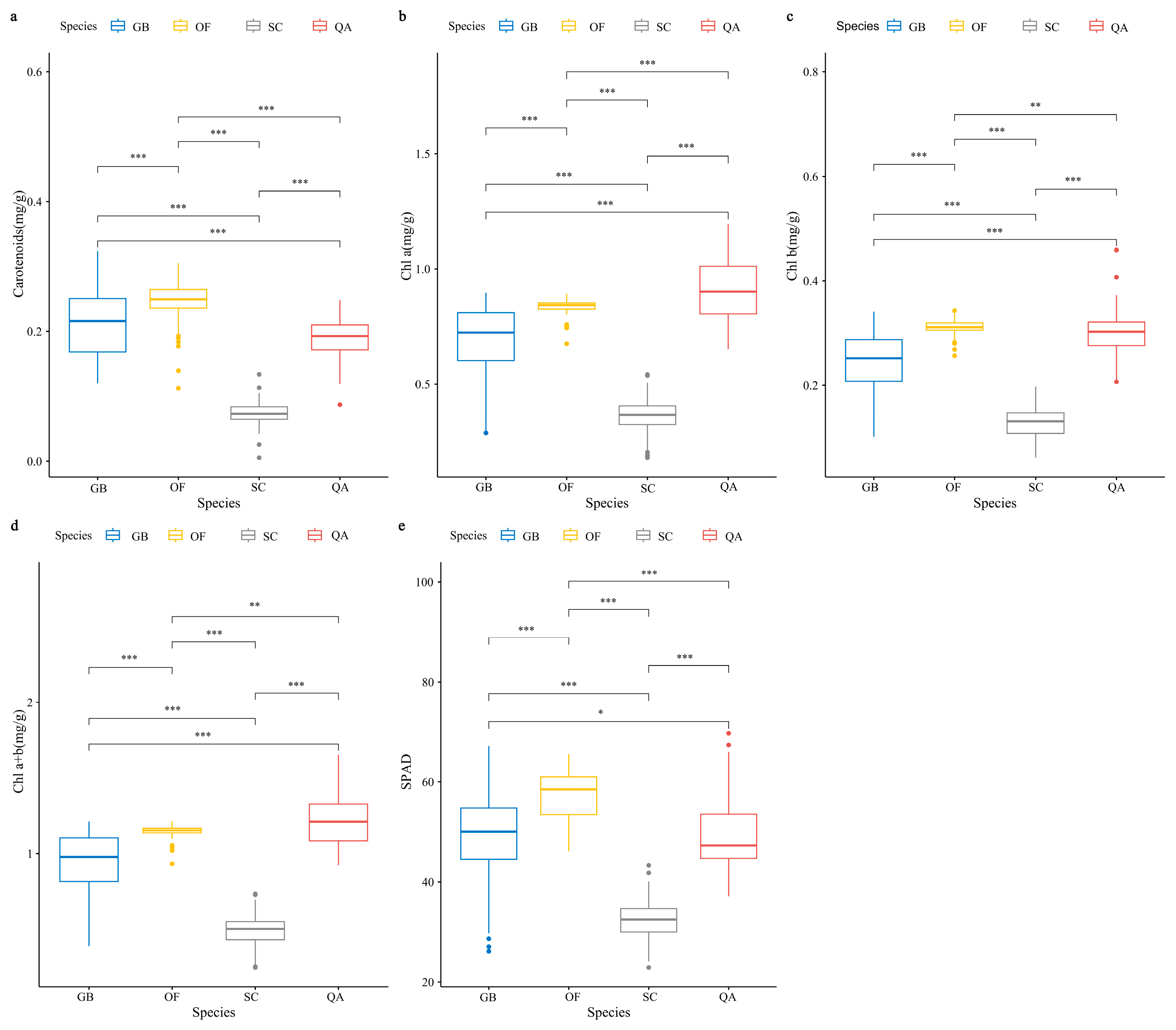
Based on the results of the Wilcoxon test, significant differences were observed between the four species in terms of the SPAD, chlorophylls, and carotenoids (Figure 2). The carotenoid concentrations in the fresh leaves of Ginkgo biloba (GB), Osmanthus fragrans (OF), Sabina chinensis ‘Kaizuca’ (SC), and Quercus acutissima (QA) were 0.21 ± 0.05, 0.25 ± 0.03, 0.07 ± 0.02, and 0.19 ± 0.03, respectively (Mean ± SD, Figure 2a). The chlorophyll a concentrations were 0.91 ± 0.12, 0.84 ± 0.03, 0.36 ± 0.08, and 0.91 ± 0.12, respectively (Mean ± SD, Figure 2b). The chlorophyll b concentrations were 0.91 ± 0.12, 0.84 ± 0.03, 0.36 ± 0.08, and 0.91 ± 0.12, respectively (Mean ± SD, Figure 2c). The chlorophyll a + b concentrations were 0.94 ± 0.20, 1.15 ± 0.04, 0.49 ± 0.10, and 1.12 ± 0.15, respectively (Mean ± SD, Figure 2d). The SPAD values were 49.4 ± 8.00, 57.47 ± 4.76, 32.25 ± 3.73, and 48.87 ± 6.75, respectively (Mean ± SD, Figure 2e).
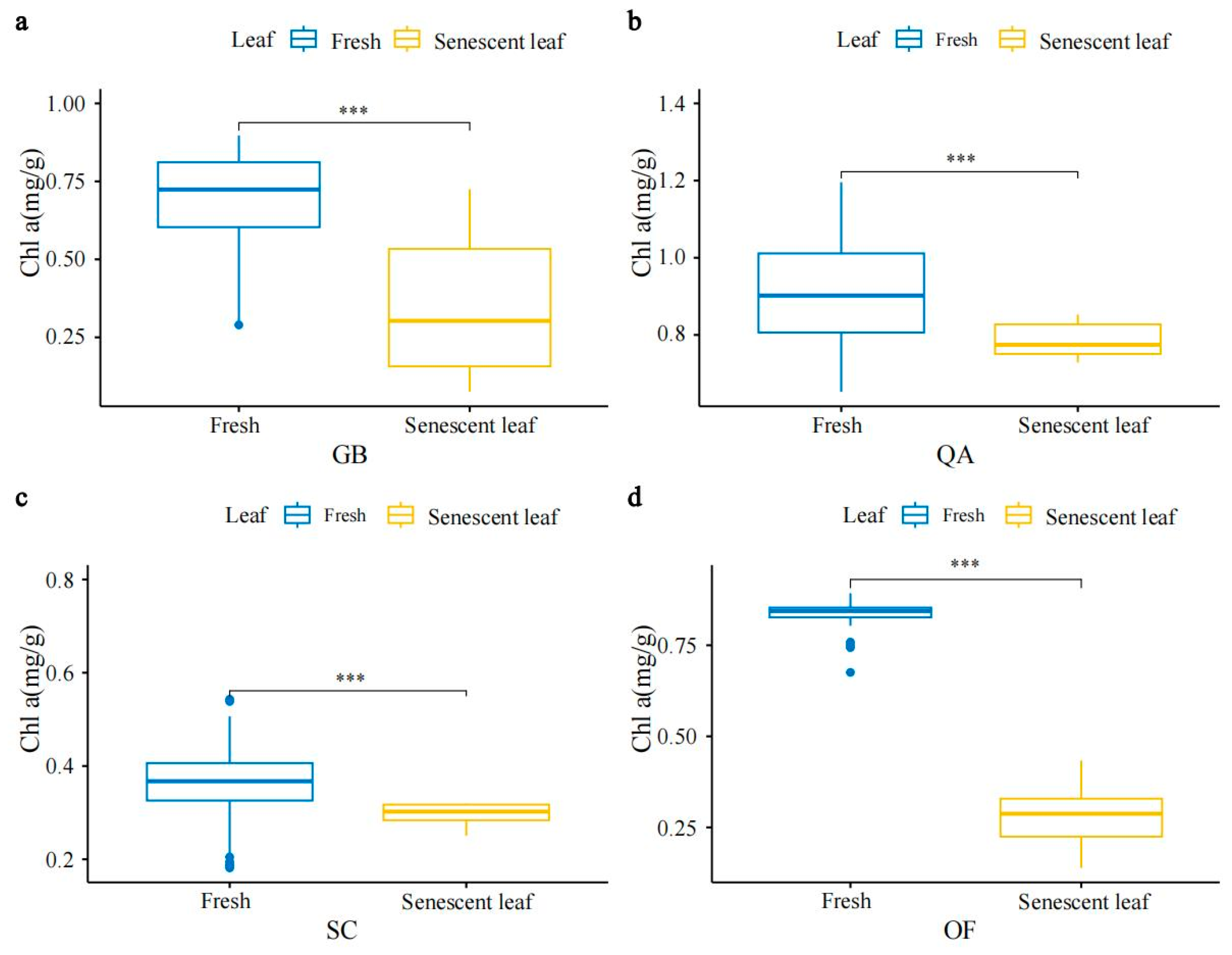
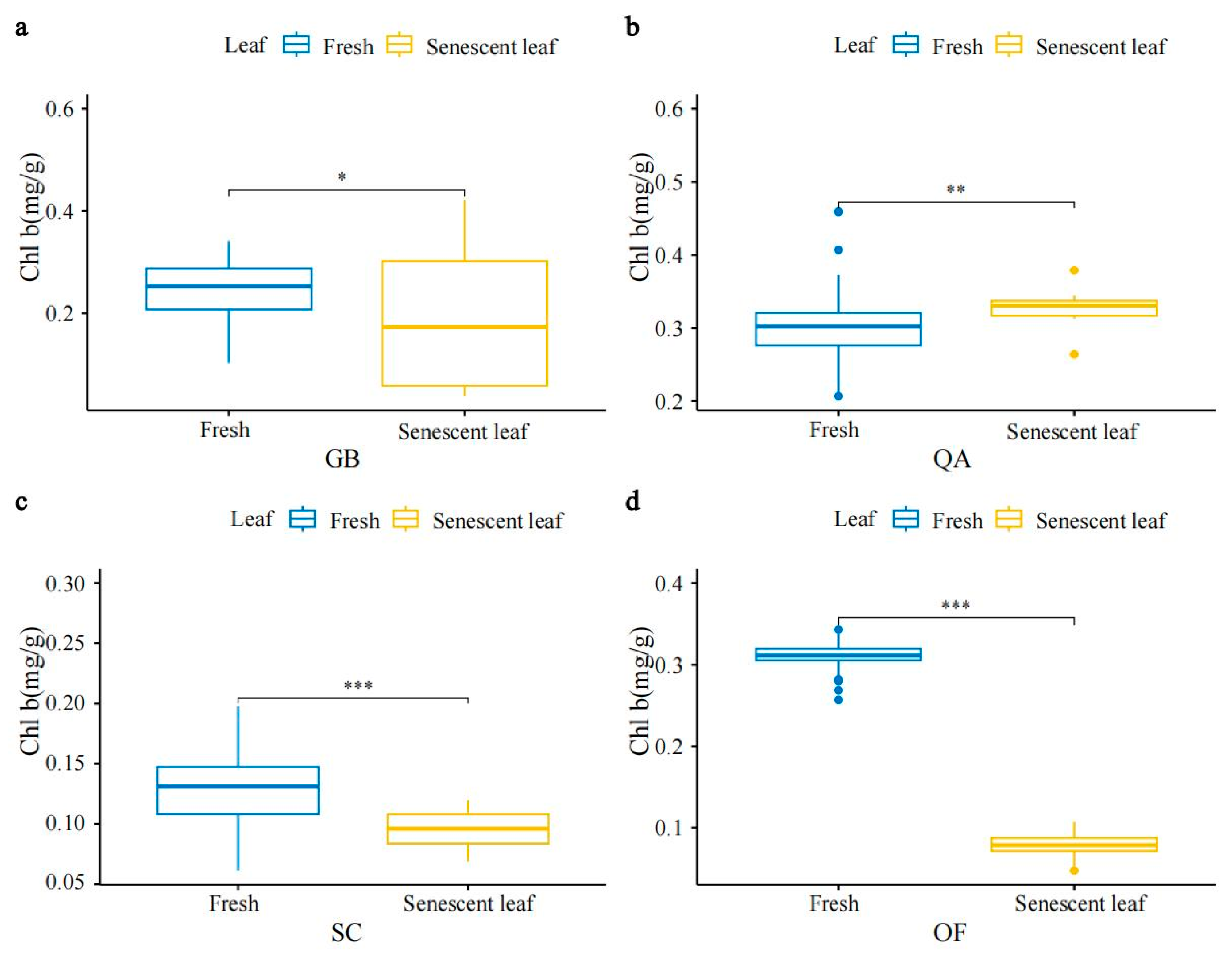
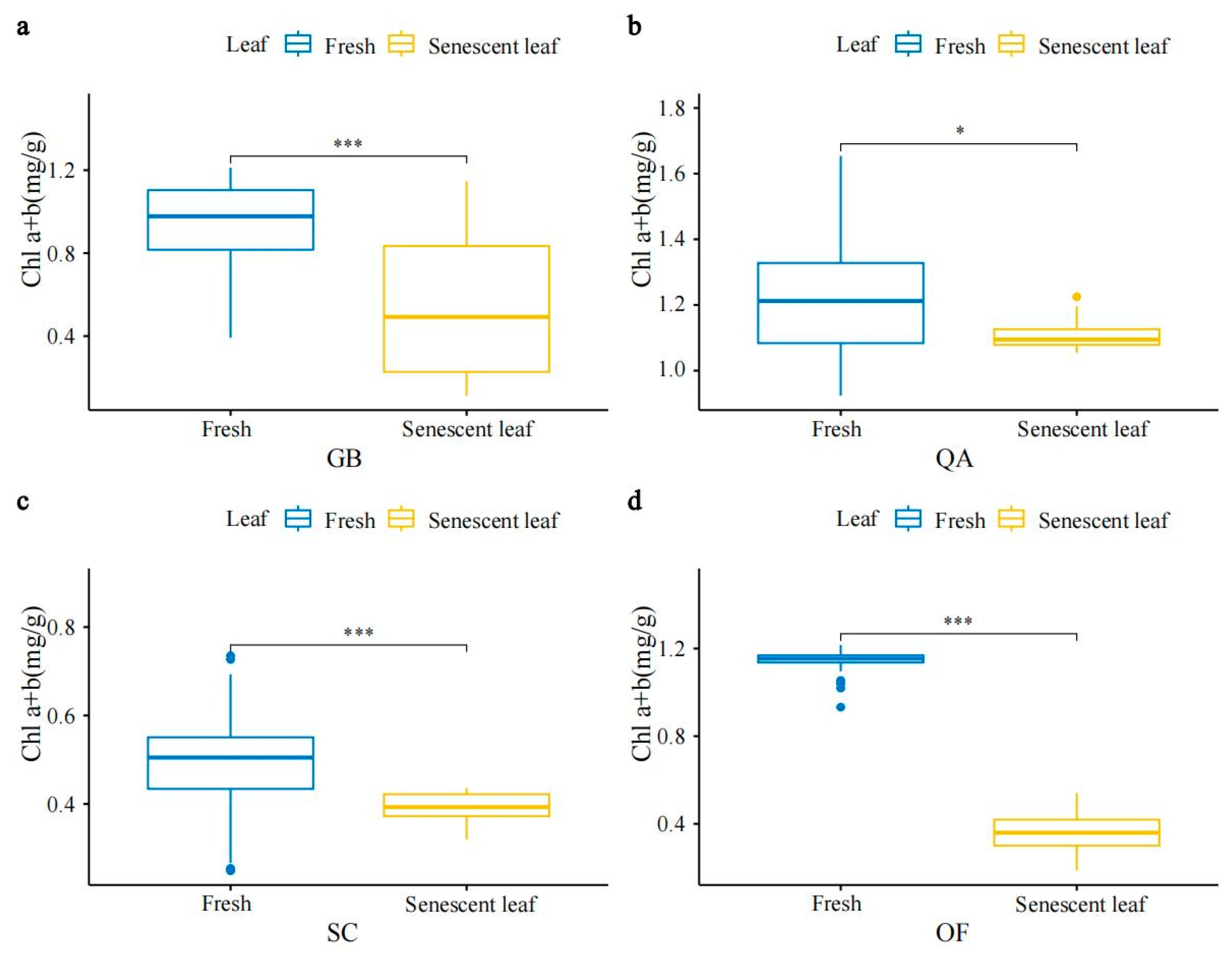
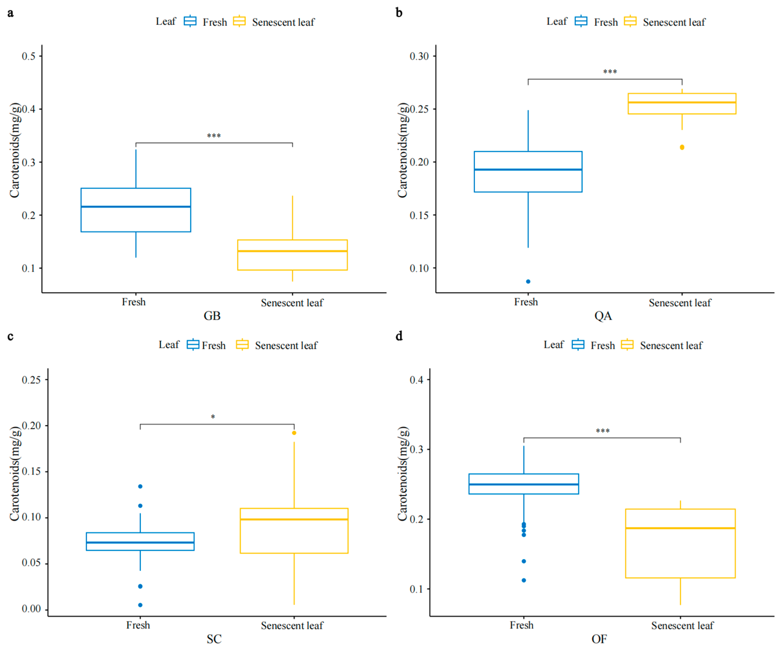
Significant differences were also observed between fresh leaves and senescent leaves. The results of the Wilcoxon test indicated that the SPAD, chlorophyll, and carotenoid concentrations were significantly higher in fresh leaves compared to senescent leaves (Figure 3 and Figure A1,Figure A2,Figure A3,Figure A4). Regarding the Chlorophyll Enrichment Index (CEI), Ginkgo biloba (GB) exhibited the highest value, while Quercus acutissima (QA) showed the lowest (Table 2). Additionally, the Normal Chlorophyll Concentration Threshold (NCCT) results indicated that QA had the highest value, whereas Sabina chinensis ‘Kaizuca’ (SC) had the lowest (Table 2).

3.2. The Relationship between SPAD, Chlorophyll, and Carotenoid Concentrations in Four Ornamental Plants

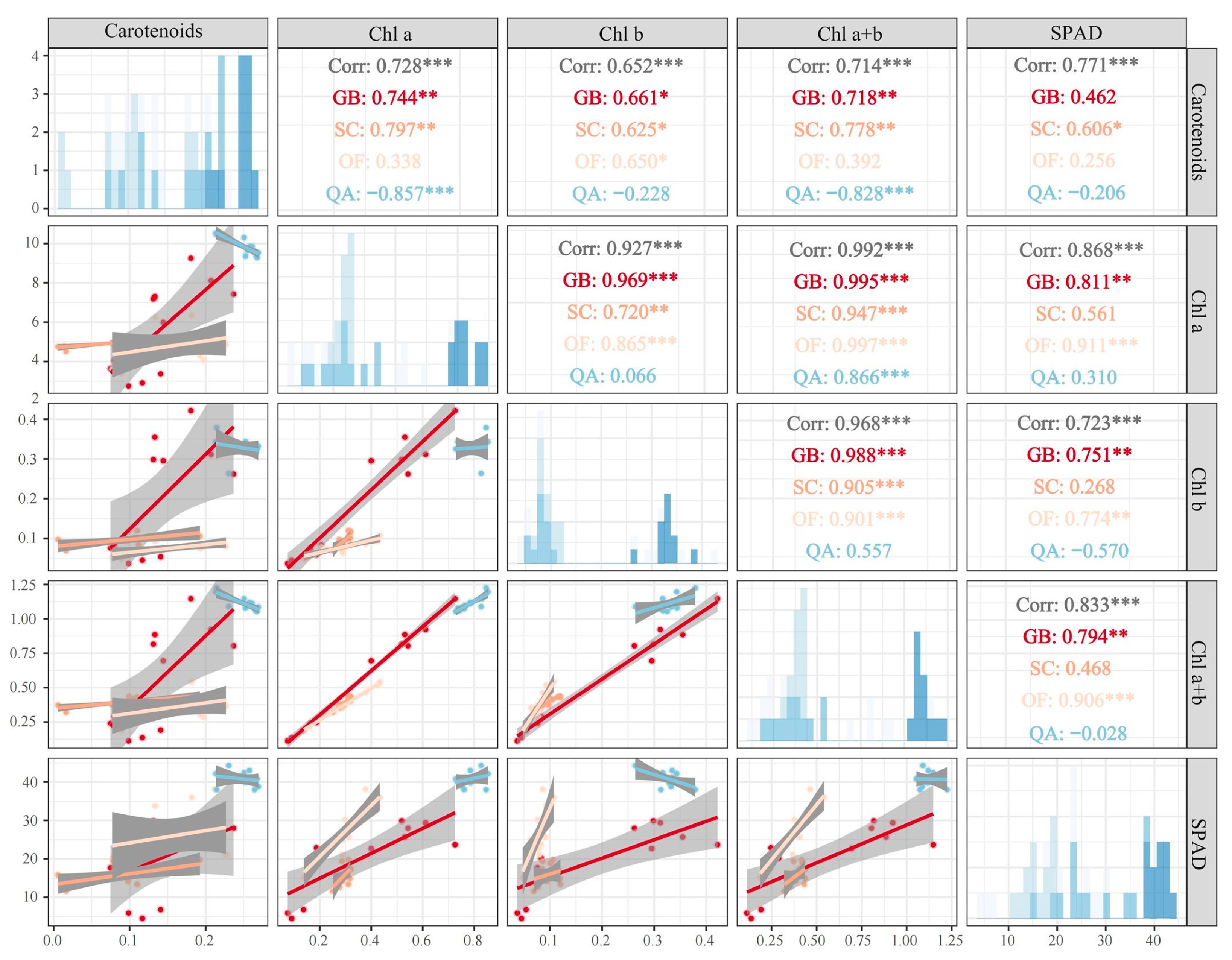
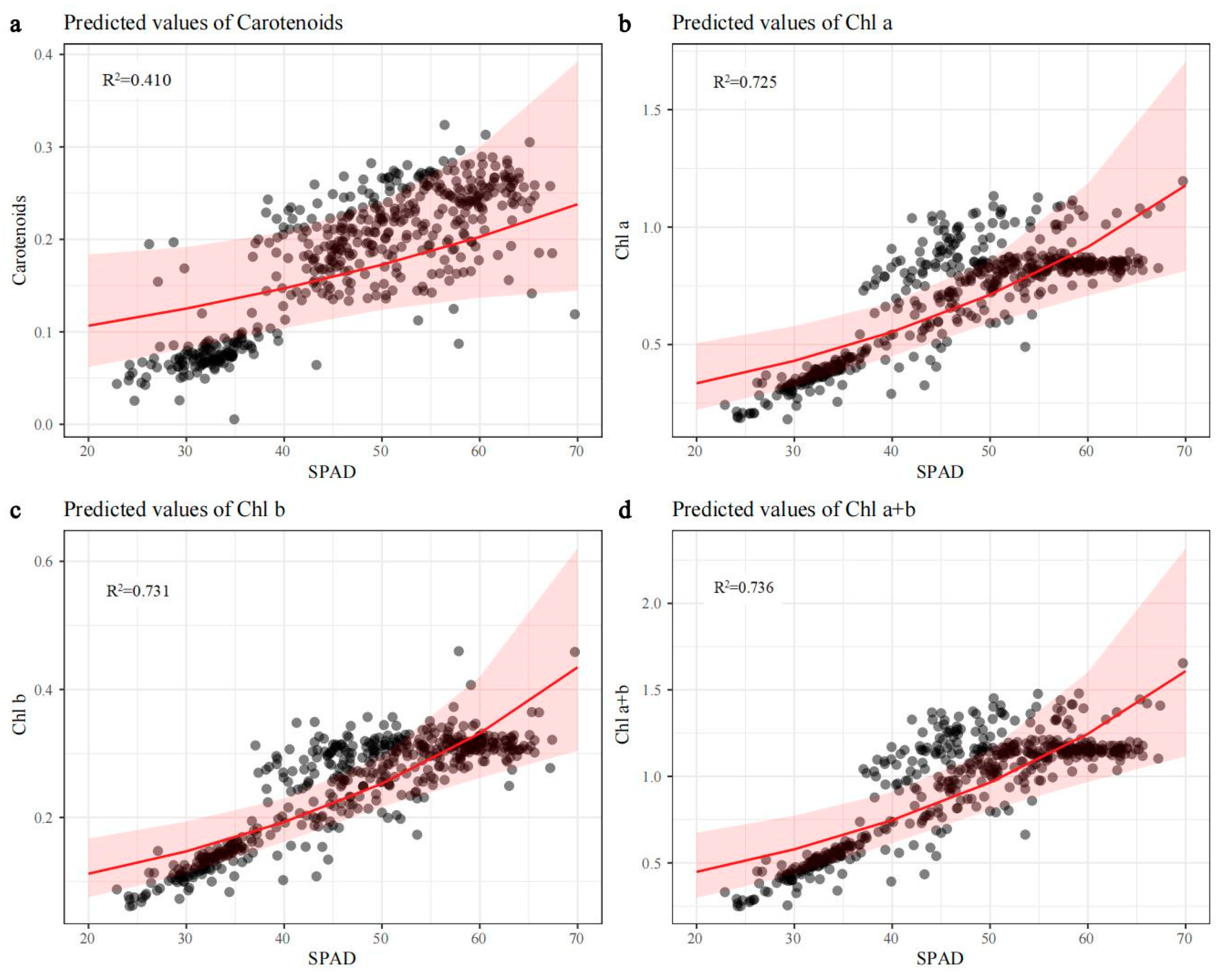
In the results of the simple linear regression, our study found a negative correlation between the carotenoid concentrations and SPAD values in fresh leaves of QA, while in other species, there was a negative correlation between the SPAD values and chlorophyll concentrations (Figure 4). In the senescent leaves of QA, a negative correlation was found between SPAD values, carotenoids, chlorophyll b, and total chlorophyll (Figure 5). The results of the generalized linear mixed effects model indicated an overall positive correlation between leaf color parameters and chlorophyll concentrations. However, there were differences in the predictive ability of SPAD values, which could effectively predict chlorophyll a, chlorophyll b, and total chlorophyll (Figure 6b,c). However, its predictive ability for carotenoids was relatively weak (R2 = 0.410) (Figure 6a).

3.3. Correlations between Leaf Color Parameters, Chlorophylls, and Carotenoids

According to the Ducan analysis, there were no significant differences in the L*, a*, and b* values between fresh and senescent leaves among the four tree species (p > 0.05), while there were significant differences in the L*, a*, and b* values between fresh and senescent leaves (p < 0.05). This indicates that although the leaf color of fresh leaves is roughly the same under different environmental conditions, the color of senescent leaves varies between different tree species. Based on the color parameters, it is easy to determine whether the leaves are fresh or senescent. For the fresh and senescent leaves of the four tree species, the a* value (+a* for red direction; −a* for green direction) ranged from the least in the senescent leaves of Quercus acutissima to the most in the fresh leaves of Osmanthus fragrans, from 0.23 to 0.98 (Table 3). The b* value (+b* for yellow direction; −b* for blue direction) was the lowest in the senescent leaves of Ginkgo biloba (1.22) and the highest in the fresh leaves of Sabina chinensis ‘Kaizuca’ (1.94). The range of L* for color brightness was the highest in the fresh leaves of Osmanthus fragrans (99.89) and the darkest in the senescent leaves of Sabina chinensis ‘Kaizuca’ (93.87). The values of L*, a*, and b* may reflect the different concentrations of photosynthetic pigment.
From Table 4, it can be observed that the L* value of fresh leaves shows a significant positive correlation with carotenoids and a negative correlation with chlorophyll. However, for senescent leaves, the L* value did not exhibit a significant correlation with either chlorophylls or carotenoids. The a* value of fresh leaves was significantly and positively correlated with both chlorophylls and carotenoids. In contrast, the a* value of senescent leaves was negatively correlated with Chl a and the sum of Chl a+b. In fresh leaves, the b* value was significantly and negatively correlated with chlorophyll and carotenoids, whereas in senescent leaves, it showed a significant positive correlation with both.

4. Discussion

4.1. SPAD Value as a Good Predictor for Photosynthetic Pigment Concentrations

A considerable amount of SPAD measurement data has been reported for cereals (e.g., Hordeum vulgare var. coeleste, 26.30–55.95 [43]), vegetables (e.g., Spinacia oleracea, 23.48–39.08 [27]; Solanum tuberosum, 26.45–41.66 [37]), and horticultural crops (e.g., Vitis vinifera, 22.6–34.3 [20]; Vaccinium corymbosum, 57.4–73.1 [36]). Compared to herbaceous crops, the SPAD value of tree species seems to be much higher. For example, for mangrove plants, a report showed that the SPAD reading ranges for Avicennia marina, Rhizophora stylosa, Bruguiera gymnorhiza, and Aegiceras corniculatum were 41.50–56.8, 70.40–80.10, 52.80–69.90, and 42.30–73.60, respectively [42], while another study recorded that the average values for Bruguiera sexangula, Ceriops tagal, and Rhizophora apiculata were 58.60, 61.56, and 66.57, respectively [23]. Intrinsic plant factors (such as developmental stages) and external factors (such as light intensity) can both affect SPAD values. The SPAD values for the seedlings of Quercus petraea and Prunus serotina were lower and more variable, with reading ranges of 10–60 and 10–45 [49]. Under the 0%, 25%, and 50% shade levels, the SPAD readings in Hopea hainanensis were 18.39, 25.76, and 31.81, respectively [50]. Furthermore, more comprehensive empirical research involving more species was conducted on multiple crops. The lowest SPAD value (36.6, Mango) and the highest value (70.2, Coffee) were documented for 13 crops (atemoya, bean, corn, coffee, grape, mango, passion fruit, peach, pear, quinoa, rice, soybean, and sugarcane) [33]. Finally, a comprehensive SPAD dataset covering 20 native and exotic trees and shrubs indicated a range of variation from 25.0 to 78.6 [40]. Our SPAD measurement data were among the empirical data range reported above. Compared to previous studies [40], there were certain differences in the measurement data of the same species or same generic species, e.g., G. biloba (49.40) vs. G. biloba (32.9–35.5), O. fragrances (57.47) vs. O. decorus (62.8–71.8), and Q. acussima (48.87) vs. Q. pubescens (32.1–46.6).
Based on the analysis of mixed data from four tree species, we found that the SPAD values of fresh leaves were significantly and positively correlated with the measurements of various photosynthetic pigments. However, the correlation between the SPAD values and photosynthetic pigments in Quercus acutissima was different from the other three species: its SPAD values were negatively correlated with the concentration of carotenoids. Conversely, a strong positive and statistically significant relationship between SPAD values and chlorophyll concentrations, as well as between SPAD values and carotenoid concentrations, was established in Triticum aestivum based on a large measurement dataset (n = 277) [32]. In a few previous reports [18,37], SPAD values were assumed to be linearly related to chlorophyll a, b, and a+b concentrations. However, this relationship for SPAD values versus photosynthetic pigment concentrations was mostly verified as a polynomial model [24,31,32,39,51] or an exponential model [29,52,53].
We integrated measurement data from fresh and senescent leaves and established a generalized linear mixed model between SPAD values and pigment concentrations. Subsequently, we found that the SPAD value was a good predictor for chlorophyll a, b, and a + b concentrations rather than carotenoid concentrations. Evidently, the generalized linear mixed model of the relationship between the SPAD values and carotenoids was not so well fitted as those between the SPAD and total Chl content (R2 = 0.41 for carotenoids, R2 = 0.725 for Chl a, R2 = 0.731 for Chl b, R2 = 0.736 for Chl a+b). In fact, using SPAD values to estimate chlorophyll concentrations has been applied in many crops and relevant calibration equations have been established [25,31,38,41,54,55]. Although SPAD values can predict chlorophyll concentrations, caution is needed for a specific species. More samples should be obtained when establishing calibration equations, as the relationship between SPAD values and chlorophyll concentrations was described as crop-, site-, or season-dependent [21,40,41].

4.2. Association Patterns of Leaf Color Parameters with Chlorophyll and Carotenoid Concentrations

Interesting patterns of the relationship between functional leaf color parameters and chlorophyll or carotenoid concentrations for four tree species were found in our analysis. The L* value was significantly and negatively correlated with chlorophyll concentrations and significantly and positively with carotenoid concentrations. The relationship between the a* value and chlorophyll or carotenoid concentrations was significantly positive, while the b* value was significantly and negatively associated with chlorophyll or carotenoid concentrations. If the data of senescent leaves were integrated, the significant correlation between a* value and chlorophyll or carotenoid concentrations, as well as the significant correlation between L* value or b* value and carotenoid concentrations, was maintained; although the association patterns for other variable pairs were weakened.
Unfortunately, so far, we have not found any documentation on the correlation between leaf color parameters and carotenoid concentrations, but there have been a few studies on fruits. An association of a* and b* values with carotenoid concentrations was significantly positive, while it was negative for the relationship between L* values and carotenoid concentrations in Cucurbita fruit [56]. On the contrary, the correlations between color parameters and carotenoid concentrations were negative for L* and b* values, and positive for a* values in cherry tomatoes [57]. Similarly, there have been no reports on the correlation between leaf color parameters and chlorophyll concentrations.
We did not analyze the correlation between SPAD values and leaf color parameters, as a few previous studies have not yet yielded consistent patterns. In Hordeum vulgare var. coeleste [43], Oryza sativa [58], and Eucalyptus species [59], SPAD values were negatively related to both L* and b* values and positively to a* values. In a study involving 13 crops, however, the patterns for the correlation between SPAD values and leaf color parameters were variable and species-dependent based on multiple linear regression [33]. SPAD values may relate to L*, a*, or b* values both positively and negatively, with a common pattern of negative effects for L* or b* values and positive effects for a* values. This trend was also supported by the full dataset that included 13 crops.
Overall, leaf color parameters can predict chlorophyll and carotenoid concentrations to a certain extent and are closely related to SPAD. However, more empirical studies and data accumulation are needed.

4.3. Characteristics of SPAD, Chlorophyll, and Carotenoid Concentrations in Four Tree Species

We first measured the SPAD value of senescent leaves and found a marked contrast to fresh leaves. For all four tree species, fresh leaves were significantly higher than senescent leaves in SPAD values. The senescent leaves we measured were lifeless leaves that had recently fallen to the ground. Based on these scientific data, we speculate that there should be a threshold SPAD value between functional leaves and senescent leaves. In fact, threshold SPAD values have been used for crop nutrition diagnosis and fertilization management. A threshold SPAD value can guide N management for crops [60]. The practice of using threshold SPAD values in nutritional diagnosis has been widely reported in rice [61,62,63], maize [60,64], wheat [65], potato [66,67], cassava [68], pumpkins [69], tomato [70], strawberries [71], and other crops such as Zizania, soybean, peanut, and cotton [72]. However, there are relatively few reports on tree species. An investigation of Vaccinium corymbosum confirmed the rationality and importance of using SPAD as a nutrition management tool [36]. Based on chlorophyll and SPAD measurements, Fe sufficiency threshold values were established in these two clones of hybrid poplar [73]. The SPAD value of green tea leaves can positively respond to nitrogen addition [74]. The rapid diagnosis of nutritional status using SPAD measurement and threshold reference was also applied to Acer pseudoplatanus, Ligustrum ovalifolium, Prunus laurocerasus, Tilia cordata, and Vaccinium angustifolium [75,76].
Unfortunately, these studies were unable to compare and analyze SPAD values between normal functional leaves and lifeless senescent leaves. This is crucial for perennial woody plants, especially evergreen species or trees with long leaf lifespans. In the current study, we first proposed an enrichment index (CEI) and a normal chlorophyll concentration threshold (NCCT) based on SPAD values from fresh and senescent leaves. The CEI is the ratio of SPAD values between fresh and senescent leaves, while the NCCT is calculated by multiplying the sum of SPAD values of fresh and senescent leaves by half. Using the above simple calculation method, we obtained the CEI and NCCT values of four tree species (Table 2). The NCCT values can serve as more accurate indicators for evaluating the growth potential of old trees. In the evaluation criteria for the growth potential of old trees in China [77], leaf quantity is the main diagnostic basis for the growth potential of old trees, and it is divided into four levels: normal plants with over 95% of normal leaves, weak plants with 50–95% of normal leaves, endangered plants with less than 50% of normal leaves, and dead plants without normal leaves. Now, we can add NCCT into the evaluation criteria for the growth potential of old trees. The leaves with SPAD readings greater than or equal to the NCCT can be identified as normal leaves, while the leaves with SPAD readings less than the NCCT are diagnosed as abnormal leaves.
On the other hand, the CEI can be applied for the comparison of SPAD values between different species. A high CEI value indicates a significant difference in SPAD values between fresh and senescent leaves. In our study, the CEI (1.19) of Q. acutissima was significantly lower than the other three tree species (CEI > 2), which provided us with clues to explore the pigment composition of leaves in depth. Investigations on the chemical composition of oak leaves showed that leaf tissues were rich in phenolic active substances such as catechins, rutin, ellagic acid, quercetin, and kaempferol [78,79]. Phenols are defensive compounds [80,81], and high levels of phenols may make the leaf coloration more complex, thereby affecting SPAD values.

5. Conclusions

Hand-held, non-destructive optical meters like the SPAD-502 offer an alternative to traditional, destructive sampling methods. Our findings indicate that chlorophyll and carotenoid concentrations substantially vary among species, and this variation is mirrored in SPAD readings. The SPAD values of fresh leaves were significantly positively correlated with various pigment measurements (SPAD values versus chlorophyll a concentrations, SPAD values versus chlorophyll b concentrations, SPAD values versus chlorophyll a + b concentrations, and SPAD values versus carotenoid concentrations), despite there being a few inconsistent patterns when analyzing individual tree species. The outlier species was mainly Q. acussima, and its SPAD values were negatively correlated with carotenoid concentrations. The main inconsistent pattern was that the correlation between SPAD values and carotenoid concentrations was not significant. By employing a generalized linear mixed model, we have demonstrated the practicality of using SPAD to predict photosynthetic pigments and developed an initial framework for assessing tree growth potential.
Although we have clarified that SPAD values can not only be used for predicting photosynthetic pigments and evaluating tree growth potential, empirical investigations with larger samples are necessary for future exploration due to the limited number of tree species and samples measured in this experiment. Fortunately, we have discovered an effective pathway for the rapid and non-destructive detection of photosynthetic pigments in woody plants. By combining this pathway with other new methods, it is expected to achieve the remote real-time monitoring of photosynthetic pigments or nutritional status.

Author Contributions

Conceptualization, L.W. and Y.F.; Methodology, Y.F., L.W. and L.L.; Literature search: L.W., Y.S., X.R. and Y.L.; Figures: L.W. and L.L.; Data collection: L.W., Y.S. and X.R.; Data analysis: L.W., L.L., Y.S. and X.R.; Formal analysis, L.W., L.L. and Y.L.; Data interpretation: L.W., L.L., Y.S. and X.R.; Resources, Y.L.; Writing—Original Draft Preparation, L.W.; Writing—Review and Editing, Y.F. and L.W.; Supervision, Y.L. and Y.F. All authors have read and agreed to the published version of the manuscript.

Funding

This research was funded by the Research and Integration of Health Diagnosis and Protection and Revitalization Technology for Ancient and Famous Trees, Jiangsu Province Forestry Science and Technology Innovation and Promotion Project, LYKJ[2021]27.

Data Availability Statement

Data are contained within the article.

Acknowledgments

We would like to thank the Jiangsu Academy of Forestry for kindly providing support for the SPAD-502 chlorophyll meter.

Conflicts of Interest

The authors declare no conflicts of interest.

Appendix A

Figure A1. Analysis of Chl a concentrations and differences between four tree species:(a) represents Chl a concentrations and differences between fresh and senescent leaves of Ginkgo biloba; (b) represents Chl a concentrations and differences between fresh and senescent leaves of Quercus acutissima; (c) represents Chl a concentrations and differences between fresh and senescent leaves of Sabina chinensis ‘Kaizuca’; (d) represents Chl a concentrations and differences between fresh and senescent leaves of Osmanthus fragrans; Note: ‘***’ represent significant correlations at the 0.001 levels.
Horticulturae 10 00548 g0a1
Figure A2. Analysis of Chl b concentrations and differences between four tree species: (a) represents Chl b concentrations and differences between fresh and senescent leaves of Ginkgo biloba; (b) represents Chl b concentrations and differences between fresh and senescent leaves of Quercus acutissima; (c) represents Chl b concentrations and differences between fresh and senescent leaves of Sabina chinensis ‘Kaizuca’; (d) represents Chl b concentrations and differences between fresh and senescent leaves of Osmanthus fragrans; Note: ‘*’, ‘**’, and ‘***’ represent significant correlations at the 0.5, 0.01, and 0.001 levels, respectively.
Horticulturae 10 00548 g0a2
Figure A3. Analysis of Chl a+b concentrations and differences between four tree species: (a) represents Chl a+b concentrations and differences between fresh and senescent leaves of Ginkgo biloba; (b) represents Chl a+b concentrations and differences between fresh and senescent leaves of Quercus acutissima; (c) represents Chl a+b concentrations and differences between fresh and senescent leaves of Sabina chinensis ‘Kaizuca’; (d) represents Chl a+b concentrations and differences between fresh and senescent leaves of Osmanthus fragrans; Note: ‘*’ and ‘***’represent significant correlations at the 0.5 and 0.001 levels, respectively.
Horticulturae 10 00548 g0a3
Figure A4. Analysis of SPAD concentrations and differences between four tree species: (a) represents SPAD concentrations and differences between fresh and senescent leaves of Ginkgo biloba; (b) represents SPAD concentrations and differences between fresh and senescent leaves of Quercus acutissima; (c) represents SPAD concentrations and differences between fresh and senescent leaves of Sabina chinensis ‘Kaizuca’; (d) represents SPAD concentrations and differences between fresh and senescent leaves of Osmanthus fragrans; Note: ‘***’ represent significant correlations at the 0.001 levels.
Horticulturae 10 00548 g0a4

References

  1. Sun, B.; Jiang, M.; Zheng, H.; Jian, Y.; Huang, W.; Yuan, Q.; Zheng, A.; Chen, Q.; Zhang, Y.; Lin, Y.; et al. Color-related chlorophyll and carotenoid concentrations of Chinese kale can be altered through CRISPR/Cas9 targeted editing of the carotenoid isomerase gene BoaCRTISO. Hortic. Res. 2020, 7, 161. [Google Scholar] [CrossRef] [PubMed]
  2. Cho, Y.B.; Boyd, R.A.; Ren, Y.D.; Lee, M.B.; Jones, S.I.; Ruiz-Vera, U.M.; Mcgrath, J.; Masters, M.D.; Ort, D.R. Reducing chlorophyll levels in seed-filling stages results in higher seed nitrogen without impacting canopy carbon assimilation. Plant Cell Environ. 2024, 47, 278–293. [Google Scholar] [CrossRef] [PubMed]
  3. Demmig Adams, B.; Stewart, J.J.; LópezPozo, M.; Polutchko, S.K.; Adams, W.W. Zeaxanthin, a molecule for photoprotection in many different environments. Molecules 2020, 25, 5825. [Google Scholar] [CrossRef] [PubMed]
  4. Zeng, Y.L.; Hao, D.L.; Huete, A.; Dechant, B.; Berry, J.; Chen, J.M.; Joiner, J.; Frankenberg, C.; Bond-Lamberty, B.; Ryu, Y.; et al. Optical vegetation indices for monitoring terrestrial ecosystems globally. Nat. Rev. Earth Environ. 2022, 3, 477–493. [Google Scholar] [CrossRef]
  5. Li, L.; Yuan, H. Chromoplast biogenesis and carotenoid accumulation. Arch. Biochem. Biophys. 2013, 539, 102–109. [Google Scholar] [CrossRef] [PubMed]
  6. Paciulli, M.; Palermo, M.; Chiavaro, E.; Pellegrini, N. Chlorophylls and colour changes in cooked vegetables. In Fruit and Vegetable Phytochemicals: Chemistry and Human Health, 2nd ed.; John Wiley & Sons: Hoboken, NJ, USA, 2017; Volume 2, pp. 703–719. [Google Scholar]
  7. Maoka, T. Carotenoids as natural functional pigments. J. Nat. Med. 2020, 74, 1–16. [Google Scholar] [CrossRef] [PubMed]
  8. Archetti, M.; Döring, T.F.; Hagen, S.B.; Hughes, N.M.; Leather, S.R.; Lee, D.W.; Lev-Yadun, S.; Manetas, Y.; Ougham, H.J.; Schaberg, P.G.; et al. Response to Sinkkonen: Ultraviolet reflectance in autumn leaves and the un-naming of colours. Trends Ecol. Evol. 2009, 24, 237–238. [Google Scholar] [CrossRef]
  9. Hörtensteiner, S.; Lee, D.W. Chlorophyll Catabolism and Leaf Coloration. Annu. Plant Rev. 2007, 26, 12–38. [Google Scholar]
  10. Wang, J.Q.; Gu, X.Y.; Dong, Y.L.; Wang, T.; Sun, Q.; Fu, S.; Yang, Y.; Huang, J.; Liang, C.; Xie, X.; et al. Advances in the endogenous and exogenous regulation of anthocyanins–the key to color change in eudicots. Crit. Rev. Plant Sci. 2023, 42, 217–238. [Google Scholar] [CrossRef]
  11. Wen, B.B.; Li, C.; Fu, X.L.; Li, D.; Li, L.; Chen, X.; Wu, H.; Cui, X.; Zhang, X.; Shen, H.; et al. Effects of nitrate deficiency on nitrate assimilation and chlorophyll synthesis of detached apple leaves. Plant Physiol. Biochem. 2019, 142, 363–371. [Google Scholar] [CrossRef]
  12. Huang, P.; Li, Z.; Guo, H. New advances in the regulation of leaf senescence by classical and peptide hormones. Front. Recent Dev. Plant Sci. 2022, 13, 923136. [Google Scholar] [CrossRef] [PubMed]
  13. Zhao, Y.Q.; Han, Q.H.; Ding, C.B.; Huang, Y.; Liao, J.; Chen, T.; Feng, S.; Zhou, L.; Zhang, Z.; Chen, Y.; et al. Effect of low temperature on chlorophyll biosynthesis and chloroplast biogenesis of rice seedlings during greening. Int. J. Mol. Sci. 2020, 21, 1390. [Google Scholar] [CrossRef] [PubMed]
  14. Mittelberger, C.; Yalcinkaya, H.; Pichler, C.; Gasser, J.; Scherzer, G.; Erhar, E.; Schumacher, S.; Holzner, B.; Janik, K.; Robatscher, R.; et al. Pathogen-induced leaf chlorosis: Products of chlorophyll breakdown found in degreened leaves of phytoplasma-infected apple (Malus × domestica Borkh.) and apricot (Prunus armeniaca L.) trees relate to the pheophorbide a oxygenase/phyllobilin pathway. J. Agric. Food Chem. 2017, 65, 2651–2660. [Google Scholar] [CrossRef] [PubMed]
  15. Swapnil, P.; Meena, M.; Singh, S.K.; Dhuldhaj, U.P.; Harish; Marwal, A. Vital roles of carotenoids in plants and humans to deteriorate stress with its structure, biosynthesis, metabolic engineering and functional aspects. Curr. Plant Biol. 2021, 26, 100203. [Google Scholar] [CrossRef]
  16. Zhang, R.R.; Wang, Y.H.; Li, T.; Tan, G.F.; Tao, J.P.; Su, X.J.; Xu, Z.S.; Tian, Y.S.; Xiong, A.S. Effects of simulated drought stress on carotenoid contents and expression of related genes in carrot taproots. Protoplasma 2021, 258, 379–390. [Google Scholar] [CrossRef] [PubMed]
  17. Li, C.; Ji, J.; Wang, G.; Li, Z.; Wang, Y.; Fan, Y. Over-expression of LcPDS, LcZDS, and LcCRTISO, genes from wolfberry for carotenoid biosynthesis, enhanced carotenoid accumulation, and salt tolerance in tobacco. Front. Recent Dev. Plant Sci. 2020, 11, 119. [Google Scholar] [CrossRef] [PubMed]
  18. Villegas-Velázquez, I.; Zavaleta-Mancera, H.A.; Arévalo-Galarza, M.L.; Suarez-Espinosa, J.; Garcia-Osorio, C.; Padilla-Chacon, D.; Galvan-Escobedo, I.G.; Jimenez-Bremont, J.F. Chlorophyll measurements in Alstroemeria sp. using SPAD-502 meter and the color space CIE L*a*b*, and its validation in foliar senescence. Photosynthetica 2022, 60, 230–239. [Google Scholar] [CrossRef]
  19. Wang, S.; Liu, X.; Liu, X.; Xue, J.; Ren, X.; Zhai, Y.; Zhang, X. The red/blue light ratios from light-emitting diodes affect growth and flower quality of Hippeastrum hybridum ‘Red Lion’. Front. Plant Sci. 2022, 13, 1048770. [Google Scholar] [CrossRef] [PubMed]
  20. Cantwell, M.I.; Hong, G.; Albornoz, K.; Berlanga, M. Fresh grapevine (Vitis vinifera L.) leaves: Postharvest biology and handling recommendations. Sci. Hortic. 2022, 292, 110627. [Google Scholar] [CrossRef]
  21. Pinkard, E.A.; Patel, V.; Mohammed, C. Chlorophyll and nitrogen determination for plantation-grown Eucalyptus nitens and E. globulus using a non-destructive meter. For. Ecol. Manag. 2006, 223, 211–217. [Google Scholar] [CrossRef]
  22. Martín, I.; Alonso, N.; López, M.C.; Prieto, M.; Cadahia, C.; Eymar, E. Estimation of leaf, root, and sap nitrogen status using the SPAD-502 chlorophyll meter for ornamental shrubs. Commun. Soil. Sci. Plant Anal. 2007, 38, 1785–1803. [Google Scholar] [CrossRef]
  23. Li, H.Z.; Cui, L.J.; Dou, Z.G.; Wang, J.J.; Zhai, X.J.; Li, J.; Zhao, X.S.; Lei, Y.R.; Wang, J.Z.; Li, W. Hyperspectral analysis and regression modeling of SPAD measurements in leaves of three mangrove species. Forests 2023, 14, 1566. [Google Scholar] [CrossRef]
  24. Percival, G.C.; Keary, I.P.; Noviss, K. The potential of a chlorophyll content SPAD meter to quantify nutrient stress in foliar tissue of sycamore (Acer pseudoplatanus), English oak (Quercus robur), and European beech (Fagus sylvatica). Arboric. Urban. For. 2008, 34, 89–100. [Google Scholar] [CrossRef]
  25. Marenco, R.A.; Antezana-Vera, S.A.; Nascimento, H.C.S. Relationship between specific leaf area, leaf thickness, leaf water content and SPAD-502 readings in six Amazonian tree species. Photosynthetica 2009, 47, 184–190. [Google Scholar] [CrossRef]
  26. Afonso, S.; Arrobas, M.; Ferreira, I.Q.; Angelo Rodrigues, M. Assessing the potential use of two portable chlorophyll meters in diagnosing the nutritional status of plants. J. Plant Nutr. 2018, 41, 261–271. [Google Scholar] [CrossRef]
  27. Agarwal, A.; Gupta, S.D. Assessment of spinach seedling health status and chlorophyll content by multivariate data analysis and multiple linear regression of leaf image features. Comput. Electron. Agric. 2018, 152, 281–289. [Google Scholar] [CrossRef]
  28. Jiang, X.P.; Zhen, J.N.; Miao, J.; Zhao, D.; Wang, J.; Jia, S. Assessing mangrove leaf traits under different pest and disease severity with hyperspectral imaging spectroscopy. Ecol. Indic. 2021, 129, 107901. [Google Scholar] [CrossRef]
  29. Li, G.Y.; Aubrey, D.P.; Sun, H.Z. Predictive capability of a leaf optical meter for determining leaf pigment status during senescence. Photosynthetica 2017, 55, 543–552. [Google Scholar] [CrossRef]
  30. Tan, X.; Wang, W.H.; Gao, L.; Wei, J.; Zhang, W.; Li, L.; Wu, J.J.; Wang, J.R.; Zhang, X.D.; Liao, X.; et al. The difference in leaf color quality of Cotinus coggygria during the coloration peak period affected by soil and topographic heterogeneity. Catena 2023, 228, 107140. [Google Scholar] [CrossRef]
  31. Hunt, E.R.; Daughtry, C.S.T. Chlorophyll meter calibrations for chlorophyll content using measured and simulated leaf transmittances. Agron. J. 2014, 106, 931–939. [Google Scholar] [CrossRef]
  32. Shah, S.H.; Houborg, R.; McCabe, F. Response of chlorophyll, carotenoid and spad-502 measurement to salinity and nutrient stress in wheat (Triticum aestivum L.). Agronomy 2017, 7, 61. [Google Scholar] [CrossRef]
  33. Borges, C.; Vega, R.R.A.; Chakraborty, S.; Weindorf, D.C.; Lopes, G.; Guilherme, L.R.G.; Curi, N.; Li, B.; Ribeiro, B.T. Pocket-sized sensor for controlled, quantitative and instantaneous color acquisition of plant leaves. J. Plant Physiol. 2022, 272, 153686. [Google Scholar] [CrossRef] [PubMed]
  34. Guo, Y.; Chen, S.; Li, X.; Cunha, M.; Jayavelu, S.; Cammarano, D.; Fu, Y. Machine learning-based approaches for predicting SPAD values of maize using multi-spectral images. Remote Sens. 2022, 14, 1337. [Google Scholar] [CrossRef]
  35. Wang, W.; Cheng, Y.; Ren, Y.; Zhang, Z.; Geng, H. Prediction of chlorophyll content in multi-temporal winter wheat based on multispectral and machine learning. Front. Plant Sci. 2022, 13, 896408. [Google Scholar] [CrossRef]
  36. Pinzón-Sandoval, E.H.; Balaguera-López, H.E.; Almanza-Merchán, P.J. Evaluation of SPAD index for estimating nitrogen and magnesium contents in three blueberry varieties (Vaccinium corymbosum L.) on the Andean Tropics. Horticulturae 2023, 9, 269. [Google Scholar] [CrossRef]
  37. Loayza, H.; Calderón, R.A.; Gutiérrez, R.R.O.; Céspedes, F.E.; Quiroz, R. Estimation of relative chlorophyll concentrations in potato (Solanum tuberosum L.) leaflet using vegetation reflectance techniques. Ecol. Apl. 2022, 21, 91–101. [Google Scholar] [CrossRef]
  38. Coste, S.; Baraloto, C.; Leroy, C.; Marcon, E.; Renaud, A.; Richardson, A.D.; Roggy, J.C.; Schimann, H.; Uddling, J.; Herault, B. Assessing foliar chlorophyll contents with the SPAD-502 chlorophyll meter: A calibration test with thirteen tree species of tropical rainforest in French Guiana. Ann. For. Sci. 2010, 67, 607. [Google Scholar] [CrossRef]
  39. Bielinis, E.; Jozwiak, W.; Robakowski, P. Modelling of the relationship between the SPAD values and photosynthetic pigments content in Quercus petraea and Prunus serotina leaves. Dendrobiology 2015, 73, 125–134. [Google Scholar] [CrossRef]
  40. Atar, F.; Güney, D.; Bayraktar, A. Seasonal change of chlorophyll content (SPAD value) in some tree and shrub species. Turk. J. For. Sci.-Prague 2020, 4, 245–256. [Google Scholar] [CrossRef]
  41. Donnelly, A.; Yu, R.; Rehberg, C.; Meyer, G.; Young, E.B. Leaf chlorophyll estimates of temperate deciduous shrubs during autumn senescence using a SPAD-502 meter and calibration with extracted chlorophyll. Ann. For. Sci. 2020, 77, 30. [Google Scholar] [CrossRef]
  42. Wang, J.J.; Xu, Y.; Wu, G.F. The integration of species information and soil properties for hyperspectral estimation of leaf biochemical parameters in mangrove forest. Ecol. Indic. 2020, 115, 106467. [Google Scholar] [CrossRef]
  43. Liu, Y.; Hatou, K.; Aihara, T.; Kurose, S.; Akiyama, T.; Kohno, Y.; Lu, S.; Omasa, K. A robust vegetation index based on different UAV RGB images to estimate SPAD Values of naked barley leaves. Remote Sens. 2021, 13, 686. [Google Scholar] [CrossRef]
  44. Liu, J.J.; Lindenmayer, D.B.; Yang, W.J.; Ren, Y.; Campbell, M.J.; Wu, C.; Luo, Y.; Zhong, L.; Yu, M. Diversity and density patterns of large old trees in China. Sci. Total Environ. 2019, 655, 255–262. [Google Scholar] [CrossRef] [PubMed]
  45. Xie, C.P.; Li, M.; Jim, C.Y.; Liu, D. Environmental factors driving the spatial distribution pattern of venerable trees in Sichuan Province, China. Plants 2022, 11, 3581. [Google Scholar] [CrossRef] [PubMed]
  46. Huang, L.; Jin, C.; Pan, Y.J.; Zhou, L.H.; Hu, S.W.; Guo, Y.P.; Meng, Y.Y.; Song, K.; Pang, M.Y.; Li, H.; et al. Human activities and species biological traits drive the long-term persistence of old trees in human-dominated landscapes. Nat. Plants 2023, 9, 898–906. [Google Scholar] [CrossRef] [PubMed]
  47. Jan, N.; Jitka, P.; Jiří, R.; Martina, S. SPAD chlorophyll meter reading can be pronouncedly affected by chloroplast movement. Photosynth. Res. 2010, 105, 265–271. [Google Scholar]
  48. Gao, J.F. Experimental Techniques of Plant Physiology; The World Book Publishing Company: Xi’an, China, 2000; pp. 99–103. [Google Scholar]
  49. Yuan, Y.; Wang, X.F.; Shi, M.M.; Wang, P. Performance comparison of RGB and multispectral vegetation indices based on machine learning for estimating Hopea hainanensis SPAD values under different shade conditions. Front. Plant Sci. 2022, 13, 928953. [Google Scholar] [CrossRef] [PubMed]
  50. Fang, X.; Wang, K.; Sun, X.C.; Wang, Y.H.; Zheng, P.F.; Shi, F.C. Characteristics of chlorophyll fuorescence in ten garden shrub species under fooding stress. Biologia 2022, 77, 339–350. [Google Scholar] [CrossRef]
  51. Hawkins, T.S.; Gardiner, E.S.; Comer, G.S. Modeling the relationship between extractable chlorophyll and SPAD-502 readings for endangered plant species research. J. Nat. Conserv. 2009, 17, 123–127. [Google Scholar] [CrossRef]
  52. Cerovic, Z.G.; Masdoumier, G.; Ben, G.N.; Latouche, G. A new optical leaf-clip meter for simultaneous non-destructive assessment of leaf chlorophyll and epidermal flavonoids. Physiol. Plant 2012, 146, 251–260. [Google Scholar]
  53. Shibaeva, T.G.; Mamaeva, A.V.; Sherudilo, E.G. Evaluation of a spad-502 plus chlorophyll meter to estimate chlorophyll content in leaves with interveinal chlorosis. Russ. J. Plant Physiol. 2020, 67, 690–696. [Google Scholar] [CrossRef]
  54. Silva, V.D.; Dos, A.L.; Brito-Rocha, E.; Dalmolin, A.C.; Mielke, M.S. Calibration of a multi-species model for chlorophyll estimation in seedlings of Neotropical tree species using hand-held leaf absorbance meters and spectral reflectance. iForest 2016, 9, 829–834. [Google Scholar] [CrossRef]
  55. Brown, L.A.; Williams, O.; Dash, J. Calibration and characterisation of four chlorophyll meters and transmittance spectroscopy for non-destructive estimation of forest leaf chlorophyll concentration. Agric. For. Meteorol. 2022, 323, 109059. [Google Scholar] [CrossRef]
  56. Itle, R.A.; Kabelka, E.A. Correlation Between L*a*b* Color Space Values and Carotenoid Content in Pumpkins and Squash (Cucurbita spp.). Hortscience 2009, 44, 633–637. [Google Scholar] [CrossRef]
  57. Shilpa, P.; Sadashiva, A.T.; Shivashankar, K.S. Carotenoid content in cherry tomatoes correlated to the colour space values L*, a*, b*: A non-destructive method of estimation. J. Hortic. Sci.-India 2020, 15, 27–34. [Google Scholar]
  58. Wang, Y.; Wang, D.Y.; Shi, P.H.; Omasa, K.J. Estimating rice chlorophyll content and leaf nitrogen concentration with a digital still color camera under natural light. Plant Methods 2014, 10, 36. [Google Scholar] [CrossRef]
  59. Valverde, J.C. Estimation of leaf nitrogen content from nondestructive methods in Eucalyptus tereticornis and Eucalyptus saligna plantations. Rev. Fac. Nac. Agron. Univ. Antioq. 2021, 74, 9655–9666. [Google Scholar] [CrossRef]
  60. Jagdeep-Singh; Varinderpal-Singh. Chlorophyll meter based precision nitrogen management in spring maize. J. Plant Nutr. 2023, 46, 17–27. [Google Scholar] [CrossRef]
  61. Ghosh, M.; Roychowdhury, A.; Dutta, S. SPAD Chlorophyll Meter-Based Real-Time Nitrogen Management in Manure-Amended Lowland Rice. J. Soil Sci. Plant Nutr. 2023, 23, 5993–6005. [Google Scholar] [CrossRef]
  62. Alridiwirsah; Tampubolon, K.; Basyuni, M.; Mustamu, N.E. SPAD Total Chlorophyll as an Initial Indicator of the Effect of 2,4-D Dimethyl Amine Herbicide for Lowland Rice and Barnyardgrass Weed. IOP 2023, 1241, 012007. [Google Scholar]
  63. Wu, G.Z.; Chen, X.Y.; Zang, Y.G.; Ye, Y.; Qiao, X.Q.; Zhang, W.Y.; Zhang, H.; Liu, L.J.; Zhang, Z.J.; Wang, Z.Q.; et al. An optimized strategy of nitrogen-split application based on the leaf positional differences in chlorophyll meter readings. J. Integr. Agric. 2023. [Google Scholar] [CrossRef]
  64. Maltese, N.E.; Maddonni, G.A.; Melchiori, R.J.M.; Caviglia, O.P. Plant nitrogen status at flowering and kernel set efficiency in early- and late-sown maize crops. Field Crops Res. 2021, 270, 108216. [Google Scholar] [CrossRef]
  65. Guizani, A.; Babay, E.; Askri, H.; Sialer, M.F.; Gharbi, F. Screening for drought tolerance and genetic diversity of wheat varieties using agronomic and molecular markers. Mol. Biol. Rep. 2024, 51, 432. [Google Scholar] [CrossRef] [PubMed]
  66. Yang, H.; Hu, Y.H.; Zheng, Z.Z.; Qiao, Y.C.; Hou, B.R.; Chen, J. A New Approach for Nitrogen Status Monitoring in Potato Plants by Combining RGB Images and SPAD Measurements. Remote Sens. 2022, 14, 4814. [Google Scholar] [CrossRef]
  67. Liu, N.; Liu, G.; Sun, H. Real-Time Detection on SPAD Value of Potato Plant Using an In-Field Spectral Imaging Sensor System. Sensors 2020, 20, 3430. [Google Scholar] [CrossRef]
  68. Byju, G.; Anand, M.H. Leaf color chart and chlorophyll-meter-based leaf nitrogen estimation and their threshold values for real-time nitrogen management in cassava. Commun. Soil. Sci. Plant Anal. 2009, 40, 2816–2832. [Google Scholar] [CrossRef]
  69. Swiader, J.M.; Moore, A. SPAD-chlorophyll response to nitrogen fertilization and evaluation of nitrogen status in dryland and irrigated pumpkins. J. Plant Nutr. 2002, 25, 1089–1100. [Google Scholar] [CrossRef]
  70. Zhou, Y.; Yang, L.Y.; Wang, J.; Guo, L.J.; Huang, J.S. Synergistic Effect between Trichoderma virens and Bacillus velezensis on the Control of Tomato Bacterial Wilt Disease. Horticulturae 2021, 7, 439. [Google Scholar] [CrossRef]
  71. Wu, G.S.; Jiang, Q.Y.; Bai, Y.H.; Tian, C.F.; Pan, W.; Jin, X.X.; Zhang, B.H. Nitrogen status assessment for multiple cultivars of strawberries using portable FT-NIR Spectrometers combined with cultivar recognition and multivariate analysis. IEEE ACCESS 2020, 8, 126039–126050. [Google Scholar] [CrossRef]
  72. Xiong, D.L.; Chen, J.; Yu, T.T.; Gao, W.L.; Ling, X.X.; Li, Y.; Peng, S.B.; Huang, J.L. SPAD-based leaf nitrogen estimation is impacted by environmental factors and crop leaf characteristics. Sci. Rep. 2015, 5, 13389. [Google Scholar] [CrossRef]
  73. Lombard, K.; O’Neill, M.; Mexal, J.; Ulery, A.; Onken, B.; Bettmann, G.; Heyduck, R. Can soil plant analysis development values predict chlorophyll and total Fe in hybrid poplar? Agrofor. Syst. 2010, 78, 1–11. [Google Scholar] [CrossRef]
  74. Xiao, F.; Yang, Z.Q.; Huang, H.J.; Yang, F.; Zhu, L.Y.; Han, D. Nitrogen fertilization in soil affects physiological characteristics and quality of green tea leaves. Hortscience 2018, 53, 715–722. [Google Scholar] [CrossRef]
  75. Bracke, J.; Elsen, A.; Adriaenssens, S.; Schoeters, L.; Vandendriessche, H.; Van, L.M.C. Application of proximal optical sensors to fine-tune nitrogen fertilization: Opportunities for woody ornamentals. Agronomy 2020, 9, 408. [Google Scholar] [CrossRef]
  76. Lafond, J. Rapid diagnosis of the nitrogen status of the wild lowbush blueberry. Int. J. Fruit. Sci. 2023, 23, 13–24. [Google Scholar] [CrossRef]
  77. LY/T 2737-2016; Regulation for Identification of Old and Notable Trees. The State Forestry Administration: Beijing, China, 2016.
  78. Shen, H.L.; Zhang, J.J.; Guo, C.S.; Gao, X.Y.; Chen, J.Y.; Chang, C.; Han, X.L.; Wang, L.J. Characterization and optimization of hydrothermal extraction of quercetin from Quercus leaves using response surface methodology. Can. Soc. Chem. Eng. 2021, 100, 598–606. [Google Scholar] [CrossRef]
  79. Wang, L.M.; Du, X.F.; Yue, D.M.; Chen, X.H. Catechin, rutin and quercetin in Quercus mongolica Fisch leaves exert inhibitory effects on multiple cancer cells. J. Food. Chem. 2022, 46, e14486. [Google Scholar] [CrossRef] [PubMed]
  80. Hikosaka, K.; Takashima, T.; Kabeya, D.; Hirose, T.; Kamata, N. Biomass Allocation and Leaf Chemical Defence in Defoliated Seedlings of Quercus serrata with Respect to Carbon–Nitrogen Balance. Ann. Bot. 2005, 95, 1025–1032. [Google Scholar] [CrossRef]
  81. Wang, X.; Liu, J.F.; Gao, W.Q. Defense pattern of Chinese cork oak across latitudinal gradients: Inffuences of ontogeny, herbivory, climate and soil nutrients. Sci. Rep. 2016, 6, 27269. [Google Scholar] [CrossRef]
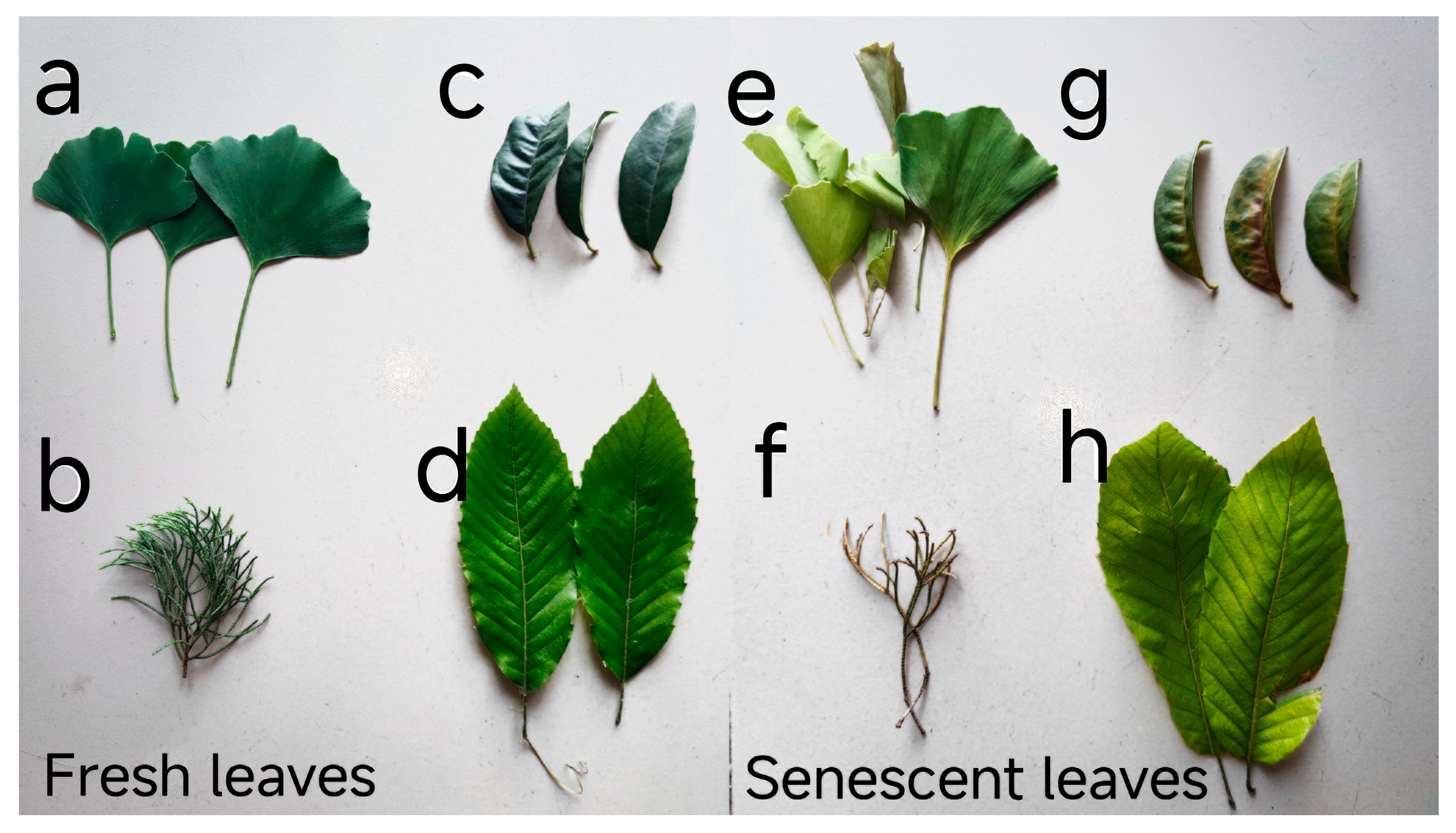
Figure 1. The fresh and senescent leaves of four plant species: (a) represents the fresh leaves of Ginkgo biloba; (b) represents the fresh leaves of Sabina chinensis ‘Kaizuca’; (c) represents the fresh leaves of Osmanthus fragrans; (d) represents the fresh leaves of Quercus acutissima; (e) represents the senescent leaves of Ginkgo biloba; (f) represents the senescent leaves of Sabina chinensis ‘Kaizuca’; (g) represents the senescent leaves of Osmanthus fragrans; (h) represents the senescent leaves of Quercus acutissima.
Horticulturae 10 00548 g001
Figure 2. The fresh leaves’ SPAD and chlorophyll concentration data distributions of four plant species were analyzed, and differences between each pair were compared using the Wilcoxon test: (a) represents the fresh leaves’ carotenoid concentrations among different species; (b) represents the fresh leaves’ Chl a concentrations among different species; (c) represents the fresh leaves’ Chl b concentrations among different species; (d) represents the fresh leaves’ Chl a+b concentrations among different species; (e) represents the fresh leaves’ SPAD concentrations among different species; Note: ‘*’, ‘**’, and ‘***’ represent significant correlations at the 0.5, 0.01, and 0.001 levels, respectively. GB: Ginkgo biloba, OF: Osmanthus fragrans, SC: Sabina chinensis ‘Kaizuca’, QA: Quercus acutissima.
Horticulturae 10 00548 g002
Figure 3. Analysis of carotenoid concentrations and differences between fresh and senescent leaves among different species: (a) represents carotenoid concentrations and differences between fresh and senescent leaves of Ginkgo biloba; (b) represents carotenoid concentrations and differences between fresh and senescent leaves of Quercus acutissima; (c) represents carotenoid concentrations and differences between fresh and senescent leaves of Sabina chinensis ‘Kaizuca’; (d) represents carotenoid concentrations and differences between fresh and senescent leaves of Osmanthus fragrans; Note: ‘*’ and ‘***’ represent significant correlations at the 0.5 and 0.001 levels, respectively.
Horticulturae 10 00548 g003
Figure 4. The correlation between fresh leaves’ SPAD, chlorophyll, and carotenoid concentrations across different plant species. The varying colors represent the linear relationships and Pearson correlation coefficient values between different species. Note: ‘*’, ‘**’, and ‘***’ represent significant correlations at the 0.5, 0.01, and 0.001 levels, respectively.
Horticulturae 10 00548 g004
Figure 5. The correlation between senescent leaves’ SPAD, chlorophyll, and carotenoid concentrations across different plant species. The varying colors represent the linear relationships and Pearson correlation coefficient values between different species. Note: ‘*’, ‘**’, and ‘***’ represent significant correlations at the 0.5, 0.01, and 0.001 levels, respectively.
Horticulturae 10 00548 g005
Figure 6. Relationship between SPAD, chlorophyll, and carotenoid concentrations.
Horticulturae 10 00548 g006
Table 1. Botanical name, life form, and classification systems of each species.
Botanical NameLife FormPhylumFamilyGenus
Ginkgo bilobadeciduous treeGymnospermaeGinkgoaceaeGinkgo
Osmanthus fragransEvergreen tree or shrubAngiospermaeOleaceaeOsmanthus
Sabina chinensis ‘Kaizuca’Evergreen treeGymnospermaeCupressaceaeJuniperus
Quercus acutissimadeciduous treeAngiospermaeFagaceaeQuercus
Table 2. CEI and NCCT values of each species.
SpeciesCEINCCT
GB2.49721988234.59333333
OF2.18863852741.86416667
SC2.03373620624.055
QA1.19834286944.827875
Table 3. Analysis of color parameters, L*, a*, and b*, measured in fresh and senescent leaves of four species. Note: different letters indicate the difference among different plants, fresh leaves, and senescent leaves (p < 0.05).
SpeciesLeafLeaf Color Parameter
L*a*b*
GBFresh99.8321 ± a0.9064 ± ab1.7913 ± ab
Senescent94.1925 ± c0.2575 ± d1.2183 ± d
OFFresh99.8961 ± a0.9758 ± a1.6533 ± b
Senescent93.9725 ± cd0.2525 ± d1.22 ± d
SCFresh99.8101 ± a0.8247 ± b1.944 ± a
Senescent93.9025 ± d0.4467 ± c1.2442 ± d
QAFresh98.6988 ± b0.9412 ± a1.7459 ± b
Senescent93.8725 ± d0.2325 ± d1.4317 ± c
Table 4. The correlation analysis of Leaf color parameters and photosynthetic pigments of fresh leaves and senescent leaves. Note: ‘*’, ‘**’ represent significant correlations at the 0.5, 0.01 levels, respectively.
LeafLeaf Color ParameterCorrelation
CarotenoidsChl aChl bChl a+b
FreshL*0.120 **−0.324 **−0.182 **−0.298 **
a*0.279 **0.242 **0.275 **0.251 **
b*−0.309 **−0.246 **−0.296 **−0.262 **
Senescent leavesL*0.064−0.177−0.035−0.138
a*−0.108−0.324 *−0.203−0.291 *
b*0.334 *0.460 **0.389 **0.450 **
TotalL*0.123 **−0.085−0.045−0.078
a*0.256 **0.340 **0.289 **0.332 **
b*−0.225 **−0.052−0.136 **−0.073
Disclaimer/Publisher’s Note: The statements, opinions and data contained in all publications are solely those of the individual author(s) and contributor(s) and not of MDPI and/or the editor(s). MDPI and/or the editor(s) disclaim responsibility for any injury to people or property resulting from any ideas, methods, instructions or products referred to in the content.