Article

Enhancing Secondary Metabolite Production in Pelargonium graveolens Hort. Cell Cultures: Eliciting Effects of Chitosan and Jasmonic Acid on Bioactive Compound Production

1
Laboratoire d’Amélioration des Productions Agricoles, Biotechnologie et Environnement (LAPABE), Faculté des Sciences, Université Mohammed Premier, Oujda 60000, Morocco
2
Centre de l’Oriental des Sciences et Technologies de l’Eau et de l’Environnement (COSTEE), Université Mohammed Premier, Oujda 60000, Morocco
3
Laboratory of Bioresources, Biotechnology, Ethnopharmacology and Health, Faculty of Sciences, Mohammed First University, Boulevard Mohamed VI, B.P. 717, Oujda 60000, Morocco
4
Euromed Research Center, Euromed Polytechnic School, Euromed University of Fes (UEMF), Fès 30000, Morocco
5
UMRT INRAE 1158 BioEcoAgro, Laboratoire BIOPI, University of Picardie Jules Verne, 80000 Amiens, France
*
Authors to whom correspondence should be addressed.
Horticulturae 2024, 10(5), 521; https://doi.org/10.3390/horticulturae10050521
Submission received: 22 April 2024 / Revised: 13 May 2024 / Accepted: 15 May 2024 / Published: 17 May 2024

Abstract

:
This study explores the effects of chitosan (CHT) and jasmonic acid (JA) elicitors on rose-scented geranium (Pelargonium graveolens Hort.) cell suspension cultures, aiming to enhance the production of phenolics and flavonoids and antioxidant properties. Elicitation with CHT and JA resulted in varied biomass yields and callus characteristics, with higher concentrations generally leading to increased phenolic accumulation. Optimal biomass was achieved with CHT4 (75 mg/mL) and JA3 (50 µM) treatments. HPLC-DAD analysis revealed changes in phenolic compound composition and quantities, with specific compounds induced by either CHT4 or JA3. For instance, gallic acid content increased significantly in CHT4-treated cells, while catechin content increased notably in both CHT4 and JA3 treatments. Antioxidant enzyme activities like superoxide dismutase and peroxidase increased with elicitor concentration, particularly in CHT4 and JA3 treatments. Both treatments exhibited potent antioxidant activity, with JA3 exhibiting the lowest IC50 value in the DPPH assay and highest total antioxidant capacity (TAC) values. Surprisingly, both CHT4 and JA3 extracts effectively inhibited tyrosinase activity. These findings underscore the efficacy of CHT and JA elicitors in enhancing phenolic and flavonoid production, boosting antioxidant capacity, and inhibiting tyrosinase activity in P. graveolens cultures, offering promising implications for further research and industrial applications in pharmaceutical and cosmetic sectors.

1. Introduction

Plants are recognized for their ability to produce a wide range of natural substances, which are also known as secondary metabolites [1]. Plant secondary metabolites, also referred to as PSMs, are chemicals that are produced through different metabolic routes than primary metabolites and their precursors [2]. PSMs are a valuable collection of resources that have great importance in multiple industries, such as the pharmaceutical industry, cosmetics, and food additives [3,4]. Most pharmaceutically significant secondary metabolites are derived from wild or cultivated plants [1]. However, despite various efforts, their chemical synthesis has generally not been economically viable [5]. Plant cell culture technology offers several advantages as a viable alternative for producing plant secondary metabolites [3,5]. It surpasses traditional field cultivation and chemical synthesis, especially for natural compounds derived from slow-growing plants or those that are challenging to synthesize chemically [6,7].
Plant secondary metabolites (SMs), such as phenolics and alkaloids, are recognized as the most effective oxygen radical scavengers. These molecules play a crucial role in combating environmental stress [8,9]. The production rate of SMs in many plants is frequently low, typically less than 1% of the plant’s dry weight. This rate is influenced by the physiological and developmental stages of the plant, as well as environmental factors [10]. Plants employ a diverse range of defense mechanisms to counteract pathogen invasion. Pathogen resistance is achieved by the activation of both pre-existing (constitutive) and induced defensive mechanisms [11]. Inducible defense responses are activated upon the detection of certain chemical substances known as ‘elicitors’ [12]. This term was initially used to refer to molecules that may trigger the formation of phytoalexins. However, it is now routinely used to describe substances that stimulate any form of plant defense [13,14]. Ultimately, the activation of defense mechanisms can result in increased resistance. They could originate from both living and non-living sources. Elicitors function as signaling molecules at low concentrations, conveying information to the plant to initiate defense mechanisms [15]. These compounds have the ability to trigger different types of plant defense mechanisms, such as the generation of reactive oxygen species (ROS), the hypersensitive response, and the creation of phytoalexins, which are secondary chemicals with antibacterial properties [16]. The activation of phytoalexin biosynthesis has become particularly significant in biotechnological strategies aimed at enhancing the synthesis of secondary metabolites.
Pelargonium graveolens Hort., also referred to as ‘rose-scented geranium’, is a member of the Geraniaceae family. Its essential oil is extensively utilized in the fields of cosmetic sand aromatherapy and as a food additive [17]. The genus Pelargonium comprises around 250 species that are extensively found in North Africa [18]. This perennial herb, native to South Africa, is characterized by its attractive, deeply lobed leaves and delicate, rose-scented flowers. It has been extensively utilized in traditional medicine for the treatment of several ailments, and numerous investigations have documented its chemical composition. The leaves of P. graveolens are extensively utilized for their antioxidant capabilities, as well as other pharmacological properties, and find several applications in the food and cosmetic industries [19]. The herb has been utilized for its antioxidant [20], anti-inflammatory, analgesic [21], anti-parasitic [22], and anti-tuberculotic effects [23]. In addition, the plant has been found to exhibit antibacterial properties against many pathogenic bacteria and fungi [21,24,25,26,27]. However, the traditional methods of cultivating and harvesting P. graveolens often fail to fully exploit the potential of these valuable bioactive compounds. As a result, alternative approaches have gained prominence in the pursuit of sustainable, controlled production. One of these approaches, of particular interest, is the use of plant cell cultures. These cultures provide an efficient, environmentally friendly method for producing high-value secondary metabolites while mitigating the challenges associated with traditional agriculture.
Chitosan, a derivative of chitin found naturally in crustacean shells, has shown promise as an elicitor in plant cell cultures by activating the plant’s defense mechanisms [28,29]. This activation prompts the synthesis of secondary metabolites as a protective response. In the current study, chitosan was utilized as a biotic stressor. Conversely, jasmonic acid, a plant hormone involved in regulating various physiological processes, including defense against biotic stress, was employed as an abiotic elicitor [30]. When applied as an elicitor, jasmonic acid can stimulate the production of bioactive compounds.
The primary aim of this research is to investigate and enhance the production of bioactive compounds in Pelargonium graveolens Hort. cell suspension cultures by strategically employing biotic and abiotic elicitors, notably chitosan and jasmonic acid. This study focuses on several aspects: (i) examining the impact of biotic and abiotic stressors on the growth and accumulation of polyphenols and flavonoids in P. graveolens suspension cultures; (ii) evaluating the response of antioxidant enzymatic defense in elicited calli under both biotic and abiotic stress conditions; (iii) analyzing changes in the phytochemical profile of elicited cell suspension cultures through HPLC-DAD quali-quantitative analysis; and (iv) assessing the bioactivity of elicited cultures, particularly their antioxidant and anti-tyrosinase activities.

2. Materials and Methods

2.1. Seed Germination

Seeds of P. graveolens were obtained from Clorofila E-nursery (Casablanca, Morocco). Seed sterilization followed a protocol adapted from Abbasi et al. [31], involving rinsing with distilled water, and treatment with 10% sodium hypochlorite for 30 s followed by 70% ethanol for 2 min. Following thorough rinsing with distilled water, the seeds were positioned on sterilized filter paper and then shifted onto MS0 basal medium, which was enriched with 3% sucrose and 0.8% agar and adjusted to a pH range of 5.5–5.7 [32]. Subsequently, the culture flasks were placed in a growth chamber set at 25 ± 1 °C with a light intensity of approximately 40 μmol·m–2·s−1 for a photoperiod of 16 h light and 8 h darkness (L/D).

2.2. Callus Establishment

To induce callogenesis, leaf explants (1 cm2) from 3-week-old seed germinated plantlets were cultured on MS medium supplemented with 3% sucrose, 0.8% agar, and TDZ (4.4 μM). The culture medium was prepared at a specific pH (5.5–5.7) and sterilized by autoclaving. Then, the cultures were incubated at a constant temperature (25 ± 1 °C) with alternating periods of light and darkness 16/8 h (Light/Dark).

2.3. Inducing the Formation of Callus with Elicitors

2.3.1. Preparation of Elicitors

Commercially available chitosan (C611NO4, deacetylating grade 70–85%, Merck Chemicals, Saint-Quentin Fallavier, France) was dissolved in a 0.1% acetic acid solution at 50 °C with continuous stirring for 5 h. Jasmonic acid (J2500, Sigma-Aldrich, St. Louis, MO, USA) was prepared at a 1 mM concentration by dissolving it in distilled water, followed by stirring for 2 h. Varying concentrations of chitosan (5, 25, 50, 75, and 100 mg/L) and jasmonic acid (5, 25, 50, 75, and 100 µM) were then introduced into the MS-derived culture medium. Each 1 mL solution dissolved in MS medium was added to the respective MS-derived medium. A control medium without elicitors (with 1 mL of fresh MS medium) was used for comparison (see Table 1).

2.3.2. Callus Inoculation

Leaf-derived calli (0.5 g fresh weight) were introduced to Murashige and Skoog (MS) liquid medium [32]. Erlenmeyer flasks (with a volume of 250 mL), each containing 100 mL of liquid MS medium supplemented with 4.45 µM of TDZ and 30 g of sucrose, were enriched with different concentrations of chitosan (CHT) (5, 25, 50, 75, 100 mg/mL) and jasmonic acid (JA) (5, 25, 50, 75, 100 μM). These cultures were then placed in a growth room and maintained at 25 ± 1 °C under a 16/8 h (light/dark) photoperiod, with a light intensity of 40–50 μmol·m−2·s−1. The flasks were placed on a gyratory shaker set at 140 rpm for a period of six weeks. After every 7 days throughout the six-week period, calli samples from each treatment with elicitors were collected. The morphological characteristics of the calli were recorded, and then they were dried overnight in an oven after their fresh weights were measured and then stored for subsequent analysis.

2.4. Sample Extraction

In accordance with the methodology outlined by Ahmad et al. [33], methanolic extracts of calli were prepared. In brief, for effective mixing and extraction, 100 mg of finely ground samples were combined with 10 mL of methanol (80%). This was followed by sonication for 10 min (Toshiba, Tokyo, Japan) and vortexing for 20 min. After three repetitions of the procedure, the resulting mixtures underwent 15 min of centrifugation at 13,000 rpm. Additional analysis was conducted on the supernatants, which were then stored at 4 °C.

2.4.1. Total Phenolic Contents

Slight modifications were made to the Folin–Ciocalteu reagent protocol as described by Slinkard and Singleton (1977) in order to ascertain the total phenolic contents [34]. In 96-well plates, a volume of 20 μL of the samples was added to each well, followed by the addition of 90 μL of the Folin–Ciocalteu reagent. Following this, 90 µL of sodium carbonate was added to each well from the 6% stock solution, and the plates were left at room temperature for 90 min under standard room lighting. As positive and negative controls, methanol (20 µL) and gallic acid (1 mg/mL) were employed, respectively. The standard gallic acid was utilized to generate the calibration curve (0–100 μg/mL, R2 = 0.9798); the TPC was denoted in gallic acid equivalents (GAE) per gram of dried weight. To estimate total phenolic production (TPP) in mg GAE/L, the subsequent formula was applied.
T P P   ( m g / L ) = D W   ( g / L ) × T P C   ( m g / g )

2.4.2. Total Flavonoid Contents

The method described by Chang et al. [35] was used to determine the total flavonoid contents, using aluminum chloride. A 10% solution of aluminum trichloride (10 μL) and potassium acetate (10 μL) was introduced into the reaction well, along with 20 μL of the samples. The total volume was increased to 200 μL by adding 160 μL of distilled water and left at room temperature for 30 min. A calibration curve was constructed using quercetin (with a range of 0–40 μg/mL) and a coefficient of determination (R2) of 0.9981. The total flavonoid content (TFC) was quantified as milligrams of quercetin equivalent (QE) per gram of dry weight. The calculation of total flavonoid production (TFP) was performed using the following formula, and the results are represented in milligrams of quercetin equivalents per liter (mg QE/L).
T F P   ( m g / L ) = D W   ( g / L ) × T F C   ( m g / g )

2.5. Quantification of Phenolic Compounds Using HPLC-DAD

The phenolic compounds in the methanol extracts were analyzed using High-Performance Liquid Chromatography (HPLC) with an Agilent 1200 system (Agilent Technologies, Palo Alto, CA, USA) linked to a diode array UV detector (DAD, Bruker, Berlin, Germany). The experiment involved injecting 20 µL of each extract into a Zorbax XDB-C18 column (porosity of 5 µm, dimensions of 250 × 4.6 mm; Agilent Technologies series 1100 (Palo Alto, CA, USA)). The column was equipped with a 4 × 3 mm C18 cartridge precolumn (Agilent Technologies). The elution process involved a gradient of solvents A and B, transitioning through different compositions over time. Solvent A (water/0.5% phosphoric acid) and B (methanol) were used as the mobile phases. The elution started with 80% A and 20% B, shifting gradually to 100% B and then back to 100% A, with intermediate compositions in between. The elution was conducted at a flow rate of 1 mL/min. The spectrophotometric detection was carried out at 254, 280, 320, and 340 nm. The identification of the compounds was accomplished by comparing their retention periods and UV spectra with those of the authentic standards (Table S1). The quantification was performed by utilizing calibration curves containing various concentrations of standards in ethanol (the standards used, caffeic acid, syringic acid, 3-hydroxybenzoic acid, p-coumaric acid, sinapic acid, ferulic acid, 3-hydroxyflavone, salicylic acid, rutin, cinnamic acid, quercetin 3-O-β-D-glucoside, 3-hydroxycinnamic acid, kaempferol, and chalcone) (0.2; 0.4; 0.6; 0.8; 1 mg/mL). The results are presented in Table S1. The quantification of all chemicals was performed using 5-point calibration curves with a coefficient of determination (R2) greater than 0.99. The samples were analyzed in triplicate, and the results were represented as milligrams per 100 g of dry weight of the sample. The limit of detection (LOD) and limit of quantification (LOQ) [36] were calculated for each molecule using Formulas (1) and (2), respectively.
L O D = 3.3 × S D   o f   s i g n a l   i n t e n s i t y   f r o m   l o w C o n c e n t r a t i o n   s a m p l e s S l o p e   o f   t h e   c a l i b r a t i o n   c u r v e
L O Q = 10 × S D   o f   s i g n a l   i n t e n s i t y   f r o m   l o w C o n c e n t r a t i o n   s a m p l e s S l o p e   o f   t h e   c a l i b r a t i o n   c u r v e

2.6. Extraction of Samples, and Enzymes Activities

With minor adjustments, the sample extraction procedure outlined by Taimoor et al. [37] was implemented for the quantification of POD. In summary, 1% PVP was added to 1 mL of potassium phosphate buffer (50 mM, pH 7) to homogenize 100 milligrams of the fresh sample. After 30 min of centrifugation at 15,000 rpm, the supernatant was extracted for additional analysis.

2.6.1. Peroxidase (POD) Activity

POD activity was evaluated using the protocol established by Lagrimini in 1980 [38], with slight adjustments. The assay mixture comprised 40 μL of potassium phosphate buffer (50 mM; pH 7), 20 μL of guaiacol (100 mM; 10× concentration), 20 μL of freshly prepared sample extract, 100 μL of distilled water, and 20 μL of hydrogen peroxide (27.5 mM; 10× concentration). A control sample was also prepared, containing the same quantity of all reagents except for the sample extract.
A = E · L · C
A represents Absorbance, E is the Extinction coefficient with a value of 6.39 mM−1·cm−1, L stands for Length of wall which is 0.25 cm, and C represents enzyme concentration calculated in nM/min/mg FW.

2.6.2. Superoxide Dismutase (SOD) Activity

A modified approach based on the method outlined by Taimoor et al. [37] was employed for the SOD assay. Briefly, the procedure involved mixing 20 μL of EDTA (1 mM), 20 μL of methionine (130 mM), 20 μL of NBT (0.75 mM), 78 μL of phosphate buffer (50 mM, pH 7), 2 μL of riboflavin (0.02 mM), and 60 μL of freshly prepared sample extract. Additionally, a control was created using the identical mixture, but excluding the sample extract. After exposure to fluorescent light for 7 min, the amount of light absorbed was measured at a wavelength of 660 nm using a microplate reader (Thermo Scientific Multiskan GO, Waltham, MA, USA). The enzymatic activity was assessed using the formula analogous to that employed for quantifying POD activity.

2.7. DPPH Activity

The assessment of antioxidant activity was conducted using a modified DPPH technique, following standard procedures [39,40]. The DPPH-MeOH solution was prepared by dissolving 2 mg of DPPH in 100 mL of methanol. Subsequently, a volume of 2.5 mL of the DPPH mixture was introduced into each of the distinct solutions, and the overall volume was then adjusted to 3 mL. Following 30 min incubation at room temperature, the absorbance of the combination was determined at a wavelength of 517 nm, relative to a blank sample. The formula used to calculate the percentage of DPPH free radical scavenging activity (FRSA) was as follows:
F R S A % = A b l a n k A s a m p l e A b l a n k × 100
The control reaction, which included all reagents except the extract, was assigned an absorbance value labeled as Ablank. The absorbance of the extract at different concentrations was denoted as Asample. In order to ascertain the IC50 value, a graph was constructed by plotting the percentage of inhibition versus the concentrations of the extract. Ascorbic acid was utilized as a reference control.

2.8. Anti-Tyrosinase Activity

The tyrosinase inhibitory activity was evaluated using the procedure outlined by Bouyahya et al. [41,42]. A 25 µL measure of the sample was combined with 100 µL of tyrosinase solution (333 units per milliliter, 50 mM phosphate buffer, pH 6.5) and maintained in a pre-incubation phase at a temperature of 37 °C for a duration of 10 min. Next, 300 µL of L-DOPA solution with a concentration of 5 mM was introduced into the mixture, which was then subjected to incubation at a temperature of 37 °C for a duration of 30 min. The spectrophotometer was used to measure the absorbance at a wavelength of 510 nm. The extent of tyrosinase inhibition was assessed throughout a range of doses spanning from 10 to 500 µg/mL, and the IC50 values were established. The positive control utilized in the study was Kojic acid (Sigma, St. Louis, MO, USA).

2.9. Statistical Analysis

All trials were executed with no fewer than three duplications, and the outcomes are delineated as mean ± standard deviation from at least three replications. Charts were created utilizing GraphPad Prism software v8.0 (GraphPad Software, San Diego, CA, USA) showcasing average data values and error bars for standard deviation. Statistical significance at a significance level of p < 0.05 was determined through SPSS v19.0 (IBM Corp., Armonk, NY, USA) for computing means and standard deviations, as well as for the analysis of variance. The correlogram plot was constructed using RStudio version 1.0.13 (RStudio, Boston, MA, USA).

3. Results and Discussion

3.1. Preliminary Tests with Different Concentrations of Elicitors

Table 2 presents the effects of different elicitors on growth parameters and the accumulation of secondary metabolites, specifically polyphenols and flavonoids, in cell suspension cultures. The elicitors examined include CHT (chitosan) and JA (jasmonic acid), each tested at various concentrations.
The initiation day of callus cultures varied depending on the elicitor concentration, ranging from the 3rd to the 7th day. Callus characteristics were found to be influenced by the elicitor type and the concentration (Table 2). For instance, callus initiated with TDZ typically exhibited a light green color and friable texture, while those initiated with higher concentrations of CHT and JA tended to be darker in color (dark green—DG) and more compact in texture.
For cell suspensions treated with chitosan, the optimal biomass (DW = 12.34 ± 0.73 g/100 mL) was achieved on MS media supplemented with CHT4 (75 mg/mL), while lower biomass (DW = 6.80 ± 0.19 g/100 mL) was observed with CHT1 (5 mg/mL) treatment, exhibiting light-brown compact features. Similarly, for JA, the maximum biomass was recorded with the treatment JA3 (50µM), and JA4 (75 µM), yielding DW = 15.62 ± 0.98 g/100 mL, and DW = 16.65 ± 0.70 g/100 mL, respectively, with dark green compacted calli for JA3, and dark green friable calli for JA4. Higher concentrations of CHT (75 mg/mL and 100 mg/mL) and JA (50 µM and 75 µM) led to the highest biomass accumulation, with dark green (DG) or green-brown (GB) callus characteristics. In contrast, lower biomass was observed in cultures treated with lower concentrations of CHT and JA, as well as in the control (TDZ). In comparison to callus cultures stimulated by TDZ, those influenced by varying concentrations of CHT and JA exhibited a range of colors and callus yields. Concentrations of JA and CHT hindered callus proliferation, possibly due to stress induced by elevated JA and CHT levels.
When considering CHT4 (75 mg/mL), the optimal levels (TPC: 80.94 ± 2.38 μg GAE/g DW, TPP: 9.99 ± 0.23 mg/mL) were observed during the 4th week. In contrast, for calli derived from JA3 (50 μM), the peak production (TPC: 86.41 ± 1.62 μg GAE/g DW, TPP: 13.49 ± 0.24 mg/mL) occurred during the 3rd week compared to the control (TPC: 53.24 ± 0.52 μg GAE/g DW, TPP: 3.00 ± 0.15 mg/mL) at the same time point (see Figure 1A). Additionally, the investigation extended to flavonoid content and production. The findings revealed that the highest levels of TFC and TFP (TFC: 38.45 ± 1.40 μg QE/g DW, TFP: 4.74 ± 0.09 mg/mL) were observed in CHT4 cultures during the 4th week. This was followed by TFC and TFP (TFC: 37.61 ± 0.53 μg QE/g DW, TFP: 5.87 ± 0.11 mg/mL) in cultures derived from JA3 compared to the control (TFC: 24.10 ± 0.70 μg QE/g DW, TFP: 1.380 ± 0.08 mg/mL) (refer to Figure 1B).
The sustainable production of phenolics and flavonoids in response to elicitors may rely on various factors, including the age of cultures, elicitor dosage, and duration of contact [43,44]. Callus characteristics such as color and texture were also influenced by the elicitor type and concentration [29]. This observation is in line with prior evidence by Taimoor et al. [37]. The increased response seen with JA compared to CHT may be attributed to JA’s role as a plant growth regulator involved in modulating plant responses to various stresses. On the other hand, CHT triggers plant defense responses, stimulating secondary metabolism and consequently producing phenolic compounds and flavonoids, albeit in lesser quantities compared to JA. Elicitors exerted a notable influence on the accumulation of polyphenolic and flavonoid compounds compared to control conditions, with higher concentrations generally leading to increased phenolic and flavonoid contents [45].

3.2. Phenolic Compounds Quantification Using HPLC-DAD

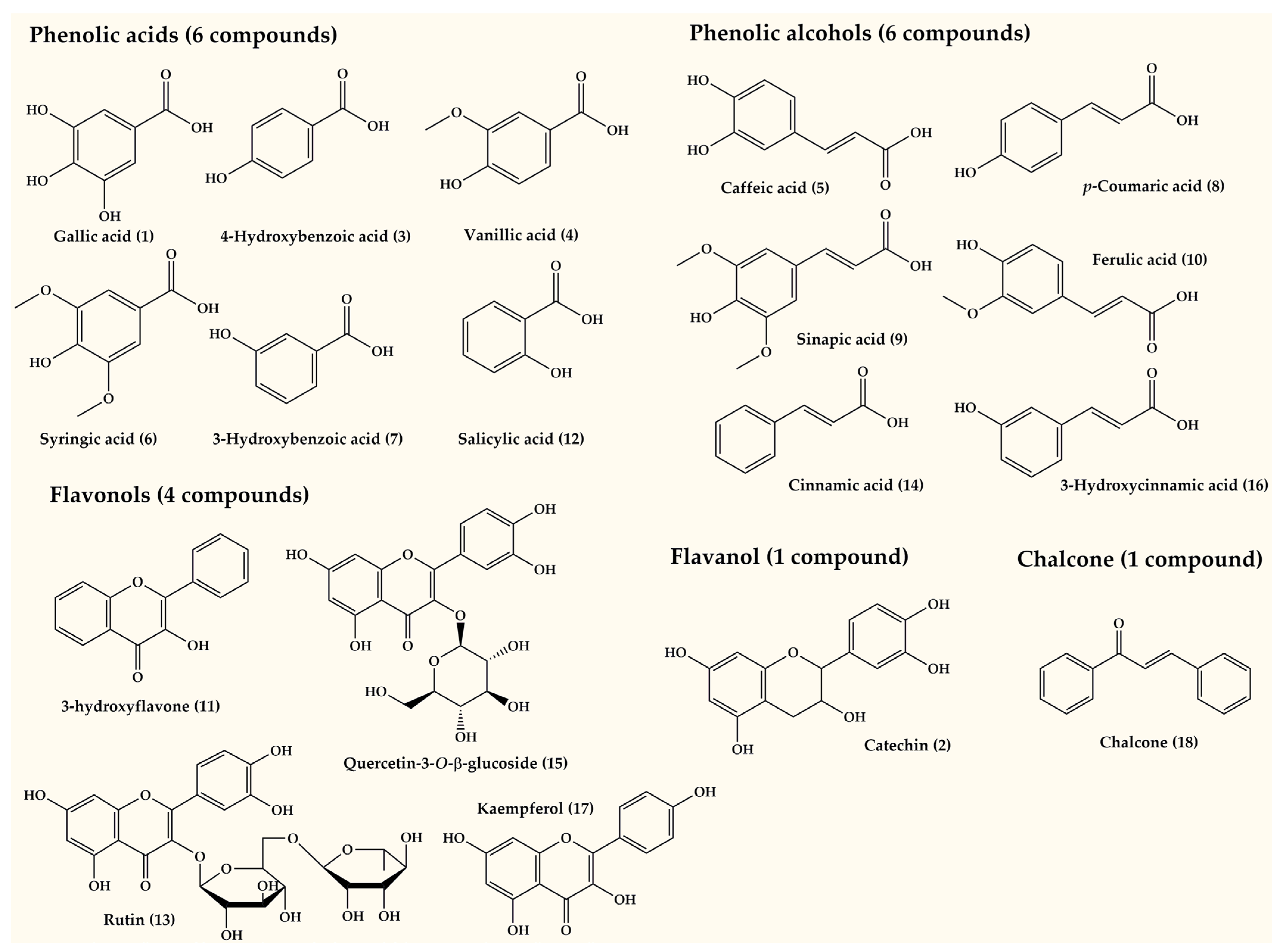
In this study, we quantified a total of 18 metabolites in cell suspension cultures of P. graveolens treated with elicitors, as shown in Table 3 and Figure 2 and Figure 3. High-performance liquid chromatography (HPLC) analysis indicated that the overall production of these metabolites increased significantly in both CHT (13,602.01 ± 6.447 mg/100 g DW) and JA (24,891.40 ± 4.30 mg/100 g DW) treatments compared to controls (8216.37 ± 8.0661 mg/100 g DW) (Table 3). The examination of the compounds present in control (CTRL), CHT4-treated, and JA3-treated samples revealed noticeable differences in bioactive molecules among the treatments.
The Venn diagram (Figure 4A) depicts the distribution of molecules across the different treatments. In the CTRL circle, 11 molecules are found, while the CHT and JA circles contain 12 and 14 molecules, respectively. Overlaps between CTRL and CHT, CTRL and JA, and CHT and JA contain 9, 10, and 10 common compounds, respectively. The intersection of CTRL, CHT4, and JA3 encompasses eight compounds. The Venn diagram representation succinctly illustrates both the unique responses and shared characteristics of molecules under different treatments, shedding light on the specific effects induced by chitosan and jasmonic acid treatments compared to the control condition. A differentially accumulated metabolite (DAM) analysis was performed to identify the number of compounds that were upregulated or downregulated by the treatments (Figure 4A). CHT4 treatment resulted in the upregulation of 12 compounds and the downregulation of 3 compounds compared to CTRL. JA3 treatment resulted in the upregulation of 10 compounds and the downregulation of 5 compounds compared to CTRL.
Table 3 presents the results of the HPLC-DAD quantification of the most abundant phenolic compounds in the elicited cell cultures, with data expressed as means ± standard error. The variations in the quantities of phenolic compounds between the control (CTRL) and groups treated with CHT4 (75 mg/mL) and JA3 (50 µM) are notable and provide insights into the metabolic responses of the cells to these elicitors.
Treating cell suspension cultures with JA3 (at a concentration of 50 µM) resulted in the neo-synthesis of two phenolic acids—syringic acid (6) and 3-hydroxybenzoic acid (7)—along with two phenolic alcohols—ferulic acid (10) and cinnamic acid (14)—and one flavonol, 3-hydroxyflavone. Conversely, the application of CHT4 (at a concentration of 75 mg/mL) led to the production of three compounds: two phenolic alcohols—ferulic acid (10) and cinnamic acid (14)—and one phenolic acid, gallic acid (1).
Gallic acid (1) was not detected in the control but showed a significant increase in quantity in the CHT4-treated cells (0.91 ± 0.28 mg/100 g DW), suggesting an induction of its biosynthesis pathway in response to chitosan. Catechin (2), a flavan-3-ol flavonoid, exhibited substantial increases in both CHT4 and JA3 treatments compared to the control. The magnitude of increase was much higher in the CHT treatment (1279.84 ± 0.07 mg/100 g DW) compared to JA3 (911.83 ± 0.05 mg/100 g DW), indicating a stronger elicitation effect by chitosan. 4-Hydroxybenzoic acid also showed an increase by 96.4% from 208.92 ± 0.01 mg/100 g DW in the control to 410.41 ± 0.01 mg/100 g DW in the CHT4 treatment and by 40.1% in the JA3 treatment with 292.75 ± 0.02 mg/100 g DW. This augmentation might be linked to the activation of phenylpropanoid pathway enzymes by both elicitors. Vanillic acid was detected in traces in the treatments, indicating its very low abundance in the cell cultures under the experimental conditions.
Caffeic acid exhibited a decrease by 53.1% from 299.32 ± 0.48 mg/100 g DW in the control to 140.25 ± 0.12 mg/100 g DW in the CHT4 treatment but increased by 98.2% to 278.16 ± 0.96 mg/100 g DW in the JA3 treatment comparable to the control. This variability could result from differential regulatory mechanisms induced by chitosan and jasmonic acid. Syringic acid was absent in the control and CHT4 treatments but appeared at 60.87 ± 0.03 mg/100 g DW in the JA3 treatment, suggesting a specific induction by jasmonic acid. 3-hydroxybenzoic acid was not detected in the control or CHT4 treatments but was present at 921.37 ± 0.00 mg/100 g DW in the JA3 treatment, indicating a selective response to jasmonic acid. p-coumaric acid exhibited a decrease from 286.93 ± 0.41 mg/100 g DW in the control to absence in the CHT4 treatment but showed a substantial increase by 683.7% to 2248.33 ± 0.53 mg/100 g DW in the JA3 treatment, possibly due to the specific induction of its biosynthesis pathway by jasmonic acid. Sinapic acid was present in the control and absent in both the CHT4 and JA3 treatments, suggesting a possible downregulation of its biosynthesis pathway by the elicitors. Ferulic acid was absent in the control, appeared at 350.75 ± 0.18 mg/100 g DW in the CHT4 treatment, and significantly increased by 345.5% to 1562.87 ± 1.75 mg/100 g DW in the JA3 treatment, indicating differential regulation by chitosan and jasmonic acid. 3-hydroxyflavone was not detected in the control or JA3 treatments but appeared at 1012.41 ± 0.00 mg/100 g DW in the CHT4 treatment, suggesting specific induction by chitosan. Salicylic acid showed an increase by 217.2% from 18.28 ± 0.07 mg/100 g DW in the control to 57.99 ± 0.03 mg/100 g DW in the CHT4 treatment and further increased by 3685.9% to 692.10 ± 0.21 mg/100 g DW in the JA3 treatment. Rutin was present in trace amounts in the control and CHT4 treatments but was absent in the JA3 treatment, suggesting a specific downregulation by jasmonic acid.
Cinnamic acid was absent in the control, appeared at 54.67 ± 0.29 mg/100 g DW in the CHT4 treatment, and increased to 395.11 ± 1.45 mg/100 g DW in the JA3 treatment, indicating specific induction by both elicitors. Quercetin 3-O-β-D-glucoside exhibited an increase from 2760.51 ± 6.22 mg/100 g DW in the control to 3328.10 ± 2.07 mg/100 g DW in the CHT4 treatment and further increased to 8177.46 ± 2.07 mg/100 g DW in the JA3 treatment, suggesting a cumulative effect of chitosan and jasmonic acid on its accumulation. 3-hydroxycinnamic acid showed an increase from 2799.25 ± 4.83 mg/100 g DW in the control to 6834.61 ± 6.03 mg/100 g DW in the CHT4 treatment and further increased to 6377.09 ± 2.41 mg/100 g DW in the JA3 treatment, indicating a strong induction by both elicitors.
Kaempferol exhibited an increase from 223.37 ± 0.83 mg/100 g DW in the control to 886.99 ± 0.41 mg/100 g DW in the CHT4 treatment and further increased to 1031.81 ± 1.24 mg/100 g DW in the JA3 treatment, suggesting a synergistic effect of chitosan and jasmonic acid on its accumulation. Chalcone showed an increase from 0.79 ± 0.03 mg/100 g DW in the control to 557.91 ± 0.02 mg/100 g DW in the CHT4 treatment and further increased to 927.35 ± 0.03 mg/100 g DW in the JA3 treatment, indicating a cumulative effect of both elicitors.
The variations in the quantities of phenolic compounds suggest differential metabolic responses of the cell cultures to chitosan and jasmonic acid elicitation, with some compounds showing specific induction by one elicitor and others responding to both elicitors, albeit to varying degrees. These results were found to be perfectly aligned with recent reports [46,47]. The plant produces a diverse range of secondary metabolites in response to different signaling molecules. Plants react to stress by diverting primary metabolites (PMs) and triggering the formation of SMs such as phenolic compounds, flavonoids, tannins, and other SMs. This response also stimulates the activities of antioxidant enzymes [8]. CHT has been reported to stimulate the accumulation of phenylpropanoids in germinated buckwheat (Fagopyrum esculentum Moench.) [48], phenolic acids in cell suspension cultures of Malus × domestica Borkh. [49], flavonoids in callus culture of Rumex vesicarius L. [50], and flavonolignans in Silybum marianum (L.) Gaertn. cell suspension cultures [51]. JA also exhibits positive effects on plant metabolism by enhancing the accumulation of stilbenes, trans-resveratrol, and viniferins in vitro cultures of Vitis vinifera [52,53]. Increased levels of rosmarinic acid were observed in Mentha × piperita cell suspension cultures following elicitation with jasmonic acid and methyl jasmonate [54]. Additionally, Gadzovska et al. reported that JA-elicited cell suspensions of Hypericum perforatum L. enhanced the production of phenylpropanoids and naphtodianthrones [55].
These findings provide a valuable foundation for the modulation of phenolic compound biosynthesis pathways in response to elicitor treatments in cell cultures.

3.3. Antioxidant Enzymes Quantification

SOD and POD enzymes are isoforms with distinct physical, chemical, and structural properties; they collaborate synergistically to maintain cellular redox homeostasis and protect plants from oxidative stress-induced damage. SOD catalyzes the dismutation of superoxide radicals into molecular oxygen and hydrogen peroxide, whereas POD catalyzes the reduction of hydrogen peroxide using various substrates, generating water and oxidized substrates in the process. The influence of chitosan (CHT) and jasmonic acid (JA) on the enzymatic activities of SOD and POD was also examined in cell suspension cultures of P. graveolens. These activities were observed to increase with increased elicitor concentration [37,56,57,58]. Optimal enzyme activities (POD: 4.84 ± 0.02; SOD: 0.779 ± 0.012 nM/min/mg FW) were observed in cultures elicited with CHT4 and JA3 (POD: 4.97 ± 0.04; SOD: 0.812 ± 0.017 nM/min/mg FW) compared to the control (POD: 2.31 ± 0.01; SOD: 0.55 ± 0.007 nM/min/mg FW) (see Figure 5A,B). Earlier research findings suggest that chitosan and jasmonic acid potentially enhances SOD and POD activities in cell suspension cultures, as well as in callus cultures of different plant species [28,37,59,60,61,62]. A report by Fan et al. indicates the involvement of POD and SOD in mediating chitosan-induced stress in cell suspension cultures of Betula platyphylla Suk. [28]. According to Zaragoza-Martinez et al., the application of jasmonic acid triggered oxidative reactions in Jatropha curcas cell suspension culture, leading to elevated levels of hydrogen peroxide (H2O2), along with an increase in malondialdehyde levels. Additionally, the study observed heightened activity of catalase, peroxidases, and ascorbate peroxidase in response to JA treatment [59].
The exposure of plant cells to elicitor treatment triggers a cascade of events, starting with signal transduction from the plasma membrane’s surface and leading to the production of reactive oxygen species (ROS) [63]. This process activates the plant’s defense mechanisms and promotes the enhancement of secondary metabolism by regulating specific enzymes involved in the synthesis of targeted secondary metabolites [64]. The oxidative stress induced by elicitor treatment results in the accumulation of ROS, which can be counteracted by antioxidant enzymes such as superoxide dismutase (SOD), peroxidase (POD), ascorbate peroxidase (APX), and catalase (CAT) [65]. When oxidative damage surpasses the capacity of these antioxidant enzymes, it can lead to a decrease in cell viability and eventual cell death.
The findings of our study highlight the distinct roles of CHT and JA with respect to ROS for systemic signaling in plants to combat abiotic stress. Both types of elicitors showed peak activity in the CHT4 and JA3 treatments positively correlating with biomass, polyphenols, and flavonoid levels [66,67,68].

3.4. Antioxidant Activity

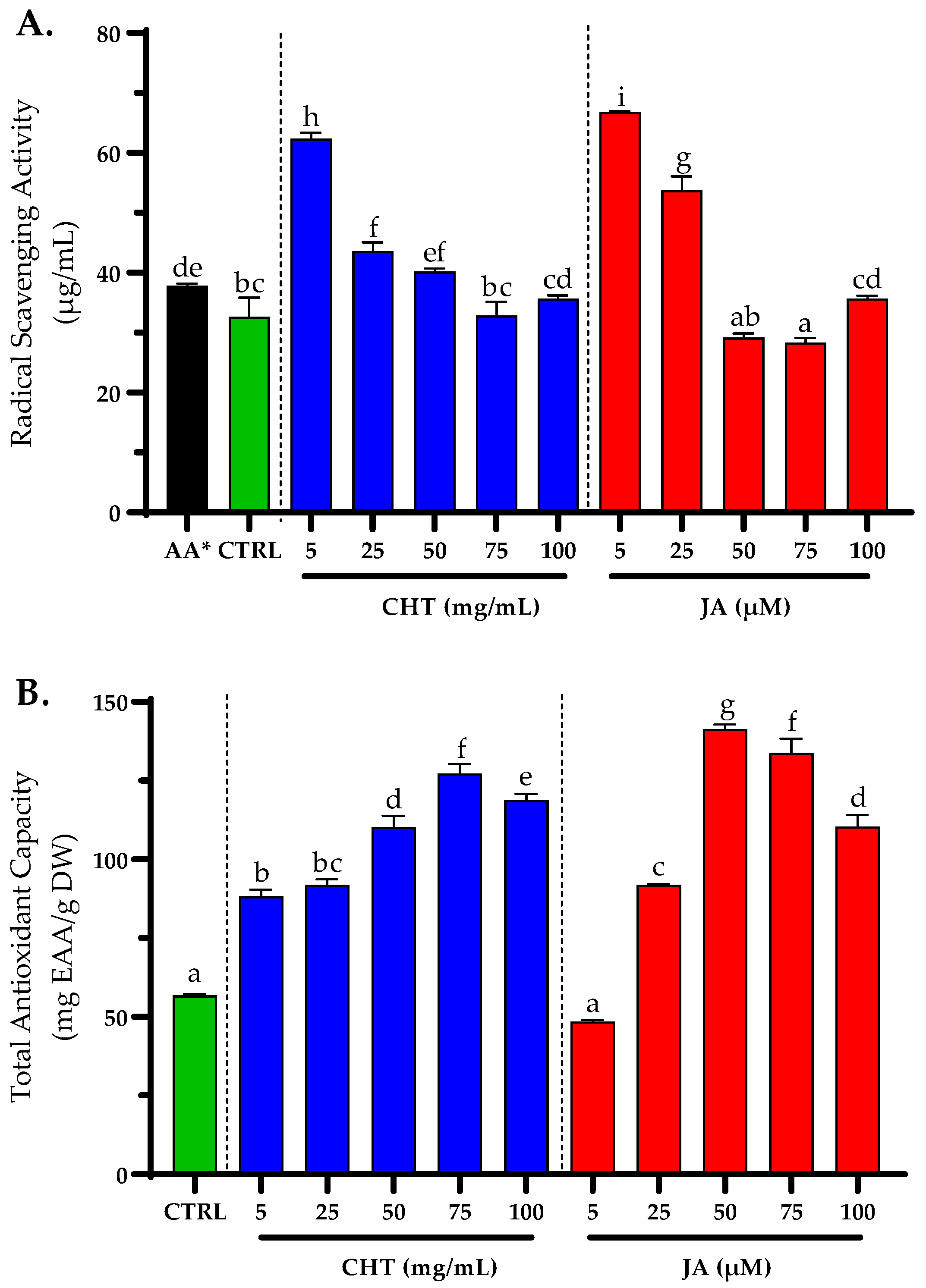
Typically, the antioxidant activity is measured by employing DPPH, a free radical, followed by assessing the sample’s half-inhibitory concentration (IC50) [69]. The optimal IC50 values of DPPH activity was detected in JA3- and JA4-treated cultures (29.19 ± 0.65, and 28.32 ± 0.78 µg/mL, respectively) and controls (32.67 ± 3.19 µg/mL), followed by CHT4 cultures (32.83 ± 2.30 µg/mL), (Figure 6A). This is attributed to the fact that phenolics and flavonoids mainly contribute to the antioxidant potential of plants [70,71].
The total antioxidant capacity (TAC) assay employing the phosphomolybdenum method relies on the reduction of Mo(VI) to Mo(V) facilitated by the analyte present in the sample, followed by the formation of a green phosphate/Mo(V) complex under acidic conditions. This method is particularly adept at detecting antioxidants such as various phenolics, ascorbic acid, α-tocopherol, and carotenoids [72]. The findings indicated notably elevated TAC values in CHT4 and JA3 cultures, measuring 127.23 ± 2.91 and 141.32 ± 1.50 mg EAA/g DW, respectively, compared to the control, which exhibited a comparatively modest value of 56.80 ± 0.51 mg EAA/g DW (Figure 6B).
Plants maintain their equilibrium by neutralizing free radicals with phytochemicals that function as antioxidants [73,74,75]. Phenolic compounds are one of the primary classes of these molecules known for scavenging free radicals [76]. It has been reported that JA, and CHT stimulates certain genes of antioxidant enzymes such as SODs, catalases, and ascorbate peroxidases to shield cells in response to ROS [30,77,78]. Furthermore, earlier reports have demonstrated high antioxidant activities of plants exposed to both CHT or JA [78,79].

3.5. Tyrosinase Inhibition

In this study, cell extracts from CHT4 and JA3 exhibited significantly enhanced inhibition of tyrosinase activity compared to the control extract (with IC50 values of 39.41 ± 0.88 µg/mL and 31.00 ± 0.57 µg/mL, respectively), whereas kojic acid, a well-known tyrosinase inhibitor, demonstrated the highest inhibitory efficacy with an IC50 value of 22.54 ± 0.09 µg/mL (Figure 7). These findings suggest that elicitation with CHT at a concentration of 75 mg/mL and JA at 50 µM elicited a favorable response in cell suspension cultures, leading to potent inhibition of tyrosinase activity. This phenomenon may be attributed to the enhanced biosynthesis and impact of the latter elicitors at those concentrations on phenolic compounds. Tyrosinase (EC 1.14.18.1), also known as polyphenol oxidase, plays a pivotal role in melanogenesis by catalyzing the initial two rate-limiting steps of melanin biosynthesis [80]. The inhibition of tyrosinase activity is correlated with a reduction in melanogenesis, rendering it a commonly employed strategy in depigmentation agents. Various manifestations of skin aging, including wrinkles, roughness, dryness, and specific forms of hyperpigmentation such as age spots, solar lentigos, and melasma, prompt the demand for skin-lightening cosmetic formulations. To mitigate concerns regarding skin irritation often associated with synthetic compounds, natural alternatives are sought after [81].
The results conclusively indicate that extracts from P. graveolens cell suspension cultures elicited with CHT or JA harbor efficacious tyrosinase inhibitors, thereby presenting promising potential for integration into skin-whitening formulations within cosmetic products.

3.6. Correlation Analysis

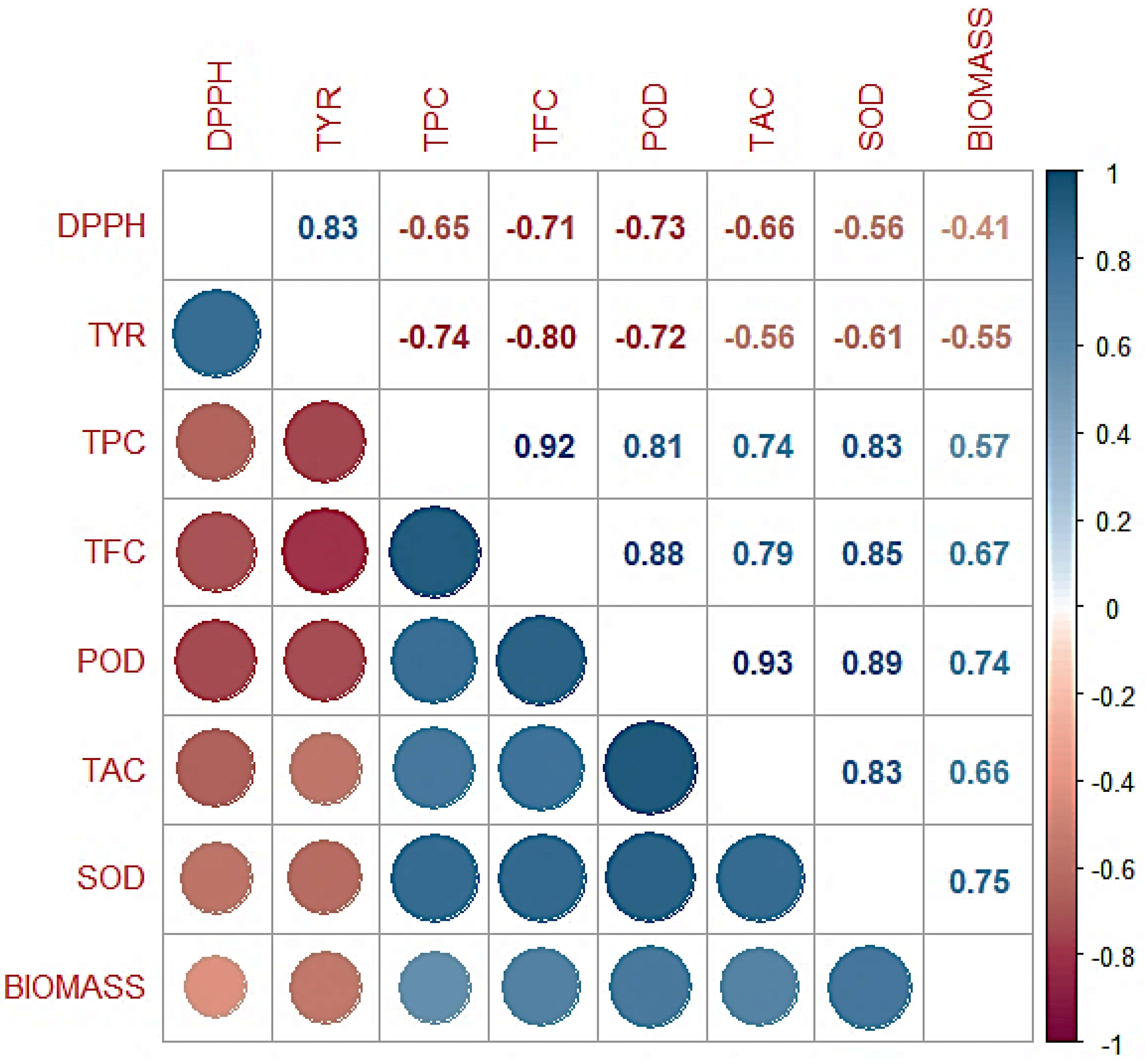
A Pearson correlation analysis was conducted to explore the interrelationships between the secondary metabolites’ contents and antioxidant enzymes with the biological activities of the elicited cultures in this study. This analysis included total antioxidant capacity (TAC), superoxide dismutase activity (SOD), peroxidase (POD), total flavonoid content (TFC), total phenolic content (TPC), biomass, tyrosinase inhibitory activity (TYR), and the free radical scavenging activity against the stable free radical 2,2-diphenyl-1-picrylhydrazyl (DPPH). The results are presented as a correlogram in Figure 8.
The correlation analysis revealed a network of statistically significant positive correlations. Notably, strong positive correlations were observed between TAC and SOD (r = 0.83, p < 0.001), TFC (r = 0.92, p < 0.001), and TPC (r = 0.83, p < 0.001). These findings suggest a potential co-occurrence or synergistic interaction between these specific antioxidant compounds. Samples exhibiting higher overall antioxidant capacity (TAC) also tended to possess elevated levels of these individual antioxidant constituents (SOD, TFC, TPC), potentially contributing to their superior free radical scavenging abilities. Furthermore, a moderate positive correlation was observed between biomass and TFC (r = 0.68, p < 0.01), suggesting a possible association between sample quantity and flavonoid content.
A strong positive correlation (r = 0.83, p < 0.001) has been observed between the DPPH (antioxidant activity) and tyrosinase inhibition (TYR) values, indicating a potential association between the capacity to scavenge free radicals, as assessed by DPPH, and the inhibition of the enzyme tyrosinase. This observation is consistent with numerous studies that have highlighted a relationship between the ability to scavenge radicals and the inhibition of tyrosinase activity by polyphenol and flavonoid antioxidants [82,83]. However, it is essential to recognize that other factors may also play a role in tyrosinase inhibition [84].
Intriguingly, a moderate negative correlation was identified between DPPH IC50 value and TPC (r = −0.71, p < 0.01). As the DPPH IC50 value increases (indicating lower antioxidant activity), TPC levels tend to decrease. This observation implies that samples enriched with total phenolics may exhibit superior free radical scavenging activity, potentially due to the presence of specific phenolic compounds with potent antioxidant properties [85]. It is well documented that various phenolic compounds possess diverse mechanisms of action for free radical scavenging, including hydrogen atom transfer and single electron transfer [86]. These findings provide a preliminary understanding of the intricate relationships between various antioxidant properties within the investigated samples. The negative correlation between DPPH IC50 and TPC highlights the potential role of phenolic compounds in free radical scavenging activity. Future studies employing targeted analyses of individual phenolic compounds and their corresponding antioxidant mechanisms are warranted to elucidate the underlying interactions and contributions of these bioactive molecules within the samples.

4. Conclusions

The utilization of biotechnological approaches, notably elicitation, to augment the synthesis of secondary metabolites holds paramount importance in modern scientific endeavors. With the increasing demand for natural products with therapeutic and commercial value, the ability to manipulate metabolic pathways within plant cells presents unprecedented opportunities for innovation. Elicitation serves as a potent tool to induce specific biochemical responses, thereby enhancing the production of desirable secondary metabolites such as phenolics and flavonoids.
The current study provides valuable insights into the influence of chitosan (CHT) and jasmonic acid (JA) elicitors on the enhancement of phenolics and flavonoid production, as well as antioxidant properties, in Pelargonium graveolens cell suspension cultures. The findings demonstrate that both elicitors induce significant changes in biomass yield, callus characteristics, and secondary metabolite accumulation, with higher concentrations generally resulting in increased production of polyphenols and flavonoids. Specifically, optimal biomass yields were achieved with CHT4 (75 mg/mL) and JA3 (50 µM) treatments, accompanied by alterations in phenolic compound composition, such as the induction of gallic acid by CHT and catechin by both CHT and JA treatments. Furthermore, the study highlights the role of elicitor concentration in enhancing antioxidant enzyme activities, including superoxide dismutase (SOD) and peroxidase (POD), with CHT4 and JA3 treatments exhibiting optimal activity levels. Both CHT and JA treatments also demonstrated potent antioxidant activity, with JA3 treatment showing the lowest IC50 value in the DPPH assay and the highest total antioxidant capacity (TAC) value. Surprisingly, both elicitors effectively inhibited tyrosinase activity, indicating their potential applications in cosmetic industries.
Nevertheless, it is imperative to acknowledge the inherent limitations of this study, including the necessity for further elucidation of the intricate signaling pathways mediating elicitor-induced metabolic changes. Additionally, while the focus on P. graveolens offers valuable insights into the elicitor response dynamics within this species, broader investigations encompassing diverse plant cell cultures are warranted to extrapolate the findings and optimize elicitation strategies for broader industrial applications.
Overall, these findings not only enrich our understanding of elicitation strategies in plant cell cultures but also pave the way for the development of innovative biotechnological interventions with far-reaching implications in pharmaceutical, cosmetic, and nutraceutical industries.

Supplementary Materials

The following supporting information can be downloaded at https://www.mdpi.com/article/10.3390/horticulturae10050521/s1, Table S1: Validation characteristics data of the phenolic standards, and sensitivity of the HPLC-DAD approach.

Author Contributions

Conceptualization, A.E. and M.A.; Data curation, A.E., A.B., M.H. and M.A.; Formal analysis, A.E., M.T., A.B., M.H., E.H.L., A.A. and M.A.; Funding acquisition, A.E., A.A. and M.A.; Investigation, A.E., E.H.L. and A.A.; Methodology, A.E., M.T., A.B., M.H., F.M. and M.A.; Project administration, M.A.; Resources, F.M. and M.A.; Software, A.E., M.T., A.B., M.H., A.A. and M.A.; Supervision, M.A.; Validation, A.E., M.T. and M.A.; Visualization, A.E., F.M. and M.A.; Writing—original draft, A.E., M.T., A.B., M.H., E.H.L. and M.A.; Writing—review and editing, A.E., M.T., M.H., A.A., F.M. and M.A. All authors have read and agreed to the published version of the manuscript.

Funding

This research received no external funding.

Data Availability Statement

The raw data supporting the conclusions of this article will be made available by the authors on request.

Conflicts of Interest

The authors declare no conflicts of interest.

References

  1. Yeshi, K.; Crayn, D.; Ritmejerytė, E.; Wangchuk, P. Plant Secondary Metabolites Produced in Response to Abiotic Stresses Has Potential Application in Pharmaceutical Product Development. Molecules 2022, 27, 313. [Google Scholar] [CrossRef] [PubMed]
  2. Kessler, A.; Kalske, A. Plant Secondary Metabolite Diversity and Species Interactions. Annu. Rev. Ecol. Evol. Syst. 2018, 49, 115–138. [Google Scholar] [CrossRef]
  3. Marchev, A.S.; Yordanova, Z.P.; Georgiev, M.I. Green (Cell) Factories for Advanced Production of Plant Secondary Metabolites. Crit. Rev. Biotechnol. 2020, 40, 443–458. [Google Scholar] [CrossRef]
  4. Gupta, S.; Chauhan, D.; Mehla, K.; Sood, P.; Nair, A. An Overview of Nutraceuticals: Current Scenario. J. Basic Clin. Pharm. 2010, 1, 55. [Google Scholar] [PubMed]
  5. Wawrosch, C.; Zotchev, S.B. Production of Bioactive Plant Secondary Metabolites through in Vitro Technologies—Status and Outlook. Appl. Microbiol. Biotechnol. 2021, 105, 6649–6668. [Google Scholar] [CrossRef] [PubMed]
  6. Zhao, J.; Verpoorte, R. Manipulating Indole Alkaloid Production by Catharanthus roseus Cell Cultures in Bioreactors: From Biochemical Processing to Metabolic Engineering. Phytochem. Rev. 2007, 6, 435–457. [Google Scholar] [CrossRef]
  7. Zárate, R.; Verpoorte, R. Strategies for the Genetic Modification of the Medicinal Plant Catharanthus roseus (L.) G. Don. Phytochem. Rev. 2007, 6, 475–491. [Google Scholar] [CrossRef]
  8. Humbal, A.; Pathak, B. Influence of Exogenous Elicitors on the Production of Secondary Metabolite in Plants: A Review (‘“VSI: Secondary Metabolites”’). Plant Stress 2023, 100166. [Google Scholar] [CrossRef]
  9. Ahmed, I.M.; Nadira, U.A.; Bibi, N.; Cao, F.; He, X.; Zhang, G.; Wu, F. Secondary Metabolism and Antioxidants Are Involved in the Tolerance to Drought and Salinity, Separately and Combined, in Tibetan Wild Barley. Environ. Exp. Bot. 2015, 111, 1–12. [Google Scholar] [CrossRef]
  10. Bourgaud, F.; Gravot, A.; Milesi, S.; Gontier, E. Production of Plant Secondary Metabolites: A Historical Perspective. Plant Sci. 2001, 161, 839–851. [Google Scholar] [CrossRef]
  11. Anil, K.; Das, S.N.; Podile, A.R. Induced Defense in Plants: A Short Overview. Proc. Natl. Acad. Sci. India Sect. B Biol. Sci. 2014, 84, 669–679. [Google Scholar] [CrossRef]
  12. Hammond-Kosack, K. Responses to Plant Pathogens. Biochem. Mol. Biol. Plants 2000, 1102–1109. [Google Scholar]
  13. Eder, J.; Cosio, E.G. Elicitors of Plant Defense Responses. In International Review of Cytology; Elsevier: Amsterdam, The Netherlands, 1994; Volume 148, pp. 1–36. ISBN 0074-7696. [Google Scholar]
  14. Ramachandra Rao, S.; Tripathi, U.; Ravishankar, G.A. Biotransformation of Digitoxin in Cell Cultures of Capsicum Frutescens in the Presence of β-Cyclodextrin. Biocatal. Biotransform. 2002, 20, 137–143. [Google Scholar] [CrossRef]
  15. Boller, T. Chemoperception of Microbial Signals in Plant Cells. Annu. Rev. Plant Biol. 1995, 46, 189–214. [Google Scholar] [CrossRef]
  16. Montesano, M.; Brader, G.; Palva, E.T. Pathogen Derived Elicitors: Searching for Receptors in Plants. Mol. Plant Pathol. 2003, 4, 73–79. [Google Scholar] [CrossRef] [PubMed]
  17. Ćavar, S.; Maksimović, M.; Vidic, D.; Parić, A. Chemical Composition and Antioxidant and Antimicrobial Activity of Essential Oil of Artemisia annua L. from Bosnia. Ind. Crops Prod. 2012, 37, 479–485. [Google Scholar] [CrossRef]
  18. Gomes, P.B.; Mata, V.G.; Rodrigues, A.E. Characterization of Portuguese-Grown Geranium Oil (Pelargonium Sp.). J. Essent. Oil Res. 2004, 16, 490–495. [Google Scholar] [CrossRef]
  19. Lohani, A.; Mishra, A.K.; Verma, A. Cosmeceutical Potential of Geranium and Calendula Essential Oil: Determination of Antioxidant Activity and in Vitro Sun Protection Factor. J. Cosmet. Dermatol. 2019, 18, 550–557. [Google Scholar] [CrossRef] [PubMed]
  20. El Aanachi, S.; Gali, L.; Nacer, S.N.; Bensouici, C.; Dari, K.; Aassila, H. Phenolic Contents and in Vitro Investigation of the Antioxidant, Enzyme Inhibitory, Photoprotective, and Antimicrobial Effects of the Organic Extracts of Pelargonium graveolens Growing in Morocco. Biocatal. Agric. Biotechnol. 2020, 29, 101819. [Google Scholar] [CrossRef]
  21. Lalli, J.Y.Y.; Van Zyl, R.L.; Van Vuuren, S.F.; Viljoen, A.M. In Vitro Biological Activities of South African Pelargonium (Geraniaceae) Species. S. Afr. J. Bot. 2008, 74, 153–157. [Google Scholar] [CrossRef]
  22. Cock, I.E.; Selesho, M.I.; Van Vuuren, S.F. A Review of the Traditional Use of Southern African Medicinal Plants for the Treatment of Selected Parasite Infections Affecting Humans. J. Ethnopharmacol. 2018, 220, 250–264. [Google Scholar] [CrossRef] [PubMed]
  23. Kardan-Yamchi, J.; Mahboubi, M.; Kazemian, H.; Hamzelou, G.; Feizabadi, M.M. The Chemical Composition and Anti-Mycobacterial Activities of Trachyspermum copticum and Pelargonium graveolens Essential Oils. Recent Pat. Anti-Infect. Drug Discov. 2020, 15, 68–74. [Google Scholar] [CrossRef] [PubMed]
  24. Malik, T.; Singh, P.; Pant, S.; Chauhan, N.; Lohani, H. Potentiation of Antimicrobial Activity of Ciprofloxacin by Pelargonium graveolens Essential Oil against Selected Uropathogens. Phytother. Res. 2011, 25, 1225–1228. [Google Scholar] [CrossRef]
  25. Hsouna, A.B.; Hamdi, N. Phytochemical Composition and Antimicrobial Activities of the Essential Oils and Organic Extracts from Pelargonium graveolens Growing in Tunisia. Lipids Health Dis. 2012, 11, 167. [Google Scholar] [CrossRef] [PubMed]
  26. Boukhatem, M.N.; Kameli, A.; Saidi, F. Essential Oil of Algerian Rose-Scented Geranium (Pelargonium graveolens): Chemical Composition and Antimicrobial Activity against Food Spoilage Pathogens. Food Control 2013, 34, 208–213. [Google Scholar] [CrossRef]
  27. Singh, P.; Srivastava, B.; Kumar, A.; Kumar, R.; Dubey, N.K.; Gupta, R. Assessment of Pelargonium graveolens Oil as Plant-based Antimicrobial and Aflatoxin Suppressor in Food Preservation. J. Sci. Food Agric. 2008, 88, 2421–2425. [Google Scholar] [CrossRef]
  28. Fan, G.; Li, X.; Wang, X.; Zhai, Q.; Zhan, Y. Chitosan Activates Defense Responses and Triterpenoid Production in Cell Suspension Cultures of Betula platyphylla Suk. Afr. J. Biotechnol. 2010, 9, 2816. [Google Scholar]
  29. Elateeq, A.A.; Saad, Z.; Eissa, M.; Ullah, S. Effect of Chitosan and Light Conditions on the Production of Callus Biomass, Total Flavonoids and Total Phenolics in Ginkgo biloba L. Al-Azhar J. Agric. Res. 2021, 46, 28–42. [Google Scholar] [CrossRef]
  30. Lucho-Constantino, G.G.; Zaragoza-Martínez, F.; Ponce-Noyola, T.; Cerda-García-Rojas, C.M.; Trejo-Tapia, G.; Esparza-García, F.; Ramos-Valdivia, A.C. Antioxidant Responses under Jasmonic Acid Elicitation Comprise Enhanced Production of Flavonoids and Anthocyanins in Jatropha curcas Leaves. Acta Physiol. Plant. 2017, 39, 165. [Google Scholar] [CrossRef]
  31. Abbasi, B.H.; Khan, M.A.; Mahmood, T.; Ahmad, M.; Chaudhary, M.F.; Khan, M.A. Shoot Regeneration and Free-Radical Scavenging Activity in Silybum marianum L. Plant Cell Tissue Organ Cult. (PCTOC) 2010, 101, 371–376. [Google Scholar] [CrossRef]
  32. Murashige, T.; Skoog, F. A Revised Medium for Rapid Growth and Bio Assays with Tobacco Tissue Cultures. Physiol. Plant. 1962, 15, 473–497. [Google Scholar] [CrossRef]
  33. Ahmad, I.; Mehmood, Z.; Mohammad, F. Screening of Some Indian Medicinal Plants for Their Antimicrobial Properties. J. Ethnopharmacol. 1998, 62, 183–193. [Google Scholar] [CrossRef] [PubMed]
  34. Slinkard, K.; Singleton, V.L. Total Phenol Analysis: Automation and Comparison with Manual Methods. Am. J. Enol. Vitic. 1977, 28, 49–55. [Google Scholar] [CrossRef]
  35. Chang, C.-C.; Yang, M.-H.; Wen, H.-M.; Chern, J.-C. Estimation of Total Flavonoid Content in Propolis by Two Complementary Colorimetric Methods. J. Food Drug Anal. 2002, 10, 3. [Google Scholar]
  36. Loukili, E.L.H.; Abrigach, F.; Bouhrim, M.; Bnouham, M.; Fauconnier, M.; Ramdani, M. Chemical Composition and Physicochemical Analysis of Opuntia Dillenii Extracts Grown in Morocco. J. Chem. 2021, 2021, 8858929. [Google Scholar] [CrossRef]
  37. Khan, T.; Khan, T.; Hano, C.; Abbasi, B.H. Effects of Chitosan and Salicylic Acid on the Production of Pharmacologically Attractive Secondary Metabolites in Callus Cultures of Fagonia indica. Ind. Crops Prod. 2019, 129, 525–535. [Google Scholar] [CrossRef]
  38. Lagrimini, L. Plant Peroxidases: Under-and over-Expression in Transgenic Plants and Physiological Consequences. Plant Peroxidases 1980, 1990, 59–69. [Google Scholar]
  39. Taibi, M.; Elbouzidi, A.; Ou-Yahia, D.; Dalli, M.; Bellaouchi, R.; Tikent, A.; Roubi, M.; Gseyra, N.; Asehraou, A.; Hano, C.; et al. Assessment of the Antioxidant and Antimicrobial Potential of Ptychotis verticillata Duby Essential Oil from Eastern Morocco: An In Vitro and In Silico Analysis. Antibiotics 2023, 12, 655. [Google Scholar] [CrossRef]
  40. Elbouzidi, A.; Taibi, M.; Laarej, S.; El Hassania, L.; Haddou, M.; El Hachlafi, N.; Naceiri Mrabti, H.; Baraich, A.; Bellaouchi, R.; Asehraou, A. Chemical Profiling of Volatile Compounds of the Essential Oil of Grey-Leaved Rockrose (Cistus albidus L.) and Its Antioxidant, Anti-Inflammatory, Antibacterial, Antifungal, and Anticancer Activity In Vitro and In Silico. Front. Chem. 2024, 12, 1334028. [Google Scholar] [CrossRef]
  41. Bouyahya, A.; Lagrouh, F.; El Omari, N.; Bourais, I.; El Jemli, M.; Marmouzi, I.; Salhi, N.; Faouzi, M.E.A.; Belmehdi, O.; Dakka, N. Essential Oils of Mentha viridis Rich Phenolic Compounds Show Important Antioxidant, Antidiabetic, Dermatoprotective, Antidermatophyte and Antibacterial Properties. Biocatal. Agric. Biotechnol. 2020, 23, 101471. [Google Scholar] [CrossRef]
  42. El Aanachi, S.; Gali, L.; Rammali, S.; Bensouici, C.; Aassila, H.; Dari, K. In Vitro Study of the Antioxidant, Photoprotective, Anti-Tyrosinase, and Anti-Urease Effects of Methanolic Extracts from Leaves of Six Moroccan Lamiaceae. J. Food Meas. Charact. 2021, 15, 1785–1795. [Google Scholar] [CrossRef]
  43. Namdeo, A.G.; Ingawale, D.K. Ashwagandha: Advances in Plant Biotechnological Approaches for Propagation and Production of Bioactive Compounds. J. Ethnopharmacol. 2021, 271, 113709. [Google Scholar] [CrossRef] [PubMed]
  44. Murthy, H.N.; Lee, E.-J.; Paek, K.-Y. Production of Secondary Metabolites from Cell and Organ Cultures: Strategies and Approaches for Biomass Improvement and Metabolite Accumulation. Plant Cell Tissue Organ Cult. (PCTOC) 2014, 118, 1–16. [Google Scholar] [CrossRef]
  45. Ali, A.M.A.; El-Nour, M.E.M.; Yagi, S.M. Total Phenolic and Flavonoid Contents and Antioxidant Activity of Ginger (Zingiber officinale Rosc.) Rhizome, Callus and Callus Treated with Some Elicitors. J. Genet. Eng. Biotechnol. 2018, 16, 677–682. [Google Scholar] [CrossRef] [PubMed]
  46. Gadzovska Simic, S.; Tusevski, O.; Maury, S.; Delaunay, A.; Joseph, C.; Hagège, D. Effects of Polysaccharide Elicitors on Secondary Metabolite Production and Antioxidant Response in Hypericum perforatum L. Shoot Cultures. Sci. World J. 2014, 2014, 609649. [Google Scholar] [CrossRef] [PubMed]
  47. Naik, P.M.; Al-Khayri, J.M. Abiotic and Biotic Elicitors-Role in Secondary Metabolites Production through in Vitro Culture of Medicinal Plants. In Abiotic and Biotic Stress in Plants—Recent Advances and Future Perspectives; InTech: Rijeka, Croatia, 2016; Volume 17, pp. 247–277. [Google Scholar]
  48. Park, C.H.; Yeo, H.J.; Park, Y.E.; Chun, S.W.; Chung, Y.S.; Lee, S.Y.; Park, S.U. Influence of Chitosan, Salicylic Acid and Jasmonic Acid on Phenylpropanoid Accumulation in Germinated Buckwheat (Fagopyrum esculentum Moench). Foods 2019, 8, 153. [Google Scholar] [CrossRef] [PubMed]
  49. Cai, Z.; Kastell, A.; Smetanska, I. Chitosan or Yeast Extract Enhance the Accumulation of Eight Phenolic Acids in Cell Suspension Cultures of Malus × domestica Borkh. J. Hortic. Sci. Biotechnol. 2014, 89, 93–99. [Google Scholar] [CrossRef]
  50. Sayed, M.; Khodary, S.E.A.; Ahmed, E.S.; Hammouda, O.; Hassan, H.M.; El-Shafey, N.M. Elicitation of Flavonoids by Chitosan and Salicylic Acid in Callus of Rumex vesicarius L. In Proceedings of the IX International Symposium on In Vitro Culture and Horticultural Breeding 1187, Giza, Egypt, 13 March 2016; pp. 165–176. [Google Scholar]
  51. Shah, M.; Jan, H.; Drouet, S.; Tungmunnithum, D.; Shirazi, J.H.; Hano, C.; Abbasi, B.H. Chitosan Elicitation Impacts Flavonolignan Biosynthesis in Silybum marianum (L.) Gaertn Cell Suspension and Enhances Antioxidant and Anti-Inflammatory Activities of Cell Extracts. Molecules 2021, 26, 791. [Google Scholar] [CrossRef] [PubMed]
  52. Orlita, A.; Sidwa-Gorycka, M.; Paszkiewicz, M.; Malinski, E.; Kumirska, J.; Siedlecka, E.M.; Łojkowska, E.; Stepnowski, P. Application of Chitin and Chitosan as Elicitors of Coumarins and Furoquinolone Alkaloids in Ruta graveolens L. (Common Rue). Biotechnol. Appl. Biochem. 2008, 51, 91–96. [Google Scholar] [CrossRef]
  53. Taurino, M.; Ingrosso, I.; D’amico, L.; De Domenico, S.; Nicoletti, I.; Corradini, D.; Santino, A.; Giovinazzo, G. Jasmonates Elicit Different Sets of Stilbenes in Vitis vinifera Cv. Negramaro Cell Cultures. SpringerPlus 2015, 4, 49. [Google Scholar] [CrossRef]
  54. Krzyzanowska, J.; Czubacka, A.; Pecio, L.; Przybys, M.; Doroszewska, T.; Stochmal, A.; Oleszek, W. The Effects of Jasmonic Acid and Methyl Jasmonate on Rosmarinic Acid Production in Mentha × piperita Cell Suspension Cultures. Plant Cell Tissue Organ Cult. (PCTOC) 2012, 108, 73–81. [Google Scholar] [CrossRef]
  55. Gadzovska, S.; Maury, S.; Delaunay, A.; Spasenoski, M.; Joseph, C.; Hagege, D. Jasmonic Acid Elicitation of Hypericum perforatum L. Cell Suspensions and Effects on the Production of Phenylpropanoids and Naphtodianthrones. Plant Cell Tissue Organ Cult. (PCTOC) 2007, 89, 1–13. [Google Scholar] [CrossRef]
  56. Liu, Y.; Xu, R.; Tian, Y.; Wang, H.; Ma, F.; Liu, C.; Liang, W.; Li, C. Exogenous Chitosan Enhances the Resistance of Apple to Glomerella Leaf Spot. Sci. Hortic. 2023, 309, 111611. [Google Scholar] [CrossRef]
  57. Ali, M.; Ayyub, C.M.; Silverman, E.; Hussain, Z.; Iqbal, S.; Ayyub, S.; Akram, B. Antioxidant, Lipid Peroxidation and Cell Membrane Stability Influence Yield in Cucumis sativus L. by Chitosan Application under Different Sowing Times. JPAA 2020, 5, 59–69. [Google Scholar]
  58. Al-Dhabaan, F.A.; Mostafa, M.; Almoammar, H.; Abd-Elsalam, K.A. Chitosan-Based Nanostructures in Plant Protection Applications. Nanobiotechnology Appl. Plant Prot. 2018, 2018, 351–384. [Google Scholar]
  59. Zaragoza-Martínez, F.; Lucho-Constantino, G.G.; Ponce-Noyola, T.; Esparza-García, F.; Poggi-Varaldo, H.; Cerda-García-Rojas, C.M.; Trejo-Tapia, G.; Ramos-Valdivia, A.C. Jasmonic Acid Stimulates the Oxidative Responses and Triterpene Production in Jatropha curcas Cell Suspension Cultures through Mevalonate as Biosynthetic Precursor. Plant Cell Tissue Organ Cult. (PCTOC) 2016, 127, 47–56. [Google Scholar] [CrossRef]
  60. Woch, N.; Laha, S.; Gudipalli, P. Salicylic Acid and Jasmonic Acid Induced Enhanced Production of Total Phenolics, Flavonoids, and Antioxidant Metabolism in Callus Cultures of Givotia moluccana (L.) Sreem. Vitr. Cell. Dev. Biol. -Plant 2023, 59, 227–248. [Google Scholar] [CrossRef]
  61. Samadi, S.; Saharkhiz, M.J.; Azizi, M.; Samiei, L.; Ghorbanpour, M. Exploring Potential of Multi-Walled Carbon Nanotubes to Establish Efficient Callogenesis, Elicitation of Phenolic Compounds and Antioxidative Activities in Thyme Plants (Thymus daenensis): An in Vitro Assay. S. Afr. J. Bot. 2023, 157, 602–613. [Google Scholar] [CrossRef]
  62. Khan, H.; Khan, T.; Ahmad, N.; Zaman, G.; Khan, T.; Ahmad, W.; Batool, S.; Hussain, Z.; Drouet, S.; Hano, C. Chemical Elicitors-Induced Variation in Cellular Biomass, Biosynthesis of Secondary Cell Products, and Antioxidant System in Callus Cultures of Fagonia indica. Molecules 2021, 26, 6340. [Google Scholar] [CrossRef]
  63. Benhamou, N. Elicitor-Induced Plant Defence Pathways. Trends Plant Sci. 1996, 1, 233–240. [Google Scholar] [CrossRef]
  64. Zhao, J.; Davis, L.C.; Verpoorte, R. Elicitor Signal Transduction Leading to Production of Plant Secondary Metabolites. Biotechnology Advances 2005, 23, 283–333. [Google Scholar] [CrossRef] [PubMed]
  65. Fujita, M.; Hasanuzzaman, M. Approaches to Enhancing Antioxidant Defense in Plants. Antioxidants 2022, 11, 925. [Google Scholar] [CrossRef] [PubMed]
  66. Du Jardin, P. Plant Biostimulants: Definition, Concept, Main Categories and Regulation. Sci. Hortic. 2015, 196, 3–14. [Google Scholar] [CrossRef]
  67. Zheng, L.-Y.; Zhu, J.-F. Study on Antimicrobial Activity of Chitosan with Different Molecular Weights. Carbohydr. Polym. 2003, 54, 527–530. [Google Scholar] [CrossRef]
  68. Wasternack, C.; Parthier, B. Jasmonate-Signalled Plant Gene Expression. Trends Plant Sci. 1997, 2, 302–307. [Google Scholar] [CrossRef]
  69. Elbouzidi, A.; Taibi, M.; Ouassou, H.; Ouahhoud, S.; Ou-Yahia, D.; Loukili, E.H.; Aherkou, M.; Mansouri, F.; Bencheikh, N.; Laaraj, S.; et al. Exploring the Multi-Faceted Potential of Carob (Ceratonia siliqua Var. Rahma) Leaves from Morocco: A Comprehensive Analysis of Polyphenols Profile, Antimicrobial Activity, Cytotoxicity against Breast Cancer Cell Lines, and Genotoxicity. Pharmaceuticals 2023, 16, 840. [Google Scholar] [CrossRef] [PubMed]
  70. Akbari, B.; Baghaei-Yazdi, N.; Bahmaie, M.; Mahdavi Abhari, F. The Role of Plant-derived Natural Antioxidants in Reduction of Oxidative Stress. BioFactors 2022, 48, 611–633. [Google Scholar] [CrossRef]
  71. Hano, C.; Tungmunnithum, D. Plant Polyphenols, More than Just Simple Natural Antioxidants: Oxidative Stress, Aging and Age-Related Diseases. Medicines 2020, 7, 26. [Google Scholar] [CrossRef]
  72. Prieto, P.; Pineda, M.; Aguilar, M. Spectrophotometric Quantitation of Antioxidant Capacity through the Formation of a Phosphomolybdenum Complex: Specific Application to the Determination of Vitamin E. Anal. Biochem. 1999, 269, 337–341. [Google Scholar] [CrossRef]
  73. Li, J.-K.; Liu, X.-D.; Shen, L.; Zeng, W.-M.; Qiu, G.-Z. Natural Plant Polyphenols for Alleviating Oxidative Damage in Man: Current Status and Future Perspectives. Trop. J. Pharm. Res. 2016, 15, 1089–1098. [Google Scholar] [CrossRef]
  74. Girgih, A.; Ichoron, N.; Akinsola, A.; Igoli, J. Free Radicals and Antioxidant Quenching Properties of Plant Phytochemicals in the Management of Oxidative Stress. In Plant Food Phytochemicals and Bioactive Compounds in Nutrition and Health; CRC Press: Boca Raton, FL, USA, 2024; pp. 202–240. [Google Scholar]
  75. Engwa, G.A. Free Radicals and the Role of Plant Phytochemicals as Antioxidants against Oxidative Stress-Related Diseases. In Phytochemicals: Source of Antioxidants and Role in Disease Prevention; IntechOpen: London, UK, 2018; Volume 7, pp. 49–74. [Google Scholar]
  76. Sánchez-Moreno, C.; Larrauri, J.A.; Saura-Calixto, F. A Procedure to Measure the Antiradical Efficiency of Polyphenols. J. Sci. Food Agric. 1998, 76, 270–276. [Google Scholar] [CrossRef]
  77. Ali, M.B.; Yu, K.-W.; Hahn, E.-J.; Paek, K.-Y. Methyl Jasmonate and Salicylic Acid Elicitation Induces Ginsenosides Accumulation, Enzymatic and Non-Enzymatic Antioxidant in Suspension Culture Panax ginseng Roots in Bioreactors. Plant Cell Rep. 2006, 25, 613–620. [Google Scholar] [CrossRef] [PubMed]
  78. Chong, T.M.; Abdullah, M.A.; Fadzillah, N.M.; Lai, O.M.; Lajis, N.H. Jasmonic Acid Elicitation of Anthraquinones with Some Associated Enzymic and Non-Enzymic Antioxidant Responses in Morinda Elliptica. Enzym. Microb. Technol. 2005, 36, 469–477. [Google Scholar] [CrossRef]
  79. Piotrowska, A.; Bajguz, A.; Czerpak, R.; Kot, K. Changes in the Growth, Chemical Composition, and Antioxidant Activity in the Aquatic Plant Wolffia arrhiza (L.) Wimm. (Lemnaceae) Exposed to Jasmonic Acid. J. Plant Growth Regul. 2010, 29, 53–62. [Google Scholar] [CrossRef]
  80. Pillaiyar, T.; Manickam, M.; Namasivayam, V. Skin Whitening Agents: Medicinal Chemistry Perspective of Tyrosinase Inhibitors. J. Enzym. Inhib. Med. Chem. 2017, 32, 403–425. [Google Scholar] [CrossRef] [PubMed]
  81. Momtaz, S.; Mapunya, B.M.; Houghton, P.J.; Edgerly, C.; Hussein, A.; Naidoo, S.; Lall, N. Tyrosinase Inhibition by Extracts and Constituents of Sideroxylon inerme L. Stem Bark, Used in South Africa for Skin Lightening. J. Ethnopharmacol. 2008, 119, 507–512. [Google Scholar] [CrossRef] [PubMed]
  82. Mapoung, S.; Semmarath, W.; Arjsri, P.; Umsumarng, S.; Srisawad, K.; Thippraphan, P.; Yodkeeree, S.; Limtrakul (Dejkriengkraikul), P. Determination of Phenolic Content, Antioxidant Activity, and Tyrosinase Inhibitory Effects of Functional Cosmetic Creams Available on the Thailand Market. Plants 2021, 10, 1383. [Google Scholar] [CrossRef]
  83. Ali, L.; Khan, S.; Nazir, M.; Raiz, N.; Naz, S.; Zengin, G.; Mukhtar, M.; Parveen, S.; Shazmeen, N.; Saleem, M. Chemical Profiling, in Vitro Biological Activities and Pearson Correlation between Phenolic Contents and Antioxidant Activities of Caragana Brachyantha Rech. f. S. Afr. J. Bot. 2021, 140, 189–193. [Google Scholar] [CrossRef]
  84. El-Nashar, H.A.S.; El-Din, M.I.G.; Hritcu, L.; Eldahshan, O.A. Insights on the Inhibitory Power of Flavonoids on Tyrosinase Activity: A Survey from 2016 to 2021. Molecules 2021, 26, 7546. [Google Scholar] [CrossRef]
  85. Dalvand, H.; Hamdi, S.M.M.; Ahmadvand, H. Phytochemicals Analysis and Antioxidant Potential of Hydroalcoholic Extracts of Fresh Fruits of Pistacia Atlantica and Pistacia Khinjuk. Plant Sci. Today 2024, 11, 513–520. [Google Scholar] [CrossRef]
  86. Gulcin, İ. Antioxidants and Antioxidant Methods: An Updated Overview. Arch. Toxicol. 2020, 94, 651–715. [Google Scholar] [CrossRef]
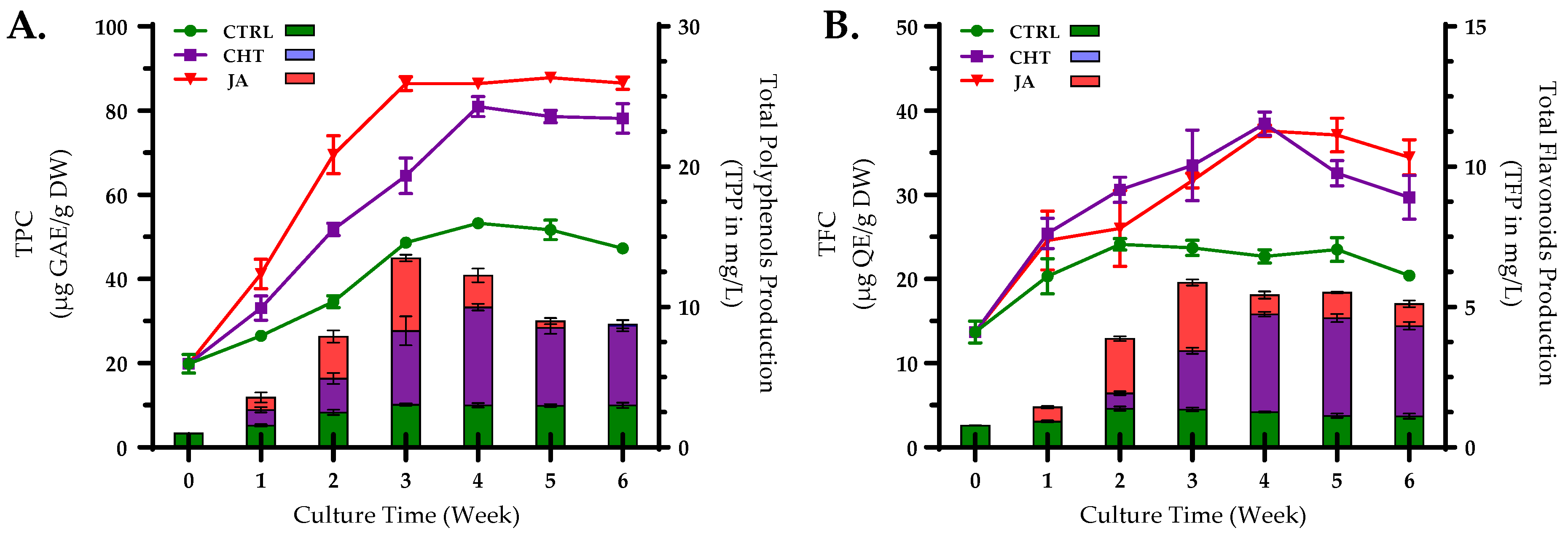
Figure 1. Accumulation of total polyphenols (A) and flavonoids (B) under elicitation with chitosan (CHT) at a concentration of 75 mg/mL and jasmonic acid (JA) at 50 µM in suspension culture of P. graveolens on distinct growth periods. Values represent means ± standard deviations from triplicates. Bars represent total polyphenols production (A) and total flavonoid production (B); lines represent the total polyphenols content (A) and total flavonoid content (B).
Horticulturae 10 00521 g001
Figure 2. HPLC-DAD chromatograms at 285 nm of the control (A,A’), the elicited cultures with CHT4 (B,B’), and JA3 (C,C’).
Horticulturae 10 00521 g002
Figure 3. Chemical structures of the identified compounds in P. graveolens cell suspension cultures through HPLC-DAD method.
Horticulturae 10 00521 g003
Figure 4. Venn diagram illustrating the phenolic composition of cultures treated with elicitors (CHT4 for CHT-treated, and JA3 for JA-treated cultures) (A). Bar graph depicting the count of DAMs that are upregulated and downregulated across pairwise comparisons (B).
Horticulturae 10 00521 g004
Figure 5. Peroxidase (POD; (A)) and superoxide dismutase (SOD; (B)) activities in suspension cultures of P. graveolens treated with different concentrations of chitosan (CHT) and acid (JA) in comparison with the CTRL group. Data are means ± standard error; different letters indicate significant differences as determined by the Tukey test (p ≤ 0.05).
Horticulturae 10 00521 g005
Figure 6. Effect of different concentrations of chitosan (CHT) and jasmonic acid (JA) on the antioxidant activity of P. graveolens calli in suspension cultures. (A) radical scavenging activity through DPPH assay; (B) TAC assay. Data are means ± standard error; different letters indicate significant differences as determined by the Tukey test (p ≤ 0.05). * Values were compared to the DPPH (IC50 of ascorbic acid) = 37.87 ± 0.28 µg/mL.
Horticulturae 10 00521 g006
Figure 7. Tyrosinase inhibition of the methanolic extracts of cell suspension cultures under elicitation with chitosan (CHT), and jasmonic acid (JA) at different concentrations. Kojic acid was used as a positive control. IC50 values are presented as means ± standard error; different letters indicate significant differences as determined by the Tukey test (p ≤ 0.05).
Horticulturae 10 00521 g007
Figure 8. Correlogram representing the correlation among the examined parameters and their associated biological activities. Correlations with p-values greater than 0.05 are deemed insignificant. The color and size of circles are indicative of the correlation coefficients, with positive correlations depicted in various shades of blue (with dark blue indicating the strongest correlation, where r = 1), while negative correlations are shown in shades of red (ranging from light red to dark red, with dark red indicating the weakest correlation, where r = −1). DPPH: 2,2-diphenyl-1-picrylhydrazyl radical scavenging activity (IC50 values where considered), TYR: tyrosinase inhibition (IC50 values where considered), TPC: total polyphenols content, TFC: total flavonoid content, POD: peroxidase activity, TAC: total antioxidant content, SOD: superoxide dismutase activity, and BIOMASS: biomass changes.
Horticulturae 10 00521 g008
Table 1. Concentrations of chitosan and jasmonic acid used in the elicitation study.
ElicitorsConcentrationsTags
No-elicitor
(control)
-CTRL
Chitosan (mg/mL)5CHT1
25CHT2
50CHT3
75CHT4
100CHT5
Jasmonic acid (µM)5JA1
25JA2
50JA3
75JA4
100JA5
Table 2. Effect of elicitors on growth parameters, secondary metabolites, namely, polyphenols, and flavonoid accumulation.
ElicitorConcentrationsInitiation DayCallus CharacteristicsMaximum Biomass DW (g/100 mL)Optimum Values
ColorTextureTPC
(µg GAE/g DW)
TFC
(µg QE/g DW)
CTRL-3rd LGF5.73 ± 0.23 a53.24 ± 0.52 b24.10 ± 0.70 cd
CHT (mg/mL)57thLBC6.80 ± 0.19 a51.72 ± 1.23 b20.33 ± 1.06 b
257thLBC8.67 ± 0.65 b42.11 ± 1.40 a16.56 ± 2.45 a
503rdDGF13.32 ± 0.38 e62.41 ± 0.33 d28.59 ± 0.51 e
754thGBC12.34 ± 0.73 de80.94 ± 2.38 f38.45 ± 0.62 h
1003rdDGC10.04 ± 0.64 bc82.67 ± 1.02 f32.23 ± 1.21 fg
JA (µM)54thLGC12.22 ± 0.32 de54.03 ± 0.46 bc21.61 ± 0.23 bc
255th LBC11.09 ± 0.41 cd52.19 ± 0.83 b26.88 ± 0.74 de
503rdDGC15.62 ± 0.98 f86.41 ± 1.62 g37.61 ± 0.53 h
753rdDGF16.65 ± 0.70 f73.62 ± 1.04 e33.54 ± 0.48 g
1003rd DGC13.43 ± 0.94 e57.13 ± 0.95 c29.32 ± 0.81 ef
DG = dark green; LG = light green, LB = light brown; C: compact, F: friable; TPC = total phenolic contents, TFC = total flavonoid contents. Data are means ± standard error; Distinct characters in every column signify notable variances as identified by the Tukey test (p < 0.05).
Table 3. HPLC-DAD quantification of the most abundant phenolic compounds in the elicited cell cultures. Data are means ± standard error.
No.CompoundsQuantity (mg/100 g DW of Sample)
CTRLCHT (75 mg/mL)JA (50 µM)
1Gallic acid-0.91 ± 0.28-
2Catechin79.27 ± 0.681279.84 ± 0.07911.83 ± 0.05
34-Hydroxybenzoic acid208.92 ± 0.01410.41 ± 0.01292.75 ± 0.02
4Vanillic acid---
5Caffeic acid299.32 ± 0.48140.25 ± 0.12278.16 ± 0.96
6Syringic acid--60.87 ± 0.03
73-hydroxybenzoic acid--921.37 ± 0.00
8p-Coumaric acid286.93 ± 0.41-2248.33 ± 0.53
9Sinapic acid669.40 ± 1.24--
10Ferulic acid-350.75 ± 0.181562.87 ± 1.75
113-hydroxyflavone--1012.41 ± 0.00
12Salicylic acid18.28 ± 0.0757.99 ± 0.03692.10 ± 0.21
13Rutin0.53 ± 0.070.59 ± 0.64-
14Cinnamic acid-54.67 ± 0.29395.11 ± 1.45
15Quercetin 3-O-β-D-glucoside2760.51 ± 6.223328.10 ± 2.078177.46 ± 2.07
163-hydroxycinnamic acid2799.25 ± 4.836834.61 ± 6.036377.09 ± 2.41
17Kaempferol223.37 ± 0.83886.99 ± 0.411031.81 ± 1.24
18Chalcone0.79 ± 0.03557.91 ± 0.02927.35 ± 0.03
Total Phenolic acids (mg/100 g)8216.37 ± 8.066113,602.01 ± 6.44724,891.40 ± 4.30
-: trace, below LOQ.
Disclaimer/Publisher’s Note: The statements, opinions and data contained in all publications are solely those of the individual author(s) and contributor(s) and not of MDPI and/or the editor(s). MDPI and/or the editor(s) disclaim responsibility for any injury to people or property resulting from any ideas, methods, instructions or products referred to in the content.