Article

Effect of UV Stress on the Antioxidant Capacity, Photosynthetic Activity, Flavonoid and Steviol Glycoside Accumulation of Stevia rebaudiana Bertoni

by
Natalia A. Semenova
1,2,
Alina S. Ivanitskikh
1,
Nadezhda I. Uyutova
1,
Alexander A. Smirnov
1,
Yuri A. Proshkin
1,
Dmitry A. Burynin
1,
Sergey A. Kachan
1,
Alexander V. Sokolov
1,
Alexey S. Dorokhov
1 and
Narek O. Chilingaryan
1,*
1
Federal State Budgetary Scientific Institution “Federal Scientific Agroengineering Center VIM” (FSAC VIM), 109428 Moscow, Russia
2
Prokhorov General Physics Institute of the Russian Academy of Sciences, Vavilov St., 38, 119991 Moscow, Russia
*
Author to whom correspondence should be addressed.
Horticulturae 2024, 10(3), 210; https://doi.org/10.3390/horticulturae10030210
Submission received: 30 January 2024 / Revised: 20 February 2024 / Accepted: 21 February 2024 / Published: 23 February 2024
(This article belongs to the Special Issue Breeding, Cultivation, and Metabolic Regulation of Medicinal Plants)

Abstract

:
Lighting conditions are an important controller of plant growth and development, and they affect secondary metabolite synthesis. In this research, we explored the effect of additional UV irradiation of various ranges in addition to the main one at PPFD 160 µmol m−2 s−1 on the accumulation of some secondary metabolites of stevia (Stevia rebaudiana Bertoni). The fresh weight of leaves was slightly higher under additional UV-A and UV-B irradiation compared with the control variant, and the leaf surface area was significantly larger, respectively, by 23.3 and 20.7% than in the control variant, while the rate of photosynthesis did not decrease. Plants under additional UV-B and UV-C irradiation were under the greatest light stress, as evidenced by a decrease in antioxidant capacity by an average of 30% compared to the control and UV-A. The total flavonoid content was significantly higher (by 74%) under UV-B irradiation. The highest concentration of steviol glycoside was observed during budding and flowering under UV-B and UV-C irradiation (by 13.2 and 11.3%, respectively). Analysis of hyperspectral images, chlorophyll fluorescence, and vegetation indices showed light stress increasing under UV-C irradiation, which caused an increase in the relative chlorophyll content, scorches, leaf morphology changes, a CO2 absorption rate decrease, and plant growth inhibition. UV-B irradiation can be used as an optimal type of irradiation based on a set of indicators.

1. Introduction

The overconsumption of sugar can impair human health [1], which is the reason for many decades of searching for sugar substitutes, most of which turn out to be unsafe for health. Steviol glycoside (SG) sweeteners are extracted and purified from the stevia plant (Stevia rebaudiana Bertoni) of the Compositae family, native to South America, where it has been used as a sweetener for a long time. Stevia rebaudiana contains SGs such as stevioside (St), rebaudioside (Rb) A, B, C, D and E, dulcoside A and steviol biosides [2,3,4,5,6]. The flavor of rebaudioside A has been described as bitter, metallic, and astringent, with an unnaturally sweet aftertaste compared to 5% sucrose, which exhibits a high sweetness intensity [7]. However, there are ways to mask unwanted flavors, and the long-lasting sweetness of stevia is possible without disturbing the natural composition of the sweetener [8]. The sweet taste of stevia is 50–350 times sweeter than sugar and at the same time contains a minimum number of calories [1,9,10,11]. The chemical structure of SGs remains unchanged at high temperatures, so it can be used in hot cooking. In addition, stevioside can accelerate wound healing, has anti-allergenic and anti-inflammatory effects [12,13,14], and plays an important role in medical research for the treatment of diabetes, obesity, high blood pressure, tooth decay, and skin problems [12,15,16,17]. The safety of stevia extracts has been confirmed by various toxicity studies [18,19,20]. For this reason, there is growing interest in the secondary metabolites of stevia plants and their biologically active substances.
Stevia is a slow-growing herbaceous plant, and its biomass yield is low due to the slow initial growth rate of the plant, small number of leaves, and leaf surface area [21]. Plant age and harvest period influence the content of macro- and microelements, chlorophyll, and SGs in stevia plants [22]. The biosynthesis of SGs predominantly occurs in stevia leaves and then it transfers to the rest of the plant, so its highest accumulation is observed in the leaves [3]. It has been revealed that the plant developmental phase also influences SG content in stevia leaves. SG levels are highest during bud formation [23].
It is known that light conditions are important plant growth and development regulators, and they affect the synthesis of secondary metabolites [24,25,26,27]. For stevia plants, interrupting the night phase with a short pulse of light has been found to provide a simple and inexpensive method for increasing leaf biomass production with increased SG yield [28,29]. Lengthening the dark period promotes flowering and a reduction in vegetative growth, and when artificial supplementary lighting extends the daylight hours, the plant does not bloom [30]. The morphogenetic responses of S. rebaudiana to light manipulation in in vitro culture are limited [26,31,32,33,34,35,36,37,38].
The spectral characteristics of light also affect the secondary metabolism of stevia plants. Combinations of red and blue light are known to be effective in stimulating stevia plant growth and are recommended for use during the initial stages of growth. At the same time, the blue spectrum promotes the maximum accumulation of phenolic compounds [32] and causes an increase in the SG content in stevia leaves during callus culture propagation [33]. Green light was found to improve biomass yield and rebaudioside A content, but not SG [35].
The effect of UV irradiation on stevia plants arouses special interest. It is known that UV irradiation affects the photosynthetic apparatus and the synthesis of primary and secondary metabolites of different plants [39,40,41]. UV-A prevents early flowering and increases biomass and Rb A and SG content in stevia plants [35]. However, there is extremely little information about the effect of different ranges of UV irradiation on the concentration and synthesis of Rb A and SG. Therefore, the purpose of this study is to study the effects of additional UV irradiation of various ranges (UV-A 315–400 nm, UV-B 280–315 nm, UV-C 100–280 nm) on (1) stevia plant morphological parameters; (2) antioxidant activity and accumulation of SG flavonoids in the stevia plant leaves; (3) the UV light stress level and the stevia plant photosynthetic apparatus efficiency.

2. Materials and Methods

2.1. Cultivation Conditions

Clones of one stevia plant were placed in a climate chamber with various types of UV irradiation (Table 1). S. rebaudiana clones were obtained by cutting shoots at 2 nodes. The plants were planted one by one in plastic one-liter-capacity containers filled with peat substrate Agrobalt-C (CJSC Rostorfinvest, Moscow, Russia) with added quartz sand in a ratio of 2:1 to create conditions close to optimal sandy loam soil [42].
Plants were grown in a phytochamber with an automatic microclimate system, maintaining a day/night temperature range of 24/20 ± 1.0 °C [43] and a relative air humidity of 60 ± 10% for 130 days until the flowering stage. The watering and feeding of plants was carried out by a drip irrigation system once a day. Nutrient solutions were prepared based on three-component fertilizers of the GHE Flora series (General Hydroponics Europe, Paris, France) maintaining a pH of 6.5–7.5 [42]. The photoperiod during the active growing season was maintained at a light/dark ratio of 16/8, and it was gradually changed to 12/12 10 days before sampling. Day length was shortened to stimulate the flowering process and the production of target components [22,23,28]. Within a week, the plants entered the flowering phase.

2.2. Determination of Biometric Indicators

Measuring and assessing such morphological parameters as plant height, fresh weight of shoots and leaves, dry weight, and leaf area took place on the 130th day of plant cultivation. Ten plants were selected from each group for measurements. The fresh and dry weights of the above-ground parts of plants were determined using a Sartorius LA230S balance (Sartorius Laboratory Scale, Goettingen, Germany). The selected samples were dried to a constant weight in an oven at a temperature of 105 °C for 1 h to determine the dry fraction of the substance. Leaf surface area was determined using a photoplanimeter LI-3100 AREA METER (LI-COR, Inc., Nebraska, NE, USA).

2.3. Measurement of Antioxidant Activity

The total content of antioxidants was determined on day 130 according to a well-known practice [44] using the amperometric method on a certified Tsvet-Yauza-01-AA device (JSC SPA Khimavtomatika, Moscow, Russia). Since the content of antioxidants can be unevenly distributed throughout the plant, we selected leaf groups from different tiers (upper, middle, and lower). Three samples were taken for each experimental variant.

2.4. Determination of the Total Content of Flavonoids in Terms of Quercetin

The quantitative determination of flavonoids in terms of quercetin and absolute dry primary products was carried out by the spectrophotometric method according to a well-known practice [45]. A 30 mL volume of 90% alcohol containing 1% concentrated hydrochloric acid was added to 1 g of sample, and the flask (150 mL) was connected to a reflux condenser and heated in a boiling water bath for 30 min. Then, it was cooled to room temperature and filtered. The extraction was repeated 2 times, then 1 more time with 90% alcohol for 30 min. Then, 2 mL of the solution was poured into a 25 mL volumetric flask, 1 mL of a 1% solution of aluminum chloride in 95% alcohol was added, and the volume was adjusted to the mark with alcohol. After 20 min, the optical density of the solution was measured on an SPEX SSP series 705 spectrophotometer (CJSC Spectroscopic Systems, Moscow, Russia) at a wavelength of 430 nm.

2.5. Quantification of SG

The presence of steviol glycosides was determined by HPLC [37,38].
To determine the SG content, samples were taken 2 times—during the period of active vegetative growth (on the 115th day of cultivation) and during the period of budding and flowering on the 130th day of cultivation.
To assess the accumulation of the total SG content, a sample weighing 0.05 g was taken from each comparison sample of dried stevia leaves crushed and mixed in porcelain mortars, transferred to glass test tubes, and filled with 10 mL of hot water (95 °C). Then, the samples were placed in a water bath at 95 °C and heated for 15 min. Then, they were extracted and transferred into tubes and then centrifuged for 5 min at 4000 rpm to sediment the particles. After cooling the samples to 25 °C, 1 mL of the test extracts were transferred into vials and examined on an LC-20 liquid chromatograph (Shimadzu, Kyoto, Japan) using an Eclipse Plus C18, 4.6 × 150 mm, 5 um column (Agilent, Santa Clara, CA, USA). Detection was carried out under the following conditions: flow rate—0.5 mL/min, gradient—50% acetonitrile—50% ultrapure water, temperature 30 °C, analysis time—10 min, detection wave—200 nm. Calculation of SG content was performed relative to calibration with an analytical stevioside standard (Supelco, Burlington, MA, USA).

2.6. Measuring Leaf Gas Exchange and Chlorophyll Fluorescence

Leaf CO2 exchange was measured using a Li-6800 portable photosynthetic analysis system (LI-COR, Inc., Nebraska, NE, USA) with a built-in multiphase flash fluorometer Li-6800-01A. The microclimate parameters in the chamber for measuring leaf gas exchange corresponded to the parameters in the chamber where the test plants were grown: the CO2 concentration in the measuring chamber was maintained at 450 ppm, the air temperature was 26 °C and the relative humidity was 60%. CO2 assimilation by leaves was calculated from the difference in gas concentration at the inlet and outlet of the leaf chamber. Light curves of the photosynthetic reaction were obtained by reducing PPFD from 1600 µmol m−2 s−1 to zero. Measurements were taken in the upper tier on the 4th or 5th sheet; five plants of each experimental variant were used for measurements.
Plants were adapted to dark conditions for 30 min prior to the measurement, and the maximum (Fm) and minimum fluorescence (F0) were obtained by applying a saturating light pulse (50 kHz) of 10,000 μmol m−2 s−1 PPF. The maximum PSII quantum yield (Fv/Fm) was calculated using the formula (Fm − F0)/Fm, non-photochemical quenching (NPQ) was calculated as (Fm/F′m) − 1, Y(NPQ) is the fraction of energy dissipated in the form of heat via the regulated non-photochemical quenching mechanism calculated as Fs/F′m − Fs/Fm, and the effective PSII quantum yield (Y(II)) was calculated as (F′m − Fs)/F′m.

2.7. Measuring Vegetation Indices

The spectral reflectance characteristics of leaves were measured on day 130 of cultivation using a portable hyperspectral camera Specim IQ (Specim, Spectral Imaging Ltd., Finland). Reflectance spectra were obtained for each individual image pixel in the range of 400–1000 nm (spectral resolution 7 nm, 204 spectral channels, spatial sampling 512 pixels). From these, we calculated the photochemical reflectance index (PRI) (often used as an indicator of stress). Four halogen lamps were used as the light source for the Specim IQ. They were installed symmetrically relative to the camera position. Analysis of hyperspectral images was performed using the Envi 5.2 application to calculate the PRI vegetation index. The PRI index was calculated using Formula (1):
PRI = (R531 − R570)/(R531 + R570)
The index of relative chlorophyll content in leaves was determined using a chlorophyll content meter CL-01 (Hansatech Instruments Ltd., Pentney, UK). Measurements were carried out on five leaves of the upper tiers. A plant leaf was clamped in the instrument’s arm, and the relative content of chlorophyll in the leaf was measured by measuring the absorption of light waves in two ranges—620 and 940 nm. Each sheet was measured at three points.

2.8. Data Analysis

All experiments were carried out in triplicate with statistical processing of measurement results and construction of economic graphs in MS Excel 2010. A one-way analysis of variance (ANOVA) with an innovative difference p < 0.05 was used to determine significant results.

3. Results and Discussion

3.1. The Influence of Additional UV Irradiation on the Morphological Parameters of the Stevia Plant

Biometric indicators of plants on the 130th day of cultivation revealed the advantage of UV-A and UV-B irradiation treatments. The height and fresh weight of the stems of stevia plants were slightly greater in these variants (Table 2).
The fresh weight of leaves grown under additional UV-A and UV-B irradiation was slightly higher compared with the control variant, and the leaf surface area was significantly larger, respectively, by 23.3 and 20.7% than in the control variant. Increased light stress under UV-C irradiation caused plant scorches, curling and reduction of leaf blades, and growth inhibition (Figure 1), while the percentage of dry weight to fresh weight was 37.2% higher. We obtained similar results in previous studies that examined the effects of three different sources of UV irradiation from LEDs and discharge lamps in the UV-A, UV-B, and UV-C ranges on the growth of sweet basil (Ocimum basilicum L.). Plants grown under additional UV-A and UV-B from mercury lamps were higher than the control by 90 and 53%, respectively. The weight of fresh leaves of plants under UV-A irradiation was 2.4 times higher than the fresh weight of control plants. Leaf area increased by 40% and 20%, respectively [46].

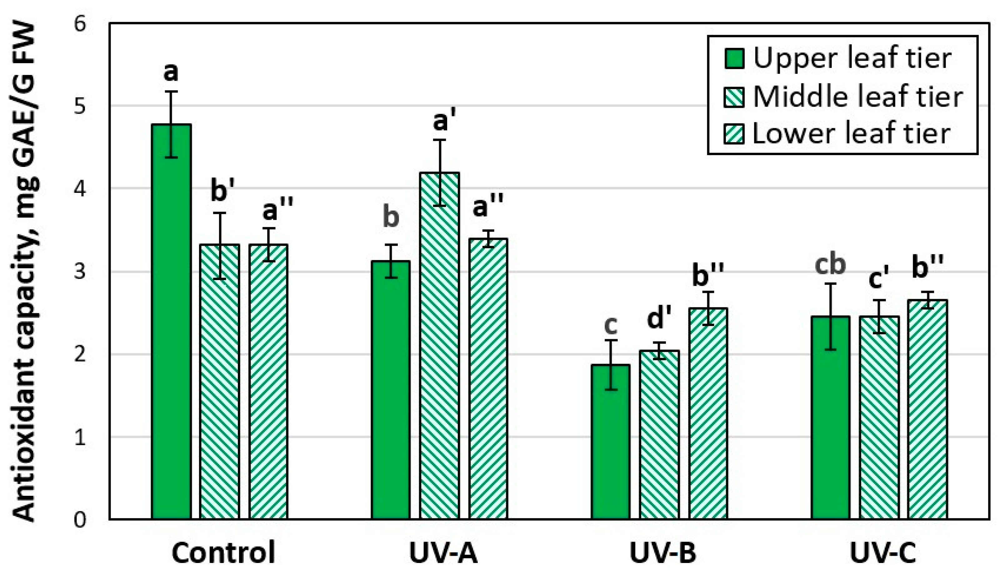
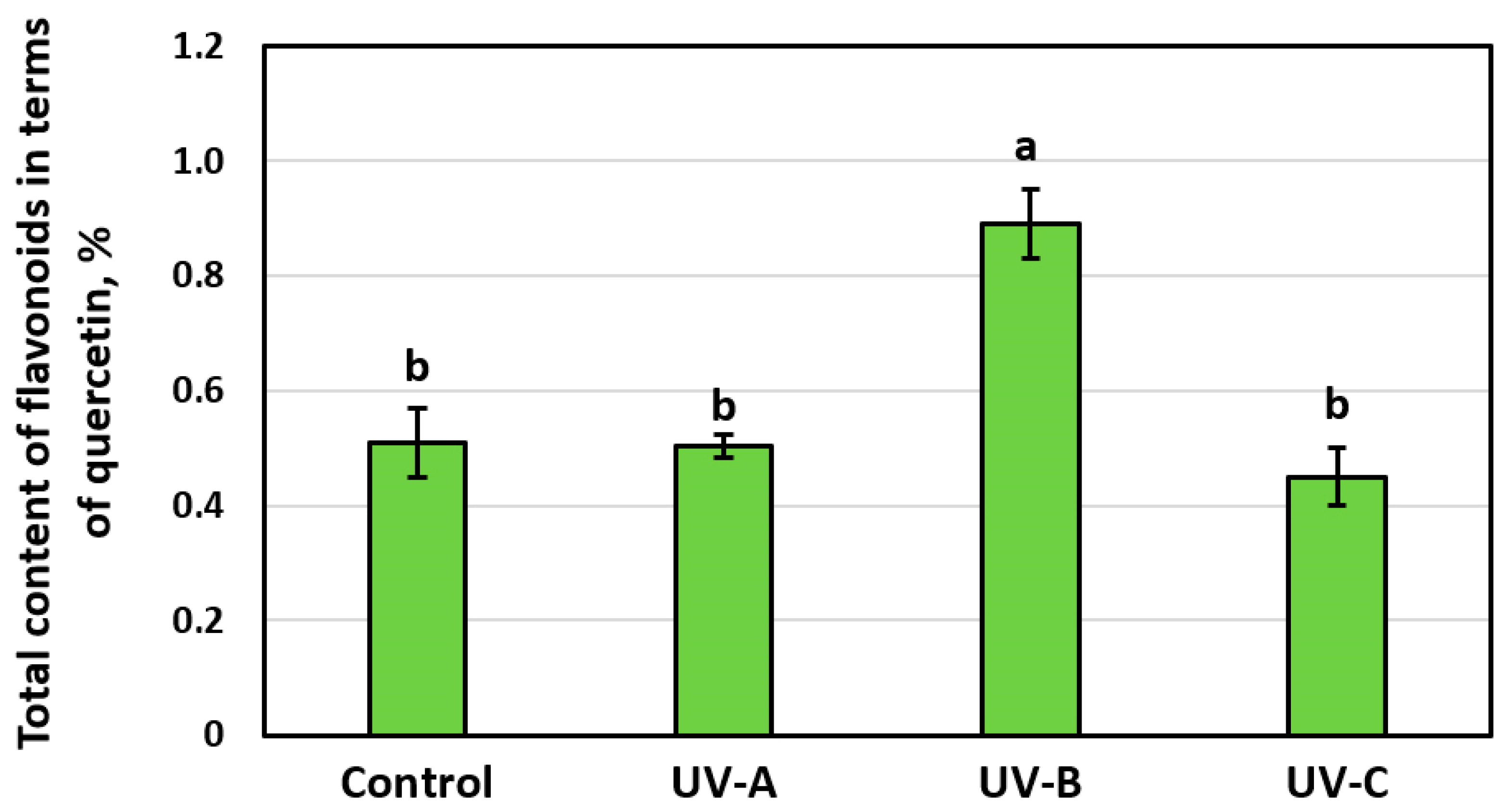
3.2. Effect of Additional UV Irradiation on Antioxidant Activity and Accumulation of SG Flavonoids in the Leaves of the Stevia Plant

A significant difference was found among experimental variants while analyzing the antioxidant capacity of leaves (Figure 2). It is known that ultraviolet (UV-B) irradiation stimulates the accumulation of antioxidants as an important abiotic stress factor. It was demonstrated in pakchoi (Brassica rapa L.) plants. In this study, irradiation with 2 µmol m−2 s−1 UV-B for 24 h or 4 µmol m−2 s−1 UV-B for 4 h increased 1,1-diphenyl-2-picrylhydrazyl and the overall reducing capacity as a result of increasing the accumulation of total polyphenols and flavonoids, without affecting the fresh weight of plants [47]. Stress factors induce the active formation of free radicals and reactive oxygen species. Then, accordingly, the greater the stress, the more active oxidation processes occur in the plant. Moreover, the processes of adaptation to unfavorable environmental conditions occur with the active participation of the antioxidant system, which controls the levels of active forms of oxygen in cells [42]. Thus, the greater the stress, the lower the antioxidant capacity of plants. Leaves of the upper and middle tiers of plants were under the greatest stress under additional UV-B and UV-C irradiation. However, plants under UV-B irradiation, unlike UV-C, did not show external signs of stress (growth inhibition, scorches). Stevia plants irradiated with additional UV-A radiation had a higher antioxidant capacity than in the control on the middle and lower tiers of leaves. The upper tier was subjected to greater stress. The average antioxidant capacity was similar in these treatments.
When analyzing the content of flavonoids, which also perform the antioxidant function of photoprotection of DNA from damage caused by UV radiation [48], it was found that a significant increase in their quantity (by 74%) compared to other experimental variants was observed only under UV-B treatment (Figure 3). Flavonoids regulate auxin catabolism, influencing key stages of plant cell growth and differentiation [49]. In the experimental variants with UV-A and UV-B treatments, the plants looked more branched, which may be due to some inhibition of auxin movement due to the accumulation of flavonoids in response to UV stress. However, judging by the concentration of flavonoids and the antioxidant activity of plants, it remains unclear whether UV-A irradiation was stressful for stevia and such a reaction is species-specific or the intensity of irradiation was not sufficient to cause specific reactions in increasing flavonoids [50]. It is also known that UV-B irradiation increases the concentration of flavonoids during the post-harvest period. It was proven in the example of peach varieties [51]. However, the effectiveness of this treatment in increasing the concentration of beneficial phenolic compounds depended on the genotype. Irradiation with a UV-B LED lamp with an intensity of 3.6 W/m2 increased the total amount of flavonoids in the collected sweet basil plants; in particular, it increased the content of quercetin, catechin, kaempferol, rutin, and luteolin compared to the irradiation with an intensity of 2.40 and 4.80 W/m2. The duration of UV-B irradiation significantly affected the concentrations of these secondary metabolites. Irradiation for 8 h resulted in an increase in the content of phenolic compounds and individual flavonoids and phenolic acids than at other durations (4, 8, and 10 h). Irradiation for 10 h significantly reduced the formation of secondary metabolites in sweet basil leaves [52]. Analysis of the main components of Citrus sinensis (L.) Osbeck showed a clear correlation between the content of flavonoid glycosides and UV-C treatment [53]. It would be logical to assume that the concentration of flavonoids under UV-C irradiation should be maximum since the stress level is higher. We observed similar effects in red-leaved basil plants. We found that UV irradiation significantly changed the secondary metabolism of sweet basil. UV-C had the greatest effect on the content of anthocyanins; they increased by 50%, while under UV-A and UV-B they increased by only 27% and 0%. Any addition of UV light did not affect the total essential oil content but changed the composition of the essential oil. Therefore, we concluded that the use of UV irradiation (other than UV-C) for basil cultivation may be justified to stimulate basil growth and optimize essential oil accumulation [46]. However, in the current experiment, UV-C irradiation did not have a significant effect on the total content of flavonoids in stevia plants.
It should be noted that most published studies of flavonoid content under UV irradiation were carried out on green and fruit crops during the post-harvest period, or additional temporary UV exposure was used to stimulate their synthesis. However, in our experiments, we used long-term low-intensity UV irradiation during the period of active growth and flowering, which allowed us to only indirectly compare the results obtained.
As for the target components (steviosides and rebaudiosides, determined in total), we confirmed that the developmental phase affects the SG content [26] and their levels are maximum during budding, but only in the case of using lighting in the UV-B and UV-C ranges (Figure 4). In the control group of plants (without additional UV irradiation), the SG content in the budding and flowering phase was even lower by 13.7%, as with UV-A irradiation. No statistical difference was observed between growth phases.
We observed the highest concentration of SGs during the budding and flowering periods under UV-B and UV-C irradiation. The accumulation of SGs was 13.2 and 11.3% higher, respectively. In view of the weaker vegetative growth and depressed state of the plant under UV-C irradiation, UV-B irradiation is the optimal irradiation variant based on a set of indicators.
Steviol glycosides, like all secondary metabolites, serve to protect plants from stressful environmental conditions. SG content was highest with minimal antioxidant capacity at maximum oxidative stress under UV-B irradiation.

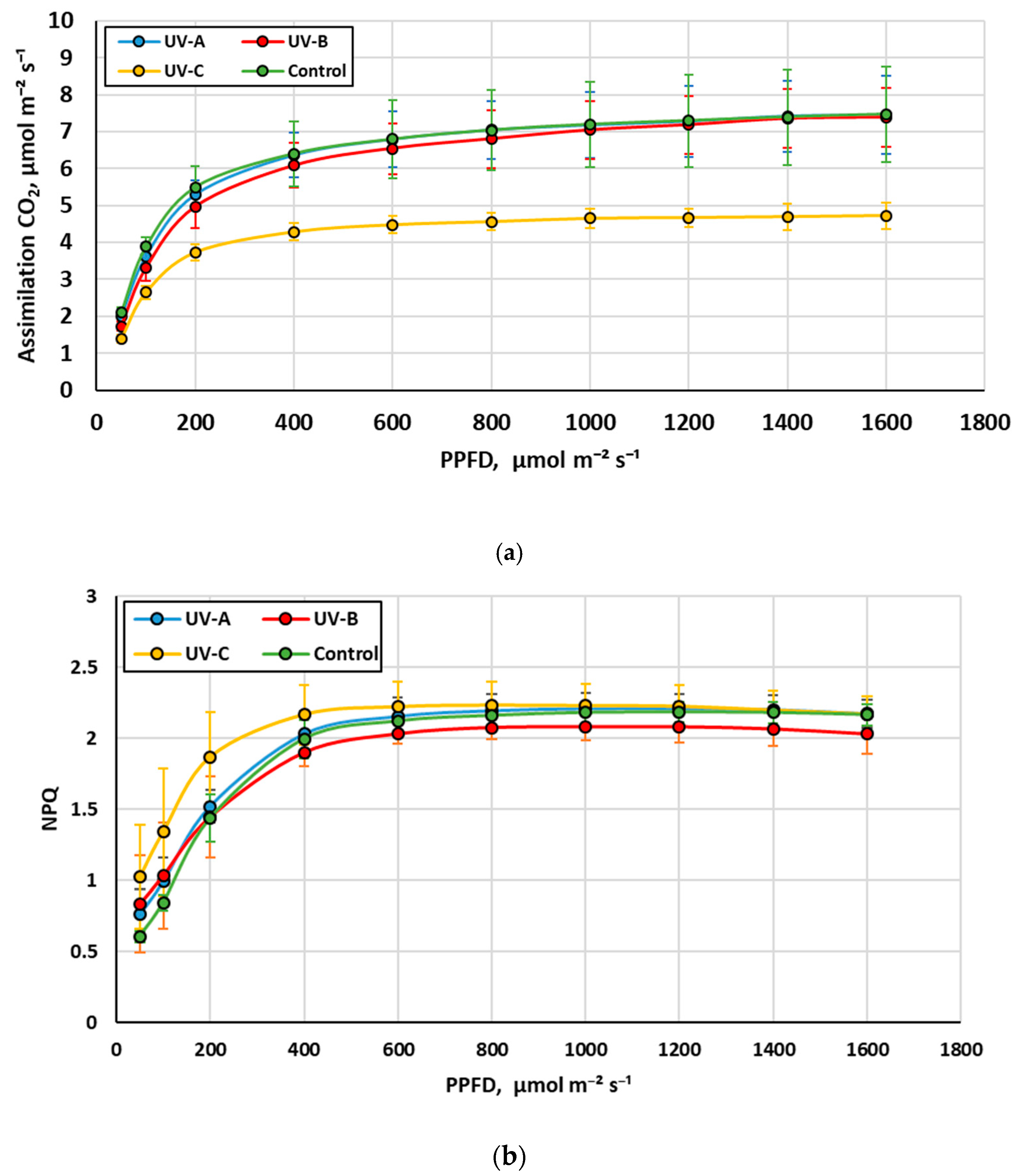
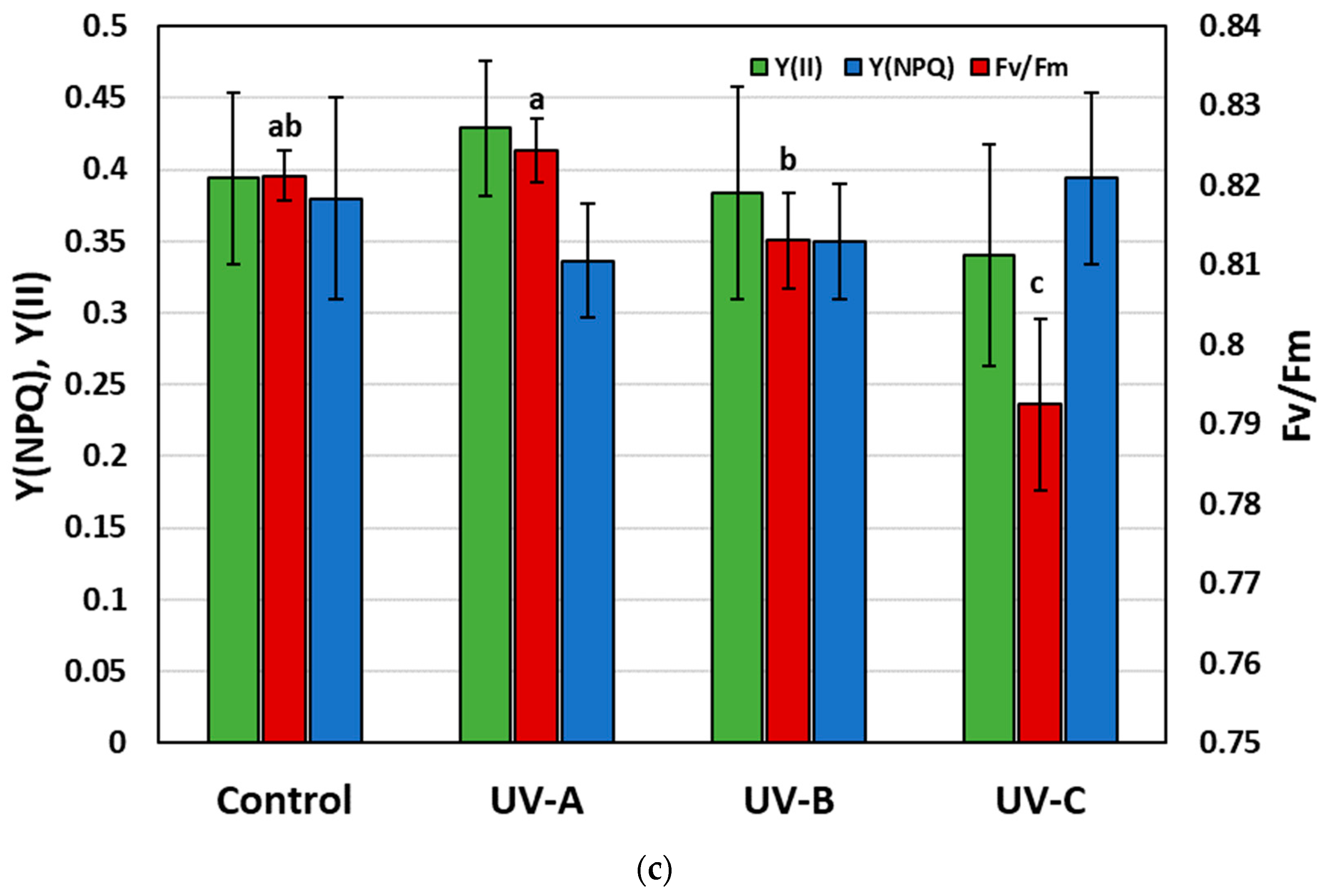
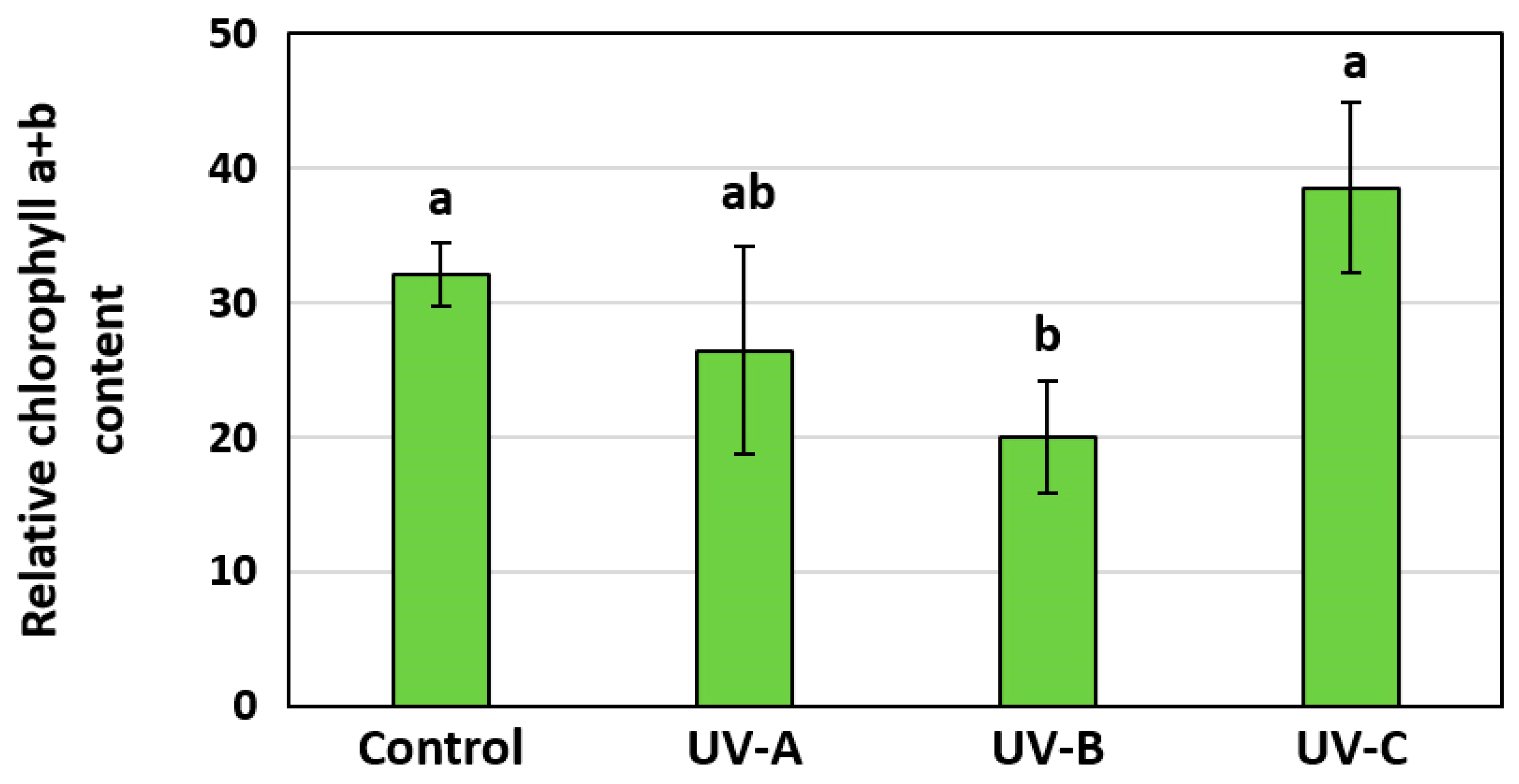
3.3. The Influence of Additional UV Irradiation on the Stress Level, Net Assimilation CO2, and Chlorophyll Fluorescence of Stevia Plants

The photochemical reflectance index (PRI) makes it possible to determine plant stress conditions associated with the photosynthetic system [54]. The PRI is sensitive to changes in the xanthophyll cycle, which protects the plant from photodamage [55]. The analysis of vegetation indices was carried out using the Envi 5.2 application. The average values of vegetation indices were obtained for the leaves of stevia plants on the 130th day of cultivation. The obtained results are presented in Figure 5.
Ultraviolet C-treated hyperspectral images of plants clearly show areas of leaves with structural changes (Figure 5a) caused by severe damage to the photosynthetic apparatus of the leaves and drying out (in these areas, the PRI index values were −0.2–0). The presented graph of average values of the PRI index (Figure 5b) shows that the greatest stress caused by UV irradiation is observed in the sample with UV-C treatment, while plants under UV-A and UV-B irradiation are subject to less stress; in the control, the stress is the least, which consistent with previous studies on Ligustrum vulgare L. and Phillyrea latifolia L. [56].
Light reaction curves (Figure 6) show that the maximum rate of photosynthesis occurred in stevia plants in the UV-A, UV-B, and control variants. The lowest rate of leaf CO2 absorption was observed in the UV-C irradiation variant, which can be associated with damage to the photosynthetic apparatus of the plant.
The results of measuring the relative chlorophyll content index are shown in Figure 7. The highest relative chlorophyll content was found in leaves treated with UV-C and in the control. High chlorophyll content under UV-C treatment was associated with changes in leaf morphology; UV-C irradiation led to a decrease in leaf area and an increase in thickness [57].
Gas exchange parameters and chlorophyll fluorescence are the most accurate indicators for UV irradiation impact assessment on the photosynthetic apparatus, which, including account data on the relative concentration of chlorophyll, allows us to assess the overall CO2 assimilation of the plant as a whole. Based on the obtained results, we can state the greatest net assimilation in the stevia plant is observed with additional treatment with UV-B irradiation, then in the sample under UV-A and control; the worst results are observed in the sample under UV-C irradiation. UV-C stress leads to a decrease in the maximum quantum yield of PSII (Fv/Fm) (Figure 6b) due to damage to the PSII active site, which ultimately leads to photoinhibition and reduced biomass [39].
Our studies have shown that PRI can be considered a good indicator of UV stress. NPQ is also the most rapid and flexible response to light stress among known photoprotective mechanisms. Y(NPQ) reflects the capacity for excess energy dissipation at the PSII action center as heat, which is usually considered a form of photoprotection. But in our case, NPQ and Y(NPQ) did not show significant changes (Figure 6b,c), although there was a tendency for the NPQ value to increase under UV-C irradiation. This may be due to the fact that in the case of photoinhibition, NPQ does not always reflect photoprotection [58]. Thus, compared with the control, leaves exposed to strong UV radiation showed a significant decrease in maximum fluorescence intensity (Fm) without changes in initial fluorescence (Fo) (data not shown).
Stevia plants exposed to UV-C treatment had a lower Fv/Fm value than UV-A, UV-B, and control plants, in which the value was maintained above 0.8. This indicates a decrease in the potential photosynthetic capacity of PSII, which also corresponded to a decrease in CO2 assimilation (Figure 6a) and a slight decrease in the Y(II) value (Figure 6c). Despite the high content of chlorophylls in the UV-C variant (at the level of the control variant), due to damage to the photosynthetic apparatus, the photosynthetic activity of plants decreased. UV irradiation is a strong stress factor, which has a significant impact on the efficiency of the plant’s photosynthetic apparatus. UV-C irradiation causes a large production of ROS, which leads to photo-oxidation, resulting in damage to the photosynthetic apparatus of the plant, specifically PSII [59], the state of which has been used in a large number of works to assess the permissible dose of UV irradiation of plants [60,61,62,63,64].
Chlorophyll degradation is the most common form of damage that can be observed after exposure to UV radiation, which can cause inhibition of photosynthesis. In our research, a decrease in chlorophyll content was observed only in the variant with UV-B irradiation, but this did not lead to a change in CO2 assimilation and PRI in this variant. Thus, PRI remote sensing can be considered a good tool for selecting UV treatment regimes.

4. Conclusions

When growing stevia in closed artificial agroecosystems, it is advisable to use additional UV-A irradiation for maximum antioxidant effect or UV-B irradiation for maximum accumulation of the target SG component. At the same time, the plants developed a more powerful shoot system and formed a larger leaf surface area by 23.3 and 20.7%, respectively. In general, the plants exhibited high net assimilation.
UV-A irradiation stimulates plant growth without causing significant changes in fresh and dry weights, while oxidative stress is not biochemically expressed, leaves do not lose antioxidant activity, and the level of steviol glycosides does not change according to growth phases, which allows stevia to be cultivated without changing phases (changes in nutrition and day length) as opposed to UV-B and UV-C irradiation. In this case, the leaves of the middle tier are the most useful to use. Lighting with the addition of UV-B spectrum is optimal for the maximum accumulation of SG (13.2% more than in the control variant before flowering) for cultivating stevia plants before the period of budding and flowering. The study of additional UV-C irradiation is promising because it increases the content of the target component SG by 11.3% while the price of UV-C LEDs is several times less than UV-B LEDs, and the negative effect on plant growth can be compensated by the frequency of treatments or application scanning mode. If the dose is incorrectly selected, the plant’s photosynthetic apparatus, specifically PSII, is degraded, which leads to low net assimilation in the plant as a whole.
The method presented in this study for assessing the effect of UV irradiation on the concentration of SG in the leaves of stevia plants allows us to evaluate the effectiveness of different ranges of UV irradiation, which makes it possible to improve stevia growing technologies and obtain higher-quality yields in totally controlled environment agriculture. Future studies will focus on the effects of different doses of UV irradiation on leaf SG concentrations and plant physiology.

Author Contributions

Conceptualization and methodology, N.A.S. and N.O.C.; validation, N.A.S. and A.A.S.; formal analysis, N.I.U. and A.V.S.; investigation and resources, A.S.I., D.A.B. and S.A.K.; writing—original draft preparation, A.A.S. and Y.A.P.; writing—review and editing, N.A.S., A.S.D., N.O.C.; visualization, Y.A.P.; supervision, A.S.D.; project administration, A.S.D. All authors have read and agreed to the published version of the manuscript.

Funding

This research was carried out at the expense of the state task of the Ministry of Science and Higher Education of the Russian Federation (FGUN-2022-0008).

Data Availability Statement

Data are contained within the article.

Conflicts of Interest

The authors declare no conflicts of interest. The funders had no role in the design of the study, in the collection, analyses, or interpretation of the data, in the writing of the manuscript, or in the decision to publish the results.

References

  1. Saraiva, A.; Carrascosa, C.; Raheem, D.; Ramos, F.; Raposo, A. Natural sweeteners: The relevance of food naturalness for consumers, food security aspects, sustainability and health impacts. Int. J. Environ. Res. Public Health 2020, 17, 6285. [Google Scholar] [CrossRef]
  2. Koyama, E.; Kitazawa, K.; Ohori, Y.; Izawa, O.; Kakegawa, K.; Fujino, A.; Ui, M. In vitro metabolism of the glycosidic sweeteners, stevia mixture and enzymatically modified stevia in human intestinal microflora. Food Chem. Toxicol 2003, 41, 359–374. [Google Scholar] [CrossRef]
  3. Bondarev, N.I.; Sukhanova, M.A.; Reshetnyak, O.V.; Nosov, A.M. Steviol glycoside content in different organs of Stevia rebaudiana and its dynamics during ontogeny. Biol. Plant 2003, 47, 261–264. [Google Scholar] [CrossRef]
  4. Muanda, F.N.; Soulimani, R.; Diop, B.; Dicko, A. Study on chemical composition and biological activities of essential oil and extracts from Stevia rebaudiana Bertoni leaves. LWT 2011, 44, 1865–1872. [Google Scholar] [CrossRef]
  5. Santini, A.; Ferrara, L.; Naviglio, D.; Aragon, A.; Ritieni, A. Stevia: Non-caloric natural sweetener. Prog. Nutr. 2008, 10, 3–10. [Google Scholar]
  6. Sharma, S.; Walia, S.; Singh, B.; Kumar, R. Comprehensive review on agro technologies of low-calorie natural sweetener stevia (Stevia rebaudiana Bertoni): A boon to diabetic patients. J. Sci. Food Agric. 2016, 96, 1867–1879. [Google Scholar] [CrossRef] [PubMed]
  7. Obst, K.; Ley, J.P.; Reichelt, K.V.; Krammer, G.E.; Engel, K.H. Sensory. Characterisation of Stevia Extracts. Stevia: Science, no fiction. In Proceedings of the 4th Stevia Symposium of the European-Stevia-Association—EUSTAS Symposium, Leuven, Belgium, 29–30 June 2010; pp. 121–134. [Google Scholar]
  8. Scholten, C.; Haji, F. Methods to influence the taste and sweetness profile of Stevia. Sensory Characterisation of Stevia Extracts. Stevia: Science, no fiction. In Proceedings of the 4th Stevia Symposium of the European-Stevia-Association—EUSTAS Symposium, Leuven, Belgium, 29–30 June 2010; pp. 111–120. [Google Scholar]
  9. Bergman, M.E.; Davis, B.; Phillips, M.A. Medically useful plant terpenoids: Biosynthesis, occurrence, and mechanism of action. Molecules 2019, 24, 3961. [Google Scholar] [CrossRef]
  10. Chen, T.H.; Chen, S.C.; Chan, P.; Chu, Y.; Yang, H.Y.; Cheng, J.T. Mechanism of the hypoglycemic effect of stevioside, a glycoside of Stevia rebaudiana. Planta Med. 2005, 71, 108–113. [Google Scholar] [CrossRef]
  11. Samuel, P.; Ayoob, K.; Magnuson, B.; Wölwer-Rieck, U.; Jeppesen, P.; Rogers, P.; Rowland, I.; Mathews, R. Stevia Leaf to Stevia Sweetener: Exploring Its Science, Benefits, and Future Potential. J. Nutr. 2018, 148, 1186S–1205S. [Google Scholar] [CrossRef]
  12. Jeppesen, P.; Gregersen, S.; Alstrup, K.; Hermansen, K. Stevioside induces antihyperglycaemic, insulinotropic and glucagonostatic effects in vivo: Studies in the diabetic Goto-Kakizaki (GK) rats. Phytomedicine 2002, 9, 9–14. [Google Scholar] [CrossRef]
  13. Starratt, A.N.; Kirby, C.W.; Pocs, R.; Brandle, J.E.; Rebaudioside, F.A. Diterpene glycoside from Stevia rebaudiana. Phytochemistry 2002, 59, 367–370. [Google Scholar] [CrossRef] [PubMed]
  14. Tadhani, M.B.; Patel, V.H.; Subhash, R. In vitro antioxidant activities of Stevia rebaudiana leaves and callus. J. Food Compos. Anal 2007, 20, 323–329. [Google Scholar] [CrossRef]
  15. Chan, P.; Tomlinson, B.; Chen, Y.J.; Liu, J.C.; Hsieh, M.H.; Cheng, J.T. A double-blind placebo-controlled study of the effectiveness and tolerability of oral stevioside in human hypertension. Br. J. Clin. Pharmacol. 2000, 50, 215–220. [Google Scholar] [CrossRef]
  16. Yasukawa, K.; Kitanaka, S.; Seo, S. Inhibitory effect of stevioside on tumor promotion by 12-O-tetradecanoylphorbol-13-acetate in two-stage carcinogenesis in mouse skin. Biol. Pharm. Bull. 2002, 25, 1488–1490. [Google Scholar] [CrossRef]
  17. Papaefthimiou, M.; Kontou, P.I.; Bagos, P.G.; Braliou, G.G. Antioxidant Activity of Leaf Extracts from Stevia rebaudiana Bertoni Exerts Attenuating Effect on Diseased Experimental Rats: A Systematic Review and Meta-Analysis. Nutrients 2023, 15, 3325. [Google Scholar] [CrossRef]
  18. Gupta, E.; Purwar, S.; Sundaram, S.; Rai, G. Nutritional and therapeutic values of Stevia rebaudiana: A review. J. Med. Plants Res. 2013, 7, 3343–3353. [Google Scholar]
  19. Panpatil, V.; Polasa, K. Assessment of stevia (Stevia rebaudiana)-natural sweetener: A review. J. Food Sci. Technol. 2008, 45, 467–473. [Google Scholar]
  20. Talevi, A. Potential medicinal effects and applications of stevia constituents. Phytochem. Rev. 2022, 21, 161–178. [Google Scholar] [CrossRef]
  21. Khandaker, M.M.; Liyana, N.; Majrashi, A.; Dalorima, T.; Alias, N.; Mat, N. Effects of kinetin on the morpho-physiological and biochemical characteristics of stevia (Stevia rebaudiana). Bulg. J. Agric. Sci. 2018, 24, 638–647. [Google Scholar]
  22. Ucar, E.; Ozyigit, Y.; Eruygur, N.; Güven, D.; Yur, S.; Turgut, K.; Özek, T.; Kütük, N. The Effect of the Plant Age and Growth Period on the Nutritional Substance, Chlorophyll and Steviol Glycoside Rates in Stevia (Stevia Rebaudiana Bertoni) Leaves. Commun. Soil Sci. Plant Anal. 2018, 49, 291–302. [Google Scholar] [CrossRef]
  23. Samsulrizal, N.H.; Khadzran, K.S.; Shaarani, S.H.; Noh, A.L.; Sundram, T.C.; Naim, M.A.; Zainuddin, Z. De novo transcriptome dataset of Stevia rebaudiana accession MS007. Data Brief 2019, 28, 1–3. [Google Scholar] [CrossRef]
  24. Tarakanov, I.G. Light control of growth and development in vegetable plants with various life strategies. Acta Hortic. 2006, 711, 315–321. [Google Scholar] [CrossRef]
  25. Tarakanov, I.; Yakovleva, O.; Konovalova, I.; Paliutina, G.; Anisimov, A. Light-emitting diodes: On the way to combinatorial lighting technologies for basic research and crop production. Acta Hortic. 2012, 956, 171–178. [Google Scholar] [CrossRef]
  26. Nakonechnaya, O.V.; Gafitskaya, I.V.; Burkovskaya, E.V.; Khrolenko, Y.A.; Grishchenko, O.V.; Zhuravlev, Y.N.; Sussotin, E.P.; Kulchin, Y.N. Effect of light intensity on the morphogenesis of Stevia rebaudiana under in vitro conditions. Russ. J. Plant Physiol. 2019, 66, 656–663. [Google Scholar] [CrossRef]
  27. Muthu, S.; Schuurmans, F.J.; Pashley, M.D. Red, green, and blue LEDs for white light illumination. IEEE J. Sel. Top. Quantum. Electron. 2002, 8, 333–338. [Google Scholar] [CrossRef]
  28. Rivera-Avilez, J.A.; Jarma-Orozco, A.; Pompelli, M.F. Stevia rebaudiana Bertoni: The Interaction of Night Interruption on Gas Exchange, Flowering Delay, and Steviol Glycosides Synthesis. Horticulturae 2021, 7, 543. [Google Scholar] [CrossRef]
  29. Stijn, C.; Stefaan, W.; Geuns, J. Stimulation of steviol glycoside accumulation in Stevia rebaudiana by red LED light. J. Plant Physiol. 2012, 169, 749–752. [Google Scholar]
  30. Abdulameer, D.; Osman, M.; Sulaiman, Z.; Yusop, M.; Abdullah, S.; Azizi, P.; Muttaleb, Q. Assessment of Stevia rebaudiana Bertoni Genotypes via Morpho-Agronomic Traits under Two Light Conditions. Am. J. Plant Sci. 2018, 9, 1403–1413. [Google Scholar] [CrossRef]
  31. Bondarev, N.I.; Reshetnyak, O.V.; Nosov, A.M. Influence of photoperiod and irradiation intensity on the development of Stevia rebaudiana shoots in vitro and synthesis of steviol glycosides. Izv. TSKhA 2008, 4, 102–107. [Google Scholar]
  32. Ahmad, N.; Rab, A.; Ahmad, N. Light-induced biochemical variations in secondary metabolite production and antioxidant activity in callus cultures of Stevia rebaudiana (Bert). J. Photochem. Photobiol. 2016, 154, 6–51. [Google Scholar] [CrossRef]
  33. Yoneda, Y.; Nakashima, H.; Miyasaka, J.; Ohdoi, K.; Shimizu, H.J.P. Impact of blue, red, and far-red light treatments on gene 1093 expression and steviol glycoside accumulation in Stevia rebaudiana. Phytochemistry 2017, 137, 57–65. [Google Scholar] [CrossRef]
  34. Shevchenko, Y.; Wendt, A.; Smetanska, I. Sprout culture of Stevia rebaudiana Bertoni. In Proceedings of the 4th Stevia Symposium of the European-Stevia-Association—EUSTAS Symposium. Stevia: Science, Leuven, Belgium, 29–30 June 2010; pp. 5–26. [Google Scholar]
  35. Rengasamy, N.; Othman, R.Y.; Che, H.S.; Harikrishna, J.A. Beyond the PAR spectra: Impact of light quality on the germination, flowering, and metabolite content of Stevia rebaudiana (Bertoni). J. Sci. Food Agric. 2021, 102, 299–311. [Google Scholar] [CrossRef]
  36. Ceunen, S.; Geuns, J.M.J.P. Spatio-temporal variation of the diterpene steviol in Stevia rebaudiana grown under different photoperiods. Phytochemistry 2013, 89, 32–38. [Google Scholar] [CrossRef] [PubMed]
  37. Kovylyaeva, G.I.; Bakaleinik, G.A.; Strobykina, I.Y.; Gubskaya, V.I.; Sharipova, R.R.; Al’Fonsov, V.A.; Kataev, V.E.; Tolstikov A., G. Glycosides from Stevia rebaudiana. Chem. Nat. Compd. 2007, 43, 81–85. [Google Scholar] [CrossRef]
  38. Denev, P.N.; Uchkunov, I.I.; Uchkunov, V.I.; Kratchanova, M.G. Content of steviol glycosides in stevia (Stevia Rebaudiana B.) genotypes cultivated in Bulgaria. Bulg. Chem. Commun. 2017, 6, 121–125. [Google Scholar]
  39. Lee, M.-J.; Son, J.E.; Oh, M.-M. Growth and phenolic compounds of Lactuca sativa L. grown in a closed-type plant production system with UV-A, -B, or -C lamp. J. Sci. Food Agric. 2014, 94, 197–204. [Google Scholar] [CrossRef] [PubMed]
  40. Ullah, M.A.; Tungmunnithum, D.; Garros, L.; Drouet, S.; Hano, C.; Abbasi, B.H. Effect of Ultraviolet-C Radiation and Melatonin Stress on Biosynthesis of Antioxidant and Antidiabetic Metabolites Produced in In Vitro Callus Cultures of Lepidium sativum L. Int. J. Mol. Sci. 2019, 20, 1787. [Google Scholar] [CrossRef] [PubMed]
  41. Nazir, M.; Asad Ullah, M.; Mumtaz, S.; Siddiquah, A.; Shah, M.; Drouet, S.; Hano, C.; Abbasi, B.H. Interactive Effect of Melatonin and UV-C on Phenylpropanoid Metabolite Production and Antioxidant Potential in Callus Cultures of Purple Basil (Ocimum basilicum L. var purpurascens). Molecules 2020, 25, 1072. [Google Scholar] [CrossRef] [PubMed]
  42. Singh, S.; Rao, G.J.S.T. Stevia: The herbal sugar of 21st century. Sugar Tech 2005, 7, 17–24. [Google Scholar] [CrossRef]
  43. Huxley, A.; Griffiths, M.; Levy, M. The New RHS Dictionary of Gardening; Royal Horticultural Society: London, UK, 1992; pp. 315–318. [Google Scholar]
  44. Gins, M.S.; Gins, W.K.; Baykov, A.A.; Kononkov, P.F.; Romanova, E.V. Methodology for analyzing the total content of antioxidants in leaf and leafy vegetable crops. In Educational Manual; RUDN Publ.: Moscow, Russia, 2013; p. 40. [Google Scholar]
  45. Fedoseeva, G.M.; Mirovich, V.M.; Goryachkina, E.G.; Perelomova, M.V. Phytochemical analysis of plant raw materials containing flavonoids. In Methodological Manual on Pharmacognosy; ISMU Publ.: Irkutsk, Russia, 2009; p. 67. [Google Scholar]
  46. Semenova, N.A.; Smirnov, A.A.; Ivanitskikh, A.S.; Izmailov, A.Y.; Dorokhov, A.S.; Proshkin, Y.A.; Yanykin, D.V.; Sarimov, R.R.; Gudkov, S.V.; Chilingaryan, N.O. Impact of UV radiation on the biosynthesis of pigments and essential oils of sweet basil (Ocimum basilicum L.). Appl. Sci. 2022, 12, 7190. [Google Scholar] [CrossRef]
  47. Hao, J.; Lou, P.; Han, Y.; Zheng, L.; Lu, J.; Chen, Z.; Ni, J.; Yang, Y.; Xu, M. Ultraviolet-B Irradiation Increases Antioxidant Capacity of Pakchoi (Brassica rapa L.) by Inducing Flavonoid Biosynthesis. Plants 2022, 11, 766. [Google Scholar] [CrossRef]
  48. Kootstra, A. Protection from UV-B-induced DNA damage by flavonoids. Plant Mol. Biol. 1994, 26, 771–774. [Google Scholar] [CrossRef]
  49. Agati, G.; Brunetti, C.; Di Ferdinando, M.; Ferrini, F.; Pollastri, S.; Tattini, M. Functional roles of flavonoids in photoprotection: New evidence, lessons from the past. Plant Physiol. Biochem. 2013, 72, 35–45. [Google Scholar] [CrossRef]
  50. Agati, G.; Biricolti, S.; Guidi, L.; Ferrini, F.; Fini, A.; Tattini, M. The biosynthesis of flavonoids is enhanced similarly by UV radiation and root zone salinity in L. vulgare leaves. J. Plant Physiol. 2011, 168, 204–212. [Google Scholar] [CrossRef]
  51. Scattino, C.; Castagna, A.; Neugart, S.; Chan, H.; Schreiner, M.; Crisosto, C.; Tonutti, P.; Ranieri, A. Post-harvest UV-B irradiation induces changes of phenol contents and corresponding biosynthetic gene expression in peaches and nectarines. Food Chem. 2014, 163, 51–60. [Google Scholar] [CrossRef] [PubMed]
  52. Ghasemzadeh, A.; Ashkani, S.; Baghdadi, A.; Pazoki, A.; Jaafar, H.; Rahmat, A. Improvement in Flavonoids and Phenolic Acids Production and Pharmaceutical Quality of Sweet Basil (Ocimum basilicum L.) by Ultraviolet-B Irradiation. Molecules 2016, 21, 1203. [Google Scholar] [CrossRef] [PubMed]
  53. Liu, S.; Hu, L.; Jiang, D.; Xi, W. Effect of Post-Harvest LED and UV Light Irradiation on the Accumulation of Flavonoids and Limonoids in the Segments of Newhall Navel Oranges (Citrus sinensis Osbeck). Molecules 2019, 24, 1755. [Google Scholar] [CrossRef] [PubMed]
  54. Kohzuma, K.; Tamaki, M.; Hikosaka, K. Corrected photochemical reflectance index (PRI) is an effective tool for detecting environmental stresses in agricultural crops under light conditions. J. Plant Res. 2021, 134, 683–694. [Google Scholar] [CrossRef] [PubMed]
  55. Garbulsky, M.F.; Peñuelas, J.; Gamon, J.; Inoue, Y.; Filella, I. The photochemical reflectance index (PRI) and the remote sensing of leaf, canopy and ecosystem radiation use efficiencies: A review and meta-analysis. Remote Sens. Environ. 2011, 115, 281–297. [Google Scholar] [CrossRef]
  56. Guidi, L.; Brunetti, C.; Fini, A.; Agati, G.; Ferrini, F.; Gori, A.; Tattini, M. UV radiation promotes flavonoid biosynthesis, while negatively affecting the biosynthesis and the de-epoxidation of xanthophylls: Consequence for photoprotection. Environ. Exp. Bot. 2016, 127, 14–25. [Google Scholar] [CrossRef]
  57. Hosseini, S.; Carapetian, J.; Khara, J. The effects of UV radiation on some structural and ultrastructural parameters in pepper (Capsicum longum A. DC.). Turk. J. Biol. 2011, 35, 69–77. [Google Scholar]
  58. Peguero-Pina, J.J.; Morales, F.; Flexas, J.; Gil-Pelegrín, E.; Moya, I. Photochemistry, remotely sensed physiological reflectance index and de-epoxidation state of the xanthophyll cycle in Quercus coccifera under intense drought. Oecologia 2008, 156, 1–11. [Google Scholar] [CrossRef]
  59. Urban, L.; Chabane Sari, D.; Orsal, B.; Lopes, M.; Miranda, R.; Aarrouf, J. UV-C light and pulsed light as alternatives to chemical and biological elicitors for stimulating plant natural defenses against fungal diseases. Sci. Hortic. 2018, 235, 452–459. [Google Scholar] [CrossRef]
  60. Darras, A.I.; Tsikaloudakis, G.; Lycoskoufis, I.; Dimitriadis, C.; Karamousantas, D. Low doses of UV-C irradiation affects growth, fruit yield and photosynthetic activity of tomato plants. Sci. Hortic. 2020, 267, 109357. [Google Scholar]
  61. Darras, A.; Vlachodimitropoulou, A.; Dimitriadis, C. Regulation of corm sprouting, growth and flowering of pot Freesia hybrida L. plants by cold and UV-C irradiation forcing. Sci. Hortic. 2019, 252, 110–112. [Google Scholar] [CrossRef]
  62. Vàsquez, H.; Ouhibi, C.; Lizzi, Y.; Azzouz, N.; Forges, M.; Bardin, M.; Nicot, P.; Urban, L.; Aarrouf, J. Pre-harvest hormetic doses of UV-C radiation can decrease susceptibility of lettuce leaves (Lactuca sativa L.) to Botrytis cinerea L. Scientia. Horticulturae 2017, 222, 32–39. [Google Scholar] [CrossRef]
  63. Xu, Y.; Charles, M.; Luo, Z.; Roussel, D.; Rolland, D. Potential link between fruit yield, quality parameters and phytohormonal changes in preharvest UV-C treated strawberry. Plant Physiol. Biochem. 2017, 116, 80–90. [Google Scholar] [CrossRef] [PubMed]
  64. Oliveira, I.; Crizel, G.; Severo, J.; Renard, C.; Chaves, F.C.; Rombaldi, C.V. Preharvest UV-C radiation influences physiological, biochemical, and transcriptional changes in strawberry cv. Camarosa. Plant Physiol. Biochem. 2016, 108, 391–399. [Google Scholar] [CrossRef]
Figure 1. Appearance of stevia plants grown under different types of UV irradiation on the 130th day of cultivation: control (a), UV-A (b), UV-B (c), UV-C (d).
Horticulturae 10 00210 g001
Figure 2. Antioxidant capacity of stevia leaves on the 130th day of cultivation under different UV irradiation variants. Values represent mean and SEM (n = 3). Letters indicate significant differences among treatment and control samples (p < 0.05). Letters with one and two apostrophes are used for the middle and lower tiers of leaves, respectively.
Horticulturae 10 00210 g002
Figure 3. Total content of flavonoids, mg/100 g dry weight, of stevia leaves grown under various types of UV irradiation on 130th day of cultivation. Values represent mean and SEM (n = 3). Letters indicate significant differences among treatment and control samples (p < 0.05).
Horticulturae 10 00210 g003
Figure 4. The total content of steviol glycosides (stevioside and rebaudiosides) in 100 g of dry matter of stevia leaves grown under various types of UV irradiation on the 115th day (the period of active vegetative growth) and on the 130th day (the period of budding and flowering) of cultivation. Values represent mean and SEM (n =3). Letters indicate significant differences among treatment and control samples (p < 0.05). Letters with an apostrophe are used to indicate significant differences in the “Budding/flowering period” variant.
Horticulturae 10 00210 g004
Figure 5. The vegetation index PRI of stevia leaves on the 130th day of cultivation; false-color image (a) and relative value chart (b). Values represent mean SEM (n = 3). Letters indicate significant differences among treatment and control samples (p < 0.05). Damaged leaf sections were not taken into account in the calculations.
Horticulturae 10 00210 g005
Figure 6. Light response curves of CO2 assimilation rate (a), non-photochemical quenching (NPQ) (b), and Fv/Fm, Y(NPQ), and Y(II) ratio (c) in stevia leaves under various types of UV irradiation on the 130th day of cultivation. Values represent mean SEM (n = 6). Letters indicate significant differences among treatment and control samples (p < 0.05).
Horticulturae 10 00210 g006aHorticulturae 10 00210 g006b
Figure 7. Relative content of chlorophyll in stevia leaves under various types of UV irradiation on the 130th day of cultivation. Values represent mean SEM (n = 3). Letters indicate significant differences among treatment and control samples (p < 0.05).
Horticulturae 10 00210 g007
Table 1. Spectral properties of irradiation sources.
Growth Light
Variant
Photon Flux Density (µmol Photons·m−2·s−1)
Blue LightGreen LightRed LightFar Red LightUV-CUV-BUV-A
Control2044869---
UV-A2044869--1.5
UV-B2044869-0.015-
UV-C20448690.010--
Table 2. Biometric indicators of stevia plants on the 130th day of cultivation. Values represent mean and SEM (n = 10). Letters indicate significant differences between experimental variants compared to the control group of plants (p < 0.05) according to a two-way ANOVA.
Growth Light
Variant
Plant Height
(cm)
Fresh Weight
(g)
Dry Weight/Fresh Weight
(%)
Leaf Surface Area (cm2)
StemsLeaves
Control60.6 ± 3.5 b16.7 ± 4.3 ab28.2 ± 4.5 b18.0 ± 1.2 a1225.4 ± 123.0 b
UV-A68.3 ± 4.6 b20.8 ± 5.1 b31.7 ± 4.8 b17.5 ± 1.6 a1597.4 ± 138.3 c
UV-B63.8 ± 2.8 b20.0 ± 5.3 b32.1 ± 5.1 b19.3 ± 1.3 a1544.9 ± 152.2 c
UV-C49.0 ± 4.2 a11.4 ± 3.8 a11.6 ± 6.9 a28.6 ± 2.5 b542.9 ± 225.0 a
Disclaimer/Publisher’s Note: The statements, opinions and data contained in all publications are solely those of the individual author(s) and contributor(s) and not of MDPI and/or the editor(s). MDPI and/or the editor(s) disclaim responsibility for any injury to people or property resulting from any ideas, methods, instructions or products referred to in the content.

