Article

Effects of Ozone Treatment on Postharvest Mucor Rot of Codonopsis pilosula Caused by Actinomucor elegans

1
Gansu Provincial Key Laboratory of Aridland Crop Science, Gansu Agricultural University, Lanzhou 730070, China
2
College of Science, Gansu Agricultural University, Lanzhou 730070, China
3
College of Plant Protection, Gansu Agricultural University, Lanzhou 730070, China
*
Author to whom correspondence should be addressed.
These authors contributed equally to this work.
Horticulturae 2024, 10(2), 185; https://doi.org/10.3390/horticulturae10020185
Submission received: 10 January 2024 / Revised: 10 February 2024 / Accepted: 13 February 2024 / Published: 17 February 2024
(This article belongs to the Section Plant Pathology and Disease Management (PPDM))

Abstract

:
Fresh Codonopsis pilosula is highly susceptible to fungus contamination during post-harvest storage, which not only compromises the quality of C. pilosula but also contributes mycotoxin contamination, posing a significant threat to human health. Studies have indicated that ozone treatment can inhibit post-harvest diseases in fruits and vegetables. The impact of ozone treatment on the disease incidence, disease severity index, and weight loss rate of the fresh C. pilosula infected with Actinomucor elegans was investigated through the spray inoculation of A. elegans on C. pilosula tissues. Changes in the main active ingredients of C. pilosula after ozone treatment were analyzed, and the effects of ozone treatment on the integrity of cell membranes in C. pilosula tissue and reactive oxygen species (ROS) metabolism were studied. The results showed that ozone treatment had a significant inhibitory effect on the A. elegans-induced mucor rot in C. pilosula, significantly reducing the incidence of the disease. Compared with the control group, the ozone-treated group maintained the effective components of C. pilosula well. Furthermore, ozone treatment reduced the cell membrane permeability and Malondialdehyde (MDA) content in C. pilosula, significantly increased the activity of antioxidant enzymes in the ROS metabolism pathway, prevented oxidative stress caused by the accumulation of ROS in C. pilosula tissues, and maintained the integrity of cell membranes.

1. Introduction

Codonopsis pilosula, belonging to the Campanulaceae family, is a perennial herbaceous plant whose functional component contains saponins, polyacetylenes, lignans, steroids, alkaloids, polysaccharides, and volatile ingredients [1]. It is known for its various beneficial effects, including antibacterial, anti-inflammatory, anti-tumor, antioxidant, and anti-aging effects, as well as regulation of gastrointestinal function and enhancement of immune response [2].
As a genuine medicinal material in Gansu Province, C. pilosula is also a kind of plant that embodies medicinal and edible homology and whose cultivation area accounts for 70% of the total planting area in Gansu Province [3]. However, the incidence of diseases during the post-harvest storage of fresh C. pilosula has become a critical factor that severely limits its quality and sales volume [4,5], which not only causes heavy economic losses to the traditional Chinese medicine processing industry, but, more importantly, may lead to a complete loss of medicinal value and even poses toxic effects such as carcinogenesis, teratogenicity, and mutagenicity [6].
Our research team previously found Actinomucor elegans is one kind of important pathogenic fungus causing fresh C. pilosula postharvest disease. A. elegans is widely distributed in soil, feces, grass, and air [7] and thrives under conditions of high temperature, high humidity, and poor ventilation, with the optimal growth and enzyme production environment being at temperatures of 25–30 °C, pH 6–7, and approximately 97% humidity. Similar to other fungi (like Penicillium and Monascus), A. elegans possesses strong decomposition capabilities for protein and fat [8,9]. Currently, the control of post-harvest diseases in fresh C. pilosula primarily relies on chemical agents such as sulfur dioxide fumigation. However, the use of sulfur dioxide has been prohibited in the fumigation processing of medicinal herbs in China. Therefore, proposing a green, environmentally friendly, safe, and efficient control technique is of great significance for preventing the occurrence of the diseases in fresh C. pilosula post-harvest.
Ozone, as one of the most powerful oxidizing agents in nature, exhibits strong antimicrobial properties. Its decomposition product is oxygen and it does not leave any drug residues on the treated food. Ozone has been widely employed for controlling post-harvest decay in fruits and vegetables [10] and is internationally recognized as a safe and non-toxic Generally Recognized as Safe (GRAS) agent. In 2001, the United States Food and Drug Administration (FDA) formally approved ozone as an excellent antimicrobial agent that can enter contact with food. Ozone has found extensive use in the post-harvest storage and preservation of fruits and vegetables, effectively controlling post-harvest diseases [11]. Studies by Boonkorn et al. [12] demonstrated that ozone treatment significantly inhibits the infection of citrus fruits by Penicillium spp. Liang et al. [13] indicated that ozone treatment has a good preservation effect on tomatoes. However, the aforementioned research has primarily focused on post-harvest fruits and vegetables, with limited reports on ozone treatment in the post-harvest storage of traditional Chinese medicinal herbs.
This study involved ozone treatment of C. pilosula tissues inoculated with A. elegans. The incidence rate, disease severity index, and weight loss rate of mold disease were observed and recorded. Additionally, the study investigated the changes in the main active ingredients of C. pilosula, cell membrane permeability, Malondialdehyde (MDA) content, and ROS metabolism after ozone treatment. The findings of the research can serve as a theoretical basis for the comprehensive control of post-harvest diseases in the later stages.

2. Materials and Methods

2.1. Materials

Fresh C. pilosula roots were procured from the C. pilosula planting base in Min County, Gansu Province. A. elegans was previously isolated and purified from tissues affected by C. pilosula mucor rot disease in the laboratory. The identification was carried out through morphological and molecular biological methods [14]. The isolated A. elegans was stored at 4 °C on PDA (potato dextrose agar) culture medium for future use.

2.2. Methods

2.2.1. Preparation of Spore Suspension

A. elegans was cultured on PDA medium for 7 days. Subsequently, 10 mL of sterile water containing 0.1% Tween 80 was added to the culture. Using a sterile ring, mycelia and spores were scraped and collected, and the resulted mixture was filtered through four layers of sterile gauze to obtain a spore suspension. The concentration of the spore suspension was adjusted using a hemocytometer, and sterile water was added to dilute it to the desired concentration of 1 × 10⁶ spores/mL.

2.2.2. Ozone Treatment Methods

Healthy C. pilosula tissues with no apparent mechanical damage were selected. After disinfection with 1% sodium hypochlorite for 5 min, any residual sodium hypochlorite on the surface of C. pilosula was washed off with sterile distilled water and air-dried under natural conditions. Subsequently, 10 mL of the A. elegans spore suspension (1 × 10⁶ spores/mL) was evenly sprayed onto the surface of C. pilosula using a sterile spray bottle with a spraying volume of 3 mL. An ozone generator (OSAN, Aoshan Huanbao Technology Industry Co., Ltd. China LiaoNing Dalian) was employed in the study. The ozone concentration was adjusted by controlling the air flow into the ozone generator. An ozone concentration detector was employed to monitor the ozone concentration and was connected to the outlet of the reactor to maintain the ozone concentration [15]. The ozone gas fumigation was carried out in a tightly sealed air bottle (diameter 20 cm, height 25 cm). The concentration remained constant during the treatment, and the treatment durations were 0, 1, and 2 h, respectively.

2.2.3. Assay of Disease Incidence of C. pilosula during Different Storage Periods

After the inoculated C. pilosula was subjected to ozone treatment for different durations, it was stored at 15 °C and 50% relative humidity. Photographic observations of disease development were conducted every 7 days, and the disease index and morbidity rate were calculated (see Table S1 for disease grading criteria). The C. pilosula inoculated with A. elegans but not subjected to ozone treatment served as the control.
DI = P 1 × L 1 P 2 × L 2 × 100 %
  • DI: Disease index;
  • P1: Diseased plants at each level;
  • P2: Total number of plants;
  • L1: Number of plants at that level;
  • L2: Highest disease level.
DR = P 3 P 4 × 100 %
  • DR: Disease incidence;
  • P3: Diseased plants investigated;
  • P4: Total plants investigated.

2.2.4. Assay of Weight Loss of C. pilosula during Different Storage Periods

Using the method described in Section 2.2.1, C. pilosula tissues were inoculated with A. elegans spore suspension, were air-dried at room temperature and subjected to ozone treatment for varying durations, and then were sealed in sterile bags. Storage was conducted in the dark at room temperature (15 °C, 50% RH). The weight of C. pilosula was measured every 7 days. The weight loss rate was calculated using the following formula:
WLR = W 1 W 2 W 1 × 100 %
  • WLR: Weight loss rate of ginseng;
  • W1: Initial weight of ginseng;
  • W2: Weight of ginseng at storage time node.

2.2.5. Analysis of the Main Active Ingredients of C. pilosula Inoculated with A. elegans during Different Storage Periods

Sample Preparation: Following the procedure of Section 2.2.2, C. pilosula was inoculated with A. elegans and was subjected to ozone treatment. After varying durations of ozone treatment, the samples were placed in sterile preservation bags and cultured in conditions of 15 °C and 50% RH until the onset of disease symptoms. Tissues were then collected from the tissue around the infected sites, were chopped into small pieces and ground into powder in liquid nitrogen, and finally were stored at −80 °C for subsequent analysis.
Chromatographic Conditions: Agilent 1260 High-Performance Liquid Chromatography (HPLC) system (Agilent Technologies, California, USA); Column: Symmetry C18 (250 mm × 4.6 nm, 5 μm); Detector: Diode Array Detector (DAD); Detection Wavelengths: 220 nm, 276 nm; Column Temperature: 30 °C; Injection Volume: 20 μL; Flow Rate: 1.0 mL/min; Mobile Phase: Acetonitrile (A)—0.1% Phosphoric Acid Solution (B). Gradient elution conditions of the mobile phase are detailed in Table 1.

2.2.6. Analysis of ROS Metabolism in the C. pilosula Inoculated with A. elegans during Different Storage Periods

Determination of Superoxide Anion and Hydrogen Peroxide Levels

Superoxide anion (O2−.) content was measured by referring to the method described by Nam et al. [15]. Hydrogen peroxide (H2O2) content was measured by the titanium salt colorimetric method [16]. The content of O2−. in C. pilosula tissue was expressed in nmol/g FW (fresh weight), and the content of H2O2 production was expressed in μmol/g FW.

Analysis of Enzymatic Activities Related to Reactive Oxygen Species Metabolism

Determination of NADPH Oxidases (NOX) and Superoxide Dismutase (SOD) Activities: Commercial assay kits from Suzhou Kemeing Biotechnology Co., Ltd. (Suzhou, China) were used for the measurements. Determination of CAT Activity: The method outlined by Fan et al. [17] was followed. Determination of POD Activity: The method described by Venisse et al. [18] was used. The activities of NOX, SOD, POD and CAT were expressed in U/g FW.

2.2.7. Determination of CMP

After different durations of ozone treatment, the inoculated C. pilosula was stored for 0, 7, 14, 21, 28, 42, and 56 days. Fresh tissues (3 g) were collected from the boundary between healthy and diseased areas, were washed three times with deionized water, had excess moisture removed, and then were placed in a beaker with 20 mL of deionized water. The sample was incubated at room temperature for 3 h, and the initial conductivity value (E0) and the conductivity value after 3 h (E1) were measured. Subsequently, the sample was placed in a 95 °C water bath for 30 min after cooling down, and the conductivity value (E2) was measured again. The conductivity value was calculated.
CMP = E 1 E 0 E 2 × 100 %

2.2.8. Determination of MDA Content

The MDA content was determined following the method outlined by Lesteral et al. [19]. Absorbance values were measured at 450 nm, 532 nm, and 600 nm, and the MDA content was calculated in μmol/g FW (fresh weight).
MDA ( μ mol / g   FW ) = 6.45 × ( OD 532   OD 600 ) 0.56 × OD 450 × V W
  • V: The volume of the extract solution;
  • W: Fresh weight of C. pilosula tissue.

2.2.9. Data Collection and Analysis

All experimental data were collected with a minimum of three replicates. The results are presented as mean ± standard deviation. Statistical analysis was conducted by SPSS 26.0.0.0 software, and significance was assessed at the p < 0.05 level. Graphs were generated by Origin 2022 software. The data of experiments were subjected to two-way analysis of variance (ANOVA). The principal component analysis of ozone treatment on the content of the main active ingredients of fresh C. pilosula during the storage periods was performed and displayed on the correlation heat map.

3. Results

3.1. Effect of Ozone Treatment on the Morbidity and Disease Development in the Inoculated C. pilosula during Different Storage Periods

As shown in Figure 1A, ozone treatment exhibited a significant inhibitory effect on post-harvest mucor rot of the inoculated C. pilosula, significantly inhibiting the spread of disease. For example, in the first 14 days of storage after inoculation, 3–5 mm small spots appeared on the roots of the control group C. pilosula, and 1–3 mm white mycelium spots appeared on the surface. However, the disease symptoms of the ozone-treated C. pilosula were relatively mild and only appeared in the C. pilosula tissue. A small amount of mycelial hair appeared on the surface. Between 21 and 28 days of storage time, the control group exhibited varying degrees of infection, with dense white mycelium appearing on the plant surface. As storage time extended, mycelial growth became denser, reaching lengths of 1–3 cm, and the plant epidermis showed signs of damage. However, the ozone-treated group displayed relatively milder symptoms, with mycelial bodies primarily distributed on the head and root parts of C. pilosula. By 42 days, all samples in the control group showed severe disease symptoms, with widespread infection on the plant body, ruptured epidermis, and the release of white liquid. Nevertheless, the disease development in the ozone treatment group was significantly lower than in the control group. At this point, the inhibition rates of disease for the 1 h ozone treatment group and the 2 h ozone treatment group were 9.7% and 23.6%, respectively (Figure 1B). After 56 days of storage, the disease development in the control group reached 49.7%, which was 7.4% and 13.3% higher than the 1 h and 2 h ozone treatment groups, respectively (Figure 1C). These results indicated that ozone treatment markedly inhibited the development of mucor rot caused by A. elegans, reducing the disease incidence of the fresh C. pilosula.
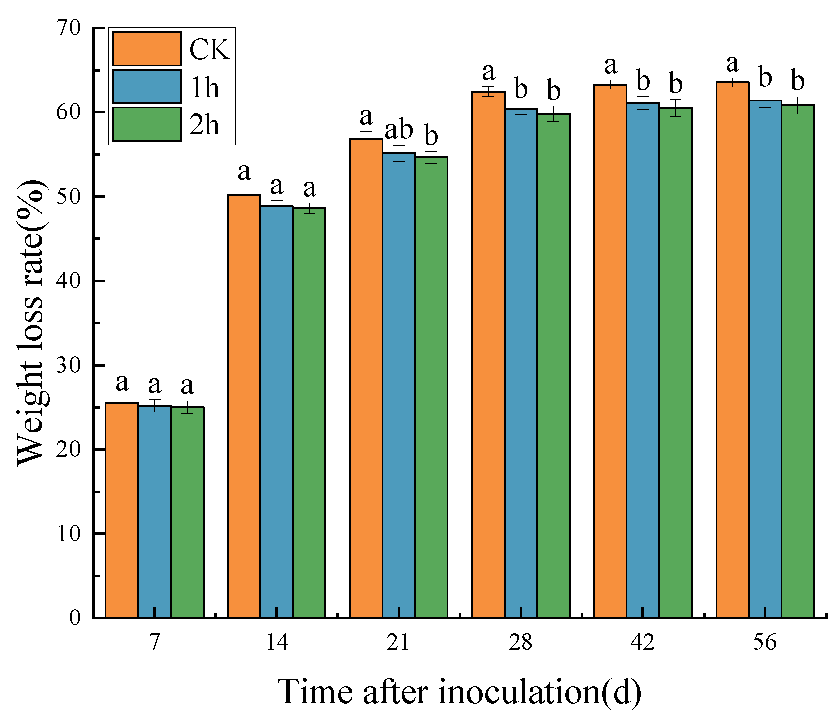
The weight loss rates of both the control group and the ozone-treated group of C. pilosula increased with prolonged storage time (Figure 2). In the initial 14 days of storage, there was a rapid increase in the weight loss rate (exceeding 48%). The ozone-treated group showed a slightly lower rate compared to the control group, but the difference was not significant. From days 21 to 28, the rate of weight loss began to slow down, and from the 28th day to the 56th day of storage, the weight loss rate gradually increased. On the 56th day of storage, the weight loss rates for the ozone-treated group were 61.43% and 60.84%, respectively. Collectively, the weight loss of C. pilosula mainly occurred in the early stages of storage. Throughout the entire storage period, the ozone-treated group exhibited a significantly lower weight loss rate compared with the control group.

3.2. The Impact of Ozone Treatment on the Contents of the Main Active Ingredients in C. pilosula

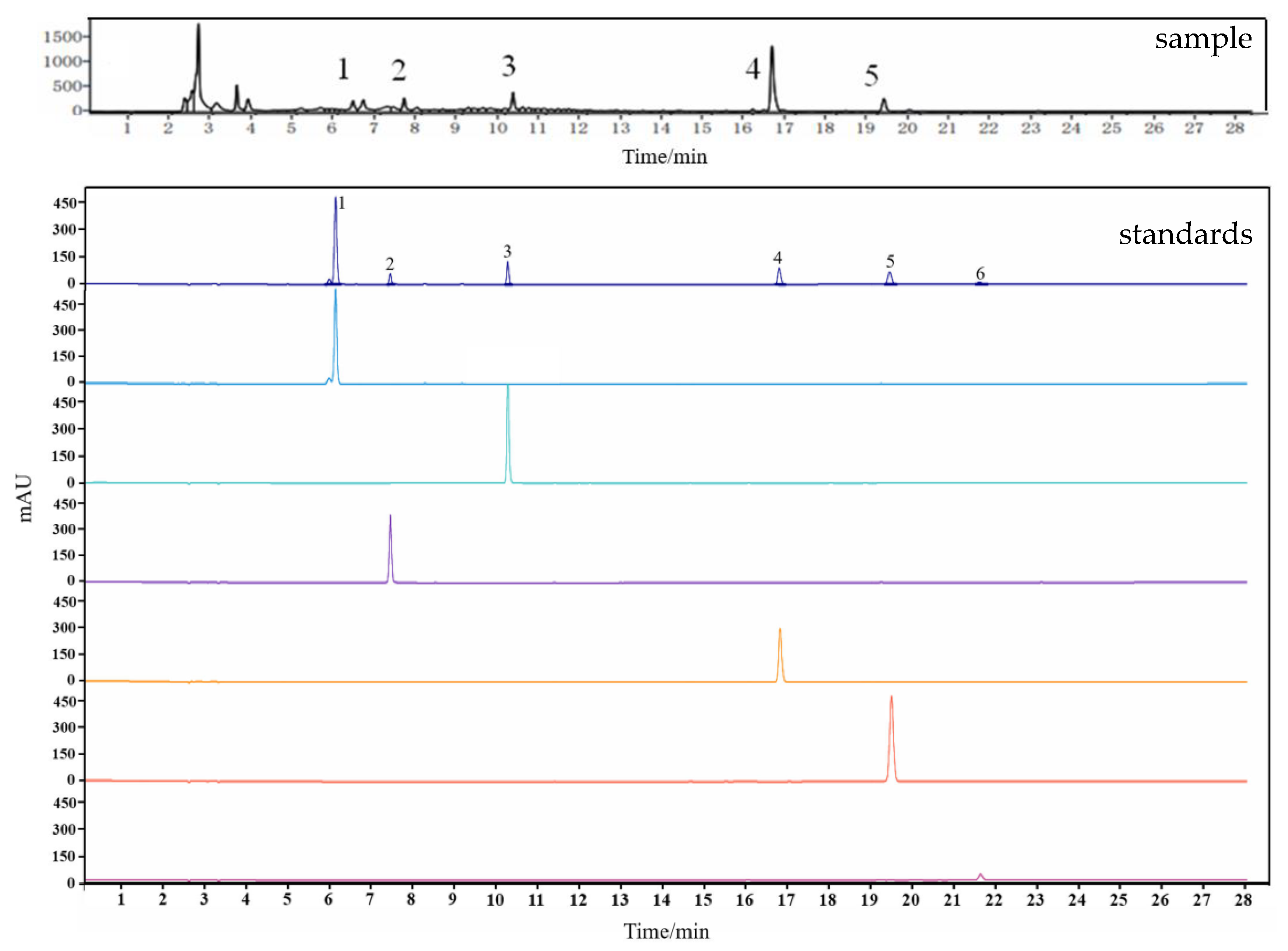
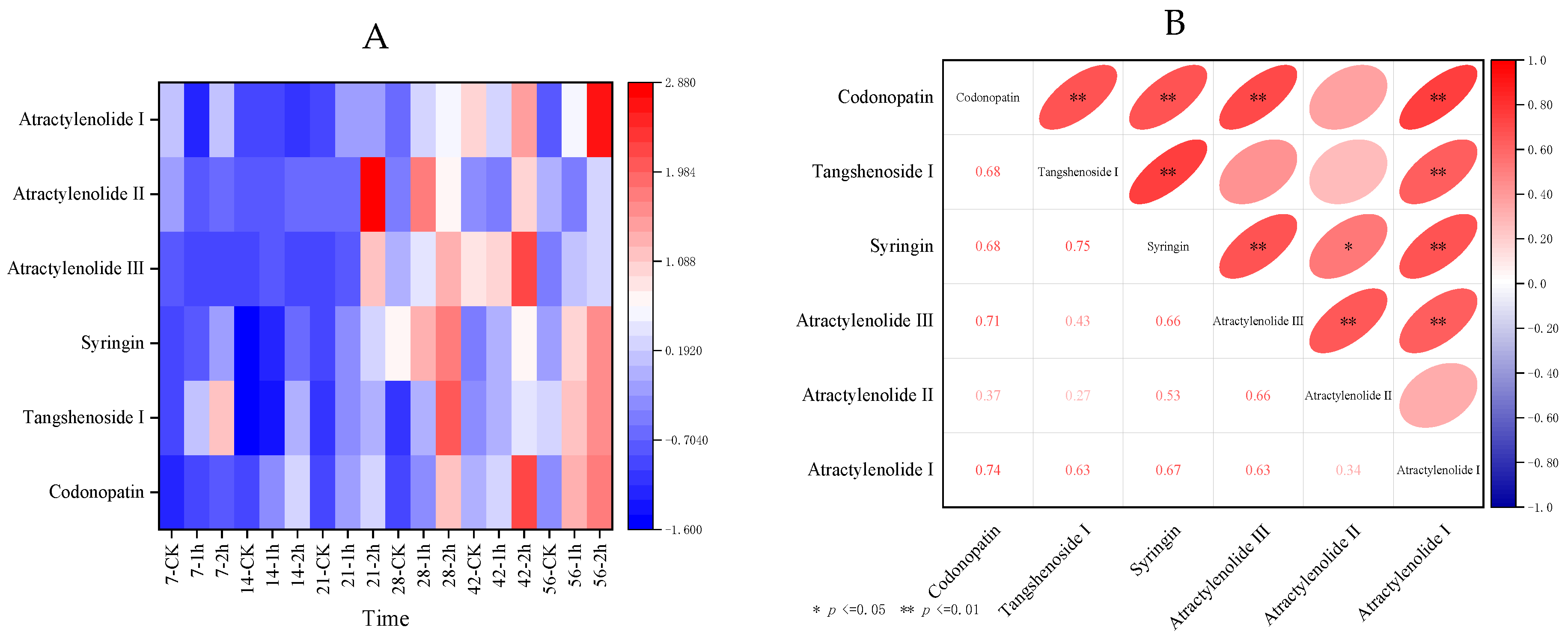
Through HPLC analysis, six active substances, including, Tangshenoside I, Syringin, Atractlenolide III, Atractlenolide II, and Atractlenolide I were primarily detected in C. pilosula (Figure 3). Following ozone treatment at different time points, the contents of the six active ingredients in C. pilosula tissues reached the highest levels after 2 h of ozone treatment, corresponding to storage durations of 42, 28, 28, 42, 21, and 56 days, respectively (Table S2). The heat map of different storage times showed that, compared with the control group, after ozone treatment of C. pilosula inoculated with A. elegans, active ingredient contents were significantly higher. The contents of four effective components in C. pilosula organization, including Codonopatin, Tangshenoside I, Syringin, and Atractlenolide III, were significantly higher than those in the control group (Figure 4A). Correlation analysis results showed a significant positive correlation between the effective component Codonopatin and Tangshenoside I, Syringin, Atractlenolide III, and Atractlenolide I (Figure 4B).
Additionally, principal component analysis (PCA) was used for the main ingredients of C. pilosula at different storage times, obtaining the characteristic values and variance contribution rates, as shown in Supplementary Materials Table S3. The characteristic values of Codonopatin, Tangshenoside I, and Syringin were 3.95285, 0.91793, and 0.4986, respectively. The cumulative contribution rate reached 89.48965%, exceeding 85%. Therefore, Codonopatin, Tangshenoside I, and Syringin can be considered as evaluation indicators for the effective components of C. pilosula at different storage times.
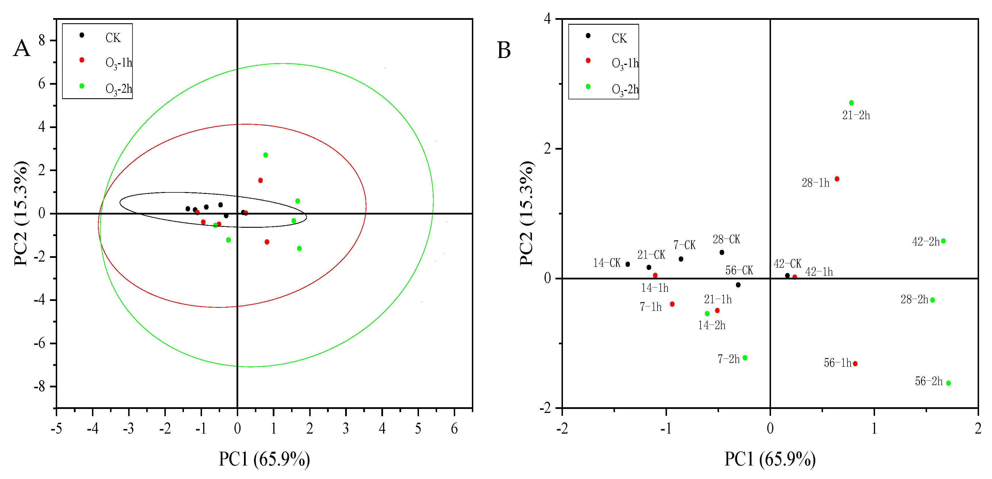
Using the variance contribution rate of each principal component as a weight, the comprehensive score (H) was calculated as follows: Comprehensive Score (H) = (65.88083F1 + 15.29881F2 + 8.31001F3)/89.48965. The ranking based on the comprehensive score is presented in Supplementary Materials S4. Combining the results with the principal component PCA score plot in Figure 5 for a comprehensive evaluation, it is observed that the group treated with ozone for 2 h on the 28th day achieved the highest score, while the control group on the 14th day obtained the lowest score. The distribution of the comprehensive evaluation of the main active ingredient of Codonopatin indicates that the group treated with ozone for 2 h has the highest score, followed by the group treated with ozone for 1 h, while the control group has the lowest comprehensive score.

3.3. The Impact of Ozone Treatment on the ROS Metabolism in C. pilosula

3.3.1. The Impact of Ozone Treatment on the Content of O2−. and H2O2 in C. pilosula

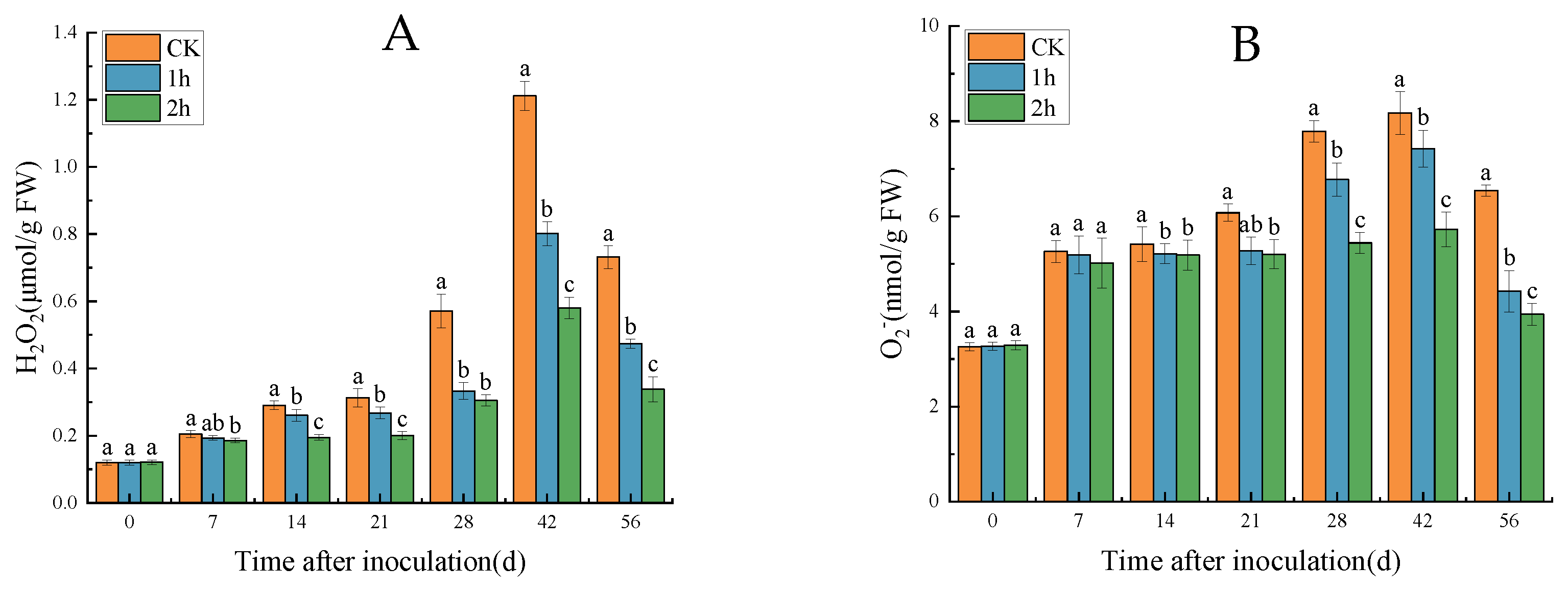
The analysis of O2−. content in C. pilosula tissues inoculated with A. elegans presented an overall trend that first increased and then decreased in both the control group and the ozone-treated groups. Meanwhile, the O2−. content in the ozone-treated group was significantly lower than that in the control group (p < 0.05). For instance, on the 42nd day of storage after inoculation, the O2−. content reached its peak, with the level of 8.17 nmol/g in the control group, while the ozone-treated groups were 7.42 nmol/g (1 h) and 5.72 nmol/g (2 h), respectively (Figure 6A). The changing trend in H2O2 content was similar to that of O2−. content, showing an initial increase followed by a decrease. On the 42nd day, the H2O2 content in the ozone-treated groups, after 1 h and 2 h, respectively, decreased by 33.86% and 52.09% compared to the control group (Figure 6B). Overall, ozone treatment significantly reduced the levels of O2−. and H2O2 in C. pilosula tissues inoculated with A. elegans.

3.3.2. The Impact of Ozone Treatment on the ROS Metabolism-Related Enzyme Activity in C. pilosula

The Impact of Ozone Treatment on the Activities of NOX and SOD in C. pilosula

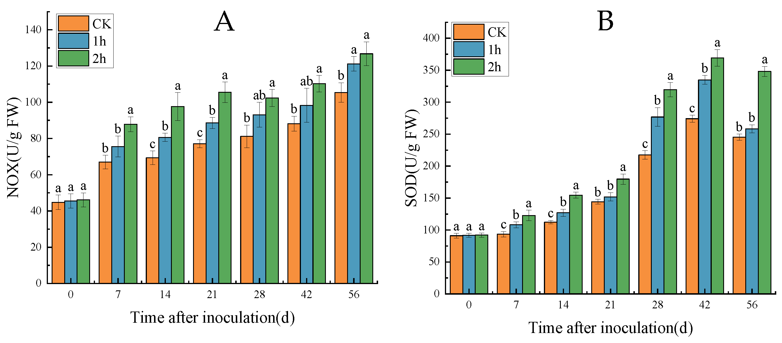
As shown in Figure 7, regardless of the control group or the treated groups, NOX activity increased with the prolonged storage time after inoculation. However, the NOX activity in the C. pilosula tissues inoculated with A. elegans and treated with ozone was significantly higher than that in the control group. Specifically, the ozone-treated group with 2 h of treatment exhibited the highest NOX activity, followed by the ozone-treated group with 1 h of treatment, and the control group had the lowest activity. For instance, on the 56th day of storage after inoculation, the NOX activity in the group treated with ozone for 2 h reached its peak at 126.76 U/g FW, was significantly higher than the control group by 16.86%, and higher than the ozone-treated group for 1 h by 4.38%. The group treated with ozone for 1 h was significantly higher than the control group by 13.05% (Figure 7A). Similarly, SOD activity showed a similar changing trend to NOX, with the group treated with ozone for 2 h exhibiting the highest peak in SOD activity, followed by the ozone-treated group with 1 h of treatment, while the control group had the lowest activity. For example, on the 42nd day of storage after inoculation, SOD activity reached its maximum value at 369.26 U/g FW, significantly higher than the control group by 25.74% and significantly higher than the ozone-treated group with 1 h of treatment by 9.34%, while the ozone-treated group with 1 h of treatment was significantly higher than the control group by 20.46% (p < 0.05).

3.3.3. The Impact of Ozone Treatment on the Activities of CAT and POD in C. pilosula

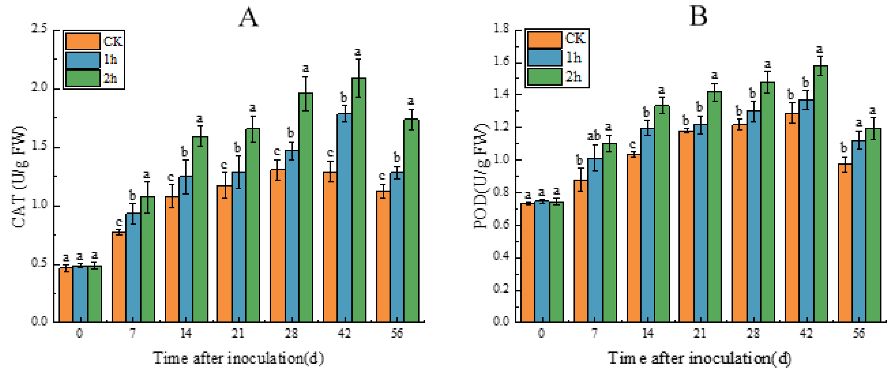
As shown in Figure 8, CAT activity in both control group and the treated group increased with the prolonged storage time after inoculation and then showed a declining trend on the 56th day. The CAT activity in the C. pilosula tissues inoculated with A. elegans and treated with ozone was significantly higher than that in the control group. Specifically, the group treated with ozone for 2 h exhibited the strongest CAT activity, followed by the group treated with ozone for 1 h, and the control group had the weakest activity. For instance, on the 42nd day of storage after inoculation, the CAT activity in the ozone-treated group with 2 h of treatment reached its peak, significantly higher than the control group by 38.13% and significantly higher than the ozone-treated group with 1 h of treatment by 14.60%, while the ozone-treated group with 1 h of treatment was significantly higher than the control group by 27.56% (Figure 8A). Similarly, POD activity showed a changing trend similar to CAT, with the ozone-treated group with 2 h of treatment exhibiting the highest peak in POD activity, followed by the ozone-treated group with 1 h of treatment, and the control group had the lowest activity. In particular, the POD activity in the ozone-treated group with 2 h of treatment was significantly higher than the control group by 18.30% and significantly higher than the ozone-treated group with 1 h of treatment by 13.15%, while the POD activity in the ozone-treated group with 1 h of treatment was significantly higher than the control group by 5.93% (Figure 8B).

3.4. The Impact of Ozone Treatment on the Cell Membrane Permeability and MDA Content of C. pilosula

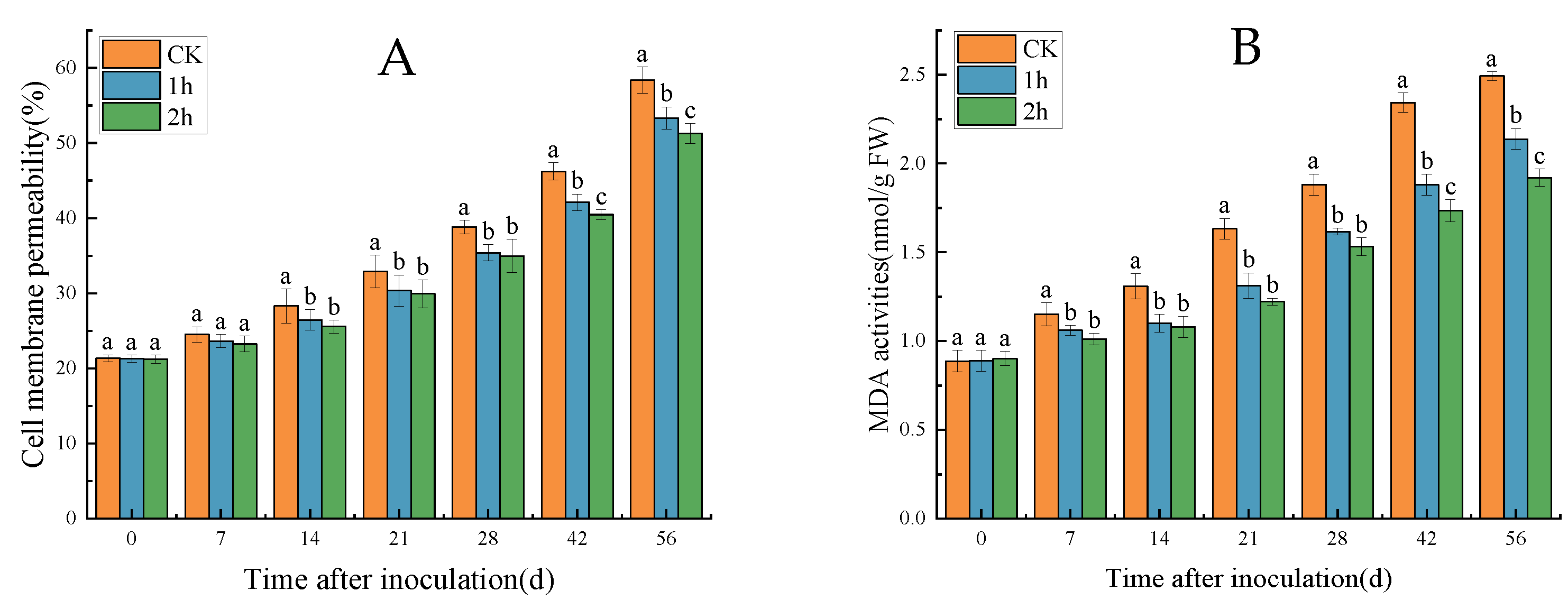
The inoculated C. pilosula showed an increasing trend in cell membrane permeability and MDA content with the prolonged storage time. However, overall, the ozone-treated groups were significantly higher than the control group, and the longer the ozone treatment time, the lower the cell membrane permeability, indicating higher cell membrane integrity and better maintenance of MDA content. For example, on the 56th day of storage after inoculation with A. elegans, the cell membrane permeability in the control group reached 58.4%, while the ozone-treated groups with 1 h and 2 h of treatment showed a decrease of 5.1% and 7.1%, respectively (Figure 9A). Correspondingly, the MDA content in the control group reached 2.49 nmol/g FW, while the MDA content in the ozone-treated groups with 1 h and 2 h of treatment were 2.14 nmol/g FW and 1.92 nmol/g FW, respectively, representing reductions of 14.06% and 22.89% (Figure 9B).

4. Discussion

A. elegans is the main pathogenic fungus causing mucor rot in C. pilosula, significantly impacting the post-harvest quality of C. pilosula. In this study, it was found that ozone treatment at 2 mg/L significantly reduced the disease incidence of C. pilosula, inhibited the development of the disease, and reduced the weight loss of C. pilosula. The effect in the 2 h treatment group was significantly better than the 1 h treatment group. Savi et al. [20] found that ozone treatment has an inhibitory effect on mycotoxin-producing fungi (Fusarium graminearum and Pseudomonas syringae) and effectively inhibits the growth of fungal colonies at a gas concentration of 3 μmol/moL. Li et al. [15] discovered that ozone treatment at 2 mg/L significantly inhibited the growth of F. sulphureum and reduced the occurrence of dry rot in potatoes, decreasing the mycotoxin accumulation in dry rot in potato tubers.
As is well known, the core function of traditional Chinese medicine is to treat diseases, and C. pilosula is no exception. The content of active ingredients in fresh C. pilosula is much higher than that of the dried C. pilosula, and the effective components of the fresh C. pilosula can be easily dissolved without spending too much time when cooking, which means the fresh C. pilosula has an immensely significant market application. However, fresh C. pilosula is highly susceptible to fungal infection, leading to mold disease during post-harvest storage. This not only significantly reduces the quality of C. pilosula but also markedly affects the content of medicinal components. After the harvest of fresh C. pilosula, mold infection occurs, resulting in a reduction in the content of active ingredients. Ozone treatment can effectively maintain the content of active ingredients in C. pilosula. For example, on the 28th day of storage, with a 2 h ozone treatment, the contents of main active ingredients in C. pilosula tissues, such as Codonopatin, Tangshenoside I, Syringin, and Atractlenolide III, were significantly higher in the ozone treatment group than in the other group. The reason for this may be that ozone treatment, by inhibiting the growth and colonization of fungus, helps to maintain the content of active ingredients in C. pilosula tissues. Moreover, the control of mucor rot by ozone treatment is closely related to the activation of ROS metabolism in C. pilosula tissues induced by ozone treatment.
This study revealed that, following inoculation with A. elegans, the contents of O2−. and H2O2 in C. pilosula tissues gradually increased with prolonged storage time. However, compared with the control group, ozone treatment reduced the content of O2−. and H2O2 in C. pilosula tissues, with a more pronounced effect observed in the 2 h ozone treatment compared with the 1 h treatment. The accumulation of ROS in C. pilosula tissues decreased after ozone treatment, which is attributable to the increased activity of antioxidant enzymes in the ROS metabolism pathway.
NOX and SOD are enzymes associated with the content of O2−. and H2O2 [6,21]. In this study, the activities of NOX and SOD in C. pilosula tissues increased with prolonged storage time, and the 2 h ozone treatment group showed significantly higher activity of these enzymes compared with the control group and the 1 h ozone treatment group. Luo et al. found that ozone treatment could enhance the activity of host defense-related enzyme POD, Polyphenoloxidase (PPO), and Phenylalamine ammonia lyase (PAL) to control postharvest diseases in kiwifruit. CAT and POD primarily convert H2O2 into O2 and H2O. Liu et al. [22] discovered that ozone treatment increased the activity of POD, SOD, and CAT enzymes, reducing O2−. and H2O2 levels and delaying browning in mushrooms. CAT and POD activities in C. pilosula tissues increased with prolonged storage time, reaching their peak on the 42nd day of storage, followed by a decline. The ozone treatment group with a 2 h exposure showed significantly higher CAT and POD activities compared with other treatment groups. Tomasz et al. [23] observed that ozone treatment induced changes in the antioxidant defense system during the storage of blueberry fruits, enhancing the activity of antioxidant enzymes and maintaining the dynamic balance of ROS metabolism. Therefore, the increased activities of these four antioxidant enzymes in the ROS metabolism pathway prevented the excessive accumulation of ROS, avoiding damage to the integrity of C. pilosula tissue cell membranes. It is expected that ozone treatment significantly reduced the integrity of cell membranes in C. pilosula tissues inoculated with A. elegans. The ozone treatment group exhibited significantly lower cell membrane permeability and MDA content than the control group, maintaining the integrity of C. pilosula tissue cell membranes better. Correspondingly, when the integrity of cell membranes in C. pilosula tissues is maintained, further invasion of the pathogen in C. pilosula tissues is prevented, thereby reducing the incidence and severity index during storage. At the same time, the preservation of cell membrane integrity also prevented the loss of moisture during the storage of C. pilosula, effectively reducing the weight loss rate of C. pilosula.

5. Conclusions

Ozone treatment (2 mg/L) significantly inhibited the expansion of mucor rot of C. pilosula inoculated with A. elegans, reducing morbidity and weight loss. Moreover, compared to the control group, the ozone treatment group exhibited significantly higher scores for the active ingredients of the A. elegans, namely, Codonopatin, Tangshenoside I, Syringin, and Atractlenolide III, all of which were significantly higher in the ozone treatment group than in other groups. Ozone treatment stimulated the ROS metabolism in A. elegans tissues, enhancing the activities of NOX, SOD, CAT, and POD in C. pilosula tissues. This suppression led to a decrease in the accumulation of O2−. and H2O2, preventing oxidative stress reactions caused by the excessive accumulation of ROS in tissues. This protective mechanism preserved cell membrane integrity, thereby controlling the occurrence of postharvest mold disease in C. pilosula.

Supplementary Materials

The following supporting information can be downloaded at: https://www.mdpi.com/article/10.3390/horticulturae10020185/s1.

Author Contributions

Conceptualization, H.X.; material preparation, data collection, and analysis, D.Z., J.C. and Z.L.; writing—original draft preparation, D.Z.; writing—review and editing, H.X. and S.S.; funding acquisition, H.X. and S.S. All authors have read and agreed to the published version of the manuscript.

Funding

This work was funded by the Project of Enterprise Supporting Plan (2022CYZC-45), the Natural Science Foundation of China (32060566) and (32360613), and the youth supervisor supporting fund (GAU-QDFC-2021-09).

Data Availability Statement

The data that support the findings of this study are available from the corresponding author upon reasonable request.

Acknowledgments

We are very thankful to the medicinal material planting base (Longxi, Gansu, China) for providing Fresh C. pilosula.

Conflicts of Interest

The authors declare no conflict of interest.

References

  1. Du, Y.E.; Lee, J.S.; Kim, H.M.; Ahn, J.-H.; Jung, I.H.; Ryu, J.H.; Choi, J.-H.; Jang, D.S. Chemical Constituents of the Roots of Codonopsis lanceolata. Arch. Pharm. Res. 2018, 41, 1082–1091. [Google Scholar] [CrossRef]
  2. Gao, S.-M.; Liu, J.-S.; Wang, M.; Cao, T.-T.; Qi, Y.-D.; Zhang, B.-G.; Sun, X.-B.; Liu, H.-T.; Xiao, P.-G. Traditional Uses, Phytochemistry, Pharmacology and Toxicology of Codonopsis: A Review. J. Ethnopharmacol. 2018, 219, 50–70. [Google Scholar] [CrossRef]
  3. Park, H.-Y.; Shin, J.-H.; Boo, H.-O.; Gorinstein, S.; Ahn, Y.G. Discrimination of Platycodon Grandiflorum and Codonopsis Lanceolata Using Gas Chromatography-Mass Spectrometry-Based Metabolomics Approach. Talanta 2019, 192, 486–491. [Google Scholar] [CrossRef]
  4. Xi, J.; Yang, D.; Xue, H.; Liu, Z.; Bi, Y.; Zhang, Y.; Yang, X.; Shang, S. Isolation of the Main Pathogens Causing Postharvest Disease in Fresh Angelica Sinensis during Different Storage Stages and Impacts of Ozone Treatment on Disease Development and Mycotoxin Production. Toxins 2023, 15, 154. [Google Scholar] [CrossRef]
  5. Jia, W.; Bi, Q.; Jiang, S.; Tao, J.; Liu, L.; Yue, H.; Zhao, X. Hypoglycemic Activity of Codonopsis pilosula (Franch.) Nannf. in Vitro and in Vivo and Its Chemical Composition Identification by UPLC-Triple-TOF-MS/MS. Food Funct. 2022, 13, 2456–2464. [Google Scholar] [CrossRef] [PubMed]
  6. Ahn, J.-H.; Jang, D.-S.; Choi, J.-H. Lancemaside A Isolated from the Root of Codonopsis lanceolata Inhibits Ovarian Cancer Cell Invasion via the Reactive Oxygen Species (ROS)-Mediated P38 Pathway. Am. J. Chin. Med. 2020, 48, 1021–1034. [Google Scholar] [CrossRef] [PubMed]
  7. Mahmud, A.; Lee, R.; Munfus-McCray, D.; Kwiatkowski, N.; Subramanian, A.; Neofytos, D.; Carroll, K.; Zhang, S.X. Actinomucor elegans as an Emerging Cause of Mucormycosis. J. Clin. Microbiol. 2012, 50, 1092–1095. [Google Scholar] [CrossRef] [PubMed]
  8. Chen, Y.; Xing, M.; Chen, T.; Tian, S.; Li, B. Effects and Mechanisms of Plant Bioactive Compounds in Preventing Fungal Spoilage and Mycotoxin Contamination in Postharvest Fruits: A Review. Food Chem. 2023, 415, 135787. [Google Scholar] [CrossRef] [PubMed]
  9. Zhao, X.; Liang, Y.; Constantine, U.; Yang, L.; Yuan, T.; Zhao, H.; Zhou, Q.; Zhang, Y.; Wang, R. First Report of Root Rot Caused by the Fusarium oxysporum Species Complex on Codonopsis pilosula in China. Plant Dis. 2021, 105, 3742. [Google Scholar] [CrossRef] [PubMed]
  10. Zapałowska, A.; Matłok, N.; Zardzewiały, M.; Piechowiak, T.; Balawejder, M. Effect of Ozone treatment on the Quality of Sea Buckthorn (Hippophae rhamnoides L.). Plants 2021, 10, 847. [Google Scholar] [CrossRef] [PubMed]
  11. Garca-Martn, J.-F.; Olmo, M.; Garca, J.-M. Effect of ozone treatment on postharvest disease and quality of different citrus varieties at laboratory and at industrial facility. Postharvest Biol. Technol. 2017, 137, 77–85. [Google Scholar] [CrossRef]
  12. Boonkorn, P.; Gemma, H.; Sugaya, S.; Setha, S.; Uthaibutra, J.; Whangchai, K. Impact of high-dose, short periods of ozone exposure on green mold and antioxidant enzyme activity of tangerine fruit. Postharvest Biol. Technol. 2012, 67, 25–28. [Google Scholar] [CrossRef]
  13. Liang, Y.-Z.; Ji, L.-L.; Chen, C.-K.; Dong, C.-H.; Wang, C.-R. Effects of ozone treatment on the storage quality of post-harvest tomato. Int. J. Food Eng. 2018, 14, 20180012. [Google Scholar] [CrossRef]
  14. Lv, B.; Yang, X.; Xue, H.; Nan, M.; Zhang, Y.; Liu, Z.; Bi, Y.; Shang, S. Isolation of Main Pathogens Causing Postharvest Disease in Fresh Codonopsis pilosula during Different Storage Stages and Ozone Control against Disease and Mycotoxin Accumulation. J. Fungi 2023, 9, 146. [Google Scholar] [CrossRef]
  15. Li, L.; Xue, H.; Bi, Y.; Zhang, R.; Kouasseu, C.J.; Liu, Q.; Nan, M.; Pu, L.; Prusky, D. Ozone Treatment Inhibits Dry Rot Development and Diacetoxyscirpenol Accumulation in Inoculated Potato Tuber by Influencing Growth of Fusarium sulphureum and Ergosterol Biosynthesis. Postharvest Biol. Technol. 2022, 185, 111796. [Google Scholar] [CrossRef]
  16. Ren, Y.; Wang, Y.; Bi, Y.; Ge, Y.; Wang, Y.; Fan, C.; Li, D.; Deng, H. Postharvest BTH Treatment Induced Disease Resistance and Enhanced Reactive Oxygen Species Metabolism in Muskmelon (Cucumis melo L. ) Fruit. Eur. Food Res. Technol. 2012, 234, 963–971. [Google Scholar] [CrossRef]
  17. Fan, M.-C.; Li, W.-X.; Hu, X.-L. Effect of micro-vacuum storage on active oxygen metabolism, internal browning and related enzyme activities in Laiyang pear (Pyrus bretschneideri Reld). LWT—Food Sci. Technol. 2016, 72, 467–474. [Google Scholar] [CrossRef]
  18. Venisse, J.; Gullner, S.-G. Brisset M N. Evidence for the involvement of an oxidative stress in the initiation of infection of pear by Erwinia amylovora. Plant Physiol. 2001, 125, 2164–2172. [Google Scholar] [CrossRef] [PubMed]
  19. Lester, G.-E.; Bruton, B.-D. Relationship of netted muskmelon fruit water loss to postharvest storage life. J. Am. Soc. Hortic. Sci. 1986, 111, 727–731. [Google Scholar] [CrossRef]
  20. Savi, G.-D.; Scussel, V.-M. Effects of ozone gas exposure on toxigenic fungi species from Fusarium, Aspergillus, and Penicillium Genera. J. Int. Ozone Assoc. 2014, 36, 144–152. [Google Scholar] [CrossRef]
  21. Simla, S.; Boontang, B.; Harakotr, B. Anthocyanin content, total phenolic content, and antiradical capacity in different ear components of purple waxy corn at two maturation stages. Aust. J. Crop Sci. 2016, 10, 675–682. [Google Scholar] [CrossRef]
  22. Liu, J.; Chang, M.-C.; Meng, J.-L.; Liu, J.-Y.; Cheng, Y.-F.; Feng, C.-P. Effect of ozone treatment on the quality and enzyme activity of Lentinus edodes during cold storage. J. Food Process. Preserv. 2020, 44, e14557. [Google Scholar] [CrossRef]
  23. Tomasz, P.; Bartosz, S.; Maciej, B. Ozone treatment induces changes in antioxidative defense system in blueberry fruit during storage. Food Bioprocess Technol. 2020, 13, 1240–1245. [Google Scholar]
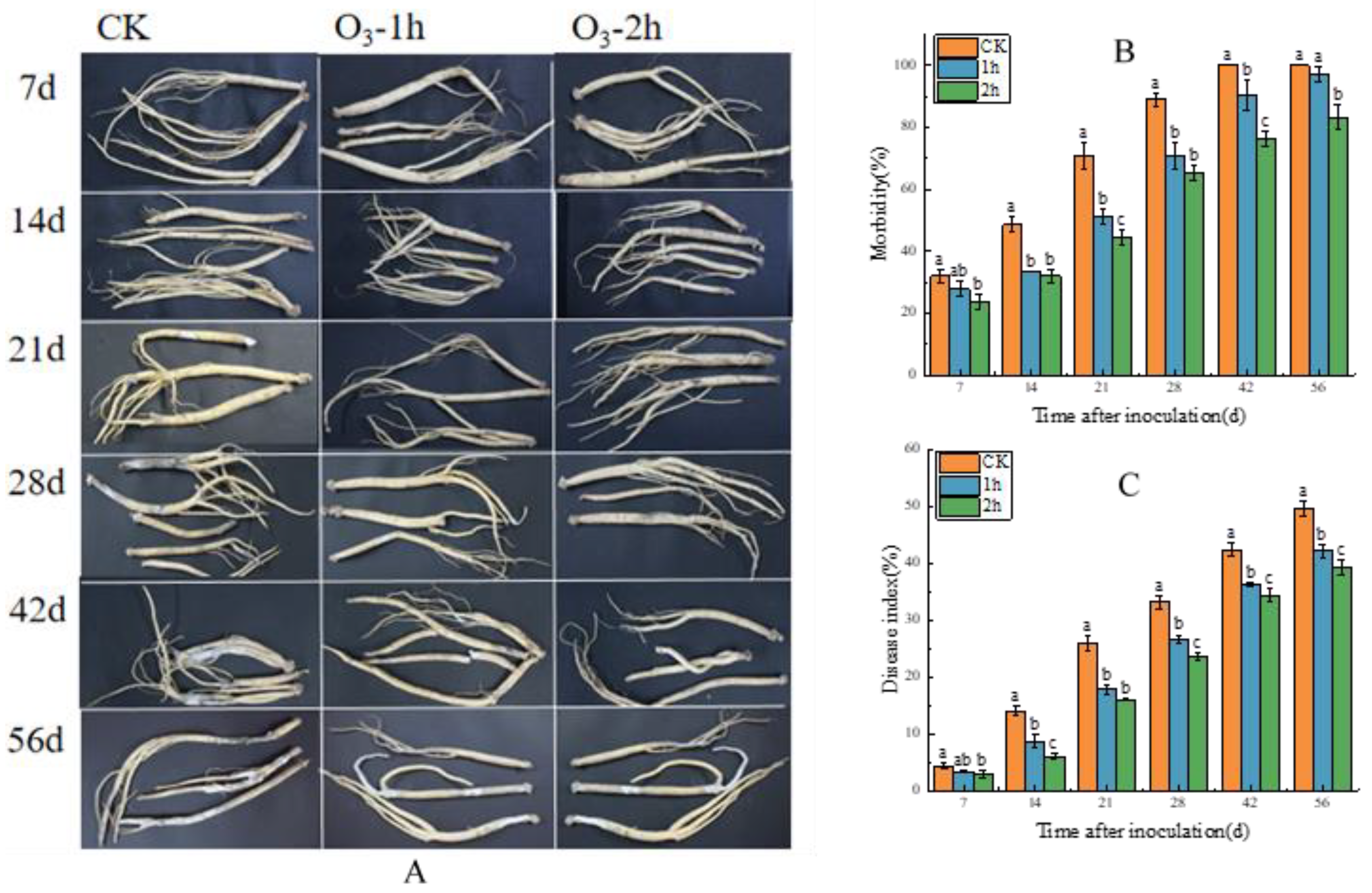
Figure 1. Effect of ozone treatment on the morbidity and disease index of the inoculated C. pilosula at different storage times. (A) Picture of the disease symptoms of the inoculated C. pilosula. (B) Morbidity of the inoculated C. pilosula. (C) Disease index of the inoculated C. pilosula (the mean values are denoted with different lowercase letters ± SEM (n = 3) showing significant differences at the 5% level, with the significance level set at p < 0.05).
Horticulturae 10 00185 g001
Figure 2. Effect of ozone treatment on weight loss of C. pilosula. (the mean values are denoted with different lowercase letters ± SEM (n = 3) showing significant differences at the 5% level, with the significance level set at p < 0.05).
Horticulturae 10 00185 g002
Figure 3. HPLC chromatograms of the six samples and standards. 1. Codonopatin; 2. Tangshenoside I; 3. Syringin; 4. Atractlenolide III; 5. Atractlenolide II; 6 Atractlenolide I.
Horticulturae 10 00185 g003
Figure 4. Effect of ozone treatment on the content of the main active ingredients of the inoculated C. pilosula. (A) Heat maps of the main active ingredient contents for different storage times; (B) Correlation analysis of the contents of main active ingredients with different storage times.
Horticulturae 10 00185 g004
Figure 5. PCA score scatter plot of Codonopsis pilosula. (A) Score chart; (B) Load chart.
Horticulturae 10 00185 g005
Figure 6. Effects of ozone treatment on O2 −. (A) and H2O2 (B) content in C. pilosula. (The mean values are denoted with different lowercase letters ± SEM (n = 3) showing significant differences at the 5% level, with the significance level set at p < 0.05).
Horticulturae 10 00185 g006
Figure 7. The effect of ozone treatment on the activities of NOX (A) and SOD (B) in C. pilosula. (The mean values are denoted by with different lowercase letters ± SEM (n = 3) showing sig-nificant differences at the 5% level, with the significance level set at p < 0.05).
Horticulturae 10 00185 g007
Figure 8. The effect of ozone treatment on the activities of CAT (A) and POD (B) in C. pilosula. (The mean values are denoted by with different lowercase letters ± SEM (n = 3) showing sig-nificant differences at the 5% level, with the significance level set at p < 0.05).
Horticulturae 10 00185 g008
Figure 9. Effects of ozone treatment on cell membrane permeability (A) and MDA content (B) of C. pilosula. (The mean values are denoted by with different lowercase letters ± SEM (n = 3) showing sig-nificant differences at the 5% level, with the significance level set at p < 0.05).
Horticulturae 10 00185 g009
Table 1. Mobile phase gradient elution procedure.
TimeA%B%
01090
52575
84555
157525
228515
259010
281090
Disclaimer/Publisher’s Note: The statements, opinions and data contained in all publications are solely those of the individual author(s) and contributor(s) and not of MDPI and/or the editor(s). MDPI and/or the editor(s) disclaim responsibility for any injury to people or property resulting from any ideas, methods, instructions or products referred to in the content.