Article

Eco-Friendly Extraction of Green Coffee Oil for Industrial Applications: Its Antioxidant, Cytotoxic, Clonogenic, and Wound Healing Properties

by
Lucas D. Dias
1,*,†,
Fernanda Mansano Carbinatto
1,
Isabelle da Silveira Almeida
1,
Kate C. Blanco
1,
Franciane Marquele-Oliveira
2,
Carla Carolina Munari
2 and
Vanderlei Salvador Bagnato
1,3,*
1
São Carlos Institute of Physics, University of São Paulo, São Carlos 13560-970, SP, Brazil
2
Eleve Science Research and Development, Innovation Laboratory, Ribeirão Preto 14056-680, SP, Brazil
3
Department of Biomedical Engineering, Texas A&M University, College Station, TX 77843, USA
*
Authors to whom correspondence should be addressed.
Current address: Laboratório de Novos Materiais, Universidade Evangélica de Goiás, Anápolis 75083-515, GO, Brazil.
Fermentation 2023, 9(4), 370; https://doi.org/10.3390/fermentation9040370
Submission received: 7 March 2023 / Revised: 1 April 2023 / Accepted: 10 April 2023 / Published: 12 April 2023
(This article belongs to the Section Microbial Metabolism, Physiology & Genetics)

Abstract

:
The development of natural oil-based cosmetic and pharmaceutical products presents great scientific and commercial interest. Herein, we aimed to extract green coffee oil from Arabic coffee by a sustainable cold-pressing method. Furthermore, this work aimed to characterize the obtained green coffee oil by Fourier-Transform Infrared (FT–IR) and ultraviolet–visible spectroscopies (UV–Vis), peroxide analysis, and fatty acids profile by gas chromatography–mass spectrometry (GC–MS). Moreover, the functional and biological properties of the obtained green coffee oil and a green-coffee oil-based commercial product (Energy up®, Dermociencia) were investigated. The green coffee oil presented linoleic and palmitic acids as the major fatty acids showing 44.8% and 35.4%, respectively. Moreover, this green coffee oil presented an antioxidant activity (EC50 7.64 mg/mL) and an absence of cytotoxic effects in keratinocyte cultures treated with up to 20 mg/mL. The obtained green coffee oil showed wound healing properties as well as clonogenic efficiency, a biological potential to induce the proliferative and migratory capacity of cells of human skin keratinocytes at 2.5 mg/mL. The samples presented high antioxidant activity and the absence of a cytotoxic effect, suggesting that green coffee oil is a promising natural product for cosmetic applications with wound healing properties. These results open new ways for the use of green coffee oil for the development of cosmetic and pharmaceutics natural-based products.

1. Introduction

Brazil is considered the world’s largest producer and exporter of coffee, with about a third of the total world production. It is the largest producer of Arabica coffee and the second largest producer of Robusta, after Vietnam [1]. The demand for processed coffee products has increased every day, with emphasis on the aroma and flavor extracts of roasted coffee due to the presence of several volatile compounds that are generated in the roasting process, namely by the Maillard reaction, which have desirable characteristics in so-called sophisticated foods [2].
The volatile compounds of roasted coffee are mainly represented by aldehydes, ketones, alcohols, esters, pyrazines, furans, acids, and compounds containing nitrogen and volatile phenolic compounds, with these compounds being variable according to the geographic location, the type of bean, and mainly the brewing process [3]. Knowing that roasted coffee oil is one of the main vehicles to concentrate the aroma and flavor, it has been widely used in the food, pharmaceutical, and also cosmetic industries, being able to act as emollients, emulsifiers, carriers, viscosity modifiers, agents spreaders, binders, and lubricants in many products [4]. Lipids present in coffee are a source of phenolic compounds, fatty acids, terpene alcohols, phytochemicals, tocopherols, carotenoids, and phytosterols, among others. These compounds have several properties such as being antioxidant and antimutagenic; having antibacterial and antiviral activity; being a stimulant of the central nervous and immune system; having analgesic and anxiolytic properties; and are auxiliary in the release of adrenaline and in the prevention of cardiovascular and rheumatological diseases, which make them of great interest in the market [5].
In this regard, coffee beans are much explored as a beverage and contain lipids (17%), proteins (11%), carbohydrates (60%), minerals (4.2%), caffeine (1.3%), trigonelline (2.0%), diterpenes (1.2%), and chlorogenic acids (up to 7.9%) [6]. Depending on the type of coffee, the content of the oil and diterpenes may change, for example, arabica coffee has a higher amount of oil and diterpene compared to robusta coffee. According to the literature, arabica and robusta coffees show 10.5–17% and 8–10% of oils, respectively. However, the industrially extracted oil content is always considered lower than that present in the coffee beans, due to technical limitations that prevent all the oil present in the beans from being accessed, reaching only levels close to 10%. In addition, the extracted oil content also depends on the chosen extraction method [7,8].
Moreover, green coffee oil and its products have been commercially explored due to the presence of bioactive compounds (e.g., diterpene esters, fatty acids, and unsaponifiable matter), its pharmacological properties (such as antioxidant and cancer-preventive properties), and consequent use in food, cosmetics, and pharmaceutical industries. Additionally, its use has been frequently applied in cosmetic industries as an emollient and moisturizer because this oil has the ability to maintain natural skin humidity due to its fatty acid content. In addition, green coffee oil was reported to protect the skin against UV radiation and may be a potential candidate to replace toxic and synthetic chemicals in sunscreens [6,9,10,11].
Regarding the extraction of green coffee oil, there are several techniques to extract this oil. Among them is extraction using supercritical fluid and CO2, extraction by ultrasonic waves, and extraction by solvent (Soxhlet methodology) in which several toxic solvents are used including hexane as well as using about 12 to 15 kg of raw coffee to produce one liter of green coffee oil [12,13,14]. However, over the past few years, there has been a great deal of interest in developing sustainable methods of analytical analysis, extraction, and synthesis that conform to the 12 Principles of Green Chemistry. These methods aim to reduce the use of toxic reagents, generating less waste for the environment [15].
Thus, in view of the expanding market for vegetable oils and the need to optimize extraction processes, the objective of this work was to evaluate the best conditions for extracting vegetable oil from green and roasted coffee, with subsequent pharmacopeial tests being carried out to sample analysis. Herein, green coffee oil was extracted by a solvent-free extraction method and characterized by FT–IR and UV–Vis spectroscopies, peroxide analysis, and fatty acids profile by GC–MS. Moreover, its biological and functional properties were studied by assessing cytotoxicity profile, antioxidant property, clonogenic efficacy and wound healing properties. Additionally, the pharmacological and physical chemical properties of a commercial product (Energy up, Dermociencia®, São Carlos, SP, Brazil) produced using the extracted oil was studied and compared to green coffee oil.

2. Materials and Methods

2.1. Chemicals and Solvents

All chemicals and solvents were commercially obtained from Sigma Aldrich (São Paulo, Brazil) and Êxodo Científica (Sumaré, São Paulo, Brazil). The green coffee oil-based commercial product (Energy up) was acquired from Dermociencia®, São Carlos, SP, Brazil. The cytotoxicity studies were carried out using a human keratinocyte cell line (HaCaT). Cells cultured in Dulbecco’s Modified Eagle Medium (DMEM) medium (Sigma Aldrich, São Paulo, Brazil) were supplemented with 10% fetal bovine serum (GIBCO) and antibiotics (0.01 mg/mL streptomycin and 0.005 mg/mL penicillin) (Sigma Aldrich, São Paulo, Brazil).

2.2. Green Coffee Oil Extraction by Cold-Pressing

Green coffee beans (Arabica coffee) were selected and screened, free from impurities (PVA—Black, broken, and impurities such as sticks, stones, and dirt) and were stored in raffia sacks at room temperature until processing. The beans were pressed to obtain the green coffee oil and the cake using a radial tubular extractor, Prensa Scottech model ERT75, (AGTTEC® company, Dois Córregos, São Paulo, Brazil). For pressing, 60 kg of green coffee beans were processed and the green coffee oil obtained was stored in an amber glass container at room temperature.

2.3. Spectroscopic Analysis

2.3.1. FT–IR

Infrared spectra were measured on an Agilent Cary 630 FTIR spectrometer (Santa Clara, CA, USA) equipped with an attenuated total reflectance (ATR) accessory, at room temperature, from 4000 to 650 cm−1, and using a spectral resolution of 16 cm−1 [16].

2.3.2. UV–Vis

UV–Vis spectra were obtained on a Cary 5000 UV–Vis–NIR spectrophotometer (Santa Clara, CA, USA) in a range between 200 and 800 nm, at room temperature, using a square quartz cuvette with an optical path of 1 cm. For a spectroscopic measurement, the samples were diluted in ethanol so that the absorbance was given between 0.1–1 A.U [17].

2.3.3. Peroxide Test

A total of 0.5 g of the sample was weighed and placed in a sealed 50 mL Erlenmeyer flask. Then, 3 mL of a v/v mixture of glacial acetic acid and chloroform (3:2 ratio) was added. The sample was stirred until the sample dissolved and 50 µL of a saturated solution of potassium iodide was added, left to rest for exactly 1 min, and then 3 mL of water and 0.6 mL of starch solution were added. It was titrated with the slow addition of 0.01 M sodium thiosulfate and without ceasing vigorous stirring until the blue color disappeared (n1 mL of 0.01 M sodium thiosulfate). A blank test was performed under the same conditions (n2 mL of 0.01 M sodium thiosulfate). The blank assay consumed no more than 0.1 mL of 0.1 M sodium thiosulfate [18].
The peroxide index was calculated by the expression:
PV = 10 ( n 1 n 2 ) m

2.3.4. Determination of the Fatty Acids Profile by GC–MS

The methodology to determine the composition of fatty acids/AOCS is according to the American Oil Chemists’ Society (AOCS) [18,19]. The Capillary Gas Chromatograph—CGC AGILENT 68650 SERIES GC SYSTEM (Santa Clara, CA, United States) Capillary column: DB-23 AGILENT (50% cyanopropyl)—methylpolysiloxane, dimensions 60 m, with inner diameter 0.25 mm, 0.25 µm film, was used. The chromatograph operating conditions were column flow = 1.00 mL/min; linear velocity = 24 cm/s; detector temperature: 280 °C; injector temperature: 250 °C; oven temperature: 110 °C (5 min), 110–215 °C (5 °C/min), and 215 °C-24 min; carrier gas: helium; and injected volume: 1.0 µL.

2.4. Biological and Functional Evaluation of Green Coffee Oil

The biological and functional tests were performed using the green coffee oil obtained by cold-pressing (100% green coffee oil) and green coffee oil-based commercial product (Energy up, from Dermociencia®, São Carlos, Brazil).

2.4.1. Antioxidant Activity

The samples’ antioxidant activity profile was measured by the scavenging of the DPPH free radical assay according to the method described by [20] and the relevant modifications. In total, 100 µL of the DPPH solution in methanol (0.25 mM) was added to 100 µL of various concentrations of green coffee oil in methanol (2.5–80 mg/mL) and allowed to stand in the dark at room temperature for 30 min. The absorbance of the sample solution was measured at 527 nm using the microplate reader SPECTRA max PLUS 384, Molecular Devices (San Jose, CA, USA). A curve of green coffee oil concentration against %DPPH was generated to estimate the concentration of green coffee oil needed to cause a 50% reduction of the initial DPPH concentration. This value of IC50/EC50 was expressed in units of µg/mL or mg/mL. The assay was performed in triplicate and the mean values were used to calculate the IC50/EC50.

2.4.2. Cytotoxicity Assay

The cytotoxicity assay was measured using an in vitro MTT (3-(4,5-dimethylthiazol-2-yl)-2,5-diphenyl-tetrazolium bromide, Sigma, São Paulo, Brazil) Colorimetric Assay to assess the metabolic capacity of the cell as an indicator of cell viability [21]. The HaCaT cells were plated onto a 96-well microplate (104 cells/well). The cell lines were exposed to different concentrations of green coffee oil or commercial product raining from 0 to 50 mg/mL for 24 h. The stock MTT solution was added to all wells of the assay and plates were incubated at 37 °C for 3 h. Afterwards, DMSO was added and mixed to all wells to dissolve the dark blue crystals and then the plates were read on a microELISA reader Kazuaki, DR-200-BS-NM-BI (Waltham, Massachusetts, United States), using a wavelength of 400 at 850 nm. The effect of the samples on cell growth was assessed as a percentage of cell viability. IC50 (concentration needed to inhibit the growth of 50%) values were estimated following 24 h treatment.

2.4.3. Clonogenic Efficiency

The clonogenic efficiency assay is an in vitro cell survival assay based on the proliferation ability of cells. The HaCaT cells were plated in 96 wells at a density of 2 × 10⁵ cells/well, and after 24 h of cellular acclimatization they were exposed to the samples (green coffee oil and commercial product) for another 24 h. After the treatment time, the cells were removed from the plate to obtain a pellet and resuspension, counted, and were again plated at an appropriate dilution for colony formation in a period of approximately 10 days, in which only one cell fraction retains the ability to reproduce by forming colonies. After the time of cultivation, colonies were fixed, stained, and analyzed using a microscope (ZEEIS, software Zen Blue) followed by the calculation of the concentration–survival fraction curve [22].

2.4.4. Wound Healing

Keratocyte strain (HaCaT) in monolayer culture were scratched manually with a yellow plastic pipette tip, washed with PBS, and treated with 0, 2.5, 10, and 20 mg/mL samples (green coffee oil and Energy up), a negative control, and Mitomycin C (MMC). A microscope ZEISS (Jena, Germany) was used to obtain images of the wound in different times of treatment (0 h, 24 h, and 48 h) [23].

2.5. Statistical Analysis

For antioxidant activity, the IC50 values were calculated by non-linear intelligent curvature. The data obtained in MTT, clonogenic efficiency, and wound healing assays were statistically analyzed by analysis of variance (ANOVA). Then, the Tukey’s method was used to compare the different treatments. The GraphPad Prism 8 program was used.

3. Results and Discussion

3.1. Extraction and Characterization of Green Coffee Oil

The green coffee oil was extracted from Arabic coffee (60 kg) by using a radial tubular extractor, namely the Prensa Scottech model ERT75, yielding 3% w/w of green coffee oil. Infrared spectroscopy was used to identify the chemical composition of coffee oil, as presented in Figure 1. The analysis of the spectra allows the identification of characteristic bands whose frequencies and intensities can clearly reveal the functional groups of the investigated samples.
The first absorption region observed in Figure 1, around 3000 cm−1, is dominated by a series of aliphatic vibrations, due to a the large number of -CH3 and -CH2 groups present in the fatty acids of the sample [16]. The vibration of stretching -CH bonds lead to high absorptions recorded around 2922 and 2852 cm−1 associated with caffeine [24]. Furthermore, the unsaturated fatty acids present in the coffee oil gave a small peak at 3009 cm−1, characteristic of the -C–H stretching vibration of the cis double bonds. The intense peak observed at 1744 cm−1 and a smaller one between 1750 and 1720 cm−1 correspond to the carbonyl group, present in the bonds between glycerol and fatty acids. In the region of 1730–1680 cm−1, the aldehyde compounds are found, whose carbonyl groups have high absorptivity and overlap with the strong band of the carbonyl ester group at 1744 cm−1 [16]. The 1400–900 cm−1 range is characterized by vibrations of various bond types, including C-H, C-O, C-N, and P-O. Chlorogenic acids show strong absorption between 1300 and 1150 cm−1. In addition, carbohydrates have already been shown to contribute to the appearance of bands in this region [24].
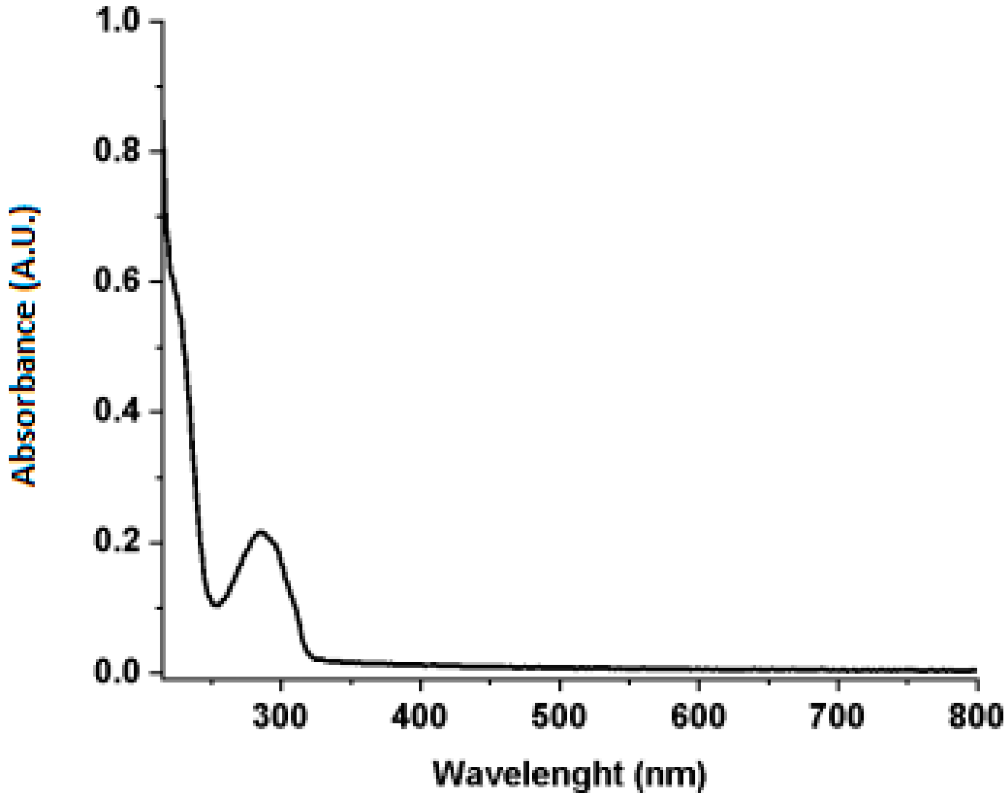
Moreover, the obtained green coffee oil was analyzed by UV–Vis spectroscopy (Figure 2). For the quality control of vegetable oils, UV–Vis spectroscopy presents itself as a quick method to evaluate the adulteration of oils. As reported by Popa et al. (2020) [25], when adulterating cold-pressed oils with refined oils, it is observed that the maximums in the absorbance spectra disappear as the percentage of adulteration increases. Didham et al. (2020) [26] reported that this can be a reliable analytical tool to detect and quantify levels greater than 10% of adulteration. Furthermore, it was described that the variations observed in the spectra can be explained by different fatty acids, lipid oxidation products, and changes in pigment profiles derived from different oil samples. According to Figure 2, it is possible to observe that the spectrum of green coffee oil is characterized by absorption in the region of 250–315 nm, with a maximum absorption peak at 275 nm. This peak at 275 nm may be related to the caffeine molecule, as described in the literature [27].
Regarding the peroxide index, the green coffee oil presented a value of 0.96 meq/kg. According to Anvisa Resolution RDC-No. 481, as of 15 March 2021, the maximum recommended Peroxide Value (PV) value is 15 meq/kg for unrefined oils. This index, also called oxidative rancidity, is one of the most used methods to measure the oxidation state of oils and fats as it quantifies hydroperoxides, which are primary products formed when a fat deteriorates. However, this method may present problems related to the lack of sensitivity to interference from colored and fat-soluble components or to the additional oxidation that occurs during lipid extraction [28].
The fatty acid composition of the coffee oil samples obtained is shown in Table 1. The data obtained are similar to those reported by [29], in which coffee oil is mainly composed of linoleic and palmitic acids, with 44.8% and 35.4%, respectively. Linoleic acid has interesting properties in relieving eczema and helping to treat and cure dermatitis, working as an excellent emollient, while palmitic acid provides good skin protection and potential application in the pharmaceutical and cosmetics industry [29].

3.2. Biological and Functional Evaluation of Green Coffee Oil

3.2.1. Antioxidant Activity

The DPPH free radical scavenging method allowed the observation of the antioxidant activity of green coffee oil and commercial product, as shown in Figure 3.
The results in Figure 3 show the dose–response curve and the antioxidant activity that was represented by the EC50 comparison, which expresses the amount of antioxidant required to reduce the initial concentration of free radial DPPH in the reaction medium by 50% in a determined time: the lower the EC50, the greater the antioxidant potential. In the human body there is a balance between antioxidants and free radicals in which antioxidant enzymes and antioxidant compounds (e.g., vitamins C and E) may protect membrane lipids, proteins, DNA, and other macromolecules against several reactive species (e.g., hydroxyl radicals (OH), superoxide anion radicals, (O2), and nitroxide radicals (NO)). Although reactive species play an important role in host defense and physiological processes, when in excess, they can disrupt the antioxidant protection and lead to a situation of oxidative stress and this condition has been associated with the development of several skin problems, such as skin accelerated aging, dermatites, eczemas, or even cancer [2].
The samples analyzed in this study are composed of natural products and, as expected, showed lower antioxidant activity when compared to the ascorbic acid standard (EC50 of 13.36 mg/mL, 7.64 mg/mL, and 8.37 mg/mL for ascorbic acid standard, the commercial product, and green coffee oil, respectively) but higher activity when compared to the others natural products such grape seed oil, which demonstrated a higher EC50 value (EC50 of 31.441 w/w DPPH) and pomegranate seed oil (EC50 1065 w/w DPPH) and Passiflora setácea seed oil (EC50 of 6054 w/w DPPH) which were both extracted by cold pressing which is the same way of extracting green coffee oil from this analysis.
The antioxidant activity related to green coffee oil can be associated with its composition which is rich in free radical scavengers such as tocopherols and linoleic acid [9,30]. These antioxidants can eliminate reactive oxygen species (ROS) contributing to the prevention of cellular damage to the skin [9]. The commercial product showed better antioxidant activity (EC50 of 7.64 mg/mL) when compared to green coffee oil (EC50 of 8.37 mg/mL), which can be related to its composition, which has a high content of green coffee oil mixed with other components that also have antioxidant activity, such as avocado oil, passion fruit oil, peppermint essential oil, and alpha bisabolol [31,32].

3.2.2. MTT Assay

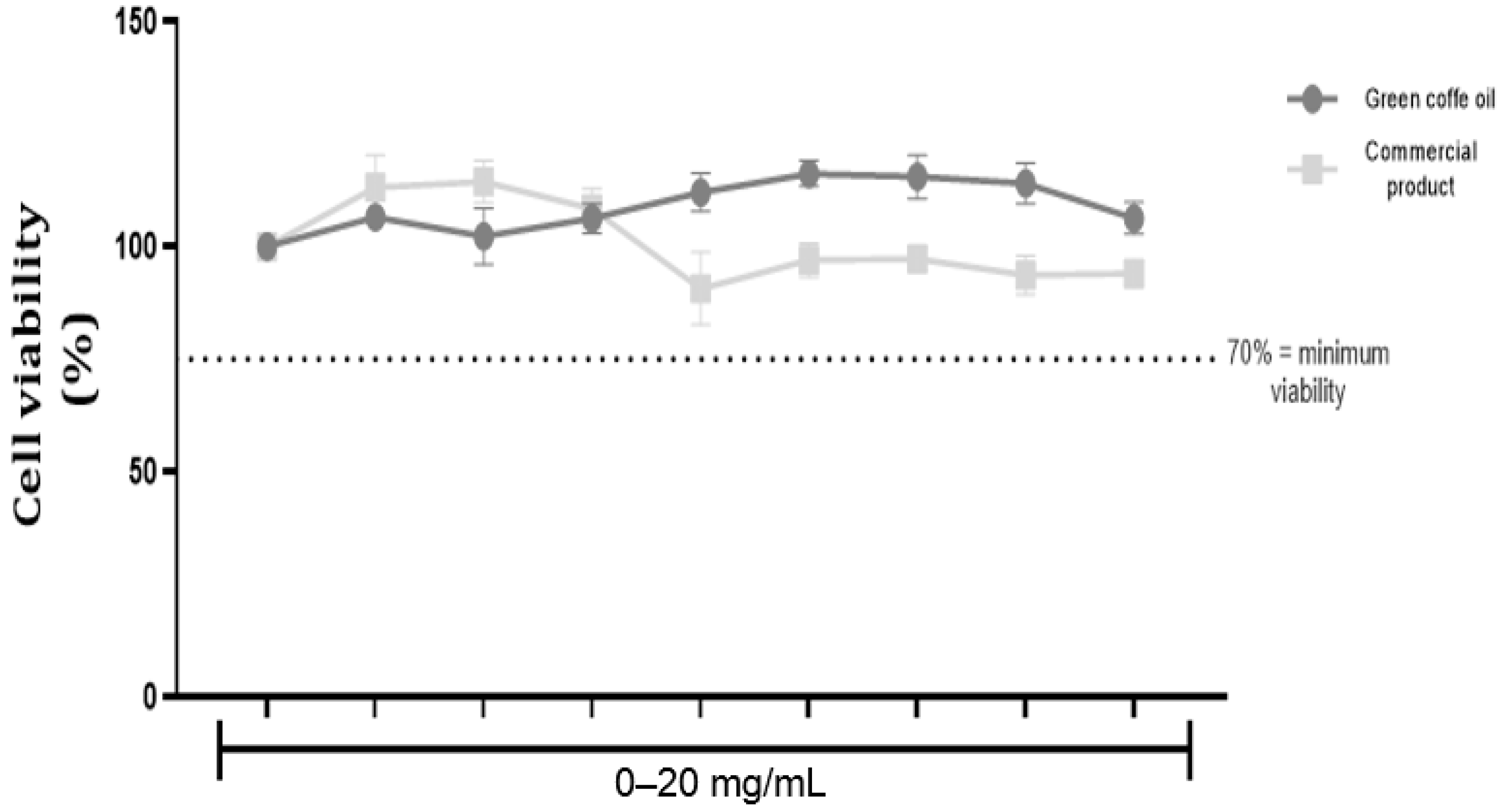
The commercial product (Energy up) and green coffee oil exhibited an absence of cytotoxic effects against human keratinocyte cells in doses corresponding to the ascending order 0–20 mg/mL, as shown in Figure 4.
Currently, there are several types of tests to assess cytotoxic effects and the MTT assay was selected because it is a valid, easy-to-use, safe, and widely used test for cytotoxicity evaluation. Moreover, it has good reproducibility and is suitable for studies in adherent cells, such as in human keratinocytes cells [29]. Green coffee oil and commercial products demonstrated the absence of cytotoxic effects in keratinocyte cultures treated with up to 20 mg/mL and indeed this result was also identified in terms of the clonogenic efficacy and cell proliferation assays and agreed with other scientific studies such as [9,29], thus confirming that its samples are safe for use in humans.

3.2.3. Clonogenic Efficiency

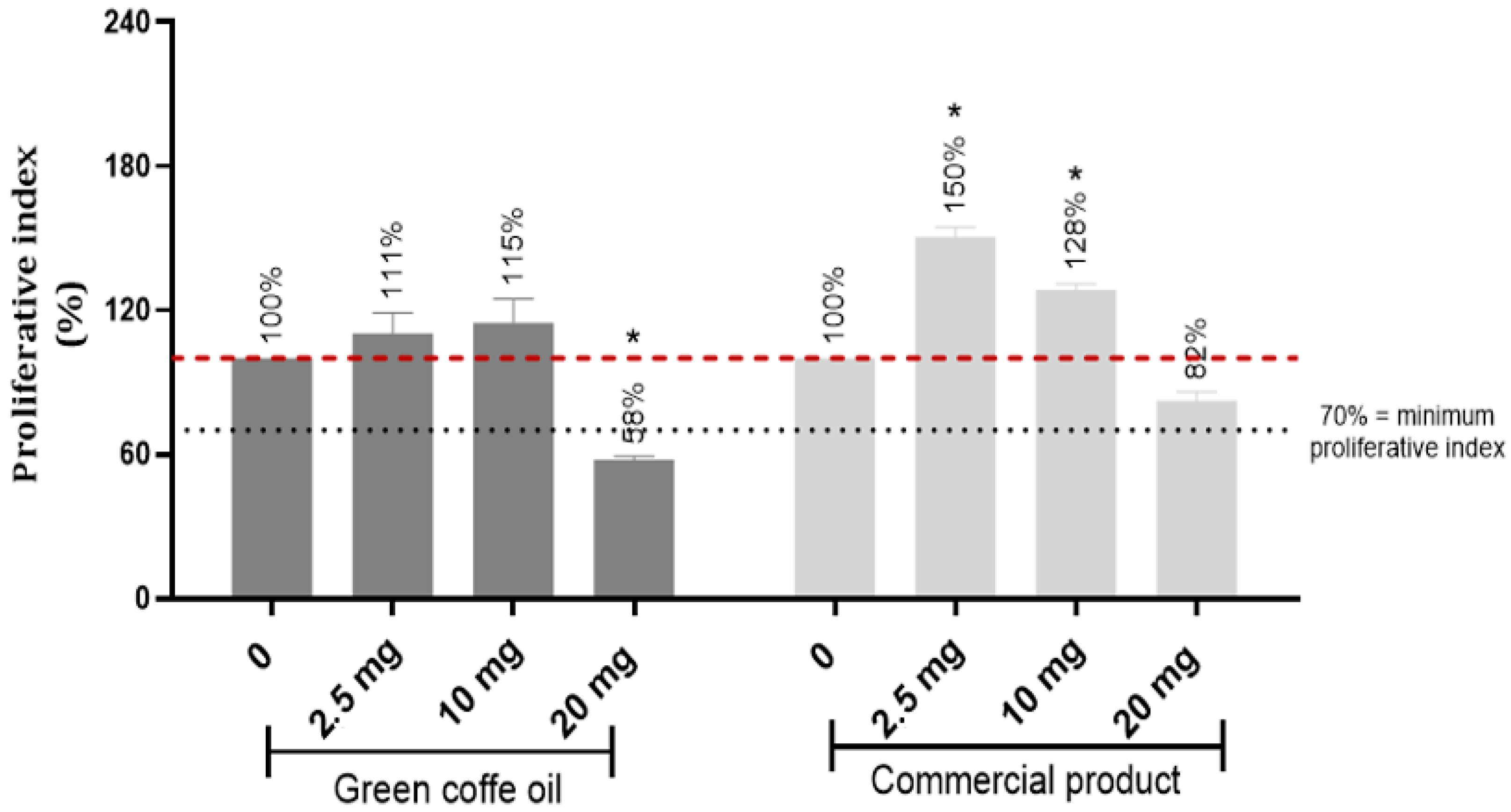
The assessment of cytotoxicity in keratinocyte cell monolayers using the clonogenic efficiency assay revealed an absence of cytotoxicity in the samples and the samples demonstrated the proliferative capacity of skin keratinocyte cells at concentrations of 2.5 and 10 mg/mL (Figure 5).

3.2.4. Wound Healing

Concentrations of 2.5, 10, and 20 mg/mL of each sample (coffee oil and commercial product) were evaluated, and it was possible to verify that the 2.5 mg/mL showed more promising results in the process of wound healing, as shown in Figure 6, corroborating clonogenic efficiency data.
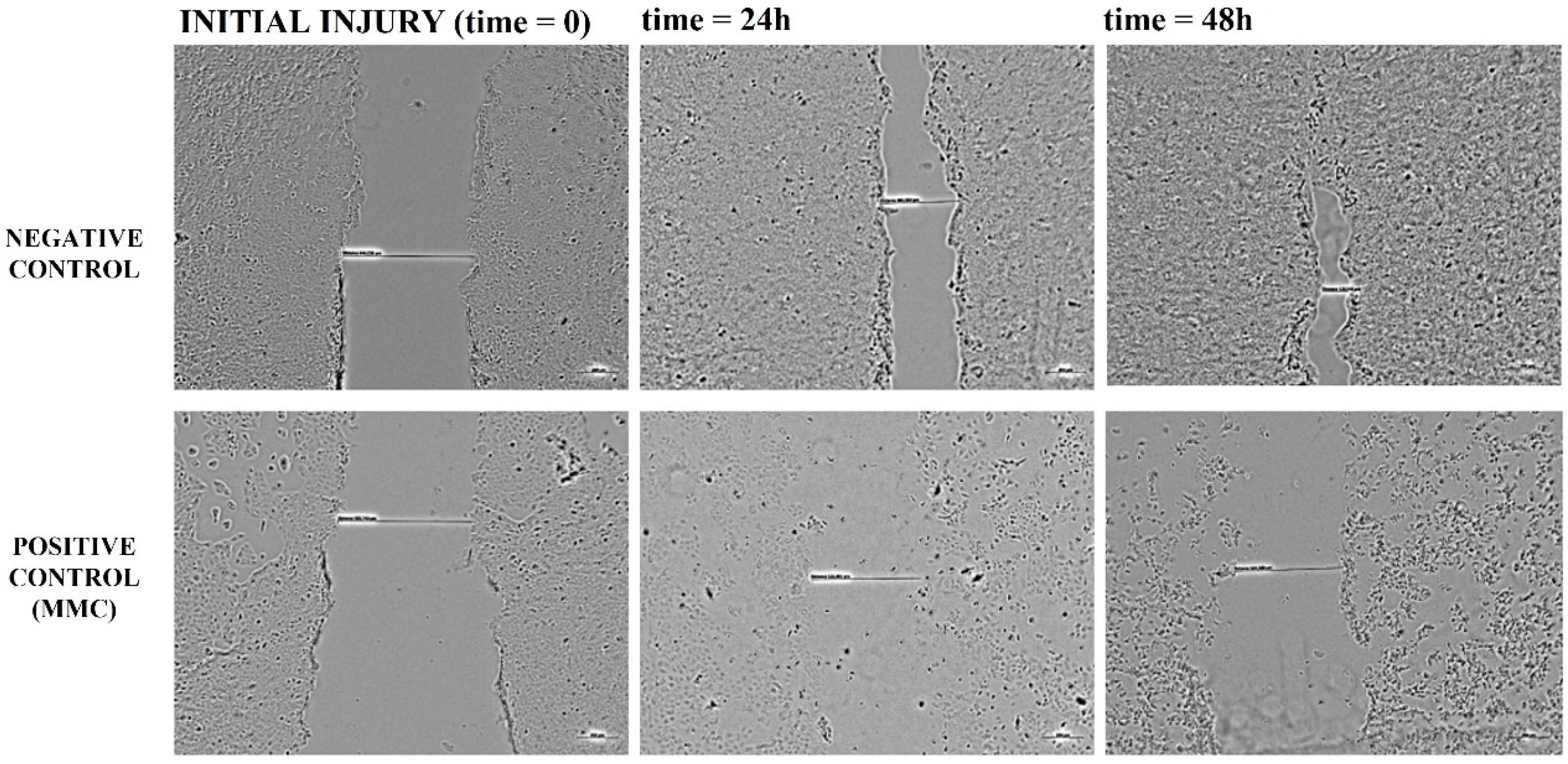
Figure 7 shows that there is a spontaneous closure of the “wound/injury” according to the process and phases of the cell cycle characteristic of human keratinocytes. Indeed, the presence of the cell proliferation inhibitor, Mitomicyn C (MMC), “delays/inhibits” this closure by impeding cell cycle progress and consequently cell proliferation. The presence of different samples in the lowest concentration (2.5 mg/mL) demonstrated complete closure of the “wound/lesion” suggesting an effect promoting tissue proliferation and recovery (Figure 8). The wound healing assay is simple and an inexpensive method that was developed to investigate directional cell migration in vitro. This method mimics cell migration during wound healing in vivo [33].
The initiation of a wound promotes complex events to repair the injured region, such as increased cell proliferation, deposition of extracellular matrix, and infiltration of inflammatory immune cells as part of the process elimination of necrotic tissue [34]. The wound-healing process begins with the polarization of cells towards the wound, followed by migration and closure of the area injured. These processes reflect the behavior of individual cells as well as tissue complexity. The evaluated samples showed similar behavior and no statistically significant difference, demonstrating that at the lowest concentration (2.5 mg/mL) there was complete closure of the “wound/lesion” suggesting an effect promoting tissue proliferation and recovery (Figure 8).
In the study conducted by [35], it was revealed that green coffee oil increases the synthesis of extracellular matrix compounds by human skin fibroblasts in vitro due to the upregulation of the production of growth factors. Green coffee oil is also able to increase iAQP-3 mRNA expression, thereby improving skin hydration [35]. The topical application of green coffee oil in wound healing was evaluated by [36] in a model of cutaneous incision in rats and the resulting fast wound healing action [36]. In addition, the use of green coffee oil formula associated with propolis was able to maintain the physical characteristics of the skin and your hydration: besides its anti-inflammatory, antioxidant, and antiaging properties, italso reduces the roughness of the skin and helps to reduce and prevent stretch marks [37].
Natural plant-based oils are gaining enormous visibility and applicability within traditional medicine. In the case of green coffee oil, which is still a little explored product, and even in cases where it is being exploited, adequate evidence is still lacking. Specifically, in the case of healing acceleration, the results create great perspectives for use in situations of bedridden patients where decubitus ulcers are relevant and difficult to solve. Another aspect of green coffee oil is its potential for use in conjunction with other techniques such as photobiostimulation and photodynamic therapy, among others. This is a new port for exploring these properties. In this regard, the pharmacological properties of green coffee oil combined with photoactive compounds could result in a synergistic effect to treat infection diseases and cancer.

4. Conclusions

The green coffee oil was obtained by cold pressing as a sustainable and green approach. The green coffee oil was fully characterized by FT–IR, UV–Vis, and GC–MS. The green coffee oil demonstrated an antioxidant activity (EC50 7.64 mg/mL) and the absence of cytotoxic effects in keratinocyte cultures treated with up to 20 mg/mL. Green coffee oil and a green-coffee oil-based commercial product (Energy up®, Dermociencia) have shown clonogenic efficiency, namely a biological potential to induce the proliferative and migratory capacity of cells of human skin keratinocytes, which was demonstrable in the wound healing assay. The samples presented high antioxidant activity and an absence of a cytotoxic effect, suggesting that green coffee oil is a promising natural product for cosmetic applications with wound healing properties. These results open new ways for the use of green coffee oil for the development of cosmetic and pharmaceutical natural-based products.

Author Contributions

Conceptualization, L.D.D., F.M.C. and V.S.B.; methodology, L.D.D., F.M.C., I.d.S.A., K.C.B., F.M.-O., C.C.M. and V.S.B.; validation, L.D.D., F.M.C., I.d.S.A., K.C.B., F.M.-O., C.C.M. and V.S.B.; formal analysis, L.D.D., F.M.C., I.d.S.A., K.C.B., F.M.-O., C.C.M., and V.S.B.; investigation, L.D.D., F.M.C., I.d.S.A., K.C.B., F.M.-O. and C.C.M.; data curation, L.D.D., F.M.C., I.d.S.A., K.C.B., F.M.-O., C.C.M. and V.S.B.; writing—original draft preparation, L.D.D., F.M.C., I.d.S.A., K.C.B., F.M.-O., C.C.M. and V.S.B.; writing—review and editing, L.D.D., F.M.C., I.d.S.A., K.C.B., F.M.-O., C.C.M. and V.S.B.; visualization, L.D.D., F.M.C., I.d.S.A., K.C.B., F.M.-O., C.C.M. and V.S.B.; supervision, L.D.D., F.M.C. and V.S.B.; project administration, L.D.D., F.M.C., and V.S.B.; funding acquisition, V.S.B. All authors have read and agreed to the published version of the manuscript.

Funding

This research was funded by MCTIC/FNDCT-CNPq/MEC–CAPES/MS-Decit/Nº 14/2016 (CNPq 440585/2016–3 and CAPES 88881.130676/2016–01); Fundação de Amparo à Pesquisa do Estado de São Paulo (FAPESP CEPOF 2013/07276–1, 2018/00106–7, and 2019/13569–8) and INCT “Basic Optics and Applied to Life Sciences” (FAPESP 2014/50857–8, CNPq 465360/2014–9). Financial support by FAPESP (Program CEPID-INCT) and EMBRAPII.

Institutional Review Board Statement

Not applicable.

Informed Consent Statement

Not applicable.

Data Availability Statement

No new data were created or analyzed in this study. Data sharing is not applicable to this article.

Conflicts of Interest

The authors declare no conflict of interest.

References

  1. Coffee Production is Expected to Reach 55.7 million Bags in the 2022 Harvest. Available online: https://www.gov.br/en/government-of-brazil/latest-news/2022/coffee-production-is-expected-to-reach-55-7-million-bags-in-the-2022-harvest (accessed on 9 April 2023).
  2. dos Santos, E.M.; de Macedo, L.M.; Tundisi, L.L.; Ataide, J.A.; Camargo, G.A.; Alves, R.C.; Oliveira, M.; Mazzola, P.G. Coffee by-products in topical formulations: A review. Trends Food Sci. Technol. 2021, 111, 280–291. [Google Scholar] [CrossRef]
  3. Caporaso, N.; Whitworth, M.B.; Cui, C.H.; Fisk, I.D. Variability of single bean coffee volatile compounds of Arabica and robusta roasted coffees analysed by SPME-GC-MS. Food Res. Int. 2018, 108, 628–640. [Google Scholar] [CrossRef] [PubMed]
  4. Wagemaker, T.A.L.; Carvalho, C.R.L.; Maia, N.B.; Baggio, S.R.; Guerreiro, O. Sun protection factor, content and composition of lipid fraction of green coffee beans. Ind. Crops Prod. 2011, 33, 469–473. [Google Scholar] [CrossRef] [Green Version]
  5. Masek, A.; Latos-Brozio, M.; Kaluzna-Czaplinska, J.; Rosiak, A.; Chrzescijanska, E. Antioxidant Properties of Green Coffee Extract. Forests 2020, 11, 557. [Google Scholar] [CrossRef]
  6. Almeida, F.S.; Dias, F.F.G.; Sato, A.C.K.; Bell, J. From solvent extraction to the concurrent extraction of lipids and proteins from green coffee: An eco-friendly approach to improve process feasibility. Food Bioprod. Process. 2021, 129, 144–156. [Google Scholar] [CrossRef]
  7. Cordoba, N.; Fernandez-Alduenda, M.; Moreno, F.L.; Ruiz, Y. Coffee extraction: A review of parameters and their influence on the physicochemical characteristics and flavour of coffee brews. Trends Food Sci. Technol. 2020, 96, 45–60. [Google Scholar] [CrossRef]
  8. Mediani, A.; Kamal, N.; Lee, S.Y.; Abas, F.; Farag, M.A. Green Extraction Methods for Isolation of Bioactive Substances from Coffee Seed and Spent. Sep. Purif. Rev. 2022, 52, 24–42. [Google Scholar] [CrossRef]
  9. Chiari, B.G.; Trovatti, E.; Pecoraro, E.; Correa, M.A.; Cicarelli, R.M.B.; Ribeiro, S.J.L.; Isaac, V.L.B. Synergistic effect of green coffee oil and synthetic sunscreen for health care application. Ind. Crops Prod. 2014, 52, 389–393. [Google Scholar] [CrossRef]
  10. de Oliveira, P.M.A.; de Almeida, R.H.; de Oliveira, N.A.; Bostyn, S.; Goncalves, C.B.; de Oliveira, A.L. Enrichment of diterpenes in green coffee oil using supercritical fluid extraction—Characterization and comparison with green coffee oil from pressing. J. Supercrit. Fluids 2014, 95, 137–145. [Google Scholar] [CrossRef]
  11. Dong, W.J.; Chen, Q.Y.; Wei, C.Q.; Hu, R.S.; Long, Y.Z.; Zong, Y.; Chu, Z. Comparison of the effect of extraction methods on the quality of green coffee oil from Arabica coffee beans: Lipid yield, fatty acid composition, bioactive components, and antioxidant activity. Ultrason. Sonochem. 2021, 74, 105578. [Google Scholar] [CrossRef]
  12. Araujo, J.M.A.; Sandi, D. Extraction of coffee diterpenes and coffee oil using supercritical carbon dioxide. Food Chem. 2007, 101, 1087–1094. [Google Scholar] [CrossRef]
  13. de Azevedo, A.B.A.; Kieckbush, T.G.; Tashima, A.K.; Mohamed, R.S.; Mazzafera, P.; de Melo, S. Extraction of green coffee oil using supercritical carbon dioxide. J. Supercrit. Fluids 2008, 44, 186–192. [Google Scholar] [CrossRef]
  14. Tsukui, A.; Santos, H.M.; Oigrnan, S.S.; de Souza, R.; Bizzo, H.R.; Rezende, C.M. Microwave-assisted extraction of green coffee oil and quantification of diterpenes by HPLC. Food Chem. 2014, 164, 266–271. [Google Scholar] [CrossRef]
  15. Kreuder, A.D.; House-Knight, T.; Whitford, J.; Ponnusamy, E.; Miller, P.; Jesse, N.; Rodenborn, R.; Sayag, S.; Gebel, M.; Aped, I.; et al. A Method for Assessing Greener Alternatives between Chemical Products Following the 12 Principles of Green Chemistry. ACS Sustain. Chem. Eng. 2017, 5, 2927–2935. [Google Scholar] [CrossRef] [Green Version]
  16. Raba, D.N.; Poiana, M.A.; Borozan, A.B.; Stef, M.; Radu, F.; Popa, M.V. Investigation on Crude and High-Temperature Heated Coffee Oil by ATR-FTIR Spectroscopy along with Antioxidant and Antimicrobial Properties. PLoS ONE 2015, 10, e0138080. [Google Scholar] [CrossRef] [Green Version]
  17. Vidal, O.L.; Santos, M.C.B.; Batista, A.P.; Andrigo, F.F.; Barea, B.; Lecomte, J.; Figueroa-Espinoza, M.C.; Gontard, N.; Villeneuve, P.; Guillard, V.; et al. Active packaging films containing antioxidant extracts from green coffee oil by-products to prevent lipid oxidation. J. Food Eng. 2022, 312, 110744. [Google Scholar] [CrossRef]
  18. Maszewska, M.; Florowska, A.; Dluzewska, E.; Wroniak, M.; Marciniak-Lukasiak, K.; Zbikowska, A. Oxidative Stability of Selected Edible Oils. Molecules 2018, 23, 1746. [Google Scholar] [CrossRef] [Green Version]
  19. Brühl, L. Official Methods and Recommended Practices of the American Oil Chemist’s Society, Physical and Chemical Characteristics of Oils, Fats and Waxes; AOCS Press: Champaign, IL, USA, 1996. [Google Scholar]
  20. Marquele-Oliveira, F.; Fonseca, Y.M.; Georgetti, S.R.; Vicentini, F.; Bronzati, V.; Fonseca, M.J.V. Evaluation of the antioxidant activity as an additional parameter to attain the functional quality of natural extracts. Lat. Am. J. Pharm. 2008, 27, 325–332. [Google Scholar]
  21. Denizot, F.; Lang, R. Rapid colorimetric assay for cell-growth and survival—modifications to the tetrazolium dye procedure giving improved sensitivity and reliability. J. Immunol. Methods 1986, 89, 271–277. [Google Scholar] [CrossRef]
  22. Munari, C.C.; de Oliveira, P.F.; Lima, I.M.D.; Martins, S.D.L.; da Costa, J.D.; Bastos, J.K.; Tavares, D.C. Evaluation of cytotoxic, genotoxic and antigenotoxic potential of Solanum lycocarpum fruits glicoalkaloid extract in V79 cells. Food Chem. Toxicol. 2012, 50, 3696–3701. [Google Scholar] [CrossRef]
  23. Borges, G.A.; Elias, S.T.; da Silva, S.M.M.; Magalhaes, P.O.; Macedo, S.B.; Ribeiro, A.P.D.; Guerra, E.N.S. In vitro evaluation of wound healing and antimicrobial potential of ozone therapy. J. Cranio-Maxillofac. Surg. 2017, 45, 364–370. [Google Scholar] [CrossRef] [PubMed]
  24. Craig, A.P.; Franca, A.S.; Oliveira, L.S. Evaluation of the potential of FTIR and chemometrics for separation between defective and non-defective coffees. Food Chem. 2012, 132, 1368–1374. [Google Scholar] [CrossRef] [PubMed] [Green Version]
  25. Popa, S.; Milea, M.S.; Boran, S.; Nitu, S.V.; Mosoarca, G.E.; Vancea, C.; Lazau, R.I. Rapid adulteration detection of cold pressed oils with their refined versions by UV-Vis spectroscopy. Sci. Rep. 2020, 10, 16100. [Google Scholar] [CrossRef] [PubMed]
  26. Didham, M.; Truong, V.K.; Chapman, J.; Cozzolino, D. Sensing the Addition of Vegetable Oils to Olive Oil: The Ability of UV-VIS and MIR Spectroscopy Coupled with Chemometric Analysis. Food Anal. Methods 2020, 13, 601–607. [Google Scholar] [CrossRef]
  27. Navarra, G.; Moschetti, M.; Guarrasi, V.; Mangione, M.R.; Militello, V.; Leone, M. Simultaneous Determination of Caffeine and Chlorogenic Acids in Green Coffee by UV/Vis Spectroscopy. J. Chem. 2017, 2017, 1–8. [Google Scholar] [CrossRef]
  28. Steele, R. Understanding and Measuring the Shelf-Life of Food; Elsevier: Amsterdam, The Netherlands, 2004. [Google Scholar]
  29. Wagemaker, T.A.L.; Rijo, P.; Rodrigues, L.M.; Campos, P.; Fernandes, A.S.; Rosado, C. Integrated approach in the assessment of skin compatibility of cosmetic formulations with green coffee oil. Int. J. Cosmet. Sci. 2015, 37, 506–510. [Google Scholar] [CrossRef]
  30. Esquivel, P.; Jimenez, V.M. Functional properties of coffee and coffee by-products. Food Res. Int. 2012, 46, 488–495. [Google Scholar] [CrossRef]
  31. Schmidt, E.; Bail, S.; Buchbauer, G.; Stoilova, I.; Atanasova, T.; Stoyanova, A.; Krastanov, A.; Jirovetz, L. Chemical Composition, Olfactory Evaluation and Antioxidant Effects of Essential Oil from Mentha x piperita. Nat. Prod. Commun. 2009, 4, 1107–1112. [Google Scholar] [CrossRef] [Green Version]
  32. Segovia, F.J.; Hidalgo, G.I.; Villasante, J.; Ramis, X.; Almajano, M.P. Avocado Seed: A Comparative Study of Antioxidant Content and Capacity in Protecting Oil Models from Oxidation. Molecules 2018, 23, 2421. [Google Scholar] [CrossRef] [Green Version]
  33. Rodriguez, L.G.; Wu, X.; Guan, J.-L. Wound-healing assay. Methods Mol. Biol. 2005, 294, 6. [Google Scholar]
  34. Poljsak, N.; Kreft, S.; Glavac, N.K. Vegetable butters and oils in skin wound healing: Scientific evidence for new opportunities in dermatology. Phytother. Res. 2020, 34, 254–269. [Google Scholar] [CrossRef] [PubMed]
  35. Pereda, M.D.V.; Dieamant, G.D.; Eberlin, S.; Nogueira, C.; Colombi, D.; Di Stasi, L.C.; Queiroz, M.L.D. Effect of green Coffea arabica L. seed oil on extracellular matrix components and water-channel expression in in vitro and ex vivo human skin models. J. Cosmet. Dermatol. 2009, 8, 56–62. [Google Scholar] [CrossRef] [PubMed]
  36. Lania, B.G.; Morari, J.; de Souza, A.L.; da Silva, M.N.; de Almeida, A.R.; Veira-Damiani, G.; Alegre, S.M.; Cesar, C.L.; Velloso, L.A.; Cintra, M.L.; et al. Topical use and systemic action of green and roasted coffee oils and ground oils in a cutaneous incision model in rats (Rattus norvegicus albinus). PLoS ONE 2017, 12, e0188779. [Google Scholar] [CrossRef] [PubMed] [Green Version]
  37. Silva, M.V.; Moura, N.G.; Motoyama, A.B. A review of the potential therapeutic and cosmetic use of propolis in topical formulations. J. Appl. Pharm. Sci. 2019, 10, 131–141. [Google Scholar] [CrossRef] [Green Version]
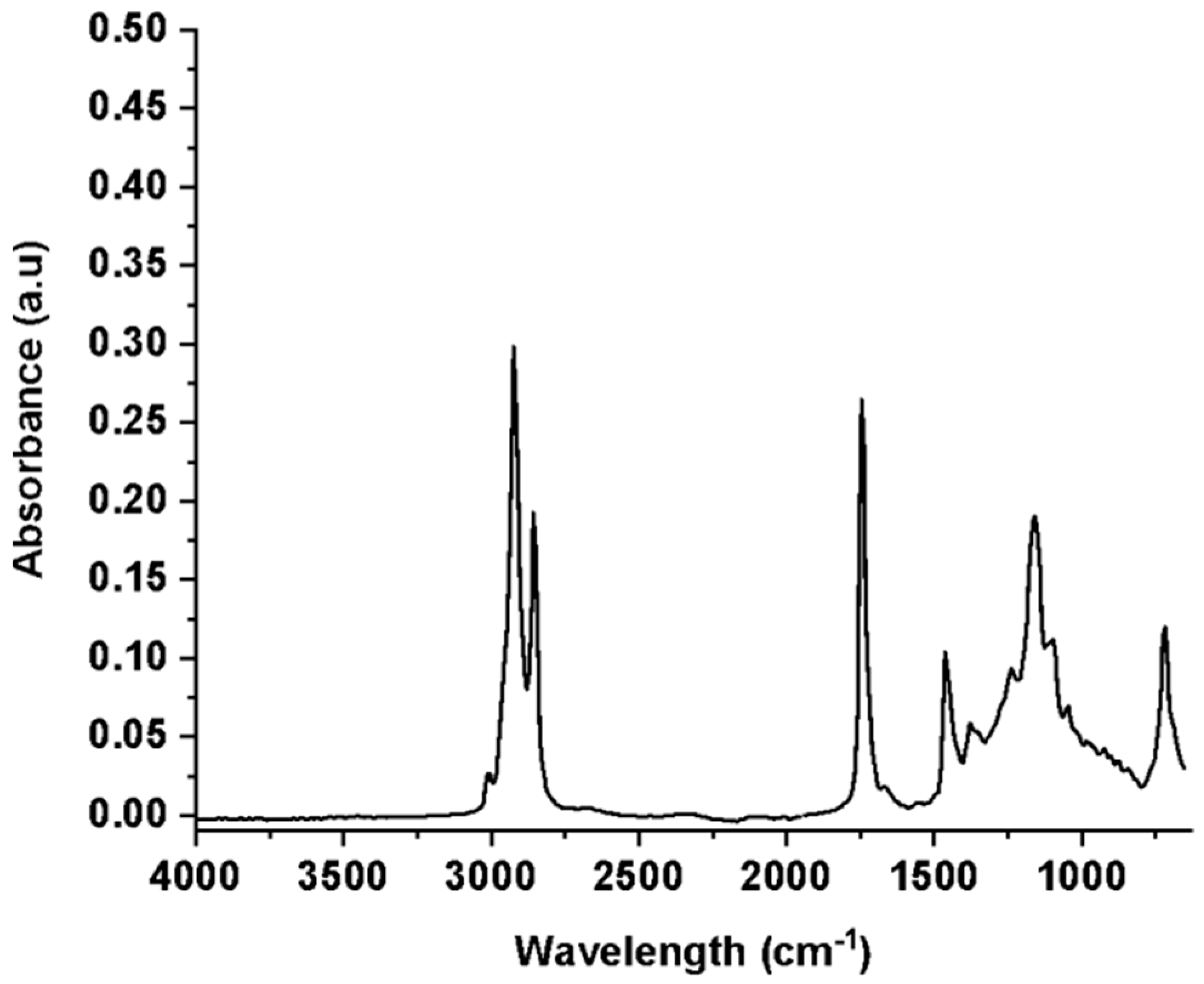
Figure 1. ATR-FTIR spectrum of green coffee oil. The analysis were performed in triplicate (n = 3).
Fermentation 09 00370 g001
Figure 2. UV/Vis absorption spectrum of green coffee oil diluted in ethanol. The analysis was performed in triplicate (n = 3).
Fermentation 09 00370 g002
Figure 3. DPPH scavenging activity for green coffee oil. The assay was performed in triplicate and the mean values were used to calculate the IC50/EC50.
Fermentation 09 00370 g003
Figure 4. Cell viability of human keratinocytes after exposure to different concentrations of green coffee oil, for a period of 24 h. The assay was performed in triplicate.
Fermentation 09 00370 g004
Figure 5. Cell survival fractions observed in human keratinocytes after 24 h of treatment with different concentrations of green coffee oil. * Significantly different from the negative control (p < 0.05). The assay was performed in triplicate.
Fermentation 09 00370 g005
Figure 6. Wound healing rate after treatment with green coffee oil for 48 h. * Significantly different from the negative control (p < 0.05). The assay was performed in triplicate.
Fermentation 09 00370 g006
Figure 7. Wound healing assay: control group photomicrographs.
Fermentation 09 00370 g007
Figure 8. Wound healing assay: samples photomicrographs.
Fermentation 09 00370 g008
Table 1. Composition in fatty acids of samples of green coffee oil by GC–MS.
Composition in Fatty Acids (% w/w)Green Coffee
C 12:0lauric-
C 14:0myristic-
C 15:0pentadecanoic-
C16:0palmitic35.4
C 16:1palmitoleic1.5
C 17:0margaric-
C 18:0stearic8.1
C 18:1oleic9.4
C 18:2 transt-linolenic-
C 18:2linoleic44.8
C 18:3linolenic1.9
C 20:0arachidic3.2
C 20:1eicosenoic-
C 22:0behenic-
C 24:0lignoceric-
Disclaimer/Publisher’s Note: The statements, opinions and data contained in all publications are solely those of the individual author(s) and contributor(s) and not of MDPI and/or the editor(s). MDPI and/or the editor(s) disclaim responsibility for any injury to people or property resulting from any ideas, methods, instructions or products referred to in the content.