Article

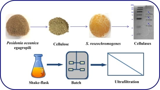
Cellulose from Posidonia oceanica Sea Balls (Egagropili) as Substrate to Enhance Streptomyces roseochromogenes Cellulase Biosynthesis

by
Odile Francesca Restaino
1,*,
Sabrina Cuomo
2,
Sergio D’Ambrosio
2,
Valentina Vassallo
2,
Seyedeh Fatemeh Mirpoor
1,
Concetta Valeria L. Giosafatto
1,
Raffaele Porta
1 and
Chiara Schiraldi
2,*
1
Department of Chemical Sciences, Montesantangelo Campus, University of Naples “Federico II”, Via Cinthia 4, 80126 Naples, Italy
2
Department of Experimental Medicine, University of Campania “Luigi Vanvitelli”, Via De Crecchio 7, 80138 Naples, Italy
*
Authors to whom correspondence should be addressed.
Fermentation 2023, 9(2), 98; https://doi.org/10.3390/fermentation9020098
Submission received: 16 December 2022 / Revised: 10 January 2023 / Accepted: 15 January 2023 / Published: 21 January 2023
(This article belongs to the Special Issue Microbial Biotechnology and Agro-Industrial By-Products Fermentation)

Abstract

:
Enhancing Streptomyces cellulase production by supplying lignocellulose biomasses has been poorly investigated so far. In this research the biosynthesis of Streptomyces roseochromogenes ATCC13400 cellulases was increased for the first time by addition of a cellulose fraction (2.5 g·L−1) to the growth medium, isolated from the marine origin Posidonia oceanica sea balls, generally called egagropili.. In shake flasks the cellulase production increased of 4.3 folds, compared to the control, up to 268 U·L−1 in 72 h, with a productivity of 3.7 U·L−1·h−1, while in batch it was further enhanced up to 347 U·L−1 in 45 h with a doubled productivity of 7.7 U·L−1·h−1 A downstream protocol was set up by coupling two ultrafiltration steps on 10 and 3 kDa membranes to recover the enzymes from the supernatant. A pool of three cellulases, having molecular weights between 115 and 47 kDa, was recovered. The optimal conditions for their enzymatic activity were 60 °C and pH 5.0, and they showed CMCase, FPase and β-glucosidase action. In conclusion, S. roseochromogenes might be considered a new cell factory for cellulase biotechnological production that might be enhanced by using the cellulose from egagropili, a well-known marine origin plant waste, as the substrate.

Graphical Abstract

1. Introduction

From an industrial and scientific point of view, Streptomyces strains are considered very interesting tools thanks to their ability to produce secondary metabolites and enzymes of biotechnological significance [1]. Xylanases, pectinases, ligninases, peroxidases, laccases and cellulases [2] are among the enzymes they can produce. Microbial cellulases have huge potential applications in various fields including pulp and paper, textile, laundry, food and feed industry, brewing and agriculture [3]. More recently, from a circular bioeconomy perspective, microbial cellulases have gained more and more attention for the bioconversion of lignocellulosic biomasses into added value products by sustainable biotechnological processes or into fermentable sugars for producing biofuel and replacing fossil fuel extraction [2,3]. In all these fields, Streptomyces cellulases might be a potential alternative to fungi enzymes, that are more frequently commercialized, as they show a wider cellulolytic activity. Cellulases, such as endo-β-glucanases (E.C. 3.2.1.4), exo-β-glucanases (E.C.3.2.1.91) and β-glucosidases (E.C. 3.2.1.21), have been reported for different Streptomyces strains, and in some cases, they could be contemporarily produced as a single pool of enzymes having molecular weights between 24 and 119 kDa [4,5,6,7,8,9]. For all these reasons, nowadays, the research of new cellulolytic Streptomyces strains, as well as of new strategies to enhance their enzyme synthesis, is very interesting [1,2,3]. A possible approach to increase the production might be by supplementing the growth medium with an appropriate carbohydrate substrate in order to induce the expression of different types of cellulases. The addition of simple substrates, such as carboxymethylcellulose (CMC), prompted the cellulase production by Streptomyces sp. CC48, Streptomyces ruber and Streptomyces griseorubens [6,8,9], while in the case of Streptomyces coelicolor A (3) SCO6548 cellulase biosynthesis was enhanced by cellobiose, glucose and Avicel (microcrystalline cellulose powder) supplementation to a Luria–Bertami medium [7]. As actinomycetes can also grow on complex substrates, the diverse cellulase production might also be boosted by the growth medium supplementation with raw materials, such as sugarcane bagasse, wheat bran or corn steep liquor, as shown for Streptomyces viridobrunneus SCPE-09 cellulase production [5]. However, so far the biotechnological production of these enzymes has been reported only at lab scale, in shake flasks and in very long processes (i.e., of 96–240 h), because these bacteria generally grow very slowly and the enzymes are biomass-related products [4,5,6,7,8,9]. Fermentations in bioreactors, with growth conditions better controlled than shake flasks, could be beneficial alternatives leading to higher cellulase production in shorter times, thus also increasing the productivity. From a perspective of a potential biotechnological application, the purification of extracellular cellulases is also a key issue and, so far, various techniques, such as precipitation with ammonium sulfate, dialysis, gel-filtration chromatography and/or ultrafiltration with 10 kDa polyethersulfone (PES) membranes, eventually used in series, have been employed [4].
In this work, for the first time, the production of extracellular cellulases by Streptomyces roseochromogenes was investigated, as well as the possibility to prompt their biosynthesis by supplementing the growth medium with a cellulose containing fraction isolated from Posidonia oceanica sea balls called egagropili. This strain has already shown a wide array of catalytic activities and it has been employed in stereospecific 16α-hydroxylation of hydrocortisone in whole cell fermentation processes, as well as in the direct whole cell bioconversion of hydrocortisone into desfluorotriamcinolone, an important anti-inflammatory agent, when coupled with Arthrobacter simplex [10,11,12]. Posidonia oceanica is a marine plant, a Mediterranean endemic species, whose stranded leaves, once accumulating on the coasts, play an important ecological role in protection from erosion, while the roots and rhizome fragments, generally called “egagropili” or sea balls, accumulate in large amounts as free-floating and brown ball-shaped materials. Egagropili are commonly considered a problem by municipalities and a waste to be discarded because they are not liked by tourists, but, instead, they might be considered a resource [13,14]. The dry form and the small salt content (lower than 2%) of the egagropili make them a non-inflammable, difficult to degrade material, and thus perfect to be used for insulation in buildings and constructions, while their lignocellulose composition makes them a possible useful source for biotechnological applications. In a previous scientific work, egagropili powder was supplemented in the growth medium of S. roseochromogenes to enhance melanin production [13]. From the same egagropili powder a lignin carbohydrate complex (LCC) and a cellulose containing fraction (32.8% and 54.2% of the dry weight, respectively) were also previously isolated [14,15]. The obtained LCC has been successfully used as reinforcement of protein-based films with the aim of replacing hemp oil in packaging system manufacturing [15]. The cellulose containing fraction, instead, has so far never been used, not even as raw material in bioprocesses. In this work, shake flask experiments were initially carried out to test the influence of the cellulose addition to a previously optimized medium on the kinetics of growth and on the cellulase production. Then, the process was scaled-up to 2-L batch experiments. The extracellular produced cellulases were purified by coupling two ultrafiltration steps on 10 and 3 kDa membranes and were characterized in terms of molecular weight, optimal pH and temperature conditions and for their enzymatic activities. Their hydrolytic ability was also tested on different substrates, such as carboxymethyl cellulose (CMC), cellobiose, lactose, maltose, dextrin and Whatman filter paper.

2. Materials and Methods

2.1. Materials

Yeast and malt extracts used in the growth medium were purchased from Organotechnie (France), while all other nutrients and salts were furnished by Sigma-Aldrich (St. Louis, MO, USA). Agar used for the plates was from Oxoid (Italy). Sodium carboxymethyl cellulose, sodium citrate, dextrin, cellobiose, maltose and lactose were also purchased by Sigma-Aldrich (St. Louis, MO, USA) as well as the Whatman filter paper (grade 1: 11 μm, medium flow filter paper). Commercial cellulases Cellic CTec2 were provided by Novozymes (Copenhagen, Denmark). The NaOH solution used to prepare the buffer for high-performance anion exchange chromatography with pulsed amperometric detection (HPAE-PAD) analyses was from J. T. Baker (The Netherlands). Sodium dodecyl sulfate (SDS), polyacrylamide, tetramethylethylenediamine, ammonium persulfate, Comassie Blue and Congo Red, used for SDS-PAGE, were from Sigma-Aldrich (St. Louis, MO, USA). Egagropili were collected on the Sardinian Poetto beach (Cagliari, Italy) and the cellulose fraction was extracted by sodium chlorite oxidation technique and then freeze-dried, as previously described [14,15].

2.2. Microorganism and Medium

S. roseochromogenes ATCC13400 bacterial strain was purchased from DSMZ (Braunschweig, Germany). The strain was stored and maintained in 20% (v/v) glycerol stock solutions at −80 °C and on agar plates of GYA medium (20.0 g·L−1 glucose, 20.0 g·L−1 yeast extract, 2.0 g·L−1 (NH4)2SO4) after being incubated at 26 °C for 48 h in a rotary air shaker (Infors HT Incubator, Bottmingen, Switzerland). S. roseochromogenes was grown in shake flasks and batch experiments on GEM III N medium (12.0 g·L−1 glucose, 6.0 g·L−1 yeast extract, 30.0 g·L−1 malt extract, 42.9 g·L−1 KH2PO4 and 17.4 g·L−1 of K2HPO4) at pH 6.0 [10,11]. For all the experiments the medium was first sterilized by autoclave without glucose and K2HPO4, and then they were both added to the media by filtration on 0.22 µm membranes (Millipore, France).

2.3. Shake Flask Experiments

S. roseochromogenes shake flask experiments were carried out in 1-L baffled flasks, containing 200 mL of GEM III N medium supplemented or not with 2.5 g·L−1 of cellulose, in triplicate, at 26 °C and 250 rpm, in a rotary air shaker (model Minitron, Infors, Bottmingen, Switzerland) for 96 h [11]. A single agar plate was used as inoculum for each shake flask. During cell growth, broth samples (15 mL) were withdrawn every 24 h.
Aliquots of 5 mL were used to determine the cell dry weight after supernatant removal at 4 °C and at 12,000 rpm for 10 min (Avanti J-20XP, Beckman Coulter, Brea, CA, USA). The obtained pellets were then washed two times with one volume of physiological solution, centrifuged at 4 °C and 12,000 rpm for 10 min (Avanti J-20XP, Beckman Coulter, Brea, CA, USA) and then put in an oven (E28, Binder, Germany) at 80 °C to reach a constant dry weight. Aliquots of 1.0 mL from the same broth samples were centrifuged at 4 °C and 12,000 rpm for 10 min (Avanti J-20XP, Beckman Coulter, Brea, CA, USA) to obtain the supernatants, then used to evaluate the glucose consumption by HPAE-PAD. The remaining volumes of the broth samples were centrifuged at 4 °C and 12,000 rpm for 10 min (Avanti J-20XP, Beckman Coulter, Brea, CA, USA) and the supernatants were used to determine the total protein content and the extracellular cellulase activity.

2.4. Batch Experiments

Batch experiments were performed in duplicate in a 2.5-L fermenter (Biostat CT plus, Sartorius group, Germany) with a working volume of 2.0 L, sterilizable in situ and equipped with pH, temperature and pO2 probes and three peristaltic pumps for the addition of alkali, acid and antifoam. The calibration of the pO2 electrode (Mettler Toledo, Switzerland) was carried out by using a pure oxygen flow as the 100% value. The batch inoculum was prepared by seeding a 1-L baffled shake flask containing 200 mL of GYA medium with a single agar plate and incubating it at 26 °C and 250 rpm for 72 h in a rotary air shaker (Infors HT Incubator, Bottingen, Switzerland). Batch experiments were run on 2 L of GEM III N medium supplemented with 2.5 g·L−1 of cellulose at 26 °C for 45 h. Initially, 200 µL·L−1 of antifoam were added to the medium. During the fermentation the pH value was kept constant at 6.0 by addition of 25% NH4OH and/or 30% H2SO4 solutions. Stirring was modulated between 300 and 480 rpm to always maintain the pO2 higher than 20% while the airflow was kept at 1.0 vvm. The process parameters were remotely controlled and collected by a digital control unit (DCU) equipped with MFCS-win software (Braun Biotech International, Sartorius Group, Göttingen, Germany). During the fermentation experiments broth samples (15 mL) were withdrawn at specific time points to determine the cell dry weight and to evaluate the glucose concentration, as previously described. The remaining volume of the samples was centrifuged to remove the biomass and the supernatants were used to evaluate the total protein content and the extracellular cellulase activity.

2.5. Extracellular Cellulase Recovery by Ultrafiltration

At the end of the shake flask runs broth supernatants were collected by centrifugation at 4 °C and 12000 rpm for 30 min (Avanti J-20XP, Beckman Coulter, Brea, CA, USA). Supernatant volumes resulting from duplicate shake flask runs were unified (400 mL) and ultrafiltrated on a 10 kDa cut-off polyethersulfone (PES) membrane with a filtration area of 0.05 m2 (PelliconXL Biomax, Millipore Corporation, Billerica, USA) by using a LabscaleTM TFF system (Millipore Corporation, Billerica, MA, USA). After concentration the sample was diafiltered with Milli-Q water. During the entire process the data of the inlet and outlet pressures and flux volumes were recorded. The 10 kDa concentrated supernatants were then further concentrated on 3 kDa centrifugal filter devices (Millipore, Molsheim, France) at 4 °C and 12,000 rpm (Z216 MK, Hermle Labortechnik GmbH, Wehingen, Germany) and then used for SDS-PAGE analysis, to determine the total protein content and the extracellular cellulase activity.

2.6. Protein Content Determination

Protein content of the supernatant samples of both shake flasks and batch cultivations, as well as of the samples from the membrane-based purification process, were determined, in terms of g·L−1, by a colorimetric assay [16] by using the Kit Protein assay Biorad (Bio-Rad Laboratories Inc, USA), according to the manufacturer’s procedure and by employing bovine serum albumin (BSA) as the standard (Sigma-Aldrich, St. Louis, MO, USA). The absorbance was measured at 595 nm by a spectrophotometer DU800, Beckman Coulter (Brea, CA, USA).

2.7. Monosaccharide Determination

The supernatants of shake flask and batch fermentation time points were concentrated ten-fold with 3 kDa centrifugal filter devices (Millipore, Molsheim, France) at 4 °C and 12000 rpm (Z216 MK, Hermle Labortechnik GmbH, Wehingen, Germany) to determine glucose consumption during the bacterial growth. Analyses were performed by HPAE-PAD (ICS-3000, Thermofisher, Waltham, MA, USA), using a Carbopac PA1 column (Thermofisher, USA) and a 237 mM NaOH buffer, at 25 °C, as previously described [17,18]. The same method was also used to detect glucose or other monosaccharides released from the disaccharide, oligosaccharide and polysaccharide substrates during the enzymatic assays performed to test cellulase activity.

2.8. SDS-PAGE and Zymogram Analyses

To evaluate the protein pattern of the final shake flask samples resulting from the 10 and 3 kDa membrane purifications, SDS-PAGE and zymogram analyses were performed by slightly modifying the protocol by Laemmli [19]. A single gel was prepared by adding 1.0% of CMC (w/v) to the resolving gel solution before pouring it into the chamber. A total amount of 40 μg of proteins was loaded for each sample in duplicate, specularly in the two half parts of the gel and then the gel was run at 120 kV for about 2 h. A 19 to 250 kDa protein ladder (Prestained Standard, Invitrogen, USA) was used as the standard. Half of the gel was stained with Coomassie blue solution (Coomassie Brilliant Blue R–250, BIORAD, USA) for 1 h and the other half with a 5% Congo Red solution (Sigma-Aldrich St. Louis, MO, USA) for 30 min. Later, the Coomassie blue stained gel was extensively washed with a 20% methanol and 7% acetic acid solution, while the Congo Red stained gel was destained with 1 M NaCl. The band’s molecular weight values were evaluated by using a Gel Doc 2000 UV System (GEL DOCTM EZ System, BIORAD, USA).

2.9. Determination of the Optimal Conditions of Cellulase Activity

Several assays were performed to investigate the optimal temperature and pH conditions for the cellulase activity. Tests at different temperatures ranging from 20 to 80 °C were performed at pH 5.0 and 600 rpm for 20 min, using a Thermomixer AG 22331 (Eppendorf, Germany), after mixing 0.3 g·L−1 total protein concentration in 2% CMC (w/v) solution prepared in 50 mM sodium citrate. Tests at different pH values were run at 60 °C and 600 rpm for 20 min, using a Thermomixer AG 22331 (Eppendorf, Germany) after mixing 0.3 g·L−1 total protein concentration in 2% CMC (w/v) solutions prepared in 50 mM sodium citrate or in 50 mM sodium phosphate buffered at pH values from 3 to 6 and from 6 to 8, respectively. In these assays the residual activity was calculated as a percentage of the maximum activity according to the following equation:
Residual activity (%) = [(U·L−1)x/(U·L−1)max] × 100
where (U·L−1)x was the produced cellulase concentration in terms of units at the condition x of temperature or pH, and (U·L−1)max was the maximum of produced cellulase concentration in terms of units at the optimal temperature or pH conditions. All other activity assays, following this optimization, were performed by mixing 0.3 g·L−1 total protein concentration in 2% CMC (w/v) solution prepared in 50 mM sodium citrate buffer at pH 5.0 and by incubating the solution at 60 °C and 600 rpm for 20 min. One unit of enzyme activity is defined as the amount of enzyme producing 1 µmole·L−1·min−1 of glucose. From the concentration of glucose released by the enzyme at each time point, determined by HPAE-PAD analyses, the eventual initial glucose concentration present in the assay samples at time zero was always subtracted. Specific activity was calculated by dividing the cellulase concentration, in terms of U·L−1, by the protein content expressed in terms of g·L−1.

2.10. Cellulase Activity Assays

Enzymatic hydrolytic ability of the purified extracellular cellulases, produced on the cellulose supplemented medium, was compared with both the activity of the cellulases produced on the control medium and with the activity of the commercial cellulases by using CMC as the substrate. For all three samples (i.e., the purified sample produced on the cellulose supplemented medium, the purified sample produced on the control medium and the commercial sample) assays were performed at 60 °C and 600 rpm by adding 0.3 g·L−1 total proteins in 2% CMC (w/v) 50 mM sodium citrate solution at pH 5.0. The released glucose was determined by HPAE-PAD after 10 and 20 min of reaction by using the analytical method reported above. Then, different types of carbohydrates including disaccharides, oligosaccharides and polysaccharides, such as cellobiose, lactose, maltose, dextrin and Whatman paper, were selected as substrates to determine the diverse hydrolytic ability of the cellulases. The experiments were carried out under the best experimental conditions at 60 °C and 250 rpm for 24 h, by incubation in a rotary air shaker (Infors HT Incubator, Switzerland) and by using 4% (w/v) of each substrate in 5 mL of 50 mM sodium citrate buffer at pH 5.0.

3. Results

3.1. Shake Flask Experiments and Cellulase Purification by Ultrafiltration Membranes

Initial shake flask experiments were performed to check the extracellular production of cellulases by S. roseochromogenes on GEM III N medium, used as the control, and on the same medium supplemented with egagropili-derived cellulose. The biomass formation increased rapidly in the presence of cellulose reaching values 9.2-fold higher than the control within 24 h (Figure 1 and Figure S1) and it maintained values 1.3–1.4-fold higher than the control during the following hours (up to 10.9 ± 0.40 gcdw·L−1 at 96 h versus 7.6 ± 0.24 gcdw·L−1 of the control), even though the consumption of glucose was similar in both conditions. In fact, the initial glucose concentration was almost completely consumed at 72 h (Figure 1). Instead, as shown in Figure 1, the extracellular cellulase activity, in the presence or not of cellulose, was different. In the absence of cellulose supplementation, the cellulase activity resulted between 23.4 ± 4.2 and 103.4 ± 9.1 U·L−1 with a maximum at 96 h, while the production of the cellulases in the supplemented medium was faster and higher in the range from 2.3 to 7.5 fold, up to a maximum of 267.8 ± 6.3 U·L−1 at 72 h of growth, with a productivity of 3.7 U·L−1·h−1 (Figure 1). A protocol for the supernatant purification was then set up to recover the extracellularly produced cellulases after removal of the biomass by centrifugation (Table 1). The supernatant volumes (400 mL) collected from the not-supplemented (control) and the supplemented shake flasks were concentrated 12 and 15 fold, respectively, with 10 kDa ultrafiltration membranes. The control sample revealed initial and final specific activities of 435 U·g−1 and 11,548 U·g−1, respectively, with a 25-fold purification increase in a single ultrafiltration step (Table 1). The sample induced with cellulose revealed an initial specific activity of 1103 U·g−1, 2.5-fold higher than the control, and a final specific activity of 24,635 U·g−1 with a 22-fold purification increase in a single ultrafiltration step (Table 1). Small aliquots of the 10 kDa concentrated samples were further ultrafiltrated with 3 kDa centrifugal filter devices to characterize the enzymes.

3.2. Determination of Cellulase Molecular Weights and Optimal Activity Conditions

SDS-PAGE and zymogram analyses were performed to determine the presence and the molecular weight of one or more cellulases in the 10 kDa and 3 kDa concentrated samples produced on the cellulose supplemented medium. The analyses revealed the presence of three enzymes that showed cellulase activities having molecular weight values of 47.34 kDa, 63.08 kDa and 115.07 kDa, respectively (Figure 2A,B). The optimal temperature and pH conditions for the activity of the cellulase pool were then tested by using CMC as the substrate (Figure 3A,B). A maximum cellulase activity was observed at 60 °C but residual activities of approximately 50.0% and 63.0% were still detected at 50 and 70 °C, respectively (Figure 3A). Then, when the activity was tested at 60 °C but at different pH values, it was noted that the maximum occurred at pH 5.0, while at pH 4.0 and 6.0 only residual activities of 12.7% and 9.2% were observed, respectively (Figure 3B).

3.3. Cellulase Activity Assays

In the optimal conditions of pH and temperature, the pool of purified cellulases, obtained with the supplemented medium, was initially tested for possible endo-β-glucanase activity by using CMC as the most suitable substrate (CMCase activity). This activity was compared to that of the commercial cellulases and of the S. roseochromogenes enzymes obtained with the not supplemented medium. The commercial enzymes were able to release a maximum of 6447.8 µmole·L−1 glucose in 20 min. The glucose released by the purified enzymes produced on the cellulose-supplemented medium was also very high (3576.9 µmole·L−1 at 20 min), up to the 63.0% of the glucose released by the commercial product (Figure 4). The supplementation of cellulose in the medium effectively increased the activity of the cellulases produced by S. roseochromogenes as the enzymes produced on the control medium released only very low amounts of glucose with a maximum of 239.2 µmole·L−1, corresponding to only 6.6% and 3.7% of the maximum glucose released by the cellulases produced on the supplemented medium and by the commercial enzyme, respectively. To determine other possible cellulase activities of the pool, and to exclude any other hydrolytic activities, such as amylase, different substrates, such as cellobiose, lactose, maltose, dextrin and the Whatman paper, were tested in different assays (Figure 5). The pool was shown to easily hydrolyze a disaccharide substrate, such as cellobiose (disaccharide of β-(1-4)-glucose) releasing 73.9% of the initial glucose in only 24 h, thus demonstrating a clear β-glucosidase activity (Figure 5). The degradation of Whatman paper, a complex substrate made of β-(1-4)-glucose fibers, released 11.9% glucose, and this result demonstrated that the action of endo-β-glucanase and of β-glucosidase was also synergic with the one of a cellobiohydrolase, such as obtaining the so-called filter paper cellulase (FPase) activity (Figure 5). Instead, from maltose (disaccharide of α-(1-4)-glucose) and linear dextrin (oligomer of α-(1-6)-glucose), very low percentages of glucose were released (0.6% and 1.6%, respectively), while an unusual catalytic activity on lactose (disaccharide of α-(1-4)-glucose and galactose) was revealed with 3.1% of initial glucose degraded (Figure 5).

3.4. Batch Experiments

To scale up the process of extracellular cellulase production, S. roseochromogenes was also grown in 2-L batch experiments on the GEM III N medium supplemented with 2.5 g·L−1 of egagropili-derived cellulose (Figure 6). The bacterial growth in the controlled batch conditions was two times faster than that observed in shake flasks, reaching a bio-mass maximum of 13.4 ± 3.1 gcdw·L−1 within 45 h (Figure 6). This was probably due to a faster glucose consumption, so that the initial glucose amount was completely up taken in 45 h (Figure 6). In the meanwhile, the cellulase production resulted in being faster and higher, with a maximum of 367.0 ± 5 U·L−1 in only 45 h, so that both production and productivity were 1.4 and 2.0 times higher, respectively (maximum productivity was 7.6 U·L−1·h−1).

4. Discussion

Microbial cellulases are widely employed in different industrial applications, such as pulp, paper, textile and food industries, as well as in biofuel production [3,20,21,22]. In the food industry, for example, cellulases, such as exo- and endo-β-glucanases, together with pectinases and xylanases, are used as macerating enzymes in the extraction and clarification of fruits to produce juices, to increase the cloud stability and to reduce the viscosity of the final products. β-glucosidases, together with pectinases, instead, are used either to improve the texture, flavor and aroma properties of fruits and vegetables or to reduce the bitterness of citrus fruits [3]. More recently, cellulases have gained greater attention as tools to degrade lignocellulose biomasses in a bioeconomy perspective. The lignocellulose component of plants is the most abundant biomass available in the world and might represent a huge opportunity as a possible waste material to valorize as a substrate for the biotechnological production of added value molecules or second-generation biofuels to replace fossil ones. Cellulose represents the major carbohydrate portion, whose representativeness could vary from 55 to 70%, and it is considered an enormous pool of energy sources for sustainable processes [23]. However, its hydrolysis requires a synergistic multi-enzymatic action by different types of cellulases, such as endo-β-glucanases, exo-β-glucanases and β-glucosidases [24]. Actinobacteria are well known for producing stable cellulases that can perform in harsh conditions and in difficult environments, that are able to degrade different and complex substrates and that could be easily recovered after production in liquid medium as extracellularly produced enzymes [3,20]. For all these reasons, in the last years cellulases from Actinobacteria have been considered a valid alternative to the fungal enzymes that are more frequently commercialized, although the long times necessary for their production and the low yields are limits for their effective applications. In literature, it has been reported that Streptomyces strains can produce both endo-β-glucanases (mainly CMCase), exo-β-glucanases as well as β-glucosidases, but only in very few cases the contemporary production of a pool of enzymes has been reported. A possible approach to boost the cellulase concentration might be the addition of substrates inducing their synthesis. CMC has been frequently used in literature, for example, supplemented in agar media, to screen the endo-β-glucanase production of Streptomyces ruber (whose highest enzyme production was about 25600 U·L−1) [8], or of Streptomyces griseorubens [9] that also showed FPase activity. Supplementation of diverse carbon sources, such as cellobiose, glucose and Avicel (microcrystalline cellulose powder) (1% each), in a semi-defined medium of Streptomyces coelicolor A (3) SCO6548 was a wise approach to induce the biosynthesis of multiple cellulases having an Avicelase, FPase and a moderate CMCase activity [7]. Studies on the use of low-cost biomass residues to produce cellulases by Actinobacteria are, instead, very scarce in literature. In few cases complex agro-industrial substrates were used to prompt the production of cellulases by Streptomyces, but the produced enzymes were only tested for their endo-β-glucanase (CMCase) activity, as this enzyme is considered the most important in breaking cellulosic material, ultimately being useful in both bioethanol production or in detergent and textile industries. For example, Streptomyces viridobrunneus SCPE-09 demonstrated a CMCase activity, in the range from 9.9 to 2000 U·L−1, when grown on a Luria–Bertami medium supplemented with wheat bran or sugar cane bagasse as carbon sources and corn steep liquor as nitrogen source [5]. The maximum production was reported at 120 h of flask shake growth by addition of 0.2% of corn step liquor and of 2.0% of wheat bran, with an increase of 2.8 folds compared to the control. The enzyme, purified from the broth supernatant, showed optimal activity at pH 5.0 and 50 °C [5]. Sugarcane bagasse is one of the main cellulosic agro-industrial waste that contains approximately 50% cellulose, 25% hemicellulose and lignin, while corn steep liquor is the major by-product of the corn wet-milling industry [24]. For these reasons these two substrates have been used for another strain, Streptomyces diastaticus PA-01 isolated from a soil cave in Brazil and added to the growth medium at percentages of 2.4% and 1.3%. Maximum endo-β-glucanase production (about 740 U·L−1) was observed after 120 h of growth at pH 4.8 and 50 °C [25]. Another strain, Streptomyces drozdowiczii, isolated from a Brazilian forest, showed both CMCase and FPase activities when grown at 30 °C on a medium containing simple substrates, such as CMC, or complex ones, such as wheat bran, or distillers’ dried grains as carbon sources and yeast extract or corn steep liquor as nitrogen sources [26]. The medium containing 1.0% CMC and 0.3% of yeast extract resulted in being the most suitable in boosting the extracellular cellulase production and the supernatant showed a maximum CMCase activity, up to 595 U·L−1, in the optimal conditions of pH 5.0 and 50 °C [26]. The production of cellulases by S. roseochromogenes has never been reported and in this paper, for the first time, we explored the possibility to also produce these enzymes by supplementation of the growth medium with a cellulose containing fraction isolated from a marine-origin plant waste called egagropili and derived from Posidonia oceanica. This novel approach aims to valorize this plant waste and its components, as well as to reduce the costs of cellulase production at the same time, by using an inexpensive material as substrate with the aim to develop more sustainable biotechnological processes. The addition of cellulose into our semidefined medium prompted both bacterial growth and cellulase production of 9.2 and 7.5 times, respectively, already in shake flasks. As cellulases are biomass-related products and Streptomyces usually grow very slowly, reducing the time of cellulase production by boosting growth could be an attractive strategy. This approach resulted in being even more effective in the controlled conditions of batch fermentation, because at same time it helped to improve production and productivity, greatly reducing the process time with a maximum of 367.0 U·L−1 in only 45 h. This process time is extremely short, considering that, so far, the processes reported in literature for these microorganisms, performed only in shake flask growths, lasted 96-240 h [4,5,6,7,8,9]. Indeed, shortening of the fermentation process time constitutes a further advantage in terms of costs. Moreover, the extracellular cellulases were easily concentrated and partially purified from the fermentation supernatant with only two tangential ultrafiltration and diafiltration steps that are very easily scalable as unit operations, compared to the previously reported precipitation, dialysis and chromatography methods. The downstream process resulted in a satisfactory recovery and sufficient purification of the cellulases, leading to a over 20-fold increase in specific activity. The purification also allowed the detection of a pool of cellulases with Mw values between 24 and 119 kDa in accordance with the data previously reported in literature for these kinds of enzymes [4,5,6,7,8,9]. Streptomyces viridobrunneus and Streptomyces sp. CC48 also produced a pool of two or three cellulases of 37 and 119 kDa and of 30, 40, and 45 kDa, respectively, when grown on agro-industrial wastes [5,6]. The S. roseochromogenes enzyme pool also showed a maximum activity at pH 5.0 and 60 °C, values similar to the ones reported before in literature [4,5,6,7,8,9]. The egagropili-derived cellulose addition was also effective in inducing only the expression of cellulases that showed a high endo-β-glucanase (CMCase) and β-glucosidase activity, as well as FPase action, without inducing the expression of other hydrolytic enzymes. In particular, the enzymatic activity on the CMC substrate resulted in being the 63.0% of the activity of a commercial enzymatic solution, which is a remarkable result considering that the latter is a cocktail containing different types of enzymes (Cellic CTec2 by Novozymes), not only β-glucosidases but also hemicellulases, as reported by the company. The contemporary expression of a pool of enzymes has been rarely reported in Streptomyces strains, so far, and it is particularly interesting for applications in the degradation of lignocellulose substrates, taking into consideration that in the case of other strains, such as various selected Streptomyces argenteolus ones, the produced endo-β-glucanases were deliberately mixed with commercial β-glucosidases and xylanases to then be applied in the hydrolyses of plant biomasses [4].

5. Conclusions

For the first time it has been demonstrated that S. roseochromogenes produces and secretes a pool of three different extra cellular cellulases when grown on a medium supplemented with cellulose isolated from egagropili, a marine waste derived from Posidonia oceanica. The addition of cellulose greatly boosted the cellulase biosynthesis in shake flask conditions, while the use of controlled parameters in batch fermentations shortened the time of cellulase production, thus also improving the productivity. The pool of enzymes showed different cellulase activities; thus, it might be potentially useful as a cocktail to degrade complex lignocellulose biomasses.

Supplementary Materials

The following supporting information can be downloaded at: https://www.mdpi.com/article/10.3390/fermentation9020098/s1, Figure S1: S. roseochromogenes shake flask growth on GEM III N medium with and without (control) 2.5 g·L−1 of cellulose supplementation.

Author Contributions

Conceptualization, O.F.R. and C.S.; methodology, O.F.R.; investigation, O.F.R., S.C., S.D., V.V., S.F.M. and C.V.L.G.; writing—original draft preparation, O.F.R. and S.C.; writing—review and editing, R.P. and C.S.; funding acquisition, C.S. All authors have read and agreed to the published version of the manuscript.

Funding

Restaino received a fellowship by the Ministry of Education, Universities and Research PON01_2464, while Cuomo was financially supported by the Project Altergon cds 000463.

Institutional Review Board Statement

Not applicable.

Informed Consent Statement

Not applicable.

Data Availability Statement

The data that support the findings of this study are available on request from the corresponding author.

Acknowledgments

The authors would like to thank Maria Fenderico for her helpful technical assistance in preparing the cellulose fraction from Posidonia oceanica egagropili.

Conflicts of Interest

The authors declare no conflict of interest.

References

  1. Barbuto Ferraiuolo, S.; Cammarota, M.; Schiraldi, C.; Restaino, O.F. Streptomycetes as platform for biotechnological production processes of drugs. Appl. Microbiol. Biotechnol. 2021, 105, 551–568. [Google Scholar] [CrossRef] [PubMed]
  2. Leo, V.V.; Asem, D.; Zothanpuia Singh, B.P. Actinobacteria: A highly potent source for holocellulose degrading enzymes. In New and Future Development in Microbial Biotechnology and Bioengineering-Actinobacteria: Diversity and Biotechnological Applications; Singh, B.P., Gupta, V.K., Passar, A.K., Eds.; Elsevier: Amsterdam, The Netherlands, 2018; pp. 191–205. [Google Scholar]
  3. Kuhad, R.C.; Gupta, R.; Singh, A. Microbial cellulases and their industrial applications. Enzym. Res. 2011, 2011, 280696. [Google Scholar] [CrossRef] [PubMed] [Green Version]
  4. Ventorino, V.; Ionata, E.; Birolo, L.; Montella, S.; Marcolongo, L.; De Chiaro, A.; Espresso, F.; Faraco, V.; Pepe, O. Lignocellulose-adapted endo-cellulase producing Streptomyces strains for bioconversion of cellulose-based materials. Front. Microbiol. 2016, 7, 2061. [Google Scholar] [CrossRef] [PubMed] [Green Version]
  5. Da Vinha, F.N.M.; Gravina-Oliveira, M.P.; Franco, M.N.; Macrae, A.; da Silva Bon, E.P.; Nascimento, R.P.; Coelho, R.R.R. Cellulase production by Streptomyces viridobrunneus SCPE-09 using lignocellulosic biomass as inducer substrate. Appl. Biochem. Biotechnol. 2011, 164, 256–267. [Google Scholar] [CrossRef] [PubMed]
  6. Celaya-Herrera, S.; Casados-Vázquez, L.E.; Valdez-Vazquez, I.; Barona-Gómez, F.; Bideshi, D.K.; Barboza-Corona, J.E. A cellulolytic Streptomyces sp. isolated from a highly oligotrophic niche shows potential for hydrolyzing agricultural wastes. BioEnergy Res. 2021, 14, 333–343. [Google Scholar] [CrossRef]
  7. Lim, J.H.; Lee, C.R.; Dhakshnamoorthy, V.; Park, J.S.; Hong, S.K. Molecular characterization of Streptomyces coelicolor A (3) SCO6548 as a cellulose 1,4-β-cellobiosidase. FEMS Microbiol. Lett. 2016, 363, fnv245. [Google Scholar] [CrossRef] [PubMed] [Green Version]
  8. El-Sersy, N.A.; Abd-Elnaby, H.; Abou-Elela, G.M.; Ibrahim, H.A.; El-Toukhy, N.M. Optimization, economization and characterization of cellulase produced by marine Streptomyces ruber. Afr. J. Biotechnol. 2010, 9, 6355–6364. [Google Scholar]
  9. Prasad, P.; Singh, T.; Bedi, S. Characterization of the cellulolytic enzyme produced by Streptomyces griseorubens isolated from Indian soil. J. King Saud Univ.-Sci. 2013, 25, 245–250. [Google Scholar] [CrossRef] [Green Version]
  10. Restaino, O.F.; Marseglia, M.; De Castro, C.; Diana, P.; Forni, P.; Parrilli, M.; De Rosa, M.; Schiraldi, C. Biotechnological transformation of hydrocortisone to 16α-hydroxy hydrocortisone by Streptomyces roseochromogenes. Appl. Microbiol. Biotechnol. 2014, 98, 1291–1299. [Google Scholar] [CrossRef]
  11. Restaino, O.F.; Marseglia, M.; Diana, P.; Borzacchiello, M.G.; Finamore, R.; Vitiello, M.; D’Agostino, A.; De Rosa, M.; Schiraldi, C. Advances in the 16α-hydroxy transformation of hydrocortisone by Streptomyces roseochromogenes. Process. Biochem. 2016, 51, 1–8. [Google Scholar] [CrossRef]
  12. Restaino, O.F.; Barbuto Ferraiuolo, S.; Perna, A.; Cammarota, M.; Borzacchiello, M.G.; Fiorentino, A.; Schiraldi, C. Biotechnological transformation of hydrocortisone into 16α-hydroxyprednisolone by coupling Arthrobacter simplex and Streptomyces roseochromogenes. Molecules 2020, 25, 4912. [Google Scholar] [CrossRef] [PubMed]
  13. Restaino, O.F.; Scognamiglio, M.; Mirpoor, S.F.; Cammarota, M.; Ventriglia, R.; Giosafatto, C.V.L.; Fiorentino, A.; Porta, R.; Schiraldi, C. Enhanced Streptomyces roseochromogenes melanin production by using the marine renewable source Posidonia oceanica egagropili. Appl. Microbiol. Biotechnol. 2022, 106, 7265–7283. [Google Scholar] [CrossRef] [PubMed]
  14. Mirpoor, S.F.; Giosafatto, C.V.L.; Di Pierro, P.; Di Girolamo, R.; Regalado-González, C.; Porta, R. Valorisation of Posidonia oceanica sea balls (egagropili) as a potential source of reinforcement agents in protein-based biocomposites. Polymers 2020, 12, 2788. [Google Scholar] [CrossRef] [PubMed]
  15. Mirpoor, S.F.; Restaino, O.F.; Schiraldi, C.; Giosafatto, C.V.L.; Ruffo, F.; Porta, R. Lignin/carbohydrate complex isolated from Posidonia oceanica sea balls (Egagropili): Characterization and antioxidant reinforcement of protein-based films. Int. J. Mol. Sci. 2021, 22, 9147. [Google Scholar] [CrossRef] [PubMed]
  16. Bradford, M.M. A rapid and sensitive method for the quantitation of microgram quantities of protein utilizing the principle of protein-dye binding. Anal. Biochem. 1976, 72, 248–254. [Google Scholar] [CrossRef] [PubMed]
  17. Restaino, O.F.; Borzacchiello, M.G.; Scognamiglio, I.; Porzio, E.; Manco, G.; Fedele, L.; Donatiello, C.; De Rosa, M.; Schiraldi, C. Boosted large-scale production and purification of a thermostable archeal phosphotriesterase-like lactonase for organophosphate decontamination. J. Ind. Microbiol. Biotechnol. 2017, 44, 363–375. [Google Scholar] [CrossRef]
  18. Restaino, O.F.; Borzacchiello, M.G.; Scognamiglio, I.; Fedele, L.; Alfano, A.; Porzio, E.; Manco, G.; Donatiello, C.; De Rosa, M.; Schiraldi, C. High yield production and purificaton of two recombinant thermostable phosphotriesterase-like lactonases from Sulfolobus acidocaldarius and Sulfolobus solfataricus useful as bioremediation tools and bioscavengers. BMC Biotechnol. 2018, 18, 18. [Google Scholar] [CrossRef] [Green Version]
  19. Laemmli, U.K. Cleavage of structural proteins during the assembly of the head of bacteriophage T4. Nature 1970, 227, 680–685. [Google Scholar] [CrossRef]
  20. Hsu, C.L.; Chang, K.S.; Lai, M.Z.; Chang, T.C.; Chang, Y.H.; Jang, H.D. Pretreatment and hydrolysis of cellulosic agricultural wastes with a cellulase-producing Streptomyces for bioethanol production. Biomass Bioenergy 2011, 35, 1878–1884. [Google Scholar] [CrossRef]
  21. Berlin, A.; Gilkes, N.; Kilburn, D.; Maximenko, V.; Bura, R.; Markov, A.; Skomarovsky, A.; Gusakov, A.; Sinitsyn, A.; Okunev, O.; et al. Evaluation of cellulase preparations for hydrolysis of hardwood substrates. Appl. Biochem. Biotechnol. 2006, 130, 528–545. [Google Scholar] [CrossRef]
  22. Merino, S.T.; Cherry, J. Progress and challenges in enzyme development for biomass utilization. In Advances in Biochemical Engineering/Biotechnology; Olsson, L., Ed.; Springer: Berlin/Heidelberg, Germany, 2007; Volume 108. [Google Scholar] [CrossRef]
  23. Takasuka, T.E.; Book, A.J.; Lewin, G.R.; Currie, C.R.; Fox, B.G. Aerobic deconstruction of cellulosic biomass by an insect-associated Streptomyces. Sci. Rep. 2013, 3, 1030. [Google Scholar] [CrossRef] [PubMed] [Green Version]
  24. Segato, F.; Dama’sio, A.R.L.; de Lucas, R.C.; Squina, F.M.; Prade, R.A. Genomics review of holocellulose deconstruction by Aspergilli. Microbiol. Mol. Biol. Rev. 2014, 78, 588–613. [Google Scholar] [CrossRef] [PubMed] [Green Version]
  25. Bispo, A.S.R.; Andrade, J.P.; Souza, D.T.; Teles, Z.N.S.; Nascimento, R.P. Utilization of agroindustrial by-products as substrate in endoglucanase production by Streptomyces diastaticus PA-01 under submerged fermentation. Braz. J. Chem. Eng. 2018, 35, 429–440. [Google Scholar] [CrossRef] [Green Version]
  26. de Lima, A.L.G.; do Nascimento, R.P.; da Silva Bon, E.P.; Coelho, R.R.R. Streptomyces drozdowiczii cellulase production using agro-industrial by-products and its potential use in the detergent and textile industries. Enzym. Microb. Technol. 2005, 37, 272–277. [Google Scholar] [CrossRef]
Figure 1. S. roseochromogenes shake flask growth on GEM III N medium supplemented with and without (control) 2.5 g·L−1 of egagropili-derived cellulose up to 96 h: biomass formation, glucose consumption and extracellular cellulase activity.
Fermentation 09 00098 g001
Figure 2. SDS-PAGE stained with Coomassie Blue (A) and zymogram stained with Congo Red (B) of the S. roseochromogenes cellulase pool produced on GEM III N medium supplemented with 2.5 g·L−1 of egagropili-derived cellulose: Lanes 1 and 4—ladder; lanes 2 and 5—broth supernatant samples concentrated on 10 kDa membranes; lanes 3 and 6—broth supernatant samples concentrated on 3 kDa membranes. The blue arrows in the Coomassie Blue-stained gel (A) indicate the three bands that in the Congo Red-stained gel (B) resulted in having a cellulase activity. These bands had Mw values of 47.34, 63.08 and 115.07 kDa, as indicated by the arrows.
Fermentation 09 00098 g002
Figure 3. Cellulase activity determined under different temperature (A) and pH (B) conditions of the S. roseochromogenes cellulase pool produced on GEM III N medium supplemented with 2.5 g·L−1 of egagropili-derived cellulose.
Fermentation 09 00098 g003
Figure 4. The activity of the cellulases produced by S. roseochromogenes on GEM III N medium with or without supplementation with 2.5 g∙L−1 of egagropili-derived cellulose in comparison with the activity of the commercial cellulases. Cellulase activity is reported as the amount of glucose (µmol·L−1) released from CMC after 10 and 20 min of assay.
Fermentation 09 00098 g004
Figure 5. Percentage of glucose released from different substrates of enzymatic hydrolysis performed by S. roseochromogenes cellulase produced on the egagropili-derived cellulose supplemented medium.
Fermentation 09 00098 g005
Figure 6. S. roseochromogenes batch on GEM III N medium supplemented with 2.5 g·L−1 of egagropili-derived cellulose up to 45 h: biomass formation, glucose consumption and extracellular cellulase production.
Fermentation 09 00098 g006
Table 1. Ultrafiltration on 10 kDa membranes of the supernatants of shake flask growths on GEM III N medium supplemented or not (control) with 2.5 g∙L−1 of egagropili-derived cellulose: volume, protein content, activity, specific activity and purification fold increase of the initial supernatant samples and of the retentate samples. All the data are averaged values of duplicate experiments with a standard deviation lower than 5.0%.
SampleVolume
(L)
Protein
(g·L−1)
Activity
(U·L−1)
Specific Activity
(U·g−1)
Purification
Fold
Supernatant of control growth w/o cellulose0.4000.227103458
10 kDa retentate of control growth w/o cellulose0.0330.29634291158425
Supernatant of growth with cellulose0.4000.2422671103
10 kDa retentate of growth with cellulose0.0260.34885732463522
Disclaimer/Publisher’s Note: The statements, opinions and data contained in all publications are solely those of the individual author(s) and contributor(s) and not of MDPI and/or the editor(s). MDPI and/or the editor(s) disclaim responsibility for any injury to people or property resulting from any ideas, methods, instructions or products referred to in the content.

