Article

Screening and Probiotic Properties of Lactic Acid Bacteria with Potential Immunostimulatory Activity Isolated from Kimchi

MEDIOGEN Co., Ltd., Biovalley 1-ro, Jecheon-si 27159, Republic of Korea
*
Author to whom correspondence should be addressed.
Fermentation 2023, 9(1), 4; https://doi.org/10.3390/fermentation9010004
Submission received: 23 November 2022 / Revised: 14 December 2022 / Accepted: 18 December 2022 / Published: 21 December 2022
(This article belongs to the Section Fermentation for Food and Beverages)

Abstract

:
The modulation of the immune system is a major mechanism through which probiotics exert beneficial effects on health. Probiotics, including lactic acid bacteria (LAB), have been reported to enhance innate immunity. The purpose of this study was to screen for LAB strains with excellent immunostimulatory activity isolated from kimchi. We selected five promising strains (Limosilactobacillus fermentum MG5489, Lactococcus lactis MG5542, Lacticaseibacillus paracasei MG5559, Latilactobacillus sakei MG5468, and Latilactobacillus curvatus MG5609) that exhibited immune-stimulating effects by inducing the production of nitric oxide (NO) and pro-inflammatory cytokines such as tumor necrosis factor (TNF)-α, interleukin (IL)-6, and IL-1β in RAW264.7 cells. The selected strains significantly increased phagocytic activity of RAW264.7 cells and nuclear factor-κB (NF-κB) activation. Furthermore, the safety of the selected strains was determined using hemolysis and antibiotic susceptibility tests. The stabilities and adhesion abilities of these strains in the gastrointestinal tract (GIT) were also determined. Taken together, these findings suggest that the strains selected in this study have the potential to be novel probiotics to enhance immunity.

1. Introduction

Macrophages, present in all tissues, are effector cells of the innate immune system that play a pivotal role in the host defense system, tissue homeostasis, and inflammatory response [1]. These cells can be activated and switch from a resting to a polarized state by various stimuli. Macrophages can be polarized into classically activated (M1) and alternatively activated (M2) macrophages in response to different stimuli [2]. M1 macrophages are induced by tumor necrosis factor (TNF)-α, interferon (IFN)-γ, and lipopolysaccharide (LPS). Activated M1 macrophages express inducible NO synthase (iNOS) and produce pro-inflammatory cytokines such as TNF-α, interleukin (IL)-1β, and IL-6 to promote Th1-mediated immunity [3]. Conversely, M2 macrophages activated by IL-4, IL-10, IL-13, and IL-33 secrete anti-inflammatory cytokines to induce Th2-associated immunity [2].
Many signaling pathways are involved in the immune response. Among the immune-related pathways, the nuclear factor-κB (NF-κB) signaling pathway is a widely studied signaling cascade that induces polarization of macrophages and is responsible for the induction of gene expression associated with the inflammatory response [1,4]. Pattern recognition receptors (PRRs) activate the NF-κB pathway by recognizing microbe-related molecular patterns. NF-κB activation is initiated by the phosphorylation of IκBα by the multi-subunit IκB kinase (IKK) complex. Phosphorylated IκBα is degraded in the cytoplasm, allowing the nuclear translocation of NF-κB, which subsequently induces the expression of pro-inflammatory mediators [5].
Probiotics are living microorganisms that confer health benefits to the host when administered in moderate amounts [6]. In particular, lactic acid bacteria (LAB) have been used in the food industry for a long time. Some of the species can be described as “generally recognized as safe” (GRAS) according to the Food and Drug Administration (FDA) [7]. Furthermore, all species belonging to the genus Lactobacillus reclassified in 2020 meet the European Food Safety Authority (EFSA) criteria for the qualified presumption of safety status (QPS) [8].
Probiotics have been reported to prevent diarrhea, improve irritable bowel syndrome, boost the immune system, and reduce various diseases such as inflammation and allergies [9]. Recently, given the interaction between probiotics and immune cells, the regulation of the host immune system by modulating macrophage activation through probiotics has attracted considerable attention. Although the mechanism of modulation of the immune system by probiotics remains unclear, it is believed to be due to nutrient competition, metabolite production, and nonspecific immune activation [10]. According to numerous studies, probiotics are involved in immune responses by modulating macrophage polarization [1]. Some strains of LAB increase the levels of pro-inflammatory markers, including TNF-α, IL-1β, and IL-6, and decrease the expression of anti-inflammatory markers (Arg 1, TGF-β, and CD206) by inducing the polarization of macrophages to the M1 phenotype [11,12]. Switching macrophage polarization from M2 to M1 is also promoted by specific strains [12]. In contrast, several LAB strains have been shown to inhibit the expression of pro-inflammatory mediators such as iNOS, COX-2, TNF-α, IL-6, and IL-1β by polarizing M1 macrophages to M2-like macrophages [13,14]. These observations suggest that the immune response to probiotic bacteria is strain-specific.
The purpose of this study was to screen LAB strains for immunostimulatory activity, which was evaluated by determining the production of NO and cytokines and phagocytosis in RAW264.7 cells. The activation of NF-κB, which is involved in macrophage polarization, was also evaluated. In addition, the safety, gastrointestinal resistance, and adhesion ability of the selected strains were investigated to assess their potential application as functional foods.

2. Materials and Methods

2.1. LAB Strains and Cultivation Conditions

All LAB strains used in this study were obtained from MEDIOGEN Co., Ltd. (Jecheon, Republic of Korea). All strains were inoculated and cultivated in MRS broth (Difco Laboratories, Detroit, MI, USA) at 37 °C under anaerobic conditions. All LAB strains tested in this study were isolated from fermented kimchi, a traditional Korean fermented food.

2.2. Cell-Free Supernatant (CFS) Preparation

CFS was prepared according to a previous study with some modifications [15]. For CFS, bacteria cultures in MRS broth were centrifuged at 4000× g for 20 min. After centrifugation, supernatants separated from bacteria were collected and filtrated through filters with 0.22 µm pore size to remove residual cells and cell debris. Thereafter, the CFS was used for the in vitro study and stored at −20 °C for further study.

2.3. Cell Culture

The RAW 264.7 and HT-29 cells were purchased from the Korea Cell Line Bank (Seoul, South Korea). RAW 264.7 and HT-29 cells were cultured in Dulbecco’s modified Eagle’s medium (DMEM; Welgene, Gyeongsan, South Korea) containing 10% heat-inactivated fetal bovine serum (FBS; Welgene) and 1% penicillin-streptomycin (PS; Welgene) at 37 °C in a humidified atmosphere with 5% CO2. Cells were subcultured when reached 70–80% confluence.

2.4. Cell Viability Assay

The 3-[4,5-Dimethylthiazole-2-yl]-2,5-diphenyltetrazolium bromide (MTT; Sigma, St. Louis, MO, USA) assay was performed to evaluate cellular cytotoxicity caused by CFS derived from LAB strains according to a previous study [15]. Briefly, RAW 264.7 cells were plated at a density of 4 × 10⁵ cells/mL in 96-well plates and maintained overnight at 37 °C. After 5% CFS treatment, the plates were incubated at 37 °C for 24 h. Thereafter, 100 µL of MTT solution was added to each well and incubated for 1 h. After incubation, the insoluble formazan crystals formed by living cells were solubilized in 100 µL of dimethyl sulfoxide (DMSO; Sigma). Cell viability was determined by measuring absorbance at 570 nm using an Epoch 2 spectrophotometer (Bio-Tek, Winooski, VT, USA). The percentage of cell viability was calculated using the following Equation (1):
Cell viability (%) = (As/Ac) × 100
where As is the absorbance of the test sample and Ac is the absorbance of the control at 570 nm.

2.5. Nitric Oxide Assay

The amount of NO secreted by CFS-stimulated RAW 264.7 cells was determined by measuring the nitrite present in the culture medium using the Griess assay according to a previous study [16]. Briefly, RAW 264.7 cells were cultured in 96-well plates (4 × 10⁵ cells/mL) and maintained overnight at 37 °C. The cells were treated with 5% CFS for 24 h. Culture supernatants were mixed with Griess reagent composed of 1% sulfanilamide and 0.1% N-(1-naphthyl)-ethylenediamine dihydrochloride. After incubation at room temperature for 15 min, the absorbance was measured at 540 nm using an Epoch 2 spectrophotometer. DMEM and lipopolysaccharide (LPS; 10 ng/mL; Sigma) were used as negative and positive control, respectively.

2.6. Cytokine Assay

For cytokine analysis, RAW 264.7 cells were plated in 24-well plates (4 × 10⁵ cells/mL) and maintained overnight at 37 °C. The cells were stimulated with 5% CFS for 24 h. The levels of cytokines present in the culture supernatants were measured using mouse TNF-α and IL-6 ELISA kits (R&D Systems, Minneapolis, MN, USA) according to the manufacturer’s protocols.

2.7. Phagocytosis Assay

The effects of CFS from LAB strains on the phagocytic activity of RAW 264.7 cells were investigated using a neutral red uptake assay [17]. RAW 264.7 cells were plated in 96-well plates (4 × 10⁵ cells/mL) and maintained overnight at 37 °C. After treatment with 5% CFS for 24 h, the cells were washed thrice with PBS. A neutral red solution was added, and the cells were further stained for 2 h. After additional washing, neutral red phagocytized by RAW264.7 cells was extracted using lysis buffer (1% acetic acid: 50% ethanol = 1:1). The absorbance of the solution was measured at 540 nm wavelength using an Epoch 2 spectrophotometer. The phagocytic activity was calculated using the following Equation (2):
Phagocytic activity = As/Ac
where As is the absorbance of the test sample and Ac is the absorbance of the control at 540 nm.

2.8. Western Blot Analysis

NF-κB activation was evaluated using a Western blot assay as previously described by Wei et al. [18]. RAW 264.7 cells were seeded at a density of 4 × 10⁵ cells/mL in 6-well plates and cultured at 37 °C overnight. After the medium was changed to fresh medium, the cells were treated with 5% CFS for 24 h. The cells were harvested and washed twice with PBS. The collected cells were resuspended in RIPA buffer (Gendepot Inc., Katy, TX, USA) containing a phosphatase inhibitor cocktail and incubated on ice for 10 min. The total protein content in the cell lysates was measured using the Bradford assay (Gendepot Inc.). Extracted proteins were separated on 10% polyacrylamide gels and transferred onto polyvinylidene fluoride membranes (PVDF; Millipore, Billerica, MA, USA) for 40 min. The membrane was blocked with 5% bovine serum albumin (BSA; Gendepot Inc.) in PBST (PBS containing 0.05% Tween 20) at room temperature for 1 h and then hybridized with specific primary antibodies (p65, p-p65, and β-actin) at 4 °C overnight. After washing, membranes were incubated with horseradish peroxidase-conjugated secondary antibodies at room temperature for 1 h. The protein bands were visualized using the ECL (ATTO, Tokyo, Japan), and densitometric analysis of the band intensities was performed using ImageJ software (National Institutes of Health, Bethesda, MD, USA).

2.9. Probiotic Properties

2.9.1. Resistance to Simulated Gastrointestinal Conditions

Probiotics must survive in the gastrointestinal tract (GIT) to exert beneficial effects on the host [19]. Therefore, the viability of the selected strains was investigated under simulated gastrointestinal conditions as previously described by Lee et al. [15]. Briefly, the cultured bacteria were harvested by centrifugation (4000× g for 20 min) and washed twice with PBS. After washing, PBS was added to adjust the optical density of the cells to 1.0 at 600 nm. The cell suspension (1 mL) was mixed with 9 mL of simulated gastric fluid (SGF; PBS containing 0.3% pepsin, pH 2.5) at 37 °C for 2 h. Ten milliliters of simulated intestinal fluid (SIF; PBS containing 1% pancreatin and 1% bile salt, pH 7.4) was added to the cell pellet obtained by centrifugation (4000× g for 20 min). After incubation at 37 °C for 2.5 h, viable cells were counted by plate count method using MRS agar and expressed as log CFU/mL. The survival rate was calculated using Equation (3):
Survival rate (%) = (Log N/Log N0) × 100
where Log N is the log number of viable cells at the end of the test and Log N0 is the log number of initial viable cells.

2.9.2. Adhesion to HT-29 Cell Line

The adhesion of the selected strains was assessed using the intestinal epithelial cell line HT-29, as previously described [20]. HT-29 cells were plated in 12-well plates (1 × 10⁵ cells/mL) and maintained at 37 °C for 24 h. The selected strains were cultured in MRS for 24 h at 37 °C and harvested by centrifugation (4000× g for 20 min). The pellets were resuspended in DMEM without FBS and PS at a concentration of 1 × 10⁸ CFU/mL. Bacterial suspensions were added to each well, and the plates were incubated for 2 h. After removing the non-adherent bacteria by washing, the cells were scraped from the plates using a scraper. The number of bacteria attached to cells was counted using a plate count method using MRS agar. The percentage of adhesion was calculated using the following Equation (4):
Adhesion rate (%) = (Log N/Log N0) × 100
where Log N is the log number of adhered bacterial cells at the end of the test and Log N0 is the log number of bacteria added.

2.9.3. Hemolytic Activity

To assess biocompatibility against blood cells, cultured bacterial cells were streaked on tryptic soy agar (Difco Laboratories) supplemented with 5% sheep blood (MB cell, Seoul, Republic of Korea) and incubated at 37 °C for 48 h. Hemolysis was evaluated based on the breakdown of red blood cells around the colonies and was expressed as α-hemolysis (greenish halo), β-hemolysis (clear halo), or γ-hemolysis (no halo) [21].

2.9.4. Antimicrobial Susceptibility Assay

The susceptibility of the selected strains was determined using the antibiotic minimum inhibitory concentration (MIC) strip method according to the manufacturer’s protocols (Liofilchem Inc., Roseto degli Abruzzi, Italy). Briefly, bacterial cells were suspended in PBS to a concentration of 0.5 McFarland standard. Then, the 0.5 McFarland bacterial suspension was swabbed onto brain infusion agar (Difco Laboratories) plates using a spreader. MIC test strips were placed in the center of the agar plate, and the plates were incubated for 24 h at 37 °C. The breakpoint values for lactobacilli species are limited in the European Committee on Antimicrobial Susceptibility Testing (EUCAST) and Clinical and Laboratory Standards Institute (CLSI) guidelines. The guidelines of EUCAST and CLSI provide breakpoint values for only four antibiotics. Therefore, the interpretation of the results was based on the established breakpoint values from EFSA [22].

2.10. Statistical Analysis

Statistical analyses were performed using SPSS software version 21 (IBM Inc., Armonk, NY, USA). Differences between groups were evaluated for statistical significance using one-way analysis of variance (ANOVA), followed by Tukey’s post hoc test at p < 0.05. All results are presented as mean ± standard deviation (SD) from three independent experiments.

3. Results

3.1. LAB Strains Induced NO Production in RAW264.7 Cells

To screen the LAB strains that have immunostimulatory activity, we evaluated cytotoxicity and NO production in RAW264.7 cells (Table 1). None of the tested strains had significant cytotoxic effects on RAW264.7 cells. However, we found that only fourteen probiotic strains, Lactiplantibacillus plantarum (L. plantarum) MG5239 (19.64 ± 1.24 μM), MG5663 (18.07 ± 0.66 μM), MG5558 (18.07 ± 1.94 μM), Limosilactobacillus fermentum (L. fermentum) MG5489 (29.96 ± 2.51 μM), MG5613 (21.21 ± 6.88 μM), Lacticaseibacillus paracasei (L. paracasei) MG5219 (8.26 ± 0.39 μM), MG5559 (15.55 ± 1.91 μM), Latilactobacillus sakei (L. sakei) MG5218 (8.13 ± 0.86 μM), MG5468 (12.60 ± 0.76 μM), Latilactobacillus curvatus (L. curvatus) MG5246 (22.35 ± 1.04 μM), MG5374 (24.11 ± 1.28 μM), MG5609 (23.92 ± 1.53 μM), Levilactobacillus brevis (L. brevis) MG5531 (10.14 ± 0.76 μM), and Lactococcus lactis (Lc. lactis) MG5542 (16.37 ± 1.11 μM), significantly induced NO production compared to the untreated control group.
NO secreted by macrophages can enhance immune responses, such as pathogen eradication, inflammation, and tumor suppression [23]. Therefore, based on these results, twelve candidate strains with higher NO production than the LPS-treated positive control (9.08 ± 0.86 μM) were screened for potential as immunostimulants, and cytokine production was analyzed using these strains.

3.2. LAB Strains Induced Pro-Inflammatory Cytokines Production in RAW264.7 Cells

To evaluate cytokine production in the primary screened strains, we investigated the levels of three cytokines (TNF-α, IL-6, and IL-1β) secreted by CFS-treated RAW264.7 cells. The concentrations of these cytokines in the cell culture medium were quantified using ELISA kits. As shown in Figure 1a, TNF-α levels were significantly higher in all CFS-treated cells than in the untreated (not detected) and LPS-treated cells (10.38 ± 5.55 ng/mL). In addition, cells treated with the CFS of all strains, except for L. plantarum MG5239, showed significantly increased IL-6 production compared to that in the untreated control group (0.14 ± 0.08 ng/mL), as shown in Figure 1b. In particular, Lc. lactis MG5542 (23.03 ± 1.45 ng/mL) showed higher induction compared to LPS (15.56 ± 5.37 ng/mL). Finally, the levels of IL-1β in cells treated with Lc. lactis MG5542 (189.28 ± 68.26 pg/mL), L. paracasei MG5559 (93.91 ± 9.40 pg/mL), L. sakei MG5468 (83.17 ± 9.25 pg/mL), and L. curvatus MG5609 (62.24 ± 5.28 pg/mL) were significantly elevated compared to those in untreated (not detected) and LPS-treated cells (10.38 ± 5.55 pg/mL), as shown in Figure 1c. The levels of TNF-α, IL-6, and IL-1β in cells treated with Lc. lactis MG5542 were significantly increased by 21.61-, 1.48-, and 31.84-fold, respectively, compared to those in LPS-treated cells.
Pro-inflammatory cytokines secreted by macrophages, such as TNF-α, IL-6, and IL-1β, induce the early defense of the organism and activate the Th1 response [24]. Based on these results, the five most promising strains, L. fermentum MG5489, Lc. lactis MG5542, L. paracasei MG5559, L. sakei MG5468, and L. curvatus MG5609 were selected, and additional studies were carried out using these strains.

3.3. The Selected Strains Induced Enhancement of Phagocytic Activity in RAW264.7 Cells

To determine the effects of the selected strains on macrophage phagocytosis, a neutral red uptake assay was performed. As shown in Figure 2, CFS-treated cells exhibited an approximately 2-fold higher phagocytic capacity than untreated control cells. This result was similar to that of LPS-treated cells.

3.4. The Selected Strains Induced NF-κB Activation in RAW 264.7 Cells

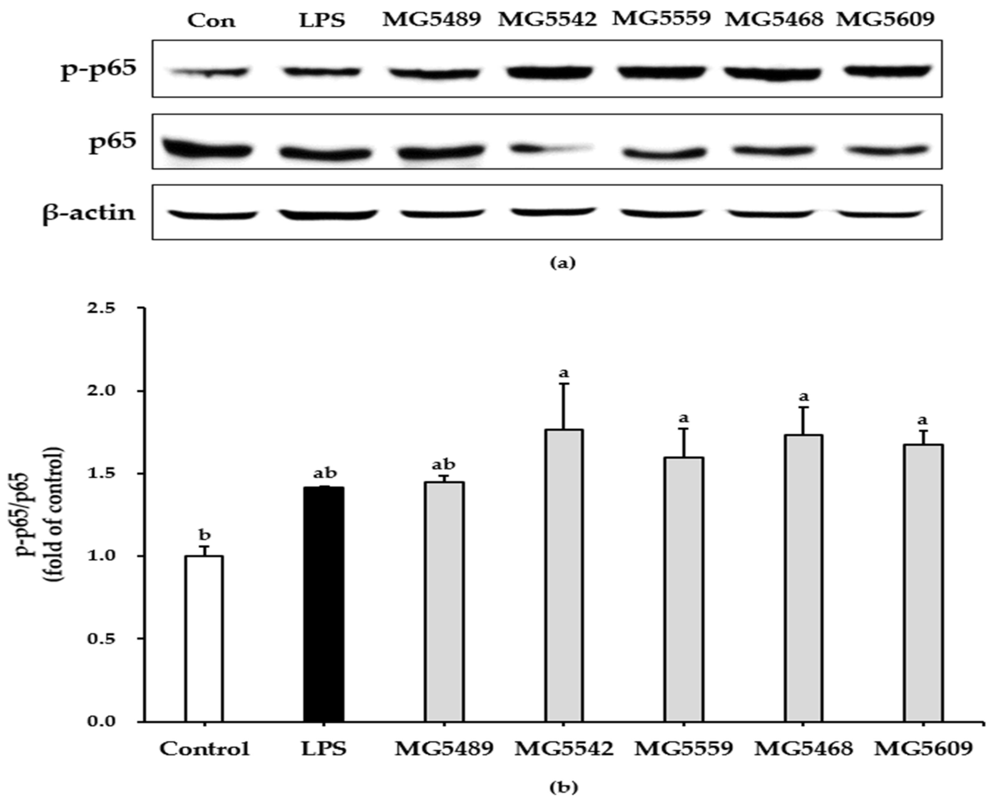
To evaluate whether the selected strains could induce NF-κB activation in cells, we performed a Western blot assay. Our results showed that the phosphorylation levels of p65 in all CFS-treated cells were significantly increased compared to those in untreated control cells (Figure 3).

3.5. Probiotic Properties of the Selected Strains

3.5.1. GIT Stability and Adhesion Ability of the Selected Strains

The resistance of the selected strains to the simulated GIT for a fixed period of time is shown in Table 2. All the tested strains survived after exposure to SGF for 2 h, but a decrease in cell viability was observed at longer incubation periods. However, most strains showed good survival rates for 2.5 h in SIF containing pancreatin and bile salts. Among the studied strains, L. fermentum MG5489 showed the highest survival rate (86.99 ± 0.29%), followed by L. sakei MG5468 (82.57 ± 0.28%) and L. paracasei MG5559 (74.75 ± 0.47%).
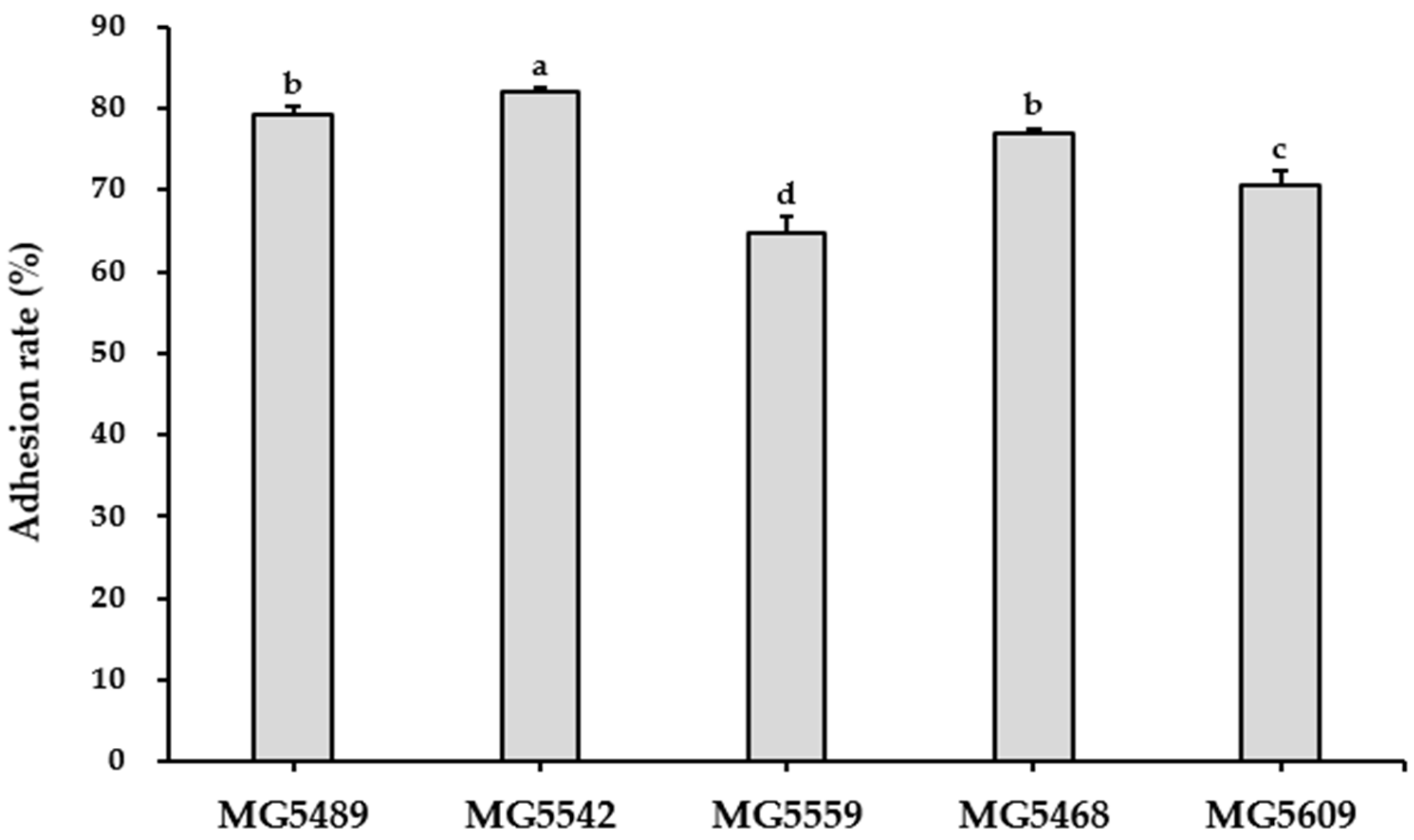
The adhesion abilities of the selected strains were assessed using HT-29 cells (Figure 4). The results revealed that the adhesion rate of the selected strains ranged from 64.82–82.04%. Among the strains studied, Lc. lactis MG5542 (82.04 ± 0.38%) showed the highest adhesion rate.

3.5.2. Safety of the Selected Strains

To assess the safety of the selected strains, we investigated their hemolytic activity. All the tested strains showed γ-hemolytic activity on blood agar plates (Figure S1), suggesting that they had no hemolytic activity.
In addition, MIC tests were performed to evaluate the antibiotic resistance of the selected strains. As shown in Table 3, L. paracasei MG5559 was resistant to chloramphenicol, while the MICs of various antibiotics against all other strains were lower than the breakpoint values indicated in Table S1.

4. Discussion

The primary function of the immune system is to protect the body from pathogens. This protective function requires an innate and adaptive immune system [25]. When the body is exposed to pathogens, the innate immune system is activated to protect it, followed by the adaptive immune system [26]. Probiotics play a role in the regulation of innate and adaptive immunity by interacting with immune cells such as dendritic cells, macrophages, T cells, and B cells [27]. Recently, numerous studies have reported that probiotics can induce early innate immune responses by activating immune cells [10]. In addition, soluble bioactive factors produced by probiotics stimulate the innate immune system through polarization into immune-stimulating macrophages [28]. Therefore, probiotics have attracted considerable interest in recent years as potential dietary supplements for immune enhancement [10].
Since probiotic strains can secrete health-promoting metabolites into the culture medium, CFS derived from probiotics has been used in vitro to evaluate their physiological benefits [28]. Most reports have shown the immunomodulatory effects of whole cells or cell wall components of probiotics on macrophages [29]. However, recent studies have investigated the immunostimulatory activity of CFS from probiotic strains on macrophages [28,30,31]. The CFS of lactobacilli might contain cell wall components such as peptidoglycan, S-layer proteins, and teichoic acids, as well as other peptides that could be responsible for the macrophage activity [32]. In addition, when ingested probiotics are stabilized in the human intestine, they produce a variety of metabolites and secrete those in the lumen of the gut [33]. It has been reported that various metabolites derived from different LAB strains exhibit differential immunomodulatory effects [28]. The present study assessed the immunostimulatory effects of CFS from LAB strains using RAW264.7 cells. In addition, we selected strains as candidates for functional probiotics and investigated their safety and properties.
Macrophages are involved in the innate immune response through the production of NO and cytokines and through phagocytosis [34]. NO is secreted by activated cells and is involved in immune regulation, inflammation, tumor destruction, and non-specific host defense [23]. According to previous studies, the culture supernatants of Lactobacillus acidophilus (L. acidophilus) and Limosilactobacillus reuteri (L. reuteri) increased NO production by approximately 12- and 18-fold, respectively, compared to untreated control cells [32]. Chang et al. [31] reported that the CFS of L. plantarum B0040 (5.28-fold), L. plantarum B0110 (5.69-fold), and Weissella cibaria B0145 (8.24-fold) induced NO secretion from RAW264.7 cells. In the present study, L. fermentum MG5489, Lc. lactis MG5542, L. paracasei MG5559, L. sakei MG5468, and L. curvatus MG5609 showed higher NO induction than the probiotics mentioned above, suggesting that they could induce a positive immune response.
Inflammation is an essential immune response caused by various factors, such as pathogens, damaged cells, and irradiation. It plays a role in removing harmful stimuli and initiating tissue repair [35]. Among a variety of inflammatory factors, the appropriate production of pro-inflammatory cytokines, which play an important role in inflammatory mechanisms, enhances the immune response [36]. Therefore, pro-inflammatory cytokines were investigated to evaluate whether CFS from the selected strains could act as immune stimulators in RAW264.7 cells. Our results showed that the CFS of the initially screened strains significantly increased the production of TNF-α and IL-6 in cells. Furthermore, some strains significantly enhanced the secretion of IL-1β. Consistent with these results, it has been previously shown that CFS from different probiotic strains significantly increases the production of TNF-α and IL-6 in cells [31]. In addition, the culture supernatants of different LAB strains upregulate the gene expression of TNF-α and IL-1β in human macrophages [28]. Based on NO and cytokine production, we selected five strains with potential immunostimulatory activities.
Phagocytosis, which is responsible for eliminating pathogens and dead cells, is essential for initiating the innate immune response [37]. In this study, phagocytic activity was enhanced when macrophages were stimulated with CFS from the selected strains, suggesting that these strains could initiate an immune response. Similarly, a previous study showed that CFS from Lacticaseibacillus rhamnosus (L. rhamnosus) GG, one of the most studied probiotic strains in the world, enhanced phagocytic activity in murine macrophages [30]. In addition, Xiu et al. [38] demonstrated that exopolysaccharides produced by Lacticaseibacillus casei (L. casei) WXD030 induced phagocytosis in RAW264.7 cells.
NF-κB signaling plays a critical role in the inflammatory response to pathogens and tumor cells and is activated by signals related to pathogens or stress [5]. In resting macrophages, the p50/p65 heterodimer, a prototypical form of NF-κB, is sequestered in the cytoplasm by binding IκBα. When cells are stimulated, IκBα is phosphorylated and degraded by the IKK complex, resulting in the nuclear translocation of NF-κB [5]. Phosphorylation of NF-κB subunit p65 activates the expression of genes associated with inflammatory response [5,39]. Phosphorylated p65 induces the production of NO and pro-inflammatory cytokines [40,41]. Our results suggest that the selected strains can enhance immune responses by inducing polarization in M1 macrophages.
Since the health benefits of ingested probiotics are diminished by exposure to gastric and intestinal conditions, their survival in the digestive tract and entry to the small and large intestines is a prerequisite to exert their beneficial effects [19]. In this study, we evaluated the stability of the selected strains in the simulated gastrointestinal fluid to confirm their resistance to gastrointestinal stress. The selected strains showed high survival rates (>50%) in the simulated gastrointestinal tract. According to a previous study, the survival rate of L. paracasei strains ranged from 37.6–79.8% under simulated gastrointestinal conditions [19]. In addition, some commercial probiotics showed survival rates similar to those observed in the current study [42]. Intestinal colonization by probiotics plays an important role in providing health benefits to the host [43]. The ability to adhere to the epithelium is a prerequisite for bacterial colonization in the digestive tract and is one of the selection criteria for probiotics [19]. Mantzourani et al. [44] reported that the adhesion rate of Pediococcus pentosaceus SP2 and L. paracasei SP5 to HT-29 cells is approximately 40%. The selected strains in this study showed higher adhesion ability than those reported in previous study. Therefore, our study suggests that all the selected strains have the potential to be used as probiotics.
Although most probiotics, including LAB, are generally considered safe, the safety of specific strains must be assessed before their use as probiotics [45]. In the present study, we found that all selected strains showed γ-hemolysis, which is considered safe. In addition, the selected species in this study have received the QPS status recognized by the EFSA. However, L. paracasei MG5559 is resistant to chloramphenicol. In general, LAB including lactobacilli and bifidobacteria are sensitive to chloramphenicol. Nevertheless, some strains show resistance to chloramphenicol [46]. Gueimonde et al. [47] reported that intrinsic resistance caused by mutations reduces the risk of horizontal transfer. Thus, L. paracasei MG5559 with chloramphenicol resistance requires further studies using transposons and plasmids to avoid potential risks.

5. Conclusions

We aimed to select strains with excellent immunostimulatory activity from 28 probiotic candidates isolated from kimchi by determining the production of inflammatory mediators and phagocytosis. In this study, we selected five probiotic strains that exhibited high production of NO, TNF-α, IL-6, and IL-1β. The five selected strains induced phagocytosis by macrophages and induced NF-κB activation. Furthermore, the selected strains showed high GIT resistance and adhesion. These effects suggest that the selected strains may improve immune response by activating macrophages and have the potential to be used as probiotics. Further studies are needed to determine the efficacy of these strains in vivo. However, the results in this study demonstrated that LAB strains, including L. fermentum MG5489, Lc. lactis MG5542, L. paracasei MG5559, L. sakei MG5468, and L. curvatus MG5609 exert positive effects on immunostimulatory activity, suggesting that they could serve as functional foods for the improvement of host innate immunity.

Supplementary Materials

The following supporting information can be downloaded at https://www.mdpi.com/article/10.3390/fermentation9010004/s1, Table S1: The microbiological breakpoint values from EFSA. Figure S1: The hemolytic activity of the selected strains.

Author Contributions

Conceptualization, C.-H.K.; methodology, S.K. and J.L.; investigation, S.K.; resources, C.-H.K.; data curation, S.K. and J.L.; writing—original draft preparation, J.L.; writing—review and editing, J.L. and C.-H.K.; supervision, C.-H.K. All authors have read and agreed to the published version of the manuscript.

Funding

This research received no external funding.

Institutional Review Board Statement

Not applicable.

Informed Consent Statement

Not applicable.

Data Availability Statement

The data presented in this study are available on request from the corresponding author.

Conflicts of Interest

The authors declare no conflict of interest.

References

  1. Wang, Y.; Liu, H.; Zhao, J. Macrophage Polarization Induced by Probiotic Bacteria: A Concise Review. Probiotics Antimicrob. Proteins 2020, 12, 798–808. [Google Scholar] [CrossRef] [PubMed]
  2. Ruytinx, P.; Proost, P.; Van Damme, J.; Struyf, S. Chemokine-Induced Macrophage Polarization in Inflammatory Conditions. Front. Immunol. 2018, 9, 1930. [Google Scholar] [CrossRef] [PubMed] [Green Version]
  3. Mosser, D.M.; Edwards, J.P. Exploring the full spectrum of macrophage activation. Nat. Rev. Immunol. 2008, 8, 958–969. [Google Scholar] [CrossRef] [PubMed]
  4. Camille, N.; Dealtry, G. Regulation of M1/M2 macrophage polarization by Sutherlandia frutescens via NFkB and MAPK signaling pathways. South Afr. J. Bot. 2018, 116, 42–51. [Google Scholar] [CrossRef]
  5. Tak, P.P.; Firestein, G.S. NF-κB: A key role in inflammatory diseases. J. Clin. Investig. 2001, 107, 7. [Google Scholar] [CrossRef]
  6. Kechagia, M.; Basoulis, D.; Konstantopoulou, S.; Dimitriadi, D.; Gyftopoulou, K.; Skarmoutsou, N.; Fakiri, E.M. Health benefits of probiotics: A review. ISRN Nutr. 2013, 2013, 481651. [Google Scholar] [CrossRef] [Green Version]
  7. Plavec, T.V.; Berlec, A. Safety Aspects of Genetically Modified Lactic Acid Bacteria. Microorganisms 2020, 8, 297. [Google Scholar] [CrossRef] [Green Version]
  8. Koutsoumanis, K.; Allende, A.; Alvarez-Ordóñez, A.; Bolton, D.; Bover-Cid, S.; Chemaly, M.; Davies, R.; De Cesare, A.; Hilbert, F.; Lindqvist, R.; et al. Update of the List of QPS-Recommended Biological Agents Intentionally Added to Food or Feed as Notified to EFSA 13: Suitability of Taxonomic Units Notified to EFSA until September 2020. EFSA J. 2021, 19, e06377. [Google Scholar] [CrossRef]
  9. Yan, F.; Polk, D.B. Probiotics and immune health. Curr. Opin. Gastroenterol. 2011, 27, 496. [Google Scholar] [CrossRef] [Green Version]
  10. Ashraf, R.; Shah, N.P. Immune system stimulation by probiotic microorganisms. Crit. Rev. Food Sci. Nutr. 2014, 54, 938–956. [Google Scholar] [CrossRef]
  11. Christoffersen, T.E.; Hult, L.T.O.; Kuczkowska, K.; Moe, K.M.; Skeie, S.; Lea, T.; Kleiveland, C.R. In vitro comparison of the effects of probiotic, commensal and pathogenic strains on macrophage polarization. Probiotics Antimicrob. Proteins 2014, 6, 1–10. [Google Scholar] [CrossRef]
  12. Guha, D.; Banerjee, A.; Mukherjee, R.; Pradhan, B.; Peneva, M.; Aleksandrov, G.; Suklabaidya, S.; Senapati, S.; Aich, P. A probiotic formulation containing Lactobacillus bulgaricus DWT1 inhibits tumor growth by activating pro-inflammatory responses in macrophages. J. Funct. Foods 2019, 56, 232–245. [Google Scholar] [CrossRef]
  13. Jang, S.E.; Hyam, S.R.; Han, M.J.; Kim, S.Y.; Lee, B.G.; Kim, D.H. Lactobacillus brevis G-101 ameliorates colitis in mice by inhibiting NF-κB, MAPK and AKT pathways and by polarizing M1 macrophages to M2-like macrophages. J. Appl. Microbiol. 2013, 115, 888–896. [Google Scholar] [CrossRef]
  14. Jang, S.E.; Han, M.J.; Kim, S.Y.; Kim, D.H. Lactobacillus plantarum CLP-0611 ameliorates colitis in mice by polarizing M1 to M2-like macrophages. Int. Immunopharmacol. 2014, 21, 186–192. [Google Scholar] [CrossRef]
  15. Lee, J.Y.; Kang, J.H.; Jung, Y.R.; Kang, C.H. Lactobacillus gasseri MG4247 and Lacticaseibacillus paracasei MG4272 and MG4577 Modulate Allergic Inflammatory Response in RAW 264.7 and RBL-2H3 cells. Probiotics Antimicrob. Proteins 2022, 1, 1–10. [Google Scholar] [CrossRef]
  16. Kang, C.H.; Kim, J.S.; Kim, H.; Park, H.M.; Paek, N.S. Heat-Killed Lactic Acid Bacteria Inhibit Nitric Oxide Production via Inducible Nitric Oxide Synthase and Cyclooxygenase-2 in RAW 264.7 Cells. Probiotics Antimicrob. Proteins 2021, 13, 1530–1538. [Google Scholar] [CrossRef]
  17. Geum, N.G.; Eo, H.J.; Kim, H.J.; Park, G.H.; Son, H.J.; Jeong, J.B. Immune-enhancing activity of Hydrangea macrophylla subsp. serrata leaves through TLR4/ROS-dependent activation of JNK and NF-κB in RAW264.7 cells and immunosuppressed mice. J. Funct. Foods 2020, 73, 104139. [Google Scholar] [CrossRef]
  18. Wei, J.; Wang, B.; Chen, Y.; Wang, Q.; Ahmed, A.F.; Zhang, Y.; Kang, W. The Immunomodulatory Effects of Active Ingredients From Nigella sativa in RAW264.7 Cells Through NF-κB/MAPK Signaling Pathways. Front. Nutr. 2022, 9, 899797. [Google Scholar] [CrossRef]
  19. Bengoa, A.A.; Zavala, L.; Carasi, P.; Trejo, S.A.; Bronsoms, S.; De los Ángeles Serradell, M.; Garrote, G.L.; Abraham, A.G. Simulated gastrointestinal conditions increase adhesion ability of Lactobacillus paracasei strains isolated from kefir to Caco-2 cells and mucin. Food Res. Int. 2018, 103, 462–467. [Google Scholar] [CrossRef]
  20. Lee, J.Y.; Kim, H.; Jeong, Y.; Kang, C.H. Lactic Acid Bacteria Exert a Hepatoprotective Effect against Ethanol-Induced Liver Injury in HepG2 Cells. Microorganisms 2021, 9, 1844. [Google Scholar] [CrossRef]
  21. Buxton, R. Blood Agar Plates and Hemolysis Protocols. Am. Soc. Microbiol. 2005, 30, 1–9. [Google Scholar]
  22. EFSA Panel on Additives and Products or Substances used in Animal Feed (FEEDAP); Rychen, G.; Aquilina, G.; Azimonti, G.; Bampidis, V.; De Lourdes Bastos, M.; Bories, G.; Chesson, A.; Cocconcelli, P.S.; Flachowsky, G.; et al. Guidance on the characterisation of microorganisms used as feed additives or as production organisms. EFSA J. 2018, 16, e05206. [Google Scholar] [CrossRef]
  23. Shreshtha, S.; Sharma, P.; Kumar, P.; Sharma, R.; Singh, S.P. Nitric Oxide: It’s Role in Immunity. J. Clin. Diagn. Res. 2018, 12, BE01–BE05. [Google Scholar] [CrossRef]
  24. Strieter, R.M.; Belperio, J.A.; Keane, M.P. Cytokines in innate host defense in the lung. J. Clin. Investig. 2002, 109, 699. [Google Scholar] [CrossRef] [PubMed]
  25. Sattler, S. The Role of the Immune System Beyond the Fight Against Infection. Adv. Exp. Med. Biol. 2017, 1003, 3–14. [Google Scholar] [CrossRef]
  26. Mowat, A.M.I.; Viney, J.L. The anatomical basis of intestinal immunity. Immunol. Rev. 1997, 156, 145–166. [Google Scholar] [CrossRef]
  27. Dargahi, N.; Johnson, J.; Donkor, O.; Vasiljevic, T.; Apostolopoulos, V. Immunomodulatory effects of probiotics: Can they be used to treat allergies and autoimmune diseases? Maturitas 2019, 119, 25–38. [Google Scholar] [CrossRef]
  28. Ren, C.; Cheng, L.; Sun, Y.; Zhang, Q.; De Haan, B.J.; Zhang, H.; Faas, M.M.; De Vos, P. Lactic acid bacteria secrete toll like receptor 2 stimulating and macrophage immunomodulating bioactive factors. J. Funct. Foods 2020, 66, 103783. [Google Scholar] [CrossRef]
  29. Yang, S.Y.; Chae, S.A.; Bang, W.Y.; Lee, M.; Ban, O.H.; Kim, S.J.; Jung, Y.H.; Yang, J. Anti-inflammatory potential of Lactiplantibacillus plantarum IDCC 3501 and its safety evaluation. Braz. J. Microbiol. 2021, 52, 2299–2306. [Google Scholar] [CrossRef]
  30. Vincenti, J.E.; Wright, D.A.; Sarker, M. The influence of cell-free Lactobacillus rhamnosus GG supernatant on the phagocytic activity of macrophages. Biosci. Horizons Int. J. Student Res. 2010, 3, 105–112. [Google Scholar] [CrossRef] [Green Version]
  31. Chang, C.K.; Wang, S.C.; Chiu, C.K.; Chen, S.Y.; Chen, Z.T.; Duh, P. Der Effect of lactic acid bacteria isolated from fermented mustard on immunopotentiating activity. Asian Pac. J. Trop. Biomed. 2015, 5, 281–286. [Google Scholar] [CrossRef]
  32. Quinteiro-Filho, W.M.; Brisbin, J.T.; Hodgins, D.C.; Sharif, S. Lactobacillus and Lactobacillus cell-free culture supernatants modulate chicken macrophage activities. Res. Vet. Sci. 2015, 103, 170–175. [Google Scholar] [CrossRef] [PubMed]
  33. Markowiak-Kopeć, P.; Śliżewska, K. The Effect of Probiotics on the Production of Short-Chain Fatty Acids by Human Intestinal Microbiome. Nutrients 2020, 12, 1107. [Google Scholar] [CrossRef] [PubMed] [Green Version]
  34. Huang, D.; Yang, L.; Wang, C.; Ma, S.; Cui, L.; Huang, S.; Sheng, X.; Weng, Q.; Xu, M. Immunostimulatory activity of protein hydrolysate from oviductus ranae on macrophage in vitro. Evid.-Based Complement. Alternat. Med. 2014, 2014, 180234. [Google Scholar] [CrossRef] [Green Version]
  35. Chen, L.; Deng, H.; Cui, H.; Fang, J.; Zuo, Z.; Deng, J.; Li, Y.; Wang, X.; Zhao, L. Inflammatory responses and inflammation-associated diseases in organs. Oncotarget 2018, 9, 7204. [Google Scholar] [CrossRef] [Green Version]
  36. Cavaillon, J.M. Cytokines and macrophages. Biomed. Pharmacother. 1994, 48, 445–453. [Google Scholar] [CrossRef]
  37. Aderem, A.; Underhill, D.M. Mechanisms of phagocytosis in macrophages. Annu. Rev. Immunol. 1999, 17, 593–623. [Google Scholar] [CrossRef]
  38. Xiu, L.; Zhang, H.; Hu, Z.; Liang, Y.; Guo, S.; Yang, M.; Du, R.; Wang, X. Immunostimulatory activity of exopolysaccharides from probiotic Lactobacillus casei WXD030 strain as a novel adjuvant in vitro and in vivo. Food Agric. Immunol. 2018, 29, 1086–1105. [Google Scholar] [CrossRef] [Green Version]
  39. Yang, F.; Tang, E.; Guan, K.; Wang, C.-Y. IKK beta plays an essential role in the phosphorylation of RelA/p65 on serine 536 induced by lipopolysaccharide. J. Immunol. 2003, 170, 5630–5635. [Google Scholar] [CrossRef] [Green Version]
  40. Jang, S.E.; Joh, E.H.; Lee, H.Y.; Ahn, Y.T.; Lee, J.H.; Huh, C.S.; Han, M.J.; Kim, D.H. Lactobacillus plantarum HY7712 ameliorates cyclophosphamide-induced immunosuppression in mice. J. Microbiol. Biotechnol. 2013, 23, 414–421. [Google Scholar] [CrossRef] [Green Version]
  41. Jang, H.J.; Yu, H.S.; Lee, N.K.; Paik, H.D. Immune-stimulating Effect of Lactobacillus plantarum Ln1 Isolated from the Traditional Korean Fermented Food, Kimchi. J. Microbiol. Biotechnol. 2020, 30, 926–929. [Google Scholar] [CrossRef]
  42. Naissinger da Silva, M.; Tagliapietra, B.L.; Flores, V.D.A.; Pereira dos Santos Richards, N.S. In vitro test to evaluate survival in the gastrointestinal tract of commercial probiotics. Curr. Res. Food Sci. 2021, 4, 320–325. [Google Scholar] [CrossRef]
  43. Reid, G. The scientific basis for probiotic strains of Lactobacillus. Appl. Environ. Microbiol. 1999, 65, 3763–3766. [Google Scholar] [CrossRef] [Green Version]
  44. Mantzourani, I.; Chondrou, P.; Bontsidis, C.; Karolidou, K.; Terpou, A.; Alexopoulos, A.; Bezirtzoglou, E.; Galanis, A.; Plessas, S. Assessment of the probiotic potential of lactic acid bacteria isolated from kefir grains: Evaluation of adhesion and antiproliferative properties in in vitro experimental systems. Ann. Microbiol. 2019, 69, 751–763. [Google Scholar] [CrossRef]
  45. Georgieva, R.; Yocheva, L.; Tserovska, L.; Zhelezova, G.; Stefanova, N.; Atanasova, A.; Danguleva, A.; Ivanova, G.; Karapetkov, N.; Rumyan, N.; et al. Antimicrobial activity and antibiotic susceptibility of Lactobacillus and Bifidobacterium spp. intended for use as starter and probiotic cultures. Biotechnol. Biotechnol. Equip. 2015, 29, 84–91. [Google Scholar] [CrossRef]
  46. Sharma, P.; Tomar, S.K.; Goswami, P.; Sangwan, V.; Singh, R. Antibiotic resistance among commercially available probiotics. Food Res. Int. 2014, 57, 176–195. [Google Scholar] [CrossRef]
  47. Gueimonde, M.; Sánchez, B.; De los Reyes-Gavilán, C.G.; Margolles, A. Antibiotic resistance in probiotic bacteria. Front. Microbiol. 2013, 4, 202. [Google Scholar] [CrossRef]
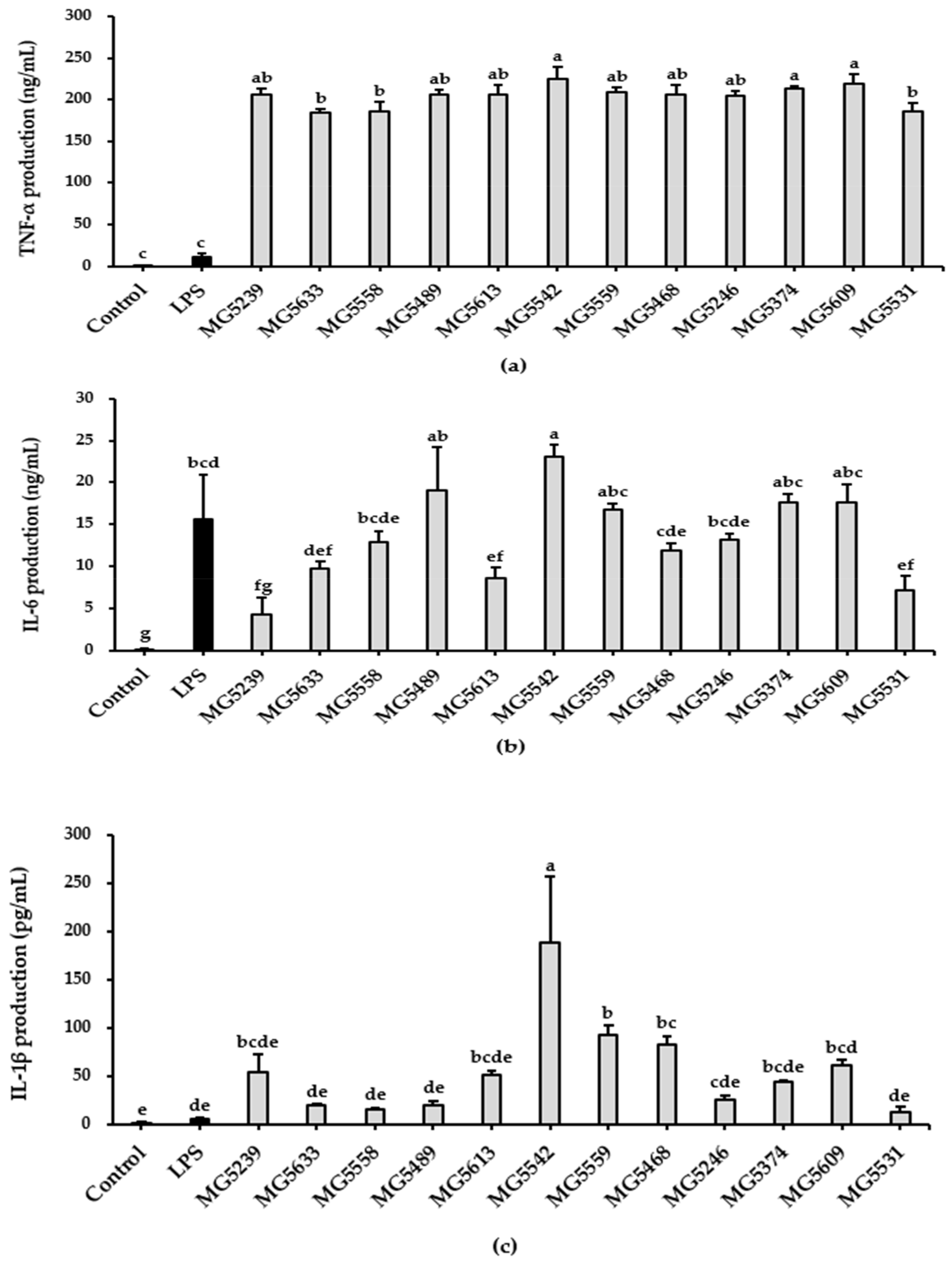
Figure 1. Effect of the initially screened strains on expression levels of TNF-α (a), IL-6 (b), and IL-1β (c) in RAW264.7 cells. Cells were cultured with LPS (10 ng/mL) or 5% CFS for 24 h. Data are presented as mean ± SD of three independent experiments (n = 3). Different letters on the column indicate significant differences between means at p < 0.05 based on Tukey’s post hoc test.
Fermentation 09 00004 g001
Figure 2. Effect of the selected strains on the phagocytosis of RAW264.7 cells. Phagocytosis was evaluated using the neutral red uptake assay and was quantified by measuring the logarithmic intensity using a microplate reader. Results were expressed as phagocytic activity relative to untreated cells. Data are presented as mean ± SD of three independent experiments (n = 3). Different letters on the column indicate significant differences between means at p < 0.05 based on Tukey’s post hoc test.
Fermentation 09 00004 g002
Figure 3. Effect of the selected strains on NF-κB activation in RAW264.7 cells. Western blots (a) and phosphorylation levels of p65 (b). The phosphorylation levels of p65 were expressed as fold changes of control. Data are presented as mean ± SD of three independent experiments (n = 3). Different letters on the column indicate significant differences between means at p < 0.05 based on Tukey’s post hoc test.
Fermentation 09 00004 g003
Figure 4. Adhesion ability of the selected strains to HT-29 cells. Data are presented as mean ± SD of three independent experiments (n = 3). Different letters on the column indicate significant differences between means at p < 0.05 based on Tukey’s post hoc test.
Fermentation 09 00004 g004
Table 1. Effect of LAB strains on cell viability and NO production in RAW264.7 cells.
SpeciesStrainsCell Viability (%)NO (μM)
Untreated100.00 ± 6.440.65 ± 0.11
LPS (10 ng/mL)101.99 ± 5.479.08 ± 0.86 **
Lactiplantibacillus plantarumMG5239107.69 ± 12.2419.64 ± 1.24 ***
MG5363103.29 ± 9.135.18 ± 1.11
MG5420101.72 ± 2.664.04 ± 0.29
MG5633100.11 ± 1.5318.07 ± 0.66 ***
MG5558101.75 ± 3.1818.07 ± 1.94 ***
Limosilactobacillus fermentumMG5489103.91 ± 7.2729.96 ± 2.51 ***
MG5613103.84 ± 4.9421.21 ± 6.88 ***
Lacticaseibacillus paracaseiMG5219101.86 ± 5.858.26 ± 0.39 *
MG5559101.74 ± 6.2015.55 ± 1.91 ***
Latilactobacillus sakeiMG521899.94 ± 1.978.13 ± 0.86 *
MG5235102.02 ± 2.324.55 ± 1.32
MG5468101.38 ± 5.1212.60 ± 0.76 ***
MG5496101.03 ± 3.077.57 ± 1.15
MG5557100.88 ± 1.254.23 ± 2.40
Latilactobacillus curvatusMG5246100.24 ± 4.7222.35 ± 1.04 ***
MG5374100.35 ± 4.8624.11 ± 1.28 ***
MG5609101.74 ± 3.2023.92 ± 1.53 ***
Levilactobacillus brevisMG5250100.55 ± 3.182.85 ± 0.82
MG5264100.59 ± 2.221.97 ± 0.58
MG5291100.36 ± 6.451.72 ± 0.19
MG5426100.49 ± 4.401.47 ± 0.71
MG5531100.23 ± 1.9510.14 ± 0.76 **
Lactococcus lactisMG5542101.72 ± 1.5116.37 ± 1.11 ***
Weissella cibariaMG5223100.10 ± 4.832.28 ± 0.82
MG5234103.89 ± 5.401.97 ± 0.11
MG5285104.11 ± 6.012.03 ± 0.66
MG5362100.15 ± 6.442.22 ± 0.39
MG5569102.56 ± 5.582.22 ± 0.11
The results are presented as mean ± SD of three independent experiments (n = 3). Significance was based on Tukey’s test; * p < 0.05; ** p < 0.01; *** p < 0.001 compared with the same column of the untreated control. NO, nitric oxide.
Table 2. The survival rate of the selected strains in the simulated gastrointestinal tract.
StrainsInitial Counts
(log CFU/mL)
SGF
(log CFU/mL)
SIF
(log CFU/mL)
Survival
Rate (%)
L. fermentum MG54898.82 ± 0.007.68 ± 0.017.67 ± 0.0386.99 ± 0.29
Lc. lactis MG55427.84 ± 0.034.61 ± 0.084.20 ± 0.1953.63 ± 2.44
L. paracasei MG55597.61 ± 0.045.96 ± 0.105.69 ± 0.0474.75 ± 0.47
L. sakei MG54688.33 ± 0.016.96 ± 0.016.87 ± 0.0282.57 ± 0.28
L. curvatus MG56097.44 ± 0.065.42 ± 0.044.54 ± 0.1161.01 ± 1.43
Data are presented as mean ± SD of three independent experiments (n = 3). SGF: simulated gastric fluid; SIF: simulated intestinal fluid.
Table 3. Antimicrobial susceptibility of the selected strains.
AntimicrobialsSusceptibility (S/R)
MG5489MG5542MG5559MG5468MG5609
AmpicillinSSSSS
(0.19)(0.5)(0.75)(0.5)(1)
GentamicinSSSSS
(0.25)(2)(2)(2)(0.75)
KanamycinSSSSS
(16)(8)(32)(8)(2)
StreptomycinSSSSS
(6)(24)(24)(32)(6)
TetracyclineSSSSS
(1.5)(0.25)(1)(3)(1.5)
ChloramphenicolSSRSS
(3)(6)(6)(3)(2)
ErythromycinSSSSS
(0.75)(0.38)(0.094)(0.125)(0.094)
Vancomycin-S---
(n.r)(0.25)(n.r)(n.r)(n.r)
ClindamycinSSSSS
(0.125)(0.094)(0.25)(0.125)(0.016)
Antimicrobial susceptibility was determined according to EFSA guidelines [22]. Probiotic strains are classified as susceptible (S) or resistant (R) according to the EFSA breakpoint values. Minimum inhibitory concentrations are shown in parentheses (µg/mL). n.r.: not required.
Disclaimer/Publisher’s Note: The statements, opinions and data contained in all publications are solely those of the individual author(s) and contributor(s) and not of MDPI and/or the editor(s). MDPI and/or the editor(s) disclaim responsibility for any injury to people or property resulting from any ideas, methods, instructions or products referred to in the content.