Article

Fermentation in Minimal Media and Fungal Elicitation Enhance Violacein and Deoxyviolacein Production in Two Janthinobacterium Strains

by
Andri Frediansyah
1,*,
Yosephine Sri Wulan Manuhara
2,
Alfinda Novi Kristanti
3,
Arif Luqman
4,* and
Anjar Tri Wibowo
2,*
1
Research Center for Food Technology and Processing (PRTPP), National Research and Innovation Agency (BRIN), Yogyakarta 55861, Indonesia
2
Department of Biology, Faculty of Science and Technology, Airlangga University, Kampus C, Mulyorejo, Surabaya 60115, Indonesia
3
Department of Chemistry, Faculty of Science and Technology, Airlangga University, Kampus C, Mulyorejo, Surabaya 60115, Indonesia
4
Department of Biology, Institut Teknologi Sepuluh Nopember, Surabaya 60111, Indonesia
*
Authors to whom correspondence should be addressed.
Fermentation 2022, 8(12), 714; https://doi.org/10.3390/fermentation8120714
Submission received: 23 November 2022 / Revised: 3 December 2022 / Accepted: 5 December 2022 / Published: 7 December 2022
(This article belongs to the Section Microbial Metabolism, Physiology & Genetics)

Abstract

:
Violacein and its biosynthesis by-product deoxyviolacein are valuable natural pigments with different biological activities. Various efforts have been made to enhance violacein and deoxyviolacein production in microbes. However, the effect of different culture media, agitation, and fungal elicitation on biosynthesis in Janthinobacterium has not been evaluated. In this study, the effect of eight different culture media, agitation, and fungal elicitation by Agaricus bisporus on violacein and deoxviolacein production in Janthinobacterium agaricidamnosum DSM 9628 and Janthinobacterium lividum DSM 1552 were examined. The results showed that violacein and deoxviolacein are produced at high-levels when Janthinobacterium is cultivated in minimal media such as Davis minimal broth with glycerol (DMBgly), shipworm basal medium (SBM), and MM9 media. A 50-fold increase was observed in violacein production when Janthinobacterium was cultivated in these media compared to cultivation in Luria–Bertani (LB), nutrient broth (NB), and King’s B (KB). Agitation reduces violacein and deoxyviolacein production, while fungal elicitation decreases violacein but increases deoxyviolacein when Janthinobacterium is cultured in KB media, SBM, and modified SBM (MSBM). An antibacterial assay using various pathogenic bacteria showed that violacein and deoxyviolacein extracted from Janthinobacterium are effective against both Gram-positive and Gram-negative pathogens, confirming their functionality as antibacterial agents. The findings suggest that cultivation in minimal media and fungal elicitation might invoke a stress response, enhancing the production of violacein and deoxviolacein in Janthinobacterium.

Graphical Abstract

1. Introduction

Microbes can produce natural pigments that can be used as coloring agents in the food, cosmetics, and textile industries [1,2]. One naturally occurring pigment is violacein and its derivative deoxyviolacein, bis-indole pigments formed by the condensation of two tryptophan molecules [3,4,5]. These compounds have purple–blue color and are known to exhibit a variety of biological activities such as anticancer [6,7], antifungal [8,9], antiviral [10,11], antioxidant [12,13], and antibacterial activities against Staphylococcus aureus [14], Klebsiella pneumonia [14], Pseudomonas aeruginosa [15], and other Gram-positive pathogens [16]. Due to its valuable properties, many studies focused on the isolation of violacein-producing bacteria [17], the cloning and expression of violacein biosynthesis genes [18,19], and the optimization of culture conditions for violacein production [20,21].
Violacein and deoxyviolacein are known to be produced by various Gram-negative bacteria, including from the genus Chromobacterium [22], Collimonas [23], Duganella [14], Massilia [24], Pseudoalteromonas [19,25], and Janthinobacterium [26]. The production of these compounds varies between species, and previous works showed that media composition [27,28] and culture conditions [27,29] could significantly affect violacein production in different bacterial strains. This study evaluated the effect of different media compositions, agitation, and fungal elicitation on violacein and deoxyviolacein production in Janthinobacterium agaricidamnosum DSM 9628 and Janthinobacterium lividum DSM 1552. Preliminary bioinformatics analysis showed that these two bacterial strains possess a gene cluster for violacein biosynthesis, but J. agaricidamnosum’s capacity for violacein and deoxyviolacein production has not been tested. Moreover, the effect of media composition, agitation, and elicitors on violacein and deoxyviolacein biosynthesis in Janthinobacterium has not been evaluated.
In this study we cultivated J. agaricidamnosum DSM 9628 and J. lividum DSM 1552 in eight different media. The results showed that violacein and deoxyviolacein production are highest when Janthinobacterium strains are cultivated in minimal media such as Davis minimal broth with glycerol (DMBgly), shipworm basal medium (SBM), and MM9 media. Agitation negatively affects the production, while fungal elicitation using champignon (Agaricus bisporus) could enhance deoxyviolacein biosynthesis when Janthinobacterium is cultured in KB media, SBM, and MSBM. Furthermore, violacein and deoxyviolacein produced by Janthinobacterium retain their functional antibacterial activities against various pathogenic bacteria.

2. Materials and Methods

2.1. Media for Bacterial Cultivation

J. agaricidamnosum DSM 9628 and J. lividum DSM 1552 were cultivated in five-liter-scale fermentation using eight different media, as described in Table 1. The media were prepared using distilled water (ddH2O) and autoclaved for 15 min at 121 °C and pressure of 2 bars (Systec, VX-150, Systex GmBH, Linden, Germany).

2.2. Bacterial Strains

Pathogenic bacteria Yersinia pseudotuberculosis YPIII was provided by Prof. P. Dersch (Institute of Infectiology, University of Münster, Germany), while Dr. Niraj Aryal (Pharmaceutical Biology Department, University of Tübingen) and Prof. Dr. Götz (Microbial Genetics Department, University of Tübingen) gave Mycobacterium phlei and Staphylococcus aureus USA300 LAC, respectively. Janthinobacterium for the production of violacein and deoxyviolacein are listed in Table 2. The other strains were obtained from the DSMZ—German Collection of Micro-organisms and Cell Cultures GmbH, Braunschweig, Germany, or the ATCC—American Type Culture Collection, Manassas, United States, and maintained under the instructions provided.

2.3. Cryo-Culture Preparation

Each bacterial strain (J. agaricidamnosum DSM 9628 and J. lividum DSM 1552) was prepared in cryo-culture by combining 500 µL of the culture in Luria–Bertani (LB) growth media with 500 µL of sterile glycerol at a concentration of 50% (v/v). Subsequently, the cryo-culture was stored at −86 °C [30].

2.4. Pre-Culture Preparation

A defrosted cryo stock of bacterial strain was used for pre-culture cultivation by inoculating the strains on an LB plate at 30 °C for 48 h, followed by inoculation into 6 mL of LB media in a 15 mL sterile tube (Sarstedt, Nümbrecht, Germany). The pre-culture was incubated for 24 h at 30 °C and 160 rpm in a VWR orbital shaker model 3500. Furthermore, the seed culture was prepared by dilution to an OD600 of 0.06 in a new 15 mL sterile tube.

2.5. Bacterial Growth Measurement

The viable count was performed at different intervals for 48 hours of fermentation [31]. To perform a viable count, 10 sterile 15 mL test tubes were filled with 9 mL sterilized phosphate buffer saline (PBS). Serial 10-fold dilutions in PBS were prepared in the test tubes using 1 mL of fermented juice as the starter. The viable count was next performed using the pour plate method in duplicate, and 1 mL of solution from each dilution series was mixed with 25 mL tempered (47 °C) Plate Count Agar (OXOID®, Basingstoke, England). The plate was incubated for 72 h at 30 °C ± 2 °C, and colony numbers were calculated using a colony counter. Plates with 15 to 300 colonies were considered for colony-forming unit calculation. The viable colonies were converted into weighted mean forming units per milliliter (CFU/mL) using the following Equation:
N = ∑C/[(n1 + 0.1n2)d]
where N is the number of colonies in the plate, ∑C is the sum of plates containing 15 to 300 colonies, n1 and n2 are the number of plates retained in the first and second dilutions, respectively, and d is the first dilution factor. Furthermore, the viable colonies were converted into log CFU/mL.

2.6. Large-Scale Fermentation

The pre-cultured bacteria were used for large-scale fermentation to obtain sufficient quantities of the desired compounds. The seed cultures of J. agaricidamnosum DSM 9628 and J. lividum DSM 1552 were inoculated into a 15 mL sterile tube containing 6 mL LB and re-incubated at 30 °C, at 160 rpm for 48 h. About 2 mL of 48 h seed culture was inoculated into two 5-liter Erlenmeyer jars containing 2.5 liters of each medium (Table 1). For cultivation with champignon (Agaricus bisporus), 100 g of sterile champignon cut into 2 cm2 pieces was added for every 1 L medium. The culture was incubated at 30 °C for 48 h under static conditions or with a 160-rpm agitation using Multitron Pro orbital incubator shaker (INFORS HT, Bottmingen, Switzerland).

2.7. Extraction, Fractionation, and Isolation of Violacein and Deoxyviolacein

To obtain violacein and deoxyviolacein, the broth from the 5 L fermentation of J. agaricidamnosum DSM 9628 and J. lividum DSM was extracted using 30 g/L of Diaion® HP 20 resin, followed by 3 days of shaking at 5 °C and 120 rpm in a Multitron Pro orbital incubator shaker (INFORS HT, Bottmingen, Switzerland). Subsequently, the resin was filtered from the supernatant, washed twice with deionized water, and separated with a vacuum liquid chromatography (VLC) system to obtain 5 fractions (A to E).
The fraction C (50:50 methanol/water) from the VLC was then run through a Water system HPLC operated by the Waters Millennium Software 4.0 consisting of a 1525 pump, 996 photodiode array detectors, a Rheodyne 7725i injector, and a Kromega vacuum degasser series A50010. To purify violacein and deoxyviolacein, fraction C was subjected to a linear gradient from 10:90 acetonitrile (ACN): H2O to 100% ACN containing 0.1% TFA for 40 min, followed by an isocratic stage at 100% ACN for 5 min using Luna® Omega 3 µm Polar C18 100, 250 × 4.6 mm (Phenomenex®, Aschaffenburg, Germany) packing, 0.8 mL/min flow rate, and UV monitoring at 232 nm. In addition, the concentration is determined by the weight of the product isolated from 1 L of fermentation broth after evaporation in vacuo followed by freeze-drying.

2.8. Mass Spectrometry Analysis

For LC-MS analysis of violacein and deoxyviolacein, an 1100 Series HPLC system was equipped with a G1322A degasser, G1312A binary pump, G1329A autosampler, and G1315A diode array detector. An ABSCIEX 3200 QTRAP LC/MS/MS mass spectrometer was coupled to the Agilent HPLC components. In addition, high-resolution mass spectra were collected using a Bruker maXis 4G HR-ESI-TOF-MS mass spectrometer.

2.9. Antibacterial Assay

Antibacterial assays against pathogenic bacteria were conducted in 96-well plates using the broth dilution method [32]. Each well was cultured with 100 µL of Mueller–Hinton broth and 5 µL of each bacterial pathogen (OD600 = 0.2). About 200 µL of a solution containing 256 g/mL of pure compounds dissolved in 10% MeOH was added to the first vertical line of the 96-well plate. Using serial dilutions, 10 pure compound concentrations were obtained in each well at 128, 64, 32, 16, 8, 4, 2, 1, 0.5, and 0.25 g/mL, with 10% MeOH as a negative control. The 96-well plates were incubated for 24 h at 30 °C, and growth inhibition was measured using the Tecan Plate Reader Infinite M200 at 600 nm. For each bacterial pathogen, the MIC (minimum inhibitory concentration) was determined in triplicate independently.

2.10. Statistical Analysis

All measurements were conducted using at least three independent replicates. The analysis output was presented as mean value ± SD, and statistical analysis was performed using two-way ANOVA with Tukey’s multiple comparison test by GraphPad Prism 7. A probability (p-value) less than 0.05 was considered statistically significant.

3. Results

3.1. Bioinformatic Analysis of the Strains and Isolation of Violacein and Deoxyviolacein

Violacein biosynthesis requires the action of five enzymes called VioA, B, E, D, and C, encoded by the vioABCDE operon. To evaluate the presence of this gene cluster in J. agaricidamnosum DSM 9628 and J. lividum DSM 1552, a BLAST analysis was performed (antiSMASH 6.0 in default setting) by comparing the genome of these bacteria against well-known violacein-producing strain Chromobacterium violaceum ATCC 12472. The results showed that J. agaricidamnosum DSM 9628 and J. lividum DSM 1552 had 14 predicted Biosynthetic Gene Clusters (BGCs), including vioABCDE operon (Figure 1A, Supplementary Figure S1). The configurations of the violacein gene cluster between C. violaceum ATCC 12472 with J. agaricidamnosum DSM 9628 and J. lividum DSM 1552 are identical (Figure 1A). However, there are only 53.12% to 72.73% and 53.85% to 72.73% amino acid sequence similarity values between C. violaceum ATCC 12472 and J. agaricidamnosum DSM 9628 and between C. violaceum ATCC 12472 and J. lividum DSM 1552 (Figure 1B, Supplementary Figure S2), respectively. Despite the amino acid differences, the predicted vioABCDE operon from Janthinobacterium strains can produce functional indole-based violacein.
These two bacterial strains were cultivated in 5 L scale fermentation using eight different culture media to isolate the predicted violacein and deoxyviolacein. Extraction and fractionation from the bacterial cultures yielded five fractions of A to E. The fraction C, as shown in Figure 2, contains violacein (RT 23.1 min) and deoxyviolacein (RT 24.1 min). In detail, the pure forms of isolated violacein and deoxyviolacein have negative HR-ESI-MS m/z 342.0888 [M+H] (calc. for C20H13N3O3 342.0887, Δ = 0.9 ppm) and HR-ESI-MS m/z 326.0935 [M+H] (calc. for C20H12N3O3 326.0939, Δ = 1.4 ppm), as shown in Supplementary Figure S3.

3.2. The Growth Rate of Janthinobacterium agaricidamnosum DSM 9628 and Janthinobacterium lividum DSM 1552 in Different Culture Media

To evaluate the effect of different media compositions on violacein and deoxyviolacein production, J. agaricidamnosum DSM 9628 and J. lividum DSM 1552 were cultivated in eight different culture media. J. lividum grows fastest when cultivated in nutrient-rich LB, NB, and KB media; relatively slower in DMBgly and modified DMBgly (MDMBgly); and slowest in SBM, modified SBM (MSBM), and MM9. Across all media, J. lividum reached maximum growth after 36 to 48 h of fermentation, and following 48 h, J. lividum showed similar viable counts across all media (Figure 3A). Similarly, J. agaricidamnosum grows faster when cultivated in LB, NB, and KB media than in others. Across all culture media, it reached the stationary phase after 30 h of fermentation and showed similar viable counts (Figure 3B). Therefore, despite the strains growing faster in nutrient-rich media, in all media the maximum growth is reached in the same period, and similar viable counts are observed after 48 h of culture.

3.3. The Production Rate of Violacein and Deoxyviolacein in Different Culture Media

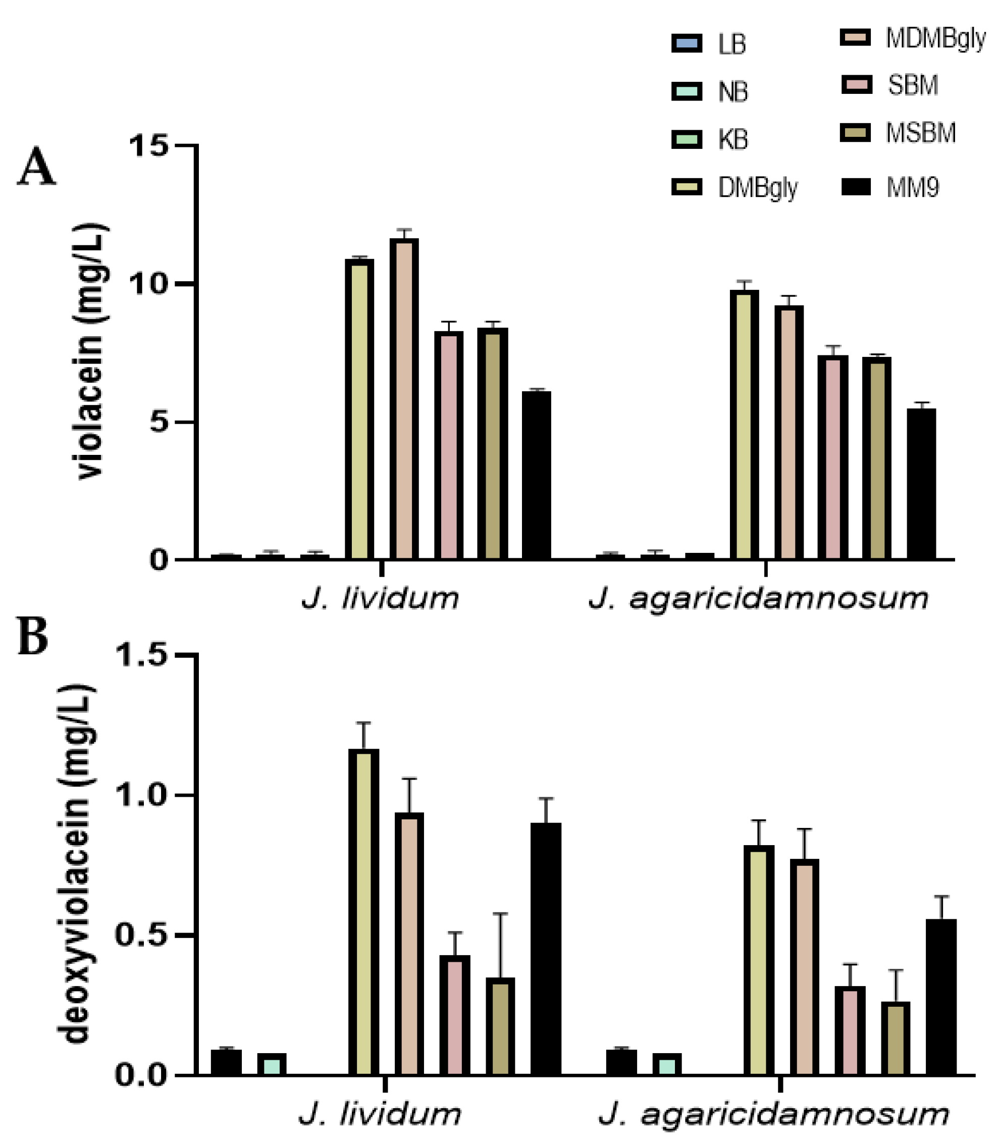
Violacein and deoxyviolacein production in J. agaricidamnosum DSM 9628 and J. lividum DSM 1552 were measured following 48 h of cultures in eight different media. The results showed that they are produced at very low levels when the bacterial strains are cultivated in standard-nutrient-rich LB, NB, and KB media (Figure 4 and Supplementary Table S1). They are produced at a much higher level, more than a 50-fold-increase, when J. agaricidamnosum DSM 9628 and J. lividum DSM 1552 are cultivated in DMBgly, MDMBgly, SBM, MSBM, and MM9. Violacein and deoxyviolacein were produced at the highest level when J. agaricidamnosum DSM 9628 was cultivated in DMBgly, producing 9.8 mg/L and 0.82 mg/L of violaceins and deoxyviolacein, respectively. For J. lividum DSM 1552, the highest production was observed when it was cultivated in MDMBgly (10.89 mg/L) and DMBgly (1.17 mg/L), as seen in Figure 4 and Supplementary Table S1. The results showed that media composition could significantly affect the production rate in Janthinobacterium, where DMBgly and MDMBgly appear to be the most suitable media to facilitate violacein and deoxyviolacein.

3.4. Agitation during Culture and Fungal Elicitation Could Not Increase Violacein Production

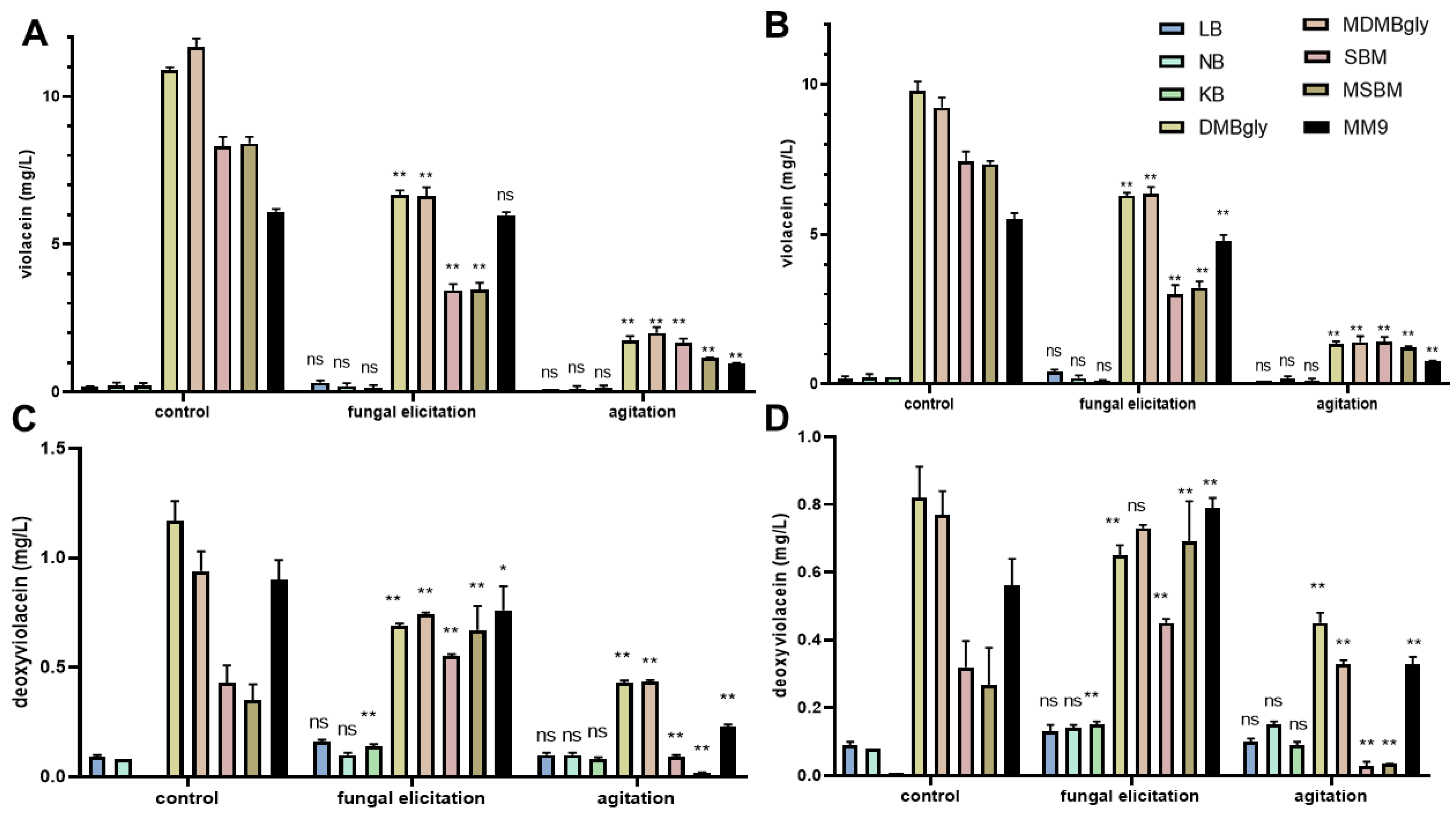
Previous work reported that agitation during culture could affect violacein production in Pseudoalteromonas luteoviolacea [25] and Chromobacterium sp. [8]. J. agaricidamnosum is naturally found as a pathogen in champignon (Agaricus bisporus), causing soft-rot disease in the mushroom [33,34,35]. Here we would like to examine whether the addition of champignon in the culture media can enhance violacein and deoxyviolacein production. To evaluate the effects of agitation and fungal elicitation by A. bisporus on violacein and deoxyviolacein biosynthesis in Janthinobacterium, the production in J. agaricidamnosum DSM 9628 and J. lividum DSM 1552 was compared in static and agitated culture (160 rpm, constant), with and without champignon addition. In general, both agitation and fungal elicitation negatively affect violacein production. The negative effect of fungal elicitation was apparent in bacterial strains that are cultivated in DMBgly, MDMBgly, SBM, and MSBM. Meanwhile, the negative effect of agitation can be observed across all culture media, as seen in Figure 5 and Supplementary Table S1. Agitation and fungal elicitation can have positive or negative effects on deoxyviolacein production, depending on the culture media. Agitation positively affects deoxyviolacein production in bacterial strains cultivated in King’s B (KB) while having no negative effect on those cultivated in other culture media. Similarly, fungal elicitation positively affects deoxyviolacein production when Janthinobacterium is cultivated in King’s B (KB) media. It also positively affects deoxyviolacein production in Janthinobacterium grown in SBM and MSBM.

3.5. Antibacterial Activities of Violacein and Deoxyviolacein Isolated from J. agaricidamnosum DSM 9628 and J. lividum DSM 1552

Violacein extracted from Chromobacterium has been reported to have antibacterial activities against various pathogenic bacteria, including Staphylococcus aureus, Klebsiella pneumoniae, Pseudomonas aeruginosa, Vibrio cholerae, Bacillus subtilis, Salmonella typhi [15,20,36]. To evaluate whether violacein and deoxyviolacein from J. agaricidamnosum DSM 9628 and J. lividum DSM 1552 possess similar biological functions, antibacterial activities against various pathogenic bacterial strains were examined, as shown in Table 3. These include Pseudomonas aeruginosa DSM 50071, Yersinia tuberculosis YPIII, Paenibacillus sp. AD64, Bacillus subtilis ATCC 6051, Mycobacterium phlei 01, and Staphylococcus aureus USA300 LA. The assay demonstrated that both violacein and deoxyviolacein are effective against Pseudomonas aeruginosa, Paenibacillus sp., Bacillus subtilis, and Staphylococcus aureus but ineffective against Yersinia tuberculosis and Mycobacterium phlei. Therefore, the violacein and deoxyviolacein antibacterial activity are specific and limited to certain bacterial taxa. The antibacterial strength against pathogenic bacteria is comparable, and these two compounds showed similar MIC values against all tested pathogens.

4. Discussion

Violacein and deoxyviolacein are valuable natural pigments with various biological activities. Different efforts have been made to optimize the production of different bacterial strains. However, the effect of different culture media on violacein and deoxyviolacein production has not been evaluated. This study analyzed the effect of eight different culture media on violacein and deoxyviolacein production in two bacterial strains, J. agaricidamnosum DSM 9628 and J. lividum DSM 1552. The results showed that violacein and deoxyviolacein are produced at much higher levels when the bacteria are cultivated in minimal media such as DMBgly, MDMBgly, SBM, MSBM, and MM9 media. Compared to cultivation in rich media such as LB, NB, and KB media, a more than 50-fold increase was observed in minimal media. These findings suggested that violacein and deoxyviolacein are synthesized in response to environmental stresses since exposure to stresses is known to induce secondary metabolite production in bacteria [29,30]. Cultivation in minimal media (DMBgly, MDMBgly, SBM, MSBM, and MM9) might cause nutritional stress because they primarily contain salt or trace elements while lacking proteins and amino acids. As we are aware, this is the first study to investigate the violacein and deoxyviolacein production in minimal media. More research is needed to determine the effect of minimal media on other violacein-producing bacteria like Chromobacterium and Duganella sp. Previous study suggested that bacterial cells are engaged in rapid growth when cultivated in rich media while primed for survival in minimal media, mainly by producing higher levels of sugars, fatty acids, and secondary metabolites [37]. Previous work reported that violacein and biofilm production are essential in the J. lividum survival mechanism, specifically in resisting environmental stresses [26]. The results suggest that cultivation in minimal media invokes the stress response in Janthinobacterium, which triggers the production of violacein and deoxyviolacein. The highest violacein production was observed when Janthinobacterium was cultivated in DMBgly. The presence of glycerol in DMBgly might further enhance violacein production. These data support previous findings that glycerol in culture media could increase violacein production in C. violaceum [38] and J. lividum [26]. It is also reported to increase deoxyviolacein in genetically modified Escherichia coli [7].
Agitation during culture was reported to affect violacein production in Pseudoalteromonas luteoviolacea [25] and Chromobacterium sp. [8]. Here we found that agitation during culture decreases violacein production in all media. This result is in accordance with previous study of P. luteoviolacea, where violacein production is higher at static conditions than under agitation [25]. It is suggested bacterial aggregation might be essential for the production of violacein and such aggregation is affected by agitation, thus inhibiting violacein production [25]. In addition, agitation during culture can cause shear stress [39] and increased dissolved oxygen in the media that negatively affect metabolite production [40], which are diametrically opposed in the production of antidiabetic compounds by L. plantarum [31].
The preliminary bioinformatics analysis showed only 53–72% amino acid sequence similarity between violacein biosynthesis enzymes in J. lividum and J. agaricidamnosum with Chromobacterium violaceum. Studies regarding violacein antibacterial activity were mainly performed using the product isolated from Chromobacterium. Meanwhile, the antibacterial activity of violacein from Janthinobacterium is poorly understood. Here, we showed that violacein and deoxyviolacein isolated from Janthinobacterium are also effective antimicrobial agents. They could effectively inhibit the growth of various pathogenic bacteria, including Pseudomonas aeruginosa, Paenibacillus sp., Bacillus subtilis, and Staphylococcus aureus. The antibacterial activity is attributed to their ability to disrupt microbial cytoplasmic membranes [16]. In this study, the MIC values of violacein from J. lividum and J. agaricidamnosum against S. aureus USA300 LAC (the clinical methicillin-resistant Staphylococcus aureus strain) were 7.89–8 µg/mL and against Pseudomonas aeruginosa DSM 50071, the values were 19.5–19.8 µg/mL. Previous studies reported that violacein produced by Chromobacterium violaceum Bergonzini has a MIC value against S. aureus MTCC 3160 of 5.7 µg/mL and against P. aeruginosa MTCC 1688 of 18.5 µg/mL [15], while the violacein from Duganella violaceinigra NI28 has a MIC against S. aureus isolates of 5.14 µg/mL and against P. aeruginosa PAO1 of 10.28 µg/mL [14]. Overall, we can see that the antibacterial activity of violacein from J. lividum and J. agaricidamnosum is slightly lower but comparable to that of violacein originating from other bacterial strains. Please note that the previous work used different pathogen strains to perform the MIC tests. Furthermore, synthetized violacein inhibits the growth of other Gram-positive bacteria, such as Staphylococcus epidermidis and Bacillus subtilis. It also has antibacterial efficacy against Gram-negative bacteria such as Klebsiella pneumoniae, Vibrio cholerae, and Salmonella typhi [20]. Since violacein could target both Gram-positive and Gram-negative bacteria, it remains unclear how the bacteria could prevent self-damage as Gram-negative microbes.

5. Conclusions

This study showed that violacein and deoxyviolacein are produced at much higher levels when J. agaricidamnosum DSM 9628 and J. lividum DSM 1552 were cultivated in minimal media. Minimal media might invoke nutritional stress, which induces the production of various secondary metabolites. Furthermore, violacein and deoxyviolacein isolated from Janthinobacterium could effectively inhibit the growth of pathogenic Gram-positive and Gram-negative bacteria, confirming their functionality as antibacterial agents.

Supplementary Materials

The following supporting information can be downloaded at: https://www.mdpi.com/article/10.3390/fermentation8120714/s1, Figure S1: Biosynthetic Gene Clusters (BGCs) found in Janthinobacterium agaricidamnosum DSM 9628 and Janthinobacterium lividum DSM 1552 through antiSMASH v6.0 analysis; Figure S2: Amino acid sequence alignment between violacein ABCDE operon in Chromobacterium violaceum ATCC 12472 with Janthinobacterium agaricidamnosum DSM 9628 and J. lividum DSM 1552; Figure S3: Extracted ion chromatograms (EIC) of violacein (A) and deoxyviolacein (B) in J. lividum; Table S1: Yield of violacein and deoxyviolacein in J. lividum and J. agaricidamnosum.

Author Contributions

Conceptualization, A.F.; methodology, A.F., A.T.W. and A.L.; formal analysis, A.F., A.T.W. and A.L.; investigation, A.F. and A.L.; resources, A.F., A.L., A.T.W., A.N.K. and Y.S.W.M.; data curation, A.F. and A.L.; writing—original draft preparation, A.F. and A.T.W.; writing—review and editing, A.F., A.L., A.T.W., A.N.K. and Y.S.W.M.; supervision, A.F. and A.T.W.; project administration, A.F., A.L., A.T.W., A.N.K. and Y.S.W.M.; funding acquisition, A.F., A.L., A.T.W., A.N.K. and Y.S.W.M. All authors have read and agreed to the published version of the manuscript.

Funding

Airlangga University Hibah Riset Mandat Grant (387/UN3.14/PT/2020) from Universitas Airlanggga, Indonesia.

Institutional Review Board Statement

Not applicable.

Informed Consent Statement

Not applicable.

Data Availability Statement

Not applicable.

Acknowledgments

The authors are grateful to Harald Gross at the Pharmaceutical Institute, University of Tübingen, Germany; Petra Dersch at the Institute of Infectiology, University of Münster, Germany; Niraj Aryal at the Pharmaceutical Biology Department, University of Tübingen; and Friedrich Götz at Microbial Genetics Department, University of Tübingen for providing all necessary equipment, bacterial strains, and access for this study.

Conflicts of Interest

The authors declare no conflict of interest.

References

  1. Sen, T.; Barrow, C.J.; Deshmukh, S.K. Microbial pigments in the food industry—Challenges and the way forward. Front. Nutr. 2019, 6, 7. [Google Scholar] [CrossRef] [PubMed] [Green Version]
  2. Tuli, H.S.; Chaudhary, P.; Beniwal, V.; Sharma, A.K. Microbial pigments as natural color sources: Current trends and future perspectives. J. Food Sci. 2015, 52, 4669–4678. [Google Scholar] [CrossRef] [Green Version]
  3. Kanelli, M.; Mandic, M.; Kalakona, M.; Vasilakos, S.; Kekos, D.; Nikodinovic-Runic, J.; Topakas, E. Microbial production of violacein and process optimization for dyeing polyamide fabrics with acquired antimicrobial properties. Front. Microbiol. 2018, 9, 1495. [Google Scholar] [CrossRef] [PubMed] [Green Version]
  4. Füller, J.J.; Röpke, R.; Krausze, J.; Rennhack, K.E.; Daniel, N.P.; Blankenfeldt, W.; Schulz, S.; Jahn, D.; Moser, J. Biosynthesis of violacein, structure and function of l-Tryptophan oxidase VioA from Chromobacterium violaceum. J. Biol. Chem. 2016, 291, 20068–20084. [Google Scholar] [CrossRef] [PubMed] [Green Version]
  5. Wille, G.; Steglich, W. A short synthesis of the bacterial pigments violacein and deoxyviolacein. Synthesis 2001, 2001, 759–762. [Google Scholar] [CrossRef]
  6. Hashimi, S.M.; Xu, T.; Wei, M.Q. Violacein anticancer activity is enhanced under hypoxia. Oncol. Rep. 2015, 33, 1731–1736. [Google Scholar] [CrossRef] [Green Version]
  7. Rodrigues, A.L.; Trachtmann, N.; Becker, J.; Lohanatha, A.F.; Blotenberg, J.; Bolten, C.J.; Korneli, C.; de Souza Lima, A.O.; Porto, L.M.; Sprenger, G.A. Systems metabolic engineering of Escherichia coli for production of the antitumor drugs violacein and deoxyviolacein. Metab. Eng. 2013, 20, 29–41. [Google Scholar] [CrossRef]
  8. Sasidharan, A.; Sasidharan, N.K.; Amma, D.B.N.S.; Vasu, R.K.; Nataraja, A.V.; Bhaskaran, K. Antifungal activity of violacein purified from a novel strain of Chromobacterium sp. NIIST (MTCC 5522). J. Microbiol. 2015, 53, 694–701. [Google Scholar]
  9. Wang, H.; Wang, F.; Zhu, X.; Yan, Y.; Yu, X.; Jiang, P.; Xing, X.-H. Biosynthesis and characterization of violacein, deoxyviolacein and oxyviolacein in heterologous host, and their antimicrobial activities. Biochem. Eng. J. 2012, 67, 148–155. [Google Scholar] [CrossRef]
  10. Andrighetti-Fröhner, C.; Antonio, R.; Creczynski-Pasa, T.; Barardi, C.; Simões, C. Cytotoxicity and potential antiviral evaluation of violacein produced by Chromobacterium violaceum. Memórias Inst. Oswaldo Cruz 2003, 98, 843–848. [Google Scholar] [CrossRef]
  11. Tsukimoto, A.; Saito, R.; Hashimoto, K.; Takiguchi, Y.; Miyano-Kurosaki, N. Violacein induces various types of cell death in highly differentiated human hepatocellular carcinoma and hepatitis C virus replicon cell lines. Int. J. Anal. Bio-Sci. 2021, 9, 46–56. [Google Scholar]
  12. Konzen, M.; De Marco, D.; Cordova, C.A.; Vieira, T.O.; Antonio, R.V.; Creczynski-Pasa, T.B. Antioxidant properties of violacein: Possible relation on its biological function. Bioorganic Med. Chem. 2006, 14, 8307–8313. [Google Scholar] [CrossRef] [PubMed]
  13. Vishnu, T.; Palaniswamy, M. Systematic Approach on Evaluating the in vitro Antioxidant Activity of Violacein; Novel Isolate Chromobacterium Vaccinii CV5. Biomed. Pharmacol. J. 2018, 11, 703–709. [Google Scholar]
  14. Choi, S.Y.; Kim, S.; Lyuck, S.; Kim, S.B.; Mitchell, R.J. High-level production of violacein by the newly isolated Duganella violaceinigra str. NI28 and its impact on Staphylococcus aureus. Sci. Rep. 2015, 5, 15598. [Google Scholar]
  15. Subramaniam, S.; Ravi, V.; Sivasubramanian, A. Synergistic antimicrobial profiling of violacein with commercial antibiotics against pathogenic micro-organisms. Pharm. Biol. 2014, 52, 86–90. [Google Scholar] [CrossRef] [Green Version]
  16. Cauz, A.C.; Carretero, G.P.; Saraiva, G.K.; Park, P.; Mortara, L.; Cuccovia, I.M.; Brocchi, M.; Gueiros-Filho, F. Violacein targets the cytoplasmic membrane of bacteria. ACS Infect. Dis. 2019, 5, 539–549. [Google Scholar] [CrossRef]
  17. Choi, S.Y.; Yoon, K.-H.; Lee, J.I.; Mitchell, R.J. Violacein: Properties and production of a versatile bacterial pigment. BioMed Res. Int. 2015, 2015, 465056. [Google Scholar] [CrossRef] [Green Version]
  18. Pemberton, J.M.; Vincent, K.M.; Penfold, R.J. Cloning and heterologous expression of the violacein biosynthesis gene cluster from Chromobacterium violaceum. Curr. Microbiol. 1991, 22, 355–358. [Google Scholar] [CrossRef]
  19. Zhang, X.; Enomoto, K. Characterization of a gene cluster and its putative promoter region for violacein biosynthesis in Pseudoalteromonas sp. 520P1. Appl. Microbiol. Biotechnol. 2011, 90, 1963–1971. [Google Scholar] [CrossRef]
  20. Park, H.; Park, S.; Yang, Y.-H.; Choi, K.-Y. Microbial synthesis of violacein pigment and its potential applications. Crit. Rev. Biotechnol. 2021, 41, 879–901. [Google Scholar] [CrossRef]
  21. Wang, H.; Jiang, P.; Lu, Y.; Ruan, Z.; Jiang, R.; Xing, X.-H.; Lou, K.; Wei, D. Optimization of culture conditions for violacein production by a new strain of Duganella sp. B2. Biochem. Eng. J. 2009, 44, 119–124. [Google Scholar] [CrossRef]
  22. Hoshino, T. Violacein and related tryptophan metabolites produced by Chromobacterium violaceum: Biosynthetic mechanism and pathway for construction of violacein core. Appl. Microbiol. Biotechnol. 2011, 91, 1463–1475. [Google Scholar] [CrossRef] [PubMed]
  23. Hakvåg, S.; Fjærvik, E.; Klinkenberg, G.; Borgos, S.E.F.; Josefsen, K.D.; Ellingsen, T.E.; Zotchev, S.B. Violacein-producing Collimonas sp. from the sea surface microlayer of costal waters in Trøndelag, Norway. Mar. Drugs 2009, 7, 576–588. [Google Scholar] [CrossRef] [PubMed] [Green Version]
  24. Miess, H.; Frediansyah, A.; Göker, M.; Gross, H. Draft genome sequences of six type strains of the genus Massilia. Microbiol. Resour. Announc. 2020, 9, e00226-20. [Google Scholar] [CrossRef] [PubMed]
  25. Yang, L.; Xiong, H.; Lee, O.O.; Qi, S.H.; Qian, P.Y. Effect of agitation on violacein production in Pseudoalteromonas luteoviolacea isolated from a marine sponge. Lett. Appl. Microbiol. 2007, 44, 625–630. [Google Scholar] [CrossRef] [PubMed]
  26. Pantanella, F.; Berlutti, F.; Passariello, C.; Sarli, S.; Morea, C.; Schippa, S. Violacein and biofilm production in Janthinobacterium lividum. J. Appl. Microbiol. 2007, 102, 992–999. [Google Scholar] [CrossRef]
  27. Mendes, A.S.; de Carvalho, J.E.; Duarte, M.C.; Durán, N.; Bruns, R.E. Factorial design and response surface optimization of crude violacein for Chromobacterium violaceum production. Biotechnol. Lett. 2001, 23, 1963–1969. [Google Scholar] [CrossRef]
  28. Gohil, N.; Bhattacharjee, G.; Gayke, M.; Narode, H.; Alzahrani, K.J.; Singh, V. Enhanced production of violacein by Chromobacterium violaceum using agro-industrial waste soybean meal. J. Appl. Microbiol. 2022, 132, 1121–1133. [Google Scholar] [CrossRef]
  29. Alem, D.; Marizcurrena, J.J.; Saravia, V.; Davyt, D.; Martinez-Lopez, W.; Castro-Sowinski, S. Production and antiproliferative effect of violacein, a purple pigment produced by an Antarctic bacterial isolate. World J. Microbiol. Biotechnol. 2020, 36, 120. [Google Scholar] [CrossRef]
  30. Frediansyah, A.; Straetener, J.; Brötz-Oesterhelt, H.; Gross, H. Massiliamide, a cyclic tetrapeptide with potent tyrosinase inhibitory properties from the Gram-negative bacterium Massilia albidiflava DSM 17472T. J. Antibiot. 2021, 74, 269–272. [Google Scholar] [CrossRef]
  31. Frediansyah, A.; Romadhoni, F.; Nurhayati, R.; Wibowo, A.T. Fermentation of Jamaican cherries juice using Lactobacillus plantarum elevates antioxidant potential and inhibitory activity against Type II diabetes-related enzymes. Molecules 2021, 26, 2868. [Google Scholar] [CrossRef]
  32. Jiao, J.; Du, J.; Frediansyah, A.; Jahanshah, G.; Gross, H. Structure elucidation and biosynthetic locus of trinickiabactin from the plant pathogenic bacterium Trinickia caryophylli. J. Antibiot. 2020, 73, 28–34. [Google Scholar] [CrossRef]
  33. Lincoln, S.P.; Fermor, T.R.; Tindall, B. Janthinobacterium agaricidamnosum sp. nov., a soft rot pathogen of Agaricus bisporus. Int. J. Syst. Evol. Microbiol. 1999, 49, 1577–1589. [Google Scholar] [CrossRef] [Green Version]
  34. Stein, J.; Schlosser, N.; Bardl, B.; Peschel, G.; Meyer, F.; Kloss, F.; Rosenbaum, M.A.; Regestein, L. Scalable Downstream Method for the Cyclic Lipopetide Jagaricin; Wiley Online Library: New York, NY, USA, 2021. [Google Scholar]
  35. Graupner, K.; Lackner, G.; Hertweck, C. Genome sequence of mushroom soft-rot pathogen Janthinobacterium agaricidamnosum. Genome Announc. 2015, 3, e00277-15. [Google Scholar] [CrossRef]
  36. Cazoto, L.L.; Martins, D.; Ribeiro, M.G.; Durán, N.; Nakazato, G. Antibacterial activity of violacein against Staphylococcus aureus isolated from bovine mastitis. J. Antibiot. 2011, 64, 395–397. [Google Scholar] [CrossRef]
  37. Kim, J.; Kim, K.H. Effects of minimal media vs. complex media on the metabolite profiles of Escherichia coli and Saccharomyces cerevisiae. Process Biochem. 2017, 57, 64–71. [Google Scholar]
  38. Antônio, R.V.; Creczynski-Pasa, T.B.J.G.M.R. Genetic analysis of violacein biosynthesis by Chromobacterium violaceum. Genet. Mol. Res. 2004, 3, 85–91. [Google Scholar] [PubMed]
  39. Silva-Santisteban, B.O.Y.; Maugeri Filho, F. Agitation, aeration, and shear stress as key factors in inulinase production by Kluyveromyces marxianus. Enzyme Microb. Technol. 2005, 36, 717–724. [Google Scholar] [CrossRef]
  40. Heinemann, B.; Howard, A.J.; Palocz, H. Influence of dissolved oxygen levels on production of L-asparaginase and prodigiosin by Serratia marcescens. Appl. Microbiol. 1970, 19, 800–804. [Google Scholar] [CrossRef]
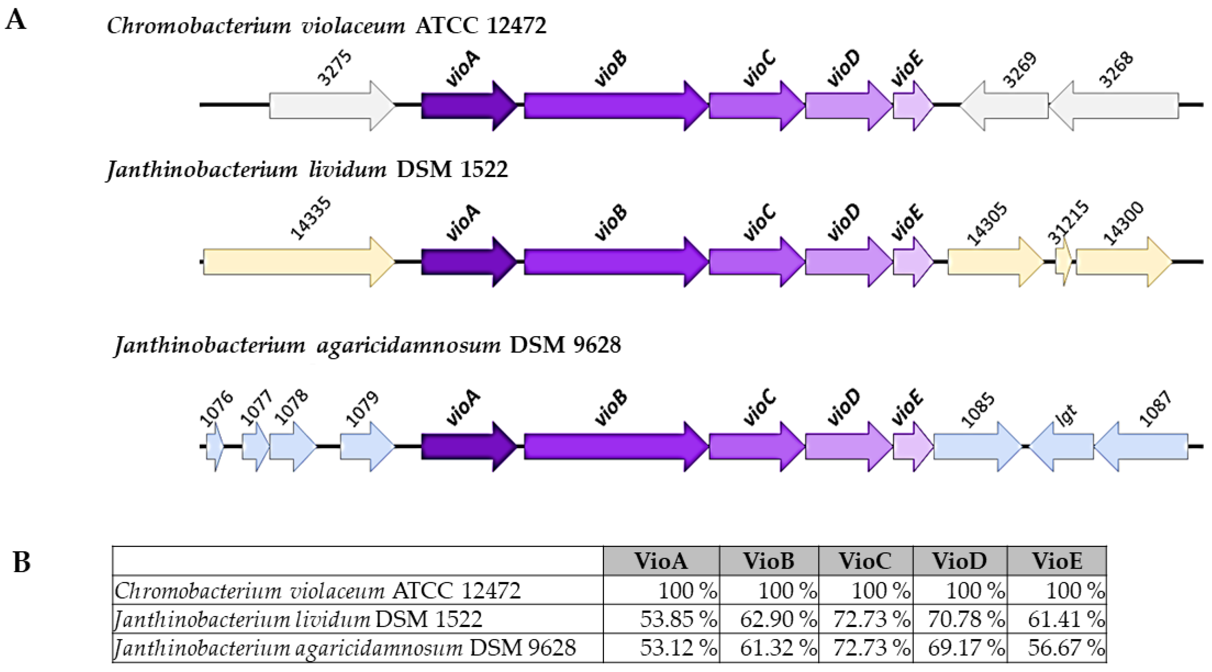
Figure 1. Violacein-producing protein cluster in J. agaricidamnosum DSM 9628 and J. lividum DSM 1552. (A). Comparison of vioABCDE protein cluster in C. violaceum ATCC 12472, J. agaricidamnosum DSM 9628, and J. lividum DSM 1552. (B). Amino acid sequence similarity of VioA, B, E, D, and C protein between C. violaceum ATCC 12,472 with J. agaricidamnosum DSM 9628 and J. lividum DSM 1552.
Fermentation 08 00714 g001
Figure 2. HPLC chromatogram of fraction C containing violacein (RT 23.1 min) and deoxyviolacein (RT 24.1 min) in J. lividum. Note: the HPLC chromatograms of violacein and deoxyviolacein in J. lividum, J. agaricidamnosum, and mixed fractions showed a similar pattern.
Fermentation 08 00714 g002
Figure 3. Viable count of (A). J. lividum and (B). J. agaricidamnosum in various media.
Fermentation 08 00714 g003
Figure 4. Violacein and deoxyviolacein production in Janthinobacterium following cultivation in different media. (A). Violacein production in J. agaricidamnosum DSM 9628 and J. lividum DSM 1552 across different culture media. (B). The production of deoxyviolacein in J. agaricidamnosum DSM 9628 and J. lividum DSM 1552 across different culture media. Violacein and deoxyviolacein were measured following 48 hours of fermentation in 5 L media, and the concentrations presented are the mean values of three independent replicates. Abbreviations: Luria–Bertani (LB), nutrient broth (NB), King’s B (KB), Davis minimal broth with glycerol (DMBgly), modified DMBgly (MDMBgly), shipworm basal medium (SBM), modified SBM (MSBM).
Fermentation 08 00714 g004
Figure 5. The effect of agitation and fungal elicitation on violacein and deoxyviolacein production in Janthinobacterium. The effect of agitation and fungal elicitation on violacein production in J. lividum (A) and J. agaricidamnosum (B). The effect of agitation and fungal elicitation on deoxyviolacein production in J. lividum (C) and J. agaricidamnosum (D). Violacein and deoxyviolacein were measured following 48 hours of fermentation in 5 L media, and the concentrations presented are the mean values of three independent replicates. Abbreviations: Luria–Bertani (LB), nutrient broth (NB), King’s B (KB), Davis minimal broth with glycerol (DMBgly), modified DMBgly (MDMBgly), Shipworm basal medium (SBM), modified SBM (MSBM). Note: * p ≤ 0.05; ** p ≤ 0.01; and ns: not-significant.
Fermentation 08 00714 g005
Table 1. Media used for Janthinobacterium fermentation.
Cultivation MediaConstituent per L
Luria–Bertani (LB)10 g tryptone, 5 g yeast extract, 10 g NaCl; final pH 7.5 at 25 °C
Nutrient Broth (NB)5 g peptone, 2 g yeast extract, 5 g NaCl; final pH 7.4 at 25 °C
King’s B Medium (KB)20 g proteose peptone, 1.5 g K2HPO4, 1.5 g MgSO4, 15 g glycerol; final pH 7.5 at 25 °C
Davis Minimal Broth with Glycerol (DMBgly)10.6 g Davis minimal broth without dextrose, 1.83 mL of 100% glycerol; pH adjustment not necessary
Modified DMBgly (MDMBgly)5.3 g Davis minimal broth without dextrose, 0.56 g HEPES buffer, 5.1 g sucrose, 1 mg methionine, 1 mL trace metal mix A5 with Co; final pH 6.6 at 25 °C
Shipworm Basal Medium (SBM) without sea salt15.3 mg KH2PO4, 10 mg Na2CO3, 2.5 mg Na2MoO4.2H2O, 0.5 mg EDTA, 3 mg C6H8FeNO7, 5.2 g HEPES buffer, 1 mL trace metal mix A5 with Co; pH adjustment not necessary
Modified SBM (MSBM) without sea salt15.3 mg KH2PO4, 10 mg Na2CO3, 2.5 mg Na2MoO4.2H2O, 0.5 mg EDTA, 5.2 g HEPES buffer, 1 mL trace metal mix A5 with Co, 0.267 g NH4Cl, 5 g sucrose, 5 mg L-methionine; final pH 7.2 at 25 °C
MM9:900 ml solution C, 20 ml solution A, 20 ml solution B, 16.7 ml 100mM L-leucine, 5 ml 60mM L-histidine, 10 ml 100 mM L-lysine, 10 ml 40 mM L-tryptophan, 10 ml 40 mM L-methionine, 20 ml 50% (w/v) glucose, 1 mL trace metal mix A5 with Co; pH adjustment not necessary
Notes: Trace metal mix A5 with Co (2.86 g H3BO3, 1.81 g MnCl2·4H2O, 0.222 g ZnSO4·7H2O, 0.390 mg Na2MoO4·2H2O, 79 mg CuSO4·5H2O, 49 mg Co(NO3).6H2O); solution A (100 g KH2PO4, 350 g K2HPO4); solution B (50 g (NH2)·2SO4, 5 g MgSO4); solution C (2 g of each amino acid (L-arginine, L-alanine, L-asparagine, L-aspartate, L-cysteine, L-glutamine, L-glutamate, L-glycine, L-isoleucine, L-proline, L-serine, L-threonine, L-tyrosine, L-valine, and L-phenylalanine) or 30 g casamino acid, final pH 7.2 at 25 °C).
Table 2. General genomic features of the Janthinobacterium strain used in this study.
Bacterial StrainSubmitterSourcesNCBI Reference SequenceGenome Size (Gb)GC Content (%)
J. agaricidamnosum
DSM 9628
NITE Biological Resource Center, Japanmushroom Agaricus bisporusNZ_BCTH00000000.15.8861.1
J. lividum DSM 1552Georg-August-University Goettingen, Germanyfrom the soil, Michigan, USANZ_LRHW00000000.16.7162.4
Table 3. Antibacterial activities of violacein and deoxyviolacein from Janthinobacterium strains against pathogenic bacteria.
Antibacterial ActivitiesMIC (µg/mL)
ViolaceinDeoxyviolaceinTetracycline (+)
JlJaJlJa
Pseudomonas aeruginosa DSM 50071 19.81 19.5 24.79 23.4 1.3
Yersinia tuberculosis YPIII >64 >64 >64 >64 1.4
Paenibacillus sp. AD64 14.49 14.2 15.35 16 1.1
Bacillus subtilis ATCC 6051 16 16 16 16 1.1
Staphylococcus aureus USA300 LAC 7.89 8 8.02 8 4.2
Mycobacterium phlei 01 >64 >64 >64 >64 5.2
Note: Jl (J. lividum) and Ja (J. agaricidamnosum).
Publisher’s Note: MDPI stays neutral with regard to jurisdictional claims in published maps and institutional affiliations.