Article

Phytochemical Analysis, Antibacterial and Antibiofilm Activities of Aloe vera Aqueous Extract against Selected Resistant Gram-Negative Bacteria Involved in Urinary Tract Infections

by
Mbarga M. J. Arsene
1,2,*,
Podoprigora I. Viktorovna
1,2,
Goriainov V. Sergei
2,3,
Fadi Hajjar
3,
Yashina N. Vyacheslavovna
1,
Zhigunova A. Vladimirovna
1,
Vasilyeva E. Aleksandrovna
1,
Senyagin A. Nikolayevich
1 and
Nadezhda Sachivkina
1
1
Department of Microbiology V.S. Kiktenko, Peoples Friendship University of Russia (RUDN University), 117198 Moscow, Russia
2
Research Institute of Molecular and Cellular Medicine, Рeoples’ Friendship University of Russia (RUDN University), 117198 Moscow, Russia
3
Research and Educational Resourse Center Pharmacy, Peoples Friendship University of Russia (RUDN University), 117198 Moscow, Russia
*
Author to whom correspondence should be addressed.
Fermentation 2022, 8(11), 626; https://doi.org/10.3390/fermentation8110626
Submission received: 27 October 2022 / Revised: 6 November 2022 / Accepted: 9 November 2022 / Published: 11 November 2022
(This article belongs to the Section Microbial Metabolism, Physiology & Genetics)

Abstract

:
In bacterial infections, including urinary tract infections (UTIs), the gap between the development of new antimicrobials and antimicrobial resistance is dramatically increasing, especially in Gram-negative (Gram–) bacteria. All healthy products that can be used per se or that may be sources of antibacterial compounds should be considered in the fight against this major public health threat. In the present study, the phytochemical composition of Aloe vera extract was investigated by HPLC–MS/MS, and we further evaluated its antibacterial and antibiofilm formation activity against selected resistant Gram– bacteria involved in UTIs, namely, Achromobacter xylosoxidans 4892, Citrobacter freundii 426, Escherichia coli 1449, Klebsiella oxytoca 3003, Moraxella catarrhalis 4222, Morganella morganii 1543, Pseudomonas aeruginosa 3057, and a reference strain E. coli ATCC 25922. Inhibition zones (IZs) of the extract were determined using the well diffusion method, minimum inhibitory (MIC), and bactericidal (MBC) concentration by the two-fold serial microdilution assay, and antibiofilm formation activity by the crystal violet attachment assay. Aloe-emodin and its derivatives were the major constituent (75.74%) of A. vera extract, the most important of them being aloesin (30.22%), aloe-emodin-diglucoside (12.58%), and 2′-p-methoxycoumaroylaloeresin B (9.64%). The minerals found in the extract were sulfur (S), silicon (Si), chlorine (Cl), potassium (K), and bromine (Br). Except for the clinical strain E. coli 1449, which was totally non-susceptible, A. vera demonstrated noteworthy antibacterial activity with MIC and MBC values ranging from 0.625 to 5 mg/mL and 5 to 10 mg/mL, respectively. A. vera also demonstrated dose-dependent antibacterial effects, and the reference strain E. coli ATCC 25922 was the most susceptible with MIC = 0.625 and IZ = 19 mm at 20 mg/mL. The antibiofilm formation potential of A. vera extract was strong at 2MIC and MIC (93–100% of biofilm formation inhibition), moderate at MIC/2 (32–41%), weak at MIC/4 (14–21%), and nil at MIC/8.

1. Introduction

Antibiotic resistance is defined as the ability of a bacteria to resist the inhibitory or destructive activity of an antimicrobial to which it was not resistant [1,2]. This problem poses serious public health concerns because it negatively impacts the treatment of infections requiring antibiotics. A recent report from the World Health Organization (WHO) estimates that more than 730,000 deaths are directly associated with antibiotic resistance annually [3]. In addition to the increase in mortality, several researchers have reported that antibiotic resistance inevitably leads to an increase in medical expenses resulting from the failure of primary antibiotic treatments and prolonged hospitalizations [2]. No bacterial infections, including urinary tract infections (UTIs), are spared from this major public health issue. UTIs are more serious because they are very common and widespread infections that affect most people throughout their lifespans. It has been reported that women are more prone to these infections than men; on average 1 in 2 women experience a UTI in their lifetime [4,5].
Before the emergence of antibiotic resistance, the treatment of UTIs was relatively simple and involved conventional antibiotics. Nitrofurantoin, ciprofloxacin, fosfomycin, trimethoprim/sulfamethoxazole (TMP–SMX), or trimethoprim alone were very often recommended as first-line treatment (for 3–5 days) for acute uncomplicated UTIs [5,6]. However, for patients previously exposed to TMP–SMX and ciprofloxacin or at risk of being infected with extended-spectrum β-lactamase (ESBL)-producing bacteria, the use of these antibiotics was precluded as empiric treatments for UTIs [7]. Certain antibiotics such as β-lactams (amoxicillin-clavulanate) and oral cephalosporins (fluoroquinolones; cefixime) were sometimes used as second-line treatments, but despite the panoply of existing antibiotics, treatment options are now very limited [7] due to antibiotic resistance in bacteria involved in UTIs, especially in Gram-negative bacteria [7]. UTIs are caused mainly by Enterobacteriaceae, and the so-called uropathogenic Escherichia coli (UPEC) has been reported to be the pathogen involved in 80–90% of these infections. Other germs such as Pseudomonas aeruginosa, Klebsiella pneumoniae, Proteus mirabilis, and Acinetobacter baumannii have also been reported as causative agents in UTIs [5,6]. The bacteria mentioned above are particularly difficult to treat when involved in UTIs, because as Gram– bacteria, they easily acquire resistance genes (i.e., β-lactamase genes) and mostly have efflux pumps that allow them to expel antibacterials from the cells, thus making them less sensitive or resistant to several types of antibiotics [2]. In general, antibiotic resistance has led to a major mobilization in the search for new antimicrobial compounds and alternative treatments. The most mentioned alternatives to conventional antibiotics are nanostructures and nanocomplexes (i.e., with sizes less than 100 nm) [8], phage therapy [9], antimicrobial peptides [10], probiotics [11,12], and medicinal plants [5,12]. Due to their availability, fewer reported side effects, cost, high tolerance toward patients, and lack of bacterial resistance, medicinal plants represent the alternative with the most advantages [13,14]. Many plants of the families Amaryllidaceae (Allium sativum), Apiaceae (Apium graveolens L., Coriandrum sativum, Petroselinum crispum var. crispum, Trachyspermum copticum), Asteraceae (Bidens pilosa L., Cichorium intybus, Taraxacum officinale, Arctium lappa), Combretaceae (Terminalia chebula), Lamiaceae (Prunella vulgaris, Ocimum sanctum, Mentha piperita), Malvaceae (Hibiscus rosa-sinensis, Malva sylvestris), Myrtaceae (Syzygium cumini), Ranunculaceae (Hydrastis canadensis), Theaceae (Camellia sinensis), and Zingiberaceae (Zingiber officinale) have already been reported for their successful use in the treatment of urinary tract infections [5]. However, little research has been devoted to the possibility of using widespread and well-known plants such as Aloe vera in the management of pathogens involved in UTIs.
Aloe vera is a plant of the Liliaceae family whose medicinal properties have been known since ancient times. Collenchyma cells and thin-walled cells from the parenchyma of its leaves contain mucilaginous transparent gel, which is referred to as aloe vera gel [15]. A. vera gel, extracts, juice, and powder have become popular and generally recognized as safe (GRAS) substances for applications in food, dietary supplements, and Ayurvedic drugs [15]. Its anti-inflammatory, antibacterial, antioxidant, antiviral, anti-ulcer, wound-healing, lipid-lowering, anti-diabetic, anti-hypertensive, and immune regulatory properties have been demonstrated in several studies [15,16,17,18].
The present study aims at assessing the antimicrobial activity of aqueous extracts from Aloe vera against selected resistant Gram-negative bacteria involved in UTIs. We also attempted to assess the phytochemical composition of the extract.

2. Materials and Methods

2.1. Collection of Plant Material

Aloe vera was collected in December 2021 in Nlobison II, central region of Cameroon (VJ5J + C3 Yaounde, Cameroon). The mobile professional version of PictureThis-Plant Identifier (Glority LLC, 2021; Hong Kong, China) was used to identify the plant. The plant was packaged in airtight plastic bags and transported to the Microbiology Laboratory of RUDN University on 5 January 2022.

2.2. Extraction

Fifty grams (50 g) of A. vera leaf was weighed and added to distilled water (450 mL) in a conical flask, covered tightly, and shaken at 300 rpm for 2 h at 25 °C in a shaker incubator (Heidolph Inkubator 1000 coupled with Heidolph Unimax 1010®, Schwabach, Germany). The mixture was then filtered using No. 1 Whatman filter paper and concentrated at 40 °C in a rotary evaporator (IKA RV8) equipped with an IKA HB10 (IKA Werke, Staufen, Germany) water bath and an IKA MVP10 (IKA Werke, Staufen, Germany) vacuum pumping unit. The extract was collected when the volume was small enough to avoid losses and placed in petri dishes previously weighed and then incubated open at 40 °C until complete evaporation (this operation was carried out because total drying of the solution in the flask would make it difficult to completely recover the crude extract) [19,20].

2.3. Analysis of the Extract by HPLC–MS/MS

2.3.1. Sample Preparation

A 1.0 mg sample was placed in an Eppendorf, and then 1.0 mL of a mixture of methanol:water (70:30) was added, and extraction was carried out in an ultrasonic bath for 30 min [20,21]. The complete dissolution of the sample was noted, and it was transferred to a chromatographic vial for analysis.

2.3.2. Analysis Conditions

The extract was analyzed exactly as in our previous studies [20,21] by 6030 series HPLC–MS/MS (Agilent, Santa Clara, CA, USA). HPLC (Agilent 1290), consisting of a binary pump, an autosampler, and a thermostatted column compartment, was performed with Shim-pack FC-ODS C18, 150 mm × 2.0 mm × 3.0 µm. The flow rate was 0.25 mL/min. The sample cooler and the column temperature were set at 5 °C and 30 °C, respectively. The injection volume was 10 μL. Gradient elution was performed with (A) 0.1% (v/v) formic acid and (B) acetonitrile. The gradient of mobile phase B was used: 10% (5 min)–30% (20 min)–90% (22 min)–90% (28 min)–10% (29 min)–10% (35 min). Mass spectrometric detection was achieved with an ESI source operating in positive and negative modes using nitrogen as the nebulizer gas. Mass Hunter software was used to operate the mass spectrometer (Agilent, USA). The parameters of the mass spectrometer were set as follows: nebulizer gas flow, 3 L/min; drying gas, 10 L/min; drying gas temperature, 320 °C; fragmentor voltage, 135 V; capillary voltage, 4000 V; collision-induced dissociation pressure, 230 kPa [20,21]. The identification of extract components was performed by MS, MS/MS data and compared with the previously reported results in the literature. Quantification was accomplished by the area normalization method with UV-detection at λ = 280.0 nm [20,21].

2.4. Elemental Composition

Exactly as in our previous studies [20,21], an EDX-7000 Shimadzu energy dispersive X-ray fluorescence (XRF) spectrometer was used with the following settings: range of measured elements—11Na–92U; X-ray generator—a tube with an Rh-anode, air-cooled; voltage 4–50 kV, current 1–1000 μA; irradiated area—a circle of 10 mm in diameter; silicon drift detector (SDD), counting method—a digital counting filter; the content of elements according to the value of intensity; automatic change of filters emitting the wavelengths of the corresponding elements; chamber size—300 mm × 275 mm × 100 mm. The X-ray fluorescence spectrum for each measurement was recorded at the same device settings: mylar film, collimator width—10 mm, exposure time—100 s, atmosphere—air; the number of repeated measurements for one sample was n = 3. To process the obtained results, we used OriginPro 2017 software (OriginLab, Northampton, MA, USA). The results obtained using the XRF method were presented in values of irradiation intensity expressed in cps/μA [20,21].

2.5. Antibacterial Investigations

2.5.1. Preparation Bacterial Cultures and Assessment of Their Susceptibility to Common Antibiotics

The antibacterial activity of Aloe vera extract was investigated against seven Gram-negative uropathogenic bacteria and a reference strain (Escherichia coli ATCC 25922). These bacteria provided by the microbiology laboratory of RUDN University were Achromobacter xylosoxidans 4892, Citrobacter Freundii 426, Escherichia coli 1449, Klebsiella oxytoca 3003, Moraxella catarrhalis 4222, Morganella morganii 1543, and Pseudomonas aeruginosa 3057. The different strains were cultured at 37 °C for 18–24 h in 10 mL of Brain Hearth Infusion Broth (BHIB, HiMedia™ Laboratories Pvt. Ltd., Mumbai, India). After incubation, the culture media containing the cells was centrifugated (7000× g, 4 °C, 10 min), washed twice with sterile saline, and aseptically prepared in 5 mL of sterile saline to achieve a concentration equivalent to McFarland 0.5 using a DEN1 McFarland Densitometer (Grant-bio, Grant instruments Ltd., Cambridge, UK) [12,21]. Subsequently, the susceptibility of the bacteria to common antibiotics was investigated using the Kirby–Bauer disk diffusion method exactly as described in our previous study [12]. The antibiotics tested were amoxicillin, 30 μg/disk; ampicillin, 25 μg/disk; cefazolin, 30 µg/disk; cefazolin/clavulanic acid, 30/10 per disk; 30 µg/disk; ceftriaxone, 30 μg/disk; ciprofloxacin, 30 μg/disk; nitrofurantoin, 200 μg/disk; trimethoprim,30 μg/disk; fosfomycin (FO), 200 μg/disc; imipenem (IMP), 10 μg/disc; and tetracyclin (TE), 30 μg/disc. The multidrug resistance index (MDR index) was calculated by the following formula: MDR = a/b, where a represents the number of antibiotics to which the test isolate depicted resistance, and b represents the total number of antibiotics to which the test isolate was evaluated for susceptibility

2.5.2. Preparation of the Antimicrobial Solution

The dried crude extract was dissolved in 5% (v/v) DMSO (BDH Laboratories, VWR International Ltd., Radnor, PA, USA) to achieve a stock solution concentration of 20 mg/mL. The stock solution was used to prepare the different concentrations used in the analytical process and was sterilized by microfiltration (0.22 µm, Merck Millipore, Tullagreen, Cork, Ireland) prior to usage [20].

2.5.3. Antibacterial Assay by the Well Diffusion Method

The well diffusion method was performed exactly as described in our previous study [12,21]. Briefly, 100 μL of each inoculum was spread in petri dishes after pouring 15 mL of sterile Muller–Hinton agar (MHA, HiMedia™ Laboratories Pvt. Ltd., India). Then, 20 μL of extract at 20, 10, 5, and 1 mg/mL was introduced into 20 μL wells previously dug. All the trials were conducted in triplicate. The sterile 5% DMSO used for extract preparation was used as a negative control. After incubation at 37 °C for 24 h, the inhibition diameters were measured.

2.5.4. Determination of Minimum Inhibitory and Bactericidal Concentrations

MIC is the lowest concentration of antibacterial agent that completely inhibits bacterial growth, while MBC is the lowest concentration that kills 99.99% of the microorganism tested [12]. We assessed the MIC and MBC of the Aloe vera extract using the microbroth dilution method in a sterile U-bottom 96-well microplate as in our previous studies [12,22] without any modification. Briefly, the wells of a U-bottom 96-well microplate were filled with 100 µL of sterile BHIB. Then, 100 µL of extract (20 mg/mL) was added to the first row, and 100 µL of 5% DMSO and distilled water were added into wells of columns 11 and 12, respectively. Serial dilutions were performed by transferring 100 μL from the wells of the first row to the wells of the second row and so forth. Then, 10 μL of each inoculum was added in all wells of a single column. Finally, the plates were covered and incubated at 37 °C for 24 h. After incubation, MICs were considered the lowest concentration of the tested material that inhibited the visible growth of the bacteria. MBCs were determined by subculturing the wells without visible growth (with concentrations ≥ MIC) on MHA plates. Inoculated agar plates were incubated at 37 °C for 24 h. MBC was considered the lowest concentration that did not yield any bacterial growth on agar
As described in [12], the tolerance level of each of the tested bacterial strains against extract was determined using the formula:
Tolerance = MBC   MIC
The tolerance level was interpreted as follows: MBC (or MFC)/MIC ≥16 indicates the antibacterial efficacy is considered bacteriostatic, whereas MBC (or MFC)/MIC ≤4 indicates bactericidal activity.

2.6. Antibiofilm Formation Screening

A microtiter dish biofilm formation assay described by Habibipour et al. [23] was used. Briefly, 190 µL of the BHIB prepared with A. vera extract to achieve different concentrations (equivalent to MIC/8, MIC/4, MIC/2, MIC, and 2MIC) was introduced in sextuplet (3 wells for the test and 3 as the specific negative control) in a sterile 96-well microtiter plate. BHIB free of the extract was also used as a negative control. The test wells were inoculated with 10 µL of overnight culture (18 to 24 h at 37 °C and 100 rpm) of the test microorganism centrifugated and resuspended in sterile saline to obtain a turbidity equivalent to 0.5 McFarland scale. After 48 h of incubation at 37 °C, the medium was removed from the wells and replaced with 200 µL of 1% (w/v) crystal violet solution for 90 s. The wells were rinsed three times with distilled water prior to drying at 37 °C. The biofilm-bound crystal violet was solubilized in 200 µL of 99% ethanol. The OD450 was measured and used to calculate the biofilm formation inhibition percentage [23].
  I n h i b i t i o n ( % ) = O D   i n   c o n t r o l O D   i n   t r e a t m e n t O D   i n   c o n t r o l × 100

2.7. Statistical Analysis

All the results were expressed as mean ± standard error of mean (SEM) of at least three replicates. The statistical significance was set at p ≤ 0.05, and where applicable the difference between samples was assessed using the statistical software XLSTAT 2020 (Addinsof Inc., New York, NY, USA). All the graphs were plotted using Microsoft Excel (Microsoft Excel for Office 365 MSO, Microsoft Corp., Redmond, WA, USA).

3. Results

3.1. Phytochemical and Mineral Composition of the Optimized Extract (O-ECB)

Nineteen out of twenty-one compounds were tentatively identified in the Aloe vera aqueous extract, and the highlighted compounds determined included aloe-emodin and its derivatives, cinnamic acids derivatives, anthracene compounds, and flavonoids.
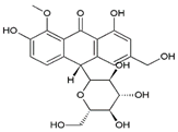
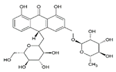
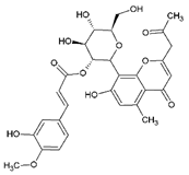
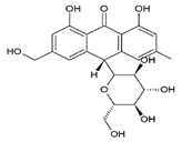
As shown on Table 1, Aloe-emodin and its derivatives constituted the largest percentage of the compounds contained in A. vera extract (about 75.74%), and the major components were aloesin (peak 2, 30.22%), aloe-emodine-diglucoside (peak 9, 12.58%), and 2′-p-methoxycoumaroylaloeresin B (peak 18, 9.64%). Aloins (B and A) at about 2.45% (peaks 15 and 17, respectively), aloinosides (B and A) at about 2.32% (peaks 10 and 12, respectively), and 8-O-methyl-7-hydroxyaloin (B and A) at about 8.43% (peaks 7 and 8, respectively) were also found in the Aloe vera water soluble extract (Figure 1). Two cinnamic acid derivatives were tentatively identified in the negative and positive modes. Peaks 1 and 3 (about 8.39%) belonged to trans-5-O-caffeoylquinic and chlorogenic acid (3-O-caffeoylquinic acid), respectively. One flavonoid compound corresponding to peak 5 was identified as luteolin 6,8-di-C-glucoside (C27H30O16, about 3.34%). Cis- and trans-isomers of 5-p-coumaroylquinic acid constituted about 9.69% of the extract.
Finally, two components with a molecular mass of about 716 Da (about 3.17%) corresponding to peaks 11 and 13 were observed in A. vera extract. The difference between the retention time of these compounds and the similarity of their MS/MS fragmentation pathways allowed us to suggest that they are also some kind of diastereomer, but no data was found about them in the literature, except that they were mentioned as unidentified compounds in [24].
In addition, the X-ray fluorescence spectrum made it possible to semi-qualitatively assess the microelement present in A. vera extract. As shown in Table 2, the minerals found were sulfur (S), silicon (Si), chlorine (Cl), potassium (K), and bromine (Br). The highest mean fluorescence intensities were recorded for K (0.2459 cps/μA) and S (0.1335 cps/μA), which could mean that these minerals are the most abundant in A. vera extract.

3.2. Susceptibility of Uropathogenic Bacteria to Common Antibiotics and Inhibition Zones of Aloe vera Extract

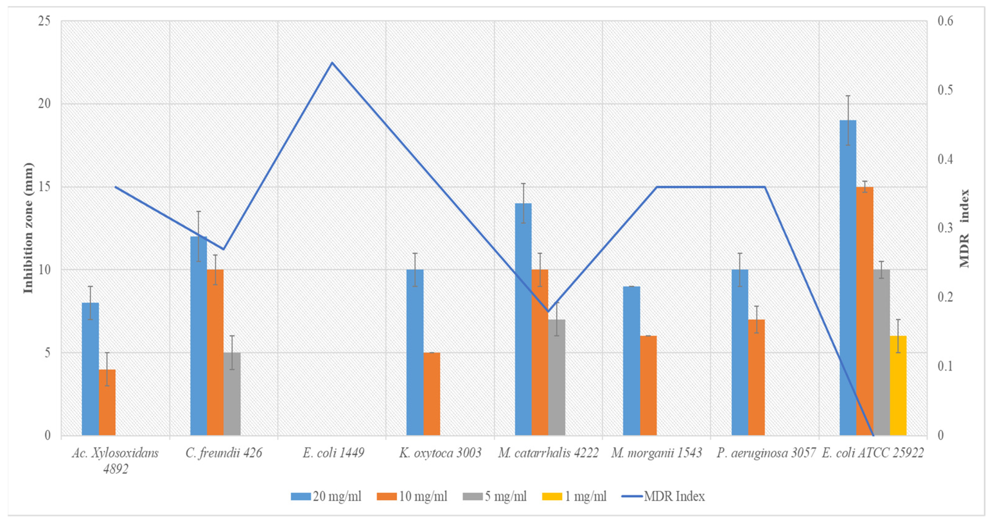
The susceptibility of the Gram-negative uropathogens to commonly used antibiotics is recorded in Table 3, and the multidrug resistance indexes were calculated. As expected, the reference strain, E. coli ATCC 25922, was susceptible to all antibiotics (MDR = 0), whereas all other clinical strains were resistant to at least two antibiotics. The most resistant bacteria were E. coli 1449 (MDR = 0.54), Ac. Xylosoxidans 4892 (MDR = 0.36), M. morganii 1543 (MDR = 0.36), P. aeruginosa 3057 (MDR = 0.36), and K. oxytoca 3003 (MDR = 0.36). Trimethoprim was the antibiotic to which uropathogens were most resistant (62.5%). This finding is consistent with the results obtained by other researchers who investigated the susceptibility to antibiotics of bacteria involved in UTIs [33]. It is well known that trimethoprim is one of the most used antibiotics as a first-line treatment in the management of UTIs, so the loss of its effectiveness is very often reported to be linked to an adaptation phenomenon due to recurrent exposures [5,6,7]. With the resurgence of antibiotic resistance, more and more failures of antibiotic treatments are observed worldwide, there are more frequent outpatient visits and hospitalizations, and there are increasing costs of second-line and third-line treatments [33,34]. Recent predictions have shown that without intervention, this threat to global health may cause 10 million deaths each year by 2050 and inflict cumulative costs of 100 trillion USD on the global economy [35,36]. Various means have been implemented in recent years to provide effective solutions to this problem, and the studies carried out target bacteria that are resistant or not. In the present study, we focused on evaluating the antibacterial potential of Aloe vera extract against the uropathogenic bacteria, the resistance profile of which is shown in Table 3. The inhibition zones (IZs) of different concentrations of A. vera extract (20, 10, 5, 1 mg/mL) obtained by the well diffusion method are depicted in Figure 2. As expected, the 5% DMSO used for diluting the dry extract showed no inhibition zone (IZ = 0 mm) against all the bacteria tested. This result was expected and agrees with the findings of Kirkwood et al. [37], who reported that DMSO had no inhibitory activity against microorganisms when used at concentrations below 10%. Furthermore, as shown in Figure 2, regardless of the bacterial strains against which an antibacterial effect was observed, it was found that the inhibition diameters decreased with a fall in the concentration, which may indicate that the antimicrobial potential of A. vera extract is dose dependent. Similar results were reported by several other authors who investigated the antibacterial activity of plant extracts [12,22]. In addition, the highest concentration (20 mg/mL) of the extract used was active on seven bacteria out of the eight tested. Indeed, unlike other clinical strains, Escherichia coli 1449 was resistant against all doses of the extract (IZ = 0 mm). Conversely, and strangely, the highest IZs was observed against E. coli ATCC 25922. This difference in susceptibility between strains of the same species can be explained by the genotypic and phenotypic differences between the two bacteria. Indeed, it is well known that the development of antimicrobial resistance is usually associated with genetic changes, either to the acquisition of resistance genes or to mutations in elements relevant to the activity of the antibiotics [38]. Unfortunately, due to limited resources, no investigation of the genetic profile of these bacteria could be conducted. However, Corona and Martinez [38] reported that in some situations, resistance can be achieved without any genetic alteration, and this was called phenotypic resistance. This was confirmed in the present study, as shown in Table 3; the E. coli 1449 strain was resistant to more than half of the conventional antibiotics tested (MDR = 0.54), while E. coli ATCC 25922 was susceptible to all the antibiotics tested (MDR = 0.00). E. coli ATCC 25922 being a reference (non-clinical) strain and E. coli 1449 being a clinical strain, as reported by others [38], it can be suggested that the phenotypic resistance of the clinical strains may have resulted from resistance acquired through exposure to various antimicrobials and other hostile conditions. Otherwise, the most sensitive (highest IZs) bacteria to A. vera extract in descending order was E. coli ATCC 25922, M. catarrhalis 4222, C. freundii 426, K. oxytoca 3003, P. aeruginosa 3057, M. morganii 1543, and Ac. xylosoxidans 4892. Interestingly, the Spearman correlation test showed that the susceptibility of bacteria to the highest concentration of A. vera extract tested strongly (p = 0.000) and negatively (Rho = −0.944) correlated with the MDR index. As shown in Figure 2, this means that the bacteria with the highest MDR index were less susceptible to the extract than those with lower ones. Therefore, this indicates that susceptibility to antimicrobials, whether new or conventional, is likely to depend on the phenotypic susceptibility of the bacteria. These observations are mainly reported on Gram negative bacteria due to the nature of their membrane and the presence of efflux pumps, which are well known to be present in Gram– bacteria. Indeed, in addition to the ability to produce antibiotic-degrading enzymes (i.e., β-lactamase genes), many Gram– bacteria possess efflux pumps that allow them to simply expel antimicrobial molecules from the cell, thus rendering the antibiotic or the antimicrobial agent ineffective [20,21]. Notwithstanding all this, medicinal plants, including A. vera, remain a credible alternative to antibiotics because they have a heterogeneous chemical composition (Table 1) that allows them to inhibit several sites in bacterial cells at the same time.

3.3. Minimum Inhibitory Concentration (MIC) and Minimum Bactericidal Concentration (MBC)

The determination of MIC and MBC is a fundamental step in the search for new antimicrobials. The MIC provides precise information on the quantity of antimicrobial necessary to inhibit the growth of bacteria, while the MBC provides information on the minimum concentration capable of killing the bacteria. In the present study, MIC was investigated by the microbroth dilution method in a sterile U-bottom 96-well microplate. As with the well diffusion method, and as expected, no bacterial inhibition was observed with the negative control (DMSO 5%). Table 4 presents the MICs and MBCs of the extract against the reference strain E. coli ATCC 25922 and the seven Gram– bacteria involved in urinary tract infections (UTIs). Overall, MICs ranged from 0.625 to >20 mg/mL, while MBCs ranged from 1.25 to >20 mg/mL. Like the results obtained by the well diffusion method, no inhibition was observed against E. coli 1449 (MIC > 20 mg/mL). This observation suggests that further analysis at concentrations higher than 20 mg/mL should be performed in order to quantify the antimicrobial activity of A. vera against E. coli 1449. Except for this resistant bacterium, A. vera extract showed interesting antibacterial activity against all the other bacteria. These results are consistent with those obtained by [14,39]. The antibacterial activity of A. vera can be directly ascribed to its chemical composition. Indeed, as revealed by the study of the chemical composition of the A. vera extract (Table 1), this plant mainly contains aloe-emodin and its derivatives, cinnamic acids derivatives, anthracene compounds, and flavonoids. Aloe-emodin and its derivatives constitute the largest percentage of the compounds contained in A. vera extract (about 75.74%) and the major components were aloesin (30.22%) and aloe-emodin-diglucoside (12.58%). Aloesin is an aromatic chromone that has applications in the cosmetics and health food industries and whose antimicrobial properties against various microorganisms have already been demonstrated [40] Añibarro-Ortega and coworkers [40] recently found MICs and MBCs close to those of ours when investigating the antibacterial activity of A. vera extract and pure aloesin against Salmonella enterica serovar Typhimurium and Listeria monocytogenes. In the same vein, Hiruy et al. [41] showed that aloesin isolated from Aloe monticola Reynolds has strong activity against Shigella boydii, Escherichia coli, S. soneii, and S. flexneri (MIC of 0.025 mg/mL) and also against Salmonella typhi, Shigella dysentery, and Staphylococcus aureus (MIC of 0.01 mg/mL).
In addition, aloe-emodin-diglucoside, which is also one of the constituents of A. vera, has already been reported for its antimicrobial activity on several microorganisms, including against Gram– bacteria such as P. aeruginosa [42]. Besides these antimicrobial properties, this anthraquinone and isomer of emodin have been reported to exhibit many pharmacological effects, including anticancer, antivirus, anti-inflammatory, antiparasitic, neuroprotective, and hepatoprotective activities [43]. Investigating the literature on biological properties of most of the compounds found in A. vera extract in the present study, it was found that each of them exhibits antimicrobial and anti-inflammatory effects, among others. This finding suggests that A. vera extract may find applications in the management of various infections, including urinary tract infections. However, the fact that uropathogenic bacteria are completely resistant to the extract indicates that this extract as such cannot play any significant role in infections involving resistant bacteria. Finally, going back to the results of the MICs and MBCs presented in Table 3, it can be concluded that except for E. coli 1449 and K. oxytoca 3003 (MBC/MIC = 8), A. vera extract exhibits bactericidal activity, since it is established in the literature that an antimicrobial compound is considered as a bactericidal against a microbial strain when the ratio MBC/MIC ≤ 4 [44,45].

3.4. Antibiofilm Formation Potential of Aloe vera Extract

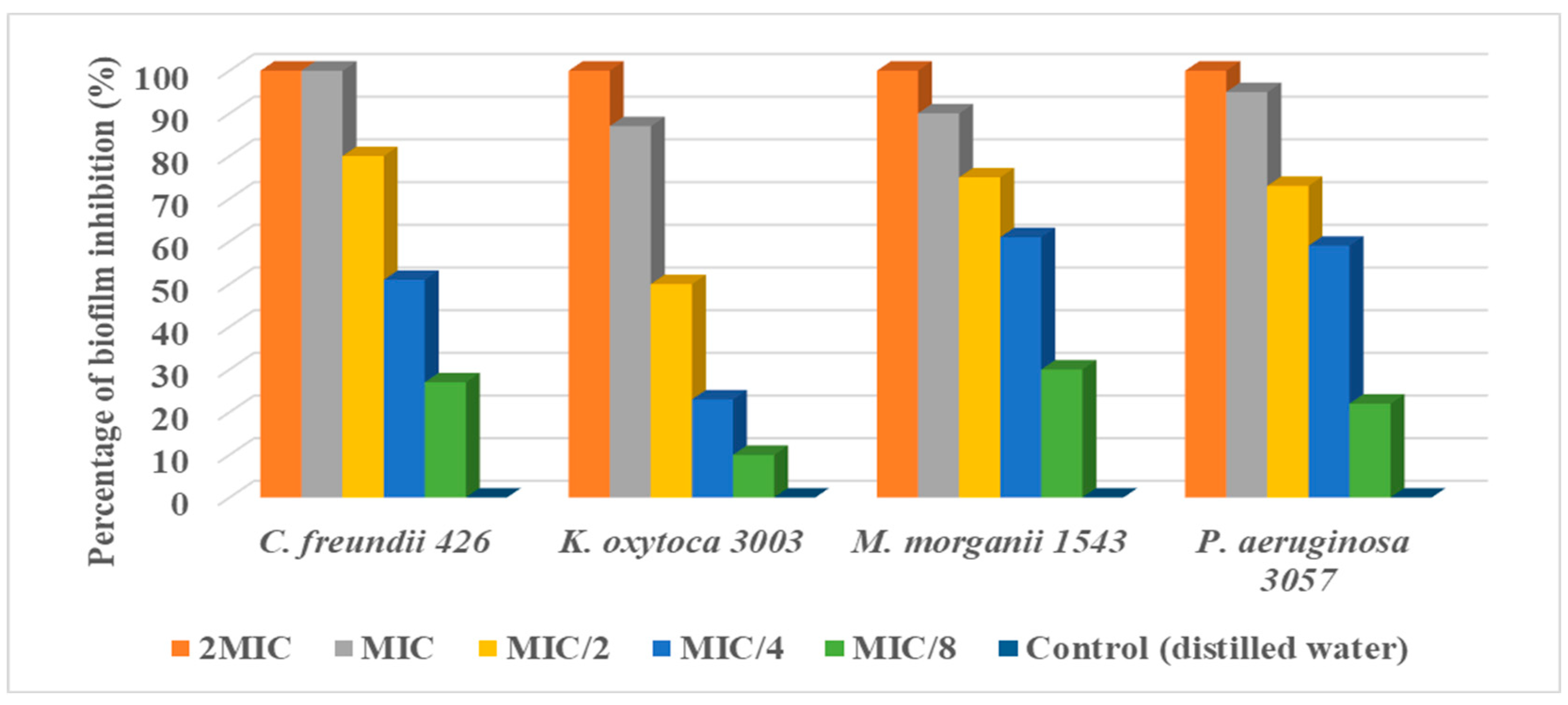
Before investigating the impact of A. vera extract on biofilm formation in selected bacteria involved in UTIs used in the present study, it was necessary to identify those that were biofilm producers. Using the crystal violet attachment assay, only C. freundii 426, K. oxytoca 3003, M. morganii 1543, and P. aeruginosa 3057 were retained as biofilm producers in the antibiofilm activity investigation. It is important to remember that biofilms are microbial consortia embedded in self-produced exopolymer matrices composed mainly of exopolysaccharides (EPS) [46]. The search for means of combating the formation of biofilms is necessary because this cellular state significantly contributes to resistance to antibiotics and the complication of infections [47]. Indeed, microbes living in these matrices benefit from nutrient and water supplies [48]; improved lateral gene transfer [49]; and protection against adverse environmental conditions such as desiccation and chemicals, including detergents, disinfectants, and antibiotics [50]. This premise was the basis of our assay on the antibiofilm formation activity of A. vera extract. As a result, regardless of the bacterial strains and as depicted in Figure 3, it was found that the antibiofilm formation potential of A. vera extract was strong at 2MIC and MIC (93–100% of biofilm inhibition), moderate at MIC/2 (32–41%), weak at MIC/4 (14–21%) and nil at MIC/8. The ability of the extract to strongly inhibit biofilms at MIC and 2MIC can be explained by the fact that the bacteria could not grow at these concentrations due to the high number of antimicrobials in the culture medium. On the other hand, this finding reveals the lack of adaptation of bacteria when exposed to the extract, because other studies have revealed that despite concentrations ≥MIC, some bacteria end up adapting after some incubation time [51]. Otherwise, it is necessary to emphasize that during this study, we attempted to investigate the ability of A. vera extract to destroy pre-formed biofilms, but no activity was observed. Under the limitations of this study, an A. vera extract at a high concentration could be recommended to prevent initial bacterial cell attachment in biofilm-related infections but must not be recommended to destroy existing biofilms.

4. Conclusions

The present study aimed to evaluate the antimicrobial activity of an aqueous extract of Aloe vera and to study its chemical composition. It was found that the A. vera extract has worthy antimicrobial activity against all uropathogenic bacteria except against a multiresistant Escherichia coli (E. coli 1449). Given the results obtained, this study constitutes a strong baseline for considering A. vera for further studies in the search for new antimicrobials in this era of antibiotic resistance.

Author Contributions

Conceptualization, M.M.J.A.; methodology, M.M.J.A. and P.I.V.; software, G.V.S.; validation, F.H., G.V.S., M.M.J.A. and P.I.V.; formal analysis, Y.N.V.; investigation, M.M.J.A.; G.V.S. and F.H.; resources, P.I.V.; data curation, Z.A.V.; writing—original draft preparation, M.M.J.A.; writing—review and editing, S.A.N., N.S. and V.E.A.; visualization, M.M.J.A.; supervision, P.I.V.; project administration, P.I.V.; funding acquisition, P.I.V. All authors have read and agreed to the published version of the manuscript.

Funding

This research received no external funding.

Institutional Review Board Statement

Not applicable.

Informed Consent Statement

Not applicable.

Data Availability Statement

All data are within the manuscript.

Acknowledgments

This study has been supported by the RUDN University Strategic Academic Leadership Program.

Conflicts of Interest

The authors declare no conflict of interest.

References

  1. Palma, E.; Tilocca, B.; Roncada, P. Antimicrobial resistance in veterinary medicine: An overview. Int. J. Mol. Sci. 2020, 21, 1914. [Google Scholar] [CrossRef] [PubMed] [Green Version]
  2. Joseph, A.M.M.; Jorelle, A.B.; Sarra, S.; Viktorovna, P.I.; Davares, A.K.; Ingrid, N.K.; Steve, A.A.F.; Andreevna, S.L.; Vyacheslavovna, Y.N.; Carime, B.Z. Short review on the potential alternatives to antibiotics in the era of antibiotic resistance. J. Appl. Pharm. Sci. 2022, 12, 29–40. [Google Scholar] [CrossRef]
  3. WHO: World Health Organization. Antibiotic Resistance. Available online: https://www.who.int/news-room/fact-sheets/detail/antibiotic-resistance.2020 (accessed on 31 May 2022).
  4. Agarwal, D.K.; Krambeck, A.E.; Sharma, V.; Maldonado, F.J.; Westerman, M.E.; Knoedler, J.J.; Rivera, M.E. Treatment of non-obstructive, non-struvite urolithiasis is effective in treatment of recurrent urinary tract infections. World J. Urol. 2020, 38, 2029–2033. [Google Scholar] [CrossRef] [PubMed]
  5. Arsene, M.M.J.; Viktorovna, P.I.; Davares, A.K.L.; Esther, N.; Nikolaevich, S.A. Urinary tract infections: Virulence factors, resistance to antibiotics, and management of uropathogenic bacteria with medicinal plants—A review. J. Appl. Pharm. Sci. 2021, 11, 001–012. [Google Scholar] [CrossRef]
  6. Gupta, K.; Hooton, T.M.; Naber, K.G.; Wullt, B.; Colgan, R.; Miller, L.G.; Moran, G.J.; Nicolle, L.E.; Raz, R.; Schaeffer, A.J.; et al. Infectious Diseases Society of America, European Society for Microbiology and Infectious Diseases. International clinical practice guidelines for the treatment of acute uncomplicated cystitis and pyelonephritis in women: A 2010 update by the Infectious Diseases Society of America and the European Society for Microbiology and Infectious Diseases. Clin. Infect. Dis. 2011, 52, e103–e120. [Google Scholar] [CrossRef] [Green Version]
  7. Bader, M.S.; Loeb, M.; Leto, D.; Brooks, A.A. Treatment of urinary tract infections in the era of antimicrobial resistance and new antimicrobial agents. Postgrad. Med. 2020, 132, 234–250. [Google Scholar] [CrossRef]
  8. Amer, M.W.; Awwad, A.M. Green synthesis of copper nanoparticles by Citrus limon fruits extract, characterization, and antibacterial activity. Chem. Int. 2021, 7, 1–8. [Google Scholar] [CrossRef]
  9. Ansaldi, M.; Boulanger, P.; Brives, C.; Debarbieux, L.; Dufour, N.; Froissart, R.; Gandon, S.; Hénaff, C.L.; Petit, M.-A.; Rocha, E.; et al. Les applications antibactériennes des bactériophages. Virologie 2020, 24, 23–36. [Google Scholar] [CrossRef]
  10. Andersson, D.I.; Hughes, D.; Kubicek-Sutherland, J.Z. Mechanisms, and consequences of bacterial resistance to antimicrobial peptides. Drug Resist. Updat. 2016, 2, 43–57. [Google Scholar] [CrossRef]
  11. Arsene, J.M.M.; Davares, A.K.; Andreevna, S.L.; Vladimirovich, E.A.; Carime, B.Z.; Marouf, R.; Khelifi, I. The use of probiotics in animal feeding for safe production and as potential alternatives to antibiotics. Vet. World 2021, 14, 319–328. [Google Scholar] [CrossRef]
  12. Manga, A.M.J.; Podoprigora, I.V.; Davares AK, L.; Razan, M.; Das, M.S.; Senyagin, A.N. Antibacterial activity of grapefruit peel extracts and green-synthesized silver nanoparticles. Vet. World 2021, 14, 1330–1341. [Google Scholar] [CrossRef]
  13. Wojnicz, D.; Kucharska, A.Z.; Sokół-Łętowska, A.; Kicia, M.; Tichaczek-Goska, D. Medicinal plants extracts affect virulence factors expression and biofilm formation by the uropathogenic Escherichia coli. Urol. Res. 2012, 40, 683–697. [Google Scholar] [CrossRef] [Green Version]
  14. Kumar, S.; Budhwar, L.; Yadav, A.; Yadav, M.; Parkash Yadav, J. Phytochemical screening and antibacterial activity of Aloe vera collected from different climatic regions of India. Nat. Prod. J. 2016, 6, 73–82. [Google Scholar] [CrossRef]
  15. Kumar, S.; Kalita, S.; Das, A.; Kumar, P.; Singh, S.; Katiyar, V.; Mukherjee, A. Aloe vera: A contemporary overview on scope and prospects in food preservation and packaging. Prog. Org. Coat. 2022, 166, 106799. [Google Scholar] [CrossRef]
  16. Sarker, A.; Grift, T.E. Bioactive properties and potential applications of Aloe vera gel edible coating on fresh and minimally processed fruits and vegetables: A review. J. Food Meas. Charact. 2021, 15, 2119–2134. [Google Scholar] [CrossRef]
  17. Nizam, N.H.M.; Rawi, N.F.M.; Ramle, S.F.M.; Abd Aziz, A.; Abdullah, C.K.; Rashedi, A.; Kassim, M.H.M. Physical, thermal, mechanical, antimicrobial, and physicochemical properties of starch-based film containing Aloe vera: A review. J. Mater. Res. Technol. 2021, 15, 1572–1589. [Google Scholar] [CrossRef]
  18. Saleem, A.; Naureen, I.; Naeem, M.; Murad, H.S.; Maqsood, S.; Tasleem, G. Aloe vera Gel Effect on Skin and Pharmacological Properties. Sch. Int. J. Anat. Physiol. 2022, 5, 1–8. [Google Scholar] [CrossRef]
  19. Mbarga, M.J.A.; Viktorovna, P.I.; Mikhaïlovitch, M.K.; Davares AK, L.; Parfait, K.; Rehailia, M.; Shommiya, D. In vitro antimicrobial activity, antibioresistance reversal properties, and toxicity screen of ethanolic extracts of Heracleum mantegazzianum Sommier and Levier (giant hogweed), Centaurea jacea L.(brown knapweed), and Chenopodium album L.(Pigweed): Three invasive plants. Open Vet. J. 2022, 12, 584. [Google Scholar] [CrossRef]
  20. Arsene, M.M.J.; Viktorovna, P.I.; Davares, A.K.L.; Parfait, K.; Andreevna, S.L.; Mouafo, H.T.; Rehailia, M.; Vyacheslavovna, Y.N.; Pavlovna, S.I.; Manga, I.A.M.; et al. Antimicrobial and Antibiotic-Resistance Reversal Activity of Some Medicinal Plants from Cameroon against Selected Resistant and Non-Resistant Uropathogenic Bacteria. Front. Biosci.-Elite 2022, 14, 25. [Google Scholar] [CrossRef]
  21. Arsene, M.M.J.; Viktorovna, P.I.; Alla, M.V.; Mariya, M.A.; Sergei, G.V.; Cesar, E.; Davares, A.K.L.; Parfait, K.; Wilfrid, K.N.; Nikolay, T.S.; et al. Optimization of Ethanolic Extraction of Enantia chloranta Bark, Phytochemical Composition, Green Synthesis of Silver Nanoparticles, and Antimicrobial Activity. Fermentation 2022, 8, 530. [Google Scholar] [CrossRef]
  22. Khar’kov, Y.K.; Arsene, M.M.; Aliya, M.V.; Viktorovna, P.I.; Elena, V.G.; Azova, M.M.; Amira, A.A. Assessment of Antimicrobial Activity of Ethanolic and Aqueous Extracts of Aesculus hippocastanum L. (Horse Chestnut) Bark against Bacteria Isolated from Urine of Patients Diagnosed Positive to Urinary Tract Infections. Front. Biosci.-Sch. 2022, 14, 11. [Google Scholar] [CrossRef]
  23. Habibipour, R.; Moradi-Haghgou, L.; Farmany, A. Green synthesis of AgNPs@ PPE and its Pseudomonas aeruginosa biofilm formation activity compared to pomegranate peel extract. Int. J. Nanomed. 2019, 14, 6891–6899. [Google Scholar] [CrossRef] [PubMed] [Green Version]
  24. El Sayed, A.M.; Ezzat, S.M.; El Naggar, M.M.; El Hawary, S.S. In vivo diabetic wound healing effect and HPLC–DAD–ESI–MS/MS profiling of the methanol extracts of eight Aloe species. Rev. Bras. Farmacogn. 2016, 26, 352–362. [Google Scholar] [CrossRef] [Green Version]
  25. Solaberrieta, I.; Jiménez, A.; Garrigós, M.C. Valorization of Aloe vera Skin By-Products to Obtain Bioactive Compounds by Microwave-Assisted Extraction: Antioxidant Activity and Chemical Composition. Antioxidants 2022, 11, 1058. [Google Scholar] [CrossRef]
  26. Quispe, C.; Villalobos, M.; Bórquez, J.; Simirgiotis, M. Chemical Composition and Antioxidant Activity of Aloe vera from the Pica Oasis (Tarapac´a, Chile) by UHPLC-Q/Orbitrap/MS/MS. J. Chem. 2018, 2018, 6123850. [Google Scholar] [CrossRef] [Green Version]
  27. Añibarro-Ortega, M.; Pinela, J.; Barros, L.; C´iric´, A.; Silva, S.P.; Coelho, E.; Mocan, A.; Calhelha, R.C.; Sokovic´, M.; Coimbra, M.A.; et al. Compositional Features and Bioactive Properties of Aloe vera Leaf (Fillet, Mucilage, and Rind) and Flower. Antioxidants 2019, 8, 444. [Google Scholar] [CrossRef] [Green Version]
  28. Debora, S.; Giuseppe, P.; Paolo, B. Aloe exudate: Characterization by reversed phase HPLC and headspace GC-MS. J. Agric. Food Chem. 2001, 49, 26–30. [Google Scholar] [CrossRef]
  29. Aida, P.U.I.A.; Chedea, V.S.; Levai, A.M.; Bocsan, I.C.; Buzoianu, A.D. Pot Aloe vera gel—A natural source of antioxidants. Not. Bot. Horti Agrobot. Cluj-Napoca 2022, 50, 2. [Google Scholar] [CrossRef]
  30. Lee, S.; Do, S.-G.; Kim, S.Y.; Kim, J.; Jin, Y.; Lee, C.H. Mass Spectrometry-Based Metabolite Profiling and Antioxidant Activity of Aloe vera (Aloe barbadensis Miller) in Different Growth Stages. J. Agric. Food Chem. 2012, 60, 11222–11228. [Google Scholar] [CrossRef]
  31. Aldayel, T.S.; Grace, M.H.; Lila, M.A.; Yahya, M.A.; Omar, U.M.; Alshammary, G. LC-MS Characterization of Bioactive Metabolites from Two Yemeni Aloe Spp. with Antioxidant and Antidiabetic Properties. Arab. J. Chem. 2020, 13, 5040–5049. [Google Scholar] [CrossRef]
  32. Samira, B.; Ismahene, B.; Aicha, T.; Radia, D.; Abbes, B.; Chawki, B. Analysis of Phytochemical Constituents by using LC-MS, Antifungal and Allelopathic Activities of Leaves Extracts of Aloe Vera. Jordan J. Biol. Sci. 2022, 15, 21–28. [Google Scholar] [CrossRef]
  33. Mortazavi-Tabatabaei SA, R.; Ghaderkhani, J.; Nazari, A.; Sayehmiri, K.; Sayehmiri, F.; Pakzad, I. Pattern of antibacterial resistance in urinary tract infections: A systematic review and meta-analysis. Int. J. Prev. Med. 2019, 10, 169. [Google Scholar] [CrossRef] [PubMed]
  34. Hadi, U.; Van den Broek, P.; Kolopaking, E.P.; Zairina, N.; Gardjito, W.; Gyssens, I.C. Cross-sectional study of availability and pharmaceutical quality of antibiotics requested with or without prescription (Over The Counter) in Surabaya, Indonesia. BMC Infect. Dis. 2010, 10, 203. [Google Scholar] [CrossRef] [PubMed] [Green Version]
  35. De Kraker, M.E.; Stewardson, A.J.; Harbarth, S. Will 10 million people die a year due to antimicrobial resistance by 2050? PLoS Med. 2016, 13, e1002184. [Google Scholar] [CrossRef] [Green Version]
  36. Arsène MM, J.; Davares AK, L.; Viktorovna, P.I.; Andreevna, S.L.; Sarra, S.; Khelifi, I.; Sergueïevna, D.M. The public health issue of antibiotic residues in food and feed: Causes, consequences, and potential solutions. Vet. World 2022, 15, 662. [Google Scholar] [CrossRef]
  37. Kirkwood, Z.I.; Millar, B.C.; Downey, D.G.; Moore, J.E. Antimicrobial effect of dimethyl sulfoxide and N, N-Dimethylformamide on Mycobacterium abscessus: Implications for antimicrobial susceptibility testing. Int. J. Mycobacteriol. 2018, 7, 134. [Google Scholar] [CrossRef]
  38. Corona, F.; Martinez, J.L. Phenotypic resistance to antibiotics. Antibiotics 2013, 2, 237–255. [Google Scholar] [CrossRef] [Green Version]
  39. Das, P.; Srivastav, A.K. Phytochemical extraction and characterization of the leaves of Aloe vera barbadensis for its anti-bacterial and antioxidant activity. Int. J. Sci. Res. 2015, 4, 658–661. [Google Scholar]
  40. Añibarro-Ortega, M.; Pinela, J.; Ćirić, A.; Lopes, E.; Molina, A.K.; Calhelha, R.C.; Soković, M.; Ferreira, O.; Ferreira, I.C.F.R.; Barros, L. Extraction of aloesin from Aloe vera rind using alternative green solvents: Process optimization and biological activity assessment. Biology 2021, 10, 951. [Google Scholar] [CrossRef]
  41. Hiruy, M.; Bisrat, D.; Mazumder, A.; Asres, K. Two chromones with antimicrobial activity from the leaf latex of Aloe monticola Reynolds. Nat. Prod. Res. 2021, 35, 1052–1056. [Google Scholar] [CrossRef]
  42. Li, T.; Lu, Y.; Zhang, H.; Wang, L.; Beier, R.C.; Jin, Y.; Wang, W.; Li, H.; Hou, X. Antibacterial Activity and Membrane-Targeting Mechanism of Aloe-Emodin Against Staphylococcus epidermidis. Front. Microbiol. 2021, 12, 621866. [Google Scholar] [CrossRef] [PubMed]
  43. Dong, X.; Zeng, Y.; Liu, Y.; You, L.; Yin, X.; Fu, J.; Ni, J. Aloe-emodin: A review of its pharmacology, toxicity, and pharmacokinetics. Phytother. Res. 2021, 34, 270–281. [Google Scholar] [CrossRef] [PubMed]
  44. Kuete, V. Potential of Cameroonian plants and derived products against microbial infections: A review. Planta Med. 2010, 76, 1479–1491. [Google Scholar] [CrossRef] [PubMed] [Green Version]
  45. Kuete, V.; Efferth, T. Cameroonian medicinal plants: Pharmacology and derived natural products. Front. Pharmacol. 2010, 1, 123. [Google Scholar] [CrossRef] [Green Version]
  46. Ouidir, T.; Gabriel, B.; Chabane, Y.N. Overview of multi-species biofilms in different ecosystems: Wastewater treatment, soil and oral cavity. J. Biotechnol. 2022, 350, 67–74. [Google Scholar] [CrossRef] [PubMed]
  47. Martinez-Gutierrez, F.; Boegli, L.; Agostinho, A.; Sánchez, E.M.; Bach, H.; Ruiz, F.; James, G. Anti-biofilm activity of silver nanoparticles against different microorganisms. Biofouling 2013, 29, 651–660. [Google Scholar] [CrossRef] [PubMed]
  48. Goller, C.C.; Romeo, T. Environmental influences on biofilm development. Bact. Biofilms 2008, 322, 37–66. [Google Scholar] [CrossRef]
  49. Mah TF, C.; O’Toole, G.A. Mechanisms of biofilm resistance to antimicrobial agents. Trends Microbiol. 2001, 9, 34–39. [Google Scholar] [CrossRef]
  50. Stewart, P.S.; Costerton, J.W. Antibiotic resistance of bacteria in biofilms. Lancet 2001, 358, 135–138. [Google Scholar] [CrossRef]
  51. Mann, R.; Holmes, A.; McNeilly, O.; Cavaliere, R.; Sotiriou, G.A.; Rice, S.A.; Gunawan, C. Evolution of biofilm-forming pathogenic bacteria in the presence of nanoparticles and antibiotic: Adaptation phenomena and cross-resistance. J. Nanobiotechnol. 2021, 19, 291. [Google Scholar] [CrossRef]
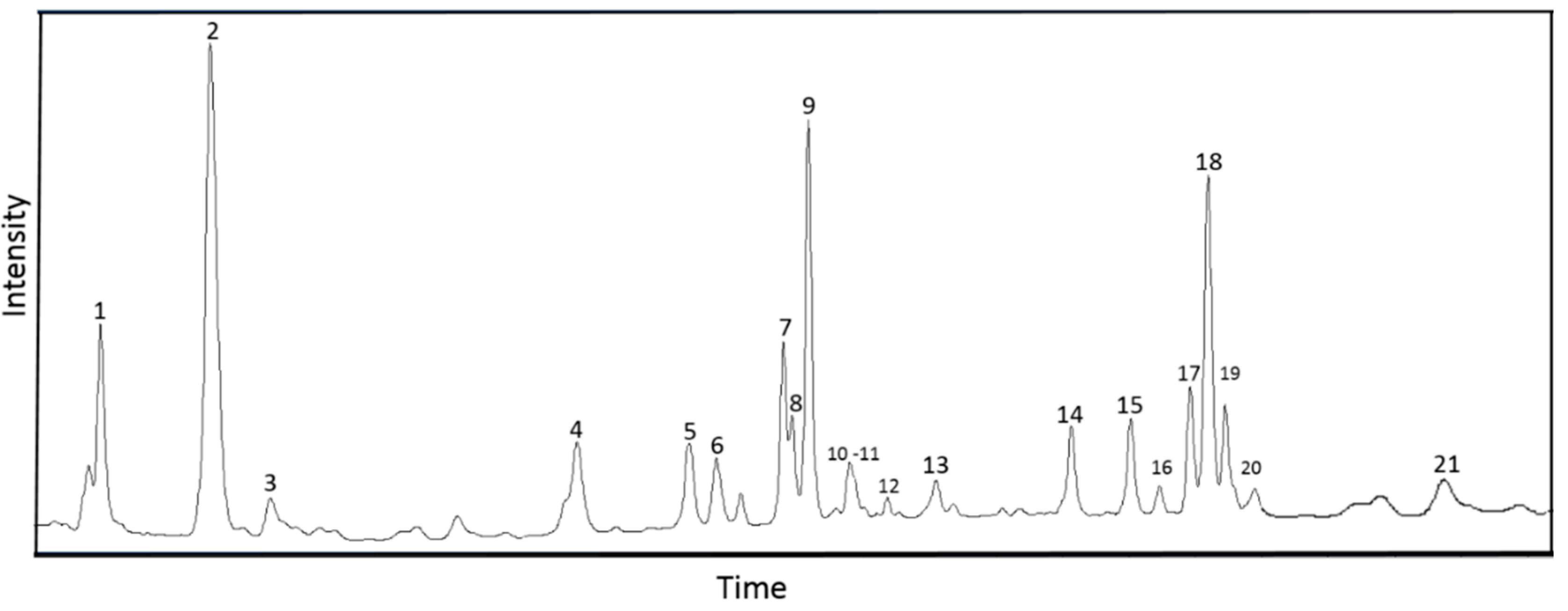
Figure 1. HPLC–MS/MS chromatogram of Aloe vera extract.
Fermentation 08 00626 g001
Figure 2. Diameter of the inhibition zone (mm) of A. vera extract against Gram-negative bacteria as a function of concentration and bacterial multidrug drug resistance (MDR) index.
Fermentation 08 00626 g002
Figure 3. Antibiofilm activity of A. vera extract against selected Gram-negative bacteria involved in urinary tract infections (UTIs).
Fermentation 08 00626 g003
Table 1. Phytochemical composition of Aloe vera extract.
No.CompoundRetention Time,
min
Molecular FormulaStructural FormulaMolecular Mass, Dam/z, Registration of Positive Ionsm/z, Registration of Negative IonsContent,
%
References
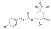
1Trans-5-O-Caffeoylquinic acid3.52C16H18O9Fermentation 08 00626 i001354355 [M + H]+, 377 [M + Na]+, 731 [2M + Na]+191 [C7H11O6], 353 [M–H], 707 [2M–H]6.68 ± 0.45[25,26,27]
2Aloesin5.34C19H22O9Fermentation 08 00626 i002394395 [M + H]+, 811 [2M + Na]+393 [M–H]30.22 ± 0.43[25,26,28,29]
3Chlorogenic acid (3-O-Caffeoylquinic acid)6.63C16H18O9Fermentation 08 00626 i003354355 [M + H]+, 377 [M + Na]+, 731 [2M + Na]+191 [C7H11O6], 353 [M–H], 707 [2M–H]1.71 ± 0.05[25,26]
4Trans-5-p-Coumaroylquinic acid10.75C16H18O8Fermentation 08 00626 i004338147 [C9H7O2]+, 339 [M + H]+337 [M–H], 675 [2M–H]6.86 ± 0.39[25,27]
5Luteolin 6,8-di-C-glucoside12.47C27H30O16Fermentation 08 00626 i005610287 [C15H11O6]+, 449 [M–glu]+,
611 [M + H]+, 633 [M + Na]+
285 [C15H9O6], 447 [M–glu], 609 [M–H]3.34 ± 0.01[24,25,26,27]
6Cis-5-p-Coumaroylquinic acid12.92C16H18O8Fermentation 08 00626 i006338147 [C9H7O2]+, 339 [M + H]+337 [M–H], 675 [2M–H]2.83 ± 0.01[25,27]
78-O-Methyl-7-hydroxyaloin B13.88C22H24O10Fermentation 08 00626 i007448449 [M + H]+, 471 [M + Na]+447 [M–H]1.03 ± 0.01[25,28,30,31]
88-O-Methyl-7-hydroxyaloin A13.91C22H24O10Fermentation 08 00626 i008448449 [M + H]+, 471 [M + Na]+447 [M–H]7.40 ± 0.08[25,28,30,31]
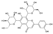
9Aloe-emodine-diglucoside14.29C27H30O15Fermentation 08 00626 i009594595 [M + H]+, 617 [M + Na]+593 [M–H]12.58 ± 0.34[24,32]
10Aloinoside B14.72C27H32O13Fermentation 08 00626 i010564565 [M + H]+563 [M–H]1.81 ± 0.01[24,28]
11Unidentified component, MM = 716 Da14.9--716717 [M + H]+715 [M–H]1.92 ± 0.08
12Aloinoside A15.2C27H32O13Fermentation 08 00626 i011564565 [M + H]+563 [M–H]0.51 ± 0.01[24,28,29]
13Unidentified component, MM = 716 Da16.18--716717 [M + H]+715 [M–H]1.25 ± 0.01
142′-O-Feruloyaloesin18.23C29H30O12Fermentation 08 00626 i012570571 [M + H]+569 [M–H]3.65 ± 0.03[24]
15Aloin B19.12C21H22O9Fermentation 08 00626 i013418419 [M + H]+, 441 [M + Na]+297 [C17H13O5], 418 [M–H]1.16 ± 0.02[24,25,26,28,29,32]
16Aloe-emodin-glucoside19.55C27H30O15Fermentation 08 00626 i014432271 [C15H11O5]+, 433 [M + H]+269 [C15H9O5], 431 [M–H]0.80 ± 0.01[24,26,29]
17Aloin A20.02C21H22O9Fermentation 08 00626 i015418419 [M + H]+, 441 [M + Na]+297 [C17H13O5], 418 [M–H]1.29 ± 0.07[24,25,26,28,29,32]
182′-p-Methoxycoumaroylaloeresin B20.28C29H30O11Fermentation 08 00626 i016554555 [M + H]+553 [M–H]9.64 ± 0.13[26,29]
196′-Malonylnataloin20.53C24H24O12Fermentation 08 00626 i017504505 [M + H]+, 527 [M + Na]+459 [C23H23O10], 503 [M–H]0.94 ± 0.04[24,26]
20Aloe-emodin-glucoside20.66C21H20O10Fermentation 08 00626 i018432271 [C15H11O5]+, 433 [M + H]+269 [C15H9O5], 431 [M–H]3.89 ± 0.15[26,29]
21Aloe-emodin24.57C15H10O5Fermentation 08 00626 i019270271 [C15H11O5]+269 [C15H9O5]0.82 ± 0.01[25,28,29]
Table 2. Elemental composition of Aloe vera extract and AgNP phytoproduct.
Chemical ElementsAverage Fluorescence Intensity, imp/µAStandard Deviation
Si0.06170.0031
S0.13350.0019
Cl0.02740.0026
K0.24590.0033
Br0.04920.0023
Table 3. Susceptibility to antibiotics of the test uropathogenic bacteria.
NITTECTRAMCFOCAZIPMCACCIPAMPTRMDR Index
Ac. Xylosoxidans 48926 ± 0 (I)11 ± 0 (R)23 ± 2 (S)36 ± 4 (S)6 ± 0 (R)16 ± 0 (I)32 ± 3 (S)16 ± 1 (I)20 ± 2 (I)20 ± 1 (S)6 ± 0 (R)0.36
C. freundii 42621 ± 1 (S)30 ± 3 (S)27 ± 2 (S)35 ± 1 (S)40 ± 2 (S)12 ± 0 (R)37 ± 1 (S)10 ± 0 (R)30 ± 2 (S)6 ± 0 (R)22 ± 2 (S)0.27
E. coli 144924 ± 3 (S)11 ± 0 (R)8 ± 0 (R)27 ± 1 (S)30 ± 1 (S)7 ± 0 (R)22 ± 2 (S)12 ± 0 (R)26 ± 1 (S)6 ± 0 (R)6 ± 0 (R)0.54
K. oxytoca 300320 ± 1 (S)8 ± 0 (R)22 ± 0 (S)24 ± 1 (S)25 ± 3 (S)15 ± 1 (I)27 ± 3 (S)6 ± 0 (R)30 ± 2 (S)6 ± 0 (R)6 ± 0 (R)0.36
M. catarrhalis 422212 ± 1 (R)22 ± 2 (S)24 ± 1 (S)36 ± 3 (S)27 ± 2 (S)16 ± 0 (I)27 ± 1 (S)21 ± 0 (S)32 ± 4 (S)10 ± 0 (R)15 ± 1 (I)0.18
M. morganii 154315 ± 0 (I)6 ± 0 (R)33 ± 2 (S)17 ± 1 (I)13 ± 0 (R)23 ± 1 (S)22 ± 0 (S)23 ± 1 (S)22 ± 2 (S)10 ± 0 (R)6 ± 0 (R)0.36
P. aeruginosa 30576 ± 0 (R)13 ± 1 (R)21 ± 1 (S)16 ± 0 (I)27 ± 1 (S)15 ± 0 (I)34 ± 4 (S)22 ± 2 (S)30 ± 1 (S)6 ± 0 (R)6 ± 0 (R)0.36
E. coli ATCC 2592221 ± 3 (S)30 ± 1 (S)24 ± 3 (S)20 ± 0 (S)40 ± 3 (S)18 ± 1 (S)32 ± 2 (S)16 ± 0 (S)24 ± 3 (S)26 ± 1 (S)24 ± 3 (S)0.00
Amoxicillin (AMC), ampicillin (AMP), cefazolin (CZ), cefazolin/clavulanic acid (CAC), ceftriaxone (CTR), ciprofloxacin (CIP), nitrofurantoin (NIT), trimethoprim (TR), fosfomycin (FO), imipenem (IMP), tetracyclin (TE).
Table 4. The minimum inhibitory concentrations (MIC) and the minimum bactericidal concentrations (MBC) of A. vera extract.
MIC (mg/mL)MBC (mg/mL)MBC/MIC
Ac. Xylosoxidans 48925102
C. freundii 4262.5104
E. coli 1449>20>20-
K. oxytoca 30031.25108
M. catarrhalis 42221.2554
M. morganii 15435102
P. aeruginosa 30572.5104
E. coli ATCC 259220.6251.252
Publisher’s Note: MDPI stays neutral with regard to jurisdictional claims in published maps and institutional affiliations.