Review

Effects of Selenium Supplementation on Rumen Microbiota, Rumen Fermentation, and Apparent Nutrient Digestibility of Ruminant Animals: A Review

by
Amin Omar Hendawy
1,2,*,
Satoshi Sugimura
1,
Kan Sato
1,3,
Mohamed Mohsen Mansour
2,
Ayman H. Abd El-Aziz
4,
Haney Samir
5,
Md. Aminul Islam
6,
A. B. M. Rubayet Bostami
7,
Ahmed S. Mandour
8,9,
Ahmed Elfadadny
10,
Rokaia F. Ragab
11,
Hend A. Abdelmageed
12,13 and
Amira Mohammed Ali
14
1
Department of Biological Production, Tokyo University of Agriculture and Technology, Tokyo 183-8509, Japan
2
Department of Animal and Poultry Production, Faculty of Agriculture, Damanhour University, Damanhour 22516, Egypt
3
Laboratory of Animal Nutrition, Division of Life Sciences, Graduate School of Agricultural Science, Tohoku University, Sendai 980-8572, Japan
4
Animal Husbandry and Animal Wealth Development Department, Faculty of Veterinary Medicine, Damanhour University, Damanhour 22511, Egypt
5
Department of Theriogenology, Faculty of Veterinary Medicine, Cairo University, Giza 12211, Egypt
6
Department of Medicine, Faculty of Veterinary Medicine and Animal Science, Bangabandhu Sheikh Mujibur Rahman Agricultural University, Gazipur 1706, Bangladesh
7
Department of Animal Science and Nutrition, Faculty of Veterinary Medicine and Animal Science, Bangabandhu Sheikh Mujibur Rahman Agricultural University, Gazipur 1706, Bangladesh
8
Department of Animal Medicine (Internal Medicine), Faculty of Veterinary Medicine, Suez Canal University, Ring road, Ismailia 41522, Egypt
9
Department of Veterinary Medicine, Tokyo University of Agriculture and Technology, Tokyo 183-8509, Japan
10
Department of Animal Internal Medicine, Faculty of Veterinary Medicine, Damanhour University, Damanhour 22511, Egypt
11
Department of Biochemistry, Faculty of Veterinary Medicine, Damanhour University, Damanhour 22511, Egypt
12
Department of Bacteriology, Animal Health Research Institute, Agriculture Research Center, Ismailia Lab, First District, Ismailia 41511, Egypt
13
Laboratory of Veterinary Microbiology, Cooperative Department of Veterinary Medicine, Faculty of Agriculture, Tokyo University of Agriculture and Technology, Tokyo 183-8509, Japan
14
Department of Psychiatric Nursing and Mental Health, Faculty of Nursing, Alexandria University, Smouha, Alexandria 21527, Egypt
*
Author to whom correspondence should be addressed.
Fermentation 2022, 8(1), 4; https://doi.org/10.3390/fermentation8010004
Submission received: 9 November 2021 / Revised: 18 December 2021 / Accepted: 20 December 2021 / Published: 24 December 2021
(This article belongs to the Special Issue Value-Added Co-products from Industrial Fermentation)

Abstract

:
Enzymes excreted by rumen microbiome facilitate the conversion of ingested plant materials into major nutrients (e.g., volatile fatty acids (VFA) and microbial proteins) required for animal growth. Diet, animal age, and health affect the structure of the rumen microbial community. Pathogenic organisms in the rumen negatively affect fermentation processes in favor of energy loss and animal deprivation of nutrients in ingested feed. Drawing from the ban on antibiotic use during the last decade, the livestock industry has been focused on increasing rumen microbial nutrient supply to ruminants through the use of natural supplements that are capable of promoting the activity of beneficial rumen microflora. Selenium (Se) is a trace mineral commonly used as a supplement to regulate animal metabolism. However, a clear understanding of its effects on rumen microbial composition and rumen fermentation is not available. This review summarized the available literature for the effects of Se on specific rumen microorganisms along with consequences for rumen fermentation and digestibility. Some positive effects on total VFA, the molar proportion of propionate, acetate to propionate ratio, ruminal NH3-N, pH, enzymatic activity, ruminal microbiome composition, and digestibility were recorded. Because Se nanoparticles (SeNPs) were more effective than other forms of Se, more studies are needed to compare the effectiveness of synthetic SeNPs and lactic acid bacteria enriched with sodium selenite as a biological source of SeNPs and probiotics. Future studies also need to evaluate the effect of dietary Se on methane emissions.

1. Introduction

The conversion of carbohydrates that exist in botanical fibers ingested by ruminants is mainly dependent on the activity of the microbial population in the rumen [1]. The rumen is abundant in astronomic numbers of various microorganisms including bacteria, methanogenic archaea, anaerobic fungi, and protozoa [2,3]. Due to the large number of enzymes excreted by these microorganisms, fibrous plant materials and non-protein nitrogen are broken down and changed into nutritious products, primarily volatile fatty acids (VFA) and microbial protein, which promote the production of milk and muscle [2]. Environmental factors such as heat stress, different management strategies (e.g., diets high in fermentable carbohydrates) and improper diets comprising inadequate supply of essential nutrients and/or improper feed formulation alter the composition of the ruminal microbial communities by promoting the propagation of pathogenic microorganisms in the rumen, resulting in a deficient fermentation process [1,4].
Dysfunctional fermentation causes a considerable loss of dietary energy and protein by promoting excessive production of ammonia and methane, which contribute to environmental hazards such as global warming [3]. As shown in Figure 1, rumen microbial alterations occurring in some diets employed by the intensive production system are associated with the development of various diseases [4,5]. These diets promote the propagation of endotoxic bacteria, which is associated with the reduction in the beneficial microflora, local inflammation in the gut, gut dysbiosis, and passage of the toxic bacteria and their metabolites into the systemic circulation to induce remote dysfunctions in various organs [6,7,8,9,10]. For example, goats on high grain diets for 50 days expressed reduced rumen pH and dysfunctional patterns of rumen bacterial composition, and significantly increased rumen and blood levels of lipopolysaccharide (LPS) [5]. LPS influx into the circulation resulted in activation of inflammatory signaling, increased production of pro-inflammatory cytokines (interleukin (IL)-1β, IL-6, tumor necrosis factor-α (TNF-α)) and matrix metalloproteinase-2, impaired animal health, and altered gait due to laminar tissue damage [5,11]. In addition, the colonization of diarrheic pathogens (bacteria, fungi, and viruses) in the bovine intestinal tract can cause considerable economic losses due to the reduction in milk yield and meat production [12].
In the past, antimicrobials have been routinely used as nutrition-boosting tools in order to promote animal health and save energy by altering methanogens. However, the use of antimicrobials as growth promoters or for prophylactic purposes in livestock production has been banned in Europe and many other parts of the world since 2006 because of the associated health hazards (e.g., antibiotic resistance) [13,14]. Ionophore antimicrobials (e.g., Monensin and Lasalocid) are excessively used in the livestock industry in the U.S. and several countries to increase feed efficiency/promote growth and to protect against coccidial and bacterial infections [15]. As growth promoters, ionophores increase performance efficiency in cattle through various related mechanisms that involve (1) altering the dynamics of ruminal fermentation, leading to increased propionate levels; (2) decreasing ruminal proteolysis and ammonia synthesis, resulting in increased chance of protein influx into the small intestine; and (3) reducing substrates involved in methane production, which decreases energy loss [16]. The antimicrobial properties of ionophores result from their ability to induce osmotic shock via a mechanism that involves facilitating the transport of ions across microbial biologic membranes: influx of Na+ and efflux of H+ and K+, resulting in intracellular alkalosis, followed by increased intracellular influx of Ca++ [17]. The latter is a multifunctional second messenger, and its dysregulation is associated with excessive homeostatic imbalance, which boosts mitochondrial-mediated cellular death [7,18]. However, there is a great debate concerning the safety of ionophores in husbandry because of the risk for microbial adaptation, toxicity due to overdose or interaction with other drugs, and environmental pollution. The latter develops because the ionophores excreted are largely unmetabolized, and they are introduced into the food chain when manure is used to fertilize croplands [15,16,17].
Since the 1950s, anabolic implants containing growth promoting chemicals such as bovine somatostatin and sex hormones (estrogenic, e.g., estradiol, androgenic, e.g., the synthetic testosterone analogue trenbolone acetate, or a combination of estrogenic and androgenic) have been widely used in the U.S. and several countries to increase body weight gain, feed conversion efficiency, and productivity [19,20]. Increased muscular growth in response to hormonal implant use is associated with a greater need for trace minerals. Therefore, trace mineral supplementation is frequently needed in animals on hormonal implants [19]. Moreover, the use of hormonal supplementation in cattle production is a subject of scrutiny. This is because the accumulation of hormonal residues in animal foods (e.g., milk and meat) increases the risk of human exposure to exogenous steroids. Additionally, residues excreted in cattle waste access raw water and the soil to be further accumulated in plant food [20,21]. Evidence denotes that chronic exposure to hormonal residues, even at low concentrations, interferes with biological activities in humans to induce metabolic and endocrinal dysfunctions [9,21].
Natural and plant-derived compounds, especially those with strong antimicrobial properties, are attracting research attention as alternative nutrition-and growth-promoting feed additives [1,13]. Most investigated effects of feed additives involve acceleration of fiber digestion, inhibition of excess release of ammonia via partial inhibition of proteolysis, and reduction in methane production [1]. Supplementing feed with growth promoters, nutraceuticals, pharmaceuticals, microminerals, and antioxidants has been adopted to enhance the productive performance of livestock [22]. This strategy may reduce input cost such as feed, vaccines, and drugs as well as counteract the adverse contributions of husbandry to environmental pollution, climate change, and diseases [23]. Selenium (Se) has been increasingly used in the last few years in order to enhance digestibility and animal performance [24,25,26].

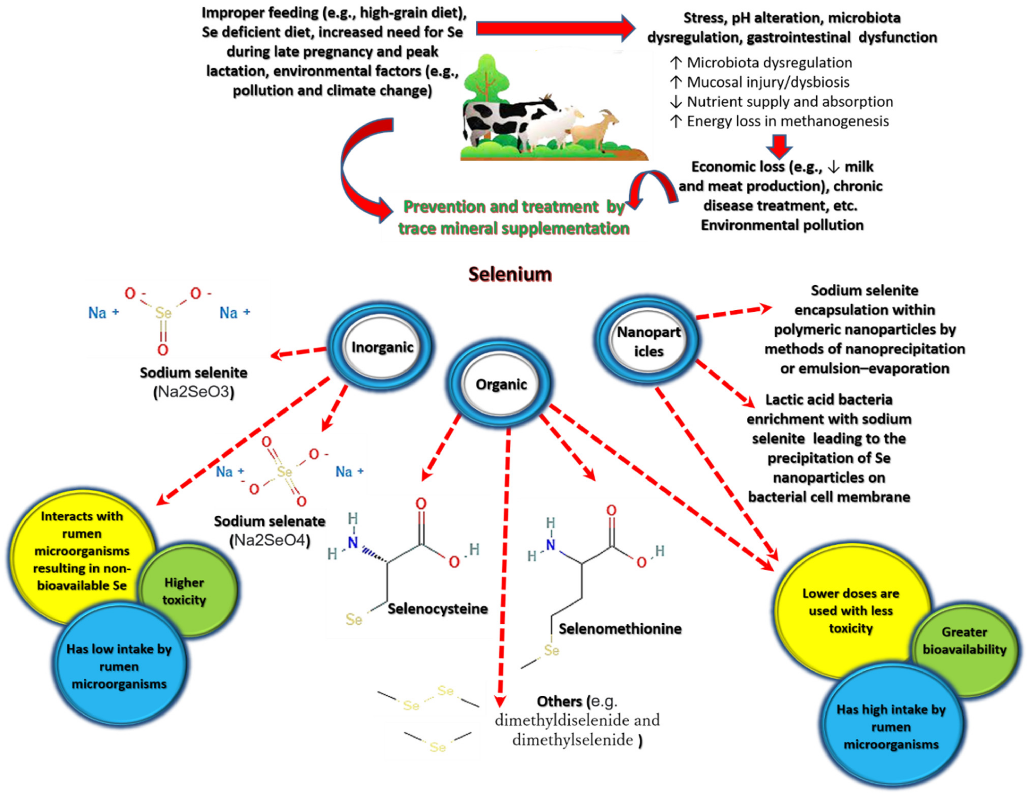
1.1. Selenium in Nature and Its Forms

Se is a potent immunomodulatory nonmetallic trace element commonly available as an amorphous brick red powder that turns into a black vitreous form when it reaches its melting point [27]. It has six different isotopes that vary according to their stabilization states, half-life, and mode of decay [27]. In nature, Se exists in inorganic forms that vary according to the redox and pH of the surrounding environmental conditions. The most common inorganic forms are sodium-selenate [Na2SeO4, Se(VI)] and sodium-selenite [Na2SeO3, Se(IV)]; selenide [Se2−, Se(−II)] and elemental Se [Se (0)] are less common while Se dioxide (SeO2) results from the combustion of elemental Se, which exists in fossil fuels and waste material [28]. Inorganic Se exists in the soil and water; it is consumed by bacteria, algae, plants, and primates, who transform it into organic Se. Se bioaccumulation in plants and animals may result from excessive and non-wise use of Se in human activities [28]. Se existing in plant food, seafood, and animals is organic Se (Se chemically bound to carbon)—taking the form of amino acids (e.g., selenomethionine (SeMet), selenocysteine, dimethylselenide, and dimethyldiselenide)[27,29,30].

1.2. Selenium Biokinetics and Bioactive Properties

The body’s daily needs of Se can be met by dietary Se (organic) or Se supplements (inorganic Se) [31]. Detailed investigations of Se biokinetics note that oral Se is absorbed to a great extent [28,31]. The bioavailability of organic Se is greater than that of Se from inorganic sources [32]. The distribution of Se is even throughout the body including breast milk. Its elimination is trivial through breath and sweat; it mostly occurs through the intestinal and urinary tracts, with trimethylselenium ions, selenosugars, and Se-methylselenoneine representing its most common urinary metabolites [31].
Counteracting oxidative stress is one of the key functions of Se [33]. Around 3% of its main form, selenoproteins, is involved in the synthesis of Se-dependent glutathione peroxidase (GSH-Px), which scavenges free radicals such as reactive oxygen species (ROS) [30,33,34,35]. Selenocystines represent a main component of the thyroxin system, which also comprises nicotinamide adenine dinucleotide phosphate [36]. All the components of this system facilitate the reduction in protein disulfides in ribonucleotide reductase, thioredoxin peroxidase, and protein disulfide-isomerase, which work harmoniously to regulate DNA synthesis and repair, antioxidant production, and proper functioning of the endoplasmic reticulum [36].
An adequate Se supply is necessary for the full expression and enzymatic activities of specific selenoproteins containing selenocysteine, which are crucial for the functioning of T lymphocytes and natural killer cells, which can kill tumor cells and attack pathogens [33,37]. Se is thought to decrease cell death following severe infections because of its inhibitory effect against transient receptor potential melastatin 2—a calcium channel involved in cell apoptosis [35]. It also exerts a direct antipathogenic effect—the conversion of inorganic forms (e.g., selenite) into the element’s divalent form oxidizes thiol groups in the active site of viral protein disulfide isomerase, converting them into inactive sulfydryl groups [38,39].
Thanks to its antioxidant properties, Se is reported to protect against coronary heart disease [30], ventilator-associated pneumonia, COVID-19, acute respiratory distress syndrome, and overall mortality as its most desired effect [35,38,40]. In fact, the outcome of COVID-19 in China has largely been associated with regional levels of Se, with greater recovery in Se-rich regions and poorer recovery in Se-deficient areas [40]. This is because Se distribution in the soil greatly varies in China, which is associated with variation in Se levels in human food originating from plant and animal sources [41]. Indeed, Keshan disease, a rare form of cardiomyopathy induced by Se deficiency, is common in Se-deficient areas in China [42]. Se deficiency is also involved in the development of metabolic disorders by promoting pancreatic atrophy [43]. Se can also promote thyroid function, wound healing, and male fertility [27]. In addition, it may protect against severe adverse effects of drugs, heavy metals, carcinogens, mycotoxins, and pesticides [44]. The use of Se as an antioxidant may be more favorable than other nutrients that are commonly used as antioxidant agents. Collective knowledge shows that the long-term use of Se in humans for preventive purposes is less likely to induce adverse effects (mortality) relative to other commonly used antioxidants (beta carotene, vitamin A, and vitamin E) [45]. However, Se has a narrow toxicity range (i.e., slight overdoses may induce toxicity) [46]. Human overexposure to Se, especially inorganic hexavalent Se, is associated with type 2 diabetes, high-grade prostate cancer, and neurodegenerative diseases (e.g., amyotrophic lateral sclerosis and Parkinson’s disease) [42]. Therefore, ensuring adequate dietary supply to humans (e.g., plant and meat/milk food with adequate levels of Se) may be necessary to avoid Se deficiency in humans as well as toxicity associated with unnecessary intake of Se supplements [46].

1.3. Selenium Deficiency in Livestock

Se dietary levels vary according to geographical location and the use of Se-containing fertilizers [29,30]. Therefore, Se is commonly used in agriculture. Plants enriched with Se are reported to be stress resistant, have more efficient biosynthesis, and are richer in amino acids, minerals, and antioxidants than non-enriched plants [47]. Because of the uneven Se levels from the soil to forage grass in different regions, different ruminants (even different breeds of a ruminant species) commonly experience Se deficiency [29].
Se deficiency in livestock is a major cause of economic losses. Se deficiency increases metabolic dysfunction (indicated by high insulin level) [48]. It also triggers thyroid dysfunction by inhibiting the conversion of thyroidal thyroxine to triiodothyronine [48,49]. This is because Se is a key structural component in selenocysteine, which exists in the active sites of enzymes involved in the thyroxin system such as iodothyronine deiodinases and thioredoxin reductases [36]. It also heightens the vulnerability to infections by promoting the evolution of pathogenic viral species [34,38], which frequently induce oxidative stress in order to direct physiological processes of host cells toward viral replication [50]. White muscle disease, retained placenta, ill-thrift, osteoporosis, and mastitis are common examples of livestock diseases induced by Se deficiency [51,52]. Inflammatory conditions associated with Se deficiency in cattle such as mastitis can considerably reduce fertility [53]. On the other hand, Se is reported to exert fertility-potentiating effects in goats [54].
Se deficiency can seriously endanger meat production. Maintaining rapid muscle growth is crucial in the animal breeding industry since it is an indicator of somatic growth and increased meat production [19,55]. Muscular growth is a dynamic process, which is based on overall balance between protein synthesis/anabolism and degradation/catabolism. Anabolism rate is greater than catabolism under normal physiological and nutritional conditions [55,56]. Muscle modeling is orchestrated by a large network of interrelated signaling cascades that regulate metabolism, oxidative stress, immune response, and autophagy, which all affect the activity of atrophy genes [57,58,59]. Dietary Se deficiency is associated with downregulation of selenoprotein encoding genes in pancreas and skeletal muscle [43]. Because fetal growth is high during the third trimester, maternal Se transfer to the growing fetus increases. Se is also involved in the synthesis of milk proteins, which may justify the Se drop during peak lactation. Lack of Se supply to pregnant and peak lactating animals may trigger Se deficiency in neonate and nursing animals [60], which is likely to affect their growth. Calves with nutritional muscular dystrophy express significantly low serum levels of Se, GSH-Px, and vitamin E, along with the development of Zenker’s necrosis, hyperglycemia and accelerated levels of creatine kinase (CK) and lactate dehydrogenase (LDH) [61]. CK and LDH are biomarkers of muscle dystrophy; the latter is associated with mitochondrial dysfunction, indicating that metabolic dysfunction evoked by oxidative stress is the mechanism underlying muscle loss in Se deficient animals [18,59,62]. Experimentally, Se deficiency (0 mg/kg body weight (BW)) and excess (16 mg/kg BW) are reported to significantly reduce weight gain rate, specific growth rate, feed efficiency, and muscle crude protein content in Rainbow Trout. In both conditions, the expression of atrophic genes (MuRF1 and Atrogin-1) and the abundance of muscle ubiquitinated proteins significantly increased. The underlying mechanism involved decreased phosphorylation of Akt Ser473 and the ratio of phosphorylated Forkhead Box O transcription factor (FOXO) 3a/FoxO3a, along with increased phosphorylation of the inhibitor of κB α and upregulation of TNF-α, IL-8, and nuclear factor kappa light chain enhancer of activated B cells (NF-κB) [55]. On the other hand, Se supplementation can restore muscle integrity by increasing antioxidant production and selenoproteins (e.g., SELENOW) in skeletal muscle, which is associated with reduced pressing muscle loss, decreased oxidative changes in frozen meat, increased muscular pH after 24 h of slaughter, cooking loss, and lower drip in broilers and pigs. Organic Se and SeNPs were significantly more effective than inorganic Se [63,64,65]. Compared with no treatment, maternal supplementation with Se-yeast boluses (105 mg of Se/week) during the third trimester was associated with increased expression of myosin and actin filament associated genes in newborn calves. These genes are involved in muscle development, potentially allowing for optimal muscle growth, functioning, and contraction [66]. In summary, Se dysregulation evokes muscular dysfunction by interfering with signaling that regulates muscle cell survival and activates atrophy genes [43,55,61], while Se supplementation during the third trimester can promote muscle growth [66], lower muscle loss, and improve meat quality [64,65]. Notably, Se supplementation during the first and second trimester has a potentially adverse effect on the development of muscle and collagen structures [66].

1.4. Selenium Use in Livestock

Because of the excessive economic losses caused by Se deficiency in the livestock industry, Se is frequently supplemented to animals in the form of inorganic salts, primarily sodium selenite [67]. Inorganic and organic trace minerals are frequently integrated into complete rations, rumen boluses or as ad libitum mineral blocks [68]. The most common route of Se administration is orally in bolus forms. However, injections are also used; subcutaneous injections of different forms of Se (e.g., sodium selenite vs. barium Se) considerably vary in the speed of absorbance [32,36]. Organic forms of Se are also commonly used as supplements (e.g., Se yeast and SeMet) [24,25,69]. Different Se forms affect Se metabolism in animals in different ways, resulting in variations in the expressed benefits or even toxicity when certain forms are used [44,47]. Se inorganic salts exhibit lower uptake by ruminal microorganisms and interact with ruminal contents, resulting in higher levels of non-bioavailable Se [32,67]. Inorganic Se can be highly toxic, and it is less likely to be transferred to milk and meat, which limits the growth of growing animals (e.g., lambs and calves) [68,70]. To avoid the adverse interactions between inorganic Se and ruminal bacteria, it is frequently administered in injectable forms [71]. However, this form of treatment is more likely to affect the general state of the ruminant rather than microbial fermentation per se. Alternatively, for oral administration, coating inorganic Se by lipids [67], encapsulating it within polymeric nanoparticles [70], or enriching lactic acid bacteria (LAB) with inorganic Se—to facilitate its transformation into elemental Se nanoparticles (SeNps)—have been recently introduced to improve treatment outcomes [68].
Nanotechnology uses methods such as nanoprecipitation and emulsion-evaporation to reduce mineral dimensions to nanoparticles of 1–100 nm [70,72]. Increasing mineral surface area reduces antagonistic behavior typically induced by traditional inorganic minerals in the gastrointestinal tract, considerably increases their bioavailability, contributes to desirable effects on metabolism and antioxidant capacity at considerably lower doses, and minimizes pollution secondary to mineral excretion into the environment [23,70]. Smaller size SeNPs (5–15 nm) at a concentration of less than 0.5 mM are suggested to minimize oxidative stress in COVID-19; they can also act as an antiviral drug carrier. SeNPs are less toxic than other forms of Se, and their bioavailability and efficiency in preventing oxidative damage are higher [33]. Thus, SeNPs represent a putative long-acting alternative to inorganic and organic Se [31]. Se encapsulation in polymeric nanoparticles facilitates Se release in pH less than four, which coincides with the intestinal conditions [70]. Aggregate data show that the use of SeNPs in poultry can considerably promote feed utilization, foster growth and reproduction, increase the relative weights of immune-related organs (burse and thymus) and enhance immunity, improve antioxidant status and GSH-Px activities, and increase the abundance of beneficial species of intestinal bacteria (at 0.9 mg/kg diet) as well as intestinal production of short chain fatty acids [23]. SeNPs have recently been introduced in ruminant breeding practice at a small scale [70,73,74,75]. This review aims to explore the available literature for the collective effect of Se on fermentation in ruminants. To obtain relevant studies for this narrative review, we conducted a systematic search in Web of Science and PubMed databases using combinations of relevant search terms: (cow OR cattle OR calf OR calves OR heifer OR buffalo OR bull OR steer OR sheep OR ewe OR lamb OR ram OR goat OR kid OR deer OR camel) AND (rumen OR fermentation OR digestibility OR CH4 emission OR methane emission OR microflora OR microbiota OR purine derivatives) AND (selenium OR selenite OR selenomethionine OR Sel-Plex). A manual search in Google Scholar was also conducted. The search included all studies published until September 2021, and was not limited by language or study type. The database search resulted in 775 studies including 59 duplicated reports. Studies included were those using Se alone to affect rumen fermentation. Thirty-nine relevant studies including four studies obtained by the manual search were included in the synthesis of the article.

2. Results

In one study, Se was supplemented to grass as a fertilizer [25]; in another study, Se was provided to animals orally [76]; and in the rest of the studies, Se was mixed with the diet. As shown in Table 1, different dietary Se species increased total VFA concentrations and molar proportions of propionate in the rumen fluid of dairy cows [24,26,69,77], Holstein bulls [78,79], Holstein dairy calves [80], goats [81,82], sheep [25,73,75,83], and lambs [84,85]. Lambs and sheep fed organic or inorganic Se diet exhibited an increase in the production of ruminal acetate, iso-butyrate, and iso-valerate compared with the control group [83,84,85]. In line, the rumen liquor of crossbred wethers fed a purified diet plus a weekly oral dose of 1 mg sodium selenite per sheep contained higher molar proportions of acetic and iso-valeric acids than that of untreated sheep [76]. The molar proportion of butyrate decreased in Se-treated ruminants compared with the controls. On the other hand, the molar proportion of butyrate was higher in the rumen of cows, bulls, and rams fed different Se supplements than in the rumen of animals fed basal diet [25,69,79]. In contrast, rumen-protected sodium selenite (4.8 mg/cow/day) or coated sodium selenite (0.1, 0.2, or 0.3 mg/kg DM) supplementation reduced the molar proportion of butyrate in the rumen of dairy cows compared with non-treatment [26,77]. Moreover, the molar proportion of acetate was lower in dairy bulls fed sodium selenite and coated sodium selenite compared with animals receiving the basal diet [79]. All types of dietary Se decreased acetate to propionate (A to P) ratio in the rumen fluid of different ruminants [24,25,26,69,73,75,77,78,79,80,86]. In few instances, inorganic and organic Se at different doses had no effects on ruminal total VFA [86,87], composition of VFA [86,87,88,89], and A to P ratio [84,86,87,88].
Se supplements decreased rumen pH and NH3-N concentration in dairy cows [24,26,69,77], steers [78,79,86], Holstein dairy calves [80], goats [81,82], sheep [73,75,83], and lambs [85]. On the other hand, ruminal NH3-N concentrations in the rumen of rams receiving the medium level of Se yeast were higher than those in high and low Se yeast groups and the control group [25]. In some studies, Se had no effect on ruminal pH [25,86,87,88,89] or NH3-N [79,80,87,88].
Se supplementation enhanced the digestibility of dry matter (DM), organic matter (OM), crude protein (CP), neutral detergent fiber (NDF), and acid detergent fiber (ADF) [24,26,29,69,73,74,75,78,79,80,83,90,91,92,93,94], ether extract [24,26,29,73,74,75,78,79,80,83,91,94,95], crude fiber [91], starch [77], microbial N efficiency [88], nutritive value digestible crude protein, and total digestible nutrients [74,94] in the rumen of different ruminant species. In one study, supplementing the steer diet with Sel-Plex (7.5, 15, and 22.5 mg/steer/day) increased ruminal microbial protein synthesis, especially higher doses (p < 0.01) [86]. In few studies, Se had no effect on digestibility [87,96,97,98,99,100,101,102,103].
Urinary excretion of allantoin and total purine derivatives (PD) were higher in Se treatments than in the control treatment [73,75,78,80,86]. Meanwhile, supplementing Tabapuã steers with Sel-Plex (5 g/head/day) reduced the urinary excretion of uric acid [87]. In some studies, different Se supplements did not affect the urinary excretion of uric acid [73,75,78,80,86], xanthine, hypoxanthine [73,75], and allantoin [87].
In a single study, microbial analysis of ruminal microbiota revealed no difference in the relative abundance (%) of bacterial phyla among treatments except for the phylum of Synergistetes, which increased in low Se treatment compared with the control group [25]. At the family level, Lachnospiraceae increased in high Se treatment relative to all other groups. At the genus level, Carnobacterium and Dysgonomonas increased in medium Se compared with other Se levels and the controls. The Lachnospiraceae XPB1014 group increased in the high Se group compared with other treatments. Moreover, Prevotella 1 was higher in the control and medium Se than those in low Se doses. The Rikenellaceae RC9 gut group increased in low Se compared with the control group. Hafnia-Obesumbacterium was lower in the high Se group than the other Se groups. Furthermore, Tax4fun metagenome analysis indicated that carbohydrate and other amino acid-related gene activities and metabolic pathways were overexpressed in the rumen microbiota of Se yeast-supplemented sheep [25]. Studies employing quantitative PCR analysis revealed that dietary Se increased total bacteria, total anaerobic fungi, total protozoa, Ruminococcus albus, Ruminococcus flavefaciens, Butyrivibrio fibrisolvens [26,77,78,80], Fibrobacter succinogenes [26,78,80], Ruminobacter amylophilus [26,77,78], and total methanogens [80]. Total methanogens decreased in another study [78]. The population of Dasytricha ruminantium, Ophryoscolex caudatus (f. tricoronatus), Polyplastron multivesiculatum, and Diploplastron affine were significantly higher in sheep supplemented with organic or inorganic Se than in animals given the basal diet alone [104]. Prevotella ruminicola decreased in three studies [26,77,78]. Moreover, total protozoa numbers and the percentage of Diplodinium and Ophryoscolex caudatum in the rumen fluid of lambs supplemented with sodium selenite (0.3 mg Se/kg feed) were higher than those fed the basal diet [85]. On the other hand, some studies revealed no effect of different Se supplements on some microbial populations including total bacteria [88], Fibrobacter succinogenes [77], Prevotella ruminicola [80], total methanogens [26], total protozoa [87,88,104], and Entodinium spp., Isotricha spp. [104].
Table 1. Effects of different sources of Se supplementation on different farm animals.
SpeciesSe SourceVFAA:P pHNH3-NDigestibilityEnzymesMicrobiotaPDRef.
Lactating dairy cowsSeY+---+NDNDND[24]
Lactating dairy cowsRPSS+---+++-ND[77]
Lactating dairy cowsHMSeBA and SS+---+NDNDND[69]
Lactating dairy cowsSS and CSS+---+++-ND[26]
Lactating dairy cowsSeYNDNDNDND0NDNDND[99]
Beef cattleSS0ND0NDNDNDNDND[89]
Beef calves and dairy heifersSS and SeYNDNDNDND0NDNDND[97]
Dairy calvesSS+--0+++-+[80]
Dairy calvesSeYNDNDNDND0NDNDND[100]
Dairy bullsSS+---+++-+[78]
Dairy bullsSS and CSS+--0+NDNDND[79]
SteersSeY+-0-NDNDND+[86]
SteersSeY00000ND00[87]
Buffalo heifersSSNDNDNDND0NDNDND[96]
Male buffalo calvesSSNDNDNDND+NDNDND[93]
Male buffalo calvesSe #NDNDNDND0NDNDND[102]
Pregnant and lactating ewesSSNDNDNDND+NDNDND[94]
SheepSeY and SeNps+---+NDND+[73]
SheepSS and SeY+00-+NDNDND[83]
SheepSeYNDNDNDND+NDNDND[92]
SheepSS and SeYNDNDNDNDND+NDND[105]
LambsSS, SeY, and SeNpsNDNDNDND+NDNDND[74]
LambsSS and SeYNDNDNDNDNDND+ND[104]
LambsSS+ND-NDNDND+ND[85]
Male lambsSSA and SeY+0NDNDNDNDNDND[84]
Male lambsSeYNDNDNDNDND+NDND[106]
Male lambsSS0000+ND0ND[88]
Male lambsSS and Jevsel-101 *NDNDNDND0NDNDND[101]
Male lambsSS and SeYNDNDNDND+NDNDND[90]
Male lambsSSNDNDNDND0NDNDND[98]
Male sheepSeNps+---+NDND+[75]
Male sheepSeYNDNDNDND+NDNDND[29]
RamsSeY+-ND+NDND+-ND[25]
RamsSS+NDNDNDNDNDNDND[76]
Lactating goatsSS and SeMet NDNDNDND+NDNDND[95]
Cashmere goatsSSNDNDNDND0NDNDND[103]
GoatsSeY+ND-NDNDNDNDND[82]
Goats SeYNDNDNDND+NDNDND[91]
Male goatsSeY+0-NDNDNDNDND[81]
Se: Selenium; VFA: Volatile fatty acids; A: P: Acetate to propionate ratio; PD: Purine derivatives; +: increase; -: decrease; 0: no effect; ND: not determined; SeY: Se yeast; SeNps: Se nanoparticles; RPSS: Rumen-protected sodium selenite; SS: sodium selenite; SSA: sodium selenate; HMSeBA: hydroxy-analog of selenomethionine; CSS: Coated sodium selenite; SeMet: Selenomethionine; #: The used Se species is not defined; *: Organic Se.
Sodium selenite at different concentrations increased the ruminal activity of xylanase, protease, and α-amylase [26,77,78,80], pectinase [77,78,80], cellobiase [26,78], and carboxymethyl-cellulase [26]. In addition, Se yeast-extract increased the activities of gamma-glutamyl transferase, glutamate dehydrogenase, and aspartate aminotransferase in the rumen fluid of lambs [105] as well as the activities of alkaline phosphatase and glutamate dehydrogenase in the ruminal fluid of Se-treated sheep [106]. Likewise, GSH-Px activity increased in the ruminal epithelium of Se-treated goats [82]. In fewer studies, Se supplementation did not exhibit a significant effect on alanine aminotransferase [105,106], alkaline phosphatase [105], aspartate aminotransferase, γ-glutamyl transferase [106], carboxymethyl cellulase [77,78,80], and cellobiase [77,80] in the ruminal fluid.
Se-yeast supplementation (6 μg/kg BW) to sheep grazed on a mixed pasture of alfalfa and tall fescue improved gross energy (GE), digestive energy (DE), and metabolic energy (ME) intakes. However, different Se levels (3, 6, 9, 12 μg/kg BW) had no significant effect on CH4 energy output per day while CH4 output as a proportion of GE, DE, and ME intakes decreased in Se treatments compared with the control. Furthermore, the N intake, fecal N, urine N, digestible N, and retained N were higher in the Se-yeast treatment (6 μg/kg BW) compared with the control [92]. Total N production, total N production/intake N, and total N production/digestible N were higher in early lactating goats receiving organic and inorganic Se (0.3 mg Se/head/day) than those in the control group [95]. Meanwhile, urinary N, urine N/intake N, and urine N/digestible N decreased in Se-treated animals. Moreover, lactating goats fed different types of Se diet exhibited improvements in production energy/ME intake as well as a decrease in urinary energy/ME intake and maintenance energy/ME intake compared with the control group [95]. Similarly, N retention increased in male lambs receiving sodium selenite or organic Se (Jevsel-101, 0.15 mg) compared with those in the control group [101]. In contrast, two studies reported no effect of dietary Se on N intake, N excretion, or N retention in cashmere goats or male buffalo calves [102,103]. Readers interested in a detailed illustration of Se treatments are encouraged to refer to Supplementary Table S1.

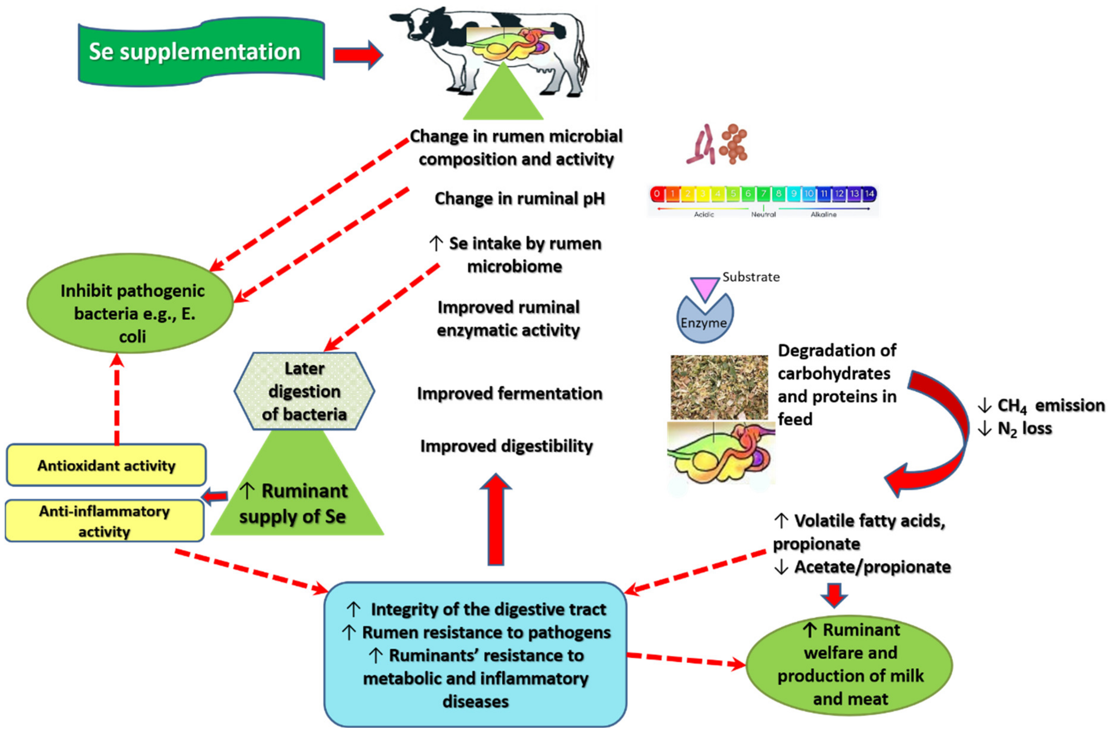
3. Mechanism of Action of Se in Ruminal Fermentation

Although no clear mechanism has been explored for increased energy production in animals receiving Se supplementation, Se is likely to promote the integrity and functioning of the digestive tract of ruminants through multiple interrelated mechanisms, resulting in more efficient ruminal fermentation. Figure 2 represents an attempt to illustrate how Se supplementation may improve fermentation and energy supply in ruminant animals.
In most studies, Se treatment resulted in more efficient transportation and absorption of VFA. The levels of VFA account for a key index of rumen fermentation [25]. In fact, Se supplementation was associated with increased abundance of bacteria that degrade cellulose (e.g., Ruminococcus albus, Ruminococcus flavefaciens, Fibrobacter succinogenes, and Butyrivibrio fibrisolvens) [26,77,78,80]. VFA act as vital nutrients for the host as well as for intestinal microbiota [107]. Their absorption largely occurs all over the host’s ruminal epithelium [25]. VFA can regulate numerous host-signaling mechanisms [107], which contribute to its unique role in promoting ruminant immunity and growth [25]. Experimental evidence shows that the growth of several isolates of the hemorrhagic Escherichia (E.) coli O157:H7 is enhanced in the ruminal fluid of fasted and poorly nourished animals, but it is inhibited in the rumen of well-fed animals [108]. Therefore, increased VFA production is likely to increase the resistance of the rumen environment to opportunistic pathogens.
In mucosal inflammatory conditions, probiotics and prebiotics are used to enhance VFA production. Alternatively, exogenous short chain fatty acid supplementation is used to restore VFA levels [109]. Metanalytic data show that short chain fatty acid supplementation can significantly reduce plasma levels of high-sensitivity C-reactive protein, LPS, and TNF-α in humans and animals [110]. The anti-inflammatory activity of VFA can be synergized by the anti-inflammatory activity exerted by Se itself. In rats treated with anticancer nedaplatin, which frequently induces diarrhea through activation of p53 or p53-regulated thrombospondin-1, SeNPs reduced diarrhea by inhibiting the activity p53 or p53-regulated thrombospondin-1 without interfering with the therapeutic activity of nedaplatin [111]. Se, along with other trace minerals, are reported to mitigate the cluster of differentiation antigens positive cells 4 (CD4+), T-cells depletion, and reduce T-cell activity in calves challenged with bovine viral diarrhea virus (BVDV2) five days after immunization [112].
Rumen bacterial species that incorporate Se in their structure mainly include Butyvibrio fibrisolvens, Selenomonas ruminantium, Streptococus sp., Lactobacillus sp., and Prevotella ruminicola [71]. Se intake into rumen bacteria in sheep receiving unprocessed soy, as a source of intact organic Se, was 5-fold higher than in sheep receiving heat-treated soy, which is low in its content of Se [113]. In ruminants, Se supplementation decreased the abundance of Prevotella ruminicola [26,77,78], which metabolize Se into elemental forms that are inaccessible to the host animals [71]. On the other hand, adequate Se intake can promote the propagation of Selenomonas ruminantium and Butyvibrio fibrisolvens, which promote the production of bioavailable selenoamino acids [26,52,71,77,78,80].
Gut microbiomes convert inorganic Se into amino acids, primarily SeMet and selenocysteine [28,71]. These amino acids catalyze GSH-Px synthesis, resulting in the attenuation of oxidative stress [30,33,34,35]—an effect documented in the intestinal mucosa of goats [114]. Cumulative evidence shows that methionine and cysteine are capable of promoting mucosal immunity and barrier function, reconstructing the structure of the damaged gut and reversing its dysfunction, along with reducing post-weaning stress in pigs [115]. Due to its antioxidant, anti-inflammatory, and probiotic-stimulating activities, Se is likely to restore optimum function of the ruminal epithelium. Experimentally, induction of Se deficiency in chicken resulted in duodenal villi cell apoptosis via an oxidative stress-induced mitochondrial apoptosis pathway associated with suppression of the activity of GSH-Px and thioredoxin reductase. In the meantime, apoptosis was potentiated via inflammatory signaling-induced death receptor pathway as a result of NF-κB activation by the accelerated levels of ROS [114]. Young goats treated with Se yeast expressed significant increases in the intestinal (duodenum and jejunum) mucosal weight, villi height and width, and villi surface area [116]. Similar results were detected in Hu lambs [117]. Additionally, Se yeast influenced the α diversity and abundances of rectal flora at the levels of the class, order, family, genus, and species [117]. Immunity modulation by Se is likely to affect bacterial growth in the lumen favoring beneficial species. In accordance, Se treatment is associated with increasing the speed and production of specific antibodies to pathogens that threaten the health of the intestinal tract such as E. coli [118] and BVDV2 [112,119] in vaccinated animals [112,118,119] or even in non-vaccinated animals [120].
Se supplementation can increase the abundance of certain species of LAB [25,71]. The intake of inorganic Se into LAB is high [51,121,122], resulting in its conversion into seleno-amino acids and SeNPs, which both exert antioxidant and antibacterial activities against pathogenic species [12,121,122]. In vitro investigations show that supplementing Lactobacillus delbrueckii ssp. bulgaricus and Streptococcus thermophilus with sodium selenite at 80 µg/mL increase the accumulation amount of Se to 12.05 ± 0.43 µg/mL and 11.56 ± 0.25 µg/mL, along with achieving the relative maximum living cells. Microscopic examination uncovered the deposition of elemental SeNPs on the cell surface of LAB [123]. Moreover, LAB exert antioxidant and anti-inflammatory effects; they represent a common form of probiotics. Apart from Se treatment, LAB metabolites promote the growth of beneficial bacteria and discourage the growth of pathogenic species [124]. Compared with the wild type, Se-enriched LAB caused extensive damage in the membrane of pathogenic Salmonella typhimurium, E. coli, Staphylococcus aureus, and Listeria monocytogenes in fermented pickles [123]. Broiler supplemented with four metabolites of various strains of Lactobacillus plantarum expressed increased fecal population of LAB and Enterobacteriaceae, along with increases in fecal VFA, small intestine villus height, final body weight, weight gain, average daily gain, and reduction in feed conversion ratio [125]. In fermented pickles, Lactobacillus plantarum enriched with organic/inorganic Se significantly inhibited the production of advanced glycation end-products (AGEs), inhibited Enterobacter, Halomonas, and Klebsiella, and increased the abundance of Lactococcus, Lactobacillus, and Leuconostoc than Se-free Lactobacillus plantarum [122]. It is noteworthy that AGEs are aggravated by ROS and cytokines, and they potentiate a vicious cycle of oxidative stress and inflammation, leading to ill-health [9,126].
E. coli inhibits the growth of beneficial bacteria in the gut [8]. It also produces toxins that alter the integrity of the gut membrane causing dysbiosis, which promotes the passage of these toxins and even the bacteria into the circulation, resulting in a systemic inflammatory response [7,9]. E. coli exchanges genetic material with other bacterial species, and possibly passes antibiotic resistance genes to transient bacterial pathogens that cause disease in humans. In fact, E. coli is a reservoir of antibiotic resistance genes and a logical indicator of the degree of antibiotic resistance within bovine microbial populations of the digestive tract because it represents 1% of the colonic bacteria in cattle, accounting for the majority of resistance in Enterobacteriaceae [6,127]. E. coli propagation in diarrheic ruminants is frequently associated with pathogenic propagation of other species such as Candida (C.) albicans [12]. Grain diets promote the propagation of the enterohemorrhagic E. coli, while switching to all hay diets can decrease E. coli survival, resulting in 1000-fold decline in E. coli within five days [6].
It is possible that Se supplementation improves fermentation by correcting gastrointestinal dysfunctions induced by pathogenic organisms. Experimentally, SeNPs in combination with antimicrobials that are potent against active against Gram-negative bacteria (E. coli, Salmonella, Shigella, Klebsiella, Enterobacter, and Pseudomonas aeruginosa) such as Polymyxin can inhibit Enterobacter cloacae 1.9 times higher than Polymyxin alone. There was no difference in treatments involving E. coli [128]. Nonetheless, the green synthesized SeNPs (0.4 and 0.3 mg/mL) inhibited E. coli and C. albicans isolated from the feed, drinking water, and feces of diarrheic buffaloes. The effect was potentiated when SeNPs were combined with cinnamon oil. The effect of SeNPs alone and in combination with cinnamon oil was significantly superior to traditional antimicrobials [12]. Compared with inorganic Se, organic Se yeast in puppies between 20 and 52 weeks of age was associated with higher number of DNA copies of Lactobacillus, a trend to lower DNA copies of E. coli, higher fecal end-fermentation products related to protein degradation in females, higher DNA concentration of Bifidobacterium in males, along with higher concentrations of total VFA, propionate, and butyrate [129]. Pregnant cows fed Se-supplemented hay exhibited increased concentrations of immunoglobulin G subclass 1 and J-5 E. coli antibody in cow colostrum and calf serum compared with non-fortified hay [120]. This effect is of particular importance in husbandry systems, which suffer excessively because of diarrhea induced by enterotoxigenic E. coli in calves [130]. One of the methods used to stimulate protective immunity is vaccinating the dam, allowing the passive transfer of protective antibodies to calves through the colostrum [130]. Thus, it is possible that ruminant supplementation with bioavailable Se may inhibit the growth of harmful bacterial species such as E.coli.
Altogether, the use of Se as a dietary supplement in ruminants can positively affect fermentation in many ways: (1) modifying ruminal microbial structure favoring the propagation of bacteria with higher Se intake, higher fiber digestibility, and probiotic/antioxidant activity (e.g., LAB); (2) increasing VFA production promotes favorable pH, increases rumen resistance to pathogens, and modulates signaling cascades of inflammation and oxidative stress; and (3) Se as an antioxidant/anti-inflammatory, along with the activity of VFA and LAB, correct oxidative stress and inflammation in the digestive tract, which are induced by improper diet and pathogens, resulting in optimal gastrointestinal functioning.

4. Discussion

Se supplementation to ruminants has recently been an attractive trace-mineral nutrition modality [29,67]. Several excellent reviews have reported that Se supplementation to ruminants can increase milk yield, milk content of Se, fat, protein, polyunsaturated fatty acids as well as improve immune response, systemic antioxidants, and feed conversion (body weight). In treated animals, Se transfer also increased to the newborn via the placenta, colostrum, and milk [36,131,132,133]. Although these results reflect improvement in digestibility and fermentation, the exact mechanism of different Se species on rumen fermentation is not clearly understood. The current review fills this gap by deeply investigating the possible effects of dietary Se on fermentation and digestibility in ruminants.
Digestive and metabolic investigations [24,26,29,69,73,74,75,78,79,80,83,90,91,92,93,94] show that Se supplementation (usually at a medium dose rate—0.3 mg/kg DM) can improve N metabolism and nutrient digestibility (DM, OM, CP, ether extract, NDF, and ADF), which are beneficial for growth and productivity in cows, sheep, and goats. In fewer studies, both organic and inorganic Se had no effect on digestibility [87,96,97,98,99,100,101,102,103]. In studies using Se yeast and SeNPs, they both produced positive effects on digestibility, total VFA, molar proportion of propionate, A to P ratio, and ruminal NH3-N as well as urinary excretion of allantoin and total PD, albeit SeNPs had the most beneficial effect [73,74,75]. Incidentally, increased urinary excretion of PD suggests increased utilization of NH3-N for the synthesis of microbial protein [80]. Sodium selenite protected by lipid microencapsulation decreased CP digestibility while protected Cu had no effect on nutrient digestibility in vitro [67]. Of interest, supplementing ruminally-cannulated steers with trace minerals (Cu, Zn, and Mn) in sulfate form decreased DM digestibility while the hydroxy form had no effect on digestibility [134]. Most reviewed studies show that Se supplementation increased total VFA concentrations and molar proportion of propionate but reduced NH3-N concentration and rumen pH to the neutrality zone (Table 1), which favors the activity of bacteria that degrade cellulose [135]. Thus, SeNPs and organic Se may represent a good source of trace minerals for enhancing digestibility in ruminants.
Studies show that improved digestibility in Se treatment is secondary to the modulation of ruminal microbiome: increases in total bacteria, total anaerobic fungi, total protozoa, Ruminococcus albus, Ruminococcus flavefaciens, Butyrivibrio fibrisolvens, Fibrobacter succinogenes, and Ruminobacter amylophilus, along with a decrease in the relative level of ruminal Prevotella ruminicola [26,77,78]. Ruminal pH values ranged between 5.40 and 6.98. However, pH below 6 was reported only in four studies [69,81,82,88], and most studies reported pH above six. The latter may be optimum for the growth of cellulolytic bacteria as well as for nutrient degradation [135]. Increased ruminal total VFA concentration following Se intake resulted in keeping the ruminal pH within the neutral range, which was consistent with the increase in nutrient digestibility [136].
Changes in microbiome composition and nutrient supply were accompanied by potentiation of microbial enzymes in the rumen: xylanase, protease, and α-amylase, pectinase, cellobiase, carboxymethyl-cellulase [137]. Cellulolytic enzymes are secreted by ruminal cellulolytic bacteria, fungi, and protozoa to degrade fibers in feed to acetate [137]. Those produced by fungi can penetrate the cuticle and lignified tissues of plant materials, leading to the degradation of the most resistant cell wall polymers [138]. Similarly, the higher propionate molar proportion may be attributed to increased activity of α-amylase and populations of Ruminobacter amylophilus and Butyrivibrio fibrisolvens—the dominant bacteria in charge of the degradation of non-fiber carbohydrates (non-structural carbohydrates e.g., starch, sugars, pectins,…etc.) in the rumen [138]. Moreover, some strains of ruminal protozoa also create amylase [139]. The production of propionate requires ruminal hydrogen, which is the major substrate for methanogen growth and methane production. Thus, the positive effect of Se treatment on propionate molar proportion was a reflection of the decrease in total methanogen population [140]. It is worth mentioning that the effect of Se supplementation on methane emission was investigated only in three studies. Two studies reported no significant effect of dietary organic and inorganic Se on methane emission [84,88]. In the other study, Se yeast had no effect on CH4 energy output per day while the CH4 output as a proportion of gross energy (GE), digestive energy (DE), and metabolic energy (ME) intakes decreased in Se treatment compared with the control [92].
The biological functions of Se result from its integral role as a catalytic entity in a variety of enzymes [28]. Therefore, the antioxidant function of dietary Se may account for the positive response of microbial population and their enzymatic activity. Specifically, ruminal microbes incorporate dietary Se to form their protein and cell wall component in the form of SeMet [141], which is capable of protecting cell membranes against oxidative damage because of its high ROS scavenging capacity [142]. In line, ruminal microbial concentration of Se increased following Se dietary supplementation in sheep fed a purified diet low in Se [76,113,133]. In another study in sheep, total Se absorption and GSH-Px activity of ruminal bacteria and protozoa significantly increased in response to dietary Se (0.4 mg/kg DM) [29]. Dietary Se (0.3 mg/kg DM) had a similar effect on ruminal protozoa in lambs [85].
Studies comparing the effects of different forms of Se favor organic Se over inorganic Se while SeNPs at considerably lower doses exerted the most desirable effects [73,74,75]. Aggregate data show that Se, particularly its inorganic form, at concentrations slightly above its nutritional levels can be extremely toxic, especially to the central nervous system [28,44,47]. A longitudinal study following humans consuming drinking water supplemented with inorganic Se (hexavalent Se/selenate, 10 μg/L) in one Italian district for 28 years reported increased risk for oropharyngeal, urinary, and lymphoid cancer compared with unexposed residents in other districts [143]. In fact, selenium sulfide is listed as an animal carcinogen by the U.S. National Toxicology Program [144]. Reports on Se toxicity in ruminants are quite scarce. In a study supplementing sheep with Se (5 mg selenite orally every two weeks for 72 weeks), along with low-protein or high-protein diets that are deficient in trace minerals, death following loss of appetite was reported in five sheep who reached a total Se cumulative doses of 70, 100, 140, 175, and 180 mg [145]. Chronic dietary exposure of steers to selenite and SeMet (0.28 and 0.8 mg Se/kg BW/day for four months) was associated with the development of dystrophic hoof lesions of chronic selenosis/alkali disease. No neurological, renal, or hepatic lesions were detected in histological examinations [146]. A recent study reports Se poisoning in Wumeng semi-fine wool sheep receiving oral SeNPs (5 mg/kg BW/day) for 30 days. In particular, SeNPs caused significant increases in serum IL-6, IL-1β, malondialdehyde, LDH, glutamic oxaloacetic transaminase, cereal third transaminase, and alkaline phosphatase, along with significant reductions in hemoglobin, erythrocyte count, packed cell volume, serum triiodothyronine, free triiodothyronine, IgG, IgM, IgA, IL-2, TNF-α, superoxide dismutase, glutathione peroxide, total antioxidant capacity, and catalase [147]. However, factors of improper diet (e.g., low in protein and minerals) [145], chronic to exposure to Se [146], and overdose [147] are likely to contribute to the negative effects reported in these studies.
Apart from its potential toxicity, inorganic Se expresses lower bioavailability [44,47]. An interesting in vitro investigation showed that rumen microorganisms separated from ewes incorporated organic SeMet (13.2-fold greater than Se-free control) significantly greater than inorganic sodium selenite or sodium selenate (3.3- and 3.5-fold greater than Se-free control). The amounts of the non-bioavailable elemental Se produced by rumen microorganisms was significantly lower in SeMet treatments compared with inorganic Se [32]. In vitro investigations showed that lipid-microencapsulation may minimize the interaction of sodium selenite with ruminal contents and limit associated production of the non-bioavailable elemental Se. In fact, relative to uncoated sodium selenite, lipid-microencapsulation of sodium selenite reduced NH3-N concentration, NH3-N flow, and CP digestibility; it also increased flows of non-ammonia N and dietary N. Nonetheless, these effects were non-significant. Meanwhile, protected sodium selenite decreased CP digestibility [67]. Other factors can affect the bioavailability of different Se species. While inorganic Se can promote the synthesis of selenoproteins, it cannot be stored in the body for later use. This is because inorganic Se has a shorter half-life compared with organic Se, which is bound to either yeast protein or bacterial protein [60,148]. Therefore, urinary loss of inorganic Se is significantly greater than organic Se [60]. The net absorption of organic Se and its transport to the tissues in an intact form is greater than inorganic Se. Se-bound amino acids act as analogs of amino acids for non-specific protein synthesis, which promotes the incorporation of organic Se into nonfunctional structural proteins (e.g., in skeletal muscle) as a direct Met replacement [63,149]. In this respect, bacterial Se is reported to considerably increase intestinal villus height, which is associated with increased Se retention more than sodium selenite [148].
In all studies, SeNPs were more effective than other Se forms, especially inorganic Se. Of interest, certain LAB species have the capacity to transform inorganic Se into predominantly elemental SeNPs, which are more bioavailable and less toxic than inorganic Se [68,121]. Experimental evidence shows that dietary supplementation of rats with Se-enriched LAB (Enterococcus faecium CCDM 922A and Streptococcus thermophilus CCDM 144) increased selenocysteine in the liver and kidney, along with reducing malondialdehyde in the tissue of various organs [121]. Enriching silage inoculant LAB with inorganic Se does not hinder LAB ability to act as silage inoculants while their yield of organic and SeNPs can be considerably increased [51]. Compared with no treatment, feeding ewes inoculated silage enriched with a supra-nutritional level of SeNPs was associated with significantly higher levels of Se in wool, muscle, blood, urine, and feces, denoting adequate absorption and bioavailability of SeNPs prepared by LAB-enrichment with inorganic Se [68]. In this respect, SeNPs introduced to livestock can be synthetic [12,70] or biologically produced through LAB-enrichment with Se salts [68]. Probiotics alone are reported to improve digestibility and reduce methane emissions in ruminants [3]. In this sense, it is necessary to compare the cost-effectiveness of synthetic SeNPs and LAB-enriched with inorganic Se in husbandry production.
Diets used in eight studies were total mixed ration [24,69,74,77,78,80,94,98]; grass was used in three studies [25,29,92] while diets in the rest of the studies comprised concentrate feed mixture and roughage [26,73,75,76,79,81,82,83,84,85,86,87,88,89,90,91,95,96,97,99,100,101,102,103,104,105,106]. Positive effects of Se supplementation on fermentation were noted in animals receiving diets low in Se [76,113,133]. Therefore, the effects of supplementary Se can be masked by dietary sufficiency of Se in the ration. In addition, changes in gut microbial flora in Se-treated animals may be confounded by diet type [6]. In this respect, Del Razo-Rodriguez et al. reported Se by grain interaction in the ruminal digestion of OM, starch, and NDF—70% grain diet supplemented with Se (0.3, 0.6, and 0.9 mg/kg DM) increased the ruminal digestion values for OM, starch, NDF, and feed N while supplementing 50% grain diet with the same levels of Se decreased ruminal digestion of OM and NDF (p < 0.05) [88]. Overall, for optimal use of Se as a dietary intervention, attention needs to be paid to genetic differences among livestock, geographical location, dietary preparations, and diet sufficiency of Se [25].

5. Conclusions

Se dietary treatment can modify the composition and potentiate the powerful metabolic activity of gut microbiome to boost the breakdown of unabsorbed carbohydrates and proteins, resulting in increased total VFA levels and propionate molar proportion, along with decreases in ruminal pH, NH3-N, and A to P ratio in ruminants. These effects are predominantly expressed by SeNPs, followed by organic and inorganic forms. More investigations are needed to compare the effectiveness of synthetic SeNPs and LAB enriched with sodium selenite as a biological source of SeNPs and probiotics as well as to thoroughly evaluate the effect of dietary Se on methane emission.

Supplementary Materials

The following are available online at https://www.mdpi.com/article/10.3390/fermentation8010004/s1, Table S1: Summary of the general data of papers evaluating the effect of different sources of Se supplementation on ruminants.

Author Contributions

Conceptualization, A.O.H., A.M.A., S.S. and K.S.; Software, A.O.H., M.M.M., R.F.R., H.A.A., A.E. and M.A.I.; Validation, A.M.A., A.H.A., H.S., A.B.M.R.B. and A.S.M.; Resources, A.M.A., H.A.A., M.M.M., A.B.M.R.B., A.S.M., M.A.I. and A.E.; Writing—original draft preparation, A.O.H., M.M.M., A.H.A., H.S., A.E., A.B.M.R.B., R.F.R., H.A.A., A.S.M. and M.A.I.; Writing—review and editing, A.M.A., S.S. and K.S.; Visualization, A.O.H., H.S., A.H.A. and R.F.R.; Supervision, A.O.H., S.S. and K.S.; Project administration, A.M.A. All authors have read and agreed to the published version of the manuscript.

Funding

This research received no external funding.

Conflicts of Interest

The authors declare no conflict of interest.

Abbreviations

A to P ratioAcetate to propionate ratio
ADFAcid detergent fiber
AGEsAdvanced glycation end-products
BVDV2Bovine viral diarrhea virus
BWBody weight
C. albicansCandida albicans
CD4+Cluster of differentiation antigens positive cells 4
CKCreatine kinase
CPCrude protein
DEDigestive energy
DMDry matter
E. coliEscherichia coli
FOXOForkhead Box O transcription factor
GSH-PxGlutathione peroxidase
GEGross energy
LABLactic acid bacteria
LDHLactate dehydrogenase
LPSLipopolysaccharide
MEMetabolize energy
NDFNeutral detergent fiber
NF-κBNuclear factor kappa light chain enhancer of activated B cells
OMOrganic matter
PDPurine derivatives
ROSReactive oxygen species
SeSelenium
SeMetSelenomethionine
SeNpsSe nanoparticles
TNF-αTumor necrosis factor-α
VFAVolatile fatty acids

References

  1. Kobayashi, Y.; Oh, S.; Myint, H.; Koike, S. Use of Asian selected agricultural byproducts to modulate rumen microbes and fermentation. J. Anim. Sci. Biotechnol. 2016, 7, 70. [Google Scholar] [CrossRef] [Green Version]
  2. Gruninger, R.J.; Ribeiro, G.O.; Cameron, A.; McAllister, T.A. Invited review: Application of meta-omics to understand the dynamic nature of the rumen microbiome and how it responds to diet in ruminants. Animal 2019, 13, 1843–1854. [Google Scholar] [CrossRef] [Green Version]
  3. Doyle, N.; Mbandlwa, P.; Kelly, W.J.; Attwood, G.; Li, Y.; Ross, R.P.; Stanton, C.; Leahy, S. Use of Lactic Acid Bacteria to Reduce Methane Production in Ruminants, a Critical Review. Front. Microbiol. 2019, 10, 2207. [Google Scholar] [CrossRef] [PubMed] [Green Version]
  4. Zeineldin, M.; Barakat, R.; Elolimy, A.; Salem, A.Z.M.; Elghandour, M.M.Y.; Monroy, J.C. Synergetic action between the rumen microbiota and bovine health. Microb. Pathog. 2018, 124, 106–115. [Google Scholar] [CrossRef] [PubMed]
  5. Zhang, R.Y.; Jin, W.; Feng, P.F.; Liu, J.H.; Mao, S.Y. High-grain diet feeding altered the composition and functions of the rumen bacterial community and caused the damage to the laminar tissues of goats. Animal 2018, 12, 2511–2520. [Google Scholar] [CrossRef] [PubMed]
  6. Callaway, T.R.; Carr, M.A.; Edrington, T.S.; Anderson, R.C.; Nisbet, D.J. Diet, Escherichia coli O157:H7, and cattle: A review after 10 years. Curr. Issues Mol. Biol. 2009, 11, 67–80. [Google Scholar] [PubMed]
  7. Ali, A.M.; Kunugi, H. Royal jelly as an intelligent anti-aging—A focus on cognitive aging and Alzheimer’s disease: A review. Antioxidants 2020, 9, 937. [Google Scholar] [CrossRef] [PubMed]
  8. Ali, A.M.; Kunugi, H. Apitherapy for age-related skeletal muscle dysfunction (sarcopenia): A review on the effects of royal jelly, propolis, and bee pollen. Foods 2020, 9, 1362. [Google Scholar] [CrossRef] [PubMed]
  9. Ali, A.M.; Kunugi, H. Intermittent fasting, dietary modifications, and exercise for the control of gestational diabetes and maternal mood dysregulation: A review and a case report. Int. J. Environ. Res. Public Health 2020, 17, 9379. [Google Scholar] [CrossRef] [PubMed]
  10. Miyai, S.; Hendawy, A.O.; Sato, K. Gene expression profile of peripheral blood mononuclear cells in mild to moderate obesity in dogs. Vet. Anim. Sci. 2021, 13, 100183. [Google Scholar] [CrossRef] [PubMed]
  11. Ali, A.M.; Kunugi, H. The effects of royal jelly acid, 10-hydroxy-trans-2-decenoic acid, on neuroinflammation and oxidative stress in astrocytes stimulated with lipopolysaccharide and hydrogen peroxide. Immuno 2021, 1, 212–222. [Google Scholar] [CrossRef]
  12. Hassan, A.A.; Yousif, M.H.; Abd-Elkhaliq, H.M.M.; Wahba, A.K.A.; El-Hamaky, A.M.a. The antimicrobial potential of selenium nanoparticles singly and in combination with cinnamon oil against fungal and bacterial causes of diarrhea in buffaloes. Adv. Anim. Vet. Sci. 2021, 9, 1238–1248. [Google Scholar] [CrossRef]
  13. Hendawy, A.O.; Shirai, M.; Takeya, H.; Sugimura, S.; Miyanari, S.; Taniguchi, S.; Sato, K. Effects of 5-aminolevulinic acid supplementation on milk production, iron status, and immune response of dairy cows. J. Dairy Sci. 2019, 102, 11009–11015. [Google Scholar] [CrossRef] [PubMed]
  14. Hendawy, A.; Mansour, M.; El-Din, A.N. Effects of medicinal plants on haematological indices, colostrum, and milk composition of ewes. J. Vet. Med. Anim. Sci. 2019, 2, 1008. [Google Scholar]
  15. Hurst, J.J.; Wallace, J.S.; Aga, D.S. Method development for the analysis of ionophore antimicrobials in dairy manure to assess removal within a membrane-based treatment system. Chemosphere 2018, 197, 271–279. [Google Scholar] [CrossRef] [PubMed]
  16. Marques, R.D.S.; Cooke, R.F. Effects of ionophores on ruminal function of beef cattle. Animals 2021, 11, 2871. [Google Scholar] [CrossRef]
  17. Novilla, M.N. Chapter 78—Ionophores. In Veterinary Toxicology, 3rd ed.; Gupta, R.C., Ed.; Academic Press: Cambridge, MA, USA, 2018; pp. 1073–1092. [Google Scholar] [CrossRef]
  18. Ali, A.M.; Kunugi, H. Hypoproteinemia predicts disease severity and mortality in COVID-19: A call for action. Diagn. Pathol. 2021, 16, 31. [Google Scholar] [CrossRef]
  19. Reichhardt, C.C.; Messersmith, E.M.; Brady, T.J.; Motsinger, L.A.; Briggs, R.K.; Bowman, B.R.; Hansen, S.L.; Thornton, K.J. Anabolic implants varying in hormone type and concentration influence performance, feeding behavior, carcass Characteristics, plasma trace mineral concentrations, and liver trace mineral concentrations of angus sired steers. Animals 2021, 11, 1964. [Google Scholar] [CrossRef]
  20. Hirpessa, B.B.; Ulusoy, B.H.; Hecer, C. Hormones and hormonal anabolics: Residues in animal source food, potential public health impacts, and methods of analysis. J. Food Qual. 2020, 2020, 5065386. [Google Scholar] [CrossRef]
  21. Kolok, A.S.; Ali, J.M.; Rogan, E.G.; Bartelt-Hunt, S.L. The fate of synthetic and endogenous hormones used in the US beef and dairy industries and the potential for human exposure. Curr. Environ. Health Rep. 2018, 5, 225–232. [Google Scholar] [CrossRef] [PubMed]
  22. Hendawy, A.O.; Khattab, M.S.; Sugimura, S.; Sato, K. Effects of 5-aminolevulinic acid as a supplement on animal performance, iron status, and immune response in farm animals: A review. Animals 2020, 10, 1352. [Google Scholar] [CrossRef]
  23. Nabi, F.; Arain, M.A.; Hassan, F.; Umar, M.; Rajput, N.; Alagawany, M.; Syed, S.F.; Soomro, J.; Somroo, F.; Liu, J. Nutraceutical role of selenium nanoparticles in poultry nutrition: A review. Worlds Poult. Sci. J. 2020, 76, 459–471. [Google Scholar] [CrossRef]
  24. Wang, C.; Liu, Q.; Yang, W.Z.; Dong, Q.; Yang, X.M.; He, D.C.; Zhang, P.; Dong, K.H.; Huang, Y.X. Effects of selenium yeast on rumen fermentation, lactation performance and feed digestibilities in lactating dairy cows. Livest. Sci. 2009, 126, 239–244. [Google Scholar] [CrossRef]
  25. Cui, X.; Wang, Z.; Tan, Y.; Chang, S.; Zheng, H.; Wang, H.; Yan, T.; Guru, T.; Hou, F. Selenium yeast dietary supplement affects rumen bacterial population dynamics and fermentation parameters of tibetan sheep (Ovis aries) in Alpine meadow. Front. Microbiol. 2021, 12, 663945. [Google Scholar] [CrossRef] [PubMed]
  26. Zhang, Z.D.; Wang, C.; Du, H.S.; Liu, Q.; Guo, G.; Huo, W.J.; Zhang, J.; Zhang, Y.L.; Pei, C.X.; Zhang, S.L. Effects of sodium selenite and coated sodium selenite on lactation performance, total tract nutrient digestion and rumen fermentation in Holstein dairy cows. Anim. Int. J. Anim. Biosci. 2020, 14, 2091–2099. [Google Scholar] [CrossRef] [PubMed]
  27. Hariharan, S.; Dharmaraj, S. Selenium and selenoproteins: It’s role in regulation of inflammation. Inflammopharmacology 2020, 28, 667–695. [Google Scholar] [CrossRef]
  28. Naderi, M.; Puar, P.; Zonouzi-Marand, M.; Chivers, D.P.; Niyogi, S.; Kwong, R.W.M. A comprehensive review on the neuropathophysiology of selenium. Sci. Total Environ. 2021, 767, 144329. [Google Scholar] [CrossRef] [PubMed]
  29. Wang, Z.F.; Tan, Y.H.; Cui, X.X.; Chang, S.H.; Xiao, X.; Yan, T.H.; Wang, H.; Hou, F.J. Effect of different levels of selenium yeast on the antioxidant status, nutrient digestibility, selenium balances and nitrogen metabolism of Tibetan sheep in the Qinghai-Tibetan Plateau. Small Rumin. Res. 2019, 180, 63–69. [Google Scholar] [CrossRef]
  30. Flores-Mateo, G.; Navas-Acien, A.; Pastor-Barriuso, R.; Guallar, E. Selenium and coronary heart disease: A meta-analysis. Am. J. Clin. Nutr. 2006, 84, 762–773. [Google Scholar] [CrossRef] [PubMed] [Green Version]
  31. Hadrup, N.; Ravn-Haren, G. Absorption, distribution, metabolism and excretion (ADME) of oral selenium from organic and inorganic sources: A review. J. Trace Elem. Med. Biol. 2021, 67, 126801. [Google Scholar] [CrossRef]
  32. Galbraith, M.L.; Vorachek, W.R.; Estill, C.T.; Whanger, P.D.; Bobe, G.; Davis, T.Z.; Hall, J.A. Rumen microorganisms decrease bioavailability of inorganic selenium supplements. Biol. Trace Elem. Res. 2016, 171, 338–343. [Google Scholar] [CrossRef] [PubMed]
  33. He, L.; Zhao, J.; Wang, L.; Liu, Q.; Fan, Y.; Li, B.; Yu, Y.-L.; Chen, C.; Li, Y.-F. Using nano-selenium to combat Coronavirus Disease 2019 (COVID-19)? Nano Today 2021, 36, 101037. [Google Scholar] [CrossRef]
  34. Seale, L.A.; Torres, D.J.; Berry, M.J.; Pitts, M.W. A role for selenium-dependent GPX1 in SARS-CoV-2 virulence. Am. J. Clin. Nutr. 2020, 112, 447–448. [Google Scholar] [CrossRef]
  35. Fakhrolmobasheri, M.; Nasr-Esfahany, Z.; Khanahmad, H.; Zeinalian, M. Selenium supplementation can relieve the clinical complications of COVID-19 and other similar viral infections. Int. J. Vitam. Nutr. Res. 2021, 91, 197–199. [Google Scholar] [CrossRef] [PubMed]
  36. Arshad, M.A.; Ebeid, H.M.; Hassan, F.-U. Revisiting the effects of different dietary sources of selenium on the health and performance of dairy animals: A review. Biol. Trace Elem. Res. 2021, 199, 3319–3337. [Google Scholar] [CrossRef]
  37. Moghaddam, A.; Heller, R.A.; Sun, Q.; Seelig, J.; Cherkezov, A.; Seibert, L.; Hackler, J.; Seemann, P.; Diegmann, J.; Pilz, M.; et al. Selenium deficiency is associated with mortality risk from COVID-19. Nutrients 2020, 12, 2098. [Google Scholar] [CrossRef]
  38. Manzanares, W.; Moreira, E.; Hardy, G. Pharmaconutrition revisited for critically ill patients with coronavirus disease 2019 (COVID-19): Does selenium have a place? Nutrition 2021, 81, 110989. [Google Scholar] [CrossRef]
  39. Jin, Z.; Du, X.; Xu, Y.; Deng, Y.; Liu, M.; Zhao, Y.; Zhang, B.; Li, X.; Zhang, L.; Peng, C.; et al. Structure of Mpro from SARS-CoV-2 and discovery of its inhibitors. Nature 2020, 582, 289–293. [Google Scholar] [CrossRef] [PubMed] [Green Version]
  40. Zhang, J.; Taylor, E.W.; Bennett, K.; Saad, R.; Rayman, M.P. Association between regional selenium status and reported outcome of COVID-19 cases in China. Am. J. Clin. Nutr. 2020, 111, 1297–1299. [Google Scholar] [CrossRef]
  41. Dinh, Q.T.; Cui, Z.; Huang, J.; Tran, T.A.T.; Wang, D.; Yang, W.; Zhou, F.; Wang, M.; Yu, D.; Liang, D. Selenium distribution in the Chinese environment and its relationship with human health: A review. Environ. Int. 2018, 112, 294–309. [Google Scholar] [CrossRef]
  42. Vinceti, M.; Filippini, T.; Wise, L.A. Environmental Selenium and Human Health: An Update. Curr. Environ. Health Rep. 2018, 5, 464–485. [Google Scholar] [CrossRef]
  43. Xu, J.; Wang, L.; Tang, J.; Jia, G.; Liu, G.; Chen, X.; Cai, J.; Shang, H.; Zhao, H. Pancreatic atrophy caused by dietary selenium deficiency induces hypoinsulinemic hyperglycemia via global down-regulation of selenoprotein encoding genes in broilers. PLoS ONE 2017, 12, e0182079. [Google Scholar] [CrossRef] [PubMed] [Green Version]
  44. Kiełczykowska, M.; Kocot, J.; Paździor, M.; Musik, I. Selenium—A fascinating antioxidant of protective properties. Adv. Clin. Exp. Med. 2018, 27, 245–255. [Google Scholar] [CrossRef]
  45. Bjelakovic, G.; Nikolova, D.; Gluud, L.L.; Simonetti, R.G.; Gluud, C. Mortality in randomized trials of antioxidant supplements for primary and secondary prevention systematic review and meta-analysis. JAMA 2007, 297, 842–857. [Google Scholar] [CrossRef]
  46. Shahid, M.; Niazi, N.K.; Khalid, S.; Murtaza, B.; Bibi, I.; Rashid, M.I. A critical review of selenium biogeochemical behavior in soil-plant system with an inference to human health. Environ. Pollut. 2018, 234, 915–934. [Google Scholar] [CrossRef]
  47. Yin, H.; Qi, Z.; Li, M.; Ahammed, G.J.; Chu, X.; Zhou, J. Selenium forms and methods of application differentially modulate plant growth, photosynthesis, stress tolerance, selenium content and speciation in Oryza sativa L. Ecotoxicol. Environ. Saf. 2019, 169, 911–917. [Google Scholar] [CrossRef]
  48. Hofstee, P.; McKeating, D.R.; Bartho, L.A.; Anderson, S.T.; Perkins, A.V.; Cuffe, J.S.M. Maternal selenium deficiency in mice alters offspring glucose metabolism and thyroid status in a sexually dimorphic manner. Nutrients 2020, 12, 267. [Google Scholar] [CrossRef] [Green Version]
  49. Lin, S.-L.; Wang, C.-W.; Tan, S.-R.; Liang, Y.; Yao, H.-D.; Zhang, Z.-W.; Xu, S.-W. Selenium deficiency inhibits the conversion of thyroidal thyroxine (T4) to triiodothyronine (T3) in chicken thyroids. Biol. Trace Elem. Res. 2014, 161, 263–271. [Google Scholar] [CrossRef]
  50. Majeed, M.; Nagabhushanam, K.; Gowda, S.; Mundkur, L. An exploratory study of selenium status in healthy individuals and in patients with COVID-19 in a south Indian population: The case for adequate selenium status. Nutrition 2020, 82, 111053. [Google Scholar] [CrossRef] [PubMed]
  51. Lee, M.R.F.; Fleming, H.R.; Cogan, T.; Hodgson, C.; Davies, D.R. Assessing the ability of silage lactic acid bacteria to incorporate and transform inorganic selenium within laboratory scale silos. Anim. Feed Sci. Technol. 2019, 253, 125–134. [Google Scholar] [CrossRef]
  52. Hudman, J.F.; Glenn, A.R. Selenium uptake by Butyrivibrio fibrisolvens and Bacteroides ruminicola. FEMS Microbiol. Lett. 1985, 27, 215–220. [Google Scholar] [CrossRef]
  53. Mansour, M.M.; Hendawy, A.O.; Zeitoun, M.M. Effect of mastitis on luteal function and pregnancy rates in buffaloes. Theriogenology 2016, 86, 1189–1194. [Google Scholar] [CrossRef] [PubMed]
  54. Mandour, A.S.; Samir, H.; El-Beltagy, M.A.; Abdel-Daim, M.M.; Izumi, W.; Ma, D.; Matsuura, K.; Tanaka, R.; Watanabe, G. Effect of supra-nutritional selenium-enriched probiotics on hematobiochemical, hormonal, and Doppler hemodynamic changes in male goats. Environ. Sci. Pollut. Res. 2020, 27, 19447–19460. [Google Scholar] [CrossRef] [PubMed]
  55. Zhang, F.; Teng, Z.; Wang, L.; Wang, L.; Huang, T.; Zhang, X. Dietary selenium deficiency and excess accelerate ubiquitin-mediated protein degradation in the muscle of rainbow trout (Oncorhynchus mykiss) via Akt/FoxO3a and NF-κB signaling pathways. Biol. Trace Elem. Res. 2021. [Google Scholar] [CrossRef]
  56. Ali, A.M.; Kunugi, H. Physical frailty/sarcopenia as a key predisposing factor to coronavirus disease 2019 (COVID-19) and its complications in older adults. BioMed 2021, 1, 11–40. [Google Scholar] [CrossRef]
  57. Ali, A.M.; Kunugi, H. Skeletal muscle damage in COVID-19: A call for action. Medicina 2021, 57, 372. [Google Scholar] [CrossRef] [PubMed]
  58. Ali, A.M.; Kunugi, H. Approaches to nutritional screening in patients with Coronavirus Disease 2019 (COVID-19). Int. J. Environ. Res. Public Health 2021, 18, 2772. [Google Scholar] [CrossRef] [PubMed]
  59. Ali, A.M.; Kunugi, H. Screening for sarcopenia (physical frailty) in the COVID-19 era. Int. J. Endocrinol. 2021, 2021, 5563960. [Google Scholar] [CrossRef] [PubMed]
  60. Hall, J.A.; Van Saun, R.J.; Bobe, G.; Stewart, W.C.; Vorachek, W.R.; Mosher, W.D.; Nichols, T.; Forsberg, N.E.; Pirelli, G.J. Organic and inorganic selenium: I. Oral bioavailability in ewes1. J. Anim. Sci. 2012, 90, 568–576. [Google Scholar] [CrossRef] [PubMed]
  61. Żarczyńska, K.; Baumgartner, W.; Sobiech, P. Coagulology, biochemical profile and muscle pathology in calves diagnosed with nutritional muscular dystrophy. Pol. J. Vet. Sci. 2017, 20, 387–394. [Google Scholar] [CrossRef] [PubMed] [Green Version]
  62. Ali, A.M.; Ali, E.M.; Ahmed, M.S.; Hendawy, A.O. Targeting gut microbiome and the recovery of muscle loss associated with cancer (cachexia): An overview of the possible effect of bee products. Med. Legal Update 2021, 21, 163–171. [Google Scholar] [CrossRef]
  63. Wang, C.; Lovell, R.T. Organic selenium sources, selenomethionine and selenoyeast, have higher bioavailability than an inorganic selenium source, sodium selenite, in diets for channel catfish (Ictalurus punctatus). Aquaculture 1997, 152, 223–234. [Google Scholar] [CrossRef]
  64. Zhang, K.; Zhao, Q.; Zhan, T.; Han, Y.; Tang, C.; Zhang, J. Effect of different selenium sources on growth performance, tissue selenium content, meat quality, and selenoprotein gene expression in finishing pigs. Biol. Trace Elem. Res. 2020, 196, 463–471. [Google Scholar] [CrossRef]
  65. Ibrahim, D.; Kishawy, A.T.Y.; Khater, S.I.; Hamed Arisha, A.; Mohammed, H.A.; Abdelaziz, A.S.; Abd El-Rahman, G.I.; Elabbasy, M.T. Effect of dietary modulation of selenium form and level on performance, tissue retention, quality of frozen stored meat and gene expression of antioxidant status in ross broiler chickens. Animals 2019, 9, 342. [Google Scholar] [CrossRef] [Green Version]
  66. Diniz, W.J.S.; Bobe, G.; Klopfenstein, J.J.; Gultekin, Y.; Davis, T.Z.; Ward, A.K.; Hall, J.A. Supranutritional maternal organic selenium supplementation during different trimesters of pregnancy affects the muscle gene transcriptome of newborn beef calves in a time-dependent manner. Genes 2021, 12, 1884. [Google Scholar] [CrossRef]
  67. Arce-Cordero, J.A.; Monteiro, H.F.; Lelis, A.L.; Lima, L.R.; Restelatto, R.; Brandao, V.L.N.; Leclerc, H.; Vyas, D.; Faciola, A.P. Copper sulfate and sodium selenite lipid-microencapsulation modifies ruminal microbial fermentation in a dual-flow continuous-culture system. J. Dairy Sci. 2020, 103, 7068–7080. [Google Scholar] [CrossRef]
  68. Lee, M.R.F.; Fleming, H.R.; Whittington, F.; Hodgson, C.; Suraj, P.T.; Davies, D.R. The potential of silage lactic acid bacteria-derived nano-selenium as a dietary supplement in sheep. Anim. Prod. Sci. 2019, 59, 1999–2009. [Google Scholar] [CrossRef] [Green Version]
  69. Wei, J.Y.; Wang, J.; Liu, W.; Zhang, K.Z.; Sun, P. Short communication: Effects of different selenium supplements on rumen fermentation and apparent nutrient and selenium digestibility of mid-lactation dairy cows. J. Dairy Sci. 2019, 102, 3131–3135. [Google Scholar] [CrossRef] [PubMed] [Green Version]
  70. Romero-Pérez, A.; García-García, E.; Zavaleta-Mancera, A.; Ramírez-Bribiesca, J.E.; Revilla-Vázquez, A.; Hernández-Calva, L.M.; López-Arellano, R.; Cruz-Monterrosa, R.G. Designing and evaluation of sodium selenite nanoparticles in vitro to improve selenium absorption in ruminants. Vet. Res. Commun. 2010, 34, 71–79. [Google Scholar] [CrossRef] [Green Version]
  71. Milewski, S.; Sobiech, P.; Błażejak-Grabowska, J.; Wójcik, R.; Żarczyńska, K.; Miciński, J.; Ząbek, K. The efficacy of a long-acting injectable selenium preparation administered to pregnant ewes and lambs. Animals 2021, 11, 1076. [Google Scholar] [CrossRef]
  72. Ali, A.M.; Kunugi, H. Propolis, bee honey, and their components protect against coronavirus disease 2019 (Covid-19): A review of in silico, in vitro, and clinical studies. Molecules 2021, 26, 1232. [Google Scholar] [CrossRef] [PubMed]
  73. Xun, W.J.; Shi, L.G.; Yue, W.B.; Zhang, C.X.; Ren, Y.S.; Liu, Q. Effect of high-dose nano-selenium and selenium-yeast on feed digestibility, rumen fermentation, and purine derivatives in sheep. Biol. Trace Elem. Res. 2012, 150, 130–136. [Google Scholar] [CrossRef]
  74. Ibrahim, E.M.; Mohamed, M.Y. Effect of different dietary selenium sources supplementation on nutrient digestibility, productive performance and some serum biochemical indices in sheep. Egypt. J. Nutr. Feeds 2018, 21, 53–64. [Google Scholar] [CrossRef]
  75. Shi, L.G.; Xun, W.J.; Yue, W.B.; Zhang, C.X.; Ren, Y.S.; Liu, Q.A.; Wang, Q.A.; Shi, L. Effect of elemental nano-selenium on feed digestibility, rumen fermentation, and purine derivatives in sheep. Anim. Feed Sci. Technol. 2011, 163, 136–142. [Google Scholar] [CrossRef]
  76. Hidiroglou, M.; Lessard, J.R. The effect of selenium or vitamin E supplementation on volatile fatty acid content of rumen liquor in sheep fed a purified diet. Int. J. Vitam. Nutr. Res. 1976, 46, 458–463. [Google Scholar]
  77. Du, H.S.; Wang, C.; Wu, Z.Z.; Zhang, G.W.; Liu, Q.; Guo, G.; Huo, W.J.; Zhang, Y.L.; Pei, C.X.; Zhang, S.L. Effects of rumen-protected folic acid and rumen-protected sodium selenite supplementation on lactation performance, nutrient digestion, ruminal fermentation and blood metabolites in dairy cows. J. Sci. Food Agric. 2019, 99, 5826–5833. [Google Scholar] [CrossRef] [PubMed]
  78. Liu, Y.J.; Wang, C.; Liu, Q.; Guo, G.; Huo, W.J.; Zhang, Y.L.; Pei, C.X.; Zhang, S.L.; Zhang, J. Effects of sodium selenite addition on ruminal fermentation, microflora and urinary excretion of purine derivatives in Holstein dairy bulls. J. Anim. Physiol. Anim. Nutr. 2019, 103, 1719–1726. [Google Scholar] [CrossRef]
  79. Liu, Y.J.; Zhang, Z.D.; Dai, S.H.; Wang, Y.; Tian, X.F.; Zhao, J.H.; Wang, C.; Liu, Q.; Guo, G.; Huo, W.J. Effects of sodium selenite and coated sodium selenite addition on performance, ruminal fermentation, nutrient digestibility and hepatic gene expression related to lipid metabolism in dairy bulls. Livest. Sci. 2020, 237, 104062. [Google Scholar] [CrossRef]
  80. Zhang, G.W.; Wang, C.; Du, H.S.; Wu, Z.Z.; Liu, Q.; Guo, G.; Huo, W.J.; Zhang, J.; Zhang, Y.L.; Pei, C.X.; et al. Effects of folic acid and sodium selenite on growth performance, nutrient digestion, ruminal fermentation and urinary excretion of purine derivatives in Holstein dairy calves. Livest. Sci. 2020, 231, 103884. [Google Scholar] [CrossRef]
  81. Abbasi, B.; Malhi, M.; Siyal, F.; Arain, M.A.; Bhutto, Z.A.; Soomro, S.; Rui, R. Influence of dietary selenium yeast on fermentation pattern mucosal growth and glutathione peroxidase (gsh-px) activity in colon of goat. J. Dairy Vet. Anim. Res. 2018, 7, 253–259. [Google Scholar] [CrossRef]
  82. Shahid, A.; Moolchand, M.; Soomro, S.A.; Giasuddin, S.M.; Kalhoro, N.H.; Kaka, A.; Mal, R.; Soomro, M.A.; Samo, S.P.; Sanjrani, M.N. Influence of dietary selenium yeast supplementation on fermentation pattern, papillae morphology and antioxidant status in rumen of goat. Pak. J. Zool. 2020, 52, 65–571. [Google Scholar] [CrossRef]
  83. Zhu, A.; Wang, F.; Feng, X.; Nie, H.; Li, F.; Wang, J.; Wang, H.; Zhu, G.; Wang, Z. Effects of different dietary selenium supplementation on growth, selenium retention in tissues and rumen fermentation in growing Hu sheep. J. Nanjing Agric. Univ. 2017, 40, 718–724. [Google Scholar]
  84. Miltko, R.; Rozbicka-Wieczorek, J.A.; Wiesyk, E.; Czauderna, M. The influence of different chemical forms of selenium added to the diet including carnosic acid, fish oil and rapeseed oil on the formation of volatile fatty acids and methane in the rumen, and fatty acid profiles in the rumen content and muscles of lambs. Acta Vet. 2016, 66, 373–391. [Google Scholar] [CrossRef] [Green Version]
  85. Naziroglu, M.; Aksakal, M. Effects of vitamin E and selenium on rumen protozoa in lambs. Turk. J. Vet. Anim. Sci. 1997, 21, 81–90. [Google Scholar]
  86. Liu, Q.; Wang, C.; Huang, Y.X.; Miao, C.H.; Gao, D.H. Effects of Sel-Plex on rumen fermentation and purine derivatives of urine in Simmental steers. J. Anim. Feed Sci. 2007, 16, 133–138. [Google Scholar] [CrossRef]
  87. Ferreira, A.V.D.; Cominotte, A.; Ladeira, M.M.; Casagrande, D.R.; Teixeira, P.D.; van Cleef, E.; Ezequiel, J.; Castagnino, P.; Neto, O.R.M. Feedlot diets with soybean oil, selenium and vitamin E alters rumen metabolism and fatty acids content in steers. Anim. Feed Sci. Technol. 2020, 260, 114362. [Google Scholar] [CrossRef]
  88. Del Razo-Rodriguez, O.E.; Ramirez-Bribiesca, J.E.; Lopez-Arellano, R.; Revilla-Vazquez, A.L.; Gonzalez-Munoz, S.S.; Cobos-Peralta, M.A.; Hernandez-Calva, L.M.; McDowell, L.R. Effects of dietary level of selenium and grain on digestive metabolism in lambs. Czech J. Anim. Sci. 2013, 58, 253–261. [Google Scholar] [CrossRef] [Green Version]
  89. Del Claro, G.R.; Zanetti, M.A.; Netto, A.S.; Vilela, F.G.; Melo, M.P.; Correa, L.B.; Freitas, J.E. The effects of copper and selenium supplementation in the diet of Brangus steers on performance and rumen fermentation. Arq. Brasil. Med. Vet. E Zootecn. 2013, 65, 255–261. [Google Scholar] [CrossRef] [Green Version]
  90. Alimohamady, R.; Aliarabi, H.; Bahari, A.; Dezfoulian, A.H. Influence of different amounts and sources of selenium supplementation on performance, some blood parameters, and nutrient digestibility in lambs. Biol. Trace Elem. Res. 2013, 154, 45–54. [Google Scholar] [CrossRef] [PubMed]
  91. Samo, S.P.; Malhi, M.; Gadahi, J.; Lei, Y.; Kaciwal, A.B.; Soomro, S.A. Effect of organic selenium supplementation in diet on gastrointestinal tract performance and meat quality of goat. Pak. J. Zool. 2018, 50, 995–1003. [Google Scholar] [CrossRef]
  92. Pan, Y.; Wang, Y.; Lou, S.; Wanapat, M.; Wang, Z.; Zhu, W.; Hou, F. Selenium supplementation improves nutrient intake and digestibility, and mitigates CH(4) emissions from sheep grazed on the mixed pasture of alfalfa and tall fescue. J. Anim. Physiol. Anim. Nutr. 2021, 105, 611–620. [Google Scholar] [CrossRef]
  93. Mudgal, V.; Garg, A.K.; Dass, R.S. Effect of dietary selenium and copper supplementation on growth and nutrient utilization in buffalo (Bubalus bubalis) calves. Anim. Nutr. Feed Technol. 2007, 7, 79–88. [Google Scholar]
  94. Ibrahim, E. The impact of dietary supplementation of iodine and selenium on nutrients digetibility and productive performance of ewes and their suckling lambs. Egypt. J. Nutr. Feeds 2016, 19, 255–263. [Google Scholar] [CrossRef] [Green Version]
  95. Taheri, Z.; Karimi, S.; Mehrban, H.; Moharrery, A. Supplementation of different selenium sources during early lactation of native goats and their effects on nutrient digestibility, nitrogen and energy status. J. Appl. Anim. Res. 2018, 46, 64–68. [Google Scholar] [CrossRef]
  96. Ganie, A.A.; Baghel, R.P.S.; Mudgal, V.; Sheikh, G.G. Effect of selenium supplementation on growth and nutrient utilization in buffalo heifers. Anim. Nutr. Feed Technol. 2010, 10, 255–259. [Google Scholar]
  97. Nicholson, J.W.G.; McQueen, R.E.; Bush, R.S. Response of growing cattle to supplementation with organically bound or inorganic sources of selenium or yeast cultures. Can. J. Anim. Sci. 1991, 71, 803–811. [Google Scholar] [CrossRef]
  98. Kumar, N.; Garg, A.K.; Mudgal, V.; Dass, R.S.; Chaturvedi, V.K.; Varshney, V.P. Effect of different levels of selenium supplementation on growth rate, nutrient utilization, blood metabolic profile, and immune response in lambs. Biol. Trace Elem. Res. 2008, 126 (Suppl. S1), S44–S56. [Google Scholar] [CrossRef]
  99. Faccenda, A.; Zambom, M.A.; de Avila, A.S.; Schneider, C.R.; Werle, C.H.; Anschau, F.A.; Almeida, A.R.E.; Lange, M.J.; dos Santos, G.T. Performance and milk composition of Holstein cows fed with dried malt bagasse and selenium-enriched Saccharomyces cerevisiae. Livest. Sci. 2020, 238, 104081. [Google Scholar] [CrossRef]
  100. Skrivanova, E.; Marounek, M.; De Smet, S.; Raes, K. Influence of dietary selenium and vitamin E on quality of veal. Meat Sci. 2007, 76, 495–500. [Google Scholar] [CrossRef] [PubMed]
  101. Kumar, N.; Garg, A.K.; Dass, R.S. Effect of dietary supplementation of inorganic and organic selenium on intake and utilization of nutrients in lambs. Anim. Nutr. Feed Technol. 2009, 9, 253–260. [Google Scholar]
  102. Shinde, P.L.; Dass, R.S.; Garg, A.K.; Bhadane, K.P. Effect of vitamin E and selenium supplementation on growth, nutrient utilization and their balance in male buffalo calves. Anim. Nutr. Feed Technol. 2008, 8, 157–165. [Google Scholar]
  103. Qin, F.; Zhu, X.P.; Zhang, W.; Zhou, J.P.; Zhang, S.W.; Jia, Z.H. Effects of dietary iodine and selenium on nutrient digestibility, serum thyroid hormones, and antioxidant status of liaoning cashmere goats. Biol. Trace Elem. Res. 2011, 143, 1480–1488. [Google Scholar] [CrossRef]
  104. Mihalikova, K.; Gresakova, L.; Boldizarova, K.; Faix, S.; Leng, L.; Kisidayova, S. The effects of organic selenium supplementation on the rumen ciliate population in sheep. Folia Microbiol. 2005, 50, 353–356. [Google Scholar] [CrossRef]
  105. Faixova, Z.; Piesova, E.; Makova, Z.; Cobanova, K.; Faix, S. Effect of dietary supplementation with selenium-enriched yeast or sodium selenite on ruminal enzyme activities and blood chemistry in sheep. Acta Vet. Brno 2016, 85, 185–194. [Google Scholar] [CrossRef] [Green Version]
  106. Faixová, Z.; Faix, Š.; Leng, Ľ.; Váczi, P.; Maková, Z.; Szabóová, R. Haematological, blood and rumen chemistry changes in lambs following supplementation with Se-yeast. Acta Vet. Brunensis 2007, 76, 3–8. [Google Scholar] [CrossRef]
  107. Ríos-Covián, D.; Ruas-Madiedo, P.; Margolles, A.; Gueimonde, M.; de Los Reyes-Gavilán, C.G.; Salazar, N. Intestinal short chain fatty acids and their link with diet and human health. Front. Microbiol. 2016, 7, 185. [Google Scholar] [CrossRef] [Green Version]
  108. Rasmussen, M.A.; Cray, W.C., Jr.; Casey, T.A.; Whipp, S.C. Rumen contents as a reservoir of enterohemorrhagic Escherichia coli. FEMS Microbiol. Lett. 1993, 114, 79–84. [Google Scholar] [CrossRef] [PubMed]
  109. Soldavini, J.; Kaunitz, J.D. Pathobiology and potential therapeutic value of intestinal short-chain fatty acids in gut inflammation and obesity. Digest. Dis. Sci. 2013, 58, 2756–2766. [Google Scholar] [CrossRef] [Green Version]
  110. Eslick, S.; Thompson, C.; Berthon, B.; Wood, L. Short-chain fatty acids as anti-inflammatory agents in overweight and obesity: A systematic review and meta-analysis. Nutr. Rev. 2021, nuab059. [Google Scholar] [CrossRef] [PubMed]
  111. Sun, F.; Wang, J.; Wu, X.; Yang, C.S.; Zhang, J. Selenium nanoparticles act as an intestinal p53 inhibitor mitigating chemotherapy-induced diarrhea in mice. Pharmacol. Res. 2019, 149, 104475. [Google Scholar] [CrossRef] [PubMed]
  112. Bittar, J.H.J.; Palomares, R.A.; Hurley, D.J.; Hoyos-Jaramillo, A.; Rodriguez, A.; Stoskute, A.; Hamrick, B.; Norton, N.; Adkins, M.; Saliki, J.T.; et al. Immune response and onset of protection from Bovine viral diarrhea virus 2 infection induced by modified-live virus vaccination concurrent with injectable trace minerals administration in newly received beef calves. Vet. Immunol. Immunopathol. 2020, 225, 110055. [Google Scholar] [CrossRef] [PubMed]
  113. Mynhardt, H.; van Ryssen, J.B.J.; Coertze, R.J. The effect of the heat processing of soybean seed on the metabolism of its selenium in lambs. Anim. Feed Sci. Technol. 2006, 128, 122–134. [Google Scholar] [CrossRef]
  114. Wang, J.; Liu, Z.; He, X.; Lian, S.; Liang, J.; Yu, D.; Sun, D.; Wu, R. Selenium deficiency induces duodenal villi cell apoptosis via an oxidative stress-induced mitochondrial apoptosis pathway and an inflammatory signaling-induced death receptor pathway. Met. Integr. Biometal Sci. 2018, 10, 1390–1400. [Google Scholar] [CrossRef]
  115. Liao, S.F. Invited review: Maintain or improve piglet gut health around weanling: The fundamental effects of dietary Amino acids. Animals 2021, 11, 1110. [Google Scholar] [CrossRef]
  116. Ahmed, Z.; Malhi, M.; Soomro, S.; Gandahi, J.; Arijo, A.; Bhutto, B.; Qureshi, T. Dietary selenium yeast supplementation improved some villi morphological characteristics in duodenum and jejunum of young goats. J. Anim. Plant Sci. 2016, 26, 382–387. [Google Scholar]
  117. Cheng, L.; Shu, Y.; He, M.; Xia, X.; Li, Y.; Feng, S.; Wang, X.; Wu, J. Effects of selenium-enriched yeast and Bacillus subtilis on intestinal mucosal morphology and rectum microflora of Hu lambs. Chin. J. Anim. Nutr. 2018, 30, 4660–4669. [Google Scholar]
  118. Panousis, N.; Roubies, N.; Karatzias, H.; Frydas, S.; Papasteriadis, A. Effect of selenium and vitamin E on antibody production by dairy cows vaccinated against Escherichia coli. Vet. Rec. 2001, 149, 643–646. [Google Scholar] [CrossRef] [PubMed]
  119. Palomares, R.A.; Hurley, D.J.; Bittar, J.H.J.; Saliki, J.T.; Woolums, A.R.; Moliere, F.; Havenga, L.J.; Norton, N.A.; Clifton, S.J.; Sigmund, A.B.; et al. Effects of injectable trace minerals on humoral and cell-mediated immune responses to Bovine viral diarrhea virus, Bovine herpes virus 1 and Bovine respiratory syncytial virus following administration of a modified-live virus vaccine in dairy calves. Vet. Immunol. Immunopathol. 2016, 178, 88–98. [Google Scholar] [CrossRef]
  120. Wallace, L.G.; Bobe, G.; Vorachek, W.R.; Dolan, B.P.; Estill, C.T.; Pirelli, G.J.; Hall, J.A. Effects of feeding pregnant beef cows selenium-enriched alfalfa hay on selenium status and antibody titers in their newborn calves. J. Anim. Sci. 2017, 95, 2408–2420. [Google Scholar] [CrossRef]
  121. Krausova, G.; Kana, A.; Vecka, M.; Hyrslova, I.; Stankova, B.; Kantorova, V.; Mrvikova, I.; Huttl, M.; Malinska, H. In Vivo Bioavailability of Selenium in Selenium-Enriched Streptococcus thermophilus and Enterococcus faecium in CD IGS Rats. Antioxidants 2021, 10, 463. [Google Scholar] [CrossRef]
  122. Zhou, M.; Zheng, X.; Zhu, H.; Li, L.; Zhang, L.; Liu, M.; Liu, Z.; Peng, M.; Wang, C.; Li, Q.; et al. Effect of Lactobacillus plantarum enriched with organic/inorganic selenium on the quality and microbial communities of fermented pickles. Food Chem. 2021, 365, 130495. [Google Scholar] [CrossRef] [PubMed]
  123. Yang, J.; Wang, J.; Yang, K.; Liu, M.; Qi, Y.; Zhang, T.; Fan, M.; Wei, X. Antibacterial activity of selenium-enriched lactic acid bacteria against common food-borne pathogens in vitro. J. Dairy Sci. 2018, 101, 1930–1942. [Google Scholar] [CrossRef] [PubMed] [Green Version]
  124. Ali, A.M.; Kunugi, H.; Abdelmageed, H.A.; Mandour, A.S.; Ahmed, M.E.; Ahmad, S.; Hendawy, A.O. Vitamin K in COVID-19—Potential anti-COVID-19 effects of vitamin K antagonists (VKA) and fermented milk fortified with bee honey as a natural source of vitamin K and probiotics. Fermentation 2021, 7, 202. [Google Scholar] [CrossRef]
  125. Thanh, N.T.; Loh, T.C.; Foo, H.L.; Hair-Bejo, M.; Azhar, B.K. Effects of feeding metabolite combinations produced by Lactobacillus plantarum on growth performance, faecal microbial population, small intestine villus height and faecal volatile fatty acids in broilers. Br. Poult. Sci. 2009, 50, 298–306. [Google Scholar] [CrossRef] [PubMed]
  126. Ali, A.M.; Kunugi, H. Apitherapy for Parkinson’s disease: A focus on the effects of propolis and royal jelly. Oxid. Med. Cell. Longev. 2020, 2020, 1727142. [Google Scholar] [CrossRef]
  127. Alexander, T.W.; Yanke, L.J.; Topp, E.; Olson, M.E.; Read, R.R.; Morck, D.W.; McAllister, T.A. Effect of subtherapeutic administration of antibiotics on the prevalence of antibiotic-resistant Escherichia coli bacteria in feedlot cattle. Appl. Environ. Microbiol. 2008, 74, 4405–4416. [Google Scholar] [CrossRef] [Green Version]
  128. Lovtsova, L.; Guliy, O.; Larionova, O.; Zabelina, M.; Uskov, K.; Lovtsov, I. Effect of a nanomodified antibiotic on field strains of E. coli and Enterobacter cloacae. IOP Conf. Ser. Earth Environ. Sci. 2021, 723, 022071. [Google Scholar] [CrossRef]
  129. Pereira, A.M.; Pinna, C.; Biagi, G.; Stefanelli, C.; Maia, M.R.G.; Matos, E.; Segundo, M.A.; Fonseca, A.J.M.; Cabrita, A.R.J. Supplemental selenium source on gut health: Insights on fecal microbiome and fermentation products of growing puppies. FEMS Microbiol. Ecol. 2020, 96, fiaa212. [Google Scholar] [CrossRef]
  130. Acres, S.D. Enterotoxigenic Escherichia coli infections in newborn calves: A review. J. Dairy Sci. 1985, 68, 229–256. [Google Scholar] [CrossRef]
  131. Ammerman, C.B.; Miller, S.M. Selenium in ruminant nutrition: A review. J. Dairy Sci. 1975, 58, 1561–1577. [Google Scholar] [CrossRef]
  132. Mehdi, Y.; Dufrasne, I. Selenium in cattle: A review. Molecules 2016, 21, 545. [Google Scholar] [CrossRef] [Green Version]
  133. Surai, P.F.; Kochish, I.I.; Fisinin, V.I.; Juniper, D.T. Revisiting oxidative stress and the use of organic selenium in dairy cow nutrition. Animals 2019, 9, 462. [Google Scholar] [CrossRef] [PubMed] [Green Version]
  134. Genther, O.N.; Hansen, S.L. The effect of trace mineral source and concentration on ruminal digestion and mineral solubility. J. Dairy Sci. 2015, 98, 566–573. [Google Scholar] [CrossRef]
  135. Russell, J.B.; Wilson, D.B. Why are ruminal cellulolytic bacteria unable to digest cellulose at low pH? J. Dairy Sci. 1996, 79, 1503–1509. [Google Scholar] [CrossRef]
  136. Kolver, E.S.; de Veth, M.J. Prediction of ruminal pH from pasture-based diets. J Dairy Sci 2002, 85, 1255–1266. [Google Scholar] [CrossRef]
  137. Dijkstra, J.; Tamminga, S. Simulation of the effects of diet on the contribution of rumen protozoa to degradation of fibre in the rumen. Br. J. Nutr. 1995, 74, 617–634. [Google Scholar] [CrossRef] [Green Version]
  138. Wang, Y.; McAllister, T.A. Rumen microbes, enzymes and feed digestion—A review. Asian-Australas. J. Anim. Sci. 2002, 15, 1659–1676. [Google Scholar] [CrossRef]
  139. Nozière, P.; Ortigues-Marty, I.; Loncke, C.; Sauvant, D. Carbohydrate quantitative digestion and absorption in ruminants: From feed starch and fibre to nutrients available for tissues. Animal 2010, 4, 1057–1074. [Google Scholar] [CrossRef] [Green Version]
  140. Moss, A.R.; Jouany, J.-P.; Newbold, J. Methane production by ruminants: Its contribution to global warming. Ann. Zootech. 2000, 49, 231–253. [Google Scholar] [CrossRef] [Green Version]
  141. Hidiroglou, M.; Heaney, D.P.; Jenkins, K.J. Metabolism of inorganic selenium in rumen bacteria. Can. J. Physiol. Pharmacol. 1968, 46, 229–232. [Google Scholar] [CrossRef]
  142. Surai, P.F. Selenium in Nutrition and Health; Nottingham University Press: Nottingham, UK, 2006. [Google Scholar]
  143. Vinceti, M.; Vicentini, M.; Wise, L.A.; Sacchettini, C.; Malagoli, C.; Ballotari, P.; Filippini, T.; Malavolti, M.; Rossi, P.G. Cancer incidence following long-term consumption of drinking water with high inorganic selenium content. Sci. Total Environ. 2018, 635, 390–396. [Google Scholar] [CrossRef] [PubMed]
  144. Barceloux, D.G. Selenium. J. Toxicol. Clin. Toxicol. 1999, 37, 145–172. [Google Scholar] [CrossRef] [PubMed]
  145. Gardiner, M.R. Chronic selenium toxicity studies in sheep. Aust. Vet. J. 1966, 42, 442–448. [Google Scholar] [CrossRef]
  146. O’Toole, D.; Raisbeck, M.F. Pathology of experimentally induced chronic selenosis (alkali disease) in yearling cattle. J. Vet. Diagn. Investig. 1995, 7, 364–373. [Google Scholar] [CrossRef] [PubMed] [Green Version]
  147. Li, Y.; He, J.; Shen, X. Effects of nano-selenium poisoning on immune function in the wumeng semi-fine wool sheep. Biol. Trace Elem. Res. 2021, 199, 2919–2924. [Google Scholar] [CrossRef]
  148. Dalia, A.M.; Loh, T.C.; Sazili, A.Q.; Samsudin, A.A. Influence of bacterial organic selenium on blood parameters, immune response, selenium retention and intestinal morphology of broiler chickens. BMC Vet. Res. 2020, 16, 365. [Google Scholar] [CrossRef]
  149. Ishii, T.; Shibata, K.; Kai, S.; Noguchi, K.; Hendawy, A.O.; Fujimura, S.; Sato, K. Dietary Supplementation with Lysine and Threonine Modulates the Performance and Plasma Metabolites of Broiler Chicken. J. Poult. Sci. 2019, 56, 204–211. [Google Scholar] [CrossRef] [Green Version]
Figure 1. Schematic illustration of possible causes of altered fermentation in ruminants and possible treatment with selenium (Se). Se deficiency can largely affect farm animals, even before they are born. This is because of increased maternal need for Se during the third trimester of pregnancy and during peak lactation. Increased Se supply is needed to promote muscular growth in bovine fetus and growing calves, and lack of Se supplementation during these two stages may evoke diseases associated with Se deficiency in bovine neonates and nursing calves (e.g., white meat disease). Several factors such as high grain diets, mineral deficiency, and stress inflected by climate change negatively affect fermentation through interference with ruminal pH and ruminal microbiota structure/activity. Poor digestibility and deficient fermentation result in excessive energy loss and decreased yield of energetic value, leading to environmental pollution and less milk and meat production. Trace minerals such as Se are supplemented in feed to promote efficient ruminal fermentation. Se is supplemented as inorganic salts, organic yeast/amino acids, or nanoparticles. In addition to its possible toxicity, inorganic Se interacts with ruminal content, resulting in lower amounts of bioavailable Se. In contrast, the other types are more taken by ruminal microbiome, produce more bioavailable Se, can be effective at small doses, and are less toxic.
Fermentation 08 00004 g001
Figure 2. Possible modifications in ruminal fermentation activities following selenium (Se) supplementation. Se treatments modify the microbial activity and composition, favoring the growth of species with high Se intake and higher digestibility of cellulose, resulting in increased energy supply to the host, primarily volatile fatty acids (VFA). VFA also maintain rumen pH at a favorable level. Additionally, VFA represent an energy source for different bacterial species in the rumen, and they regulate various signaling mechanisms that regulate immunity and growth. Because Se supplementation increases bacterial intake of Se, bacterial digestion later in the digestive tract increases the ruminants’ supply of Se, which exerts antioxidant and anti-inflammatory activities. Lactic acid bacteria (LAB) are one of the bacterial species that increase in response to Se treatment. LAB are probiotics that exert antioxidant and anti-inflammatory activities. Thus, Se operates in several interrelated ways to correct gastrointestinal dysfunctions (e.g., dysbiosis and pathogenic colonization, e.g., by Escherichia (E.) coli). Finally, Se treatment improves fermentation efficiency and digestibility, with implications for animal health and productivity.
Fermentation 08 00004 g002
Publisher’s Note: MDPI stays neutral with regard to jurisdictional claims in published maps and institutional affiliations.

