Article

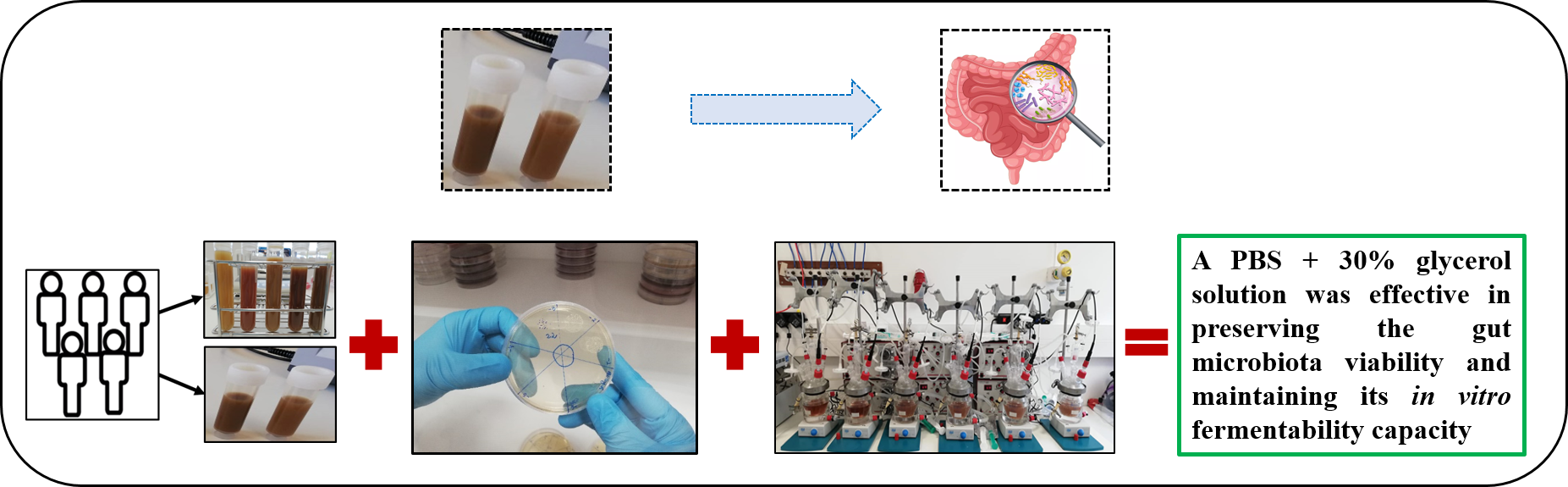
Preservation of Human Gut Microbiota Inoculums for In Vitro Fermentations Studies

by
Nelson Mota de Carvalho
1,
Diana Luazi Oliveira
1,2,*,
Mayra Anton Dib Saleh
1,
Manuela Pintado
1 and
Ana Raquel Madureira
1
1
CBQF-Centro de Biotecnologia e Química Fina-Laboratório Associado, Escola Superior de Biotecnologia, Universidade Católica Portuguesa, Rua Diogo Botelho 1327, 4169-005 Porto, Portugal
2
Amyris Bio Products Portugal, Unipessoal Lda, Rua Diogo Botelho 1327, 4169-005 Porto, Portugal
*
Author to whom correspondence should be addressed.
Fermentation 2021, 7(1), 14; https://doi.org/10.3390/fermentation7010014
Submission received: 21 December 2020 / Revised: 11 January 2021 / Accepted: 14 January 2021 / Published: 19 January 2021

Abstract

:
The use of fecal inoculums for in vitro fermentation models requires a viable gut microbiota, capable of fermenting the unabsorbed nutrients. Fresh samples from human donors are used; however, the availability of fresh fecal inoculum and its inherent variability is often a problem. This study aimed to optimize a method of preserving pooled human fecal samples for in vitro fermentation studies. Different conditions and times of storage at −20 °C were tested. In vitro fermentation experiments were carried out for both fresh and frozen inoculums, and the metabolic profile compared. In comparison with the fresh, the inoculum frozen in a PBS and 30% glycerol solution, had a significantly lower (p < 0.05) bacterial count (<1 log CFU/mL). However, no significant differences (p < 0.05) were found between the metabolic profiles after 48 h. Hence, a PBS and 30% glycerol solution can be used to maintain the gut microbiota viability during storage at −20 °C for at least 3 months, without interfering with the normal course of colonic fermentation.

Graphical Abstract

1. Introduction

A considerably large number of studies have recognized that gut microbiota is a critical factor for gut health and general host well-being, and plays a key role in the host’s gut development, mucosal immunity, food digestion, nutrient absorption [1], body detoxification, and several immune system interactions [2], not only for humans but also for other mammals and birds [3]. Microbial fermentation is seen as beneficial for the gastrointestinal tract (GIT) and host health, and is directly influenced by dietary components, which may stimulate the growth of one or more gut bacterial species [4].
Today, one of the areas of most interest for the food industry is the assessment of the prebiotic potential and gut health impact of innovative compounds incorporated in food matrices. However, before making these products commercially available, several in vitro, in vivo, and clinical trial studies are required, usually following this order. In vitro methods are considered the most suitable first-stage strategy to screen prebiotic candidates, offering the advantages of being less laborious and time-consuming, and usually raising less ethical problems, when compared to animal in vivo studies or human clinical trials [5,6]. Therefore, in a first stage, validated in vitro models are used to screen the product’s nutritional value, study the compounds digestion throughout the GIT, and evaluate their effect on the host’s gut health, namely disruptions to microbial equilibrium, such as inhibition of pathogenic bacteria growth [7,8].
Colonic fermentation in vitro models use human feces as inoculum to mimic the human gut microbiota and stimulate the fermentation, therefore, this inoculum should be representative of such ecosystems in respect to both microbial species and concentration [4]. The physical–chemical characteristics of the inoculum is one of the major concerns of in vitro studies [9]. Microbial activity variation has been linked to sampling time/day, donors and their diet, storage conditions, inoculum preparation procedures, and concentration used [9], which influences directly the microbial composition, viability, and activity [9,10,11]. The full extent of how fecal inoculum storage affects the microbiota, and which preservation method is the best for maintaining the gut microbiota as balanced and viable, is still fairly unknown [12].
The use of pooled fecal inoculums enables the reduction of inter-donor variabilities, by having a homogeneous and equilibrated mixture. In addition, having pooled fecal inoculums properly preserved and ready to use, eliminates the availability problem, reduces sample processing labor, and offers the possibility of using the same inoculum throughout several sets of experiments, hence, providing more reliable and reproducible results [4]. Moreover, frozen inoculums, enable studying the fermentation pattern of several compounds simultaneously throughout a period, without individual donor limitations. Nevertheless, mechanical damage due to ice particles and disruption of microbial cell membranes can still occur [13]. Additionally, when fecal inoculums are frozen directly, the number of viable bacterial cells (estimated by colony-forming units, CFU) can be reduced by 99.86% [14], due to different susceptibilities, e.g., the temperature of several microbial enzymes and bacteria. For example, fibrolytic gram negative bacteria are more susceptible to damage caused by freezing and thawing [13,15,16,17]. Hence, the use of a cryoprotectant prior to freezing can minimize these problems, enabling the recovery of bacterial cells to fresh equivalents [14]. In sum, the use of a well-preserved fecal inoculum is crucial for reproducing experiments and guarantee the robustness and reliability of in vitro experiments [12].
This work aimed to test the performance of a cryoprotectant solution in a human pooled fecal inoculum stored at −20 °C, by evaluating the bacterial viability and metabolic activity with storage time. The metabolic activity was assessed by investigating the gut bacteria’s capacity to carry out a colonic fermentation effectively (metabolic proof of concept). In addition, the use of a pooled fecal slurry as a fermentation inoculum was also studied, by targeting specific bacteria, e.g., bifidobacteria, acid lactic bacteria (LAB), and enterobacteria, with key roles in the host’s gut health and general well-being.

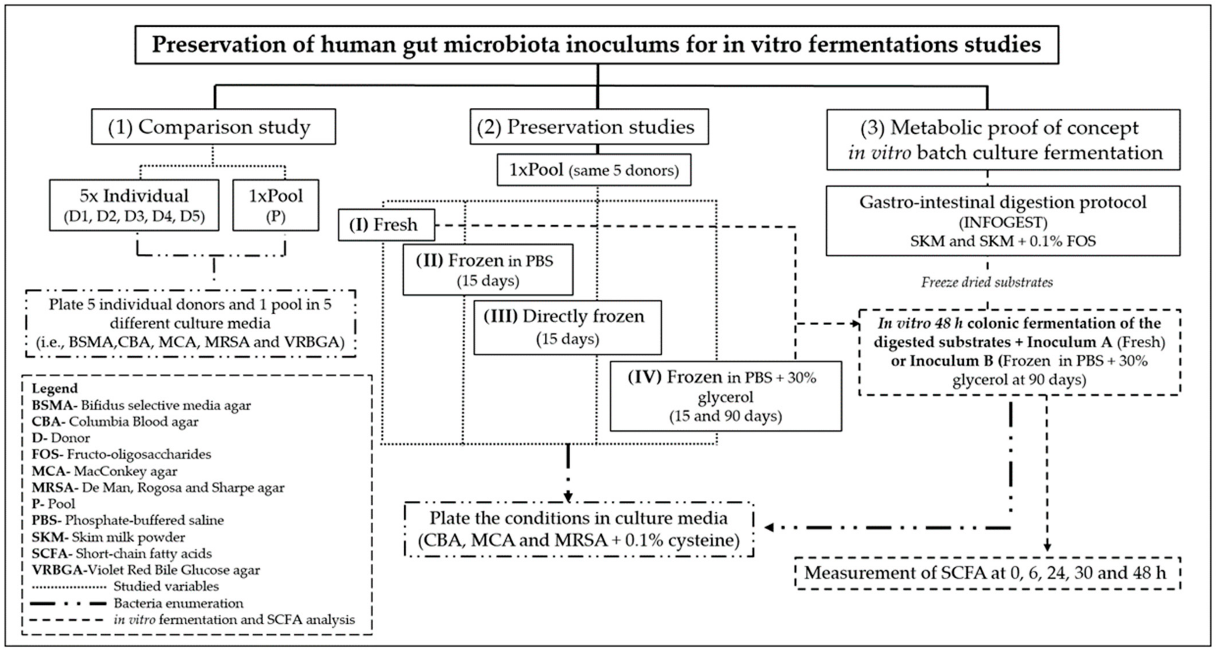
2. Materials and Methods

2.1. Reagents/Chemicals and Apparatus

The description of all reagents/chemicals and equipment used in this study are described in Appendix A. A summary of the experimental methodology is presented in Figure 1 and a detailed description of the methods/techniques used in this study are presented in the sub-sections below.

2.2. Procedures for Comparison of the Use of Individual and Pooled Inoculum

Fresh human fecal samples from five healthy adult volunteers were obtained within the premises of the Alchemy project (Universidade Católica Portuguesa, Escola Superior de Biotecnologia, Porto, Portugal). All subjects gave their informed consent for inclusion before their participation in the study (see Supplementary Information S1), conducted in accordance with the Declaration of Helsinki. The volunteers had normal omnivorous diet, without any intestinal disorders, had not ingested any antibiotics or other medicines known to affect the microbiota for at least 12 months prior to the study, and were not regular prebiotics or probiotics consumers. In addition, their last hospital admission was dated more than 12 months before the study. Volunteers were two males and three females aged 32.6 ± 4.92 years, with a body mass index (BMI) of 25.47 ± 2.31 kg/m2. Detailed information on the volunteers participating in this study is displayed in Table 1, and information on fecal sample collection is described in Appendix B.
Similar quantities of fecal sample from each donor (D) were pooled together to have a uniform and representative inoculum (pooled inoculum: P fresh). Individual fecal samples from D1 to D5, as well as P fresh, were diluted at 10% (w/w) in a 0.1 M phosphate-buffered saline pH 7.3 (PBS) solution and homogenized, first manually, and further mechanically, with a Mixwel® laboratory blender for 2 min at 460 paddles beats/min (Table 1). Sample pH was measured, using a SevenCompact pH meter, and their microbiological concentration assessed, as described in Section 2.6.

2.3. Sample Processing for Preservation Studies

In order to optimize the best preservation conditions, a new pooled inoculum was prepared and divided into four conditions, according to Table 2. Conditions I and II were mixed at 10% (w/w) with a 0.1 M PBS solution, III was kept undiluted, and IV was mixed at 10% (w/w), with a 0.1 M PBS and 30% (v/v) glycerol solution. All conditions were first homogenized manually, followed by a mechanical homogenization with a Mixwel® laboratory blender for 2 min at 460 paddles beats/min. Conditions II and III were frozen at −20 °C for 15 days, condition IV was frozen up to 90 days, whereas condition I was analyzed fresh (on the same day of preparation). To avoid repeated freezing and thawing cycles, several aliquots of the same fecal inoculum were made for each condition.
Gut bacteria viability of the four conditions was assessed with culture-dependent methods, as described in Section 2.6. Condition I was plated fresh on the same day as preparation, conditions II and III were defrosted and plated after 15 days, III was diluted at 10% (w/w) in a 0.1 M PBS solution, and IV was platted after 15 and 90 days of freezing (Table 2).

2.4. Colonic Fermentations Using Fresh and Frozen Inoculum

To evaluate the effect of storage (i.e., frozen inoculums) on the colonic fermentation performance, two inoculums, fresh (condition I) and frozen (condition IV), named from now on as inoculum A and B, respectively, were used to feed the colonic fermentation vessels. Inoculum B was previously submitted to two-glycerol wash-out cycles with a 0.1 M PBS solution. This step aimed to reduce the interference of glycerol on short-chain fatty acid (SCFA) production during fermentation, as glycerol can be used by gut bacteria as a substrate for propionate and butyrate production, which would interfere with and mislead the results. Inoculum B was first thawed in anaerobic conditions for 1 h, followed by vortexing and centrifugation (4696× g, 5 min at 4 °C). The supernatant was collected, and the pellet re-suspended in a 0.1 M PBS solution (1st glycerol wash-out cycle). The same procedure was repeated twice. After the third centrifugation (two glycerol wash-out cycles), a 0.1 M PBS solution was added to the pellet to make it up to the same initial volume as before the wash-out glycerol cycle and to complete the necessary volume for fermentation, i.e., 10% inoculum inside the fermentation vessel. The collected supernatant and inoculum B were analyzed by high-performance liquid chromatography (HPLC) (details described in Section 2.7) to quantify the glycerol concentration.
Inoculum A was submitted to in vitro batch culture fermentation while fresh, and inoculum B after 90 days frozen at −20 °C. Gut bacteria enumeration (described in Section 2.6) and pH levels were measured before fermentation. The metabolic patterns, i.e., SCFA and lactate production, over the 48 h fermentation period were compared.

2.5. Human Gastrointestinal Tract (GIT) Simulation Model Using Preserved Inoculum

An in vitro GIT simulation model adapted from the INFOGEST protocol was carried out. In a first stage the samples were submitted to gastric and intestinal digestion, followed by intestinal absorption and colonic fermentation, performed using the frozen human fecal inoculums A and B. Three conditions (in duplicates) were tested: (1) digested skim milk powder (SKM), (2) digested SKM supplemented with 0.1% (w/v) fructo-oligosaccharides (FOS), and (3) a blank control, only with fecal slurry (inoculum control—IC). The colonic fermentations were performed using two pH-controlled batch culture fermentation experiments, one using the fresh inoculum (A), and the second one using the frozen inoculum (B).

2.5.1. In vitro Gastrointestinal Digestion Protocol (INFOGEST)

An in vitro digestion method, based on the standardized European INFOGEST protocol with slight modifications, which mimics human in vivo conditions, as described by Brodkorb et al. (2019) [18], was used. SKM, i.e., the food matrix, was prepared according to the manufacturer’s instructions, i.e., 22 g of SKM in 200 mL of distilled water. The digestion protocol used 80 g of the SKM solution and followed the instructions given in the supplementary data 1 and 2 from Brodkorb et al. (2019) [18]. In addition, a dialysis step was added to simulate the nutrient absorption in the small intestine [19,20]. After gastric and intestinal digestion, all samples were transferred to a 1 kDa molecular weight cut-off regenerated cellulose dialysis tubing. The dialysis membranes were submerged in a 10 mM sodium chloride (NaCl) solution, overnight (15–20 h), at room temperature. Unabsorbed nutrients were retained inside the dialysis membrane, whereas the lower molecular weight molecules, representing the absorbed nutrients, were transferred by osmotic pressure into the NaCl solution. The retained substrates were freeze-dried and used as feed substrates in the in vitro batch culture fermentation system.

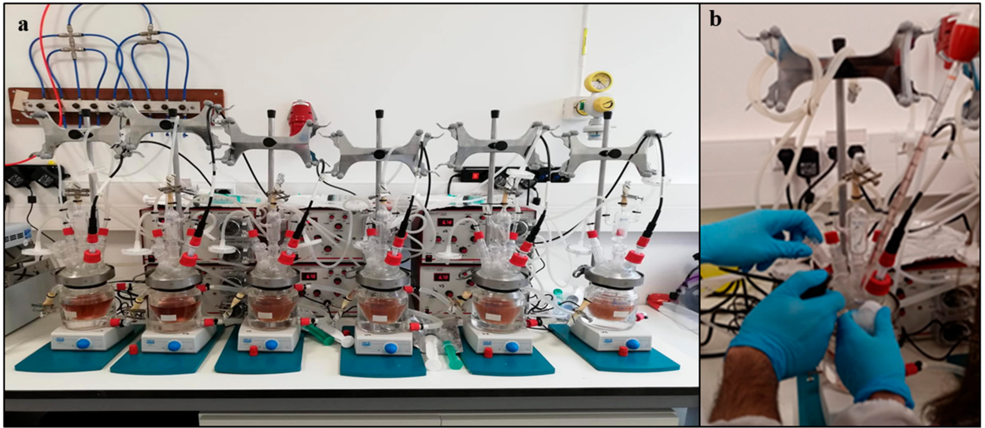
2.5.2. In vitro Batch-Culture Fermentations (Colonic Fermentation)

Six independent fermentation vessels were utilized simultaneously in two sets of experiments (set 1 and 2). The first one, with the fresh pooled fecal inoculum (inoculum A), was collected and prepared on the same day, and the second one, with the frozen pooled inoculum (inoculum B), was stored at −20 °C for 90 days in the cryoprotectant solution (i.e., 0.1 M PBS + 30% (v/v) glycerol). Sterile stirred batch culture fermentation vessels of 300 mL were set up and aseptically filled with 135 mL sterile basal nutrient medium, according to de Carvalho et al. (2019) [21], and gassed overnight with O2-free N2, and with continuous agitation (Figure 2a). Each condition was assessed in duplicate and the substrates added aseptically (by flaming the entry/sampling port). Vessels (1) and (2) had 1% (w/v) digested SKM, vessels (3) and (4) 1% (w/v) digested SKM supplemented with 0.1% (w/w) FOS (a known prebiotic), and vessels (5) and (6) were the inoculum control (IC) with no substrate added. Once the substrates were properly mixed with the basal media, each vessel was inoculated with 15 mL of fresh (set 1), or frozen (set 2), fecal slurry.
A FerMac 260 pH controller was used to maintain the pH between 6.7 and 6.9 (the pH of the human distal colon) in each vessel [22], and temperature was kept at 37 °C for 48 h with the help of a water bath. Samples (10 mL) were taken aseptically from each vessel, at 0, 6, 24, 30, and 48 h (Figure 2b). Immediately after collection, samples were placed in ice to stop the fermentation, centrifuged at 4 °C, 4696× g for 5 min, and the supernatant collected for SCFA and lactate analysis by HPLC.

2.6. Microbiological Assays

The fresh individual (D1–D5) and pooled inoculums (P fresh), subjected to the different preservation conditions (I–IV), as well as inoculum A and B, were diluted in 0.1% (w/v) peptone water (decimal dilutions) and platted in different culture media, as described in Table 3, using the Miles and Misra technique [23]. Platting and incubation were performed in anaerobic conditions using a Whitley A35 workstation at 37 °C. The gut bacteria enumeration described in Section 2.2 (D1–D5 and P fresh) used a higher number and more selective culture media, to enable a more complete study of the individual vs the pooled samples, whereas in Section 2.3 and Section 2.4 (inoculums submitted to conditions I–IV and inoculum A and B) the focus was to study the concentration of microorganisms present in the pooled inoculums, and evaluate the impact of the sample’s storage.

2.7. Determination of Short Chain Fatty Acids (SCFA), Lactate, and Glycerol/Glycerol Derivatives Produced during In Vitro Fermentation

The supernatants collected after centrifugation were filtered (0.22 μm) and directly analyzed by HPLC, using an Agilent 1260 II series HPLC instrument with a refractive index (RI) detector and diode array detector (DAD) at 220 nm, and an ion-exclusion Aminex HPX-87H column operated at 50 °C. A 5 mM sulfuric acid (H2SO4) mobile phase was used at a flow rate of 0.6 mL/min, with a running time of 30 min and an injection volume of 10 μL. Glycerol, 1-propanol, 2-propanol, 1,3-propanediol, lactate, acetate, propionate, and butyrate were identified and quantified using their corresponding calibration curves.

2.8. Statistical Analysis

Statistical analysis was carried out using TIBCO Statistica 13 software (Palo Alto, Santa Clara, CA, USA). Normality of the distributions was evaluated using Shapiro–Wilk’s test. As the samples followed normal distribution, means were compared considering a 95% confidence interval, using one-way ANOVA coupled with Tukey’s post-hoc test.

3. Results and Discussion

3.1. Differences between Using Individual and Pooled Inoculum

3.1.1. pH Variation

Table 4 presents the pH values for the conditions D1 to D5 and P fresh. The pH of the P fresh inoculum was different from the average pH value of each individual donor and closer to the donor with the most acidic pH value in study (D1). A numerical difference in the pH values was observed between each individual donor, with values ranging from 6.58 to 7.31 (Table 4). Previous studies have reported similar pH value ranges in individual feces of healthy adults [24,25,26]. These values show that the use of individual fecal inoculums may have a relevant impact on the fermentation. On the other hand, P fresh will have the same pH value for all inoculums used in the same set/experiment.
Human colon pH varies from 5 to 7 (ascending colon 5.4–5.9, traverse colon ~6.2, and distal colon 6.6 to 6.9). Despite the similarity in terms of functionality between individual fecal samples, this may not be true in terms of bacterial composition [7], resulting, for example, from the host genotype, nutrition, inflammation, antibiotics consumption, stool consistency, and pH [27,28,29]. Fecal pH, has a selective pressure on gut bacterial populations and their metabolism, especially on the fermentative gut bacteria and their metabolites production (e.g., SCFA). It influences the production of the major end product metabolites, such as propionate and acetate, of which production occurs mostly at neutral pH, and at various pH levels but by different bacteria, respectively [30].

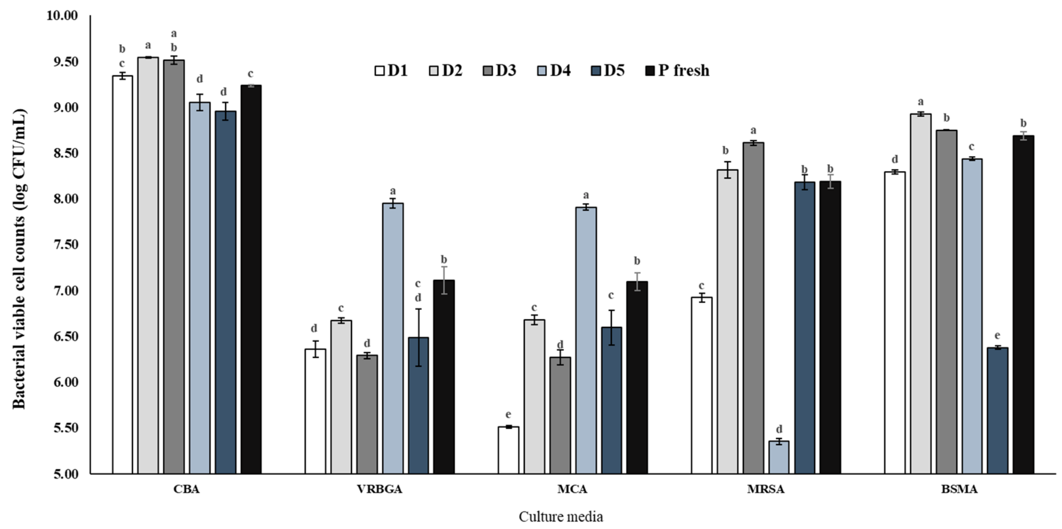
3.1.2. Bacterial Viability and Diversity on Individual and Pooled Inoculum

pH is known to modulate bacterial populations [30], so differences in the composition of bacterial populations among the volunteers were expected, as the fecal pH value for the individual donors varied significantly. To verify the inoculums bacterial population viability and composition, the fresh individual and pooled inoculums were cultivated. The bacterial groups assessed are relevant for human health and for the colonic fermentation process, namely LAB, bifidobacteria, and enterobacteria.
Figure 3, shows the bacterial viable cell counts of fresh individual and pooled inoculum from human donors obtained using the different culture media. As expected, significant differences (p < 0.05) between the bacterial populations of the individual (D1–D5) and P fresh inoculums were observed. However, for all culture media and bacteria types, P fresh inoculum presented an average cell count within the values found for all individual donors (D1–D5). Moreover, for LAB (MRSA), P fresh presented an average viable cell count similar to D5 and D2, and for bifidobacterium (BSMA), P fresh’s viable cell numbers were similar to D3. Columbia blood agar (CBA) media presented the highest bacterial cell count for P fresh, followed by Bifidus selective medium agar (BSMA), de Man, Rogosa, and Sharpe agar (MRSA), violet red bile glucose agar (VRBGA), and MacConkey agar (MCA) (9.23, 8.68, 8.19, 7.11, and 7.10 log CFU/mL, respectively). This pattern was not verified for the individual fecal inoculums (D1–D5), where a difference in the bacterial viability of the studied bacterial groups (LAB, Bifidobacteria, and Enterobacteria) was found. In addition, significant differences were found among the individual donors for the different cell culture media. Hence, the results presented in Figure 3 reinforce the idea that the use of a pooled fecal inoculum enables increasing the bacterial biodiversity and reduces the risk of outlier donors, such as D4 and D5, which may influence the normal course of a colonic fermentation.
There is an evident pattern and pH-dependent gut bacterial composition, as donors D4 and D5 had lower LAB and bifidobacteria, and higher gram-negative and enterobacteria counts, together with the highest pH values (Table 4). This is in accordance with previous findings, where LAB and bifidobacteria were also related to lowering the intestinal pH, and enterobacteria abundance associated with higher values of fecal pH [31,32,33]. Contrarily to what was observed for the individual inoculums, none of the selected bacterial populations stood out for P-fresh inoculum. The use of individual fecal inoculum (i.e., from single donors) or from a pool of the donors is still highly discussed among the experts in this field. Pooled samples are expected to have higher bacterial biodiversity, which make them more representative of the human gut microbiota, with inter-donor variations minimized [34]. This fact was also verified with the pooled fecal inoculum tested in this study, where the average viable cell count in each of the selected media was normalized in regard to the individual inoculums, Figure 3.

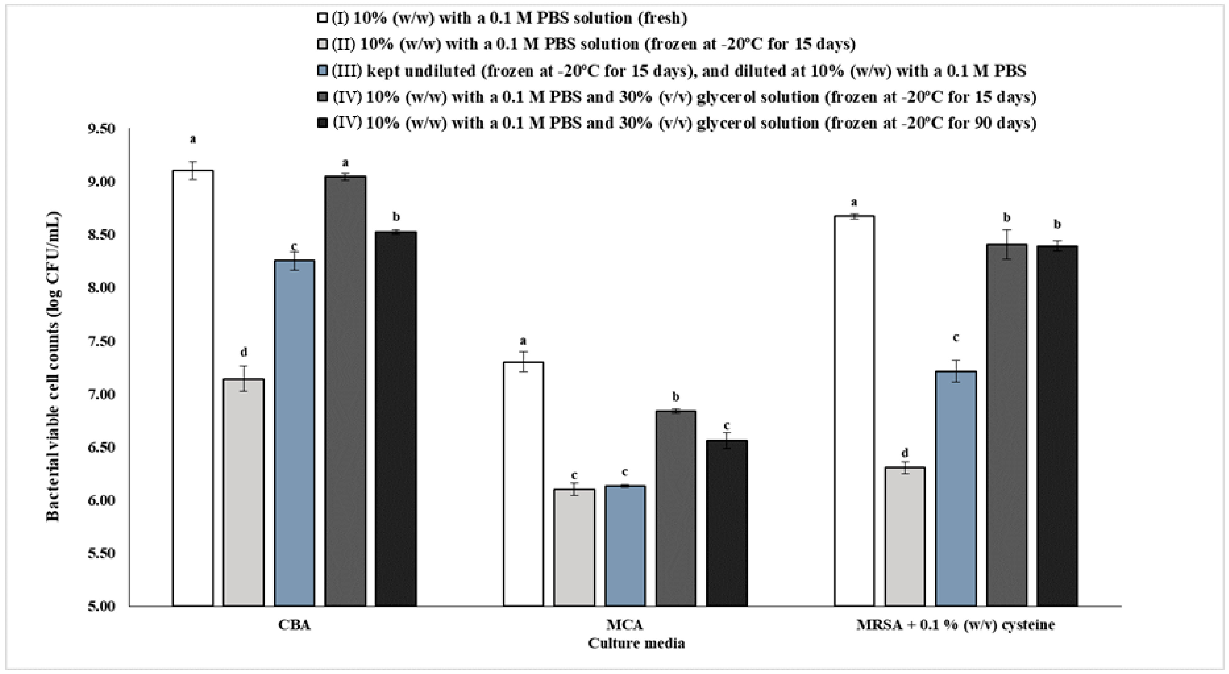
3.2. Effect of Preservation on Gut Bacteria Viability and Diversity

Gut Bacteria Viability of the Pooled Inoculums Preserved under Different Conditions (I–IV)

One of the parameters for assessing whether a preservation method, such as the one optimized in this study, is efficient in preserving fecal inoculum, is to evaluate if the bacterial concentration present in the inoculum is viable over time [35]. Hence, the bacteria viability of acid lactic bacteria (LAB), bifidobacteria, Gram negative bacteria, and total/facultative anaerobic bacteria, commonly found in a healthy gut microbiota, was assessed in the pooled fecal inoculums. Although this approach only allows quantifying a small percentage of the bacterial microbiota present (1–10%), it is still considered a valid method to assess bacteria viability in frozen inoculums, due to the relevance of the chosen bacteria (i.e., LAB, bifidobacteria, and enterobacteria) for human health [27,36].
Figure 4 presents the bacterial concentrations in the different tested preservation conditions (I–IV). As expected, the results showed that the use of a cryoprotectant solution minimized the reduction of the bacterial concentration in comparison with the other conditions, directly frozen and frozen in PBS solution. Overall, the fresh inoculum presented more viable bacteria when compared to the frozen inoculum only with PBS, which was the least efficient preservation method. The inoculum frozen with PBS + 30% glycerol had a higher log CFU/mL than the those frozen either directly, or only with PBS. At 15 days, the only differences found between fresh inoculum and frozen with PBS + 30% glycerol, were for MRSA (LAB and bifidobacteria) and MCA (enterobacteria) media (p < 0.05). However, after 90 days there was a significant difference between the fresh and frozen with PBS + 30% glycerol inoculums, in which the reduction was less than 1 log CFU/mL in the frozen inoculum, for all culture media. This decrease in bacterial concentration during the 90 days storage at −20 °C may raise the question of whether the fermentation capacity would be affected. Therefore, in vitro colonic fermentation experiments using fresh and frozen inoculums were performed (see Section 3.3) to ensure that the colonic fermentation would have similar metabolic profiles.
The initial concentration of each group of bacteria, enumerated for the condition “fresh” (in both fresh pooled inoculums used in this study), was similar to the one reported previously by Costello et al. (2015) [36]. The same study also reported a lower bacteria concentration for the “frozen” condition, compared with the “fresh” one, however, less significantly in comparison with the present study. This difference, may well be because a different cryoprotectant concentration and storage temperature were used, i.e., 10% glycerol in PBS, frozen at −80 °C. In fact, previous studies have concluded that the most suitable temperature for long-term storage of fecal samples is −70 °C [37]. Thus, it may be worth investigating this fact further, and verifying if using lower temperatures (e.g., −70 to −80 °C) improves considerably the number of viable bacteria with regard to “fresh” inoculum.
One of the aims of this study was to optimize a preservation protocol for human fecal inoculums to feed in vitro fermentation models. Using frozen, properly preserved, and viable fecal inoculums, experiment labor and time can be substantially reduced, due to the immediate availability of the fecal inoculums. Moreover, more reliable and consistent results can be achieved when using the same inoculum throughout the same or different set of experiments. Although the use of frozen fecal inoculums has been associated with less taxonomic diversity and concentrations of viable bacteria, the advantages of having such inoculums ready to use overcome the disadvantages described in the literature [9,14]. The availability of volunteers is not always guaranteed, and the bacterial composition and activity of repeated donations can be significantly different from day to day, as they are very much dependent on dietary and living practices [12]. Therefore, the use of frozen fecal inoculum would provide flexibility and consistency to in vitro fermentations studies [9]. However, and to the author’s knowledge, there is a lack of suitable approaches regarding the use of well preserved and frozen fecal inoculums. Hence, a cryoprotectant solution (0.1 M PBS solution with 30% (v/v) glycerol) was tested for the storage of human pooled fecal inoculums, to verify if the bacterial concentration and diversity was suppressed, and if the fermentation process was somehow affected by using frozen inoculums instead of fresh ones.
Glycerol is a well-known and widely used cryoprotectant, namely on bacterial culture stock solutions. As the freezing-point of biological fluids and water decreases during the freezing process, glycerol is known to protect the cells against osmotic stress, preventing eutectic crystallization and protecting the membrane vesicles containing enzymes. Moreover, glycerol has been proved to preserve cell viability, and maintain the diversity of bacterial populations [12,38]. PBS, as a hypertonic solution, is commonly used for the homogenization of fecal slurry. However, saline solutions can cause surface lesions on bacteria, and the use of glycerol contributes to suppressing this issue [12]. In the present work, glycerol behaved as a cryoprotectant of bacterial cells, and the method used was shown to be adequate for generating inoculums with sufficient viability and bacterial diversity.
Other cryoprotectant agents, such as inulin, dimethyl sulfoxide (DMSO), and polyethylene glycol 400 (PEG 400) have also been used to preserve (e.g., up to six months) fecal inoculums for in vitro fermentation experiments [39,40]. In both studies, identical metabolic profiles of SCFAs during the fermentation experiments, when using fresh and frozen inoculums, were obtained. Bircher et al. 2018 [40] observed that the use of a saline solution containing 15% glycerol stored at −80 °C for 3 months, maintained the same propionate and butyrate production at 24 h of fermentation, for both fresh and frozen inoculums. On the other hand, Gaci et al. 2017 [39] tested the impact of several cryoprotectants, i.e., DMSO, glycerol, and PEG 400, on the production of SCFAs, and concluded that DMSO was the best cryoprotective agent. However, it is important note that DMSO may be toxic, even at low concentrations, while glycerol is not [14,40]. Furthermore, Parkar et al. 2019 [41] found that fecal material preserved at −80 °C in saline solution with glycerol provided a consistent and stable inoculum for in vitro fermentations, for up to 3 years. These studies used lower storage temperatures and glycerol concentrations, and they did not mention if there was a glycerol wash out step before fermentation, as in the present study.

3.3. Impact of Using Fresh and Frozen Inoculum in Colonic Batch Fermentations

3.3.1. Glycerol Wash-out Confirmation

The HPLC results showed that after the two-glycerol wash-out cycles, with 0.1 M PBS, glycerol concentration significantly decreased, from 3258 mM (before washing the inoculum) to 7.48 mM, (glycerol concentration in inoculum B). Inoculum B was further used to feed the colonic fermentation at 10% (v/v), thus, it is expected that the glycerol concentration in the fermentation vessels decreased 10 times (i.e., 0.7 mM). Moreover, the presence of glycerol was not detected at time point 0 h in any of the fermentation experiments (data not shown), most likely due to the fact that this concentration is under the HPLC detection limits. Thus, it is expected that this low concentration of glycerol, present in inoculum B, would not interfere with the fermentation process or alter the metabolite (e.g., SCFA) production. This fact was further verified when analyzing the metabolic profiles from the fermentation experiments using both inoculums A and B (see Section 3.4), proving that the wash-out procedure applied was efficient in removing most of the glycerol from the fecal slurry before its use in in vitro batch models.
Therefore, despite glycerol’s capacity for preserving inoculum microbiota, the utilization of this cryoprotectant is inadequate, unless the glycerol is removed prior to the start of the fermentation process, as it can interfere with the metabolite production (i.e., SCFA), due to its use by the colonic bacteria [11]. Gut bacteria can use glycerol as a source of energy/nutrients, originating undesired glycerol-derived by-products (e.g., 1,3-Propanediol, 1-Propanol and 2-Propanol), and increasing the SCFA production, namely propionate and butyrate. For this reason, the glycerol wash-out step is crucial prior to fermentation.
The type of substrate present during gut fermentation will have a direct impact on the composition of the gut bacterial population and their metabolism, which is dominantly saccharolytic and/or proteolytic. The metabolites or end-products produced during fermentation include SCFA, mainly acetate, propionate, and butyrate, which represent 90–95% of the total SCFA, and at a smaller scale, branched-chain fatty acids (BCFA), such as isobutyrate and isovalerate, which represent ~5% of total SCFA production [12,42]. These metabolites play a key role in the host’s well-being, as is heavily described in the literature [28,29,42,43,44], with special importance given to the inhibition of pathogenic microorganism growth by reducing the luminal and fecal pH, and directly promoting the growth of symbionts; regulation of glucose and lipid metabolism; reduction of food intake; mineral absorption promotion; stimulation and differentiation of intestinal enterocytes; anti-oxidative functions; reduction of inflammatory disease prevalence; and also contributing to the central nervous system function (gut–brain axis) [12,21,44,45].

3.3.2. pH Variation

The pH value of the inoculum B was 6.59, and similar to the fresh pooled inoculum, as shown in Table 3. Based on these results, it was evident that the pH value was stable in the pooled inoculums upon freezing. Therefore, the use of a pooled fecal inoculum seems to be the most appropriate manner of standardizing gut microbiota populations to be used in gut fermentation experiments.

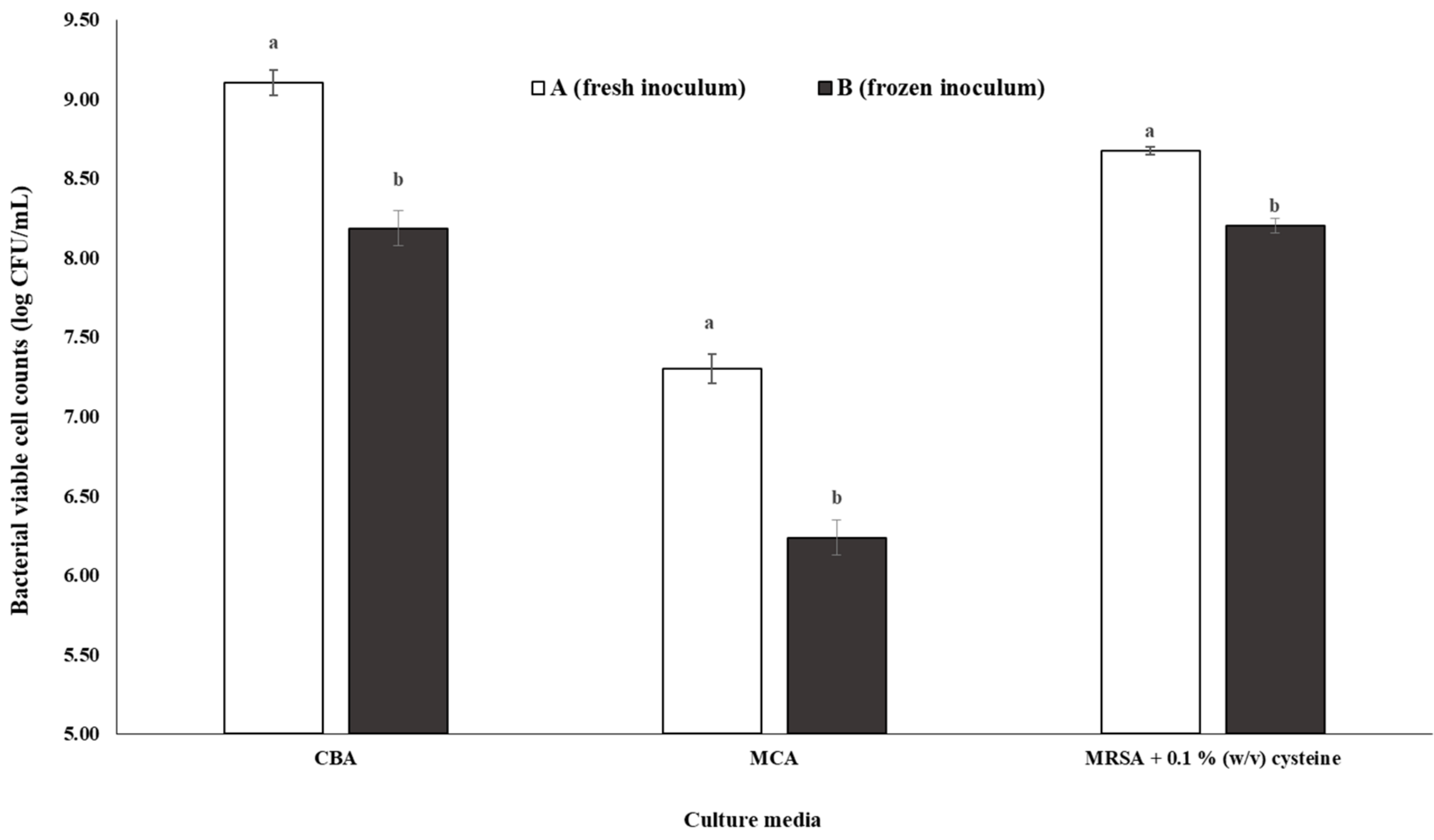
3.3.3. Gut Bacteria Enumeration of Inoculum A and B

Figure 5 shows the bacterial cell viable numbers present in fresh pooled inoculum (A) and frozen pooled inoculum (B). In both inoculums A and B, the bacterial cell viable counts were statistically different (p < 0.05). The culture media that presented the highest bacterial viability for both inoculums (A and B) was CBA (9.10 and 8.18 log CFU/mL, respectively) followed by MRSA + 0.1% (w/v) cysteine (8.67 and 8.20 log CFU/mL, respectively) and MCA (7.30 and 6.24 log CFU/mL, respectively). This trend was also verified for the pooled inoculums I and IV frozen for 90 days (Figure 4).
Despite the loss of cell viability in inoculum B, the results shown in Figure 5 support the choice of using a frozen pooled fecal inoculum for up to 90 days of storage at −20 °C, as there was still a significant number of viable cells capable of carrying out an effective in vitro colonic fermentation, as proven further (see Section 3.4). Moreover, the results obtained in the present study are in agreement with previous studies [36].

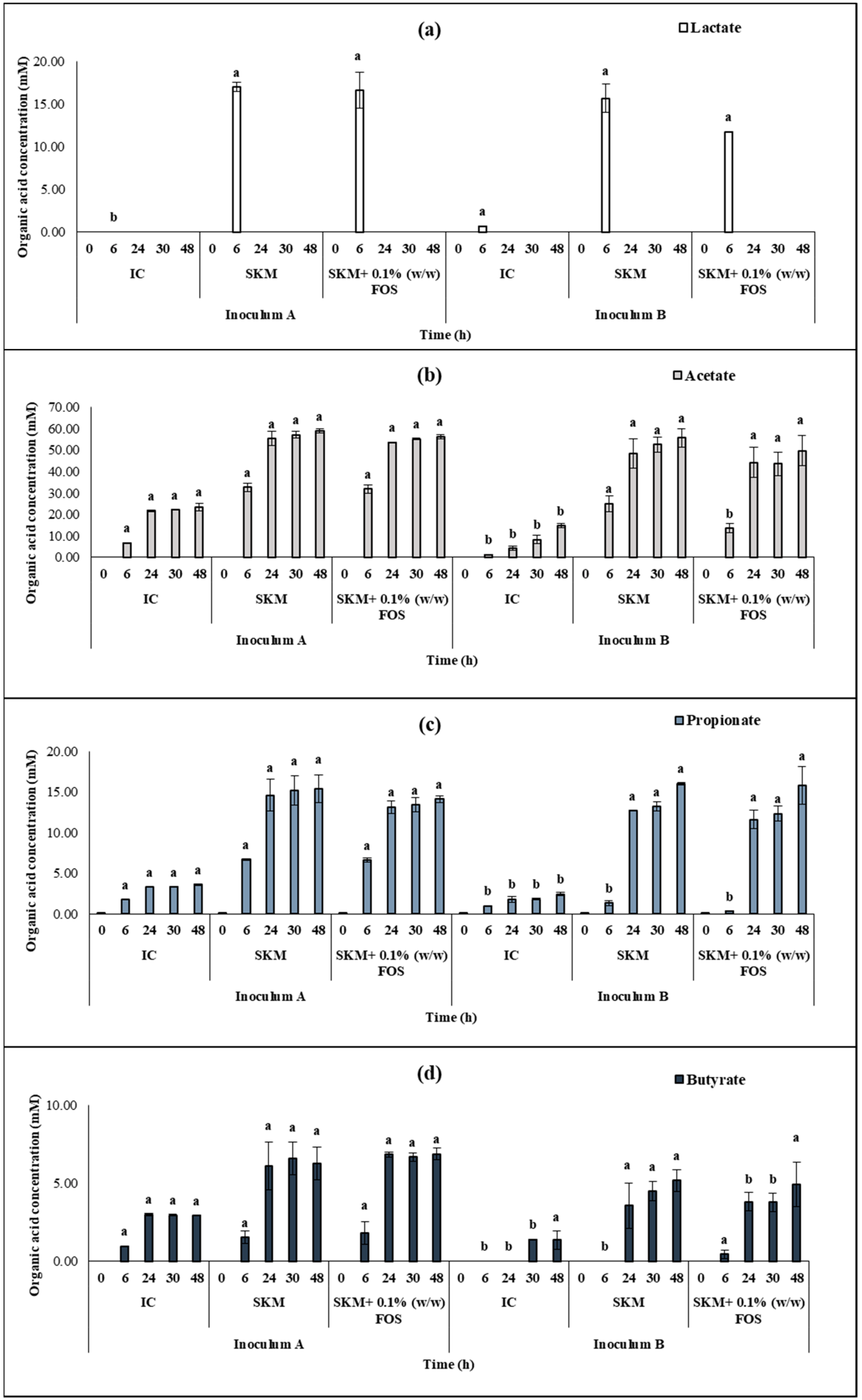
3.4. Human in vitro Colonic Fermentation Using Fresh and Frozen Inoculums

In vitro colonic fermentations were carried for both fresh (A) and frozen (B) inoculums. Figure 6, shows the SCFA and lactate production results of the fermentation experiments. Each set included three conditions (in duplicates): 1) IC (only basal media and fecal inoculum), 2) SKM condition (basal media + 1% digested SKM + fecal inoculum), and 3) SKM + 0.1 (w/w) % FOS with 1% digested SKM + 0.1% (w/w) FOS + fecal inoculum). The IC provided the background information regarding the intrinsic and already stablished gut microbiota metabolite production, without the interference of a nutrient source. In addition, it enabled comparing both the fresh and frozen inoculums used in the present study with other published studies [21,46,47,48,49].
As expected, the condition with the lowest SCFA production was the IC, for both fresh (A) and frozen (B) inoculums, since there was no addition of substrate to feed the gut bacteria. For inoculum A and at 48 h, the concentration for acetate, propionate, and butyrate was 23.48, 3.63, and 2.93 mM, respectively. For the same conditions, inoculum B had a significantly (p < 0.05) lower concentration for acetate and propionate (14.72 and 2.45, respectively), but not for butyrate (1.36 mM). The SCFA produced with the SKM and SKM + FOS 0.1% (w/w) conditions did not present significant differences (p < 0.05) for both inoculums at 48 h. Thus, the gut bacteria kept the same metabolic capacity for frozen as for the fresh inoculum, despite the preservation process. For both conditions with different inoculums, the production of the SCFA was not statistically different between them, with concentrations for acetate, propionate, and butyrate around 55 mM, 15 mM, and 5 mM, respectively. Therefore, the supplementation of FOS at 0.1% (w/w) in SKM did not increase the fermentative potential of the gut microbiota tested. This fact may be due to the lower FOS concentration used, and different results may be expected when increasing the incorporation percentage.
Regarding lactate, it is known that this organic acid usually appears within the first hours of fermentation, and disappears after a certain time (commonly at 24 h), due to its consumption by the gut bacteria [21,49]. Lactate is produced by, e.g., LAB and bifidobacteria and converted to acetate, propionate, and butyrate by other bacteria, namely e.g., Bacteroides and Roseburia, which are propionate and butyrate producing bacteria, respectively. This metabolic process, known as cross-feeding, i.e., the use of metabolic end-products of a given microorganism by others [42,43], limits the accumulation of lactate in the colon and the consequent metabolic acidosis [21]. This cross-feeding process was verified in this study, as lactate was present at concentrations between 10–20 mM (for both inoculums) at 6 h, and disappeared completely after 24 h. Simultaneously, an increase of acetate, propionate, and butyrate concentrations was observed (Figure 6).
For the IC condition, the concentration of acetate, propionate, and butyrate at 24 h, was 21.72, 3.31, and 2.99 mM, respectively, and within the concentration ranges reported in other studies, i.e., 3–25 mM, 1.5–7 mM, and 1.5–5 mM at 24 h, for the same SCFA, respectively [21,46,47,48,49]. On the other hand, no data were found for the use of SKM which could be used for comparison. At 24 h, this condition originated concentrations of acetate, propionate, and butyrate, of 55.44, 14.64, and 6.10 mM, respectively. Shen et al. (2011) [50] reported considerably lower concentrations of acetate, propionate, and butyrate, of approximately 45 mM, 6 mM, and 3 mM, respectively, using 1% of human milk oligosaccharides (HMO). These differences were most likely because SKM is a much richer substrate than HMO, and thus, higher concentrations of these acids are expected. Despite the differences in SCFA concentrations expected for different food matrixes, the molar ratio of acetate, propionate, and butyrate produced, in both fresh and frozen inoculum, was similar to other studies, ranging from 3:1:1 to 10:2:1 [42,43,51,52]. The same trend was observed for all conditions tested.
Different approaches can be used to test the prebiotic potential of a new compound. The most suitable strategy, as a first-stage, is to use in vitro models, such as the fecal batch culture system with pH control used in the present study to screen potential prebiotic candidates [6]. According to the International Scientific Association for Probiotics and Prebiotics (ISAPP), a prebiotic is a “substrate that is selectively utilized by host microorganisms conferring a health benefit”. This definition is applied for both human and animals [6,53]. The inclusion of prebiotics in human and animal nutrition has long been regarded with great interest, due to the proven beneficial impact on gut microbiota modulation, and the subsequent health-promotion effects for the host. Most of the bioactive compounds validated as prebiotics, such as FOS and galacto-oligosaccharides (GOS), which are small to medium chain carbohydrates, have been extensively studied, and are currently well-established prebiotics and commercially available [6,53,54].
The concentration of SCFA present in the distal colon can vary from 20 to 70 mM, depending on the host’s diet, which is the major carbon source available for metabolization by gut microbiota [28,29]. In the present study, the SCFA concentrations after 48 h of in vitro colonic fermentation were within this range.
Food digestion has an important role in nutrient absorption, and in determining what reaches the colon for fermentation. A standardized static in vitro simulation of gastrointestinal food digestion model (INFOGEST) [18] was used. As recommended, SKM was chosen as the food matrix, as it is one of the most complete food matrices in terms of nutrition, containing all the essential nutrients. A reduced fat content (skim) was preferred to avoid the use of complex fat digestion enzymes (i.e., lipases). Moreover, liquid foods do not require oral digestion, as there is no need for mastication. Using a well-studied food matrix will also provide useful information for results comparison. Unlike other in vitro digestion models [18,55], and in accordance with other studies [19,56,57], an additional intestinal absorption step was added to this protocol, in order to mimic as closely as possible the human complete digestive system, including the use of the unabsorbed material retained in the membranes for gut bacteria fermentation. The inclusion of a well-established prebiotic, such as FOS, in the food matrix, (i.e., SKM) will provide the necessary information on what happens when such food components are included in the diet, and this condition was the positive control of the study. The choice of the percentage to be added (0.1% FOS (w/w)) was made to verify the impact of a prebiotic-low dosage supplementation on gut microbiota.
To summarize, the use of a frozen inoculum in an in vitro batch-culture fermentation model did not show significant differences in the production of the main SCFA, i.e., acetate, propionate, and butyrate, when compared with the fresh inoculum.

4. Conclusions

The optimized preservation method (utilization of 0.1 M PBS + 30% (v/v) glycerol solution and frozen at −20 °C for 90 days) described in this study proved to be suitable for the storage of frozen pooled fecal inoculums aimed at feeding in vitro batch culture fermentation models, by maintaining the gut microbiota viability and the capability of carrying out a colonic fermentation process effectively. Pooled frozen inoculums could still be used after 90 days (~3 months) storage, although a significant decrease of viable cells was observed in the frozen inoculum compared with the fresh inoculum, and this reduction was less than 1 log CFU/mL, which was not enough to reduce the bacteria fermentative capacity. Overall, the disadvantages associated to the use of pooled frozen fecal inoculums are overcame by the numerous advantages of having well preserved, ready to use, and viable inoculums for colonic fermentation studies.

Supplementary Materials

The following are available online at https://www.mdpi.com/2311-5637/7/1/14/s1, Table S1: Bacterial viable cell counts (log CFU/mL, mean ± SD) in different culture media of the fresh fecal individual and pooled inoculums., Table S2: Bacterial viable cell counts (log CFU/mL, mean ± SD) in different culture media of different storage conditions of human pooled fecal inoculum, Table S3: Bacterial viable cell counts (log CFU/mL, mean ± SD) in different culture media of inoculum A (fresh) and B (frozen)., Table S4: Concentration (mM, means ± SD) of organic acids produced along fermentation time in inoculum A and B., S1—Informed consent form.

Author Contributions

Conceptualization, N.M.d.C., D.L.O. and A.R.M.; Data curation, N.M.d.C.; Formal analysis, N.M.d.C.; Funding acquisition, M.P. and A.R.M.; Investigation, N.M.d.C., D.L.O. and A.R.M.; Methodology, N.M.d.C., D.L.O. and M.A.D.S.; Project administration, M.P. and A.R.M.; Resources, A.R.M.; Supervision, D.L.O. and A.R.M.; Visualization, D.L.O.; Writing—original draft, N.M.d.C.; Writing—review & editing, D.L.O., M.A.D.S. and A.R.M. All authors have read and agreed to the published version of the manuscript.

Funding

This research was funded by Amyris Bio Products Portugal Unipessoal Lda and Escola Superior de Biotecnologia—Universidade Católica Portuguesa through Alchemy project-Capturing high value from industrial fermentation bio products (POCI-01-0247-FEDER-027578). We would also like to thank the scientific collaboration under the FCT project UID/Multi/50016/2019.

Institutional Review Board Statement

Ethical review and approval were waived for this study, as it was conducted according to the internal rules legally established, based on the research ethics recommendations and with the informed consent of all subjects involved in the study.

Informed Consent Statement

Informed consent was obtained from all subjects in the study.

Data Availability Statement

The data presented in this study are available in the article or supplementary material.

Acknowledgments

The authors acknowledge Amyris, Inc and CBQF—Centro de Biotecnologia e Química Fina—Laboratório Associado, Escola Superior de Biotecnologia of Universidade Católica Portuguesa.

Conflicts of Interest

The authors declare no conflict of interest.

Appendix A. Materials and Methods

Appendix A.1. Reagents/Chemicals Used in This Study

  • 1-propanol (Sigma, St. Louis, MO, USA);
  • 1,3-propanediol (Sigma, St. Louis, MO, USA);
  • 2-propanol (Honeywell Riedel-de Haen, Munich, Germany);
  • Acetic acid glacial (Sigma, St. Louis, MO, USA);
  • Ammonium Carbonate-(NH4)2CO3 (Merck KGaA, Darmstadt, Germany);
  • Bile bovine (Sigma, St. Louis, MO, USA);
  • Bile salts (Sigma, St. Louis, MO, USA);
  • Bile acid assay kit (Sigma, St. Louis, MO, USA);
  • Butyric acid (Sigma, St. Louis, MO, USA);
  • Calcium chloride dihydrate—CaCl2(H2O)2 (Carlo Erba Reagents, Barcelona, Spain)
  • Calcium chloride hexahydrate—CaCl2(H2O)6 (Sigma, St. Louis, MO, USA);
  • Defibrinated sheep blood OxoidTM (Thermo Fischer Scientific, Waltham, MA, USA)
  • Dipotassium hydrogen phosphate-K2HPO4 (Honeywell Fluka, Seelze, Germany);
  • DL-lactic acid (Sigma, St. Louis, MO, USA);
  • Fructo-oligosaccharides from chicory root (FOS) (Megazyme, Bray, Ireland);
  • Glycerol—analytical grade (Fisher Scientific, Loughborough, UK);
  • Hemin (Sigma, St. Louis, MO, USA);
  • Hemoglobin (Sigma, St. Louis, MO, USA);
  • Hydrochloric acid- HCl (Honeywell Fluka, Seelze, Germany);
  • L-cysteine HCl (Sigma-Aldrich, St. Louis, MO, USA);
  • Magnesium chloride hexahydrate-MgCl2(H2O)6 (Panreac, Barcelona, Spain);
  • Magnesium sulfate heptahydrate-MgSO4(H2O)7 (Sigma, St. Louis, MO, USA);
  • Molico skim milk powder-SKM (Nestlé S.A., Vevey, Switzerland);
  • Na-p-tosyl-L-arginine methyl esther hydrochloride—TAME (Sigma, St. Louis, MO, USA);
  • Pancreatin from porcine pancreas (Sigma, St. Louis, MO, USA);
  • Pepsin from porcine gastric mucosa powder (Sigma, St. Louis, MO, USA);
  • Peptone from animal tissue (Sigma, St. Louis, MO, USA);
  • Phosphate buffered saline (Dulbecco A) OxoidTM (Thermo Fischer Scientific, Waltham, MA, USA);
  • Potassium chloride-KCl (Honeywell Fluka, Seelze, Germany);
  • Potassium dihydrogen phosphate-KH2PO4 (Merck KGaA, Darmstadt, Germany);
  • Propionic acid (Sigma, St. Louis, MO, USA);
  • Resazurin sodium salt (Sigma, St. Louis, MO, USA);
  • Sodium chloride-NaCl (Honeywell Fluka, Seelze, Germany);
  • Sodium hydrogen carbonate-NaHCO3 (Panreac, Barcelona, Spain);
  • Sodium hydroxide—NaOH (LabChem, Zelienople, USA);
  • Sulfuric acid—H2SO4 (Honeywell Fluka, Seelze, Germany);
  • Trichloroacetic acid—TCA (Sigma, St. Louis, MO, USA);
  • Tris(hydroxymethyl)aminomethane hydrochloride (Merck KGaA, Darmstadt, Germany)
  • Tween 80 (Sigma, St. Louis, MO, USA);
  • Vitamin K1 (Sigma, St. Louis, MO, USA);
  • Yeast extract (Sigma, St. Louis, MO, USA).

Appendix A.2. Culture Media

  • Bifidus selective medium agar-BSMA (Sigma, St. Louis, MO, USA);
  • Columbia agar base-CBA (Liofilchem, Roseto degli Abruzzi, Italy);
  • de Man, Rogosa and Sharpe agar-MRSA (Biokar Diagnostics, Allonne, France);
  • MacConkey agar-MCA (Biolife, Milan, Italy);
  • Violet red bile glucose agar—VRBGA (Biokar diagnostics, Allonne, France).

Appendix A.3. Apparatus Used in This Study

  • 1 kDa molecular weight cut-off regenerated cellulose dialysis tubing Spectra/Por® 6 (Spectrum, New Brunswick, NJ, USA);
  • Agilent 1260 II series HPLC (Agilent, Santa Clara, CA, USA);
  • Alpha 2–4 LSC plus model (Martin Christ Gefriertrocknungsanlagen GmbH, Osterode am Harz, Germany)
  • Anaerobic cabinet, Whitley A35 workstation (Don Whitley Scientific, Bingley, UK);
  • FerMac 260 pH controller (Electrolab Biotech Ltd., Gloucestershire, UK);
  • Heraeus™ Megafuge™ 16R Centrifuge (Thermo Fischer Scientific, Waltham, MA, USA)
  • Ion-exclusion Aminex HPX-87H column (Biorad, Hercules, CA, USA);
  • Mixwel® laboratory blender (Alliance Bio Expertise, Guipry, France);
  • MR Hei-Tec magnetic stirrer (Heidolph Instruments GmbH & CO. KG, Schwabach, Germany)
  • MST magnetic stirrer (Velp Scientifica, Usmate Velate, Italy)
  • OxoidTM AnaeroGenTM 2.5 L sachet (Thermo Fischer Scientific, Waltham, MA, USA);
  • OxoidTM AnaeroJarTM 2.5 L (Thermo Fischer Scientific, Waltham, MA, USA);
  • Reax top vortex (Heidolph Instruments GmbH & CO. KG, Schwabach, Germany);
  • Refrigerator Beko RSNE445E33WN (Beko, Istanbul, Turkey);
  • SevenCompact pH meter (Mettler Toledo, Urdorf, Switzerland);
  • Synergy H1 Hybrid Multi-Mode Reader (BioTek, Winooski, VT, USA);
  • UV-1900 UV-VIS spectrophotometer (Shimadzu, Kyoto, Japan);
  • Tamper proof specimen 1-L containers (Sigma, St. Louis, MO, USA).

Appendix B. Human Fecal Sample Collection Protocol

The fecal samples were collected in a clean tamper-proof specimen 1 L container. The containers with the feces were placed in the OxoidTM AnaeroJarTM 2.5 L with an OxoidTM AnaeroGenTM 2.5 L sachet, closed until opened inside of the anaerobic cabinet, a Whitley A35 workstation, and used within 2 h of collection. Under the anaerobic cabinet atmosphere (nitrogen 80%, carbon dioxide 10%, hydrogen 10%), fecal content of each donor was sampled in equal amounts, placed into an empty pre-weight tamper-proof specimen 1 L container, and weighed to obtain a pooled fecal inoculum.

References

  1. Maki, J.J.; Klima, C.L.; Sylte, M.J.; Looft, T. The Microbial Pecking Order: Utilization of Intestinal Microbiota for Poultry Health. Microorganisms 2019, 7, 376. [Google Scholar] [CrossRef] [PubMed] [Green Version]
  2. Grond, K.; Sandercock, B.K.; Jumpponen, A.; Zeglin, L.H. The avian gut microbiota: Community, physiology and function in wild birds. J. Avian Biol. 2018, 49, e01788. [Google Scholar] [CrossRef] [Green Version]
  3. Yan, W.; Sun, C.; Zheng, J.; Wen, C.; Ji, C.; Zhang, D.; Chen, Y.; Hou, Z.; Yang, N. Efficacy of Fecal Sampling as a Gut Proxy in the Study of Chicken Gut Microbiota. Front. Microbiol. 2019, 10, 2126. [Google Scholar] [CrossRef] [PubMed] [Green Version]
  4. Pastorelli, G.; Faustini, M.; Attard, E. In vitro fermentation of feed ingredients by fresh or frozen pig fecal inocula. Anim. Sci. J. 2014, 85, 690–697. [Google Scholar] [CrossRef]
  5. Doke, S.K.; Dhawale, S.C. Alternatives to animal testing: A review. Saudi Pharm. J. 2015, 23, 223–229. [Google Scholar] [CrossRef] [Green Version]
  6. Amorim, C.; Silvério, S.C.; Cardoso, B.B.; Alves, J.I.; Pereira, M.A.; Rodrigues, L.R. In vitro fermentation of raffinose to un-ravel its potential as prebiotic ingredient. LWT 2020, 126, 109322. [Google Scholar] [CrossRef]
  7. Aguirre, M.; Ramiro-Garcia, J.; Koenen, M.; Venema, K. To pool or not to pool? Impact of the use of individual and pooled fecal samples for in vitro fermentation studies. J. Microbiol. Methods 2014, 107, 1–7. [Google Scholar] [CrossRef]
  8. Ashaolu, T.J.; Ashaolu, J.O.; Adeyeye, S.A.O. Fermentation of prebiotics by human colonic microbiota in vitro and short-chain fatty acids production: A critical review. J. Appl. Microbiol. 2020. [Google Scholar] [CrossRef]
  9. Murray, J.-A.M.D.; McMullin, P.; Handel, I.; Hastie, P.M. The effect of freezing on the fermentative activity of equine faecal inocula for use in an in vitro gas production technique. Anim. Feed Sci. Technol. 2012, 178, 175–182. [Google Scholar] [CrossRef]
  10. Bosch, G.; Wrigglesworth, D.J.; Cone, J.W.; Pellikaan, W.F.; Hendriks, W.H. Effects of preservation conditions of canine feces on in vitro gas production kinetics and fermentation end products. J. Anim. Sci. 2013, 91, 259–267. [Google Scholar] [CrossRef] [Green Version]
  11. Prates, A.; De Oliveira, J.; Abecia, L.; Fondevila, M. Effects of preservation procedures of rumen inoculum on in vitro microbial diversity and fermentation. Anim. Feed. Sci. Technol. 2010, 155, 186–193. [Google Scholar] [CrossRef]
  12. Aguirre, M.; Eck, A.; Koenen, M.; Savelkoul, P.H.M.; Budding, A.E.; Venema, K. Evaluation of an optimal preparation of human standardized fecal inocula for in vitro fermentation studies. J. Microbiol. Methods 2015, 117, 78–84. [Google Scholar] [CrossRef] [PubMed]
  13. Hervás, G.; Frutos, P.; Giráldez, F.J.; Mora, M.J.; Fernández, B.; Mantecón, Á.R. Effect of preservation on fermentative activity of rumen fluid inoculum for in vitro gas production techniques. Anim. Feed Sci. Technol. 2005, 123, 107–118. [Google Scholar] [CrossRef]
  14. Waite, D.W.; Deines, P.; Taylor, M.W. Quantifying the impact of storage procedures for faecal bacteriotherapy in the critically endangered New Zealand Parrot, the Kakapo (Strigops habroptilus). Zoo Biol. 2013, 32, 620–625. [Google Scholar] [CrossRef] [PubMed]
  15. Beauchemin, K.A.; Colombatto, D.; Morgavi, D.P.; Yang, W.Z.; Rode, L.M. Mode of action of exogenous cell wall degrading enzymes for ruminants. Can. J. Anim. Sci. 2004, 84, 13–22. [Google Scholar] [CrossRef]
  16. Henderson, A.R.; McDonald, P.; Anderson, D. The effect of a cellulase preparation derived fromTrichoderma viride on the chemical changes during the ensilage of grass, lucerne and clover. J. Sci. Food Agric. 1982, 33, 16–20. [Google Scholar] [CrossRef]
  17. Murray, J.M.D.; Longland, A.; Moore-Colyer, M.; Dunnett, C. The effect of feeding a low- or a high-starch diet on the in vitro fermentative capacity of equine faecal inocula. Anim. Sci. 2006, 82, 627–635. [Google Scholar] [CrossRef]
  18. Brodkorb, A.; Egger, L.; Alminger, M.; Alvito, P.; Assunção, R.; Ballance, S.; Bohn, T.; Bourlieu-Lacanal, C.; Boutrou, R.; Carrière, F.; et al. INFOGEST static in vitro simulation of gastrointestinal food digestion. Nat. Protoc. 2019, 14, 991–1014. [Google Scholar] [CrossRef]
  19. Alegría-Torán, A.; Garcia-Llatas, G.; Cilla, A. Static Digestion Models: General Introduction; Springer Science and Business Media LLC: Cham, Germany, 2015; pp. 3–12. [Google Scholar]
  20. Verhoeckx, K.; Cotter, P.; López-Expósito, I.; Kleiveland, C.; Lea, T.; Mackie, A.; Requena, T.; Swiatecka, D.; Wichers, H. The Impact of Food Bioactives on Health: In Vitro and Ex Vivo Models; Springer Nature: London, UK; Berlin, Germany; New York, NY, USA, 2015. [Google Scholar]
  21. De Carvalho, N.M.; Walton, G.; Poveda, C.; Silva, S.; Amorim, M.; Madureira, A.; Pintado, M.; Gibson, G.; Jauregi, P. Study of in vitro digestion of Tenebrio molitor flour for evaluation of its impact on the human gut microbiota. J. Funct. Foods 2019, 59, 101–109. [Google Scholar] [CrossRef]
  22. Sánchez-Patán, F.; Cueva, C.; Monagas, M.; Walton, G.E.; Gibson, G.R.; Quintanilla-López, J.E.; Lebrón-Aguilar, R.; Martin-Alvarez, P.J.; Moreno-Arribas, M.V.; Bartolomé, B. In vitro fermentation of a red wine extract by human gut microbi-ota: Changes in microbial groups and formation of phenolic metabolites. J. Agric. Food Chem. 2012, 60, 2136–2147. [Google Scholar] [CrossRef]
  23. Miles, A.A.; Misra, S.S.; Irwin, J.O. The estimation of the bactericidal power of the blood. Epidemiol. Infect. 1938, 38, 732–749. [Google Scholar] [CrossRef] [PubMed] [Green Version]
  24. Ohigashi, S.; Sudo, K.; Kobayashi, D.; Takahashi, O.; Takahashi, T.; Asahara, T.; Nomoto, K.; Onodera, H. Changes of the Intestinal Microbiota, Short Chain Fatty Acids, and Fecal pH in Patients with Colorectal Cancer. Dig. Dis. Sci. 2013, 58, 1717–1726. [Google Scholar] [CrossRef] [PubMed]
  25. Osuka, A.; Shimizu, K.; Ogura, H.; Tasaki, O.; Hamasaki, T.; Asahara, T.; Nomoto, K.; Morotomi, M.; Kuwagata, Y.; Shimazu, T. Prognostic impact of fecal pH in critically ill patients. Crit. Care 2012, 16, R119. [Google Scholar] [CrossRef] [PubMed] [Green Version]
  26. Watson, E.-J.; Giles, J.; Scherer, B.L.; Blatchford, P. Human faecal collection methods demonstrate a bias in microbiome composition by cell wall structure. Sci. Rep. 2019, 9, 1–8. [Google Scholar] [CrossRef]
  27. Prakash, O.; Nimonkar, Y.; Desai, D. A Recent Overview of Microbes and Microbiome Preservation. Indian J. Microbiol. 2020, 60, 297–309. [Google Scholar] [CrossRef]
  28. Den Besten, G.; van Eunen, K.; Groen, A.K.; Venema, K.; Reijngoud, D.-J.; Bakker, B.M. The role of short-chain fatty acids in the interplay between diet, gut microbiota, and host energy metabolism. J. Lipid Res. 2013, 54, 2325–2340. [Google Scholar] [CrossRef] [Green Version]
  29. Morrison, D.J.; Preston, T. Formation of short chain fatty acids by the gut microbiota and their impact on human metabolism. Gut Microbes 2016, 7, 189–200. [Google Scholar] [CrossRef] [Green Version]
  30. Ilhan, Z.E.; Marcus, A.K.; Kang, D.-W.; Rittmann, B.E.; Krajmalnik-Brown, R. pH-mediated microbial and metabolic inter-actions in fecal enrichment cultures. Msphere 2017, 2, e00047-17. [Google Scholar] [CrossRef] [Green Version]
  31. Henrick, B.M.; Hutton, A.A.; Palumbo, M.C.; Casaburi, G.; Mitchell, R.D.; Underwood, M.A.; Smilowitz, J.T.; Frese, S.A. El-evated fecal pH indicates a profound change in the breastfed infant gut microbiome due to reduction of Bifidobacterium over the past century. MSphere 2018, 3, e00041-18. [Google Scholar] [CrossRef] [Green Version]
  32. Mathur, H.; Beresford, T.P.; Cotter, P.D. Health Benefits of Lactic Acid Bacteria (LAB) Fermentates. Nutrients 2020, 12, 1679. [Google Scholar] [CrossRef]
  33. Saez-Lara, M.J.; Gomez-Llorente, C.; Plaza-Diaz, J.; Gil, A. The role of probiotic lactic acid bacteria and bifidobacteria in the prevention and treatment of inflammatory bowel disease and other related diseases: A systematic review of randomized human clinical trials. Biomed. Res. Int. 2015, 2015, 505878. [Google Scholar] [CrossRef] [PubMed]
  34. Aguirre, M. Fermentation of Indigestible Carbohydrates by the Gut Microbiota: One Small Step for a Microbe, a Giant Leap for Mankind? Ph.D. Thesis, Maastricht University, Maastricht, The Netherlands, 2016. [Google Scholar]
  35. Vandeputte, D.; Tito, R.Y.; Vanleeuwen, R.; Falony, G.; Raes, J. Practical considerations for large-scale gut microbiome studies. FEMS Microbiol. Rev. 2017, 41, S154–S167. [Google Scholar] [CrossRef] [PubMed]
  36. Costello, S.P.; Conlon, M.A.; Vuaran, M.S.; Roberts-Thomson, I.C.; Andrews, J.M. Faecal microbiota transplant for recur-rent Clostridium difficile infection using long-term frozen stool is effective: Clinical efficacy and bacterial viability data. Aliment. Pharmacol. Ther. 2015, 42, 1011–1018. [Google Scholar] [CrossRef] [PubMed]
  37. Achá, S.; Kühn, I.; Mbazima, G.; Colque-Navarro, P.; Möllby, R. Changes of viability and composition of the Escherichia coli flora in faecal samples during long time storage. J. Microbiol. Methods 2005, 63, 229–238. [Google Scholar] [CrossRef] [PubMed]
  38. Hubálek, Z. Protectants used in the cryopreservation of microorganisms. Cryobiology 2003, 46, 205–229. [Google Scholar] [CrossRef]
  39. Gaci, N.; Chaudhary, P.P.; Tottey, W.; Alric, M.; Brugère, J.-F. Functional amplification and preservation of human gut microbiota. Microb. Ecol. Health Dis. 2017, 28, 1308070. [Google Scholar] [CrossRef]
  40. Bircher, L.; Schwab, C.; Geirnaert, A.; Lacroix, C. Cryopreservation of artificial gut microbiota produced with in vitro fermentation technology. Microb. Biotechnol. 2018, 11, 163–175. [Google Scholar] [CrossRef]
  41. Parkar, S.G.; Davis, P.; Trower, T.M.; Hedderley, D.I.; Sutton, K.H.; Ingram, J.R. Factors affecting microbial metabolism in a human fecal fermentation model to evaluate prebiotics. J. Nutr. Intermed. Metab. 2019, 15, 55–57. [Google Scholar] [CrossRef]
  42. Ríos-Covián, D.; Ruas-Madiedo, P.; Margolles, A.; Gueimonde, M.; Reyes-Gavilán, C.G.D.L.; Salazar, N. Intestinal Short Chain Fatty Acids and their Link with Diet and Human Health. Front. Microbiol. 2016, 7, 185. [Google Scholar] [CrossRef] [Green Version]
  43. Rowland, I.; Gibson, G.; Heinken, A.; Scott, K.; Swann, J.; Thiele, I.; Tuohy, K. Gut microbiota functions: Metabolism of nu-trients and other food components. Eur. J. Nutr. 2018, 57, 1–24. [Google Scholar] [CrossRef] [Green Version]
  44. Silva, Y.P.; Bernardi, A.; Frozza, R.L. The Role of Short-Chain Fatty Acids from Gut Microbiota in Gut-Brain Communication. Front. Endocrinol. 2020, 11, 25. [Google Scholar] [CrossRef] [PubMed] [Green Version]
  45. de Carvalho, N.M.; Costa, E.M.; Silva, S.; Pimentel, L.; Fernandes, T.H.; Pintado, M.E. Fermented Foods and Beverages in Human Diet and Their Influence on Gut Microbiota and Health. Fermentation 2018, 4, 90. [Google Scholar] [CrossRef] [Green Version]
  46. Beards, E.; Tuohy, K.; Gibson, G. Bacterial SCFA and gas profiles of a range of food ingredients following in vitro fer-mentation by human colonic microbiota. Anaerobe 2010, 16, 420–425. [Google Scholar] [CrossRef] [PubMed]
  47. Liu, Y.; Gibson, G.R.; Walton, G.E. An in vitro approach to study effects of prebiotics and probiotics on the faecal microbi-ota and selected immune parameters relevant to the elderly. PLoS ONE 2016, 11, e0162604. [Google Scholar] [CrossRef]
  48. Min, F.-F.; Hu, J.-L.; Nie, S.; Xie, J.-H.; Xie, M. In vitro fermentation of the polysaccharides from Cyclocarya paliurus leaves by human fecal inoculums. Carbohydr. Polym. 2014, 112, 563–568. [Google Scholar] [CrossRef]
  49. Rodrigues, D.; Walton, G.; Sousa, S.; Rocha-Santos, T.A.; Duarte, A.C.; Freitas, A.C.; Gomes, A.M.P. In vitro fermentation and prebiotic potential of selected extracts from seaweeds and mushrooms. LWT 2016, 73, 131–139. [Google Scholar] [CrossRef]
  50. Shen, Q.; Tuohy, K.; Gibson, G.; Ward, R.E. In vitro measurement of the impact of human milk oligosaccharides on the faecal microbiota of weaned formula-fed infants compared to a mixture of prebiotic fructooligosaccharides and galactooligosaccharides. Lett. Appl. Microbiol. 2011, 52, 337–343. [Google Scholar] [CrossRef]
  51. Scott, K.P.; Gratz, S.W.; Sheridan, P.O.; Flint, H.J.; Duncan, S.H. The influence of diet on the gut microbiota. Pharmacol. Res. 2013, 69, 52–60. [Google Scholar] [CrossRef]
  52. Tan, J.; McKenzie, C.; Potamitis, M.; Thorburn, A.N.; Mackay, C.R.; Macia, L. The Role of Short-Chain Fatty Acids in Health and Disease; Elsevier BV: Amsterdam, The Netherlands, 2014; Volume 121, pp. 91–119. [Google Scholar]
  53. Gibson, G.R.; Hutkins, R.; Sanders, M.E.; Prescott, S.L.; Reimer, R.A.; Salminen, S.J.; Scott, K.; Stanton, C.; Swanson, K.S.; Cani, P.D. Expert consensus document: The International Scientific Association for Probiotics and Prebiotics (ISAPP) consensus statement on the definition and scope of prebiotics. Nat. Rev. Gastroenterol. Hepatol. 2017, 14, 491. [Google Scholar] [CrossRef] [Green Version]
  54. Scott, K.P.; Grimaldi, R.; Cunningham, M.; Sarbini, S.R.; Wijeyesekera, A.; Tang, M.L.; Lee, J.C.-Y.; Yau, Y.F.; Ansell, J.; Theis, S.; et al. Developments in understanding and applying prebiotics in research and practice—An ISAPP conference paper. J. Appl. Microbiol. 2019, 128, 934–949. [Google Scholar] [CrossRef]
  55. Minekus, M.; Alminger, M.; Alvito, P.; Ballance, S.; Bohn, T.; Bourlieu, C.; Carrière, F.; Boutrou, R.; Corredig, M.; Dupont, D.; et al. A standardised static in vitro digestion method suitable for food—An international consensus. Food Funct. 2014, 5, 1113–1124. [Google Scholar] [CrossRef] [PubMed] [Green Version]
  56. González, C.; González, D.; Zúñiga, R.N.; Estay, H.; Troncoso, E. Simulation of Human Small Intestinal Digestion of Starch Using an In Vitro System Based on a Dialysis Membrane Process. Foods 2020, 9, 913. [Google Scholar] [CrossRef] [PubMed]
  57. Mills, D.; Tuohy, K.; Booth, J.; Buck, M.; Crabbe, M.J.C.; Gibson, G.; Ames, J. Dietary glycated protein modulates the colonic microbiota towards a more detrimental composition in ulcerative colitis patients and non-ulcerative colitis subjects. J. Appl. Microbiol. 2008, 105, 706–714. [Google Scholar] [CrossRef] [PubMed]
Figure 1. Schematic model of the methodology applied in this study.
Fermentation 07 00014 g001
Figure 2. (a) In vitro Batch-Culture Fermentations Models, (b) Sampling Procedure.
Fermentation 07 00014 g002
Figure 3. Bacterial viable cell counts (log CFU/mL, mean ± SD) of fresh individual (D1–D5) and pooled inoculum (P Fresh) from human donors obtained using different culture media incubated for 72 h at 37 °C in anaerobic conditions. Different letters mark statically significant (p < 0.05) differences between conditions plated at each culture media.
Fermentation 07 00014 g003
Figure 4. Bacterial viable cell counts (log CFU/mL, mean ± SD) of the inoculum mixed at 10% (w/w) with a 0.1 M PBS solution and analyzed fresh (I) or frozen at −20 °C for 15 days (II), kept undiluted and also frozen at −20 °C for 15 days and later diluted at 10% (w/w) with 0.1 M PBS (III), 0.1 M PBS, and 30% (v/v) glycerol solution, and frozen for 15 to 90 days (IV), obtained using different culture media incubated fo 72 h at 37 °C in anaerobic conditions. Different letters mark statically significant (p < 0.05) differences between each condition when plated in each culture media.
Fermentation 07 00014 g004
Figure 5. Bacterial viable cell counts (log CFU/mL, mean ± SD) in fresh pooled inoculum (A) and frozen pooled inoculum (B) obtained using different culture media incubated for 72 h at 37 °C in anaerobic conditions. Different letters mark statically significant (p < 0.05) differences between each condition, in each culture media.
Fermentation 07 00014 g005
Figure 6. Concentration (mM, means ± SD) of the SCFA and lactate produced during 48 h of colonic fermentation using a fresh inoculum (A) and frozen inoculum (B). (a) Lactate; (b) acetate; (c) propionate; (d) butyrate. Different letters mark statically significant (p < 0.05) differences between the same conditions at each sampling time, in each inoculum. FOS—fructo-oligosaccharides; IC—inoculum control; SKM—skimed milk.
Fermentation 07 00014 g006
Table 1. Volunteer Detailed Information (1), Composition of Individual Inoculums (2), and Pooled Inoculum (P-Fresh) (3).
Donors Information (1)
DonorGenreHeight (m)Weight (kg)Age (years)BMI * (kg/m2)Dairy ConsumptionRegular Exercise
D1Male1.82972529.37YesYes
D2Female1.60603123.44YesNo
D3Female1.76813526.15YesYes
D4Male1.68723225.51YesYes
D5Female1.62604022.86YesYes
Individual Inoculums (2)
DonorIndividual Fecal Weight (g)Individual Feces Plus Dilution Solution (g)Fecal Dilution (%)
D12.0120.0810.01
D22.0120.0610.02
D32.1620.2010.69
D42.2120.0211.04
D52.1920.0710.91
Pooled Inoculum-P Fresh (3)
DonorIndividual Fecal Weight (g)Fecal Pooled Weight (g)Fecal Pooled Plus Dilution Solution (g)Fecal Dilution (%)Individual Representation (%)
D11.235.4854.869.9922.45
D21.1520.99
D31.1120.26
D40.7613.87
D51.2322.45
* BMI—body mass index.
Table 2. Detailed Information on the Composition of the Pooled Inoculum (1), and Pooled Inoculum Preparation Conditions (2).
Pooled Inoculum (1)
DonorIndividual Fecal Weight (g)Fecal Pool Weight (g)Individual Representation (%)
D12.225.113.94
D23.58.76
D34.317.13
D47.027.89
D58.132.27
Pooled Inoculum Preparation Conditions (2)
ConditionFecal Pool Weight (g)Fecal Pooled Plus Dilution Solution (g)Fecal Dilution (%)Storage SolutionStorage Time at −20 °C (Days)Fecal Pool Diluted in
I6.767.49.94------0.1 M PBS
II6.767.49.940.1 M PBS150.1 M PBS
III2.828.39.89---150.1 M PBS
IV6.666.49.940.1 M PBS + 30% glycerol15 and 900.1 M PBS + 30% glycerol
Table 3. Gut Bacteria Enumeration Platting Conditions of the Fresh Individual (D1–D5) and P Fresh (1) Inoculums and Conditions I–IV, and Inoculums A and B (2).
Microorganism Enumeration of Fresh Individual (D1–D5) and P Fresh Inoculums (1)
Culture MediaIncubation ConditionsTarget Bacteria Group
Bifidus selective medium agar (BSMA)Anaerobic
37 °C
for 72 h
Bifidobacteria
Columbia blood agar (CBA) with 5% (v/v) defibrinated sheep bloodTotal bacteria (included fastidious microorganisms)
MacConkey agar (MCA)Gram negative and enteric bacteria (e.g., Escherichia coli and Salmonella spp.)
de Man, Rogosa and Sharpe agar (MRSA)Acid lactic bacteria (LAB)
Violet red bile glucose agar (VRBGA)Enterobacteriaceae (e.g., Escherichia coli and Salmonella spp.)
Inoculums Submitted to Conditions I–IV and Inoculums A and B (2)
Culture MediaIncubation ConditionsTarget Bacteria Group
Columbia blood agar (CBA) with 5% (v/v) defibrinated sheep bloodAnaerobic37 °Cfor 72 hTotal bacteria (included fastidious microorganisms)
MacConkey agar (MCA)Gram negative and enteric bacteria (e.g., Escherichia coli and Salmonella spp.)
de Man, Rogosa and Sharpe agar (MRSA) with 0.1% (w/v) cysteineAcid lactic bacteria (LAB) and Bifidobacteria
Table 4. pH Value of the Fresh Individual (D1–D5) and Pooled Fecal (P Fresh) Inoculums.
ConditionpH
D16.58
D26.98
D36.97
D47.07
D57.31
Average *6.98 ± 0.24
P fresh6.64
* Average of pH value for individual donors (mean ± SD).
Publisher’s Note: MDPI stays neutral with regard to jurisdictional claims in published maps and institutional affiliations.

