Article

Isolation and Characterization of a Thermaerobacillus caldiproteolyticus-like Strain Producing Extracellular Amylase from the Nelumwewa Geothermal Spring, Sri Lanka

by
Sarath Bandara
1,*,
Buddhika Dharmasena
1,
Lakshani Pathirana
1,
Prasad Jayasooriya
2 and
Aruna Weerasooriya
3
1
Department of Biosystems Technology, Faculty of Technology, Sabaragamuwa University of Sri Lanka, Belihuloya 70140, Sri Lanka
2
Department of Bioprocess Technology, Faculty of Technology, Rajarata University of Sri Lanka, Mihintale 50300, Sri Lanka
3
College of Agriculture Food & Natural Resources, Prairie View A&M University (Texas A&M University System), Prairie View, TX 77446, USA
*
Author to whom correspondence should be addressed.
Fermentation 2025, 11(7), 397; https://doi.org/10.3390/fermentation11070397
Submission received: 9 May 2025 / Revised: 8 June 2025 / Accepted: 17 June 2025 / Published: 11 July 2025
(This article belongs to the Section Microbial Metabolism, Physiology & Genetics)

Abstract

The growing demand for efficient sustainable biocatalysts for industrial applications has driven the exploration of extremozymes from extremophiles, particularly those thriving in geothermal environments. This study aimed to isolate and characterize extracellular amylase-producing thermophilic bacteria from the Nelumwewa geothermal spring in Sri Lanka, an underexplored ecosystem. Among the isolated thermophilic bacterial strains, NW2 isolates exhibited a prominent extracellular amylase activity. Molecular characterization by 16S rRNA gene sequencing confirmed the close phylogenetic relationship between NW2 and Thermaerobacillus caldiproteolyticus, which is well-known for thermostable proteases. Biochemical assays revealed optimal amylase activity of NW2 isolate at 60 °C and pH 8.0, with a crude enzyme activity of 0.85 U/mL. The enzyme demonstrated efficient hydrolysis of raw cassava starch, highlighting its potential for industrial applications in food, biofuel, and detergent industries. This study reports the first T. caldiproteolyticus-like strain from Sri Lanka with significant extracellular amylase activity, emphasizing the biotechnological potential of geothermal springs as sources of novel extremozymes. These findings contribute to the growing repository of thermostable enzymes, highlighting the need for further exploration of Sri Lanka’s geothermal microbial diversity for industrial biocatalysts.

1. Introduction

Many industrial processes use large amounts of expensive and hazardous synthetic chemical catalysts, which pose serious risks to the environment and human health. In response, biocatalysts are receiving greater attention as sustainable and eco-friendly alternatives with superior selectivity, efficiency, and cost-effectiveness [1,2]. Microbial extremozymes are a unique class of biocatalysts that exhibit remarkable stability and catalytic efficiency under extreme environmental conditions, such as elevated temperatures, extreme pH, and high salinity [3,4,5]. Among the various extremozymes, thermozymes produced by thermophilic bacteria are of particular interest in modern biotechnological industries due to their high thermostability, which is essential for maintaining enzymatic activity under the harsh conditions of industrial processes [6]. Thermophilic bacteria are broadly classified based on their optimal growth temperatures into moderate (optimal growth 45–80 °C) and extreme (optimal growth > 80 °C) types [7,8]. Bacterial thermozymes, such as amylases, proteases, lipases, polymerases, cellulases, and chitinases have gained significant industrial importance due to their efficient activity under extreme conditions [6].
Among microbial extremozymes, thermostable amylases are of commercial significance in various industries representing approximately 25% of the global enzyme market. These amylases hydrolyze starch at elevated temperatures, eliminating the need for energy-intensive cooling systems and contributing to substantial reductions in processing costs [9]. They offer promising operational advantages across a range of industrial sectors, including food processing, pharmaceutical, pulp and paper, textile manufacturing, detergent, biofuel production, bioremediation, and waste management [6,10,11]. Thermostable amylases are primarily produced for industrial applications by several well-characterized bacterial genera, such as Bacillus [12] and Anoxybacillus [13,14,15], due to their high enzymatic yield, remarkable thermostability, and scalability. Moreover, numerous studies have been conducted on the isolation and characterization of bacterial species that produce thermostable amylases from diverse ecological niches. Thermostable amylases have been identified in species, such as Streptomyces erumpens, Thermobifida fusca, and Bacillus strains isolated from soil environments [16,17,18,19]. In the ongoing search for excellent thermostable amylases for industrial applications, several researchers have successfully isolated and characterized thermostable amylases from thermostable bacteria that thrive in thermal spring environments [15,20,21,22,23,24].
Despite significant advancements in the identification of novel extremophiles for industrial applications, the geothermal ecosystems of Sri Lanka remain largely underexplored. A study has been reported that explored the microbial diversity of a geothermal spring in Sri Lanka [25]. However, this study did not report the presence of well-known thermostable amylase-producing bacteria. This highlights a significant gap in the understanding of Sri Lanka’s geothermal microbial diversity in the context of industrially important extremozymes. Therefore, the present study was conducted with the objective of the isolation and molecular characterization of thermophilic bacteria producing extracellular amylases from Sri Lankan geothermal springs and assessing their potential for industrial applications.

2. Materials and Methods

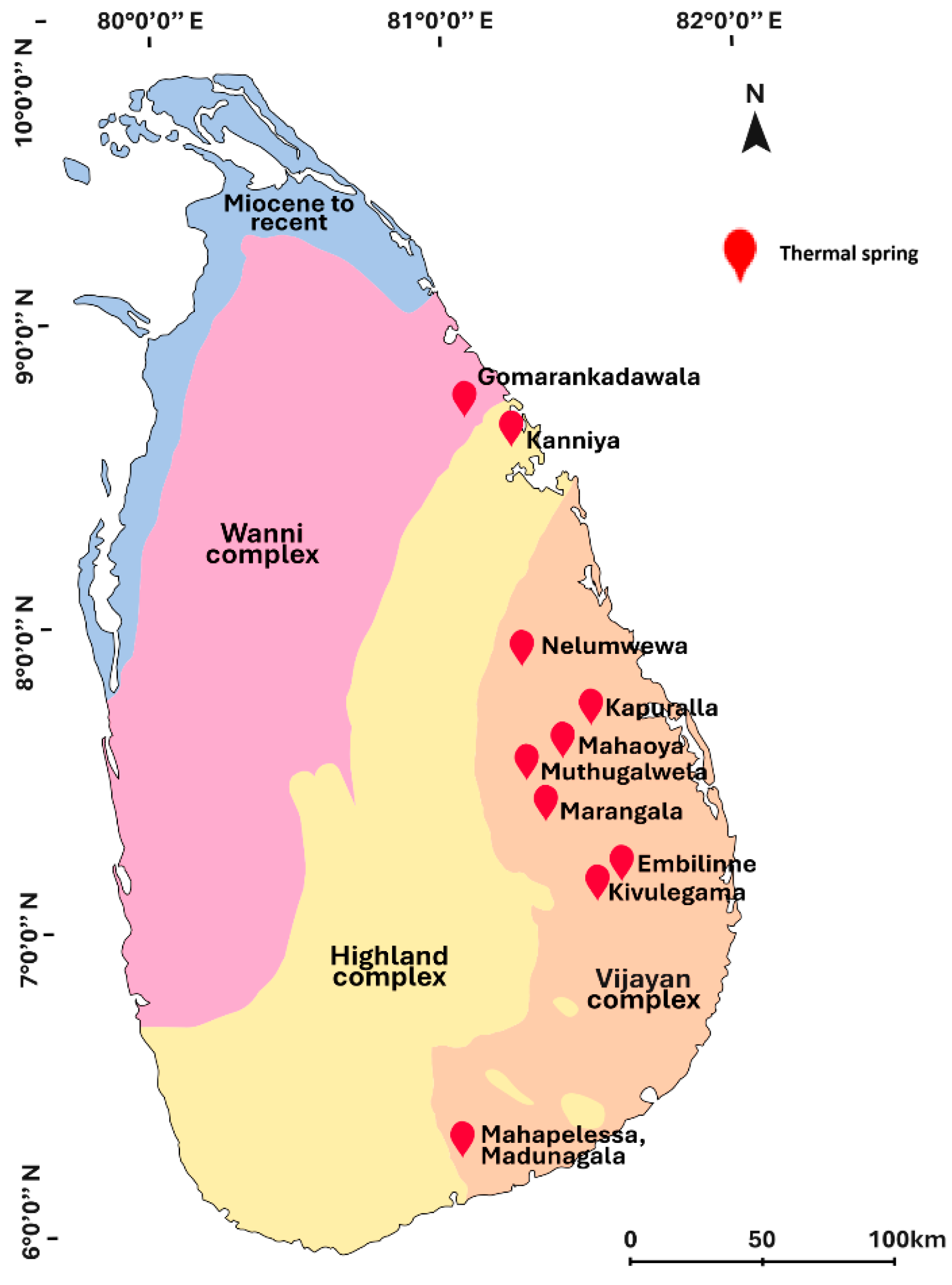
2.1. Water Sample Collection

Water samples were collected into sterile vacuum flasks from Nelumwewa geothermal spring (7°53′28.0″ N 81°11′58.2″ E) in Polonnaruwa district of Sri Lanka (Figure 1). Water temperature and pH of the hot water spring were recorded. The water samples were immediately transferred to the laboratory for thermophilic bacterial isolation.

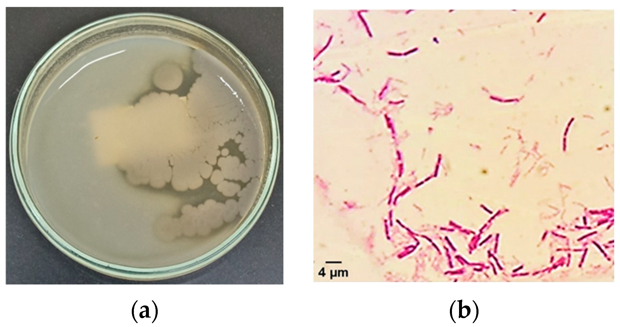
2.2. Isolation of Thermophilic Bacteria from Water Samples

An aliquot of 2 mL from each water sample was inoculated separately into 100 mL of nutrient broth (NB) (Himedia, Thane, India) and incubated overnight at 55 °C with shaking at 150 rpm for enrichment. Then, the incubated samples were serially diluted, and 100 µL from each sample was spread on individual nutrient agar plates. The bacterial colonies with different morphology were carefully selected, and pure cultures were isolated on nutrient agar plates, and incubated at 55 °C overnight. The pure cultures were prepared by streaking and subculturing and maintained on nutrient agar plates at 4 °C. For long-term storage, 20% glycerol stocks were prepared from each pure culture and stored at −80 °C. Gram’s staining was conducted to differentiate Gram-positive and Gram-negative bacterial isolates.

2.3. Catalase Test

To identify the catalase-producing bacteria, a small amount of each bacterial isolate was placed on individual clean glass slides. A drop of 3% hydrogen peroxide solution was added to each bacterial sample, and formation of bubbles was observed. The bacterial isolates that produced bubbles were identified to be catalase-producing bacteria [27].

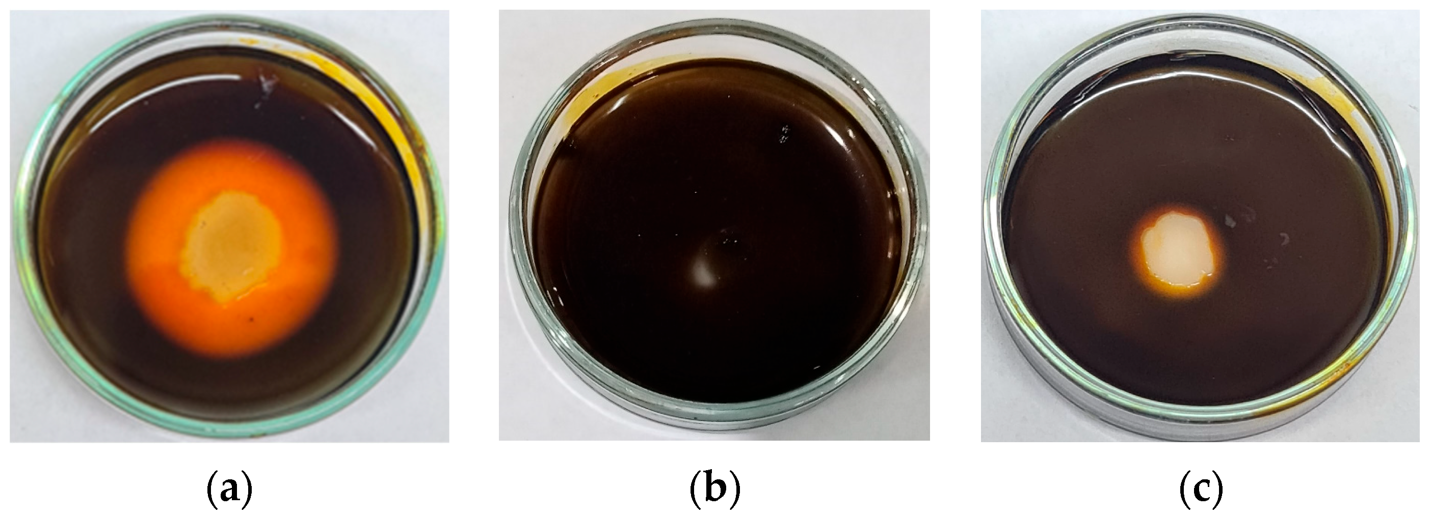
2.4. Screening of Thermophilic Bacterial Isolates for Extracellular Amylase Production

Extracellular amylase-producing bacterial isolates were screened by growing pure isolates on starch agar plates (peptone 5 g/L, NaCl 5 g/L, yeast extract 1.5 g/L, beef extract 1.5 g/L, soluble starch 2 g/L, agar 15 g/L, pH 7.5) (Himedia, India) at 55 °C overnight. Then, the plates were flooded with iodine solution (2% I2 and 0.2% KI) (Himedia, India) for 5 min, and iodine solution was decanted. The isolates that gave a clear zone around the colonies were selected and sub-cultured to maintain pure cultures, and they were identified as extracellular amylase-producing thermophilic bacteria [28].

2.5. Screening of Thermophilic Bacterial Isolates for Extracellular Protease Production

All the bacterial isolates were screened for extracellular protease by growing pure isolates on skim milk agar plates (5.0 g/L peptone, 3.0 g/L yeast extract, 1.0 g/L skim milk powder, pH 7.5) (Himedia, India) at 55 °C overnight. The thermophilic bacterial isolates that produced extracellular protease were identified by observing the clear zone around the bacterial colonies due to the proteolytic activity.

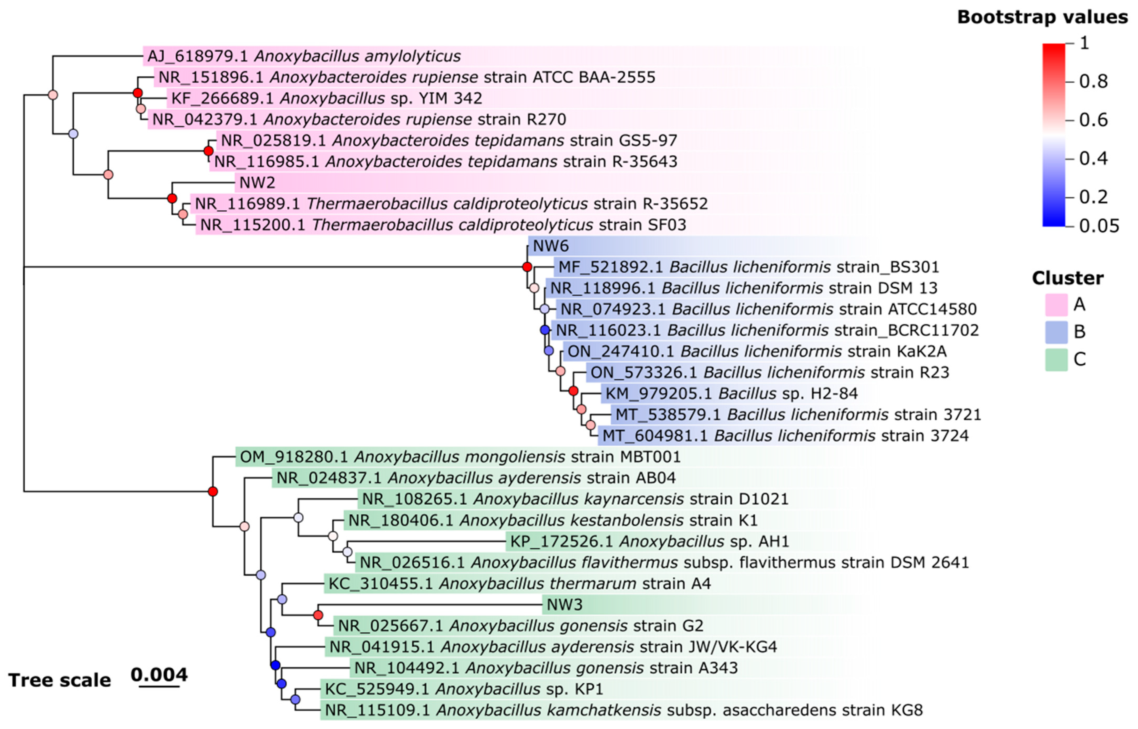
2.6. Molecular Characterization of Isolated Thermophilic Bacteria by 16S rRNA Gene Sequencing and Phylogenetic Analysis

For 16S rRNA gene bidirectional sequencing with the 27F (5′-AGTTTGATCMTGGCTCAG-3′) and 1492R (5′-TACGGCTACCTTGTTACGACTT-3′) universal primer pair, all the six pure isolates were sent to Macrogen, Inc., Seoul, Republic of Korea through Genetech Sri Lanka Pvt. Ltd., Colombo, Sri Lanka. The consensus sequences were generated using BioEdit version 7. To identify the thermophilic bacterial isolates, all the sequences were blasted against NCBI rRNA/ITS databases available online: https://blast.ncbi.nlm.nih.gov/Blast.cgi (accessed on 5 May 2025). The partial 16S rRNA gene sequences of isolated thermophilic bacteria were deposited in NCBI GenBank under the accession numbers PV489066, PV489057, and PV489038. Phylogenetic analysis was conducted using MEGA software version 12 [29]. The phylogenetic tree was constructed using the neighbor-joining method with bootstrap value of 1000, and the tree was visualized with modifications using tvBOT [30].

2.7. Determination of the Optimum Growth Temperature for the NW2 Isolate

The NW2 isolate was cultured in 20 mL of NB (beef extract 3 g/L, peptone 5 g/L, and NaCl 5 g/L, pH 7.5) at 55 °C by shaking at 150 rpm in a shaking incubator (Lab Companion, JEIO TECH, Daejeon, Republic of Korea) until the OD600 value reached 0.5. Then, 1 mL of culture was inoculated into 60 mL of NB and incubated at different temperatures (40 °C, 45 °C, 50 °C, 55 °C, 60 °C, and 65 °C) by shaking at 150 rpm. At one-hour intervals, OD600 values of each culture were measured using a spectrophotometer (UV-2600i Plus/2700i Plus, SHIMADZU, Kyoto, Japan), and growth curves were plotted. The exponential phase of each growth curve was selected, and corresponding OD600 values were converted to natural log values, and growth rates were determined by linear regression analysis using Microsoft Excel® version 2019. Each experiment was performed in triplicate.

2.8. Isolation of Crude Extracellular Amylase Enzyme from the NW2 Isolate

The crude extracellular amylase enzyme was produced and isolated from the overnight culture of NW2 strain incubated at 55 °C in 50 mL of NB (peptone 5 g/L, NaCl 5 g/L, yeast extract 1.5 g/L, beef extract 1.5 g/L, soluble starch 2 g/L, pH 7.5), with shaking at 150 rpm. Then, the culture was centrifuged at 10,000 rpm for 10 min at 4 °C using a centrifuge (Hettich, Tuttlingen, Germany). The supernatant was filtered through a bacterial filter (0.22 μm) and stored at 4 °C for conducting amylase activity assay using 3,5-Dinitrosalicylic acid (DNSA) (Sigma Aldrich, Hamburg, Germany) colorimetric assay [31].

2.9. Extracellular Amylase Activity Analysis by DNSA Assay

The extracellular amylase activity was analyzed using DNSA colorimetric assay as described previously [23,31] with slight modifications. Briefly, 0.1 mL of crude enzyme was mixed with 0.9 mL of 1% (w/v) soluble starch solution dissolved in 0.05 M phosphate buffer (pH 8) (Himedia, India) and incubated at 60 °C for 20 min. Then, 1 mL of DNSA reagent was added, and the mixture was heated in a boiling water bath for 5 min. After cooling the reaction mixture at room temperature, 8 mL of distilled water was added to the mixture, and absorbance was measured at 540 nm using a spectrophotometer. A solution without crude enzyme was used as the control. The amount of maltose (µmol) produced was determined from a maltose standard curve. All experiments were conducted in triplicate, and results are expressed as mean ± SD. One unit (U) of crude amylase activity was defined as the amount of crude enzyme that liberates 1 µmol of reducing sugars (as maltose) per minute under assay conditions, and crude extracellular amylase enzyme activity was expressed as U/mL. The crude extracellular amylase activity was calculated using the following formula [32]:
Amylase   activity   U / m L = Maltose   amount   µ m o l Reaction   time   ( min )   ×   Volume   of   crude   enzyme   mL

2.10. Determination of the Optimum Temperature for the Extracellular Amylase Activity

The optimum temperature for crude extracellular amylase activity was determined by reacting 0.1 mL of crude enzyme with 0.9 mL of 1% soluble starch solution dissolved in 0.05 M phosphate buffer (pH 7.5) for 20 min at different temperatures (40 °C, 45 °C, 50 °C, 55 °C, 60 °C, and 65 °C). Then, the activity was analyzed by DNSA assay as described above. All experiments were conducted in triplicate, and the results are expressed as mean ± SD.

2.11. Determination of Optimum pH for the Extracellular Amylase Activity

For determining optimum pH for crude extracellular amylase activity, 0.05 M sodium phosphate buffer (pH 5–8) and 0.05 M Tris-HCl buffer (pH 7–9) were used. An amount of 0.1 mL of crude enzyme was mixed with 0.9 mL of 1% soluble starch solution dissolved in different buffer solutions for 20 min at 55 °C. The crude enzyme activity of each reaction was analyzed by DNSA assay. All experiments were conducted in triplicate and the results are expressed as mean ± SD.

2.12. Determination of the Effects of Different Metal Ions on the Extracellular Amylase Activity

For determining the effects of various metal ions on the extracellular amylase activity, CaCl2, MgCl2, MnCl2, CuCl2, FeCl2, and HgCl2 (Sigma Aldrich, Hamburg, Germany) were each individually mixed with crude extracellular amylase enzyme solution at a final concentration of 0.0015 M and incubated at 60 °C for 30 min. The extracellular amylase activity was then measured by DNSA assay as described previously in Section 2.9. All experiments were conducted in triplicate. The crude amylase activity in the absence of metal ions was used as the control and considered 100% activity.

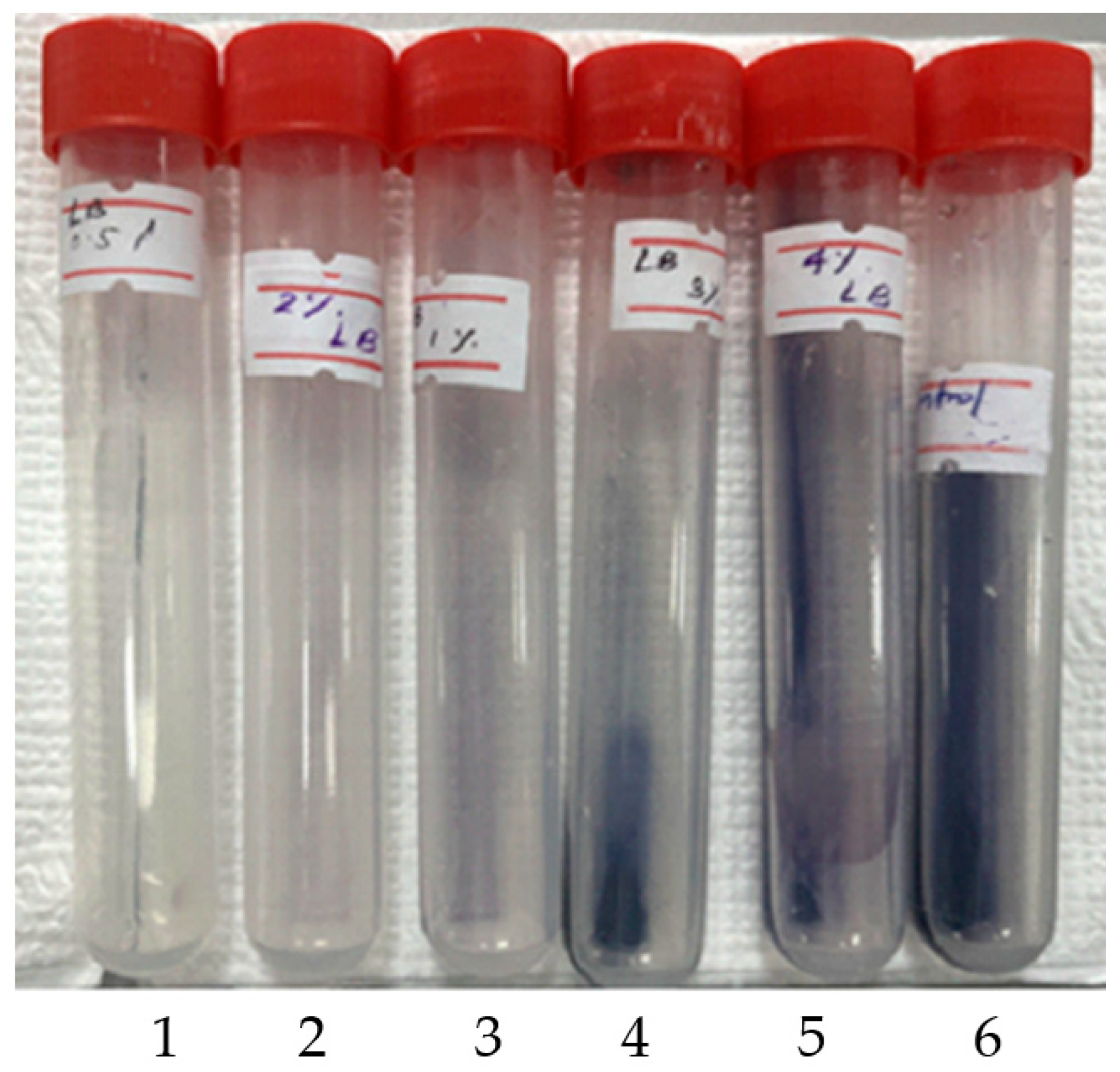
2.13. Detection of Raw Cassava Starch Hydrolysis by Crude Extracellular Amylase of NW2 Isolate Using the Iodine–Starch Test

Cassava starch was purchased from a local market. Different concentrations of cassava starch (0.5%, 1%, 2%, 3%, and 4%,) suspension were prepared by dissolving it in 0.05 M sodium phosphate buffer (pH 8.0). An amount of 0.1 mL of crude enzyme was mixed with 0.9 mL of each different cassava starch suspension separately and incubated for 4 h at 60 °C. Then, 0.1 mL of iodine solution (2% I2 and 0.2% KI) was added into each reaction mixture and mixed well to observe color differences. A control reaction prepared using 1% cassava raw starch solution without the addition of crude enzyme was included for comparison.

3. Results

3.1. Isolation and Identification of Thermophilic Bacterial Isolates That Produce Extracellular Amylase Enzyme

In this study, we isolated three thermophilic bacterial strains (NW2, NW3, and NW6) from the Nelumwewa geothermal spring in Sri Lanka. The thermal spring exhibited an average pH of 7.5, indicating slightly alkaline conditions, while average water temperature ranged from 48 °C to 55 °C across different sampling points. All isolates showed robust growth at 55 °C in NB as well as on nutrient agar plates at pH 7.5. Among the three bacterial isolates cultured on starch agar plates, only NW2 exhibited prominent clear zones around its colonies (Figure 2), with an average diameter of 25 mm, indicating a high potential for extracellular amylase production. In contrast, isolates NW3 and NW6 demonstrated only marginal amylase activity, with very narrow clear zones around their colonies (Figure 2). Therefore, due to the pronounced extracellular amylase activity of NW2 isolate, it was selected for further and detailed characterization of extracellular amylase enzyme activity. Moreover, NW2 isolate indicated extracellular protease activity as well (Figure 3). Light microscopy revealed that the NW2 isolate is a Gram-positive, rod-shaped bacterium (Figure 3). A comparison of morphological and biochemical characteristics of NW2 isolate with related bacterial species is summarized in the Table 1.

3.2. Molecular Characterization of Thermophilic Bacterial Isolates

Based on 16S rRNA gene sequence analysis of the thermophilic bacterial isolates, isolate NW2 exhibited 99.3% identity to Thermaerobacillus caldiproteolyticus strain SF03 (NR_115200.1), whereas NW6 isolate showed 99.71% identity to Bacillus licheniformis strain ATCC 14,580 (NR_074923.1). Moreover, NW3 showed 96.85% identity to Anoxybacillus gonensis strain G2 (NR_025667.1) (Table 2).
Phylogenetic analysis of the thermophilic bacterial isolates revealed that NW2, NW6, and NW3 are clustered into three separate groups A, B, and C, respectively (Figure 4). The NW2 isolate is grouped into cluster A, showing higher similarity to T. caldiproteolyticus species. Cluster B mainly consists of B. licheniformis strains, and the NW6 isolate is grouped into cluster B, exhibiting a close phylogenetic relationship with B. licheniformis. Moreover, the NW3 isolate is grouped into cluster C, with a higher similarity to A. gonensis.

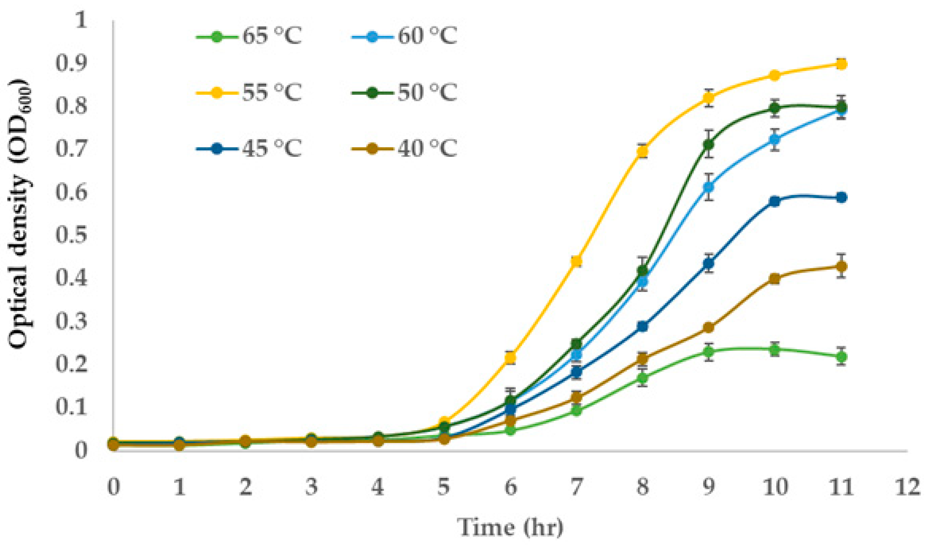
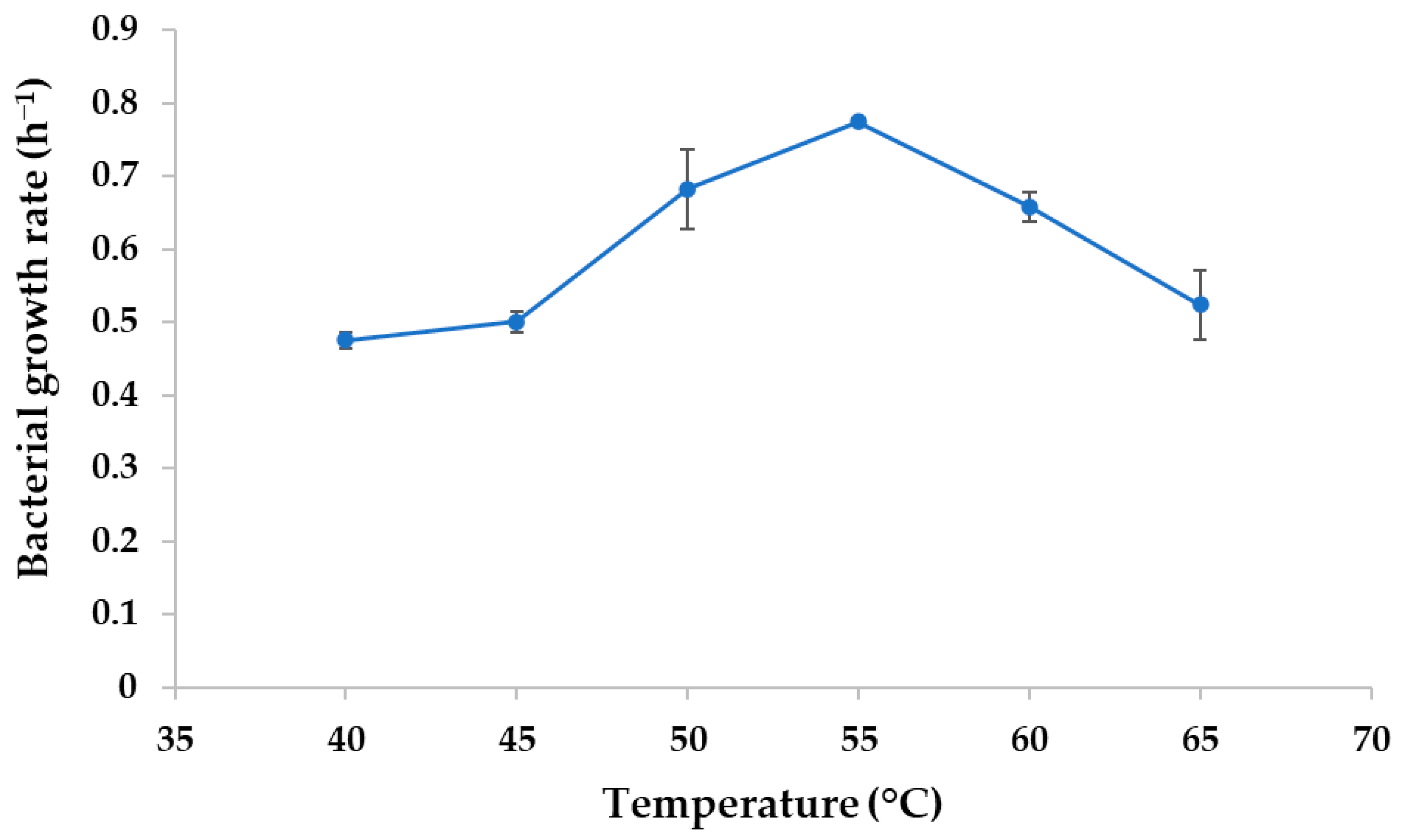
3.3. Optimum Growth Temperature of the NW2 Isolate

The NW2 isolate exhibited optimal growth characteristics at 55 °C (Figure 5). The lag phase of the growth curve lasted for about 5 h and then reached the exponential phase. The bacterial growth rates at exponential phases were determined by linear regression analysis (R2 > 0.95). The bacterial growth reached the stationary phase after about 11 h, indicating rapid growth characteristics at its optimum temperature. The NW2 isolate exhibited suboptimal growth at all other temperatures tested (Figure 5). Moreover, the maximum growth rate of the NW2 isolate was exhibited at 55 °C (Figure 6). These results indicate that the NW2 isolate is a moderate thermophile.

3.4. Characterization of Crude Extracellular Amylase Produced by NW2 Isolate

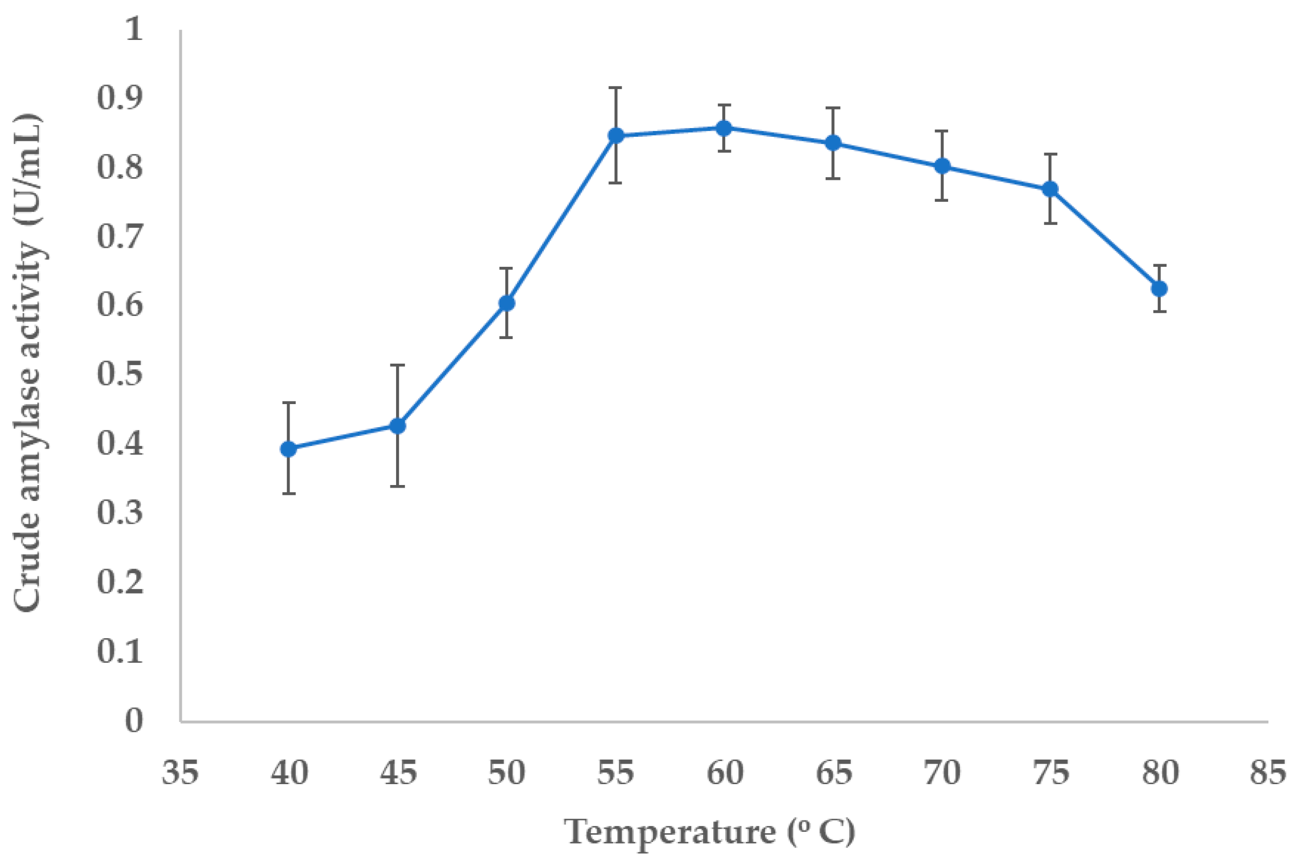
3.4.1. Determination of Optimal Temperature for the Enzyme Activity

To determine the optimal temperature of extracellular amylase produced by NW2 isolate, activity of crude amylase was analyzed by DNSA assay at different temperatures ranging from 40 °C to 80 °C. According to the results, the optimum temperature was determined to be 60 °C. However, the enzyme exhibited substantial activity up to 75 °C, which declined drastically thereafter (Figure 7). These results indicate that the extracellular amylase produced by NW2 isolates is thermostable in a wide range of temperatures from 55 °C to 75 °C.

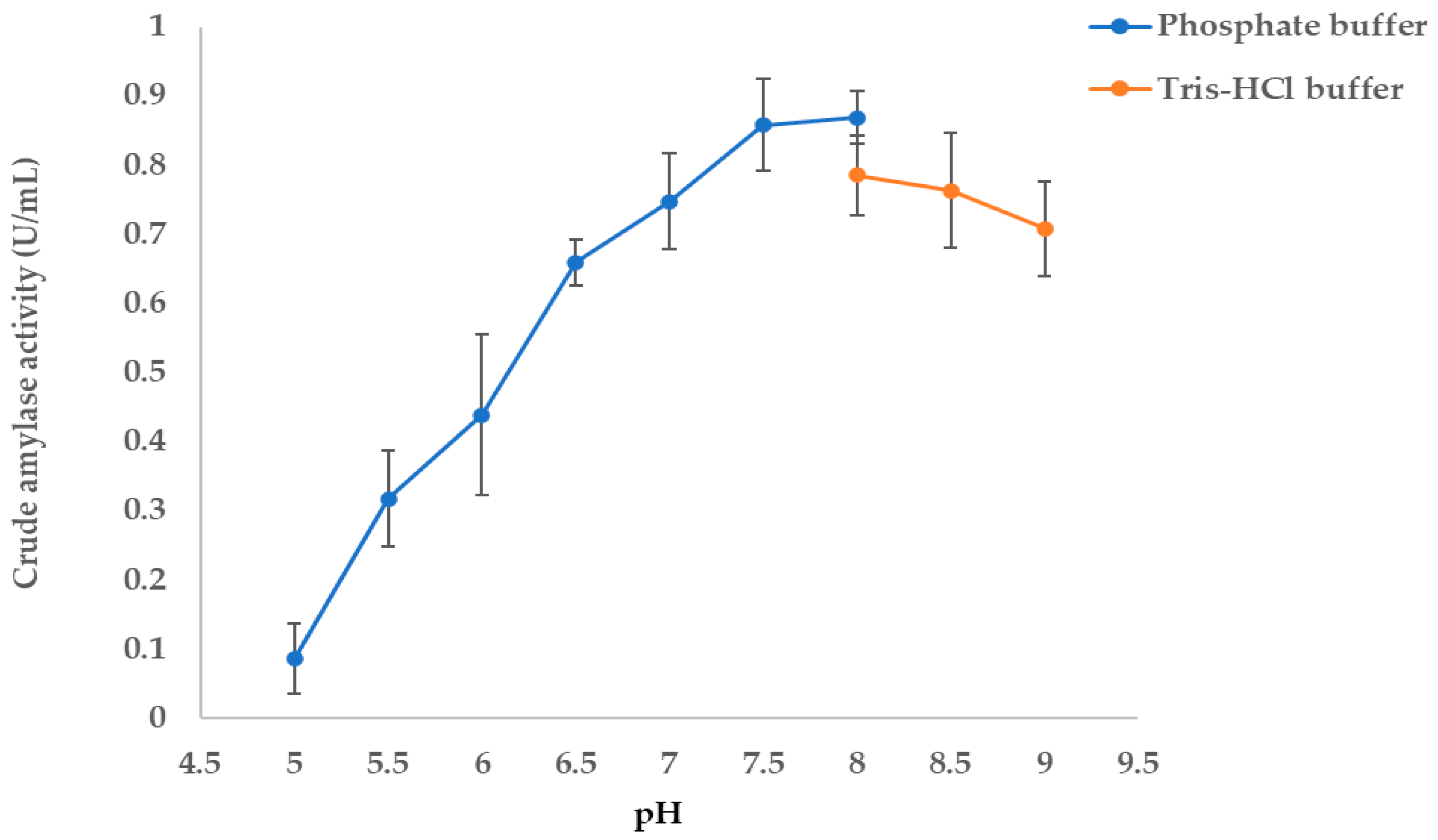
3.4.2. Determination of Optimal pH Enzyme Activity

Moreover, the amylase enzyme exhibited optimal activity at pH 8.0 in both sodium phosphate and tris-HCl buffer systems (Figure 8). Furthermore, it remains substantially active within a pH range of 7.5 to 8.5. These findings suggest that the extracellular amylase functions effectively in slightly alkaline conditions.

3.5. Extracellular Crude Amylase Activity of NW2 Isolate Under Optimum Temperature and pH

The crude extracellular amylase was harvested from the overnight culture of NM2 isolate cultured in NB (60 °C and pH 8). By DNSA assay, the crude extracellular amylase activity was quantified revealing a mean activity of 0.85 U/mL under the optimal assay conditions (60 °C and pH 8).

3.6. Effects of Different Metal Ions on the Extracellular Amylase Activity of NW2 Isolate

The effects of various metal ions on the activity of crude extracellular amylase were analyzed and expressed as a percentage relative to the control activity. Crude amylase activity was significantly enhanced by Ca2+ and Mn2+ ions. Conversely, Mg2+, Cu2+, and Fe2+ ions slightly inhibited the enzyme activity. Moreover, the Hg2+ ion strongly inhibited the activity of crude amylase (Table 3).

3.7. Hydrolysis of Raw Cassava Starch by NW2 Crude Extracellular Amylase

Under the optimum conditions, (pH 8.0 and temperature 60 °C), the crude amylase isolated from the NW2 isolate exhibited hydrolyzing of raw cassava starch (Figure 9), indicating its potential commercial application in the food, textile, and detergent industries.

4. Discussion

Extremophiles are increasingly recognized as valuable sources of industrially relevant enzymes due to their ability to produce extremozymes that remain active under extreme environmental conditions. Many thermally adapted enzymes or thermozymes, such as thermostable amylases, proteases, cellulases, and lipases, used in diverse industrial applications have been extensively derived from thermophilic and other extremophilic microorganisms [6]. Owing to their remarkable resilience and efficiency, extremozymes have enormous economic potential in the biotechnology sector. Among the microbial extremozymes, thermally stable amylases are of significant interest in all starch-related industries [6,37], while these enzymes account for nearly 25% of the world enzyme market [9]. As a result, thermozymes have received greater attention for their suitability in high-temperature industrial processes, where conventional enzymes often fail to perform optimally [19].
In the present study, three thermophilic bacterial strains were successfully isolated and molecularly characterized from the Nelumwewa geothermal spring in Sri Lanka. These isolates exhibited robust growth at 55 °C under slightly alkaline conditions (pH 7.5). However, among them, only NW2 isolate exhibited prominent extracellular amylase activity as well as extracellular protease activity, indicating it to be a promising candidate to produce thermostable industrial enzymes. Molecular characterization of NW2 isolate revealed that it shared 99.3% 16S rRNA gene sequence identity with Thermaerobacillus caldiproteolyticus strain SF03, previously classified as Anoxybacillus caldiproteolyticus or Geobacillus caldoproteolyticus [38,39], a well-documented thermophilic bacterium known for its thermostable enzyme production [40,41]. Phylogenetic analysis grouped the three isolates into three distinct clusters. The NW2 isolate clustered with T. caldiproteolyticus strains (Cluster A), indicating a close evolutionary relationship, while NW6 and NW3 were grouped into Clusters B and C, respectively, showing phylogenetic affiliation with Bacillus licheniformis and A. gonensis. A recent study on bacterial diversity analysis of Mahapelessa geothermal spring in Sri Lanka has identified some bacterial species such as Pannonibactor spp., Gulbenkiania spp., Pigmentiphaga spp., Enterobacter spp., Klebsiella spp., and Bacillus spp. [25]. However, it did not isolate any Thermaerobacillus spp., Anoxybacillus spp., or B. licheniformis, underscoring the wide bacterial diversity across different geothermal springs in Sri Lanka.
Under optimal growth conditions (55 °C and pH 7.5), NW2 isolate reached the stationary phase after about 10 h in NB medium, indicating its rapid growth characteristics. Some reports have suggested that bacterial amylase production is growth dependent [42,43]. Moreover, NW2 extracellular amylase exhibited optimal activity at 60 °C and pH 8.0 which are higher than those of optimal bacterial growth. This is a common trait found in some thermostable enzymes due to their structural rigidity [44], suggesting evolutionary adaptation to the seasonal fluctuation of temperature and pH of geothermal springs. The optimal temperature and pH of the extracellular amylase activity of NW2 isolate are almost consistent with already reported amylase activity of some Anoxybacillus spp. [22,23]. Under these optimal conditions for the enzyme activity, the crude extracellular amylase unit activity of NW2 was found to be 0.85 U/mL. The unit activity of this enzyme is somewhat consistent with previous reports on crude amylase activity from thermophilic Anoxybacillus species [23].
Numerous studies have demonstrated that extracellular amylases isolated from various bacterial species possess significant potential for industrial-scale applications. Amylases derived from the thermophilic bacterium B. licheniformis exhibit excellent amylolytic properties and have been successfully applied in the biodegradation of food waste [45]. Moreover, amylases from different bacterial sources display remarkably high thermal optima, including 135 °C for B. subtilis [46] and 85 °C for Rhodothermus marinus [47]. Other thermophilic bacteria, such as Bacillus caldolyticus [48] and Geobacillus thermoleovorans [49], exhibit peak enzymatic activity at around 70 °C, highlighting the wide range of thermostability among bacterial amylases. The thermostability and broad pH tolerance of these enzymes reduce the need for cooling steps and pH adjustments, improving process efficiency and cost-effectiveness.
The bacterial species T. caldiproteolyticus (formerly A. caldiproteolyticus) is already known for its production of extracellular proteases rather than amylase [40]. To date, only a single study has reported the production of extracellular amylase by this bacterial species [50]. However, that study lacks comprehensive details regarding the activity of the extracellular amylase produced by A. caldiproteolyticus. Furthermore, the two known bacterial strains most closely related to the NW2 isolate have not provided detailed information regarding their amylase activities [40,50], suggesting the need for further studies to investigate the potential of producing extracellular amylase from this species.
The presence of different metal ions in the enzyme reaction mixture can significantly affect the activity and stability of thermostable amylases [51]. The crude amylase activity of the NW2 isolate was significantly enhanced by Ca2+ and Mn2+ ions. The Ca2+ ion has been reported to play a vital role in enhancing the activity of most of the thermostable amylases by maintaining the structural integrity and catalytic efficiency at higher temperatures [52]. However, it has been reported that the activity of some amylases is independent of Ca2+ [14]. Moreover, the Hg2+ ion strongly inhibited the activity of crude amylase of NW2. This may be due to binding to the active site of the enzyme or altering the correct three-dimensional conformation of the enzyme.
Cassava starch is extensively utilized across diverse industries, including food processing, textiles, and bioethanol production. However, its hydrolysis is often associated with high operational costs, primarily due to the energy-intensive nature of conventional processing methods. Recent studies have demonstrated the successful application of microbial amylases in the production of bioethanol from raw cassava starch [53]. In this context, the extracellular amylase produced by the NW2 isolate in the present study exhibited strong hydrolytic activity toward raw cassava starch, highlighting its potential for use in both the food and biofuel industries, where efficient and rapid starch degradation is essential. Furthermore, the enzyme’s stability under moderately alkaline conditions enhances its applicability in the detergent industry, where alkaline-tolerant amylases are required.
To the best of our knowledge, this is the first report of a T. caldiproteolyticus-like strain from Sri Lankan geothermal springs exhibiting a remarkable extracellular amylase activity. The NW2 isolate is a Gram-positive, rod-shaped, and catalase-positive bacterium. These morphological and biochemical characteristics are commonly associated with well-known amylase-producing bacteria [54]. In contrast, the NW6 isolate exhibited a very close phylogenetic relationship with 99.71% 16S rRNA gene sequence identity with B. licheniformis, a well-established industrial bacterial species recognized for its thermostable amylase production [55,56,57,58]. However, NW6 did not exhibit significant extracellular amylase activity under the tested conditions, suggesting potential strain-level variability or previously undocumented functional diversity within the species.
Overall, the findings of the present study not only reveal the biotechnological potential of the bacteria isolates from the Nelumwewa geothermal spring but also highlight the geothermal springs of Sri Lanka as a promising niche environment for exploring thermophilic enzyme-producing microbes.

5. Conclusions

This study highlights the biotechnological potential of NW2, a T. caldiproteolyticus-like strain from the Nelumwewa geothermal spring in Sri Lanka, a previously unexplored thermal ecosystem. The NW2 isolate emerged as a potential candidate for industrial applications because of its prominent extracellular amylase production with notable thermostability and activity in slightly alkaline environments. Furthermore, the extracellular amylase of NW2 demonstrated effective hydrolysis of raw cassava starch, suggesting its direct applicability in food, bioethanol, and detergent industries where robust and efficient enzymatic performance under high temperature and pH is critical. This marks the first report of a T. caldiproteolyticus-like strain from Sri Lanka with such enzymatic activity, expanding the known functional diversity of this species and underscoring the potential for strain-level variation. Moreover, this is the first detailed report characterizing the extracellular amylase from T. caldiproteolyticus. The findings of the present study reinforce the value of geothermal environments as reservoirs of novel extremozymes, suggesting further exploration to support the development of sustainable, high-performance biocatalysts for industrial applications.

Author Contributions

Conceptualization, S.B., P.J. and A.W.; methodology, S.B., L.P., B.D., P.J. and A.W.; validation, L.P. and S.B.; formal analysis, L.P., B.D. and S.B.; investigation, S.B.; data curation, L.P., B.D. and S.B.; writing—original draft preparation, B.D. and L.P.; writing—review and editing, S.B. and A.W.; supervision, S.B. and A.W.; project administration, S.B.; funding acquisition, S.B. All authors have read and agreed to the published version of the manuscript.

Funding

This research was funded by the Science and Technology Human Resource Development (STHRD) Project, Ministry of Education, Sri Lanka, under the patronage of Asian Development Bank (ADB), Grant No. CRG/R3/SB1.

Institutional Review Board Statement

Not applicable.

Informed Consent Statement

Not applicable.

Data Availability Statement

The data presented in this study are openly available in NCBI gene bank (https://www.ncbi.nlm.nih.gov/genbank/) [PV489066, PV489057, PV489038].

Conflicts of Interest

The authors declare no conflicts of interest.

Abbreviations

The following abbreviations are used in this manuscript:
NBNutrient broth
DNSA3,5-Dinitrosalicylic acid

References

  1. Raddadi, N.; Cherif, A.; Daffonchio, D.; Neifar, M.; Fava, F. Biotechnological Applications of Extremophiles, Extremozymes and Extremolytes. Appl. Microbiol. Biotechnol. 2015, 99, 7907–7913. [Google Scholar] [CrossRef] [PubMed]
  2. Cabrera, M.Á.; Blamey, J.M. Biotechnological Applications of Archaeal Enzymes from Extreme Environments. Biol. Res. 2018, 51, 37. [Google Scholar] [CrossRef] [PubMed]
  3. Marzban, G.; Tesei, D. The Extremophiles: Adaptation Mechanisms and Biotechnological Applications. Biology 2025, 14, 412. [Google Scholar] [CrossRef] [PubMed]
  4. Elleuche, S.; Schröder, C.; Sahm, K.; Antranikian, G. Extremozymes-Biocatalysts with Unique Properties from Extremophilic Microorganisms. Curr. Opin. Biotechnol. 2014, 29, 116–123. [Google Scholar] [CrossRef]
  5. Cavicchioli, R.; Siddiqui, K.S.; Andrews, D.; Sowers, K.R. Low-Temperature Extremophiles and Their Applications. Curr. Opin. Biotechnol. 2002, 13, 253–261. [Google Scholar] [CrossRef]
  6. Ashaolu, T.J.; Malik, T.; Soni, R.; Prieto, M.A.; Jafari, S.M. Extremophilic Microorganisms as a Source of Emerging Enzymes for the Food Industry: A Review. Food Sci. Nutr. 2025, 13, e4540. [Google Scholar] [CrossRef]
  7. Al-Batayneh, K.M.; Jacob, J.H.; Hussein, E. Isolation and Molecular Identification of New Thermophilic Bacterial Strains of Geobacillus pallidus and Anoxybacillus flavithermus. Int. J. Integr. Biol. 2011, 11, 39–43. [Google Scholar]
  8. Wang, Q.; Cen, Z.; Zhao, J. The Survival Mechanisms of Thermophiles at High Temperatures: An Angle of Omics. Physiology 2015, 30, 97–106. [Google Scholar] [CrossRef]
  9. Kikani, B.A.; Singh, S.P. Amylases from Thermophilic Bacteria: Structure and Function Relationship. Crit. Rev. Biotechnol. 2022, 42, 325–341. [Google Scholar] [CrossRef]
  10. Arbab, S.; Ullah, H.; Khan, M.I.U.; Khattak, M.N.K.; Zhang, J.; Li, K.; Hassan, I.U. Diversity and Distribution of Thermophilic Microorganisms and Their Applications in Biotechnology. J. Basic. Microbiol. 2022, 62, 95–108. [Google Scholar] [CrossRef]
  11. Ali, Z.; Abdullah, M.; Yasin, M.T.; Amanat, K.; Sultan, M.; Rahim, A.; Sarwar, F. Recent Trends in Production and Potential Applications of Microbial Amylases: A Comprehensive Review. Protein Expr. Purif. 2025, 227, 106640. [Google Scholar] [CrossRef] [PubMed]
  12. Prakash, O.; Jaiswal, N. α-Amylase: An Ideal Representative of Thermostable Enzymes. Appl. Biochem. Biotechnol. 2010, 160, 2401–2414. [Google Scholar] [CrossRef] [PubMed]
  13. Slavić, M.Š.; Kojić, M.; Margetić, A.; Stanisavljević, N.; Gardijan, L.; Božić, N.; Vujčić, Z. Highly Stable and Versatile α-Amylase from Anoxybacillus Vranjensis ST4 Suitable for Various Applications. Int. J. Biol. Macromol. 2023, 249, 126055. [Google Scholar] [CrossRef] [PubMed]
  14. Kikani, B.A.; Singh, S.P. The Stability and Thermodynamic Parameters of a Very Thermostable and Calcium-Independent α-Amylase from a Newly Isolated Bacterium, Anoxybacillus Beppuensis TSSC-1. Process Biochem. 2012, 47, 1791–1798. [Google Scholar] [CrossRef]
  15. Agüloǧlu Fincan, S.; Enez, B.; Özdemir, S.; Matpan Bekler, F. Purification and Characterization of Thermostable α-Amylase from Thermophilic Anoxybacillus flavithermus. Carbohydr. Polym. 2014, 102, 144–150. [Google Scholar] [CrossRef]
  16. Ammar, Y.B.; Matsubara, T.; Ito, K.; Iizuka, M.; Limpaseni, T.; Pongsawasdi, P.; Minamiura, N. New Action Pattern of a Maltose-Forming α-Amylase from Streptomyces sp. and Its Possible Application in Bakery. BMB Rep. 2002, 35, 568–575. [Google Scholar] [CrossRef]
  17. Yang, C.H.; Liu, W.H. Purification and Properties of a Maltotriose-Producing α-Amylase from Thermobifida Fusca. Enzym. Microb. Technol. 2004, 35, 254–260. [Google Scholar] [CrossRef]
  18. Burhan, A.; Nisa, U.; Gökhan, C.; Ömer, C.; Ashabil, A.; Osman, G. Enzymatic Properties of a Novel Thermostable, Thermophilic, Alkaline and Chelator Resistant Amylase from an Alkaliphilic bacillus sp. Isolate ANT-6. Process Biochem. 2003, 38, 1397–1403. [Google Scholar] [CrossRef]
  19. Ullah, I.; Khan, M.S.; Khan, S.S.; Ahmad, W.; Zheng, L.; Shah, S.U.A.; Ullah, M.; Iqbal, A. Identification and Characterization of Thermophilic Amylase Producing Bacterial Isolates from the Brick Kiln Soil. Saudi J. Biol. Sci. 2021, 28, 970–979. [Google Scholar] [CrossRef]
  20. Silva-Salinas, A.; Rodríguez-Delgado, M.; Gómez-Treviño, J.; López-Chuken, U.; Olvera-Carranza, C.; Blanco-Gámez, E.A. Novel Thermotolerant Amylase from Bacillus licheniformis Strain Lb04: Purification, Characterization and Agar-Agarose. Microorganisms 2021, 9, 1857. [Google Scholar] [CrossRef]
  21. Deljou, A.; Arezi, I. Production of Thermostable Extracellular α-Amylase by a Moderate Thermophilic Bacillus Licheniformis-AZ2 Isolated from Qinarje Hot Spring (Ardebil Prov. of Iran). Period. Biol. 2016, 118, 405–416. [Google Scholar] [CrossRef]
  22. Acer, Ö.; Pirinççioğlu, H.; Matpan Bekler, F.; Gül-Güven, R.; Güven, K. Anoxybacillus sp. AH1, an α-Amylase-Producing Thermophilic Bacterium Isolated from Dargeçit Hot Spring. Biologia 2015, 70, 853–862. [Google Scholar] [CrossRef]
  23. Sharif, S.; Shah, A.H.; Fariq, A.; Jannat, S.; Rasheed, S.; Yasmin, A. Optimization of Amylase Production Using Response Surface Methodology from Newly Isolated Thermophilic Bacteria. Heliyon 2023, 9, e12901. [Google Scholar] [CrossRef] [PubMed]
  24. Al-Harthy, O.D.; Hozzein, W.N.; Abdelkader, H.S.; Alharbi, S.A.A. Thermostable Amylase from Cytobacillus firmus: Characterization, Optimization, and Implications in Starch Hydrolysis. Egypt. J. Microbiol. 2024, 58, 35–47. [Google Scholar] [CrossRef]
  25. Samarasinghe, S.N.; Wanigatunge, R.P.; Magana-Arachchi, D.N. Bacterial Diversity in a Sri Lankan Geothermal Spring Assessed by Culture-Dependent and Culture-Independent Approaches. Curr. Microbiol. 2021, 78, 3439–3452. [Google Scholar] [CrossRef]
  26. Patabendigedara, S.; Kumara, S.M.P.G.S.; Dharmagunawardhane, H.A. Geostructural model for the Nelumwewa thermal spring: North central province, Sri Lanka. J. Geol. Soc. Sri Lanka 2014, 16, 19–27. [Google Scholar]
  27. Reiner, K. Catalase Test Protocol. References—Scientific Research Publishing. Available online: https://www.scirp.org/reference/referencespapers?referenceid=1367750 (accessed on 8 May 2025).
  28. Hols, P.; Ferain, T.; Garmyn, D.; Bernard, N.; Delcour, J. Use of Homologous Expression-Secretion Signals and Vector-Free Stable Chromosomal Integration in Engineering of Lactobacillus plantarum for Alpha-Amylase and Levanase Expression. Appl. Environ. Microbiol. 1994, 60, 1401. [Google Scholar] [CrossRef]
  29. Hall, B.G. Building Phylogenetic Trees from Molecular Data with MEGA. Mol. Biol. Evol. 2013, 30, 1229–1235. [Google Scholar] [CrossRef]
  30. Xie, J.; Chen, Y.; Cai, G.; Cai, R.; Hu, Z.; Wang, H. Tree Visualization by One Table (TvBOT): A Web Application for Visualizing, Modifying and Annotating Phylogenetic Trees. Nucleic Acids Res. 2023, 51, W587–W592. [Google Scholar] [CrossRef]
  31. Bernfeld, P. Amylases, α and β. Methods Enzym. 1955, 1, 149–158. [Google Scholar] [CrossRef]
  32. Soy, S.; Nigam, V.K.; Sharma, S.R. Enhanced Production and Biochemical Characterization of a Thermostable Amylase from Thermophilic Bacterium Geobacillus icigianus BITSNS038. J. Taibah Univ. Sci. 2021, 15, 730–745. [Google Scholar] [CrossRef]
  33. Matpan Bekler, F.; Güven, K. Isolation and Production of Thermostable α-Amylase from Thermophilic anoxybacillus sp. KP1 from Diyadin Hot Spring in Aǧri, Turkey. Biologia 2014, 69, 419–427. [Google Scholar] [CrossRef]
  34. Poli, A.; Esposito, E.; Lama, L.; Orlando, P.; Nicolaus, G.; de Appolonia, F.; Gambacorta, A.; Nicolaus, B. Anoxybacillus amylolyticus sp. Nov., a Thermophilic Amylase Producing Bacterium Isolated from Mount Rittmann (Antarctica). Syst. Appl. Microbiol. 2006, 29, 300–307. [Google Scholar] [CrossRef] [PubMed]
  35. Baltas, N.; Dincer, B.; Ekinci, A.P.; Kolayli, S.; Adiguzel, A. Purification and Characterization of Extracellular A-Amylase from a Thermophilic Anoxybacillus thermarum A4 Strain. Braz. Arch. Biol. Technol. 2016, 59, 1–14. [Google Scholar] [CrossRef]
  36. Zhang, F.; Yang, X.; Geng, L.; Zhang, Z.; Yin, Y.; Li, W. Purification and Characterization of a Novel and Versatile α-Amylase from Thermophilic anoxybacillus sp. YIM 342. Starch/Staerke 2016, 68, 446–453. [Google Scholar] [CrossRef]
  37. Yadav, A.N. Biodiversity and Bioprospecting of Extremophilic Microbiomes for Agro-Environmental Sustainability. J. Appl. Biol. Biotechnol. 2021, 9, 1–6. [Google Scholar]
  38. Patel, I.; Bello, S.; Gupta, R.S. Phylogenomic and Molecular Marker Based Studies to Clarify the Evolutionary Relationships amongst Anoxybacillus Species and Demarcation of the Family Anoxybacillaceae and Some of Its Constituent Genera. Int. J. Syst. Evol. Microbiol. 2024, 74, 006528. [Google Scholar] [CrossRef]
  39. Coorevits, A.; Dinsdale, A.E.; Halket, G.; Lebbe, L.; de Vos, P.; van Landschoot, A.; Logan, N.A. Taxonomic Revision of the Genus Geobacillus: Emendation of Geobacillus, G. Stearothermophilus, G. Jurassicus, G. Toebii, G. Thermodenitrificans and G. Thermoglucosidans (Nom. Corrig., Formerly ’Thermoglucosidasius’); Transfer of Bacillus thermantarcticus to the Genus as G. Thermantarcticus Comb. Nov.; Proposal of Caldibacillus debilis Gen. Nov., Comb. Nov. Int. J. Syst. Evol. Microbiol. 2012, 62, 1470–1485. [Google Scholar] [CrossRef]
  40. Cheng, J.H.; Wang, Y.; Zhang, X.Y.; Sun, M.L.; Zhang, X.; Song, X.Y.; Zhang, Y.Z.; Zhang, Y.; Chen, X.L. Characterization and Diversity Analysis of the Extracellular Proteases of Thermophilic anoxybacillus Caldiproteolyticus 1A02591 from Deep-Sea Hydrothermal Vent Sediment. Front. Microbiol. 2021, 12, 643508. [Google Scholar] [CrossRef]
  41. Chen, X.G.; Stabnikova, O.; Tay, J.H.; Wang, J.Y.; Tay, S.T.L. Thermoactive Extracellular Proteases of Geobacillus caldoproteolyticus, sp. Nov., from Sewage Sludge. Extremophiles 2004, 8, 489–498. [Google Scholar] [CrossRef]
  42. Simair, A.A.; Khushk, I.; Qureshi, A.S.; Bhutto, M.A.; Chaudhry, H.A.; Ansari, K.A.; Lu, C. Amylase Production from Thermophilic bacillus sp. BCC 021-50 Isolated from a Marine Environment. Fermentation 2017, 3, 25. [Google Scholar] [CrossRef]
  43. Hmidet, N.; El Hadj Ali, N.; Zouari-Fakhfakh, N.; Haddar, A.; Nasri, M.; Sellemi-Kamoun, A. Chicken Feathers: A Complex Substrate for the Co-Production of α-Amylase and Proteases by B. Licheniformis NH1. J. Ind. Microbiol. Biotechnol. 2010, 37, 983–990. [Google Scholar] [CrossRef] [PubMed]
  44. Hmidet, N.; Bayoudh, A.; Berrin, J.G.; Kanoun, S.; Juge, N.; Nasri, M. Purification and Biochemical Characterization of a Novel α-Amylase from Bacillus Licheniformis NH1. Cloning, Nucleotide Sequence and Expression of AmyN Gene in Escherichia Coli. Process Biochem. 2008, 43, 499–510. [Google Scholar] [CrossRef]
  45. Msarah, M.J.; Ibrahim, I.; Hamid, A.A.; Aqma, W.S. Optimisation and Production of Alpha Amylase from Thermophilic bacillus spp. and Its Application in Food Waste Biodegradation. Heliyon 2020, 6, e04183. [Google Scholar] [CrossRef]
  46. Konsoula, Z.; Liakopoulou-Kyriakides, M. Co-Production of α-Amylase and β-Galactosidase by Bacillus subtilis in Complex Organic Substrates. Bioresour. Technol. 2007, 98, 150–157. [Google Scholar] [CrossRef]
  47. Gomes, I.; Gomes, J.; Steiner, W. Highly Thermostable Amylase and Pullulanase of the Extreme Thermophilic Eubacterium Rhodothermus marinus: Production and Partial Characterization. Bioresour. Technol. 2003, 90, 207–214. [Google Scholar] [CrossRef]
  48. Schwab, K.; Bader, J.; Brokamp, C.; Popović, M.K.; Bajpai, R.; Berovič, M. Dual Feeding Strategy for the Production of α-Amylase by Bacillus caldolyticus Using Complex Media. N. Biotechnol. 2009, 26, 68–74. [Google Scholar] [CrossRef]
  49. Uma Maheswar Rao, J.L.; Satyanarayana, T. Improving Production of Hyperthermostable and High Maltose-Forming α-Amylase by an Extreme Thermophile Geobacillus thermoleovorans Using Response Surface Methodology and Its Applications. Bioresour. Technol. 2007, 98, 345–352. [Google Scholar] [CrossRef]
  50. Caliskan Ozdemir, S.; Coleri Cihan, A.; Kilic, T.; Cokmus, C. Optimization of thermostable alpha-amylase production from geobacillus sp. D413. J. Microbiol. Biotechnol. Food Sci. 2016, 6, 689–694. [Google Scholar] [CrossRef]
  51. Gupta, N.; Beliya, E.; Paul, J.S.; Tiwari, S.; Kunjam, S.; Jadhav, S.K. Molecular Strategies to Enhance Stability and Catalysis of Extremophile-Derived α-Amylase Using Computational Biology. Extremophiles 2021, 25, 221–233. [Google Scholar] [CrossRef]
  52. Yadav, J.K. A Differential Behavior of α-Amylase, in Terms of Catalytic Activity and Thermal Stability, in Response to Higher Concentration CaCl2. Int. J. Biol. Macromol. 2012, 51, 146–152. [Google Scholar] [CrossRef] [PubMed]
  53. Roble, N.D.; Ogbonna, J.; Tanaka, H. Simultaneous Amylase Production, Raw Cassava Starch Hydrolysis and Ethanol Production by Immobilized Aspergillus awamori and Saccharomyces cerevisiae in a Novel Alternating Liquid Phase–Air Phase System. Process Biochem. 2020, 95, 115–121. [Google Scholar] [CrossRef]
  54. Kim, S.H.; Kim, W.J.; Ryu, J.; Yerefu, Y.; Tesfaw, A. Amylase Production by the New Strains of Kocuria rosea and Micrococcus endophyticus Isolated from Soil in the Guassa Community Conservation Area. Fermentation 2025, 11, 211. [Google Scholar] [CrossRef]
  55. Aras, S.; Altıntas, R.; Aygun, E.; Goren, G.; Sisecioglu, M. Purification of α-Amylase from Thermophilic bacillus Licheniformis SO5 by Using a Novel Method, Alternating Current Magnetic-Field Assisted Three-Phase Partitioning: Molecular Docking and Bread Quality Improvement. Food Chem. 2025, 484, 144258. [Google Scholar] [CrossRef]
  56. Mahmoudnia, F. Comparison of the Synthesis of the Alpha-Amylase Enzyme by the Native Strain Bacillus licheniformis in Immobilized and Immersed Cells. Iran. J. Microbiol. 2024, 16, 827. [Google Scholar] [CrossRef]
  57. Suthar, S.; Joshi, D.; Patel, H.; Patel, D.; Kikani, B.A. Optimization and Purification of a Novel Calcium-Independent Thermostable, α-Amylase Produced by Bacillus Licheniformis UDS-5. World J. Microbiol. Biotechnol. 2024, 40, 385. [Google Scholar] [CrossRef]
  58. Divakaran, D.; Chandran, A.; Pratap Chandran, R. Comparative Study on Production of A-Amylase from Bacillus licheniformis Strains. Braz. J. Microbiol. 2011, 42, 1397–1404. [Google Scholar] [CrossRef]
Figure 1. Locations of geothermal springs across different geological zones of Sri Lanka [26].
Fermentation 11 00397 g001
Figure 2. Screening of extracellular amylase-producing thermophilic bacterial isolates. Extracellular amylase-producing isolates exhibit a clear zone around colonies. (a) NW2; (b) NW3; (c) NW6 isolates.
Fermentation 11 00397 g002
Figure 3. Characterization of NW2 isolate. (a) Protease activity observed on skim milk agar plate (clear zones indicate hydrolysis); (b) Gram-stained cells visualized under light microscopy (scale bar: 4 μm).
Fermentation 11 00397 g003
Figure 4. The phylogenetic analysis of thermophilic bacterial isolates using 16S rRNA gene sequences. The tree was constructed using the neighbor-joining method with MEGA 12 and visualized with modifications using tvBOT [30].
Fermentation 11 00397 g004
Figure 5. Growth curves of NW2 isolate at different temperatures from 40 °C to 65 °C in NB (pH 7.5). The graph shows data from triplicate experiments (mean ± SD).
Fermentation 11 00397 g005
Figure 6. Growth rate of NW2 isolate at different temperatures from 40 °C to 65 °C in NB (pH 7.5). The graph shows data from triplicate experiments (mean ± SD).
Fermentation 11 00397 g006
Figure 7. Optimum temperature for the extracellular amylase activity. The graph shows data from triplicate experiments (mean ± SD).
Fermentation 11 00397 g007
Figure 8. Optimum pH for the extracellular amylase activity. The graph shows data from triplicate experiments (mean ± SD).
Fermentation 11 00397 g008
Figure 9. Detection of raw cassava starch hydrolysis by crude extracellular amylase of NW2 isolate under optimum conditions (60 °C and pH 8.0) using the iodine–starch test. Test tubes 1 to 5 illustrate the hydrolysis of cassava starch at concentrations of 0.5% (Tube 1), 1% (Tube 2), 2% (Tube 3), 3% (Tube 4), and 4% (Tube 5), respectively, demonstrating the enzyme’s capacity to degrade starch substrates. Tube 6 serves as a control, showing the reaction with 1% cassava starch without the addition of crude amylase enzyme.
Fermentation 11 00397 g009
Table 1. Comparison of morphological and biochemical characteristics of the NW2 isolate with those of already isolated Anoxybacillus species. (+ and − signs indicate positive and negative biochemical tests results, respectively. NR—Not reported).
Bacterial CharacteristicsNW21 *2 *3 *4 *5 *6 *7 *
Optimal growth pH7.577.08.05.67.07.07.0
Optimal growth temperature (°C)6070605061556060–65
Amylase activity++++++++
Protease activity+NRNR+
Catalase activity+NR++NR++
Colony ColorOff-whiteGolden brownNRWhiteCreamNRNRYellow
Gram Stain++NR++NRNR+
Cell shapeRodRodRodRodRodRodRodRod
Culture mediumNutrient brothLuira–Bertani brothNutrient brothNutrient brothNutrient brothLuira–Bertani brothT5 mediumAnaerobic basal medium
Source of isolationThermal springsThermal springsThermal springsThermal springsThermal springsThermal springsThermal springsThermal springs
* 1—Anoxybacillus mongoliensis MBT001 [23]; 2—Anoxybacillus sp. AH1 [22]; 3—Anoxybacillus sp. KP1 [33]; 4—Anoxybacillus amylolyticus [34]; 5—Anoxybacillus thermarum A4 [35]; 6—Anoxybacillus sp. YIM 342 [36]; 7—Anoxybacillus flavithermus DSM 2641 [15].
Table 2. Molecular characterization of thermophilic bacterial isolates by 16S rRNA gene sequencing.
IsolatesAccession Number 1Closest Relative Bacterial Species 2Accession Number 3Identity 4
NW2PV489057Thermaerobacillus caldiproteolyticus strain SF03NR_115200.199.30%
NW3PV489066Anoxybacillus gonensis strain G2NR_025667.196.85%
NW6PV489038Bacillus licheniformis strain ATCC 14580NR_074923.199.71%
1 NCBI gene bank accession number of thermophilic bacteria isolated in this study. 2 The closest bacterial species with the highest percentage identity match available in the NCBI gene bank. 3 NCBI gene bank accession number of the closest bacterial species. 4 BLAST search identity of the closest bacterial species available online: https://blast.ncbi.nlm.nih.gov/Blast.cgi (accessed on 5 May 2025).
Table 3. Effects of metal ions on activity of the extracellular amylase of NW2 isolate.
Metal Ion (1.5 mM)Relative Activity 1 (%)
Control (without any metal ions)100
Ca2+120 ± 5.2
Mn2+116 ± 6.3
Mg2+85 ± 4.5
Cu2+80 ± 6.7
Fe2+76 ± 5.5
Hg2+5 ± 2.6
1 Crude extracellular amylase activity was measured at 60 °C and pH 8 by DNSA assay as described in Section 2.9. Control activity (100%) was considered as 0.85 U/mL.
Disclaimer/Publisher’s Note: The statements, opinions and data contained in all publications are solely those of the individual author(s) and contributor(s) and not of MDPI and/or the editor(s). MDPI and/or the editor(s) disclaim responsibility for any injury to people or property resulting from any ideas, methods, instructions or products referred to in the content.

