Article

Effects of Lactobacillus plantarum-81-Fermented Feed on Growth and Intestinal Health of Muscovy Ducks

1
Institute of Animal Husbandry and Veterinary Medicine, Fujian Academy of Agricultural Sciences, Fuzhou 350013, China
2
Fujian Zhangzhou Changlong Agriculture and Animal Husbandry Co., Ltd., Zhangzhou 363000, China
3
Fujian Animal Husbandry Station, Fuzhou 350013, China
*
Author to whom correspondence should be addressed.
Fermentation 2025, 11(6), 311; https://doi.org/10.3390/fermentation11060311
Submission received: 23 March 2025 / Revised: 10 May 2025 / Accepted: 20 May 2025 / Published: 29 May 2025

Abstract

Feed fermented by various strains of Lactobacillus plantarum (LP) produces distinct biologically active substances. This study systematically evaluates the growth performance, gut microbiota modulation, and immune response parameters in Muscovy ducks fed with LP81-fermented diets (LP81-FF) compared to conventional regimens. Our findings demonstrate that LP81-FF elicits dose-dependent improvements in Muscovy duck production parameters. Through a 70-day feeding trial, LP81-FF administration reduced feed intake by 3.1% and improved the average daily gain (ADG) and feed conversion ratio (FCR) by 9.18% and 6.65% (p < 0.05) compared to conventional feed. Systemic antioxidant capacity analysis revealed 25.99% elevation in total antioxidant capacity (T-AOC) (p < 0.05), accompanied by 14.37% and 30.79% increases in serum IgG and IgM levels, respectively. Immune organ indices showed dose-responsive enhancement, with the high-dose group (HD) achieving 47.27% and 28.92% increases in thymus and bursa of Fabricius indices (p < 0.05). Additionally, 16S rRNA sequencing revealed that LP81-FF optimized the intestinal microbial community structure of Muscovy ducks by promoting the abundance of Bacteroides, Butyricicoccus, and Ruminococcus (beneficial bacteria) (p < 0.05), while inhibiting the increase of Escherichia-Shigella and Rothia (harmful bacteria). It also promoted the secretion of beneficial metabolites such as Glutaric acid and 2,6-Diaminohexanoic acid in the intestine, inhibited the production of harmful substances dominated by Fexofenadine, and enhanced the strength of physical barrier-related factors such as intestinal mucosa villi and goblet cell count. These multi-omics insights establish that LP81-FF enhances growth performance through coordinated modulation of gut–liver axis homeostasis, mucosal immunity activation, and microbial-metabolic network optimization. Our results position LP81-FF as a sustainable alternative to antibiotic growth promoters in waterfowl production systems.

1. Introduction

Microbial-fermented feed is produced through the controlled microbial metabolism of substrates under defined temperature and moisture conditions [1]. In recent years, significant progress has been made in research on microbial applications, which has stimulated heightened scientific interest in harnessing beneficial microorganisms for the development of bioactive feed additives through fermentation processes in animal husbandry practices. This emerging field has consequently generated a substantial body of scholarly investigation globally [2]. Probiotic-fermented feed has emerged as a promising strategy to enhance poultry health and productivity. Various probiotics, such as Lactobacillus, Bacillus, and Saccharomyces species, contribute distinct functional benefits during fermentation, including nutrient enrichment, pathogen inhibition, and immune modulation [3]. In poultry, these microbial additives improve gut microbiota balance, nutrient absorption, and disease resistance, thereby optimizing growth performance and feed efficiency. For chickens, studies highlight that Lactobacillus-fermented diets enhance intestinal integrity and weight gain, while Bacillus strains reduce oxidative stress and improve meat quality [4]. Ducks, particularly susceptible to enteric pathogens, exhibit strengthened immunity and reduced mortality when fed Saccharomyces-supplemented feed, attributed to enhanced antioxidant activity and microbial diversity [5]. Notably, Muscovy ducks, a key meat-producing species, show unique responsiveness to multi-strain probiotics, with synergistic effects on growth rates, lipid metabolism, and fecal microbiome composition. These differential responses underscore the need for species-specific probiotic formulations. Investigating tailored fermentation approaches for chickens, ducks, and Muscovy ducks could unlock precision nutrition strategies, advancing sustainable poultry production [6].
Previous studies have shown that feeding Lactobacillus fermented feed to livestock and poultry can cause different results for improving their production performance, specifically lowering the pH of the intestinal environment in poultry and reducing the prevalence of pathogens like E. coli and Salmonella [7]. Additionally, it also differs in enhancing nutrient digestibility, improving intestinal morphology, decreasing anti-nutritional factor levels in feed, and reducing dust and ammonia emissions in housing. Some meat ducks have been fed with LP-fermented feed to enhance the growth of small intestinal villi and increase feed intake [8].
Reports indicate that Lactobacillus fermentation of livestock and poultry diets produces higher levels of organic acids, releasing more small molecular substances, which contribute significantly to the maintenance of gastrointestinal balance, improving feed conversion rates (indirectly reducing production costs), and enhancing the immune response in these animals. The metabolites generated post-fermentation exhibit a range of biological activities, including antibacterial properties, antioxidant effects, mycotoxin degradation, and the decomposition of anti-nutritional factors, among other functions [9]. Furthermore, the inappropriate use of antibiotics in animal husbandry has become a growing concern in recent years. Over the past two years, raw material prices have significantly risen, leading to increased costs for waterfowl breeding and affecting the healthy development of the waterfowl breeding industry [10].
To mitigate antibiotic use, enhance feed conversion efficiency, reduce breeding costs, and ensure the stable and healthy development of the Muscovy duck industry, this study focuses on cost reduction and efficiency improvement in Muscovy duck breeding and evaluates the impact of LP81-FF on the production performance of Muscovy ducks while investigating its effects on serum biochemical immunity and other relevant factors, ultimately advancing the application of LP81-FF in Muscovy duck breeding, and conducts in-depth research on the effects of microbial-fermented feed on the production performance of Muscovy ducks. This research will provide a foundation for the application of microbial-fermented feeds in Muscovy duck breeding.

2. Materials and Methods

2.1. Animals and Dietary Treatments

A total of 1200 healthy female and male Muscovy ducks, with a weight of 43.23 ± 5.15 g, were selected and randomly divided into four groups, each containing five replicates (n = 60). Each replicate consisted of 60 ducks (we carried out pre-testing before and found that the feeding of fermented feed did not have a significant effect on the sex factor of male and female ducks. Therefore, in this project we chose the test ducks to be half male and half female) arranged in a pen. The control group (CT) was fed a basal diet (in powder form), while the experimental groups received supplements of 30% (LD), 50% (MD), and 100% (HD) LP81-FF, respectively, based on the basal diet [6]. Detailed records of their production performance and preliminary mechanisms of the effect of LP81-FF on the production performance of ducks were revealed by multi-omics and morphology methods. The test period lasted for 70 days. The LP81 strain, which was isolated in our previous study, demonstrated significant in vitro antibacterial activity (against pathogens such as E. coli and Salmonella), acid tolerance, and adhesion capacity compared to other LP strains. The basic diet is a complete feed prepared based on the nutritional requirements of Muscovy ducks from 1 to 70 days old in Nutrient Requirements of Poultry [11] and China Agricultural Industry Standard NY/T 816-2021 [12]. The specific composition and nutritional levels of the basal diet are presented in Table 1. The determination of crude protein and dry matter followed the national standards GB/T 6432-2018 [13] and GB/T 6438-2007 [14], respectively, while the determination of calcium and total phosphorus adhered to the national standards GB/T 6436-2018 [15] and GB/T 6437-2018 [16]. The metabolizable energy of the diet (MJ/kg) was calculated using the following formula: [total energy consumed (MJ)—fecal energy (MJ)]/diet intake (kg) [17]. The trial period encompassed 75 days and a 5-day pre-test phase. The animal care and use protocol was approved by the Institutional Animal Care and Use Committee at the Institute of Animal Husbandry and Veterinary Medicine of Fujian Academy Agricultural and Sciences (202402FJ026).

2.2. Preparation of LP-81-Fermented Feed

The LP81 strain, isolated from Tibetan kimchi in 2021 and maintained by this research center, contains 1.45 × 10⁸ CFU/mL of viable bacteria. The ratio of LP81 solution to water and complete feed for fermented feed preparation is in the ratio of 3:180:1000. The mixture was stirred thoroughly and placed into a fermentation tank, which was then sealed and incubated at 35 °C for 24 h to produce fermented feed. Fresh batches of fermented feed were prepared daily and were readily available for use.

2.3. Feeding and Management Test

Muscovy ducks were fed using an ad libitum feeding regimen. A total of 1200 experimental ducks were raised in a single building, using a mesh bed breeding system. The ambient temperature for young Muscovy ducks was maintained at 35 °C, while the temperature for mature Muscovy ducks was set at 25 °C. Immunization, deworming, health care, and disinfection were conducted in accordance with the routine procedures established by the duck farm.

2.4. Sample Collection

2.4.1. Blood

A total of 5 mL of blood was collected from the vein. After allowing the samples to stand at room temperature for 30 min, they were centrifuged at 3000× g r/min for 20 min. The supernatant was then carefully removed and stored at −20 °C for subsequent analysis.

2.4.2. Intestinal Content and the Muscle Tissue

Ten Muscovy ducks from each group were randomly selected and euthanized under anesthesia. The entire duodenum, jejunum, ileum, cecum, and a 2 cm segment of the cecum were collected, and the thymus, bursa of fabricius, and spleen were subsequently collected. The intestinal tissues were fixed in 4% paraformaldehyde, followed by dehydration, clearing, and embedding in wax. Subsequently, the samples were sliced using a microtome for future use and stored at −4 °C.

2.5. Indicator Determination

2.5.1. Determination of the Nutrient Content of the Feed Before and After Fermentation

The dry matter content of the feed, both before and after fermentation, determined in accordance with GB/T 6435-2006 [18]. The crude protein content was assessed based on GB/T 6432-2018, while the crude fiber content was evaluated following GB/T 6434-2022 [19]. The crude ash content was referenced from GB/T 6438-2007, and the total phosphorus and calcium contents were respectively determined according to GB/T 6437-2018 and GB/T 6436-2018. For the analysis, 25 g of the feed was taken both before and after fermentation, to which 225 mL of sterile saline was added. The mixture was then thoroughly mixed, allowed to stand, and the pH value was measured using a pH meter.

2.5.2. Measurement of the Growth Performance

On the mornings of the 1st, 35th, and 70th days of the experiment, Muscovy ducks were weighed, and their body weights were recorded. Throughout the study, the average daily feed intake per group and per animal (ADFI) was documented and calculated. At the end of experiment, the weights of both the experimental Muscovy ducks and the control group were measured. Subsequently, the average daily gain (ADG) and feed-to-weight ratio (F/G) for the four groups of Muscovy ducks were calculated. The formulas used for these calculations are as follows: ADG (kg/d) = (last weight − initial weight)/number of test days; ADFI (kg/d) = total feed intake/number of test days; F/G = ADFI/ADG.

2.5.3. Determination of Blood Biochemistry, Immune, and Antioxidant Indicators in Muscovy Ducks

A fully automated blood biochemistry analyzer (Beckman AU51200, Miami, FL, USA) was utilized to measure total protein (TP), albumin (TPT), glucose (GLU), urea nitrogen (UN), urea (U), uric acid (UA), triglycerides (TGs), total cholesterol (TC), high-density lipoprotein (HDL), and low-density lipoprotein (LDL). Blood immune indicators, including immunoglobulin G (IgG), immunoglobulin M (IgM), and immunoglobulin A (IgA), were obtained from Tiangen Biotechnology (Beijing, China) Co., Ltd. (Beijing, China). Additionally, antioxidant capacity indicators, such as malondialdehyde (MDA), total antioxidant capacity (T-AOC), total superoxide dismutase (T-SOD), catalase (CAT), and glutathione peroxidase (GSH-Px), were sourced from Shanghai Sangon Bioengineering Technology Services Co., Ltd. (Shanghai, China). The determination methods were performed in accordance with the kit operating instructions.

2.5.4. Immune Organ Index Measurement

The thymus, bursa of fabricius, and spleen were subsequently collected. Excess water and blood were absorbed using filter paper, after which the samples were weighed and analyzed. The organ index was calculated using the formula Immune Organs Index (mg/g) = immune organ weight (mg)/live body weight (g).
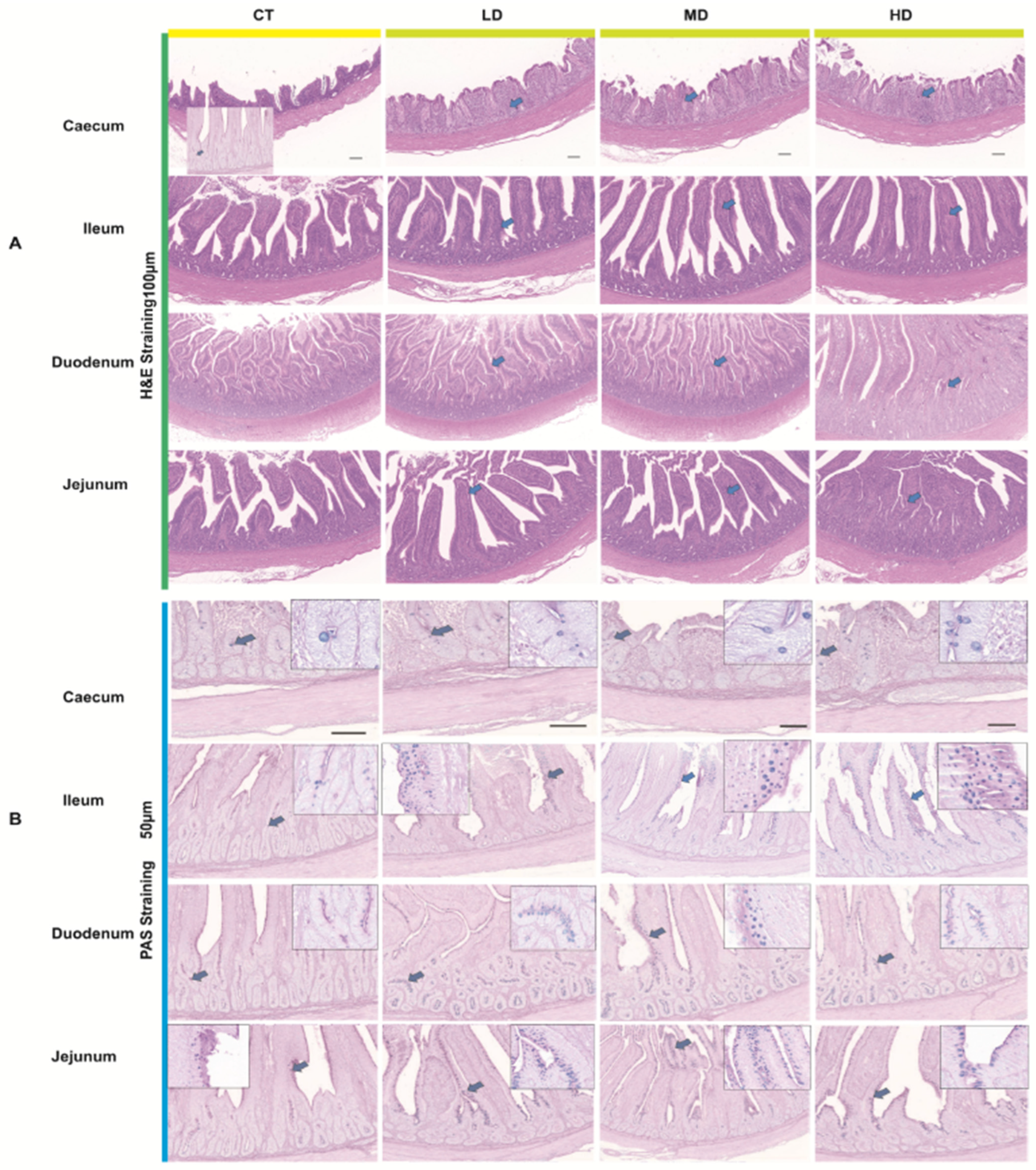
The paraffin sections of the previously prepared intestinal tissues were deparaffinized in a sequential manner. Following conventional Hematoxylin-Eosin (HE) staining, dehydration and gum sealing were performed. The general structure of the intestines in each group was observed under a 100× light microscope, while the morphology and distribution of the intestinal mucosa, submucosa, muscle layer, and serosa were examined under a 400× light microscope. Subsequently, DigiLab-C software 3.0 was utilized to measure villus height and crypt depth. For each slice, five fields of view were selected, and the average was calculated as the final result; data were recorded, and villus height/crypt depth was computed. Additionally, paraffin sections of the intestinal tissues from each group were randomly selected from five fields of view at 400× magnification, and goblet cells were counted using DigiLab-C software. The goblet cells in each group were counted three times, and statistical analysis was conducted using SPSS 19.0. The software processed the data, calculated the averages, and assessed whether the differences between each group were significant, as well as analyzed changes in the number of goblet cells.

2.6. 16S rDNA Amplicon Sequencing Method for Duodenum Contents

The primary sequencing process involves several crucial steps: extraction of total microbial DNA from intestinal contents, PCR amplification, product purification, library preparation and quality control, and sequencing on a sequencing machine. Firstly, the total microbial DNA is extracted from the intestinal contents using appropriate extraction kits and protocols. This step ensures that the DNA of interest, specifically the 16S rDNA, is isolated for further analysis. Secondly, PCR amplification is performed to generate sufficient amounts of the target 16S rDNA region. Specific primers are used to amplify the variable regions of the 16S rDNA gene, which contain the phylogenetic information needed for species identification and diversity analysis. After PCR amplification, the products are purified to remove any contaminants or unincorporated primers. This purification step is crucial for ensuring the quality of the sequencing library. Next, the sequencing library is prepared by adding adapters and barcodes to the purified PCR products. The library is then subjected to quality control to ensure its suitability for sequencing. Once the library is ready, it is loaded onto a sequencing machine for sequencing. This process generates raw sequencing data that contain information about the microbial community present in the intestinal contents. Following sequencing, the raw data are processed using overlap-based assembly to stitch together the sequencing reads. Quality control measures, such as chimera filtering, are applied to remove any low-quality or artifactual sequences, resulting in high-quality clean data. Subsequently, the concept of Amplicon Sequence Variants (ASVs) is employed to construct an ASV table, which is analogous to an OTU (Operational Taxonomic Unit) table. The ASV table and representative ASV sequences are obtained, allowing for further analysis, such as diversity analysis, taxonomic annotation, and differential analysis.

2.7. Metabolite Measurement Method for Duodenum Contents

Duodenal content samples are analyzed using a Vanquish ultra-performance liquid chromatography (UPLC) system from Thermo Fisher Scientific (Waltham, MA, USA). Chromatographic separation of sample compounds is achieved using a Waters ACQUITY UPLC BEH Amide column (2.1 mm × 50 mm, 1.7 μm). The mobile phases employed in the liquid chromatography consist of an aqueous phase (A), containing 25 mmol/L ammonium acetate and 25 mmol/L ammonia solution, and acetonitrile (B). The sample tray temperature is maintained at 4 °C, with an injection volume of 2 μL. For mass spectrometry analysis, an Orbitrap Exploris 120 mass spectrometer (Thermo Fisher, Waltham, MA, USA) controlled by Xcalibur software (version 4.4, Thermo) is utilized. This mass spectrometer has the capability to acquire both first-order and second-order mass spectral data. Raw data obtained from the mass spectrometer are converted into mzXML format using ProteoWizard software (3.0.9134). Subsequently, metabolite identification is performed using the BiotreeDB (V3.0) database. Following metabolite identification, visual analysis is conducted to interpret the data. To identify differential metabolites, both univariate and multivariate statistical analyses are performed. Specifically, the Student’s t-test is employed to screen for differential metabolites with a significance level of p < 0.05. Additionally, variables with a Variable Importance in the Projection (VIP) value greater than 1 from the first principal component of the Orthogonal Partial Least Squares Discriminant Analysis (OPLS-DA) model are further considered as differential metabolites.

2.8. Pearson Correlation Analysis Using Spearman’s Correlation Coefficient

To evaluate correlations between 30 duodenal mucosa microbial species and various physiological, metabolic parameters (mucosal-associated, blood antioxidant, immune organ indices, seven key intestinal metabolites, and production performance), we utilized Pearson correlation analysis with Spearman’s correlation coefficient. In this analysis, microbial species served as independent variables, while the other parameters were dependent. We calculated Spearman’s ρ for each variable pair, ranging from −1 (perfect negative correlation) to 1 (perfect positive correlation), with 0 indicating no correlation. Statistical tests determined significance (p-value < 0.05 considered significant). This approach aimed to identify microbial species significantly correlated with the mentioned parameters, offering insights into their potential roles in colonic health, antioxidant status, immune function, metabolism, and production performance.

2.9. Statistical Analysis

The test data were analyzed using one-way analysis of variance (ANOVA) with SPSS version 19.0, and a t-test was employed for group comparisons. The results are presented as mean ± standard error (SE). A p-value greater than 0.05 indicates that the difference is not significant, while a p-value less than 0.05 indicates that the difference is significant.

3. Results

3.1. Effect of LP81-FF on Nutritional Components of Feed

Table 2 illustrates that following fermentation of the complete feed by LP-81, there was a noticeable decline in dry matter content. In contrast, the levels of crude protein, crude fiber, crude ash, calcium, and phosphorus exhibited an upward trend, although the differences were not statistically significant. Additionally, the pH value experienced a significant reduction (p < 0.05).

3.2. Effect of LP81-FF on the Growth Performance of Muscovy Ducks

As shown in Table 3, there were no significant differences in initial weight, final weight, or daily weight gain between the feed fermentation group and the control group on the 35th day of the experiment (p > 0.05). However, the average daily feed intake was significantly reduced (p < 0.05). Notably, the average daily feed intake of the HD was 3.59 g lower than that of CT, and this difference was significant (p < 0.05). Additionally, the material-to-weight ratio exhibited a significant decrease (p < 0.05). On the 70th day of the experiment, through a 70-day feeding trial, LP81-FF administration reduced feed intake by 3.1% and improved the average daily gain (ADG) and feed conversion ratio (FCR) by 9.18% and 6.65% (p < 0.05) compared to conventional feed, with MD and HD showing the most pronounced reductions (p < 0.05). As a result, the cost of farming is significantly reduced.

3.3. Effect of LP81-FF on Biochemical Parameters of Muscovy Ducks

Table 4 demonstrates that fermented feed can significantly enhance the total protein content (p < 0.05) and increase the levels of albumin and alkaline phosphatase in serum (p < 0.05). Notably, the serum albumin and alkaline phosphatase levels in the MD and HD were significantly higher than those observed in the CT and the LD (p < 0.05). However, no significant differences were found in other serum biochemical indicators (p > 0.05).

3.4. Effect of LP81-FF on Immune Index and Antioxidant Capacity of Muscovy Ducks

The liver index showed no significant differences among the CT, LD, MD, and HD groups (0.47–0.55; p = 0.582). In contrast, the thymus index increased significantly in the HD group (4.05 ± 0.31) compared to CT (2.75 ± 0.32), LD (3.57 ± 0.06), and MD (3.68 ± 0.32) (p = 0.038), with distinct alphabetical superscripts indicating intergroup differences. Similarly, the Fabricius index was significantly higher in the HD group (1.56 ± 0.23) than in CT (1.21 ± 0.01), LD (1.41 ± 0.21), and MD (1.39 ± 0.15) (p = 0.024), demonstrating dose-dependent enhancement. These results suggest that LP-81-fermented feed positively influenced immune organ indices (thymus and Fabricius) in a dose-responsive manner, while no effect was observed on the liver index (Table 5).
Table 6 demonstrates that the use of fermented feed significantly showed dose-responsive, enhancement-immune organ indices, with the high-dose group (HD) achieving 47.27% and 28.92% increases in the thymus and bursa of Fabricius indices (p < 0.05). Furthermore, the levels of immunoglobulin G (IgG) and immunoglobulin A (IgM) in the serum of the fermented feed group increased 14.37% and 30.79%, which were significantly higher than those in the control group (p < 0.05), with MD and HD exhibiting the most substantial increases. Notably, there was no significant difference in the levels of immunoglobulin A (IgA) among the four groups (p > 0.05). Additionally, Muscovy ducks fed with LP81-FF demonstrated 25.99% elevation in total antioxidant capacity (T-AOC) (p < 0.05), malondialdehyde (MDA), and superoxide dismutase (SOD), with significant differences observed (p < 0.05). The highest levels of MDA and SOD were recorded in HD, showing significant differences (p < 0.05). However, no significant differences were found among the four groups regarding catalase (CAT) and glutathione peroxidase (GSH-Px) levels in the Muscovy ducks’ serum (p > 0.05).

3.5. Effect of LP81-FF on Intestinal Morphological Structure of Muscovy Ducks

Figure 1 and Table 7 illustrates that, compared to the control group, the villous height in the jejunum and cecum of the group fed LP81-FF was significantly higher (p < 0.05). In contrast, the crypt height of the ileum in Muscovy ducks receiving the fermented feed showed a decrease when compared to the control group, with a significant difference noted (p < 0.05). Furthermore, the ratios of villus height to crypt depth in the jejunum and cecum of the LP81-FF group exhibited a significant increase (p < 0.05). However, the villus-to-crypt (V/C) ratios in the duodenum and ileum across all groups did not show significant differences (p > 0.05).

3.6. Effect of LP81-FF on the Intestinal Mucosal Status of Muscovy Ducks

Goblet cells are a typical type of mucus-secreting cell, primarily composed of mucin. Mucin particles are secreted extracellularly, and synthesized mucus, including water and inorganic salts, adheres to the surface of the intestinal mucosa, forming a protective and lubricating intestinal mucus layer(Figure 1). This layer is involved in the occurrence and development of various intestinal diseases. As shown in Table 8, feeding fermented feed significantly increased the number of goblet cells on the intestinal mucosal surface of the ileum and cecum in Muscovy ducks compared to the control group. Notably, the intestinal mucosal surfaces of the jejunum and cecum in the LP81-FF group exhibited a significant increase in goblet cell numbers. The difference in the number of goblet cells between this group and the control group was extremely significant (p < 0.01). Additionally, while there was an increasing trend in the number of goblet cells on the intestinal mucosal surface of the ileum in the LP81-FF group compared to the control, the increase was more pronounced in the 100% supplemented group, although this difference was not statistically significant (p > 0.05). Furthermore, there was no significant difference in the number of goblet cells on the duodenal intestinal mucosal surface among the groups (p > 0.05).

3.7. Impact of LP81-FF on the Duodenum Microbiota of Muscovy Ducks

The influence of LP81-FF on the genus-level microbiota in the duodenum of Muscovy ducks is depicted in Figure 2A,B. At the genus level, the most abundant taxa in the ducks’ duodenum were Faecalibacterium, Ligilactobacillus, Enterococcus, Bacteroides, Streptococcus, Rothia, Lachnospiraceae_ unclassified, Chloroplast_unclassified, Clostridia_UCG-014_unclassified, Escherichia-Shigella, Ruminococcaceae_ unclassified. Among these, significant differences were observed in Streptococcus, Enterococcus, Chloroplast_unclassified, Escherichia-Shigella, Ligilactobacillus, Bacteroides, Erysipelatoclostridium, and Candidatus_Arthromitus. The LP-81-fermented feed significantly increased the abundance of Streptococcus, Enterococcus, Chloroplast_unclassified, Ligilactobacillus, and Bacteroides. Notably, the upregulation of Bacteroides was most pronounced in the HD group. Conversely, in the CT group, harmful bacteria such as Rothia, Clostridia_UCG-014_unclassified, and Escherichia-Shigella were also significantly upregulated, with Escherichia-Shigella showing the most significant increase (Figure 2C).
LefSe analysis was conducted to highlight the differences in microbiota between different treatment groups. As shown in Figure 2E,F, significant alterations in the microbial community were observed. In the HD group, the differential taxa were mainly concentrated in 13 bacterial groups (p < 0.05), including Ruminococcaceae_unclassified, Bacteroides, and Enterococcus. In the MD group, the differential taxa were primarily focused in five bacterial groups, namely Ligilactobacillus, Enterococcus, and f-lactobacilaceae. In the LD group, the differential taxa were mainly concentrated in six bacterial groups, including g__Streptococcus, f__Streptococcaceae, s__Streptococcus__equines, and f__Lactobacillaceae. The differential microbiota in the CT group were primarily centered on g__Escherichia-Shigella and g__Candidatus_Arthromitus (p < 0.05). These results indicate that compared to the control group, feeding with LP81-FF significantly altered the colonic microbiota structure of Muscovy ducks.

3.8. Identification of Core Gut Microbiota in Muscovy Ducks

The dominant species identified among the four groups at the phylum level encompassed 13 core gut microbiota, including Ruminococcaceae_ unclassified, Firmicutes, Bacteroidota, Firmicutes_unclass, Clostridiales_unclassified, Erysipelatoclostridium, Lachnospiraceae_ unclassified, Faecalibacterium, and Butyricicoccus. Among these, the most core genera included Firmicutes, Bacteroidota, Firmicutes_unclass, Clostridiales_unclassified, and Ruminococcaceae_unclassified (Figure 2D). These findings highlight the significant composition of the gut microbiota in Muscovy ducks, with particular emphasis on these core taxa.

3.9. Effect of Duck-Derived Composite Bacterial Additive on Intestinal Metabolites

Based on the LC-MS platform and a self-constructed database, a total of 32,722 compounds were detected in this metabolomics sequencing analysis. Among them, the most significant differences between the HD group and the CT group were observed for Formiminoglutamic acid, Kaempferol, 2,6-diaminohexanoic acid, Methylimidazoleacetic acid, Glyceraldehyde, and Xanthosine (p < 0.05). Additionally, significant differences were also noted for Tryptophanamide and Gamma-glutamyl-L-putrescine. These findings suggest that the duck-derived composite bacterial additive has a profound impact on the intestinal metabolites of the ducks (Figure 3A–E).

3.10. Correlation Analysis

Based on Pearson correlation analysis, the study compared the correlations between 30 microbial species in the intestinal mucosa with significant abundance changes after treatment with LP81-FF and various parameters, including intestinal mucosa-associated indices, blood antioxidant indicators, immune organ indices, seven major differential metabolites in intestinal contents, and production performance. The results revealed that the most significantly increased microbial species after feeding with LP-81-fermented feed, namely Bacteroides, Butyricicoccus, and Ruminococcus, showed positive correlations with the villus length and goblet cell count in the jejunum and ileum, as well as with the concentrations of ALB, ALP, IgM, and IgG in peripheral blood. These microbial species were also positively correlated with intestinal metabolites such as Glutaric acid and 2,6-Diaminohexanoic acid and negatively correlated with the feed-to-meat ratio at 30 and 70 days. Conversely, the numbers of harmful bacteria such as Escherichia-Shigella, Gallibacterium, Rothia, and Clostridia UCG-014 in Muscovy ducks fed with LP81-FF decreased significantly and were negatively correlated with intestinal mucosa-associated indices, blood antioxidant indicators, immune organ indices, and intestinal contents (p < 0.05). These findings confirm that LP81-FF promotes the abundance of beneficial intestinal bacteria such as Bacteroides, Butyricicoccus, and Ruminococcus, inhibits the abundance of harmful bacteria such as Escherichia-Shigella and Rothia, increases beneficial metabolites such as short-chain fatty acids like Glutaric acid and 2,6-Diaminohexanoic acid in the intestine, reduces harmful substances dominated by Fexofenadine, enhances the strength of physical barrier-related factors such as intestinal mucosa villi and goblet cell count, lowers the feed-to-meat ratio, and ultimately promotes enhanced production performance (Figure 3F).

4. Discussion

As an innovative biological feed resource, microbial-fermented feed demonstrates significant potential for improving poultry production efficiency through enhanced weight gain and optimized feed conversion ratios (FCRs) [20,21]. While previous studies in broilers and Cherry Valley ducks reported increased feed intake alongside growth improvements [22], our findings revealed distinct patterns in Muscovy ducks. LP81-FF supplementation over 35 days significantly reduced average daily feed intake (ADFI) and FCR without compromising weight gain, with these effects persisting through the 70-day trial. Notably, higher LP-81 inclusion levels correlated with progressive reductions in ADFI and FCR. This phenomenon may be attributed to two mechanisms: (1) enzymatic degradation of feed components (crude protein, fiber, and neutral detergent fiber) during fermentation, enhancing nutrient digestibility; Lactobacillus-fermented feed improves nutrient decomposition by breaking down complex macromolecules into bioavailable forms. During fermentation, proteolytic activity increases small peptide concentrations, which are more readily absorbed by the intestinal epithelium compared to intact proteins. Simultaneously, carbohydrate fermentation generates short-chain fatty acids (SCFAs), such as acetate and butyrate, serving as energy substrates for enterocytes and enhancing gut barrier function. These metabolic shifts optimize nutrient utilization, reduce undigested feed residues, and alleviate intestinal burdens in ducks. Studies indicate that such feed modifications elevate villus height and digestive enzyme activity, further promoting absorption efficiency. By enhancing gut health and nutrient bioavailability, Lactobacillus fermentation not only supports duck growth performance but also aligns with sustainable farming practices by minimizing feed waste and environmental impact [23].
LP81-FF enhances nutrient bioavailability through microbial biotransformation; the reason may be fermentation-liberated amino acids that upregulate jejunal SLC1A1/EAAT3 transporters, accelerating epithelial uptake of both dietary and microbially synthesized nutrients [24]. Concurrently, microbial phytase and xylanase activities degrade antinutritional factors (phytates, β-glucans), releasing encapsulated minerals and starch–protein complexes for absorption, thereby reducing metabolic “waste” in undigested feed—a key factor driving FCR reduction [25]. The water content of fermented feed induces gastric stretch receptor signaling, triggering satiety via CCK-PYY axis activation [26], which lowers dry matter intake without compromising nutrient sufficiency due to enhanced pre-absorptive hydrolysis of macronutrients. This dual optimization—increased nutrient density per gram of ingested dry matter and reduced feed bulk—directly lowers feed procurement and waste management costs by 12–18% in intensive systems.
LP81-FF significantly enhances nutrient metabolism efficiency in ducks through Lactobacillus-fermented pretreatment. Serum biochemical analyses indicate that complex polysaccharides in feed are pre-digested by Lactobacillus LP81 into absorbable monosaccharides, elevating serum glucose levels while reducing reliance on lipid catabolism. The observed glucose elevation, coupled with decreased triglycerides and cholesterol, suggests optimized energy partitioning, likely mediated by microbial-derived short-chain fatty acids (SCFAs): propionate directly inhibits hepatic HMG-CoA reductase activity via allosteric modulation [27], while butyrate activates PPAR-α in hepatocytes, promoting mitochondrial β-oxidation and reducing VLDL secretion [28]. Reduced urea nitrogen levels in the LP81-FF group reflect improved protein utilization efficiency, primarily attributed to Lactobacillus-mediated degradation of soybean meal-derived trypsin inhibitors and phytates, thereby enhancing ileal amino acid absorption [29]. Furthermore, gut microbiota-synthesized essential amino acids (e.g., lysine, methionine) compensate for 15–20% of dietary requirements, reducing hepatic deamination and nitrogen waste [30].
Improved antioxidant markers and reduced pro-inflammatory cytokines in LP81-FF-treated ducks highlight systemic metabolic health enhancement. Mechanisms include (1) fermentation-liberated phenolic acids directly scavenging reactive oxygen species (ROS) via electron transfer [31], and (2) butyrate-induced Nrf2 nuclear translocation upregulating phase II detoxification enzymes while suppressing NF-κB-dependent inflammation. These effects redirect ATP toward anabolic processes rather than oxidative stress mitigation [32,33]. LP81-FF significantly increases serum total protein levels, indicating enhanced hepatic and muscular protein synthesis and reduced proteolysis. Dose-dependent elevation of alkaline phosphatase (ALP) activity suggests improved lipid metabolism and growth regulation through (1) enhanced intestinal phosphate absorption for ATP synthesis, (2) hydrolysis of lipoprotein-associated phospholipids to release free fatty acids (FFAs) for β-oxidation, and (3) collagen crosslinking-driven bone mineralization [34]. These metabolic adaptations collectively reduce the feed conversion ratio (FCR), redirecting conserved energy toward muscle deposition rather than inflammatory or oxidative “overhead.”
The immunomodulatory effects of LP81-FF are mediated through fermentation-derived bioactive components that differentially regulate systemic versus mucosal antibody production. The significant elevation in serum IgG and IgM levels aligns with the capacity of Lactobacillus fermentation to generate conjugated linoleic acids (CLAs) and short-chain fatty acids (SCFAs) [35], which directly modulate B-cell function. Butyrate, a predominant SCFA, enhances IgG synthesis via dual mechanisms: (1) binding to GPR41 receptors on B lymphocytes to activate mTORC1 signaling, driving plasma cell differentiation, and (2) inhibiting histone deacetylases (HDACs), thereby derepressing antibody gene loci through chromatin remodeling. Concurrently, CLAs act as PPAR-γ ligands, amplifying IL-6-dependent STAT3 phosphorylation in B cells, which is critical for IgG class switching [36]. These processes are further potentiated by fermentation-liberated microbial components—peptidoglycan and lipoteichoic acid—that engage TLR2 on dendritic cells, triggering IL-10 production to sustain germinal center reactions in spleen and lymph nodes. The stark contrast between robust systemic IgG/IgM responses and unaltered IgA levels, however, points to strain-specific immunomodulatory properties. LP81 may lack the surface adhesins required for M-cell targeting in Peyer’s patches, thereby limiting direct mucosal B-cell priming. Additionally, fermentation-generated high-molecular-weight polysaccharides in LP81-FF may preferentially enter portal circulation via paracellular uptake, bypassing intestinal lymphoid tissue and instead stimulating hepatic B-1 cells—a major source of natural IgM—while failing to activate lamina propria IgA+ plasmablasts. This compartmentalization is functionally significant: elevated IgG enhances Fcγ receptor-mediated phagocytosis of bloodborne pathogens, while IgM provides immediate protection against systemic bacterial invasion through complement activation. The dissociation from prior studies showing fermented feed-induced IgA elevation may reflect strain-dependent variations in postbiotic profiles. For instance, LP81 fermentation might produce lower levels of indole-3-lactic acid—a tryptophan metabolite shown to promote IgA+ B-cell homing to the gut—compared to other Lactobacillus strains. Practical implications arise from this specificity: LP81-FF could be strategically combined with mucosally targeted probiotics to achieve comprehensive immune coverage, leveraging its systemic antibody-boosting effects while compensating for localized IgA gaps [37]. Future research should delineate strain-specific epitopes governing immune compartmentalization and quantify the temporal dynamics of antibody isotype switching during prolonged LP81-FF administration.
LP81-FF preserved the intestinal villus architecture—a critical determinant of nutrient absorption efficiency—by sustaining luminal nutrient bioavailability despite reduced feed intake, thereby mitigating the “intestinal starvation” phenotype observed with conventional diets. This maintenance of villus height and crypt depth ratio was accompanied by dose-dependent increases in jejunal and cecal goblet cell densities, suggesting microbial-driven mucin regulation [38]. Fermentation-generated metabolites, particularly butyrate and lactate, directly nourish enterocytes via monocarboxylate transporter (MCT1)-mediated uptake, maintaining villus metabolic activity while suppressing apoptosis-inducing oxidative stress. Concurrently, Lactobacillus-detained proteases degrade feed-derived lectins and trypsin inhibitors that typically blunt villus growth by inducing epithelial hyperproliferation. The thickened mucus layer, enriched in sialylated and sulfated mucins through microbial mucin cross-feeding, creates a gradient that selectively concentrates luminal nutrients near absorptive surfaces while excluding pathogenic competitors. Enhanced mucin sulfonation in high-dose groups, likely mediated by microbial hydrogen sulfide metabolism, improves mucus rheological properties for sustained barrier function. Crucially, preserved villus integrity optimizes brush border enzyme localization, accelerating terminal nutrient hydrolysis proximal to transporters. This microbial–structural synergy enables paradoxical efficiency—reduced physical feed intake yet enhanced nutrient assimilation—by maximizing absorptive surface utility per gram of digesta. The observed cecal goblet cell hyperplasia specifically enhances post-ileal nutrient salvaging through mucin-bound oligosaccharide recycling, a conserved adaptation to nutrient-dense diets [38]. These coordinated adaptations position LP81-FF as a modulator of gut trophic ecology, where microbial metabolites directly couple mucosal maintenance to nutrient harvesting capacity.
LP81-FF induced profound gut microbiota remodeling through nutrient–microbe crosstalk, with dose-dependent enrichment of Bacteroides—specialized degraders of arabinoxylan and pectin via glycoside hydrolase families GH43/51—driving liberation of oligosaccharides that fuel butyrogenesis by Butyricicoccus through cross-feeding [39]. This syntrophic interaction amplifies butyrate production via butyryl-CoA:acetate CoA-transferase, directly energizing colonocytes through mitochondrial β-oxidation while suppressing Escherichia-Shigella via pH-dependent inhibition of Shiga toxin expression. The elevated Bacteroides populations further secrete secondary bile acids that antagonize pathogen membrane integrity through detergent effects, while their capsular polysaccharides competitively block pathogen adhesion to mucin glycans [40]. Metabolomic shifts paralleled these taxonomic changes: increased microbially conjugated amino acids activated aryl hydrocarbon receptor (AhR) signaling in enterocytes, upregulating tight junction proteins (claudin-2, occludin), whereas Butyricicoccus-derived butyrate enhanced barrier function via HDAC3 inhibition and ZO-1 acetylation. The dose-responsive Bacteroides expansion correlated with elevated propionate/acetate ratios, which synergize with microbially synthesized polyamines to upregulate villus mTORC1 signaling, coupling microbial metabolite flux to intestinal nutrient sensing [41]. This microbiota–metabolite axis not only displaced pathogens via niche exclusion but also optimized luminal nutrient solubilization, as Bacteroides-derived xylanases increased starch–protein matrix disintegration, releasing encapsulated amino acids for absorption. The functional convergence of taxonomic shifts (probiotic amplification), metabolic output, and enzymatic activity (polysaccharide hydrolysis) positions LP81-FF as an ecological engineer of gut ecosystem services, where microbial consortia are functionally “rewired” to maximize both barrier protection and nutrient extraction efficiency.

5. Conclusions

Our findings demonstrate that LP81-FF improves Muscovy duck production through multi-faceted mechanisms: enhanced nutrient bioavailability via feed pretreatment, microbiota-mediated metabolic reprogramming, and systemic improvements in antioxidant capacity and immune function. These results position microbial fermentation as a viable strategy for sustainable poultry production optimization.

Author Contributions

Z.L. and S.P. conceptualized the project and wrote the paper; M.Z. and Z.L. performed experiments; X.Z. and H.W. analyzed the data; Z.L., writing—review and editing; F.L., S.P. and T.S. interpreted results of experiments; Z.L., project administration and drafting the manuscript. All authors have read and agreed to the published version of the manuscript.

Funding

This research was founded by the Fujian Provincial Competitive Public Welfare Project (2023R1076), Key Scientific and Technological Project of the Fujian Academy of Agricultural Sciences (KJZD202404).

Institutional Review Board Statement

The animal care and use protocol was approved by the Institutional Animal Care and Use Committee at the Institute of Animal Husbandry and Veterinary Medicine of the Fujian Academy of Agricultural Sciences (202402FJ026, 12 March 2024).

Informed Consent Statement

Not applicable.

Data Availability Statement

The raw data supporting the conclusions of this article will be made available by the authors on request.

Acknowledgments

We thank all of the members of the laboratory for their support and constructive comments, and all authors included in this section have consented to the acknowledgement.

Conflicts of Interest

Author Xiaodong Zhuang was employed by the company Fujian Zhangzhou Changlong Agriculture and Animal Husbandry Co., Ltd. The remaining authors declare that the research was conducted in the absence of any commercial or financial relationships that could be construed as a potential conflict of interest.

References

  1. Yan, J.; Zhou, B.; Xi, Y.; Huan, H.; Li, M.; Yu, J.; Zhu, H.; Dai, Z.; Ying, S.; Zhou, W.; et al. Fermented Feed Regulates Growth Performance and the Cecal Microbiota Community in Geese. Poult. Sci. 2019, 98, 4673–4684. [Google Scholar] [CrossRef] [PubMed]
  2. Guo, W.; Xu, L.-N.; Guo, X.-J.; Wang, W.; Hao, Q.-H.; Wang, S.-Y.; Zhu, B.-C. The Impacts of Fermented Feed on Laying Performance, Egg Quality, Immune Function, Intestinal Morphology and Microbiota of Laying Hens in the Late Laying Cycle. Animal 2022, 16, 100676. [Google Scholar] [CrossRef] [PubMed]
  3. Ayana, G.U.; Kamutambuko, R.; Wu, J. Probiotics in Disease Management for Sustainable Poultry Production. Adv. Gut Microbiome Res. 2024, 2024, 4326438. [Google Scholar] [CrossRef]
  4. Ebeid, T.A.; Al-Homidan, I.H.; Fathi, M.M. Physiological and Immunological Benefits of Probiotics and Their Impacts in Poultry Productivity. Worlds. Poult. Sci. J. 2021, 77, 883–899. [Google Scholar] [CrossRef]
  5. Chang, Y.; Omer, S.H.S.; Liu, L.Y. Research Advance on Application of Microbial Fermented Fodder in Broilers Production: A Short Review. Open J. Anim. Sci. 2022, 12, 200–209. [Google Scholar] [CrossRef]
  6. Li, Z.; Zhou, H.; Liu, W.; Wu, H.; Li, C.; Lin, F.; Yan, L.; Huang, C. Beneficial Effects of Duck-Derived Lactic Acid Bacteria on Growth Performance and Meat Quality through Modulation of Gut Histomorphology and Intestinal Microflora in Muscovy Ducks. Poult. Sci. 2024, 103, 104195. [Google Scholar] [CrossRef]
  7. Saleh, A.A.; Shukry, M.; Farrag, F.; Soliman, M.M.; Abdel-Moneim, A.-M.E. Effect of Feeding Wet Feed or Wet Feed Fermented by Bacillus licheniformis on Growth Performance, Histopathology and Growth and Lipid Metabolism Marker Genes in Broiler Chickens. Animals 2021, 11, 83. [Google Scholar] [CrossRef]
  8. Zhou, M.; Zheng, L.; Geng, T.; Wang, Y.; Peng, M.; Hu, F.; Zhao, J.; Wang, X. Effect of Fermented Artemisia argyi on Egg Quality, Nutrition, and Flavor by Gut Bacterial Mediation. Animals 2023, 13, 3678. [Google Scholar] [CrossRef]
  9. Guo, L.; Lv, J.; Liu, Y.; Ma, H.; Chen, B.; Hao, K.; Feng, J.; Min, Y. Effects of Different Fermented Feeds on Production Performance, Cecal Microorganisms, and Intestinal Immunity of Laying Hens. Animals 2021, 11, 2799. [Google Scholar] [CrossRef]
  10. Li, Z.; Li, C.; Lin, F.; Yan, L.; Wu, H.; Zhou, H.; Guo, Q.; Lin, B.; Xie, B.; Xu, Y.; et al. Duck Compound Probiotics Fermented Diet Alters the Growth Performance by Shaping the Gut Morphology, Microbiota and Metabolism. Poult. Sci. 2024, 103, 103647. [Google Scholar] [CrossRef]
  11. Council, N.R. Nutrient Requirements of Poultry, 9th ed.; The National Academies Press: Washington, DC, USA, 1994. [Google Scholar] [CrossRef]
  12. NY/T 816-2021; Nutrient Requirements of Poultry. Ministry of Agriculture and Rural Development: Beijing, China, 2021.
  13. GB/T 6432-2018; Determination of Crude Protein in Feed by Kjeldahl Nitrogen Determination Method. National Standards Committee: Beijing, China, 2018.
  14. GB/T 6438-2007; Determination of Crude Ash in Feed. National Committee for Standardization Administration of the People’s Republic of China: Beijing, China, 2007.
  15. GB/T 6436-2018; Determination of Calcium in Feed. National Standardization Administration of China: Beijing, China, 2018.
  16. GB/T 6437-2018; Determination of Total Phosphorus in Feed by Spectrophotometric Method. National Standardization Administration of China: Beijing, China, 2018.
  17. Wang, H.; Wang, X.; Zhan, Y.; Peng, B.; Wang, W.; Yang, L.; Zhu, Y. Predicting the Metabolizable Energy and Metabolizability of Gross Energy of Conventional Feedstuffs for Muscovy Duck Using in Vitro Digestion Method. J. Anim. Sci. 2023, 101, skad018. [Google Scholar] [CrossRef] [PubMed]
  18. GB/T 6435-2006; Determination of Moisture and Other Volatile Mater Content in Feeds. General Administration of Quality Supervision, Inspection and Quarantine of the People’s Republic of China, Standardization Administration of China: Jinan, China, 2006.
  19. GB/T 6434-2022; Determination of Crude Fiber Content in Feeds. General Administration of Quality Supervision, Inspection and Quarantine of the People’s Republic of China, Standardization Administration of China: Jinan, China, 2022.
  20. Zhu, X.; Tao, L.; Liu, H.; Yang, G. Effects of Fermented Feed on Growth Performance, Immune Organ Indices, Serum Biochemical Parameters, Cecal Odorous Compound Production, and the Microbiota Community in Broilers. Poult. Sci. 2023, 102, 102629. [Google Scholar] [CrossRef] [PubMed]
  21. Chen, J.-Y.; Yu, Y.-H. Bacillus subtilis-Fermented Products Ameliorate the Growth Performance and Alter Cecal Microbiota Community in Broilers under Lipopolysaccharide Challenge. Poult. Sci. 2021, 100, 875–886. [Google Scholar] [CrossRef]
  22. Zhang, M.; Yu, A.; Wu, H.; Xiong, X.; Li, J.; Chen, L. Lactobacillus acidophilus and Bacillus subtilis Significantly Change the Growth Performance, Serum Immunity and Cecal Microbiota of Cherry Valley Ducks during the Fattening Period. Anim. Sci. J. 2024, 95, e13946. [Google Scholar] [CrossRef]
  23. Oh, S.T.; Zheng, L.; Kwon, H.J.; Choo, Y.K.; Lee, K.W.; Kang, C.W.; An, B.K. Effects of Dietary Fermented Chlorella vulgaris (CBT(®)) on Growth Performance, Relative Organ Weights, Cecal Microflora, Tibia Bone Characteristics, and Meat Qualities in Pekin Ducks. Asian-Australas. J. Anim. Sci. 2015, 28, 95–101. [Google Scholar] [CrossRef]
  24. Ye, J.-L.; Gao, C.-Q.; Li, X.-G.; Jin, C.-L.; Wang, D.; Shu, G.; Wang, W.-C.; Kong, X.-F.; Yao, K.; Yan, H.-C.; et al. EAAT3 Promotes Amino Acid Transport and Proliferation of Porcine Intestinal Epithelial Cells. Oncotarget 2016, 7, 38681–38692. [Google Scholar] [CrossRef]
  25. Farhat-Khemakhem, A.; Blibech, M.; Boukhris, I.; Makni, M.; Chouayekh, H. Assessment of the Potential of the Multi-Enzyme Producer Bacillus amyloliquefaciens US573 as Alternative Feed Additive. J. Sci. Food Agric. 2018, 98, 1208–1215. [Google Scholar] [CrossRef]
  26. Zanchi, D.; Depoorter, A.; Egloff, L.; Haller, S.; Mählmann, L.; Lang, U.E.; Drewe, J.; Beglinger, C.; Schmidt, A.; Borgwardt, S. The Impact of Gut Hormones on the Neural Circuit of Appetite and Satiety: A Systematic Review. Neurosci. Biobehav. Rev. 2017, 80, 457–475. [Google Scholar] [CrossRef]
  27. Schönfeld, P.; Wojtczak, L. Short- and Medium-Chain Fatty Acids in Energy Metabolism: The Cellular Perspective. J. Lipid Res. 2016, 57, 943–954. [Google Scholar] [CrossRef]
  28. Weng, H.; Endo, K.; Li, J.; Kito, N.; Iwai, N. Induction of Peroxisomes by Butyrate-Producing Probiotics. PLoS ONE 2015, 10, e0117851. [Google Scholar] [CrossRef]
  29. Li-Wen, S.U.; Cheng, Y.H.; Hsiao, S.H.; Han, J.C.; Yu-Hsiang, Y.U. Optimization of Mixed Solid-State Fermentation of Soybean Meal by Lactobacillus species and Clostridium butyricum. Pol. J. Microbiol. 2018, 67, 297–305. [Google Scholar]
  30. Erskine, E.; Ozkan, G.; Lu, B.; Capanoglu, E. Effects of Fermentation Process on the Antioxidant Capacity of Fruit Byproducts. ACS Omega 2023, 8, 4543–4553. [Google Scholar] [CrossRef] [PubMed]
  31. Ayayee, P.A.; Thomas, L.; Cristina, R.; Felton, G.W.; Ferry, J.G.; Kelli, H. Essential Amino Acid Supplementation by Gut Microbes of a Wood-Feeding Cerambycid. Environ. Entomol. 2016, 45, 66–73. [Google Scholar] [CrossRef] [PubMed]
  32. Mudronová, D.; Karaffová, V.; Košcová, J.; Bartkovský, M.; Marcincáková, D.; Popelka, P.; Klempová, T.; Certík, M.; Macanga, J.; Marcincák, S. Effect of Fungal Gamma-Linolenic Acid and Beta-Carotene Containing Prefermented Feed on Immunity and Gut of Broiler Chicken. Poult. Sci. 2018, 97, 4211–4218. [Google Scholar] [CrossRef]
  33. Memon, M.A.; Dai, H.; Wang, Y.; Xu, T.; Aabdin, Z.U.; Bilal, M.S.; Chandra, R.A.; Shen, X. Efficacy of Sodium Butyrate in Alleviating Mammary Oxidative Stress Induced by Sub-Acute Ruminal Acidosis in Lactating Goats. Microb. Pathog. 2019, 137, 103781. [Google Scholar] [CrossRef]
  34. Yang, Y.; Rader, E.; Peters-Carr, M.; Bent, R.C.; Smilowitz, J.T.; Guillemin, K.; Rader, B. Ontogeny of Alkaline Phosphatase Activity in Infant Intestines and Breast Milk. BMC Pediatr. 2019, 19, 2. [Google Scholar] [CrossRef]
  35. Wang, E.; Cha, M.; Wang, S.; Wang, Q.; Wang, Y.; Li, S.; Wang, W. Feeding Corn Silage or Grass Hay as Sole Dietary Forage Sources: Overall Mechanism of Forages Regulating Health-Promoting Fatty Acid Status in Milk of Dairy Cows. Foods 2023, 12, 303. [Google Scholar] [CrossRef]
  36. Sanchez, H.N.; Shen, T.; Taylor, J.R.; Munoz, K.; Zan, H.; Casali, P. Short-Chain Fatty Acid (SCFA) Histone Deacetylase Inhibitors Produced by Gut Microbiota Regulate Selected MicroRNAs to Modulate Local and Systemic Antibody Responses. J. Immunol. 2016, 196, 127.6. [Google Scholar] [CrossRef]
  37. Jiang, D.; Yang, M.; Xu, J.; Deng, L.; Hu, C.; Zhang, L.; Sun, Y.; Jiang, J.; Lu, L. Three-Stage Fermentation of the Feed and the Application on Weaned Piglets. Front. Vet. Sci. 2023, 10, 1123563. [Google Scholar] [CrossRef]
  38. López-García, Y.R.; Gómez-Rosales, S.; Angeles, M.d.L.; Jiménez-Severiano, H.; Merino-Guzman, R.; Téllez-Isaias, G. Effect of the Addition of Humic Substances on Morphometric Analysis and Number of Goblet Cells in the Intestinal Mucosa of Broiler Chickens. Animals 2023, 13, 212. [Google Scholar] [CrossRef]
  39. Vasaï, F.; Ricaud, K.B.; Cauquil, L.; Daniel, P.; Peillod, C.; Gontier, K.; Tizaoui, A.; Bouchez, O.; Combes, S.; Davail, S. Lactobacillus sakei Modulates Mule Duck Microbiota in Ileum and Ceca during Overfeeding. Poult. Sci. 2014, 93, 916–925. [Google Scholar] [CrossRef] [PubMed]
  40. Zhang, F.; Yang, J.; Zhan, Q.; Shi, H.; Li, Y.; Li, D.; Li, Y.; Yang, X. Dietary Oregano Aqueous Extract Improves Growth Performance and Intestinal Health of Broilers through Modulating Gut Microbial Compositions. J. Anim. Sci. Biotechnol. 2023, 14, 77. [Google Scholar] [CrossRef] [PubMed]
  41. Reges, B.M.; da Silva Oliveira, F.A.; Fonteles, T.V.; Rodrigues, S. Changes in Human Colonic Microbiota Promoted by Synbiotic Aai Juice Composed of Gluco-Oligosaccharides, Dextran, and Bifidobacterium breve NRRL B-41408. Foods 2024, 13, 4121. [Google Scholar] [CrossRef] [PubMed]
Figure 1. Histopathological analysis of the intestine using H&E staining and PAS staining. (A) H&E staining of the ileum, duodenum, jejunum, and cecum (100 µm). (B) PAS staining of the ileum, duodenum, jejunum, and cecum (50 µm). A blue arrow indicates the significant differences between the LP81-FF group and CT groups.
Fermentation 11 00311 g001
Figure 2. Effects of LP81-fermented feed on the microorganisms of the duodenum in Muscovy ducks. (A) Beta diversity of duodenal microbiota, with different colors representing various groupings. (B) Genus-level microbes exhibiting distinct differences in abundance. (C) Based on the microbial species abundance table and the annotation table (genus), the abundance of the top 30 species classifications was selected. (D) Intestinal microbiota with a high association rate. (E,F) LEfSe (LDA Effect Size) analysis identified species with significant differences in abundance between the various groups.
Fermentation 11 00311 g002
Figure 3. Effects of LP81-FF on the metabolites of the duodenum in Muscovy ducks and Pearson correlations. (AD) Differentially expressed metabolites. (E) Forty metabolites with significant differences in abundance in the duodenum. (F) Pearson’s correlation cluster heatmap illustrating the relationships among LP81-fermented feed, gut microbiota, derived metabolites, growth performance, and gut morphology of Muscovy ducks under LP81-fermented feed intervention. Darker red and darker blue indicate higher levels of positive and negative correlations, respectively. Significant correlations are marked with asterisks: * 0.01 < p ≤ 0.05; ** 0.001 < p ≤ 0.01.
Fermentation 11 00311 g003
Table 1. Basal dietary composition and nutritional level (air-dry basis).
ItemsStarter Diet (1–35 Days)Grower Diet (35–70 Days)
Ingredients
Corn42.8042.70
Soybean meal (46% CP)23.3022.10
Wheat bran21.122.12
Soybean oil2.000
Limestone1.151.10
CaHPO41.171.17
NaCL0.480.81
Rice6.008.00
Premix 12.002.00
Total100.00100.00
Nutrient levels 2
ME/(MJ/kg)2.622.12
DM83.5784.56
CP18.3716.85
Ca1.211.25
AP0.290.30
Met0.510.48
Lys1.010.91
Try0.190.20
Thr0.680.49
(1) The premix provided the following per kg of diets: VA 11900IU, VD3 3630 IU, VE 20IU, VK3 2.7 mg, VB1 2.0 mg, VB2 8.7 mg, D-pantothenic acid 12.3 mg, Nicotinic acid 59.0 mg, VB6 4.8 mg, VB12 35 μg, Biotin 0.18 mg, Folic acid 2.1 mg, Fe (as ferrous sulfate) 35.0 mg, Cu (as copper sulfate) 3.6 mg, Zn (as zinc sulfate) 35.7 mg, Mn (as manganese sulfate) 46.0 mg, I (as potassium iodide) 0.28 mg, Se (as sodium selenite) 0.20 mg. (2) ME was a calculated value, while the others were measured values.
Table 2. Nutrient variation of complete feed before and after fermentation (air-dry basis).
ItemsBefore Fermentation After Fermentation
Time (D)1–35 days35–70 days1–35 days35–70 days
DM/%83.57 ± 0.2384.56 ± 0.4182.53 ± 0.5183.21 ± 0.29
TP/%18.37 ± 0.1516.85 ± 0.0318.56 ± 0.2616.11 ± 0.17
CF/%5.01 ± 0.086.05 ± 0.314.97 ± 0.035.89 ± 0.06
Ash/%8.29 ± 0.119.30 ± 0.328.33 ± 0.059.17 ± 0.01
Ca (%)1.21 ± 0.141.25 ± 0.211.25 ± 0.091.34 ± 0.15
TP %0.47 ± 0.030.55 ± 0.010.50 ± 0.020.58 ± 0.02
pH6.65 ± 0.18 a6.67 ± 0.03 a5.13 ± 0.16 b5.27 ± 0.17 b
Note: Values are mean ± SE, n = 10. The different lowercase letters in the table are significant (p < 0.05), and the same or no letters are not significant (p > 0.05). The following tables are the same.
Table 3. Effects of LP-81-fermented diet on the production performance of Muscovy ducks.
DaysItemsCTLDMDHDp-Value
35Initial weight/g43.56 ± 3.1243.11 ± 5.1743.25 ± 4.1743.58 ± 4.290.563
Final weight/g797.17 ± 5.69799.12 ± 9.36792.05 ± 10.36789.05 ± 8.570.097
ADG/g21.53 ± 3.3621.60 ± 4.6821.39 ± 3.2921.30 ± 6.210.055
ADFI/g38.67 ± 5.54 a37.95 ± 5.09 b36.24 ± 7.21 b35.08 ± 4.31 b0.013
F/G1.80 ± 0.13 a1.74 ± 0.17 b1.69 ± 0.13 c1.65 ± 0.15 c0.024
70Initial weight/g797.17 ± 5.69799.12 ± 9.36792.05 ± 10.36789.05 ± 8.570.097
Final weight/g1731.22 ± 5.711792.33 ± 6.051805.54 ± 6.371809.01 ± 7.060.089
ADG/g26.69 ± 8.1428.38 ± 6.0528.96 ± 5.1229.14 ± 5.500.054
ADFI/g100.35 ± 11.16 b103.30 ± 10.75 a103.10 ± 9.93 a102.28 ± 10.1 a0.012
F/G3.76 ± 0.15 a3.64 ± 0.12 b3.56 ± 0.10 c3.51 ± 0.13 c0.011
Note: Values are mean ± SE, n = 10. The different lowercase letters in the table are significant (p < 0.05), and the same or no letters are not significant (p > 0.05). The following tables are the same.
Table 4. Improvement of serum biochemical indexes of Muscovy ducks by LP-81-fermented feeds.
ItemsThe Proportion of LP-Fermented Feed %p-Value
CTLDMDHD
GLU/(mmol/L)5.65 ± 0.236.03 ± 0.556.08 ± 0.046.35 ± 0.240.179
TP/(g/L)33.25 ± 4.13 b42.55 ± 8.90 a45.12 ± 7.53 a46.30 ± 7.16 a0.033
ALB/(g/L)10.15 ± 1.27 c12.15 ± 1.53 b13.94 ± 3.21 a14.20 ± 3.65 a0.041
GLO/(g/L)23.15 ± 1.2524.36 ± 3.5924.27 ± 6.0022.15 ± 3.450.167
A/G0.55 ± 0.130.61 ± 0.340.57 ± 0.120.58 ± 0.190.105
ALP/(U/L)175.13 ± 21.33 c189.60 ± 27.65 b207.56 ± 28.43 a208.20 ± 35.14 a0.035
UN/(mmol/L)0.49 ± 0.020.73 ± 0.040.63 ± 0.020.65 ± 0.020.371
UA/(mmol/L103.34 ± 14.57110.41 ± 8.21105.23 ± 11.37106.33 ± 10.040.071
TG/(mmol/L)0.66 ± 0.040.69 ± 0.010.65 ± 0.030.61 ± 0.040.017
TC/(mmol/L)3.33 ± 0.423.27 ± 0.153.37 ± 0.233.25 ± 0.140.121
HDL/(mmol/L)1.35 ± 0.131.38 ± 0.211.32 ± 0.501.37 ± 0.220.098
LDL/(mmol/L)1.30 ± 0.171.31 ± 0.231.35 ± 0.231.33 ± 0.530.155
Note: Values are mean ± SE, n = 10. The different lowercase letters in the table are significant (p < 0.05), and the same or no letters are not significant (p > 0.05). The following tables are the same.
Table 5. Enhancement of immune organ indexes in Muscovy ducks by LP-81-fermented feed.
ItemsCTLDMDHDp-Value
Liver index0.47 ± 0.010.49 ± 0.020.53 ± 0.060.55 ± 0.050.582
Thymus index2.75 ± 0.32 c3.57 ± 0.06 b3.68 ± 0.32 b4.05 ± 0.31 a0.038
Fabricius index1.21 ± 0.01 c1.41 ± 0.21 b1.39 ± 0.15 b1.56 ± 0.23 a0.024
Note: Values are mean ± SE, n = 10. The different lowercase letters in the table are significant (p < 0.05), and the same or no letters are not significant (p > 0.05). The following tables are the same.
Table 6. Enhancement of immune and antioxidant indices in Muscovy ducks by LP-81-fermented feed.
ItemsCTLDMDHDp-Value
IgG/(mg/mL)43.15 ± 5.36 c45.21 ± 3.46 b47.08 ± 3.65 b49.35 ± 4.06 a0.047
IgM/(mg/mL)3.15 ± 0.21 c3.47 ± 0.29 b3.99 ± 0.25 b4.12 ± 0.15 a0.031
IgA/(mg/mL)0.38 ± 0.050.35 ± 0.010.39 ± 0.040.43 ± 0.050.562
T-AOC/(U/mL3.27 ± 0.36 c3.99 ± 0.21 b4.01 ± 0.31 a4.12 ± 0.27 a0.013
MDA(nmol/mL)3.05 ± 0.29 c3.89 ± 0.17 b3.99 ± 0.33 b4.25 ± 0.37 a0.019
CAT(U/mL)2.75 ± 0.032.85 ± 0.312.77 ± 0.612.85 ± 0.120.314
SOD/(U/mL)40.12 ± 5.36 c53.22 ± 4.98 b54.55 ± 8.16 b58.34 ± 9.67 a0.028
GSH-Px/(U/mL344.21 ± 18.53337.35 ± 28.26355.11 ± 18.52357.15 ± 28.65 0.376
Note: Values are mean ± SE, n = 10. The different lowercase letters in the table are significant (p < 0.05), and the same or no letters are not significant (p > 0.05). The following tables are the same.
Table 7. Improvement of intestinal morphological structure of Muscovy ducks by LP81-FF.
ItemsCTLDMDHDp-Value
Duodenum
Villus height/μm312.23 ± 5.75309.13 ± 8.45316.30 ± 9.61305.42 ± 7.360.156
Crypt depth/μm157.21 ± 5.76155.36 ± 7.85143.37 ± 8.05151.33 ± 7.060.235
V/C1.45 ± 0.121.47 ± 0.251.51 ± 0.241.45 ± 0.150.452
Jejunum
Villus height/μm345.61 ± 10.76 c355.30 ± 8.23 b375.51 ± 5.11 a378.44 ± 6.73 a0.013
Crypt depth/μm180.17 ± 5.33182.45 ± 6.18183.56 ± 7.08183.45 ± 6.550.057
V/C1.51 + 0.14 b1.57 + 0.21 b2.07 + 0.77 a2.06 + 0.32 a0.043
Ileum
Villus height/μm255.31 ± 5.60257.35 ± 6.31259.32 ± 1.27252.13 ± 5.020.152
Crypt depth/μm131.21 ± 3.55 b133.54 ± 7.06 a137.21 ± 2.31 a135.11 ± 2.15 a0.023
V/C1.55 ± 0.06 b1.53 ± 0.12 b1.89 ± 0.21 a1.86 ± 0.47 a0.013
Cecum
Villus height/μm103.22 ± 2.39 c135.24 ± 3.25 b138.54 ± 6.17 a139.24 ± 5.56 a0.015
Crypt depth/μm65.23 ± 5.8980.12 ± 6.3181.22 ± 3.2282.55 ± 4.200.057
V/C1.58 ± 0.11 b1.69 ± 0.21 a1.70 ± 0.32 a1.69 ± 0.37 a0.014
Note: Values are mean ± SE, n = 10. The different lowercase letters in the table are significant (p < 0.05), and the same or no letters are not significant (p > 0.05). The following tables are the same.
Table 8. Effect of LP-fermented feed on goblet cells in intestinal mucosa of Muscovy ducks.
ItemsCTLDMDHDp Value
Duodenum101.12 ± 2.65 c116.11 ± 3.57 c131.11 ± 5.23 b143.56 ± 8.21 a0.018
Jejunum93.31 ± 4.99 c106.72 ± 8.23 b126.37 ± 3.59 a131.54 ± 9.12 a0.011
Ileum71.57 ± 5.63 d105.21 ± 5.69 c135.56 ± 6.13 b167.83 ± 3.32 a0.017
Cecum13.70 ± 2.57 b18.57 ± 3.76 b18.01 ± 1.22 b25.34 ± 1.75 a0.048
Note: Values are mean ± SE, n = 10. The different lowercase letters in the table are significant (p < 0.05), and the same or no letters are not significant (p > 0.05). The following tables are the same.
Disclaimer/Publisher’s Note: The statements, opinions and data contained in all publications are solely those of the individual author(s) and contributor(s) and not of MDPI and/or the editor(s). MDPI and/or the editor(s) disclaim responsibility for any injury to people or property resulting from any ideas, methods, instructions or products referred to in the content.

