Article

Non-Targeted Metabolomics Analysis Unravels Changes in Non-Volatile Metabolites in Folium nelumbinis (Lotus Leaf) Induced by Aspergillus cristatus-Mediated Fermentation

1
Key Laboratory of Plant Resource Conservation and Germplasm Innovation in Mountainous Region (Ministry of Education), College of Life Sciences/Institute of Agro-Bioengineering, Guizhou University, Guiyang 550025, China
2
Guizhou Key Laboratory of Agricultural Microbiology, Guizhou Academy of Agricultural Sciences, Guiyang 550009, China
*
Authors to whom correspondence should be addressed.
Fermentation 2025, 11(5), 279; https://doi.org/10.3390/fermentation11050279
Submission received: 6 April 2025 / Revised: 29 April 2025 / Accepted: 7 May 2025 / Published: 13 May 2025

Abstract

:
This study employed non-targeted metabolomics to investigate the impact of solid-state fermentation by Aspergillus cristatus on the major non-volatile metabolites in lotus leaves (Folium nelumbinis). Sensory evaluation and metabolomic analysis of the leaves before and after fermentation revealed that the fermentation process significantly enhanced the sensory quality of lotus leaf tea, resulting in a brighter infusion color, a mellow flavor profile, and a notable reduction in astringency. The fermentation also induced the production of several potentially bioactive metabolites, including chlorogenic acid and sphondin, and stimulated the expression of genes related to the phenylpropanoid pathway, thereby promoting the synthesis of chlorogenic acid. Additionally, the fermentation led to a marked decrease in the content of flavonoids, while the content of alkaloids remained relatively unchanged. This study provides a theoretical basis for the in-depth development and utilization of lotus leaves and offers a novel approach to applying microbial fermentation technology to medicinal and edible materials.

1. Introduction

Nelumbo nucifera Gaertn., commonly known as the lotus, is an aquatic perennial plant belonging to the Nelumbonacee family, widely cultivated in Asia, the Americas, and Oceania for its various uses, particularly in traditional Chinese medicine [1]. Different parts of the lotus, especially the leaves (F. nelumbinis), have been valued for their medicinal properties for centuries, with the book Key to Diagnosis and Treatment highlighting their potential in managing obesity [2]. Recognized as both a functional food and a medicinal ingredient, lotus leaves are rich in bioactive compounds such as alkaloids, flavonoids, volatile oils, and organic acids [3,4]. In 2002, lotus leaf was listed as a homologous substance of food and medicine in the No.51 document issued by the Ministry of Health of the People’s Republic of China [5]. In recent years, many studies have proved that lotus leaves have diverse applications in biology and pharmacology. For instance, they possess anti-inflammatory, anti-oxidation, anti-cancer, and hypolipidemic properties, and they lower blood pressure and alleviate atherosclerosis [6,7,8,9]. Among these, alkaloids, known for their structural complexity and diverse chemical properties, often exhibit a bitter taste [10]. Lotus leaves contain a variety of monomeric alkaloids, including nuciferine, pronuciferine, O-nornuciferine, N-nornuciferine, roemerine, liensinine, neferine, isoliensinine, anonaine, liriodenine, armepavine, N-norarmepavine, coclaurine, O-norcoclaurine, N-methylcoclaurine, N-methylisococlaurine, caaverine, dehydroroemerine, dehydronuciferine, tomatidenol, nelumnucine, and lysicamine [11]. Recent studies have demonstrated that nuciferine ameliorates glucose and lipid metabolic disorders in obese mice by modulating the gut–liver axis [12], and has also been shown to prevent obesity through activation of brown adipose tissue [13]. Flavonoids, ubiquitously present in nearly all green plants, are predominantly distributed among higher vascular species. These secondary metabolites frequently exhibit a bitter taste profile as a distinctive organoleptic property. Monomeric flavonoids isolated from lotus leaves include quercetin, isoquercitrin, nelumboside, rutin, and kaempferol [14]. Among the identified flavonoids, quercetin constitutes the predominant constituent, with a substantial array of derivatives structurally based on quercetin and kaempferol aglycone cores, predominantly existing as glycosidic conjugates [3]. Quercetin 3-O-glucuronide has been demonstrated to attenuate lipid accumulation in 3T3-L1 adipocytes and enhance lipolytic activity in diet-induced obese murine models [15]. In brief, lotus leaf possesses high medicinal properties and health benefits and is a kind of functional foodstuff with substantial development potential. Though the annual production of lotus leaves exceeds 800,000 tons in China, only a small portion is used for tea and medicinal purposes, while the more significant portion is discarded as agricultural waste [16]. It is thus crucial to develop strategies to take full advantage of the resource. Flavonoids and phenolic acids, such as quercetin and tannin contained in lotus leaves, are the source of bitterness and astringency [17], which limit their exploitation and application [18]. Through the following studies, we postulate that the bioconversion of lotus leaves through microbial fermentation—such as solid-state fermentation with food-safety-grade fungus—could release bioactive substances to improve the efficacy of substrates [19]. He et al. demonstrated that fermentation of lotus leaf supernatants using probiotic Enterococcus strains yielded two bioactive fractions exhibiting dual hypolipidemic activity. In vitro, these fermented supernatants suppressed adipogenesis and reduced lipid accumulation in 3T3-L1 adipocytes. In vivo studies further revealed significant reductions in serum and hepatic total cholesterol (TC) and triglycerides (TGs), alongside inhibited hepatic steatosis in high-fat-diet-induced rat models [20]. Yu et al. concluded that mechanical shaking and controlled fermentation during the processing of lotus leaf tea (Nelumbo nucifera) significantly enhance its organoleptic quality by intensifying aromatic volatile compounds and optimizing flavor complexity [21]. Similarly, a recent investigation employing black tea fermentation protocols to process lotus leaf tea systematically characterized dynamic shifts in bioactive constituents and microbial communities during fermentation. The findings demonstrated that this bioprocessing strategy augmented both functional phytochemical profiles and biological efficacy compared to non-fermented controls [22].
A. cristatus, the sole dominant fungus in Fuzhuan brick tea, is widely recognized as a food-safety-grade fungus, also known as Eurotium cristatum or the golden-flower fungus [23]. Extensive evidence supports its use as a fungal probiotic against human obesity for approximately six centuries [12,24]. It demonstrates environmental plasticity by biosynthesizing functionally distinct secondary metabolites with bioactive potential under varying cultivation parameters [25]. A. cristatus decomposes various compounds in tea and forms derivatives such as purine alkaloids, polyphenolic compounds, and terpenoids, thereby reducing the bitterness and astringency as well as giving the tea a unique ‘fungal flower’ aroma [26]. Fermentation processing using A. cristatus can significantly deepen the infusion color of Taxilli Herba (Sang Ji-Sheng) tea, imparting a medicinal aroma and enhancing its sweet aftertaste. It also degrades lectins into nitrogen and carbon sources through the action of extracellular enzymes, thereby antagonizing their inhibitory effect on α-amylase [27]. Solid-state fermentation of Ginkgo biloba leaves using A. cristatus has been shown to significantly increase the flavonoid content, antioxidant activity, and aroma compound retention in G. biloba leaves [28]. Collectively, we postulate that solid-state fermentation with A. cristatus is a feasible method to improve the taste and quality of lotus leaves.
Herein, lotus leaves were subjected to solid-state fermentation using A. cristatus to elucidate the impact of this fungal fermentation on the sensory attributes and primary bioactive compounds of the leaves. Subsequent untargeted metabolomic analysis was employed to ascertain the alterations in metabolic profiles before and after fermentation. This study provides a valuable reference for the development and utilization of A. cristatus-fermented lotus leaf products, particularly those aimed at lipid-lowering, and enhances our understanding of microbial fermentation technology in the context of dual-purpose food–medicine applications.

2. Materials and Methods

2.1. Experimental Materials

The lotus leaves were purchased from Guqingtang Pharmaceutical Company Ltd. in Bozhou, Anhui, China. Aspergillus cristatus (CGMCC 7.193) is preserved at the Culture Collection Center of the Chinese Academy of Sciences and the Agricultural Biology Institute of Guizhou Province.

2.2. Fermentation Processing

A. cristatus was activated in Malt Yeast Agar (MYA) medium, after which the medium was rinsed with sterile water to obtain a spore suspension with a concentration of 106–107 spores/mL, as measured using an optical microscope. Lotus leaves (5 g) were sterilized for 20 min. at 121 °C, cooled to room temperature, and then inoculated with the spore suspension, resulting in an inoculation amount of 47% (2.35 mL). The mixture was incubated for 7 days at 32 °C. The unfermented lotus leaf (UL) and fermented lotus leaf (FL) samples were subsequently stored in sterile plastic bags at −80 °C awaiting analysis. Each experiment had 3–6 biological replicates.

2.3. Sensory Evaluation

The sensory attributes, including appearance, aroma, taste, infusion color, and leaf residue, were evaluated by a panel of ten experienced members (5 males and 5 females, aged 20 to 40 years) from the Guizhou Institute of Biotechnology. All the panel members had no sensory defects. This assessment followed the sensory evaluation standards outlined in the Chinese standard GB/T 23776-2018 [29,30]. The samples were scored based on the evaluation criteria presented in Table 1. Briefly, 3 g of UL and FL samples were placed in a specialized tea cup, followed by the addition of 150 mL of boiling water for infusion for 8 min. The tea infusion was then poured into a tea cup and randomly presented to a trained sensory evaluation panel for assessment. Samples were coded using double digits and analyzed using one-way analysis of variance (ANOVA).

2.4. Determination of Total Flavonoids and Alkaloids

The total flavonoids were determined using the NaNO2-Al(NO3)3-NaOH colorimetric method, following a previously reported protocol [31]. The results were expressed as rutin equivalents in mg/g dry lotus leaves. The total alkaloids were determined using the bromocresol green microplate assay. The results were expressed as nuciferine equivalents in mg/g dry lotus leaves.

2.5. Metabolite Extraction

Metabolites were extracted from 50 mg of UL and FL samples. The samples were put in a 2 mL centrifuge tube containing a 6 mm diameter grinding bead and 400 μL of extraction solution (methanol: water = 4:1 (v/v)) containing 0.02 mg/mL of internal standard (L-2-chlorophenylalanine). The samples were ground using a Wonbio-96c frozen tissue grinder (Shanghai Wonbio Biotechnology Co., Ltd., Shanghai, China) for 6 min (−10 °C, 50 Hz), followed by low-temperature ultrasonic extraction for 30 min (5 °C, 40 kHz). The samples were then maintained at −20 °C for 30 min, centrifuged for 15 min (4 °C, 13,000× g), and the supernatant was subsequently transferred to an injection vial for LC-MS/MS analysis [32].

2.6. UHPLC-MS/MS) Analysis

The LC-MS/MS analysis of the extracted samples was conducted on a Thermo UHPLC-Q Exactive HF-X system equipped with an ACQUITY HSS T3 column (100 mm × 2.1 mm i.d., 1.8 μm; Waters Corporation, Milford, MA, USA) at Majorbio Bio-Pharm Technology Co. Ltd. (Shanghai, China).
The mobile phases consisted of 0.1% formic acid in water: acetonitrile (95:5, v/v) (solvent A) and 0.1% formic acid in acetonitrile: isopropanol: water (47.5:47.5, v/v) (solvent B). The positive ion mode separation gradient was as follows: 0–3 min, mobile phase B increased from 0% to 20%; 3–4.5 min, mobile phase B increased from 20% to 35%; 4.5–5 min, mobile phase B increased from 35% to 100%; 5–6.3 min, mobile phase B maintained at 100%; 6.3–6.4 min, mobile phase B decreased from 100% to 0%; 6.4–8 min, mobile phase B maintained at 0%. The separation gradient in the negative ion mode was as follows: 0–1.5 min, mobile phase B increased from 0 to 5%; 1.5–2 min, mobile phase B increased from 5% to 10%; 2–4.5 min, mobile phase B increased from 10% to 30%; 4.5–5 min, mobile phase B increased from 30% to 100%; 5–6.3 min, mobile phase B linearly maintained at 100%; 6.3–6.4 min, the mobile phase B decreased from 100% to 0%; 6.4–8 min, the mobile phase B linearly maintained at 0%. The flow rate was 0.40 mL/min, while the column temperature was 40 °C.
The mass spectrometric data were collected using a Thermo UHPLC-Q Exactive HF-X Mass Spectrometer equipped with an electrospray ionization (ESI) source operating in the positive and negative modes. The optimal MS conditions were set as follows: source temperature was 425 °C; sheath gas flow rate was 50 arb; Aux gas flow rate was 13 arb; ion-spray voltage floating (ISVF) was −3500 V in negative mode and 3500 V in positive mode, respectively; normalized collision energy, and 20-40-60 V rolling for MS/MS. The full MS resolution was 60,000, while the MS/MS resolution was 7500. Data acquisition was performed in the Data Dependent Acquisition (DDA) mode, with the detection carried out over a mass range of 70–1050 m/z [33].

2.7. Data Analysis

The pretreatment of LC/MS raw data was performed using Progenesis QI (Waters Corporation, Milford, MA, USA) software, followed by the export of a three-dimensional data matrix in CSV format. The three-dimensional matrix contained the sample information, metabolite name, and mass spectral response intensity. Internal standard peaks and any known false positive peaks, including noise, column bleed, and derivatized reagent peaks, were removed from the data matrix to make it non-redundant, followed by the pooling of the peaks. The metabolites were identified through a search in the HMDB (http://www.hmdb.ca/, accessed on 12 December 2024), Metlin (https://metlin.scripps.edu/, accessed on 12 December 2024), and Majorbio Databases.
The data matrix was obtained after the database search was uploaded to the Majorbio cloud platform (https://cloud.majorbio.com, accessed on 12 December 2024) for data analysis. The data matrix was first pre-processed by retaining 80% of the metabolic features detected in any set of samples. The minimum metabolite value for specific samples with metabolite levels below the lower limit of quantification was estimated after filtering, and each metabolic signature was normalized to the sum. The response intensities of the sample mass spectrometry peaks were normalized using the sum normalization method to reduce the errors caused by sample preparation and instrument instability and obtain the normalized data matrix. Variables of QC samples with relative standard deviation (RSD) > 30% were excluded and subjected to log10 logarithmic scaling to obtain the final data matrix for subsequent analysis.
The R package “ropls” (Version 1.6.2, Majorbio) was used to perform principal component analysis (PCA), orthogonal least partial squares discriminant analysis (OPLS-DA), and 7-cycle interactive validation to evaluate the stability of the model. The metabolites with VIP > 1.5 and p < 0.05 were denoted as significantly different metabolites based on the variable importance in the projection (VIP) obtained by the OPLS-DA model and the p-value generated by the student’s t-test.
The differential metabolites between the two groups were mapped into their biochemical pathways through metabolic enrichment and pathway analysis in the KEGG database (http://www.genome.jp/kegg/, accessed on 7 January 2025). These metabolites were classified according to the pathways they were involved in or the functions they performed. Enrichment analysis was used to analyze a group of metabolites in the presence or absence of a function node. The principle was that the annotation analysis of a single metabolite developed into an annotation analysis of a group of metabolites. The Python (Version 3.8.5) package “scipy.stats” (https://docs.scipy.org/doc/scipy, accessed on 23 January 2025) was used to perform enrichment analysis to obtain the most relevant biological pathways for experimental treatments. The data were analyzed using the free online platform of majorbio cloud platform (cloud.majorbio.com, accessed on 23 January 2025) [34].

3. Results and Discussion

3.1. Effect of A. cristatus Fermentation on the Sensory Quality of Lotus Leaf

The sensory evaluation scores are summarized in Table 2. A. cristatus-mediated fermentation resulted in a brighter brew, displaying a distinctive “golden ring” similar to that found in Fuzhuan black tea (Figure 1). The infusion color transitioned from light yellow to amber, while the flavor profile improved significantly, yielding a rich, mellow taste with reduced bitterness, accompanied by a floral aroma characteristic of traditionally fermented Fuzhuan brick tea. These color changes in the tea are primarily due to changes in tea pigments, which are polymers formed through the oxidation and polymerization of catechins, commonly referred to as catechin oxidation polymers [35].

3.2. Effect of A. cristatus Fermentation on the Main Active Components of Lotus Leaf

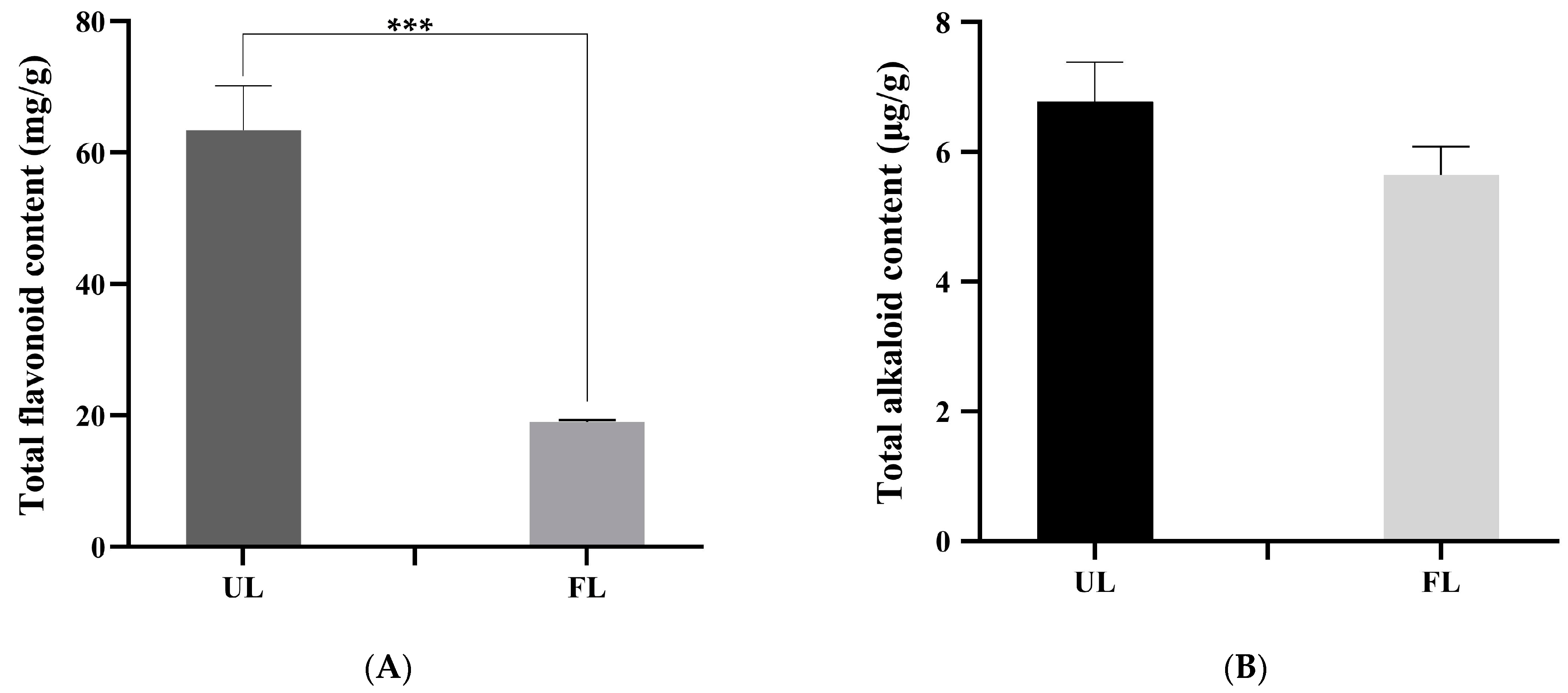
The content of total flavonoids in UL were 63.40 mg/g, while that in FL were 19.05 mg/g (Figure 2A). Fermentation significantly reduced the flavonoid content in lotus leaves by approximately two-thirds (p < 0.001). This reduction was attributed to oxidation and polymerization of the phenolic hydroxyl and carboxyl groups [36]. Flavonoids are recognized as primary bitter-tasting constituents [17]. Their diminished levels may link to the observed amelioration of astringent bitterness in the fermented tea infusion. In contrast, A. cristatus-mediated fermentation did not significantly affect the total alkaloids of the lotus leaves. The content of total alkaloids in UL were 6.78 μg/g, while that in FL were 5.65 μg/g (Figure 2B). Alkaloids are key bioactive compounds driving their lipid-lowering effects [37]. This suggests fermentation improves the taste of lotus leaves while preserving its primary health benefits.

3.3. UHPLC–MS/MS Metabolic Profile Analysis

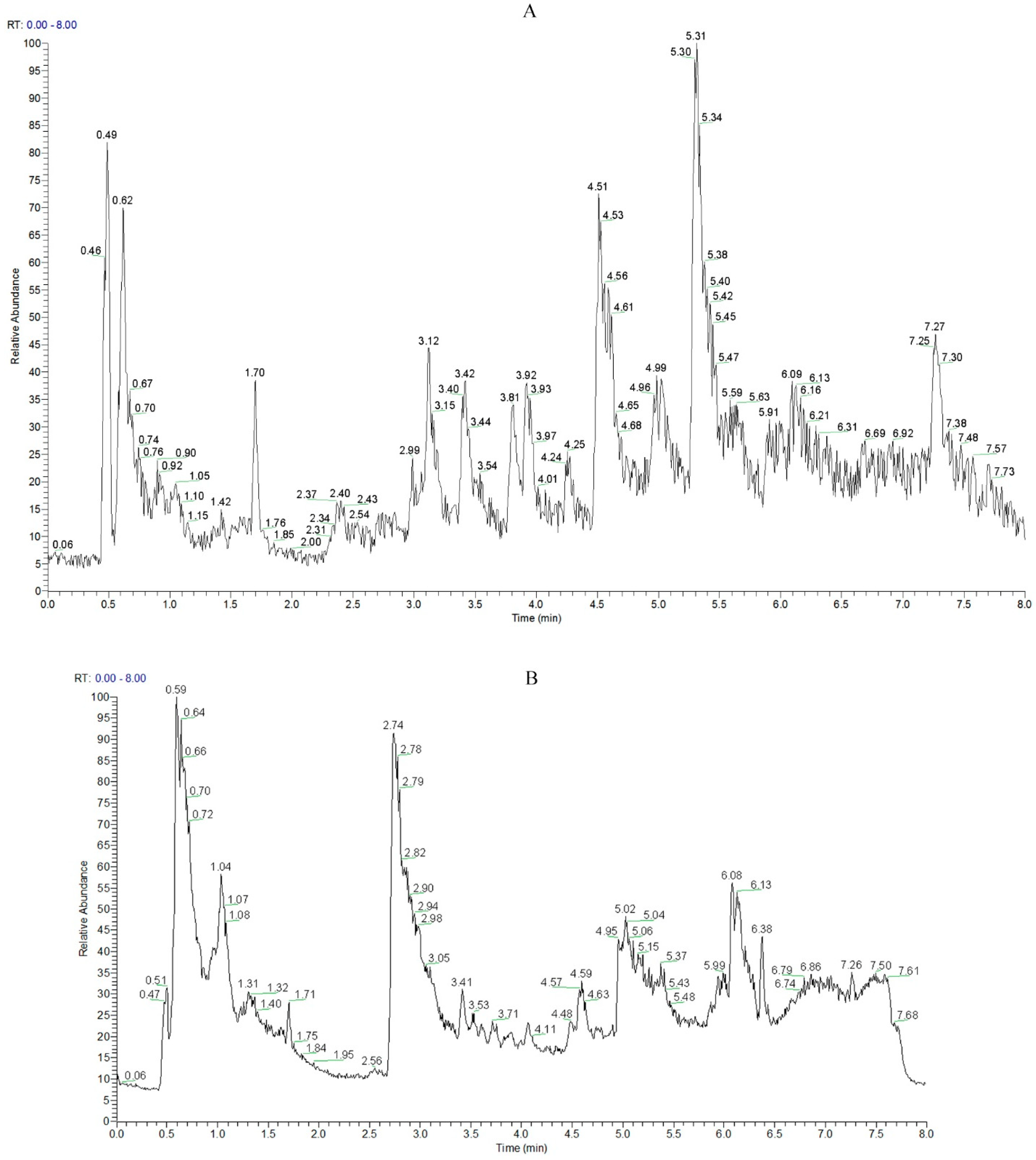
Twelve samples of UL and FL were analyzed using LC-MS/MS in both positive and negative ion modes (Figure 3). Component annotation and data processing yielded 12,511 mass spectral peaks. Among them, 3365 metabolites were identified, with 3200 metabolites annotated in HMDB and KEGG databases. Among them, there were 495 kinds of lipids and lipid molecules (16.72%), 415 kinds of organic oxygen compounds (13.72%), 474 kinds of organic heterocyclic compounds (13.56%), 327 kinds of amino acids, peptides and analogues (10.32%), 308 kinds of benzene compounds (9.80%), 266 kinds of flavonoids (9.66%), 218 kinds of phenylpropanoids (7.09%), 27 kinds of terpenoids (4.51%), 134 kinds of organic acids and derivatives.

3.4. Multivariate Statistical Analysis

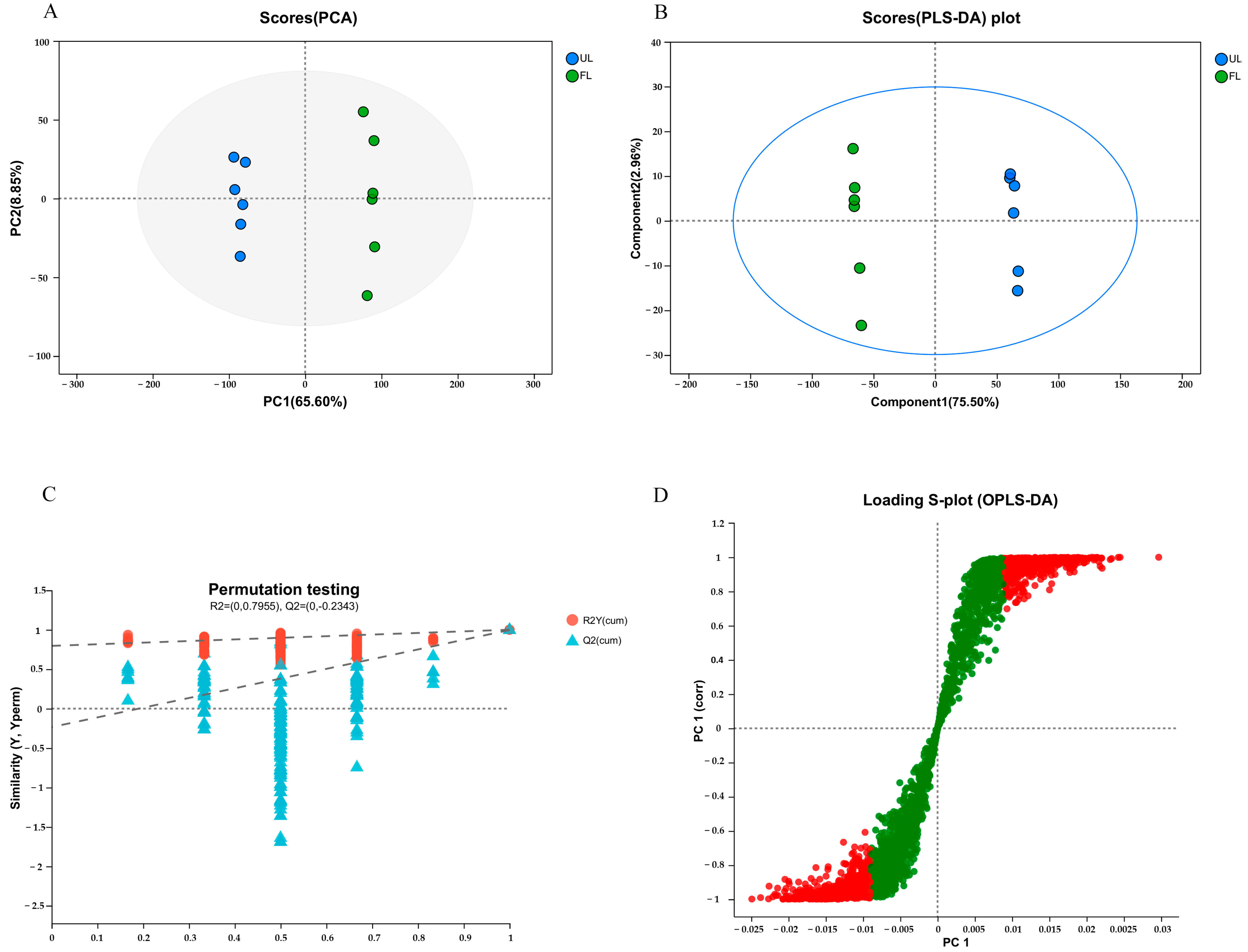
Principal component analysis revealed that UL and FL samples were distributed within a 95% confidence interval, with a significant distinction between the two groups (Figure 4). Replicates of each sample clustered together, with the first two principal components (PC1 and PC2) accounting for 74.45% of the variance in material composition distribution between the two samples. This result suggests that fermentation significantly affected the separation of lotus leaf samples, with samples from the same treatment exhibiting similar material distribution and composition trends.
Supervised partial least squares discriminant analysis (PLS-DA) further separated the samples more effectively. Figure 4B illustrates a comparative analysis of metabolites between unfermented and fermented lotus leaves using PLS-DA. The two groups exhibited significant separation, with all six samples of unfermented lotus leaves clustering on the left and all six samples of fermented lotus leaves clustering on the right. This finding indicates that fermentation substantially impacted the lotus leaves, providing compelling evidence for the differentiation of fermented lotus leaf samples during processing. A permutation test revealed that the blue Q2 points, moving from left to right, were all lower than the original red Q2 point on the far right (Q2-0). This observation confirmed that overfitting was not present and further validated the reliability of the established model.
The S-plot revealed significant clustering of the metabolites at both ends of the chart, highlighting their role in distinguishing fermented and unfermented samples. This finding suggests that the metabolites potentially play an important role in biological processes.

3.5. Analysis of Differential Metabolites

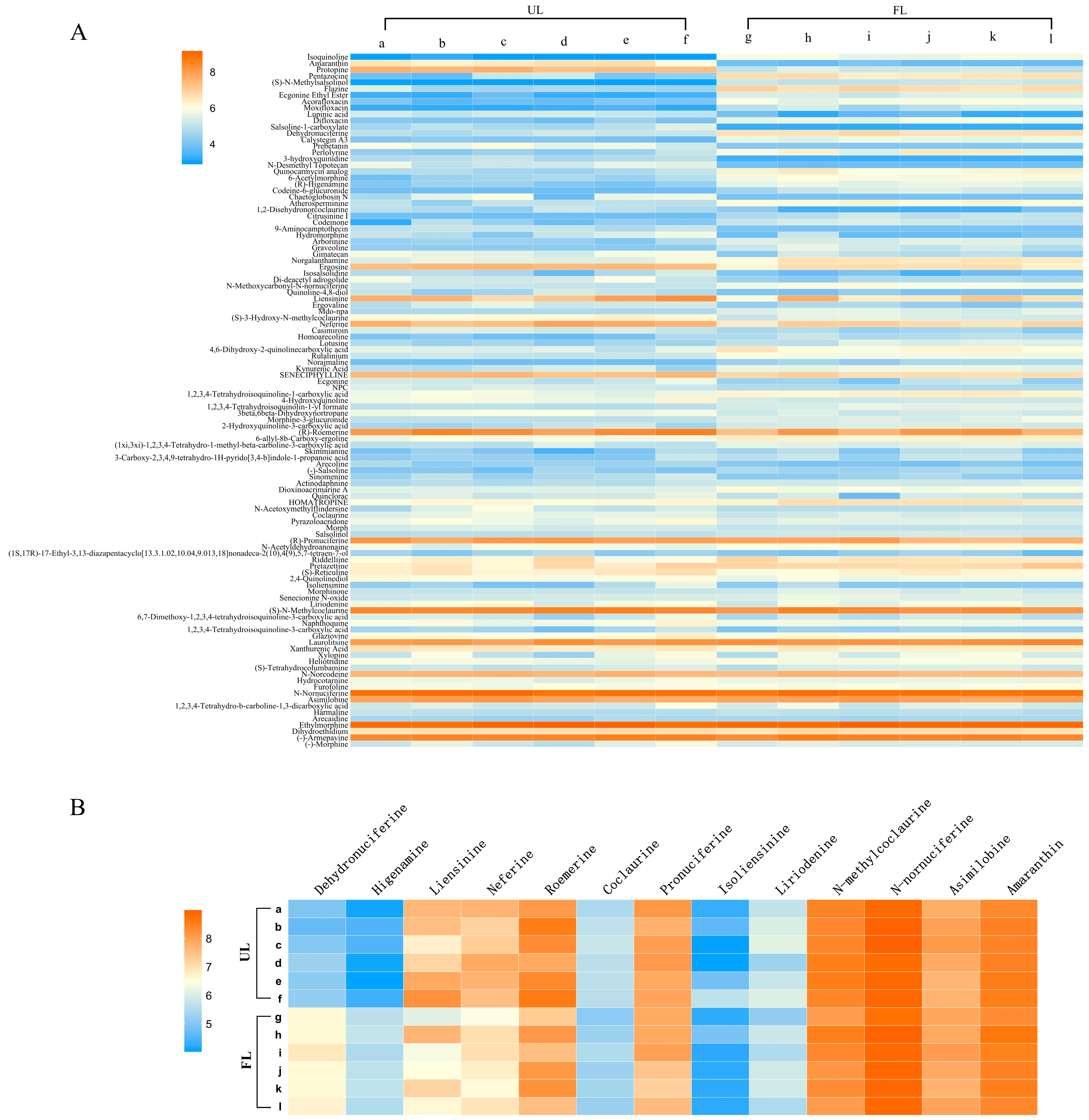
Alkaloids are key bioactive compounds driving their lipid-lowering effects [13]. To elucidate the impact of fermentation on alkaloid profiles in lotus leaves, we conducted a comprehensive analysis of 108 alkaloids identified across 12 experimental samples (Figure 5A). Only approximately one-third of the alkaloidal metabolites exhibited statistically significant alterations (p < 0.05), with 23 compounds demonstrating marked upregulation and 14 showing pronounced downregulation. Among them were 13 monomers previously reported to be isolated from lotus leaves. The monomers included hehydronuciferine, higenamine, liensinine, neferine, coclaurine, pronuciferine, isoliensinine, liriodenine, N-methylcoclaurine, N-nornuciferine, asimilobine, and armepavine (Figure 5B). Notably, only dehydronuciferine and higenamine were upregulated (p < 0.05), while the others showed no significant changes. Nuciferine is an aporphine-type alkaloid extracted from lotus leaves. It has a long history of use and exhibits strong anti-inflammatory and antioxidant properties. It regulates glucose and lipid metabolism and significantly protects multiple organs in the body. It is commonly used clinically for lipid reduction and weight loss [7]. Pronuciferine is a naturally occurring isoquinoline alkaloid that serves as a neuroprotectant, preventing apoptosis in neurodegenerative diseases [38]. Roemerin is a naturally occurring alkaloid, structurally similar to Mianserin, Moxifloxacin, Venlafaxine, Diphenhydramine, and Ofloxacin drugs. It has antidepressant effects [39]. Liensinine exerts anti-cancer effects in liver cancer by inhibiting the Kv10.1 channel [40] and can also inhibit oxidative stress. N-nornuciferine is an alkaloid extracted from lotus seeds and harbors multiple pharmacological activities, including antioxidant, anti-inflammatory, antithrombotic, and anti-apoptotic effects. Clinical studies postulate that N-nornuciferine prevents cancer, obesity, and cardiovascular diseases [39]. Isoliensinine is an active compound in lotus leaves harboring various bioactivities, including cell protection by inhibiting oxidative stress. It also acts as an antihypertensive and an aortic protective agent [41]. Liriodenine has anti-cancer activity and can induce apoptosis by activating the intrinsic pathways involving caspase-3 and caspase-9 [42]. Armepavine has anti-inflammatory effects and exhibits immunosuppressive activity on T lymphocytes and lupus nephritis mice, demonstrating potential as an antifibrotic drug for the liver [43]. Coclaurine is a tetrahydroisoquinoline alkaloid isolated from various plant sources. It possesses blood pressure-lowering, anti-cancer, and anti-aging activities [44]. Lysicamine inhibits the growth of thyroid and breast cancer cells and hepatocellular carcinoma cells. It also exhibits some iron-reducing and antimicrobial activities [45].
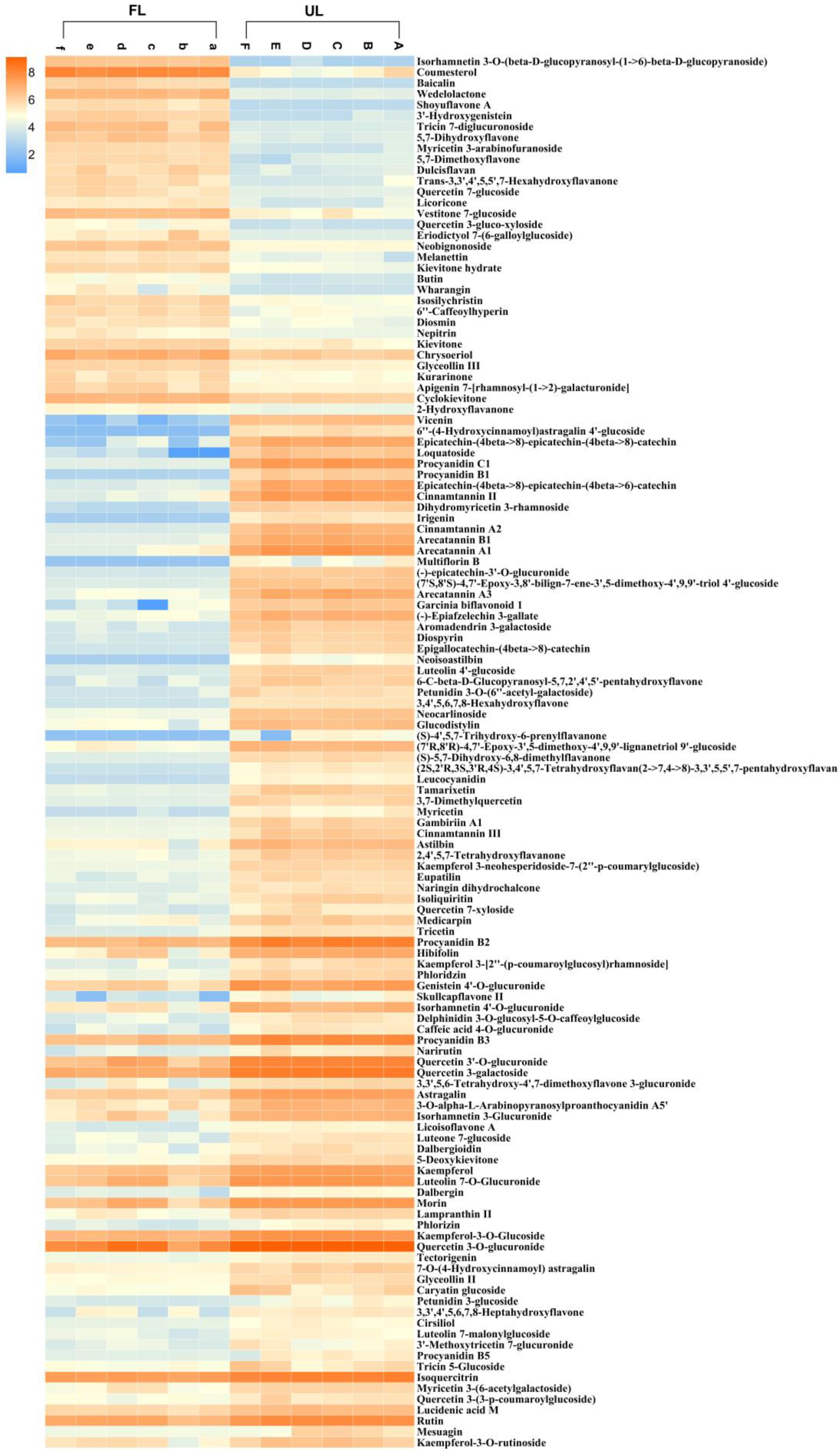
Flavonoids, recognized as the primary contributors to the bitter taste profile of lotus leaves, were systematically investigated to elucidate fermentation-induced modifications [10]. Among them, 128 were differentially expressed: 33 were significantly upregulated, while 95 were significantly downregulated (p < 0.05). The relative abundance of the majority of flavonoid compounds exhibited a marked reduction following fermentation treatment (Figure 6).
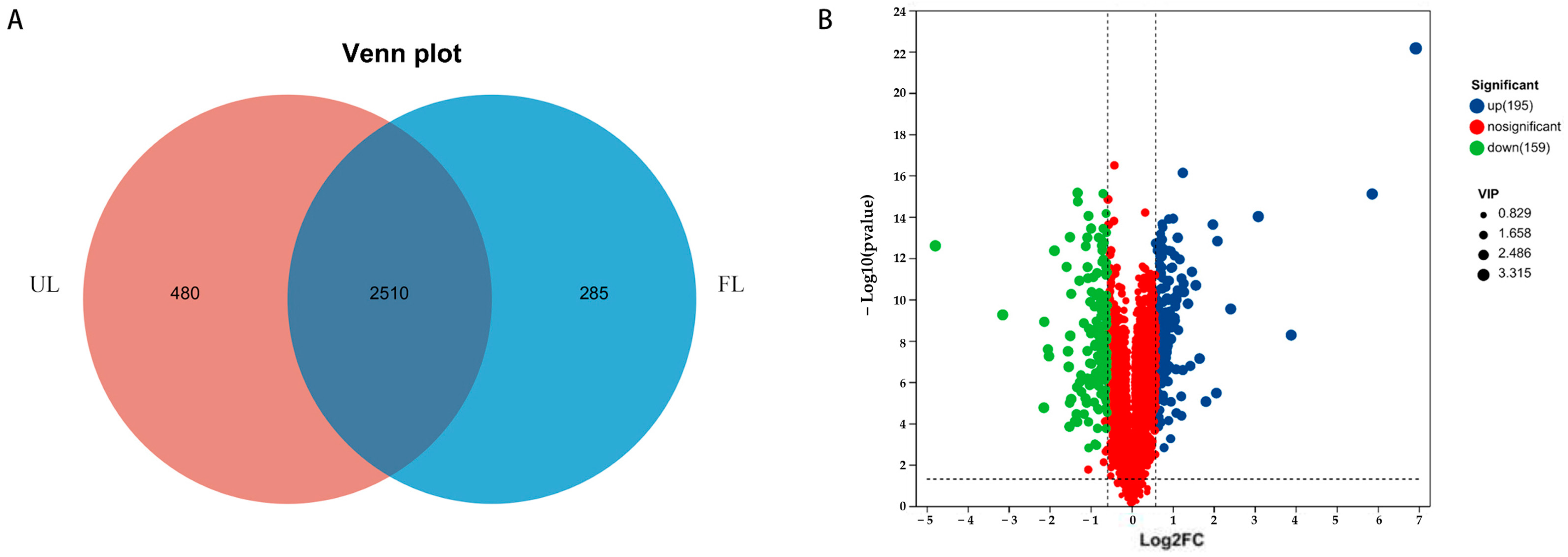
To elucidate the fermentation-induced alterations in the metabolic profile of lotus leaves, a comprehensive metabolomic analysis was conducted. LC/MS metabolomics analysis identified 480 metabolites unique to non-fermented lotus leaves and 285 metabolites unique to fermented lotus leaves (Figure 7). All the identified metabolites were screened based on the criteria of p < 0.05, VIP > 1.5, and a fold change of >1.5 (upregulated/downregulated). A volcano plot was used for visualization, resulting in the identification of 354 differential metabolites: 195 were significantly upregulated, while 159 were significantly downregulated.
Crucial metabolites with potential biological activity were further selected from the significantly upregulated differential metabolites through database search and reviewing of literature. The top 20 metabolites, ranked based on VIP value, are highlighted in Table 3. The main bioactive effects of the key metabolites included antioxidant, anti-cancer, anti-inflammatory, and antimicrobial activities, among other properties. For example, chlorogenic acid and its derivatives exhibit bioactivities, such as antioxidant, antitumor, antifungal, and regulation of lipid metabolism [46]. Sphondin, a furanocoumarin derivative isolated from Angelica dahurica, has anti-inflammatory properties and is also a natural antiviral agent [47]. Cumambrin A, a natural compound isolated from chrysanthemums, belongs to the sesquiterpene lactone class and is commonly used in traditional medicine to treat inflammation [48]. Furanodiene exhibits broad-spectrum antitumor cell proliferation activity in vitro and analgesic, antimicrobial, antiviral, hepatoprotective, and anti-inflammatory pharmacological activities [49]. 3,4-Dihydroxymandelic acid possesses antioxidant activity and is an essential intermediate in synthesizing various pharmaceuticals and fragrances [50]. Ascorbic acid, also known as Vitamin C, is widely found in citrus fruits, strawberries, tomatoes, broccoli, green peppers, red peppers, and other leafy vegetables. It plays an important biological role in antioxidant, anti-cancer, immune regulation, and reduction of atherosclerosis [51,52]. Beta-glucogallin, a plant-derived polyphenolic ester, is found in Phyllanthus emblica, pomegranate, raspberry, mango, and Chinese white olive. It has several plant medicinal activities, primarily antioxidant and anti-inflammatory [53]. Gallic acid can scavenge ROS and enhance the antioxidant defense system. It confers beneficial effects on oxidative stress-related diseases, such as diabetes, hyperlipidemia, cancer, hypertension, and metabolic syndromes [36]. Tea leaves rich in Theogallin help prevent cognitive and mood disorders. Studies postulate that drinking green tea rich in Theogallin can reduce the risk of depression [54]. Bellidifolin, an oxygenated anthraquinone compound extracted from Gentiana triflora, exhibits antidiabetic, antioxidant, anti-inflammatory, antitumor, and hepatoprotective properties [55]. Beta-Lapachone, a naphthoquinone compound isolated from Solanum eleagnifolium, inhibits various malignancies, including lung and pancreatic cancer and melanoma. It also possesses anti-inflammatory, anti-obesity, antioxidant, neuroprotective, and nephroprotective properties [56].

3.6. Metabolic Pathway Analysis

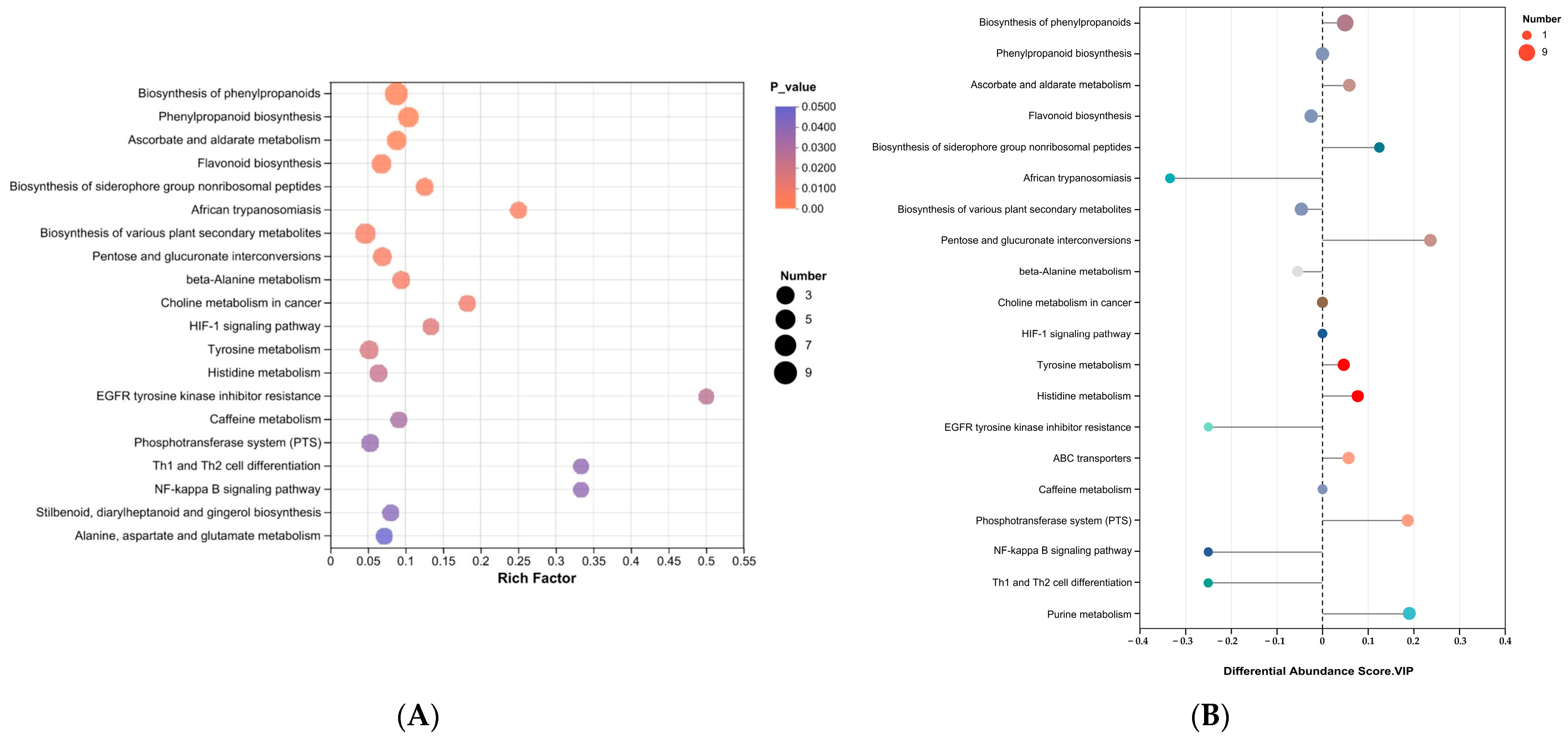
The metabolic pathways and regulatory mechanisms within organisms are highly complex and interconnected. They work together to regulate cellular biochemical processes. Conducting a comprehensive and systematic analysis of metabolism and regulatory pathways helps gain a deeper understanding of how changes in experimental conditions affect biological processes. A search in relevant databases followed by metabolic pathway enrichment analysis was performed, and the enrichment ratios and p-values were subsequently calculated to screen pathways containing differential metabolites. Figure 8A,B shows the top 20 pathways ranked based on the p-value from the smallest to the largest. KEGG pathway enrichment analysis and differential abundance analysis revealed significant enrichment in the biosynthesis pathways of phenylpropanoid compounds, ascorbic acid, aldonic acid metabolism, and the biosynthesis of iron carrier group non-ribosomal peptides. Of note, the overall trend of these pathways was upregulation. No significant changes were observed in the phenylpropanoid biosynthesis pathway. In contrast, the flavonoid biosynthesis pathway showed an overall downregulation trend. In the samples tested, 22 metabolites, of which 6 were identified as differential metabolites, were detected in the phenylpropanoid biosynthesis pathway (Figure 8). Spermidine and 5-Coumaroylquinic acid decreased by 0.43-fold, and 1-O-mustard oil-β-D-glucose decreased by 1-fold. Trans-Chlorogenic acid increased by 121.2-fold, and Artemisinin increased by 0.6-fold. Chlorogenic acid is a kind of phenylpropanoid synthesized by plant cells through shikimic acid pathway, which is the product of plant secondary metabolism phenylpropanoid pathway. During the fermentation process, A. cristatus may stimulate the expression of phenylpropanoid pathway-related genes in lotus leaf cells by secreting metabolites, thereby promoting the synthesis of chlorogenic acid, or indirectly enhancing the activity of chlorogenic acid synthase.

4. Conclusions

In this study, lotus leaves underwent solid-state fermentation mediated by A. cristatus. A comprehensive sensory evaluation of the fermented leaves revealed a substantial enhancement in the sensory attributes and flavor profile of the resulting lotus leaf tea. Analysis of the bioactive components showed a notable reduction in the total flavonoid content, whereas the levels of alkaloids remained relatively unchanged. Subsequent metabolomic analysis revealed that the fermentation process induced the production of several metabolites with potential functional activities, including chlorogenic acid, strychnine, kamanbulin A, and caffeoyl tartaric acid. These findings suggest that A. cristatus fermentation may endow lotus leaves with novel pharmacological properties, thereby providing a foundation for future investigations into the fermentation of lotus leaves by this fungus.

Author Contributions

Conceptualization, W.D.; Investigation, Y.T.; Data curation, S.H.; Funding acquisition, L.Z.; Methodology, X.R.; Project administration, Y.H.; Supervision, W.D.; Writing—original draft, W.D.; Writing—review and editing, L.S. and Y.L. All authors have read and agreed to the published version of the manuscript.

Funding

This research was funded by the Research and Application of the Core Technique on Diversified Utilization of Agricultural Biomass (QianNongKe JBGS[2024]05), Central government guidance for local science and technology development projects for Guizhou province ([2023]027) and Guizhou Key Laboratory of Agricultural Microbiology (QianKeHePingTai[2025]029).

Institutional Review Board Statement

Not applicable.

Informed Consent Statement

Informed consent was obtained from all subjects involved in the study.

Data Availability Statement

The original contributions presented in this study are included in the article. Further inquiries can be directed to the corresponding authors.

Conflicts of Interest

The authors declare no conflict of interest.

References

  1. Wang, G.; Ma, F.; Xie, K.; Li, X.; Tan, X.; Xia, Y.; Wang, Y.; Dong, J. Liensinine alleviates mouse intestinal injury induced by sepsis through inhibition of oxidative stress, inflammation, and cell apoptosis. Int. Immunopharmacol. 2024, 127, 111335. [Google Scholar] [CrossRef] [PubMed]
  2. Wang, Z.; Cheng, Y.; Zeng, M.; Wang, Z.; Qin, F.; Wang, Y.; Chen, J.; He, Z. Lotus (Nelumbo nucifera Gaertn.) leaf: A narrative review of its Phytoconstituents, health benefits and food industry applications. Trends Food Sci. Technol. 2021, 112, 631–650. [Google Scholar] [CrossRef]
  3. Feng, J.; Wang, J.; Bu, T.; Ge, Z.; Yang, K.; Sun, P.; Wu, L.; Cai, M. Structural, in vitro digestion, and fermentation characteristics of lotus leaf flavonoids. Food Chem. 2023, 406, 135007. [Google Scholar] [CrossRef]
  4. Guo, Y.; Chen, X.; Qi, J.; Yu, B. Simultaneous qualitative and quantitative analysis of flavonoids and alkaloids from the leaves of Nelumbo nucifera Gaertn. using high-performance liquid chromatography with quadrupole time-of-flight mass spectrometry. J. Sep. Sci. 2016, 39, 2499–2507. [Google Scholar] [CrossRef] [PubMed]
  5. Luo, J.; Xiao, W.; Liu, Z. Research progress of alkaloids in lotus leaves. Today Pharm. 2008, 3, 9–12+17. [Google Scholar]
  6. Huang, C.; Peng, X.; Pang, D.-J.; Li, J.; Paulsen, B.S.; Rise, F.; Chen, Y.-L.; Chen, Z.-L.; Jia, R.-Y.; Li, L.-X.; et al. Pectic polysaccharide from Nelumbo nucifera leaves promotes intestinal antioxidant defense in vitro and in vivo. Food Funct. 2021, 12, 10828–10841. [Google Scholar] [CrossRef]
  7. Qi, Q.; Li, R.; Li, H.-Y.; Cao, Y.-B.; Bai, M.; Fan, X.-J.; Wang, S.-Y.; Zhang, B.; Li, S. Identification of the anti-tumor activity and mechanisms of nuciferine through a network pharmacology approach. Acta Pharmacol. Sin. 2016, 37, 963–972. [Google Scholar] [CrossRef] [PubMed]
  8. Zhang, H.; Chen, G.; Zhang, Y.; Yang, M.; Chen, J.; Guo, M. Potential hypoglycemic, hypolipidemic, and anti-inflammatory bioactive components in Nelumbo nucifera leaves explored by bioaffinity ultrafiltration with multiple targets. Food Chem. 2022, 375, 131856. [Google Scholar] [CrossRef]
  9. Zhou, G.; Feng, X.; Tao, A. Explore the lipid-lowering and weight-reducing mechanism of lotus leaf based on network pharmacology and molecular docking. Evid.-Based Complement. Altern. Med. 2021, 2021, 1464027. [Google Scholar] [CrossRef]
  10. Wu, N.; Xie, H.; Fang, Y.; Liu, Y.; Xi, X.; Chu, Q.; Dong, G.; Lan, T.; Wei, Y. Isolation and purification of alkaloids from lotus leaves by ionic-liquid-modified high-speed countercurrent chromatography. J. Sep. Sci. 2018, 41, 571–577. [Google Scholar] [CrossRef]
  11. Wang, C.; Wang, L.; Yang, Y.; Hu, Y.; Cai, H.; Lu, Y. Research progress on alkaloid active ingredients in Nelumbinis Folium against non-alcoholic fatty liver disease. Shanghai J. Tradit. Chin. Med. 2023, 57, 76–82. [Google Scholar] [CrossRef]
  12. Zhu, X.; Hao, R.; Lv, X.; Zhou, X.; Li, D.; Zhang, C. Nuciferine ameliorates high-fat diet-induced disorders of glucose and lipid metabolism in obese mice based on the gut–liver axis. Food Front. 2023, 5, 188–201. [Google Scholar] [CrossRef]
  13. Yan, C.; Zhan, Y.; Yuan, S.; Cao, Y.; Chen, Y.; Dong, M.; Zhang, H.; Chen, L.; Jiang, R.; Liu, W.; et al. Nuciferine prevents obesity by activating brown adipose tissue. Food Funct. 2024, 15, 967–976. [Google Scholar] [CrossRef]
  14. Jiang, H.; Zhang, Q.; Yang, J.; Shu, Z.; Zhang, H. Research Progress on Extraction Technologies and Pharmacological Activities of Flavonoids from Lotus Leaf. Shandong Chem. Ind. 2022, 51, 79–81. [Google Scholar] [CrossRef]
  15. Wang, Z.; Xue, C.; Wang, X.; Zeng, M.; Wang, Z.; Chen, Q.; Chen, J.; Christian, M.; He, Z. Quercetin 3-O-glucuronide-rich lotus leaf extract promotes a Brown-fat-phenotype in C3H10T1/2 mesenchymal stem cells. Food Res. Int. 2023, 163, 112198. [Google Scholar] [CrossRef]
  16. Limwachiranon, J.; Huang, H.; Shi, Z.; Li, L.; Luo, Z. Lotus Flavonoids and Phenolic Acids: Health Promotion and Safe Consumption Dosages. Compr. Rev. Food Sci. Food Saf. 2018, 17, 458–471. [Google Scholar] [CrossRef] [PubMed]
  17. Zhang, L.; Zhou, Q.-M.; Xu, L.; Xie, X.; Wang, P.-X.; Xie, Z.-H.; Li, J.-L.; Tu, Z.-C. Extraction optimization and identification of four advanced glycation-end products inhibitors from lotus leaves and interaction mechanism analysis. Food Chem. 2023, 414, 135712. [Google Scholar] [CrossRef]
  18. Wang, Z.; Xue, C.; Yang, T.; Zeng, M.; Wang, Z.; Chen, Q.; Chen, J.; He, Z. Miquelianin, a main functional flavonoid of lotus leaf, induces thermogenic signature via p38-PINK1-PARKIN-mediated mitophagy and mimicking NRF2 signaling during brown adipocyte differentiation. Food Front. 2023, 4, 1831–1844. [Google Scholar] [CrossRef]
  19. Xie, Y.; Li, C.; Feng, H.; Jia, M.; Wei, X. Research progress on microbial fermentation of active ingredients of traditional Chinese medicine. Chin. Tradit. Pat. Med. 2025, 47, 182–187. [Google Scholar]
  20. He, Y.; Tao, Y.; Qiu, L.; Xu, W.; Huang, X.; Wei, H.; Tao, X. Lotus (Nelumbo nucifera Gaertn.) Leaf-Fermentation Supernatant Inhibits Adipogenesis in 3T3-L1 Preadipocytes and Suppresses Obesity in High-Fat Diet-Induced Obese Rats. Nutrients 2022, 14, 4348. [Google Scholar] [CrossRef] [PubMed]
  21. Yu, Y.; Huang, J.; Li, C.; Wang, J.; Jiang, X. Effects of shaking and fermentation on the quality of lotus leaf tea. Newsl. Seric. Tea 2023, 4, 1–7. [Google Scholar]
  22. Gao, X.; Kan, X.; Du, F.; Sun, L.; Li, X.; Liu, J.; Liu, X.; Yao, D. The Manufacturing Process of Lotus (Nelumbo Nucifera) Leaf Black Tea and Its Microbial Diversity Analysis. Foods 2025, 14, 519. [Google Scholar] [CrossRef]
  23. Li, Q.; Jin, Y.; Jiang, R.; Xu, Y.; Zhang, Y.; Luo, Y.; Huang, J.; Wang, K.; Liu, Z. Dynamic changes in the metabolite profile and taste characteristics of Fu brick tea during the manufacturing process. Food Chem. 2021, 344, 128576. [Google Scholar] [CrossRef]
  24. Kang, D.; Su, M.; Duan, Y.; Huang, Y. Eurotium cristatum, a potential probiotic fungus from Fuzhuan brick tea, alleviated obesity in mice by modulating gut microbiota. Food Funct. 2019, 10, 5032–5045. [Google Scholar] [CrossRef] [PubMed]
  25. Xie, L.; Zhou, L.; Zhang, R.; Zhou, H.; Yang, Y. Material Composition Characteristics of Aspergillus cristatus under High Salt Stress through LC–MS Metabolomics. Molecules 2024, 29, 2513. [Google Scholar] [CrossRef]
  26. Deng, J.; Li, Y.; Yuan, Y.; Yin, F.; Chao, J.; Huang, J.; Liu, Z.; Wang, K.; Zhu, M. Secondary Metabolites from the Genus Eurotium and Their Biological Activities. Foods 2023, 12, 4452. [Google Scholar] [CrossRef] [PubMed]
  27. Wu, W.; Qin, Z.; Liu, J.; Chen, L.; Xia, Y.; Li, X.; Liu, X.; Li, Y. Effect of Processing Technology on Quality and Functional Activity of Taxilli Herba Tea. Sci. Technol. Food Ind. 2023, 44, 149–157. [Google Scholar] [CrossRef]
  28. Zou, M.; Zhang, W.; Dong, Q.; Tang, C.; Cao, F.; Su, E. Submerged fermentation of Ginkgo biloba seed powder using Eurotium cristatum for the development of ginkgo seeds fermented products. J. Sci. Food Agric. 2021, 101, 1782–1791. [Google Scholar] [CrossRef]
  29. Zhang, D.; Huang, Y.; Fan, X.; Zeng, X. Effects of solid-state fermentation with Aspergillus cristatus (MK346334) on the dynamics changes in the chemical and flavor profile of dark tea by HS-SPME-GC–MS, HS-GC-IMS and electronic nose. Food Chem. 2024, 455, 139864. [Google Scholar] [CrossRef]
  30. GB/T 23776-2018; Methodology for Sensory Evaluation of Tea. China National Standardization Management Committee: Beijing, China, 2018.
  31. Shi, G.; Xiang, Q.; Fan, Y.; Zhao, W.; Liu, Y. Optimization of Process on Extracting Flavonoids from Dry Onion Skins by Alkaline Method. Open Chem. Eng. J. 2015, 9, 39–46. [Google Scholar] [CrossRef]
  32. Kong, X.; Guo, Z.; Yao, Y.; Xia, L.; Liu, R.; Song, H.; Zhang, S. Acetic acid alters rhizosphere microbes and metabolic composition to improve willows drought resistance. Sci. Total Environ. 2022, 844, 157132. [Google Scholar] [CrossRef] [PubMed]
  33. Li, C.; Al-Dalali, S.; Zhou, H.; Xu, B. Influence of curing on the metabolite profile of water-boiled salted duck. Food Chem. 2022, 397, 133752. [Google Scholar] [CrossRef]
  34. Li, Z.; Wu, Y.; Hu, J.; Yang, G.; Wang, Z.; Sun, J. Dissection of the response mechanism of alfalfa under phosphite stress based on metabolomic and transcriptomic data. Plant Physiol. Biochem. 2022, 192, 35–49. [Google Scholar] [CrossRef]
  35. Du, Y.; Yang, W.; Yang, C.; Yang, X. A comprehensive review on microbiome, aromas and flavors, chemical composition, nutrition and future prospects of Fuzhuan brick tea. Trends Food Sci. Technol. 2022, 119, 452–466. [Google Scholar] [CrossRef]
  36. Xiang, Z.; Guan, H.; Zhao, X.; Xie, Q.; Xie, Z.; Cai, F.; Dang, R.; Li, M.; Wang, C. Dietary gallic acid as an antioxidant: A review of its food industry applications, health benefits, bioavailability, nano-delivery systems, and drug interactions. Food Res. Int. 2024, 180, 114068. [Google Scholar] [CrossRef]
  37. Sun, J.; Xia, P.; Liang, Z. Pharmacological efficacy of Nelumbinis folium and development of functional food products. J. Zhejiang Agric. Sci. 2021, 62, 1874–1881. [Google Scholar] [CrossRef]
  38. Bayazeid, O.; Nemutlu, E.; Eylem, C.C.; Yalçın, F.N. Neuroactivity of naturally occurring proaporphine alkaloid, pronuciferine. J. Biochem. Mol. Toxicol. 2020, 34, e22601. [Google Scholar] [CrossRef] [PubMed]
  39. Bayazeid, O.; Nemutlu, E.; Eylem, C.C.; İlhan, M.; Küpeli-Akkol, E.; Karahan, H.; Kelicen-Uğur, P.; Ersoz, T.; Yalçın, F.N. Neuroactivity of the naturally occurring aporphine alkaloid, roemerine. Nat. Prod. Res. 2021, 35, 6147–6152. [Google Scholar] [CrossRef]
  40. Ma, B.; Shi, S.; Guo, W.; Zhang, H.; Zhao, Z.; An, H. Liensinine, a Novel and Food-Derived Compound, Exerts Potent Antihepatoma Efficacy via Inhibiting the Kv10.1 Channel. J. Agric. Food Chem. 2024, 72, 4689–4702. [Google Scholar] [CrossRef]
  41. Manogaran, P.; Anandan, A.; Padma, V.V. Isoliensinine augments the therapeutic potential of paclitaxel in multidrug-resistant colon cancer stem cells and induced mitochondria-mediated cell death. J. Biochem. Mol. Toxicol. 2023, 37, e23395. [Google Scholar] [CrossRef]
  42. Levorato-Vinche, A.D.; Melhem, M.d.S.C.; Bonfietti, L.X.; De-La-Cruz-Chacón, I.; Boaro, C.S.F.; Fabro, A.T.; Ferreira, G.; Silva, J.d.F.d.; dos Santos, D.C.; Pereira, B.A.S.; et al. Antifungal activity of liriodenine on clinical strains of Cryptococcus neoformans and Cryptococcus gattii species complexes. J. Venom. Anim. Toxins Incl. Trop. Dis. 2022, 28, e20220006. [Google Scholar] [CrossRef] [PubMed]
  43. Weng, T.; Shen, C.; Chiu, Y.; Lin, Y.; Huang, Y. Effects of armepavine against hepatic fibrosis induced by thioacetamide in rats. Phytotherapy Res. 2012, 26, 344–353. [Google Scholar] [CrossRef] [PubMed]
  44. Al-Zahrani, M.H.; Alghamdi, R.A. In Silico Molecular Docking Analysis of the Potential role of Reticuline and Coclaurine as Anti-colorectal Cancer Alkaloids. J. Pharm. Res. Int. 2022, 34, 33–42. [Google Scholar] [CrossRef]
  45. Rodrigues, M.T.; Michelli, A.P.P.; Caso, G.F.; de Oliveira, P.R.; Rodrigues-Junior, D.M.; Morale, M.G.; Júnior, J.M.; Bortoluci, K.R.; Tamura, R.E.; da Silva, T.R.C.; et al. Lysicamine Reduces Protein Kinase B (AKT) Activation and Promotes Necrosis in Anaplastic Thyroid Cancer. Pharmaceuticals 2023, 16, 1687. [Google Scholar] [CrossRef]
  46. Nguyen, V.; Taine, E.G.; Meng, D.; Cui, T.; Tan, W. Chlorogenic Acid: A Systematic Review on the Biological Functions, Mechanistic Actions, and Therapeutic Potentials. Nutrients 2024, 16, 924. [Google Scholar] [CrossRef]
  47. Ren, F.; Hu, J.; Dang, Y.; Deng, H.; Ren, J.; Cheng, S.; Tan, M.; Zhang, H.; He, X.; Yu, H.; et al. Sphondin efficiently blocks HBsAg production and cccDNA transcription through promoting HBx degradation. J. Med. Virol. 2023, 95, e28578. [Google Scholar] [CrossRef]
  48. Zhou, L.; Liu, Q.; Hong, G.; Song, F.; Zhao, J.; Yuan, J.; Xu, J.; Tan, R.X.; Tickner, J.; Gu, Q.; et al. Cumambrin A prevents OVX-induced osteoporosis via the inhibition of osteoclastogenesis, bone resorption, and RANKL signaling pathways. FASEB J. 2019, 33, 6726–6735. [Google Scholar] [CrossRef]
  49. Gao, T.-H.; Liao, W.; Lin, L.-T.; Zhu, Z.-P.; Lu, M.-G.; Fu, C.-M.; Xie, T. Curcumae rhizoma and its major constituents against hepatobiliary disease: Pharmacotherapeutic properties and potential clinical applications. Phytomedicine 2022, 102, 154090. [Google Scholar] [CrossRef]
  50. Li, C.; Zhang, Z.; Wang, J. A Thermophilic Biofunctional Multienzyme Cascade Reaction for Cell-Free Synthesis of Salvianic Acid A and 3,4-Dihydroxymandelic Acid. ACS Sustain. Chem. Eng. 2019, 7, 18247–18253. [Google Scholar] [CrossRef]
  51. Reang, J.; Sharma, P.C.; Thakur, V.K.; Majeed, J. Understanding the Therapeutic Potential of Ascorbic Acid in the Battle to Overcome Cancer. Biomolecules 2021, 11, 1130. [Google Scholar] [CrossRef]
  52. Zou, M.-Y.; Nie, S.-P.; Yin, J.-Y.; Xie, M.-Y. Ascorbic acid induced degradation of polysaccharide from natural products: A review. Int. J. Biol. Macromol. 2020, 151, 483–491. [Google Scholar] [CrossRef] [PubMed]
  53. Singh, R.; Chandel, S.; Ghosh, A.; Matta, T.; Gautam, A.; Bhattacharya, A.; Babu, S.S.; Sukla, S.; Nag, D.; Ravichandiran, V.; et al. Glucogallin Attenuates the LPS-Induced Signaling in Macrophages and Protects Mice against Sepsis. Int. J. Mol. Sci. 2022, 23, 11254. [Google Scholar] [CrossRef] [PubMed]
  54. Nomura, S.; Monobe, M.; Ema, K.; Yoshida, K.; Yamashita, S.; Ogino, A.; Nesumi, A. Effects of a Tea Cultivar “MK5601” on Behaviors and Hippocampal Neurotrophin-3 Levels in Middle-Aged Mice. J. Nutr. Sci. Vitaminol. 2021, 67, 170–179. [Google Scholar] [CrossRef] [PubMed]
  55. Zhou, D.; Liu, W.; Zhang, J.; Dong, Y.; Wu, J.; Dai, C.; Zhang, T.; Yang, G.; Zhang, Y.; Li, A. Bellidifolin ameliorates isoprenaline-induced cardiac hypertrophy by the Nox4/ROS signalling pathway through inhibiting BRD4. Cell Death Discov. 2023, 9, 279. [Google Scholar] [CrossRef]
  56. Woo, S.H.; Lee, S.-H.; Moon, S.-J.; Han, J.; Seo, K.-S.; Lee, H.; Lee, C.-H.; Hwang, J.H. Beta-lapachone ameliorates the progression of primary sclerosing cholangitis pathogenesis in rodent models. Life Sci. 2024, 337, 122342. [Google Scholar] [CrossRef]
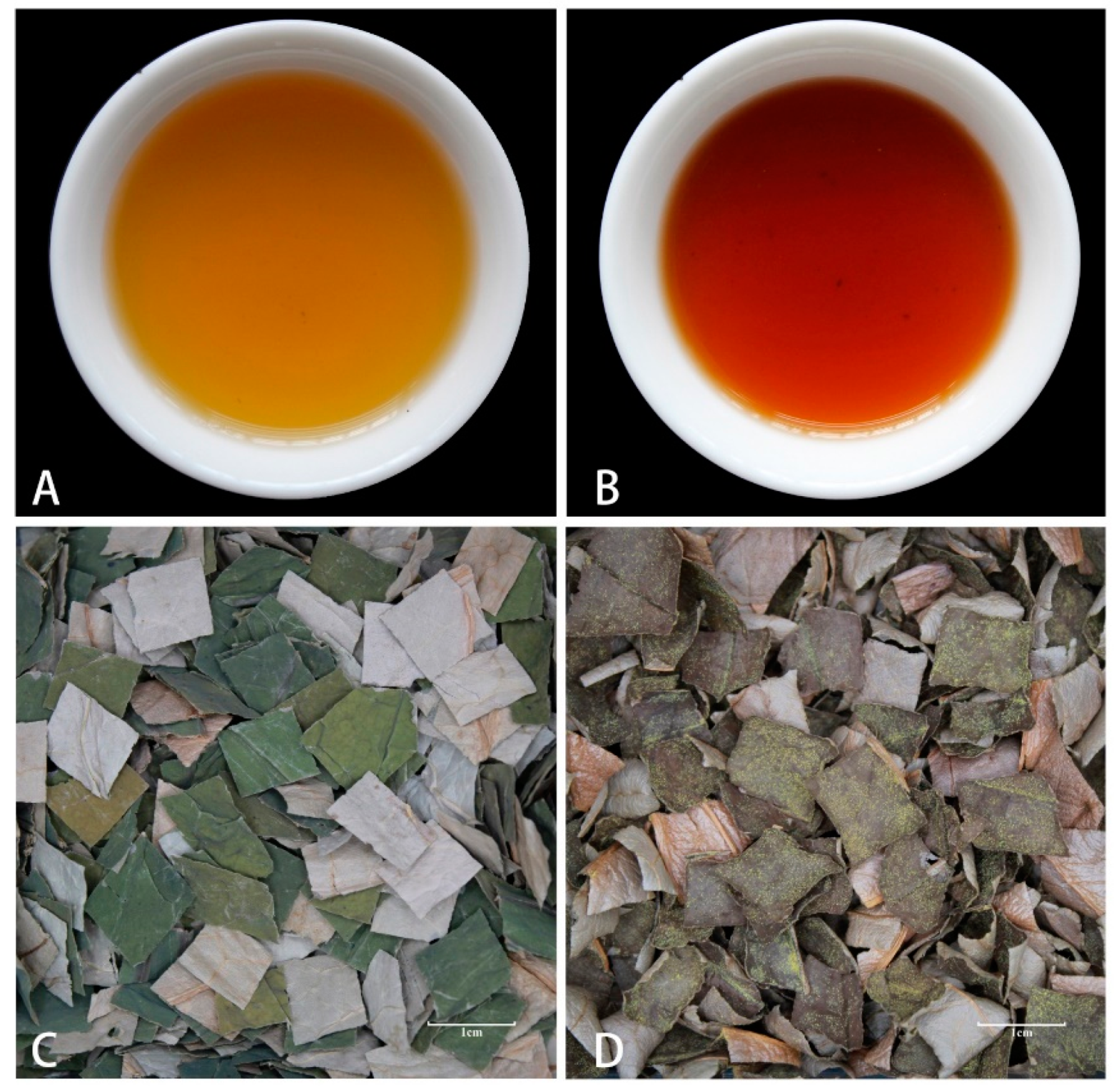
Figure 1. The color and appearance of UL and FL infusions: (A) UL infusion color; (B) FL infusion color; (C) UL infusion appearance; (D) FL infusion appearance. Note: UL, the unfermented lotus leaf; FL, the fermented lotus leaf.
Fermentation 11 00279 g001
Figure 2. Concentration of main active components: (A) The total flavonoids of UL and FL; (B) The total alkaloids of UL and FL. Note: UL, the unfermented lotus leaf; FL, the fermented lotus leaf; *** means the flavonoids or alkaloids between UL and FL show significantly different values at the p < 0.001.
Fermentation 11 00279 g002
Figure 3. Total ion current diagram of UL and FL: (A) Positive-ion mode analysis; (B) Negative-ion mode analysis.
Fermentation 11 00279 g003
Figure 4. PCA, PLS-DA score plots and permutation test, and OPLS-DA S-plot of the samples before and after fermentation: (A) PCA score plots; (B) PLS-DA score plots; (C) PLS-DA permutation test; (D) OPLS-DA S-plot; The red dots indicate that the VIP values of these metabolites are greater than or equal to 1, and the green dots indicate that the VIP values of these metabolites are less than 1.
Fermentation 11 00279 g004
Figure 5. Heatmap of (A) all alkaloids and (B) reported alkaloids. Note: UL, the unfermented lotus leaf; FL, the fermented lotus leaf; lowercase letters indicate different sample (a–l); the color gradient in the legend corresponds to the relative abundance of the metabolite.
Fermentation 11 00279 g005
Figure 6. Heatmap of all flavonoids. Note: UL, the unfermented lotus leaf; FL, the fermented lotus leaf; capital letters indicate different sample of UL (A–F); lowercase letters indicate different sample of FL (a–f); the color gradient in the legend corresponds to the relative abundance of the metabolite.
Fermentation 11 00279 g006
Figure 7. Venn plots (A) and volcanos plots (B) for UL vs. FL. Note: UL, the unfermented lotus leaf; FL, the fermented lotus leaf.
Fermentation 11 00279 g007
Figure 8. Enrichment analysis of pathways associated with the differentially expressed metabolites: (A) Pathway enrichment analysis diagram; (B) Pathway difference abundance score chart.
Fermentation 11 00279 g008
Table 1. Sensory evaluation standards of UL and FL.
ProjectStandard for EvaluationScore (Points)
Appearance (15%)Shade of yellow, with the surface covered with A. cristatus.11~15
Shade of yellow, with less A. cristatus scattered on the surface.6~10
Jade green, without A. cristatus.0~5
Aroma
(35%)
Unique A. cristatus aroma and fragrance of lotus leaf, with both exhibiting richness and potency.31~35
The unique aroma of A. cristatus is mild, with a rich fragrance of lotus leaf.21~30
Mild A. cristatus and lotus leaf aroma.11~20
Only one aroma is smelled.0~10
Taste
(35%)
Pronounced unique flavor of A. cristatus, providing a stimulating experience for the taste buds without any astringency.28~35
Slightly unique flavor of A. cristatus, with a faintly bitter and astringent taste.15~27
Off-flavor, with a bitter and astringent taste.0~14
Infusion color
(10%)
Orange-yellow, clear, and bright.6~10
Pale yellow, clear and bright.0~5
Leaf residue
(5%)
Reddish-brown, fairly uniform.3~5
Green, not sufficiently uniform.0~2
Note: UL, the unfermented lotus leaf; FL, the fermented lotus leaf.
Table 2. The sensory evaluation scores.
GroupScoreTotal Score
AppearanceAromaTasteInfusion ColorLeaf Residue
UL4.90 ± 0.329.60 ± 0.8412.20 ± 1.994.90 ± 0.322.00 ± 0.0033.60 ± 2.12
FL9.30 ± 1.70 *28.50 ± 5.68 *24.80 ± 3.62 *7.80 ± 0.79 *3.30 ± 0.48 *70.90 ± 7.81 *
Note: UL, the unfermented lotus leaf; FL, the fermented lotus leaf; * means sensory property between UL and FL show significantly different values at the p < 0.05.
Table 3. The top 20 metabolites ranked based on the VIP value.
MetaboliteClassificationFormulaVIPDifference Multiple
Trans-Chlorogenic acidphenylpropanoidC16H18O93.3151122.2079
SphondinphenylpropanoidC12H8O42.588614.848
Cumambrin AterpenesC17H22O52.46072.4171
Byssochlamic acidphenylpropanoidC18H20O62.34431.9447
FuranodieneterpenesC15H20O2.31352.38
MatricarinlactonesC17H20O52.23491.7867
Isorhamnetin 3-O-(beta-D-glucopyranosyl-(1->6)
-beta-D-glucopyranoside)
phenolsC28H32O172.21832.1373
Thiophene-4,5-epoxidephenylpropanoidC14H12O4S2.20012.0397
3-Oxoadipic acidorganic acidsC6H8O52.18921.8412
3,4-Dihydroxymandelic AcidphenolsC8H8O52.18572.0714
Cis-Caffeoyl tartaric acidphenylpropanoidC13H12O92.17022.0788
Ascorbic acidorganic acidsC6H8O62.16982.3275
D-Xylono-1,5-lactonelactonesC5H8O52.15112.4134
Beta-GlucogallinphenylpropanoidC13H16O102.12122.1199
Gallic AcidphenylpropanoidC7H6O52.11971.5084
TheogallinphenylpropanoidC14H16O102.10452.0177
4-ButylphenolphenolsC10H14O2.10291.962
BellidifolinquinonesC14H10O62.09521.793
Beta-LapachonequinonesC15H14O32.08891.9392
10-Hydroxy-3-methoxy-1,3,5,7
-cadinatetraen-9-one
terpenesC16H20O32.08541.8185
Disclaimer/Publisher’s Note: The statements, opinions and data contained in all publications are solely those of the individual author(s) and contributor(s) and not of MDPI and/or the editor(s). MDPI and/or the editor(s) disclaim responsibility for any injury to people or property resulting from any ideas, methods, instructions or products referred to in the content.