Article

Efficient Synthesis of High-Active Myoglobin and Hemoglobin by Reconstructing the Mitochondrial Heme Synthetic Pathway in Engineered Saccharomyces cerevisiae

by
Xiaoyan Sun
1,2,3,4,
Yunpeng Wang
1,2,3,4,
Yijie Wang
1,2,3,4,
Jingwen Zhou
1,2,3,4,
Jianghua Li
1,2,3,4,
Jian Chen
1,2,3,4,
Guocheng Du
1,2,3,4,5 and
Xinrui Zhao
1,2,3,4,*
1
Science Center for Future Foods, Jiangnan University, 1800 Lihu Road, Wuxi 214122, China
2
Key Laboratory of Industrial Biotechnology, Ministry of Education, School of Biotechnology, Jiangnan University, 1800 Lihu Road, Wuxi 214122, China
3
Jiangsu Province Engineering Research Center of Food Synthetic Biotechnology, Jiangnan University, 1800 Lihu Road, Wuxi 214122, China
4
Engineering Research Center of Ministry of Education on Food Synthetic Biotechnology, Jiangnan University, 1800 Lihu Road, Wuxi 214122, China
5
Key Laboratory of Carbohydrate Chemistry and Biotechnology, Ministry of Education, Jiangnan University, 1800 Lihu Road, Wuxi 214122, China
*
Author to whom correspondence should be addressed.
Fermentation 2025, 11(5), 246; https://doi.org/10.3390/fermentation11050246
Submission received: 2 April 2025 / Revised: 25 April 2025 / Accepted: 30 April 2025 / Published: 1 May 2025
(This article belongs to the Section Microbial Metabolism, Physiology & Genetics)

Abstract

:
Currently, various types of myoglobins and hemoglobins are widely used in the fields of food additives and biocatalytic applications. However, the limited availability of heme constrains the biosynthesis of these high-activity hemoproteins in microbial chassis cells. In this work, a new heme synthetic pathway was reconstructed in the mitochondria by eliminating the spatial barrier during heme synthesis in Saccharomyces cerevisiae, resulting in a significant enhancement in intracellular heme supply. To further enhance the supply of the essential precursor for heme synthesis (5-aminolevulinate, ALA), the special ALA exporter in the mitochondrial membrane (Ort1p) was identified and knocked out. Moreover, the mitochondrial heme exporter (Ygr127wp) was overexpressed to promote the transport of heme to the cytoplasm to participate in the synthesis of various myoglobins and hemoglobins. Based on these strategies in the engineered strain, the binding ratios of heme in porcine myoglobin (52.4 ± 4.9%) and soybean hemoglobin (75.5 ± 2.8%) were, respectively, increased by 2.4-fold and 3.6-fold, and the titers of porcine myoglobin (130.5 ± 2.8 mg·L−1) and soybean hemoglobin (152.8 ± 2.6 mg·L−1), respectively, increased by 31.1% and 42.1%. Furthermore, the engineered strain presents great potential in the efficient synthesis of other heme-binding proteins and enzymes in S. cerevisiae.

1. Introduction

Myoglobins (Mbs) and hemoglobins (Hbs) are essential hemoproteins that serve critical functions in transporting and storing oxygen [1], binding and clearing toxic nitric oxide [2], and regulating the levels of intracellular pH [3]. Mb is a monomeric protein that can store oxygen and promote its diffusion in muscles [1], while Hb is a tetramer protein composed of four subunits that can transport oxygen in the blood. Recent advances in Mb and Hb research have expanded their application as iron fortifiers, food-grade colorants, and nutritional additives, showing their huge potential in the fields of nutrition and health [4]. Notably, biosynthetic leghemoglobin (LegH) has received FDA approval as a food additive to simulate the real color and flavor in artificial meat [5] and myoglobin, such as bovine myoglobin and porcine myoglobin (pMb), also play important roles in the improvement of meat color and texture [6]. Due to the increasing demand for Mb and Hb, current time-consuming and expensive extraction methods cannot meet the requirements of large-scale green manufacture [7]. With the development of synthetic biology, the biosynthesis of heterologous proteins by microbial cell factories is a more economical and environmentally friendly approach for industrial production. As a GRAS (generally recognized as safe) and easy-to-use model host, Saccharomyces cerevisiae has been applied in the synthesis of various chemicals [8] and can be developed as an ideal platform for the efficient biosynthesis of Mb and Hb.
Heme is an essential cofactor of Mb and Hb, but the intracellular supply of heme is insufficient to efficiently express heterologous proteins, resulting in the weak activities of Mb and Hb [9]. Although exogenous addition of heme can slightly increase the heme-binding rates of proteins, the cost will be added, and it is not suitable for the large-scale industrial production [10]. Consequently, augmenting endogenous heme biosynthesis represents a critical challenge for producing high-activity hemoproteins [11]. In S. cerevisiae, the heme synthetic pathway is compartmentalized between the mitochondria and the cytoplasm [9]. This spatial isolation phenomenon is a big barrier to efficiently synthesizing heme because the intermediates need to cross the double membrane of the mitochondria. The first step in the heme synthetic pathway takes place in the mitochondria and is catalyzed by Hem1p which converts succinyl-CoA and glycine into 5-aminolevulinic acid (ALA). ALA is subsequently exported to the cytoplasm, where Hem2p, Hem3p, Hem4p, and Hem12p successively catalyze ALA to form porphobilinogen (PBG), hydroxymethylbilane (HMB), uroporphyrinogen III (UPG III), and coproporphyrinogen III (CPG III). Next, CPG III is transported back to the mitochondria and transformed into protoporphyrinogen IX (PPG IX) by Hem13p. PPG IX is further oxidized to protoporphyrin IX (PP IX) by Hem14p, and finally, Hem15p catalyzes the reaction of PP IX and Fe2+ to form heme [12] (Figure 1).
The inefficiency of intracellular heme synthesis and utilization is closely related to the spatial compartmentalization of the heme biosynthetic pathway [13]. To address this limitation, Xue et al. identified the subcellular localization of eight key enzymes in the heme synthetic pathway by visualization analysis and by reconstructing the heme synthetic pathway in the cytoplasm. Their spatial rearrangement strategy led to a 2.5-fold increase in the intracellular heme content [13]. However, the lack of precursor (succinyl-CoA) in the cytoplasm resulted in inefficient ALA synthesis in the cytoplasm and the heme supply could not be guaranteed. Thus, retaining Hem1p in the mitochondria remains critical for the stability and availability of the new pathway.
Mitochondria are important organelles in eukaryotes, producing metabolic intermediates for diverse biosynthetic pathways [14], which rely on mitochondrial transporters to facilitate the transport of intermediates between the mitochondria and the cytoplasm. In particular, several mitochondrial transporters transport ALA from the mitochondria to the cytoplasm [9] and transport heme from the cytoplasm to the mitochondria [15]. However, the transport rates are limited during these processes, resulting in lower intracellular heme levels. Therefore, the mitochondrial transporters associated with heme and ALA are critical to the efficiency of heme utilization in the synthesis of high-active Mb and Hb. In addition, the application of mitochondria as powerful organelles for the efficient synthesis of terpenes and their precursor compounds has been verified [16,17]. Based on the combination of cytoplasmic metabolic engineering and mitochondrial compartmentalization, the titer of the heme precursor (CPG III) was increased in S. cerevisiae, reaching 402.8 ± 9.3 mg·L−1 at the fermenter level. Thus, it is feasible to create a new pathway to synthesize heme in mitochondria [18].
Herein, the mitochondrial heme synthetic pathway was reconstructed to eliminate the spatial isolation of the original synthetic pathway in S. cerevisiae. Concurrently, the intracellular supply of heme was further enhanced by mining the important mitochondrial ALA and heme transporters to overcome the low efficiency of heme synthesis and utilization. Finally, by combining the newly reconstructed heme biosynthetic pathway with an appropriate expressional platform for globins, significant increases in the titers and functional activities of pMb and LegH were achieved. Depending on the strategies and engineered S. cerevisiae strain, the effective synthesis of other hemoproteins (such as P450s, catalase, and peroxidase) with high titers and activities can be achieved.

2. Materials and Methods

2.1. Materials

TransStart® FastPfu DNA Polymerase (TransGen Biotech, Beijing, China) and Green Taq Mix DNA polymerase (Vazyme, Nanjing, China) were used for PCR and colony PCR, respectively. The kits for plasmid miniprep purification and PCR purification were purchased from TransGen Biotech (Beijing, China). The TIANGEN pre-kit (Tiangen, Beijing, China) was used to purify genomic DNA from S. cerevisiae. Restriction enzymes Bsa I and EcoR I, used for Golden Gate assembly, were purchased from Beyotime (Shanghai, China). The standards of 5-aminolevulinic acid hydrochloride and hemin were purchased from Sigma-Aldrich (St. Louis, MO, USA). Mitochondrion-specific fluorescent dye (Mito-Tracker Green FM) was purchased from Beyotime (Shanghai, China). Hemoglobin-ref (bovine hemoglobin) and Myoglobin-ref (equine skeletal muscle myoglobin) were purchased from Yuanye Bio-Technology (Shanghai, China).

2.2. Strains and Cultural Conditions

The wild-type S. cerevisiae strain CEN.PK2-1C was used as the host and the E. coli DH5α strain was used to construct vectors. The related strains are shown in Table S2. Luria–Bertani (LB) medium (10 g·L−1 NaCl, 10 g·L−1 peptone, and 5 g·L−1 yeast extract) with 100 mg·L−1 ampicillin or kanamycin was used to routinely cultivate the engineered E. coli DH5α strains. YPD medium (20 g·L−1 glucose, 20 g·L−1 peptone, and 10 g·L−1 yeast extract) was used for the culture of S. cerevisiae strains. The strains harboring plasmids with pGAL1 promoter were cultivated in YPDG medium (2% galactose was added after 18 h of cultivation in YPD medium). SD-Ura medium (6.7 g·L−1 yeast nitrogen base, 20 g·L−1 glucose, 40 mg·L−1 leucine, 40 mg·L−1 histidine, and 40 mg·L−1 tryptophan) was used for the screening of the constructed S. cerevisiae strains. SD-Ura-Trp medium (6.7 g·L−1 yeast nitrogen base, 20 g·L−1 glucose, 40 mg·L−1 leucine, and 40 mg·L−1 histidine) was used for the screening of the constructed strains that transporter genes knocked out. YPD medium with 1 g·L−1 5-fluoroorotic acid (5-FOA) was used to remove the Ura3 marker during the manipulation of gene editing in S. cerevisiae.

2.3. Plasmids and the Cassettes for Gene Knocking-In/Out Construction

All plasmids were constructed by Gibson assembly using pY26 or pESC as the backbones. Plasmids containing knock-in or knock-out cassettes were generated by Golden Gate assembly. All constructed plasmids were verified by sequencing (Sangon Biotech, Shanghai, China). The pRS426-Cas9-ura plasmid utilized in the CRISPR-Cas9 system for gene editing was preserved in our laboratory. The plasmids and primers used in this study are listed in Tables S1 and S2.

2.4. Fluorescent Visualization and Analysis by Microscopy

The heme synthetic enzymes (Hem2p, Hem3p, Hem4p, Hem12p, and Hem13p) were fused with an N-terminal mitochondrial localization signal (MLS) and C-terminal red fluorescent protein mScarlet. The LiAc method was employed for the transformation of S. cerevisiae. The engineered S. cerevisiae strains were cultured in YPD medium with shaking at 30 °C and 220 rpm for 24 h. Subsequently, strains in the logarithmic growth phase were harvested. The cells were diluted by PBS buffer (pH 7.4) to a final concentration of 106 cells·mL−1. The cell pellets were collected after centrifugation and resuspended in PBS buffer with 100 nM Mito-Tracker Green FM. Following a 30 min staining at 30 °C in the dark, the residual dye was removed by washing three times with PBS buffer. The stained cells were subsequently observed using an ECLIPSE Ci-L fluorescent microscope (Nikon, Tokyo, Japan) with a 100× oil lens. The Mito-Tracker Green FM was excited at 490 nm and emitted at 516 nm by fluorescent illuminator, while the mScarlet was excited at 570 nm and emitted at 605 nm by fluorescent illuminator. The bright-field images captured in the same field of view served as controls.

2.5. Qualitative and Quantitative Analysis of Fluorescent Localization

The software ImageJ (V1.8.0.112) was used to process and analyze the monochromatic fluorescent images to obtain scatter plot results and overlap coefficient values [19]. In ImageJ software, the red fluorescence images and the green fluorescence images were analyzed separately according to the Analyze–Colocalization–Colocalization Threshold process. The Rcoloc value in the Section 3 was used as the fluorescence overlap coefficient.

2.6. Intracellular Heme Measurements

The intracellular titers of heme were performed as previously mentioned [20]. The cells (OD600 × mL = 8) were collected in brown centrifuge tubes away from light. The cells were centrifuged at 8000 rpm for 5 min to collect the yeast cells, and then washed twice with PBS buffer. After centrifugation, we discarded the supernatant and suspended the yeast cells in 500 μL of 20 mM oxalic acid solution. The mixture was mixed evenly and placed in refrigerator at 4 °C for 16 h overnight. After the reaction, 500 μL of 2 M oxalic acid solution was mixed evenly with the overnight reaction solution, and then, 500 μL of the mixed solution was incubated in the water bath at 95 °C for 30 min. The remaining mixed solution was used as a control and placed at room temperature. After incubation, the mixed solution was centrifuged at 8000 rpm for 5 min, and the supernatant was used for fluorescence measurements. Fluorescence was detected by BioTek Synergy H1 (Agilent, Santa Clara, CA, USA) under excitation at λ = 400 nm and emission at λ = 620 nm. A standard curve for heme concentration was drawn using Hemin (Aladdin reagent, Shanghai, China).

2.7. Isolation of Mitochondria

Based on the mitochondrial extraction method from Meisinger et al. [21] and the mitochondrial isolation buffer optimized by Corcelli et al. [22], in this work, we used the mitochondrial extraction protocol optimized by Pan et al. [23] to separate the mitochondria from the cytoplasm. After incubation of the yeast cells in DTT buffer, yeast cells were washed with KClEM buffer (180 mM KCl, 1 mM EDTA, and 5 mM MOPS-KOH) (pH 7.2) and treated with yeast lytic enzyme Zymolyase-20T. Subsequently, the cells were resuspended and washed in cold KClEM buffer. Centrifugation was then performed at increasing speeds (250× g, 2 min; 1000× g, 4 min; and 1900× g, 4 min) to precipitate the nuclei. The supernatant was centrifuged at high speed at 16,800× g for 15 min, and the precipitate was regarded as the mitochondrial part, while the supernatant was regarded as the cytoplasm.

2.8. The Measurements of ALA in Mitochondria and Cytoplasm

The collected mitochondria were resuspended in PBS buffer and lysed by ultrasonic treatment. The mitochondrial lysate and the isolated cytoplasmic solution were prepared as samples for ALA quantification [24,25]. Specifically, 100 μL of acetylacetone and 200 μL of sodium acetate buffer (pH 4.6) were added, sequentially, to 400 μL of sample. The well-mixed solution was heated in boiling water for 15 min, and then cooled to room temperature. Then, 700 μL of Ehrlich’s reagent (30 mL of glacial acetic acid, 1 g of DMAB, and 8 mL of 70% perchloric acid added sequentially, dissolved, diluted with glacial acetic acid to 50 mL volume, and stored in the dark) was added. After a 20-min lightproof reaction, the absorbance was measured at 554 nm by using BioTek Synergy H1 (Agilent, Santa Clara, CA, USA). The standard curve was constructed using ALA hydrochloride as the reference standard.

2.9. Expression and Purification of Myoglobin and Hemoglobin

The single colony of engineered S. cerevisiae strain was inoculated into 5 mL of SD-ura medium and routinely cultivated at 30 °C and 220 rpm for 20 h. The 1% seed solution was inoculated into a 250 mL shaking flask with 50 mL YPD medium and then incubated at 30 °C and 220 rpm for 18 h. After adding 2% galactose as an inducer, the fermentation proceeded for 48 h at 30 °C and 220 rpm. The cells were collected by centrifugation at 4 °C after the incubation, and the strains were resuspended in 50 mL pre-cooled binding buffer (20 mM phosphate-buffered saline buffer, PBS) with a final concentration of 1 mM PMSF solution. The cells were lysed using a High-Pressure Cell Crusher (Union Biotech, Shanghai, China) at 1000 bar. The crude protein extract was obtained by collecting the supernatant after centrifugation at 4 °C. Protein purification was performed using His-Affinity (Ni NTA beads) agarose (Dianchuang Biotech, Shanghai, China). PBS buffer containing 50 mM imidazole was used as the washing buffer, and PBS buffer containing 1 M imidazole served as the elution buffer. The purified protein was resuspended in LDS sample buffer (Invitrogen Novex, Shanghai, China) at 98 °C for 10 min, and the samples were loaded to polyacrylamide gel electrophoresis (12%) for SDS PAGE. Image Lab Software for PC Version 6.1 was used to perform grayscale analysis of SDS-PAGE to calculate the proportion of the target protein band out of the total soluble protein. The Bradford protein Assay Kit (Beyotime Biotech, Shanghai, China) was used to measure the concentration of purified proteins.

2.10. Determination of Heme-Binding Ratios in Purified Myoglobin and Hemoglobin

The heme-binding ratios in proteins were characterized by analyzing the differential spectra between oxidized and reduced samples [26]. First, 100 μL of purified sample and 100 μL of solution I (0.2 M NaOH, 40% (v/v) pyridine and 500 µM potassium ferricyanide) were added to a 96-well plate. After thorough mixing, the sample was scanned in 500–600 nm intervals (wavelength spacing of 1 nm) to obtain the oxidized spectrum. Then, 2 μL of solution II (0.5 M sodium dithionite in 0.5 M NaOH) was added to the oxidized sample. After it was mixed well again, the sample turned pink and was scanned in the 500–600 nm interval to obtain the reduced spectrum. The heme-binding ratios were determined using Beer’s law, A = ε × c × l (absorbance = extinction coefficient × concentration × pathlength), ε550–535 = 23.8 mM−1 cm−1 for heme b.

2.11. Determination of Functional Activities for Synthesized Myoglobin and Hemoglobin

Determination of characteristic absorption peaks: 1 μM of purified myoglobin or hemoglobin was incubated in PBS buffer for 15 min. The samples were scanned in the 300–700 nm interval using BioTek Synergy H1 (Agilent, Santa Clara, CA, USA) to obtain the characteristic absorption peaks.
Determination of specific peroxidase activities: the specific peroxidase activities of purified myoglobin or hemoglobin were determined using ABTS [2,2-azino-di-(3-ethylbenzthiazoline sulfonic acid)] (Solarbio, Shanghai, China) as the substrate, according to the previous method [13].

3. Results and Discussion

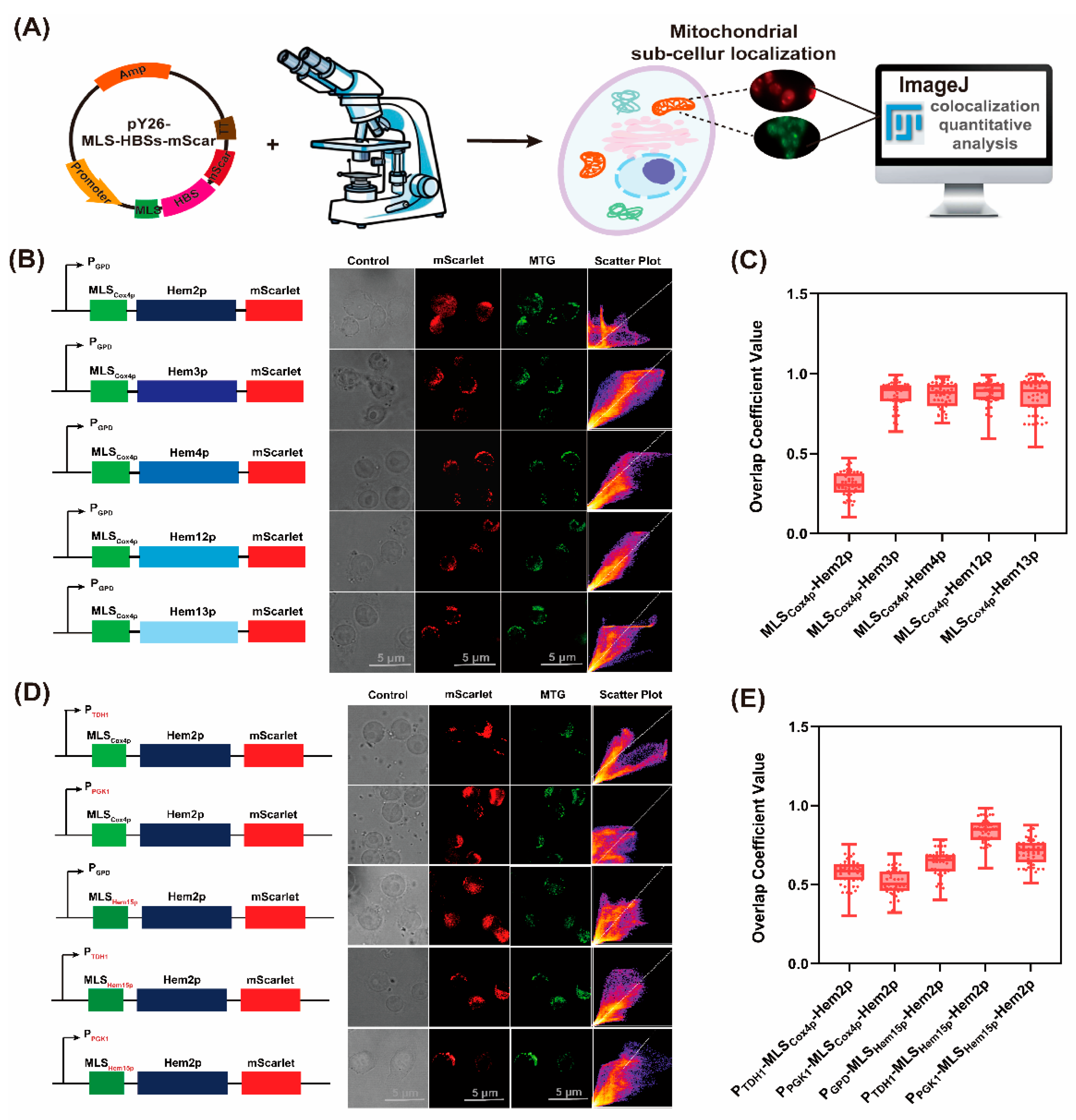
3.1. The Effective Mitochondrial Localization of Cytoplasmic Heme Synthetic Enzymes in S. cerevisiae

The subcellular localization of eight heme synthetic enzymes in S. cerevisiae was determined, revealing that three enzymes (Hem1p, Hem14p, and Hem15p) were localized in the mitochondria, and the other five (Hem2p, Hem3p, Hem4p, Hem12p, and Hem13p) were localized in the cytoplasm [13]. When the MLS was removed, Hem1p was retained in the cytoplasm, resulting in inefficient synthesis of ALA due to the lack of the precursor (succinyl-CoA) in the cytoplasm. Thus, the localization of Hem1p should be maintained in the mitochondria to ensure ALA supply for the biosynthesis of heme. To address the spatial isolation issue during heme biosynthesis in S. cerevisiae, it is necessary to select the appropriate MLS and relocate the five cytoplasmic enzymes (Hem2p, Hem3p, Hem4p, Hem12p, and Hem13p) into the mitochondria. In this study, these five enzymes were firstly located to the mitochondria by applying the MLS from cytochrome C oxidase subunit IV (MLSCox4p) [27] in the N-terminal. Additionally, a monomeric red fluorescent protein (mScarlet) [28] was fused in the C-terminal to monitor their localization status (MLS-HemXp-mScarlet) (Figure 2A).
To evaluate the localization effect of MLSCox4p for each enzyme, the recombinant strains that expressed MLS-HemXp-mScarlet were stained using Mito-Tracker Green FM (MTG). The red fluorescence of mScarlet and the green fluorescence of MTG were analyzed qualitatively and quantitatively using ImageJ (Figure 2A) [29]. A higher density of scatter points along the diagonal line indicated better mitochondrial localization, and the overlap coefficient values for red and green fluorescence (ranging from 0 to 1) were used to assess the localization efficiency of specific heme synthetic enzymes [19]. The overlap coefficient values of MLSCox4p for Hem3p, Hem4p, Hem12p, and Hem13p were all nearly 0.90, suggesting the better mitochondrial localization effect of MLSCox4p on these four enzymes. However, the overlap coefficient value of MLSCox4p-Hem2p was only 0.30 ± 0.08 and the scattered points in the scatter plot indicated that MLSCox4p could not accurately localize Hem2p in the mitochondria (Figure 2B,C).
To enhance the mitochondrial localization efficiency of MLSCox4p, strong constitutive promoters PTDH1 and PPGK1 [30] were selected to replace the original promoter PGPD. Fluorescent localization analysis revealed that the mitochondrial localization efficiency of MLSCox4p-Hem2p improved under the control of PTDH1 and PPGK1, with overlap coefficient values of 0.57 ± 0.08 and 0.51 ± 0.09, respectively (Figure 2D,E). This indicated that stronger promoters increased the mitochondrial localization efficiency of MLSCox4p-Hem2p, but the improvement was still limited. Thus, it was necessary to select other more suitable MLSs to accurately localize Hem2p in the mitochondria.
In our previous work, TPpred 3.0 was employed as a bioinformatic method to accurately predict the sequences of MLSs in Hem15p (31 residues in the N-terminal) [31]. Thus, the MLSCox4p was replaced with MLSHem15p. The results of fluorescent localization showed that the combination of PTDH1 and MLSHem15p increased the value of the overlap coefficient for Hem2p to 0.84 ± 0.08, which was higher than the value obtained for the combination of PPGK1 and MLSHem15p (0.71 ± 0.08) (Figure 2D,E). Based on the previous findings [19], when the values of the overlap coefficient exceeded 0.8, it could be considered that the heme synthetic enzymes had been successfully localized in the mitochondria. Therefore, PTDH1 and MLSHem15p were selected to localize Hem2p, and PGPD and MLSCox4p were selected to localize Hem3p, Hem4p, Hem12p, and Hem13p, ensuring effective mitochondrial localization for these five enzymes.

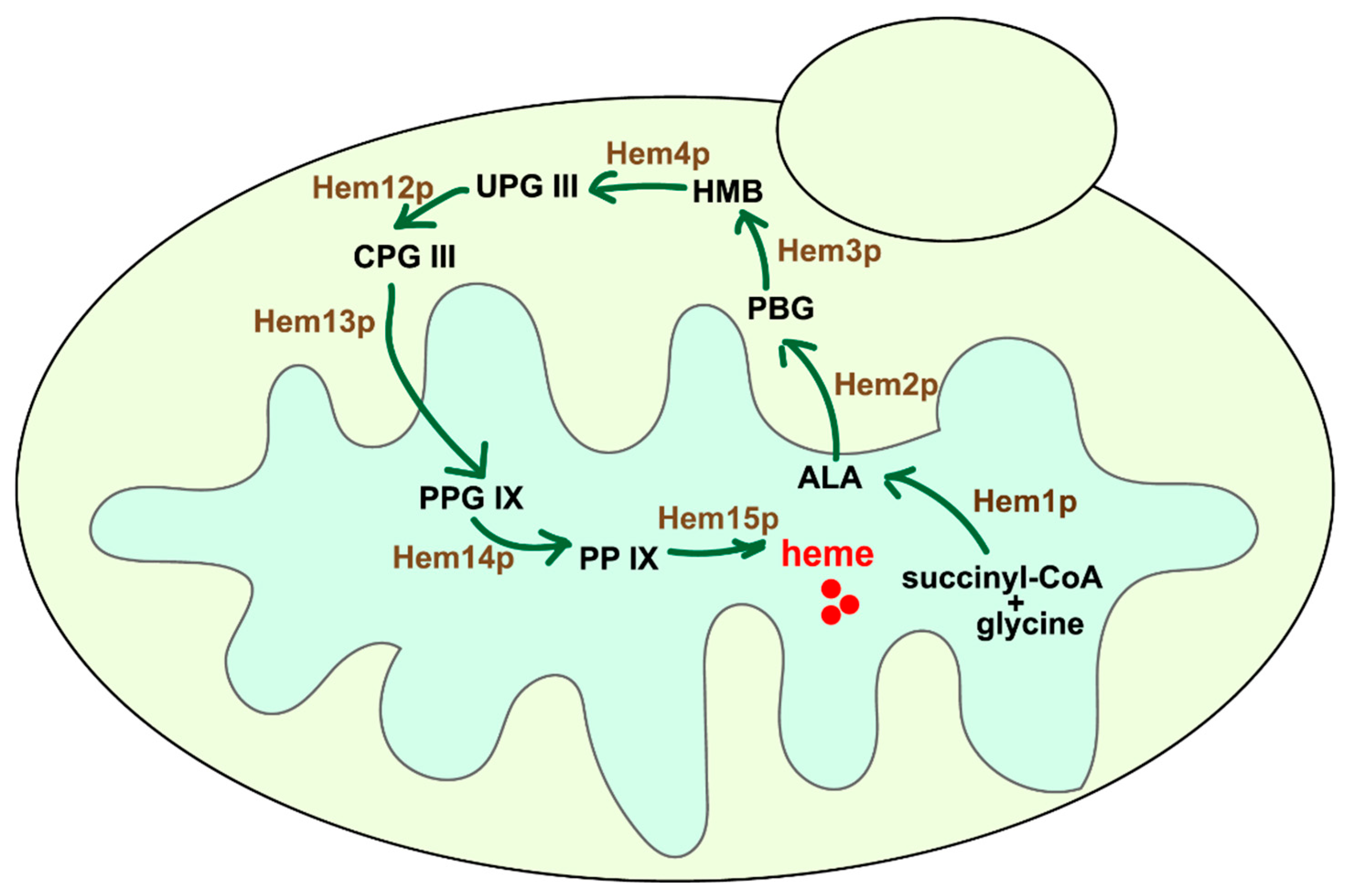
3.2. The Reconstruction of Heme Synthetic Pathway in the Mitochondria

Based on the appropriate selection of MLS and promoters, the cytoplasmic heme synthetic enzymes can be moved to the mitochondria (Figure 3A), and all the modified enzymes can be integrated into the genome to stabilize their expression [32,33]. The fragments of PTDH1-MLSHem15p-Hem2p-TCYC1, PGPD-MLSCox4p-Hem3p-TCYC1, PGPD-MLSCox4p-Hem4p-TCYC1, PGPD-MLSCox4p-Hem12p-TCYC1, and PGPD-MLSCox4p-Hem13p-TCYC1 were, respectively, integrated into five different sites in the S. cerevisiae CEN.PK2-1C genome to obtain the engineered HEME-1 strain (Figure 3B). Since ALA is transported to the cytoplasm and participates in the heme biosynthesis in the wild-type S. cerevisiae strain, it was necessary to knock out the native cytoplasmic Hem2p to maintain more heme synthetic precursors in the mitochondria. Therefore, the engineered HEME-2 strain (HEME-1-∆hem2) was obtained by in situ knocking out the HEM2 gene in the genome of the HEME-1 strain (Figure 3B). However, according to the status of cell growth, the biomass of the HEME-2 strain (OD600 = 7.01 ± 0.11) was, respectively, reduced by 57.6% and 55.1% (Figure 3C), which was significantly lower than the values for the wild-type S. cerevisiae CEN.PK2-1C strain (OD600 = 16.52 ± 0.41) and the HEME-1 strain (OD600 = 15.61 ± 0.38). It indicated that preserving the original cytoplasmic pathway was essential for maintaining the normal growth of strains.
To evaluate the effect of the reconstructed mitochondrial pathway on intracellular heme biosynthesis, the intracellular heme levels were measured. The results demonstrated that the titer of heme in the HEME-1 strain at 24 h was 7.6-fold higher than the titer in the CEN.PK2-1C control, reaching 0.40 ± 0.04 mg·L−1 (Figure 3D). To verify whether the increase in heme titer in the HEME-1 strain was attributable to the reconstructed new heme synthetic pathway in the mitochondria or the elevated copy number of key genes involved in heme biosynthesis, an engineered HEME-3 strain was constructed by integrating five cytoplasmic heme synthetic enzymes without MLSs (Figure 3B). The results showed that the intracellular heme titer in the HEME-3 strain was 2.8-fold higher than the heme level in the CEN.PK2-1C control, reaching 0.15 ± 0.02 mg·L−1 (Figure 3D). Thus, while increasing the copy number of key genes related to heme biosynthesis could enhance the intracellular heme titer to some extent, the reconstruction of the heme synthetic pathway in the mitochondria led to a significantly greater increase in the intracellular heme titer, which was 1.3-fold higher than the heme level achieved in the previously reconstructed cytoplasmic heme synthetic pathway (0.3 mg·L−1) [13]. The results showed that the supply of intracellular heme in the HEME-1 strain was effectively enhanced and could be used to synthesize high-active Mb and Hb.

3.3. The Mining and Modifications of Mitochondrial ALA and Heme Transporters to Enhance Heme Supply

Since the original cytoplasmic heme synthetic pathway remained active in the HEME-1 strain, it would lead to the unnecessary consumption of ALA in the cytoplasm and negatively affected the efficiency of heme synthesis in the mitochondria. To enhance the metabolic flux of ALA in the reconstructed mitochondrial pathway, it is critical to mine the mitochondrial ALA transporter and reduce the transport of mitochondrial ALA to the cytoplasm by knocking out the transporter. However, the specific mitochondrial ALA transporter in S. cerevisiae has not yet been reported [9].
In S. cerevisiae, the mitochondrial proteome comprises 986 distinct proteins [34], including 35 types of mitochondrial transporters [35]. Among these mitochondrial transporters, most have been functionally characterized [36]. Additionally, the known cytoplasmic ALA transporter (Uga4p) is functionally related to the transport of amino acids in S. cerevisiae [37]. Based on their functional annotations, eight possible mitochondrial ALA transporters (Agc1p, Hem25p, Mtm1p, Odc1p, Odc2p, Ort1p, Ymc1p, and Ymc2p) related to the transport of amino acids were selected. To verify their functions in ALA transport, these eight genes were knocked out in the wild-type CEN.PK2-1C strain, obtaining the engineered CEN-∆agc1, CEN-∆hem25, CEN-∆mtm1, CEN-∆odc1, CEN-∆odc2, CEN-∆ort1, CEN-∆ymc1, and CEN-∆ymc2 strains, respectively (Figure 4B).
After culturing the engineered strains with 20 mg·L−1 ALA supplementation, mitochondria were isolated and extracted (Figure 4A). The results showed that the intramitochondrial ALA content in the CEN-∆ort1 strain reached 1.19 ± 0.17 mg·L−1, which was 3.4-fold higher than the value in the CEN.PK2-1C control (0.35 ± 0.08 mg·L−1). Meanwhile, the cytoplasmic ALA content in the CEN-∆ort1 strain was decreased by 14.2% (4.61 ± 0.06 mg·L−1) compared with the CEN.PK2-1C control (5.37 ± 0.15 mg·L−1). Additionally, intramitochondrial ALA content did not increase significantly in other knockout strains (Figure 4C). Notably, the human SLC25A38 protein has been reported as a potential mitochondrial ALA transporter [38]. Its homologous protein in S. cerevisiae is Hem25p, whose function is currently known as a transporter of the precursor (glycine) for ALA synthesis [39]. After knocking out the HEM25 gene, the intramitochondrial ALA content in the CEN-∆hem25 strain was significantly decreased by 25.5% (0.26 ± 0.09 mg·L−1) and the cytoplasmic ALA content was also decreased by 6.35% (5.03 ± 0.04 mg·L−1) (Figure 4C). These results indicated that the synthesis of ALA was affected by the lack of HEM25 gene, negatively affecting mitochondrial heme biosynthesis. It was worth mentioning that the large difference in ALA content between the mitochondria and cytoplasm was due to the exogenous addition of 20 mg·L−1 ALA in the culture medium. ALA in the culture medium could be transported into the cell by ALA transporter Uga4p on the cell membrane of S. cerevisiae and then distributed in the cytosol; ALA in mitochondria was almost synthesized in the mitochondria and was less affected by exogenous addition of ALA. Therefore, the ALA content in the cytoplasm was much higher than that in mitochondria. The ALA content in the mitochondria of the CEN-∆ort1 engineered strain increased exponentially compared with the CEN.PK2-1C control, providing abundant precursors for the synthesis of heme.
The ORT1 gene was knocked out in the HEME-1 strain and the intramitochondrial and cytoplasmic ALA content was measured in the engineered HEME-1-∆ort1 strain. As a result, compared with the intramitochondrial ALA content in CEN-∆ort1 strain, the content was decreased by 10.9% in the HEME-1-∆ort1 strain (0.94 ± 0.12 mg·L−1). In addition, the intracellular heme titer was increased by 6.6% in the HEME-1-∆ort1 strain (Figure 4D,E). Consistent with the above result, the intramitochondrial ALA content was also reduced in the HEME-1 strain (0.24 ± 0.10 mg·L−1) compared with the value in the CEN.PK2-1C strain (Figure 4D). These results confirm that the mitochondrial heme biosynthesis pathway functioned efficiently, utilizing ALA for heme production.
In addition to ALA transporters, mitochondrial heme transporters also affect intracellular utilization of heme, including the synthesis of downstream hemoproteins and other metabolic activities [9,40]. The mitochondrial heme transporter in S. cerevisiae has now been identified as Ygr127wp [41]. To further improve the mitochondrial heme synthetic efficiency and intracellular heme titer, the ygr127w gene was overexpressed in the HEME-1-∆ort1 strain to obtain the HEME-1-∆ort1-+ygr127w strain. The intracellular heme titer in the HEME-1-∆ort1-+ygr127w strain reached 0.43 ± 0.01 mg·L−1, which was 9.1% higher than the value in the HEME-1 strain and 8.4-fold higher than the value in the CEN.PK2-1C control (Figure 4E). Thus, the HEME-1-∆ort1-+ygr127w strain was selected as the optimal heme-supply strain to synthesize highly active pMb and LegH.

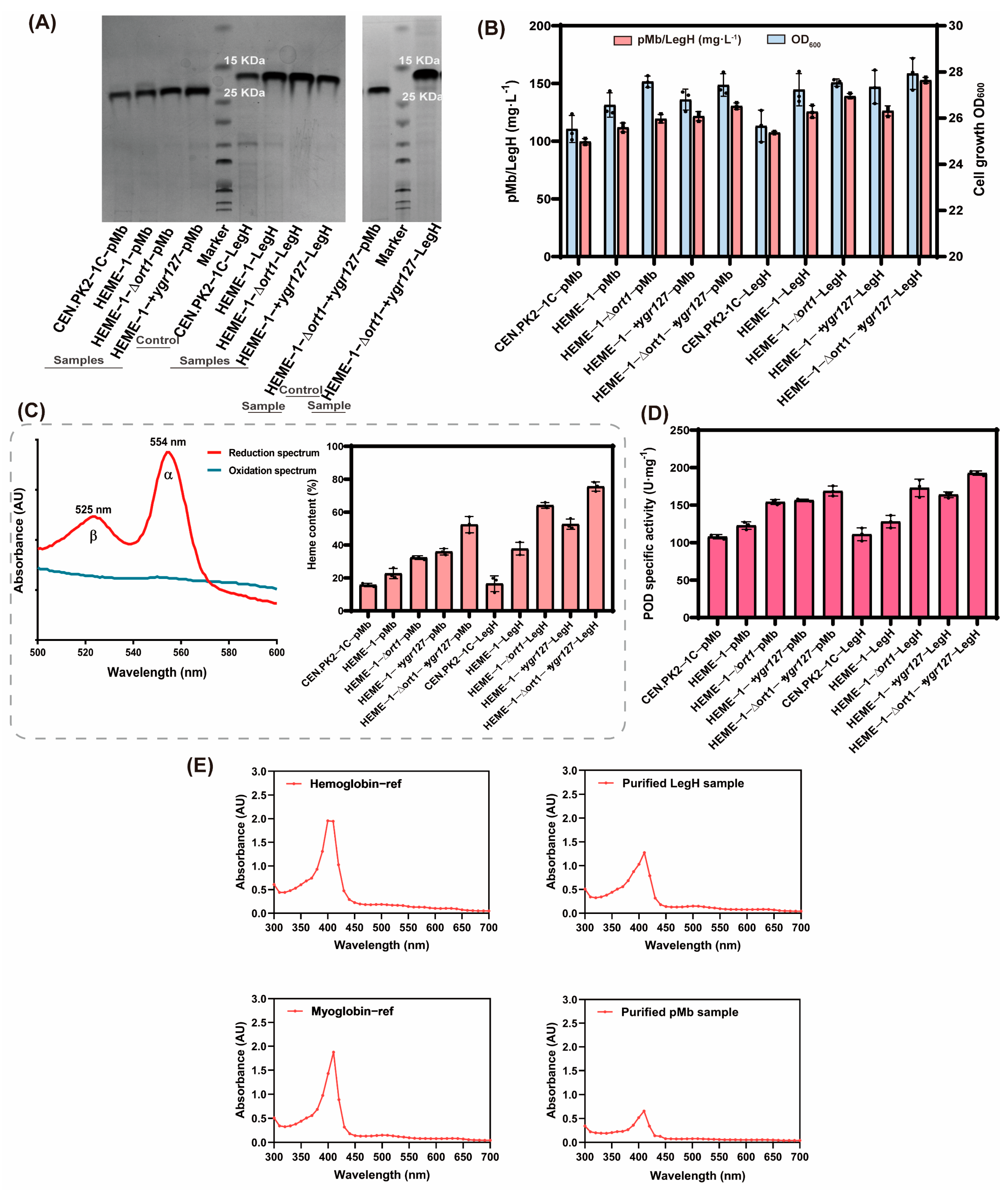
3.4. The Biochemical Properties of pMb and LegH Synthesized in the Engineered S. cerevisiae

The appropriate expressional strategies for the globins in pMb and LegH were employed in the HEME-1-∆ort1-+ygr127w strain to effectively synthesize high-active holo-hemoproteins [13]. Due to the sufficient intracellular supply of heme, the production of pMb and LegH was significantly improved in the HEME-1-∆ort1-+ygr127w strain compared with the wild-type CEN.PK2-1C control, with increases of 31.1% in pMb (130.5 ± 2.8 mg·L−1) and 42.1% in LegH (152.8 ± 2.6 mg·L−1) (Figure 5A,B). In addition, the heme-binding ratios (mol heme/mol globin) in synthesized pMb and LegH were still detected to evaluate their real activities. Applying the HEME-1-∆ort1-+ygr127w strain, the heme-binding ratios of pMb and LegH increased by 2.4-fold and 3.6-fold, reaching 52.4 ± 4.9% and 75.5 ± 2.8%, respectively (Figure 5C).
As Mb and Hb can be used in the production of colorants and flavor supplements in artificial meat, and be widely applied in high-cell-density fermentation, biocatalyst metalloenzymes, and other industrial fields [42], the biochemical properties of synthesized Mb and Hb also needed to be investigated. Firstly, commercial native Myoglobin-ref (equine skeletal muscle myoglobin) and Hemoglobin-ref (bovine hemoglobin) were used as standards and the special absorption spectra (300–700 nm) were detected for different pMb and LegH samples. Compared with the standards, the results showed that the synthesized Mb and Hb shared similar Soret band absorption spectra, with a maximum absorption peak at 410 nm (Figure 5E). This spectral property was due to the incorporation of heme in globins, which was consistent with the previous reports [43].
In addition to the special characteristic spectra, the functional activities of synthesized Mb and Hb were assessed. Since many applications of Mb and Hb depend on their peroxidase (POD) activities [44], the POD activities of synthesized pMb and LegH were determined. As a result, compared with the wild-type CEN.PK2-1C control, the specific POD activities of pMb and LegH increased by 56.4% and 73.6%, respectively, in the HEME-1-∆ort1-+ygr127w strain, reaching 168.7 ± 6.6 U·mg−1 and 192.7 ± 2.9 U·mg−1 (Figure 5D). In summary, the above results indicate that the reconstruction of the heme synthetic pathway in mitochondria can effectively improve the intracellular heme supply to synthesize high-active hemoproteins.

4. Conclusions

Herein, five heme biosynthetic enzymes originally localized in the cytoplasm were relocated to the mitochondria using efficient mitochondrial localization signal peptides and promoters, and a new heme synthetic pathway was reconstructed in the mitochondria of S. cerevisiae, eliminating the spatial isolation in the heme synthetic process. After the construction of the new pathway, the original heme synthetic pathway in the cytoplasm still functioned, resulting in the inability of the heme synthesis precursor ALA to be fully utilized for mitochondrial heme synthesis, which reduced the utilization efficiency of the new pathway. To address this issue, in this work, an important mitochondrial ALA transporter was identified. Through regulating the mitochondrial ALA transporter Ort1p and the known mitochondrial heme transporter Ygr127wp, the intracellular heme supply level was further increased, and we obtained the engineered strains which were suitable for producing myoglobin and hemoglobin. Finally, the galactose inducible system was applied to synthesize pMb and LegH at the shaking-flask level. The yield of pMb and LegH (31.1% and 42.1%), the heme-binding ratio (2.4-fold and 3.6-fold), and the specific activity of POD (56.4% and 73.6%) in the engineered strain HEME-1-∆ort1-+ygr127w were all improved to a certain extent.

Supplementary Materials

The following supporting information can be downloaded at: https://www.mdpi.com/article/10.3390/fermentation11050246/s1, Table S1: Strains and plasmids used in this study; Table S2: Primers used in this study; Table S3: Sequences of genes for heme transport in mitochondria; Table S4: Sequences of porcine myoglobin and soybean hemoglobin; Figure S1: The verification image of colony PCR.

Author Contributions

X.Z. and X.S. conceived the project; X.S., Y.W. (Yunpeng Wang), and Y.W. (Yijie Wang) performed the experiments and data analysis; J.Z., J.L., J.C. and G.D. supervised the project; X.S. wrote the manuscript; and X.Z. revised the manuscript. All authors participated in the manuscript. All authors have read and agreed to the published version of the manuscript.

Funding

The authors are thankful for the support of the Key R&D Project of Ningbo City (2025Z103), the Jiangsu Basic Research Center for Synthetic Biology (BK20233003), and the National First-Class Discipline Program of Light Industry Technology and Engineering (LITE2018-08).

Institutional Review Board Statement

Not applicable.

Informed Consent Statement

Not applicable.

Data Availability Statement

The original contributions presented in this study are included in the article/Supplementary Material. Further inquiries can be directed to the corresponding author.

Conflicts of Interest

The authors declare no conflicts of interest.

References

  1. Xing, Y.; Gao, S.; Zhang, X.; Zang, J. Dietary heme-containing proteins: Structures, applications, and challenges. Foods 2022, 11, 3594. [Google Scholar] [CrossRef] [PubMed]
  2. Foley, E.L.; Hvitved, A.N.; Eich, R.F.; Olson, J.S. Mechanisms of nitric oxide reactions with globins using mammalian myoglobin as a model system. J. Inorg. Biochem. 2022, 233, 111839. [Google Scholar] [CrossRef] [PubMed]
  3. Giardina, B. Hemoglobin: Multiple molecular interactions and multiple functions. An example of energy optimization and global molecular organization. Mol. Asp. Med. 2022, 84, 101040. [Google Scholar] [CrossRef]
  4. Ahmad, M.I.; Farooq, S.; Alhamoud, Y.; Li, C.B.; Zhang, H. Soy Leghemoglobin: A review of its structure, production, safety aspects, and food applications. Trends Food Sci. Technol. 2023, 141, 104199. [Google Scholar] [CrossRef]
  5. Simsa, R.; Yuen, J.; Stout, A.; Rubio, N.; Fogelstrand, P.; Kaplan, D.L. Extracellular heme proteins influence bovine myosatellite cell proliferation and the color of cell-based meat. Foods 2019, 8, 521. [Google Scholar] [CrossRef] [PubMed]
  6. Jolien, D.; Ann De, W.; Lore, D.; Ilse, F.; Irena, S.; Elsa, L.; Andy de, J.; Hermes, S. Improving the aromatic profile of plant-based meat alternatives: Effect of myoglobin addition on volatiles. Foods 2022, 11, 1985. [Google Scholar] [CrossRef]
  7. Zhao, X.; Zhou, J.; Du, G.; Chen, J. Recent advances in the microbial synthesis of hemoglobin. Trends Biotechnol. 2021, 39, 286–297. [Google Scholar] [CrossRef]
  8. Guirimand, G.; Kulagina, N.; Papon, N.; Hasunuma, T.; Courdavault, V. Innovative tools and strategies for optimizing yeast cell factories. Trends Biotechnol. 2021, 39, 488–504. [Google Scholar] [CrossRef]
  9. Swenson, S.A.; Moore, C.M.; Marcero, J.R.; Medlock, A.E.; Reddi, A.R.; Khalimonchuk, O. From synthesis to utilization: The ins and outs of mitochondrial heme. Cells 2020, 9, 579. [Google Scholar] [CrossRef]
  10. Shao, Y.; Xue, C.; Liu, W.; Zuo, S.; Wei, P.; Huang, L.; Lian, J.; Xu, Z. High-level secretory production of leghemoglobin in Pichia pastoris through enhanced globin expression and heme biosynthesis. Bioresour. Technol. 2022, 363, 127884. [Google Scholar] [CrossRef]
  11. Ge, J.Z.; Wang, X.L.; Bai, Y.G.; Wang, Y.R.; Wang, Y.; Tu, T.; Qin, X.; Su, X.Y.; Luo, H.Y.; Yao, B.; et al. Engineering Escherichia coli for efficient assembly of heme proteins. Microb. Cell Factories 2023, 22, 59. [Google Scholar] [CrossRef] [PubMed]
  12. Liu, L.F.; Martinez, J.L.; Liu, Z.H.; Petranovic, D.; Nielsen, J. Balanced globin protein expression and heme biosynthesis improve production of human hemoglobin in Saccharomyces cerevisiae. Metab. Eng. 2014, 21, 9–16. [Google Scholar] [CrossRef] [PubMed]
  13. Xue, J.; Zhou, J.; Li, J.; Du, G.; Chen, J.; Wang, M.; Zhao, X. Systematic engineering of Saccharomyces cerevisiae for efficient synthesis of hemoglobins and myoglobins. Bioresour. Technol. 2023, 370, 128556. [Google Scholar] [CrossRef]
  14. Malina, C.; Larsson, C.; Nielsen, J. Yeast mitochondria: An overview of mitochondrial biology and the potential of mitochondrial systems biology. FEMS Yeast Res. 2018, 18, foy040. [Google Scholar] [CrossRef]
  15. Chiabrando, D.; Marro, S.; Mercurio, S.; Giorgi, C.; Petrillo, S.; Vinchi, F.; Fiorito, V.; Fagoonee, S.; Camporeale, A.; Turco, E.; et al. The mitochondrial heme exporter FLVCR1b mediates erythroid differentiation. J. Clin. Investig. 2012, 122, 4569–4579. [Google Scholar] [CrossRef] [PubMed]
  16. Yee, D.A.; DeNicola, A.B.; Billingsley, J.M.; Creso, J.G.; Subrahmanyam, V.; Tang, Y. Engineered mitochondrial production of monoterpenes in Saccharomyces cerevisiae. Metab. Eng. 2019, 55, 76–84. [Google Scholar] [CrossRef]
  17. Jia, H.J.; Chen, T.H.; Qu, J.Z.; Yao, M.D.; Xiao, W.H.; Wang, Y.; Li, C.; Yuan, Y.J. Collaborative subcellular compartmentalization to improve GPP utilization and boost sabinene accumulation in Saccharomyces cerevisiae. Biochem. Eng. J. 2020, 164, 107768. [Google Scholar] [CrossRef]
  18. Guo, Q.D.; Xu, J.Q.; Li, J.C.; Tang, S.Y.; Cheng, Y.H.; Gao, B.; Xiong, L.B.; Xiong, J.; Wang, F.Q.; Wei, D.Z. Synergistic increase in coproporphyrin III biosynthesis by mitochondrial compartmentalization in engineered Saccharomyces cerevisiae. Synth. Syst. Biotechnol. 2024, 9, 834–841. [Google Scholar] [CrossRef]
  19. Zinchuk, V.; Wu, Y.; Grossenbacher-Zinchuk, O. Bridging the gap between qualitative and quantitative colocalization results in fluorescence microscopy studies. Sci. Rep. 2013, 3, 1365. [Google Scholar] [CrossRef]
  20. Michener, J.K.; Nielsen, J.; Smolke, C.D. Identification and treatment of heme depletion attributed to overexpression of a lineage of evolved P450 monooxygenases. Proc. Natl. Acad. Sci. USA 2012, 109, 19504–19509. [Google Scholar] [CrossRef]
  21. Meisinger, C.; Pfanner, N.; Truscott, K.N. Isolation of yeast mitochondria. Methods Mol. Biol. 2006, 313, 33–39. [Google Scholar] [PubMed]
  22. Corcelli, A.; Saponetti, M.S.; Zaccagnino, P.; Lopalco, P.; Mastrodonato, M.; Liquori, G.E.; Lorusso, M. Mitochondria isolated in nearly isotonic KCl buffer: Focus on cardiolipin and organelle morphology. Biochim. Biophys. Acta-Biomembr. 2010, 1798, 681–687. [Google Scholar] [CrossRef] [PubMed]
  23. Pan, D.Q.; Lindau, C.; Lagies, S.; Wiedemann, N.; Kammerer, B. Metabolic profiling of isolated mitochondria and cytoplasm reveals compartment-specific metabolic responses. Metabolomics 2018, 14, 59. [Google Scholar] [CrossRef]
  24. Bragagni, M.; Scozzafava, A.; Mastrolorenzo, A.; Supura, C.T.; Mura, P. Development and ex vivo evaluation of 5-aminolevulinic acid-loaded niosomal formulations for topical photodynamic therapy. Int. J. Pharm. 2015, 494, 258–263. [Google Scholar] [CrossRef]
  25. Tomokuni, K.; Hirai, Y. Factors affecting determination of delta-aminolevulinate by use of Ehrlichs reagent. Clin. Chem. 1986, 32, 192–193. [Google Scholar] [CrossRef]
  26. Barr, I.; Guo, F. Pyridine hemochromagen assay for determining the concentration of heme in purified protein solutions. Bio-Protoc. 2015, 5, e1594. [Google Scholar] [CrossRef]
  27. Hu, J.J.; Dong, L.X.; Outten, C.E. The redox environment in the mitochondrial intermembrane space is maintained separately from the cytosol and matrix. J. Biol. Chem. 2008, 283, 29126–29134. [Google Scholar] [CrossRef]
  28. Bindels, D.S.; Postma, M.; Haarbosch, L.; van Weeren, L.; Gadella, T.W.J. Multiparameter screening method for developing optimized red-fluorescent proteins. Nat. Protoc. 2020, 15, 450–478. [Google Scholar] [CrossRef] [PubMed]
  29. Dunn, K.W.; Kamocka, M.M.; McDonald, J.H. A practical guide to evaluating colocalization in biological microscopy. Am. J. Physiol.-Cell Physiol. 2011, 300, C723–C742. [Google Scholar] [CrossRef]
  30. Gao, S.; Zhou, H.R.; Zhou, J.W.; Chen, J. Promoter-library-based pathway optimization for efficient (2S)-naringenin production from p-coumaric acid in Saccharomyces cerevisiae. J. Agric. Food Chem. 2020, 68, 6884–6891. [Google Scholar] [CrossRef]
  31. Savojardo, C.; Martelli, P.L.; Fariselli, P.; Casadio, R. TPpred3 detects and discriminates mitochondrial and chloroplastic targeting peptides in eukaryotic proteins. Bioinformatics 2015, 31, 3269–3275. [Google Scholar] [CrossRef] [PubMed]
  32. Apel, A.R.; d’Espaux, L.; Wehrs, M.; Sachs, D.; Li, R.A.; Tong, G.J.; Garber, M.; Nnadi, O.; Zhuang, W.; Hillson, N.J.; et al. A Cas9-based toolkit to program gene expression in Saccharomyces cerevisiae. Nucleic Acids Res. 2017, 45, 496–508. [Google Scholar]
  33. Shaw, W.M.; Khalil, A.S.; Ellis, T. A multiplex moclo toolkit for extensive and flexible engineering of Saccharomyces cerevisiae. ACS Synth. Biol. 2023, 12, 3393–3405. [Google Scholar] [CrossRef] [PubMed]
  34. Vögtle, F.N.; Burkhart, J.M.; Gonczarowska-Jorge, H.; Kücükköse, C.; Taskin, A.A.; Kopczynski, D.; Ahrends, R.; Mossmann, D.; Sickmann, A.; Zahedi, R.P.; et al. Landscape of submitochondrial protein distribution. Nat. Commun. 2017, 8, 290. [Google Scholar] [CrossRef] [PubMed]
  35. Palmieri, F.; Agrimi, G.; Blanco, E.; Castegna, A.; Di Noia, M.A.; Iacobazzi, V.; Lasorsa, F.M.; Marobbio, C.M.T.; Palmieri, L.; Scarcia, P.; et al. Identification of mitochondrial carriers in Saccharomyces cerevisiae by transport assay of reconstituted recombinant proteins. Biochim. Biophys. Acta-Bioenerg. 2006, 1757, 1249–1262. [Google Scholar] [CrossRef]
  36. Ferramosca, A.; Zara, V. Mitochondrial carriers and substrates transport network: A lesson from Saccharomyces cerevisiae. Int. J. Mol. Sci. 2021, 22, 8496. [Google Scholar] [CrossRef]
  37. Luzzani, C.; Cardillo, S.B.; Moretti, M.B.; García, S.C. New insights into the regulation of the Saccharomyces cerevisiae UGA4 gene: Two parallel pathways participate in carbon-regulated transcription. Microbiol. Soc. 2007, 153, 3677–3684. [Google Scholar] [CrossRef]
  38. Guernsey, D.L.; Jiang, H.; Campagna, D.R.; Evans, S.C.; Ferguson, M.; Kellogg, M.D.; Lachance, M.; Matsuoka, M.; Nightingale, M.; Rideout, A.; et al. Mutations in mitochondrial carrier family gene SLC25A38 cause nonsyndromic autosomal recessive congenital sideroblastic anemia. Nat. Genet. 2009, 41, 651–653. [Google Scholar] [CrossRef]
  39. Lunetti, P.; Damiano, F.; De Benedetto, G.; Siculella, L.; Pennetta, A.; Muto, L.; Paradies, E.; Marobbio, C.M.T.; Dolce, V.; Capobianco, L. Characterization of human and yeast mitochondrial glycine carriers with implications for heme biosynthesis and anemia. J. Biol. Chem. 2016, 291, 19746–19759. [Google Scholar] [CrossRef]
  40. Li, Y.X.; Han, S.R.; Gao, H.C. Heme homeostasis and its regulation by hemoproteins in bacteria. mLife 2024, 3, 327–342. [Google Scholar] [CrossRef]
  41. Sun, F.X.; Zhao, Z.Z.; Willoughby, M.M.; Shen, S.Q.; Zhou, Y.; Shao, Y.Y.; Kang, J.; Chen, Y.T.; Chen, M.Y.; Yuan, X.J.; et al. HRG-9 homologues regulate haem trafficking from haem-enriched compartments. Nature 2022, 610, 768–774. [Google Scholar] [CrossRef] [PubMed]
  42. Hu, B.D.; Yu, H.B.; Zhou, J.W.; Li, J.H.; Chen, J.; Du, G.C.; Lee, S.Y.; Zhao, X.R. Whole-Cell P450 biocatalysis using engineered Escherichia coli with fine-tuned heme biosynthesis. Adv. Sci. 2023, 10, e2205580. [Google Scholar] [CrossRef] [PubMed]
  43. Kosmachevskaya, O.V.; Nasybullina, E.I.; Shumaev, K.B.; Topunov, A.F. Expressed soybean leghemoglobin: Effect on Escherichia coli at oxidative and nitrosative stress. Molecules 2021, 26, 7207. [Google Scholar] [CrossRef]
  44. Yu, F.; Zhao, X.R.; Zhou, J.W.; Lu, W.; Li, J.H.; Chen, J.; Du, G.C. Biosynthesis of high-active hemoproteins by the efficient heme-supply Pichia Pastoris chassis. Adv. Sci. 2023, 10, 2302826. [Google Scholar] [CrossRef] [PubMed]
Figure 1. Schematic diagram of the heme synthetic pathway in S. cerevisiae.
Fermentation 11 00246 g001
Figure 2. The selection of an effective MLS and promoter for the mitochondrial localization of cytoplasmic heme synthetic enzymes in S. cerevisiae. (A) Schematic overview of validating the mitochondrial localization efficiency of five heme synthetic enzymes by different combinations of MLS and promoter. (B) Yeast cell microscopy images and scatter plot results for the mitochondrial subcellular localization of Hem2p, Hem3p, Hem4p, Hem12p, and Hem13p by MLSCox4p. (C) The values of overlap coefficients for the mitochondrial localization of Hem2p, Hem3p, Hem4p, Hem12p, and Hem13p by MLSCox4p. (D) The yeast cell microscopy images and scatter plot results for the mitochondrial subcellular localization of Hem2p by different combinations of MLS and promoter. (E) The values of overlap coefficients for the mitochondrial localization of Hem2p by different combinations of MLS and promoter. Measurements in (C,E) were performed as mean values ± SD from fifty independent biological replicates (n = 50).
Fermentation 11 00246 g002
Figure 3. The reconstruction of heme synthetic pathway in the mitochondria. (A) The five heme synthetic enzymes in the cytoplasm (Hem2p, Hem3p, Hem4p, Hem12p, and Hem13p) were localized in the mitochondria through suitable MLSs. (B) The construction of engineered HEME-1, HEME-2, and HEME-3 strains. The HEME-1 strain integrated with the fragments of PTDH1-MLSHem15p-Hem2p-TCYC1, PGPD-MLSCox4p-Hem3p-TCYC1, PGPD-MLSCox4p-Hem4p-TCYC1, PGPD-MLSCox4p-Hem12p-TCYC1, and PGPD-MLSCox4p-Hem13p-TCYC1 expression cassettes; the HEME-2 strain knocked out the native HEM2 gene on the genome of the HEME-1 strain; and the HEME-3 strain integrated with the fragments of PTDH1-Hem2p-TCYC1, PGPD-Hem3p-TCYC1, PGPD-Hem4p-TCYC1, PGPD-Hem12p-TCYC1, and PGPD-Hem13p-TCYC1 expression cassettes. (C) Inhibition of growth induced by knockout of HEM2 gene in the HEME-1 strain. (D) The biomass and heme titer in the HEME-1 and HEME-3 strains at 24 h and 48 h. CEN represents the control CEN.PK2-1C strain. Measurements in (C,D) were performed as mean values ± SD from three independent biological replicates (n = 3).
Fermentation 11 00246 g003
Figure 4. The mining of transporters related to the heme synthetic pathway in S. cerevisiae. (A) The separation of mitochondria in S. cerevisiae. (B) The knockout of eight possible mitochondrial ALA transporters. (C) ALA contents in eight knockout strains (CEN-∆agc1, CEN-∆hem25, CEN-∆mtm1, CEN-∆odc1, CEN-∆odc2, CEN-∆ort1, CEN-∆ymc1, and CEN-∆ymc2) in mitochondria and cytoplasm. CEN.PK2-1C was used as the control strain. (D) The effects of deleting ORT1 gene in HEME-1 strain (HEME-1-∆ort1 strain) on the intramitochondrial and cytoplasmic ALA content. CEN.PK2-1C, CEN-∆ort1, and HEME-1 were used as the control strains. (E) The effects of overexpressing Ygr127w gene in HEME-1-∆ort1 strain (HEME-1-+ygr127w strain) on the intracellular heme titer. CEN.PK2-1C and HEME-1 were used as the control strains. Measurements in (C,E) were performed as mean values ± SD from three independent biological replicates (n = 3). Measurements in (D) were performed as mean values ± SD from six independent biological replicates (n = 6).
Fermentation 11 00246 g004
Figure 5. The biochemical properties of biosynthesized pMb and LegH. (A) SDS-PAGE for pMb and LegH obtained from different engineered strains. (B) Biomass and expressional levels of pMb and LegH obtained from different engineered strains. (C) The heme-binding ratios of purified pMb and LegH using the different spectra between the reduced and oxidized samples. (D) The specific POD activities of purified pMb and LegH obtained from different engineered strains. (E) The spectral characteristics of purified pMb and LegH obtained from HEME-1-∆ort1-+ygr127w strain. Myoglobin-ref (equine skeletal muscle myoglobin) and Hemoglobin-ref (bovine hemoglobin) were used as standards. All measurements were performed as mean values ± SD from three independent biological replicates (n = 3).
Fermentation 11 00246 g005
Disclaimer/Publisher’s Note: The statements, opinions and data contained in all publications are solely those of the individual author(s) and contributor(s) and not of MDPI and/or the editor(s). MDPI and/or the editor(s) disclaim responsibility for any injury to people or property resulting from any ideas, methods, instructions or products referred to in the content.

