Article

Biogas Upgrading by Wild Alkaliphilic Microalgae and the Application Potential of Their Biomass in the Carbon Capture and Utilization Technology

Applied Chemistry and Chemical Engineering Program, Graduate School of Engineering, Kogakuin University, 2665-1 Nakano-cho, Hachioji City 192-0015, Tokyo, Japan
*
Author to whom correspondence should be addressed.
Fermentation 2024, 10(3), 134; https://doi.org/10.3390/fermentation10030134
Submission received: 31 December 2023 / Revised: 19 February 2024 / Accepted: 25 February 2024 / Published: 28 February 2024
(This article belongs to the Special Issue Microbial Culture and Isolation for the Production of Biofuels)

Abstract

:
Although biogas is a renewable energy source alternative to natural gas, it contains approximately 40 vol% CO2 and, hence, a low calorific value. The sequestration of CO2 from biogas is, therefore, essential before its widespread use. As CO2 can be easily solubilized as carbonate and bicarbonate in alkaline water, in this study, we isolated and characterized alkaliphilic wild microalgae that grow under high-level CO2 conditions and evaluated their application potential in CO2-removal from biogas. For this purpose, freshwater samples were enriched with 10 vol% CO2 and an alkaline culture medium (pH 9.0), wherein almost free CO2 was converted to carbonate and bicarbonate to yield alkaliphilic and high-level CO2-tolerant microalgae. Ten microalgal strains of Micractinium, Chlorella, Scenedesmus/Tetradesmus, or Desmodesmus spp. were isolated, some of which demonstrated good growth even under conditions of >pH 10 and >30 vol% CO2. All algal strains grew well through fixing biogas-derived CO2 in a vial-scale biogas upgrading experiment, which reduced the CO2 level in biogas to an undetectable level. These strains yielded antioxidant carotenoids, including lutein, astaxanthin, zeaxanthin, and β-carotene, particularly rich in lutein (up to 7.3 mg/g dry cells). In addition, these strains contained essential amino acids, accounting for 42.9 mol% of the total amino acids on average, and they were rich in unsaturated fatty acids (comprising 62.2 wt% of total fatty acids). The present study identified strains that can contribute to biogas upgrading technology, and the present findings suggest that their biomass can serve as useful raw material across the food, nutraceutical, and feed industries.

1. Introduction

Continuous emissions of carbon dioxide (CO2) from industries and in the energy and transport sectors have been raising the atmospheric level, which is the main cause of global warming [1]. These sectors are the major source of emitted CO2 and account for approximately 75% of the total CO2 emission [2]. Besides exhaust gas, biogas contains a high concentration of CO2 [3]. Biogas is produced from the anaerobic digestion of food residues, manures, or sewage sludge and is regarded as a renewable substitute for natural gas. However, it is generally composed of 50–80 vol% methane and 20–50 vol% CO2, with some minor constituents, such as 0–5 vol% hydrogen, 0–2 vol% nitrogen, 0–1 vol% carbon monoxide, <1 vol% hydrogen sulfide, and trace amounts of ammonia, depending on the type of the fermentation substrate, thereby yielding a low calorific value (21.5 MJ/m3) in contrast to that in natural gas (35.8 MJ/m3) [3]. Therefore, the removal of CO2 from biogas is essential to improving its calorific value as an alternative energy source in sustainable social life and industry.
Some technologies for CO2 sequestration are currently under development. For instance, carbon capture and storage is employed in capturing CO2 from exhaust gas, transporting it to a storage site, and depositing it underground [4]. The key technologies for carbon capture—a process for separating CO2 from exhaust gas—include postcombustion, precombustion, and oxy-fuel combustion methods [5]. The transportation process involves sending the captured CO2 to a storage site through pipelines, ships, and trucks. Several technologies for the storage process have been studied to date, such as geological storage, ocean storage, and mineral carbonation [6]. However, the relevant practical application is hindered by the cost involved in each process as well as the related environmental impact of deposited CO2 [7]. Carbon capture and utilization is, thus, recognized as an additional strategy via which the captured CO2 can be converted to industrially valuable chemicals. However, the physicochemical conversion of CO2 to value-added chemicals in this process remains challenging for commercial viability because of the high production costs, huge energy consumption, and the need for the use of costly catalysts [8]. In contrast, the biological conversion of CO2 to value-added chemicals by microalgae has received much attention owing to its advantages, such as the low production cost, the ability to fix CO2 directly from the exhaust gas via photosynthesis, and the lack of the need for catalysts [9]. However, exhaust gases are rich in CO2, which evokes acidification of the culture medium and inhibits microalgal growth. Therefore, microalgal strains that are acclimated to high levels of CO2 are preferred for a successful process. In the culture medium used for algal production, wastewater is occasionally added to remove nitrogen and phosphorus as well as to stimulate algal growth [10,11]. Although algal biomass produced in wastewater can be used as a feedstock for biofuel production, it is not suitable for use as food/feed additives or nutraceuticals, from the hygienic standpoint. We, thus, believe that microalgal cultivation in a defined mineral salt medium under high-level CO2 can be suitable for producing algal biomass for potential application in food/feed industries.
There are several reports on the isolation of microalgal strains that acclimate to high levels of CO2 and on their evaluation for application to CO2 fixation [12]. The major potential application of this fixed CO2 is by using biomass as feedstock for biodiesel production or as animal feed, food additive, and nutraceuticals [10,13]. Several studies on photobioreactors for microalgal CO2 sequestration from exhaust gas and biogas have been reported to date [14]. However, microalgal CO2 sequestration using a photobioreactor has scope for improvement considering the low solubility of CO2 in the culture medium, because of which a major amount of CO2 is bubbled out of the reactor [15]. Much effort has been made to accelerate CO2 solubilization in culture medium via improving photobioreactor devices.
Conversely, the use of an alkaline culture medium may be useful for the same purpose considering that CO2 can dramatically increase its solubility in alkaline water by changing its form to carbonate (CO32−) or bicarbonate (HCO3) [16,17]. In water with a pH > 8.3, dissolved free CO2 rapidly changes to bicarbonate and/or carbonate and reaches a stable equilibrium, which is an advantageous condition for CO2 sequestration. For instance, the solubility of CO2 in air (0.04 vol% CO2) in water with a pH of 6.0, 7.0, 8.0, and 9.0 is 2.0 × 10⁻⁵, 8.0 × 10⁻⁵, 7.0 × 10⁻⁴, and 7.0 × 10⁻³ mol CO2 per 1.0 L of water, respectively [18]. However, extreme alkaline pH inhibits the growth of most microalgae. For instance, the cell growth of the freshwater microalga Chlamydomonas reinharditii and Auxenochlorella protothecoides is known to be inhibited at a pH of 8.5 and 9.0, respectively [19,20]. The cell growth of several marine microalgae including Cricosphaera elongate, Asterionella japonica, and Isochrysis galbana was reported to be significantly inhibited at a pH of 9.0 [21]. Therefore, the use of alkaliphilic microalgal strains that acclimate to high levels of CO2 appears to be a potential approach to efficient CO2 sequestration from exhaust gases.
In the present study, we isolated and characterized alkaliphilic wild microalgae that grow under high levels of CO2 and assessed their application potential in biogas upgrading through CO2 removal. In addition, algal biomass was evaluated as a raw material in the food/feed industries based on the analyses of its nutrition profile.

2. Materials and Methods

2.1. Chemicals

Chemicals, including salts, vitamins, and organic solvents, were purchased from Wako Pure Chemicals (Osaka, Japan). Molecular biology reagents were obtained from Toyobo (Osaka, Japan) and Nippon Gene (Toyama, Japan). Glass vials and test tubes were purchased from Maruemu (Osaka, Japan). Other labware were acquired from commercial sources.

2.2. Enrichment of Alkaliphilic Microalgae

To isolate alkaliphilic microalgal strains that could acclimate to high levels of CO2, four different freshwater samples were collected from across the Kanna River, an alkaline river with a pH in the range of 7.6–9.7, in the Gunma prefecture, Japan, while three freshwater samples and four freshwater samples were collected from different rivers and ponds, respectively, in the Hachioji City, Tokyo, as listed in Table 1. Microbial cells were harvested by centrifuging 40 mL of each freshwater sample at 16,000× g for 10 min at 4 °C and then suspending in 1.0 mL of sterile water. Then, 100 μL of the resulting suspension was inoculated into a 50 mL Maruemu glass vial containing 10 mL of SOT-C medium (the SOT medium [22] depleted of sodium carbonate), flushed with 10 vol% CO2 at a flow rate of 5 mL/s for 1 min, and immediately sealed with a butyl rubber stopper and an aluminum crimp. The vials were shaken on a reciprocal shaker (120 rpm) at 25 °C with an illumination of a 12 h light/12 h dark cycle using fluorescent light (30 μmol photons/m2/s). The SOT-C medium consisted of the following (in mg/L, unless otherwise stated): NaNO3, 2500; K2HPO4, 500; K2SO4, 1000; MgSO4·7H2O, 200; Na2EDTA·2H2O, 80; FeSO4·7H2O, 10; NaCl, 1000; CaCl2·2H2O, 40; H3BO4, 2.86; MnSO4·5H2O, 2.17; ZnSO4·7H2O, 0.222; CuSO4·5H2O, 0.079; and Na2MoO4·2H2O, 0.021. The pH of the medium was adjusted to 9.0 with 1.0 M NaOH.

2.3. Isolation of Algal Strains

In order to isolate microalgal strains, grown cultures enriched in SOT-C medium with 10 vol% CO2 were subcultured five times (2 weeks each). The subculture was performed by inoculating 100 μL aliquots of the culture into 10 mL of fresh SOT-C medium. After the five-times subculture, 20 μL aliquot of the subculture was inoculated onto SOT-C medium solidified by 1.5% agar. These samples were cultivated statically at 25 °C under an irradiance of 30 μmol photons/m2/s. Emerging green colonies were purified thrice on fresh SOT-C agar medium.

2.4. Growth Test for Microalgal Isolates at Various pH

The preculture of the algal isolates was mixed with fresh SOT-C medium with various pH values (8.0, 9.0, 10.0, 11.0, or 12.0) to prepare an algal suspension with a cell density of 1.0 × 10⁶ cells/mL. Then, 10 mL of the suspension was dispensed into a 50 mL Maruemu glass vial that was flushed with 10 vol% CO2 at a flow rate of 5 mL/s for 1 min and then sealed with a butyl rubber stopper and an aluminum crimp. The vials were shaken on a reciprocal shaker (120 rpm) at 25 °C under the illumination of a 12 h light/12 h dark cycle, as described above. Next, 10 μL of the algal culture in the glass vial was periodically sampled with a microsyringe, and its cell density was microscopically determined using a Thoma hemocytometer (Fukae Kasei, Tokyo, Japan).

2.5. Growth Test for Microalgae under Various CO2 Concentrations

The preculture of the algal isolates was mixed with fresh SOT-C medium, whose pH was adjusted to optimum for each microalgal strain, so as to obtain a cell density of 1.0 × 10⁶ cells/mL. Then, 10 mL of the cell suspension was dispensed into a 50 mL Maruemu glass vial that was flushed with different concentrations of CO2 (5, 10, 20, 30, and 50 vol%) at a flow rate of 5 mL/s for 1 min and sealed with a butyl rubber stopper and an aluminum crimp. The vials were shaken on a reciprocal shaker (120 rpm) at 25 °C under the illumination of a 12 h light/12 h dark cycle, as described earlier. The cell density of the strains during cultivation was microscopically determined, as described above.

2.6. Phylogenetic Study of the Isolates

A cell pellet harvested from the 10 mL pure culture through centrifugation (16,000× g for 10 min at 4 °C) was suspended in 200 μL of Tris-EDTA buffer (pH 8.0) containing 10.0 g/L Triton-X100 in a 2.0 mL screwcap tube (Sarstedt, Nümbrecht, Germany) and subjected to a BHA-6 bead-beating homogenizer (AS ONE, Tokyo, Japan) at 4350 rpm for 1 min, heated in boiling water for 10 min, and then chilled on an ice bath. The resulting algal homogenate was subjected to phenol–chloroform extraction [23], and the water layer was recovered as a DNA solution. PCR was carried out with the KOD One polymerase (Toyobo) and primers ITS1 (5′-TCCGTAGGTGAACCTGCGG-3′) and ITS4 (5′-TCCTCCGCTTATTGATATGC-3′), as developed by White et al. [24], to amplify the DNA fragment (approximately 700 bp) of the nuclear ribosomal gene cluster including internal transcribed spacer (ITS)-1, 5.8S, and ITS-2 regions of the algal strain, using PCR reactions of 30 cycles at 98 °C for 10 s, 54 °C for 30 s, and 68 °C for 1 min. Direct sequencing of the amplified DNA fragments was performed using the BigDye Terminator v3.1 (ThermoFisher Scientific, Waltham, MA, USA), and similarities of the sequences obtained with known species through comparison with sequence data across the GenBank, EMBL, and DDBJ databases were evaluated using the BLAST algorithm [25]. A phylogenetic tree based on the entire ITS region DNA of the algal strain and type strains of known related algal species was constructed using the MEGA11 program [26], which contains the MUSCLE algorithm [27] and the maximum-likelihood method [28] for multiple alignments of DNA sequences and construction of phylogenetic trees, respectively. The robustness of the topology on the phylogenetic trees was evaluated via bootstrap analysis with 1000 replications. The ITS region DNA sequence of the strains has been deposited in the GenBank database under accession nos. shown in Figure 1.

2.7. Biogas Upgrading Assay

The preculture of the algal isolates was mixed with fresh SOT-C medium (pH 9.0) to prepare an algal suspension with a cell density of 1.0 × 10⁶ cells/mL. Then, 10 mL of the algal suspension was dispensed in a 50 mL Maruemu glass vial, in which a Maruemu small glass test tube (8.0 mm i.d. × 50 mm) containing 15.0 mg sludge (digested sludge or excess sludge), 1.35 mL sterile water, and 0.15 mL aliquot of digested sludge-assimilating and biogas-yielding soil (DABYS) microflora seed culture was subsequently placed, as illustrated in Figure S1a. The gas phase of the vial was then replaced with nitrogen gas at a flow rate of 5 mL/s for 1 min and sealed with a butyl rubber stopper and an aluminum crimp. The cell density of the algal strains after 1 week of cultivation was microscopically determined, as described above. The composition of biogas generated in the glass vial was determined using a GC2014-TCD gas chromatography system equipped with a thermal conductivity detector (Shimadzu, Kyoto, Japan). The settings were as follows: column, Shincarbon ST column 50–80 (3.0 mm i.d. × 2.0 m, Shinwa Chemical Industries, Kyoto, Japan); injection volume, 0.5 mL; injection port temperature, 120 °C; carrier gas, argon (40.0 mL/min); column oven temperature, 120 °C; and thermal conductivity detector, 260 °C. Peak identification and quantification of methane, hydrogen, and CO2 in biogas were accomplished using standard pure gasses (GL Science, Tokyo, Japan).

2.8. Carotenoids Analysis

Further, 20 mg freeze-dried algal cells, 50 mg of 0.5 mm glass beads, 50 mg of 0.1 mm zirconia silica beads, and 500 μL of chloroform/methanol (2/1, v/v) were mixed in a Sarstedt 2.0 mL screwcap tube. The mixture was subsequently ground using a BHA-6 bead-beating homogenizer (AS ONE) at 4350 rpm for 30 s. The resultant cell homogenate was then centrifuged at 16,000× g for 10 min at 20 °C, and the supernatant was pooled into a 15 mL Maruemu glass vial. The extraction procedure was repeated thrice, and the pooled extract was evaporated to dryness in a stream of nitrogen and dissolved in 500 μL of methanol. The sample was subsequently mixed with 500 μL of 40 mM NaOH/methanol and kept for 6 h at 25 °C in the dark to allow for a complete saponification of carotenoid esters. The saponified samples were directly subjected to a high-performance liquid chromatography analysis which was equipped with a UV-970 UV/VIS detector (Jasco, Tokyo, Japan), an MD-4010 photodiode array detector (Jasco), a PU-1580 pump (Jasco), a CO-1560 column thermostat (Jasco), and a YMC Carotenoid-C30 column (4.6 mm i.d. × 250 mm, 5 μm; YMC Co., Ltd., Kyoto, Japan). The mobile phase consisted of eluents A (methanol/acetonitrile/water = 84/14/2, v/v/v) and B (dichloromethane). The following gradient procedure was used: 0% B for 14 min; first linear gradient from 0% to 5.0% B from 14 to 25 min; second linear gradient from 5.0% to 25.0% B from 25 to 30 min; third linear gradient from 25.0% to 26.0% B from 30 to 35 min; fourth linear gradient from 26.0% to 55.0% B from 35 to 50 min; fifth linear gradient from 55.0% to 100% B from 50 to 55 min; and then 100% B for 5 min. The flow rate was 1.0 mL/min, the column temperature was 40 °C, and peaks were detected through absorbance at 450 nm. The injection volume was 20 µL.

2.9. Protein and Amino Acids Analysis

The freeze-dried cells (15 mg) were mixed with 500 μL of 0.1 M NaOH in a Sarstedt 2.0 mL screwcap tube and heated at 80 °C for 1 h to lyse microalgal cells and extract protein, according to the method developed by Matsui et al. [29]. The resultant cell lysate was then centrifuged at 16,000× g for 20 min at 4 °C, and the supernatant was pooled into a 15 mL Maruemu glass vial. The extraction procedure was repeated thrice. Protein content in the extract was determined as the total protein of microalgal strains using the bicinchoninic acid (BCA) method, as described previously [30].
To analyze the amino acid composition of microalgal protein, freeze-dried cells (50 mg) were hydrolyzed with 2.5 mL of 6.0 M HCl in vacuum-sealed hydrolysis tubes (ThermoFisher Scientific, Waltham, MA, USA) at 110 °C for 24 h, after which the hydrolysate was neutralized to a pH of 7.0 by adding 1.0 M NaOH. The resultant hydrolysate was then centrifuged at 16,000× g for 20 min at 4 °C, and the supernatant was subsequently lyophilized in an FD1000 Freeze Dryer (Tokyo Rika, Tokyo, Japan). The lyophilized extract was resuspended in 1.0 mL of 0.1 M HCl and analyzed using an Acquity UPLC H-Class amino acid analyzer (Waters, Milford, MA, USA), according to the manufacturer’s instruction.

2.10. Fatty Acid Analysis

Total lipid was extracted from freeze-dried cells (20 mg), based on the method of Bligh and Dyer [31]. In brief, 20 mg freeze-dried algal cells, 300 μL of chloroform, 300 μL of methanol, and 270 μL of deionized water were mixed in a Sarstedt 2.0 mL screwcap tube. The mixture was subsequently vortexed using a BHA-6 bead-beating homogenizer (AS ONE) at 4350 rpm for 30 s. The resultant cell homogenate was then centrifuged at 16,000× g for 10 min at 20 °C, and the chloroform layer as total lipid fraction was pooled into a 15 mL Maruemu glass vial. The extraction procedure was repeated trice with the addition of 300 μL of chloroform. The extracted lipid fractions were evaporated to dryness in a stream of nitrogen and were esterified using the Fatty Acid Methylation Kit (Nacalai, Kyoto, Japan), according to the manufacturer’s instructions. The resulting fatty acid methyl esters were purified using the Fatty Acid Methyl Ester Purification Kit (Nacalai) and analyzed using a GC-2014 gas chromatograph equipped with a hydrogen flame ionization detector (Shimadzu) and a Precision SL100 hydrogen generator (Peak Scientific Instruments, Glasgow, UK) under the following conditions: column, Inert Cap 17 capillary column (0.25 mm i.d. × 30 m, GL Sciences, Tokyo, Japan); injection volume, 3 µL; carrier gas, nitrogen (18 mL/min); and temperature gradient, 220 to 250 °C for 15 min (2 °C/min) and maintained at 250 °C for 15 min.

2.11. Statistical Analysis

Unless noted otherwise, the data for microalgal growth experiments and biochemical analysis were presented as the mean value and standard deviation of three independent experiments. Statistical analyses were performed using Student’s t-test.

3. Results and Discussion

3.1. Enrichment of High-Level CO2-Tolerant Alkaliphilic Microalgae

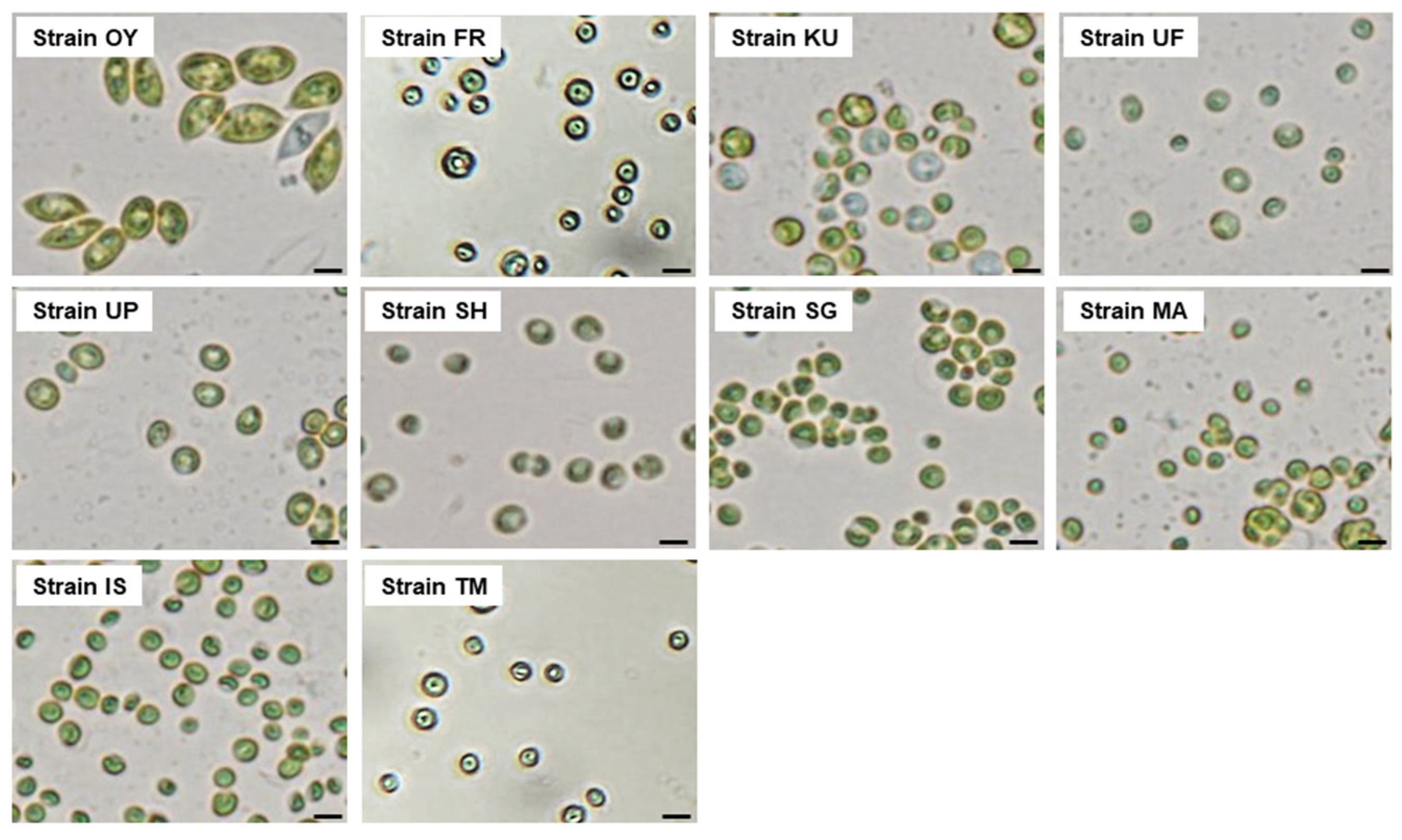
We used a glass vial for the enrichment of high-level CO2-tolerant microalgae from the collected freshwater samples because CO2 can permeate plastic bottles, and its concentration in the bottle gradually decreases during cultivation [32]. After 4 weeks of cultivation, microalgal growth (indicated by green turbidity) was observed in the vials of the 10 samples, except for the sample from Shimokubo (sample no. 4; Table 1). Then, 1.0 mL aliquot from the culture was added to a fresh SOT-C culture medium in a new vial, and subculturing was performed (4 weeks) five times under the same conditions. The enriched algal subculture displayed stable growth, and an aliquot of their fifth subculture was inoculated onto the SOT-C agar medium to isolate the algal strains. A total of 10 isolates were obtained, and their strain names are shown in Table 1. A microscopic analysis indicated that most strains were spherical to slightly ellipsoidal with a diameter of approximately 10–30 μm, although strain OY had a fusiform-shaped cell and a diameter of approximately 20–40 μm (Figure 2). The strains KU, FR, TM, MA, and SG produced sporangia containing several autospores, which are the common structures for the propagation of unicellular green microalgae [33].

3.2. Phylogenetic Identification of the Microalgal Strains

As per a recent report, the ITS region DNA is more accurate in phylogenetic analyses for microalgae than the 18S ribosomal RNA gene [34]. Thus, the ITS region DNA sequences were used to determine the phylogenetic position of the isolated strains. The phylogenetic relationship between the isolates and the known species is illustrated in Figure 1. The analysis suggested that the strains KU, FR, TM, MA, and SG belong to the genus Micractinium, and the strain IS belongs to the genus Chlorella. The strains UP, UF, and SH were suggested as species of the genus Desmodesmus, and strain OY belongs to the Scenedesmaceae family (genus Tetradesmus or Scenedesmus). Micractinium and Chlorella are closely related genera of freshwater microalgae that belong to the Chlorellaceae family. The Chrolella strains have been well studied in wastewater treatment and biofuel production as well as in physiological, ecological, and nutritional studies [35], and strains with alkaliphilic [36,37] or high-CO2-tolerant [38,39,40] abilities have also been reported. As for Micractinium species, two high-CO2-tolerant strains have been reported so far [41,42], but there is no report on alkaliphilic strains. Desmodesmus, Scenedesmus, and Tetradesmus are genera included in the Scenedesmaceae family—a green microalgal family ubiquitously found in freshwater environments like the Chlorellaceae family. They have been extensively studied as tools for wastewater treatment [43,44] and biodiesel production [45,46,47]. Alkaliphilic Scenedesmus sp. strain [48] and high-level CO2-tolerant Desmodesmus, Scenedesmus, and Tetradesmus spp. strains have also been reported [49,50,51,52,53]. However, there is no article about strains that possess both alkaliphilic- and high-CO2-preferring characteristics for these five genera.

3.3. Growth Characteristics under Various pH and CO2 Levels

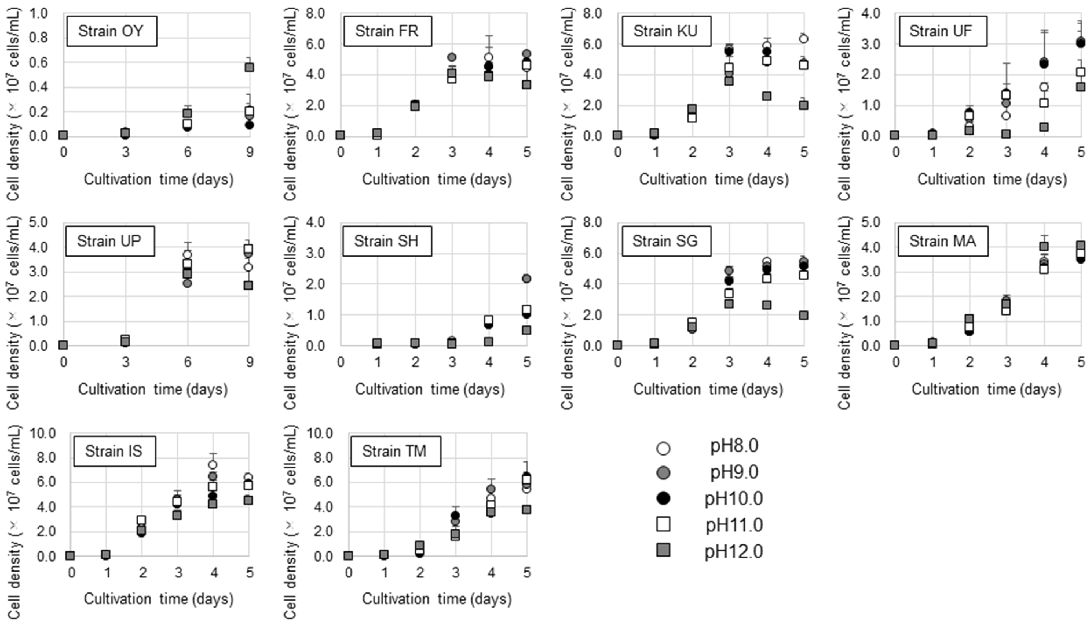
The pH range for strain growth was studied by culturing them in a medium with various pH values (8.0, 9.0, 10.0, 11.0, and 12.0) using 10 vol% CO2 to determine the optimal pH for their growth and to confirm whether they are alkaliphilic strains. The experimental results are shown in Figure 3. Although the growth rate is different among the strains, all of them could grow under the various alkaline pH conditions tested. Particularly, each strain exhibited unique growth characteristics. The optimal pH values for strains KU, SH, and IS were 8.0, 9.0, and 8.0–9.0, respectively. In contrast, the strains FR, UF, and SG were found to have an optimal pH range of 9.0–10.0, and the strains UP and TM could grow well even under a pH of 11.0. The strain MA exhibited good growth under the tested pH (8.0–12.0), implying its acclimating ability to harsh alkaline conditions. Interestingly, the strain OY grew well at a pH of 12.0, but its cell density in the 9-day culture (pH 12.0) was 0.57 × 10⁶ cells/mL, which is not as high as that for most of the other strains (in the order of 106 cells/mL in 5-day cultivation), suggesting that this strain is a slow grower. The slow growth of strain OY appears to be due to its large cell size (Figure 1), which requires a longer time for reproduction.
We subsequently cultured the strains at different CO2 concentrations (5, 10, 20, 30, and 40 vol%) under their optimal pH so as to determine the range of CO2 concentration for their growth (Figure 4). The growth of strains FR and SH was found to be enhanced under 5% CO2 but inhibited under 10% CO2. The optimal CO2 level for strains UF and IS was 10%. The stains OY, KU, UP, SG, MA, and TM were found to grow well at CO2 concentrations of up to 20%, and some of them (SG, MA, and TM) displayed good growth even under 30% CO2. Because dissolved CO2 can lower the pH of the culture medium [54], it is important to determine whether algal strains grew by favoring high levels of CO2 or by an abiotic neutralization of an alkaline medium by a CO2 flush. Therefore, the pH of the strain culture grown on the final cultivation day was analyzed (Table 2). When the strains grew well under their optimal CO2 level, the pH of grown culture for almost all strains, except strain UF, was found to be strongly alkaline (>10 or 11), suggesting that they actually favor an alkaline pH for their growth. In contrast, the growth culture of strain UF showed a pH of 6.66 on its final cultivation day, indicating that the strain could grow in a neutralized medium after a CO2 flush in the vial.

3.4. Upgrading of Biogas by the Algal Strains

In a previous study, we developed the digested sludge-assimilating and biogas-yielding soil (DABYS) microflorae that can yield biogas from sewage sludge as a substrate [55]. Accordingly, we assessed whether the algal strains could fix CO2 in biogas produced via sludge digestion by the DABYS microflora. An outline of the experiment is illustrated in Figure S1a. Excess sludge, a raw sewage sludge generated in the second wastewater treatment, and digested sludge, a sludge residue generated after anaerobic digestion of excess sludge, served as substrates for vial-scale biogas production. While algal growth during the biogas upgrading assay is depicted in Figure S1b,c, the result of the experiment is shown in Figure 5 and Figure 6. When the DABYS microflora was cultivated anaerobically after being fed with digested sludge in a test tube for 7 days, the algal strains’ seed culture in vials nos. 4 to 13 grew, as indicated by the presence of green turbidity in their culture medium (Figure S1b). A similar observation was made for the assay in which the DABYS microflora was fed with excess sludge (Figure S1c). Significant differences were found between the cell densities for the 0-day and 7-day cultures of each strain (Figure 5a,b). Green turbidity was not detected in vials nos. 1 to 3, confirming that the green turbidity observed in the vials was not due to the outgrowth of algal or cyanobacterial contaminants.
Subsequently, the gas phase of each vial was analyzed through GC-TCD to determine the methane, CO2, and hydrogen yields (Figure 6a,b). When the DABYS microflora was fed with the digested sludge or excess sludge in a small test tube, the resultant biogas was composed mainly of methane and CO2 after a 7-day incubation (vials 1 to 3 in Figure 6a or Figure 6b, respectively). The CO2 contents in the biogas of vial nos. 2 and 3 were partly decreased when compared to that in vial no. 1, although no significant difference was detected among them because of the large standard deviation, which implies that some of the CO2 was dissolved in sterile water and the SOT-C medium, respectively. In contrast, CO2 generated via anaerobic digestion in the small test tube was completely dissipated from the gas phase of the vials during the 7 days of incubation (vials nos. 4–13, Figure 6a,b). Considering that the gas phase of all vials was replaced with N2 gas at the beginning of cultivation, we believe that the algal strains in the vials were not affected by toxic biogas constituents, such as sulfide and ammonia, and that they could grow by fixing CO2 in biogas generated via the sludge digestion.
Several articles have been published on microalgal CO2 sequestration from biogas using Chlorella, Scenedesmus, and Tetraselmis monoalgal strains [56,57] or algal–bacteria/fungi consortia [58,59,60,61,62,63], and 62–99% of the CO2-removal efficiency was accomplished by improving their reaction systems. Although the vial-scale experiment in the present study is still in the fundamental research stage, CO2 was completely sequestered from biogas by algal strains, indicating their availability as a biogas-upgrading technology in combination with a sophisticated bioreactor. Furthermore, to the best of our knowledge, this is the first report on microalgal CO2 sequestration from biogas by Micractinium and Desmodesmus sp. strains.

3.5. Chemical Composition of the Algal Strains

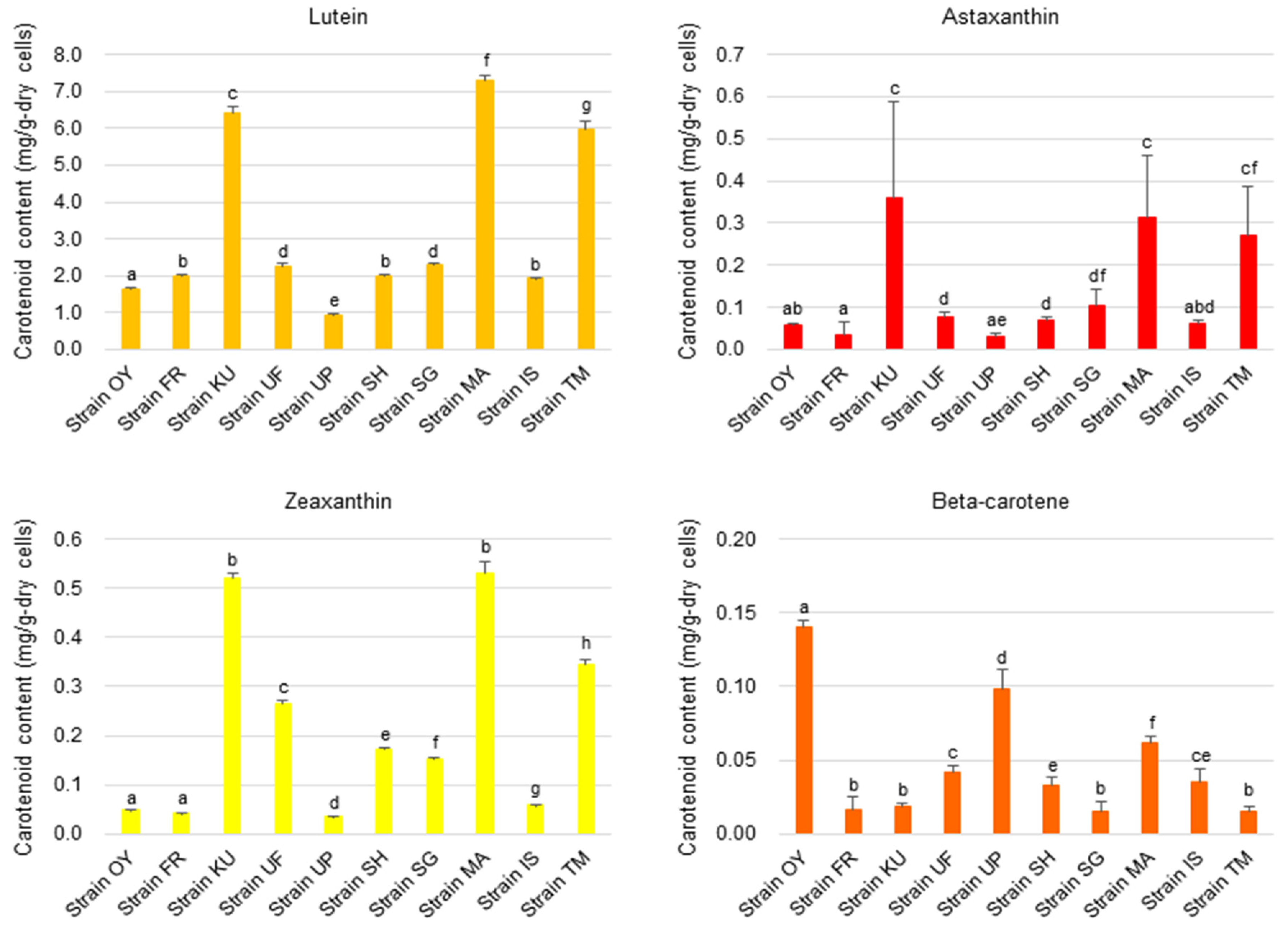
To examine the potential of algal strains for nutraceutical applications, their carotenoid contents were determined (Figure 7). The strains KU, MA, and TM, all of which belong to the genus Micractinium, were found to be rich in xanthophyll carotenoids, including lutein, astaxanthin, and zeaxanthin when compared with other strains. Particularly, their lutein contents were 6.4, 7.3, and 6.0 mg per 1.0 g of dry cell of strains KU, MA, and TM, respectively. Lutein and zeaxanthin are antioxidant carotenoids that are exclusively synthesized by plants and microalgae and are known to exert favorable effects on animal eye health and are, thus, employed in the pharmaceutical industry [64]. Astaxanthin is a potent antioxidant carotenoid produced by primary food web producers, including bacteria, yeast, and microalgae, and it is used in cosmetics and aquaculture industries as an antiultraviolet skin protectant and natural colorant, respectively [65,66]. Although there is no report on the lutein or astaxanthin content of Micractinium strains, the strains of the genus Chlorella (the closest genus for Micractinium) reportedly contain 0.069–10.4 mg lutein per 1.0 g of dry cells [67,68], implying that the strains KU, MA, and TM are good lutein producers. Inbaraj et al. reported exceptional accumulation of lutein in Chlorella pyrenoidosa-derived tablets (125 mg lutein per 1.0 g of the tablets) [69], albeit the production method for the tables was not disclosed; thus, additional sources of lutein in the tablets cannot be excluded.
The cellular protein content of the algal strains was determined (Figure 8). It was found that the strains KU, SG, MA, and TM cells were rich in protein, which was approximately or more than 300 mg per g of dry cells. In particular, strains SG and TM contained 436 and 534 mg of protein per g of dry cells, respectively, which are comparable to that for known Chlorella spp. [70]. The amino acid composition of proteins of the strains was determined (Table 3). The strains were found to possess similar amino acid compositions and contain most of the essential amino acids for animals, except tryptophan, which account for ~40 mol% (42.9 mol% on average) of the total amino acids, implying their potential use as a protein source for humans, livestock, and farmed fish. Additionally, these strains contain relatively high amounts of leucine, alanine, and glycine. Leucine is a nutritionally essential branched-chain amino acid that stimulates protein synthesis through the activation of energy metabolism, including glucose uptake, mitochondrial biogenesis, and fatty acid oxidation [71]. Interestingly, strains SH and UF were also found to be rich in arginine and glutamic acid/glutamine, respectively. Although arginine is not an essential amino acid for humans, it is essential for birds, carnivores, and some mammals, and it acts as a precursor for ornithine, an important metabolite in the urea cycle, and creatine, which plays an essential role in the energy metabolism of muscles and nerves and has specific effects on wound healing [72,73].
The fatty acid composition of the strains was determined as shown in Table 4, and 47–73 wt% (62.2 ± 9.4 wt% on average) of the total fatty acids was unsaturated fatty acids. While the most abundant unsaturated fatty acid in strain IS was oleic acid (C18:1), that in other strains was linoleic acid (C18:2). More than 30% of the fatty acids of the strains FR and UP was accounted for by linoleic acid. Linoleic acid is an essential fatty acid for humans and animals, and clinical studies have suggested that its dietary intake reduces the risk of cardiovascular diseases [74]. Thus, the algal oil from these strains may be useful as a nutrient for humans and animals.

4. Conclusions

In this study, almost all strains preferred alkaline pH for growth and propagated efficiently at high CO2 concentrations. The strains sequestered CO2 in biogas generated by the anaerobic digestion of sewage sludge in the glass vial, leading to the successful upgrading of biogas. The strains produced several valuable bioactive chemicals, such as lutein, essential amino acids, and unsaturated fatty acids, that have potential applications in the nutraceutical, food, livestock, and aquaculture industries. The strains SG, TM, and MA are particularly suitable for the purpose of CO2 use as they grow in a wide range of alkaline pH and under high levels of CO2, potentially facilitating efficient CO2 capture and fixation, thereby tackling the increasing atmospheric CO2 levels. The strains’ ability to grow in an extremely alkaline pH mineral–salt medium and under high CO2 concentrations is advantageous as it can reduce the risk of overgrowth of algal and cyanobacteria contaminants. However, to establish the optimal culture conditions to maintain efficient microalgal CO2 sequestration and improve the technologies required for the practical applications of these strains, further studies are warranted.

Supplementary Materials

The following supporting information can be downloaded at: https://www.mdpi.com/article/10.3390/fermentation10030134/s1, Figure S1: Outline of the biogas upgrading assay (a) and photographs of algal growth observed in the tested glass vials in which a small test tube dispensed with DABYS microflora and digested sludge (b) or excess sludge (c).

Author Contributions

Conceptualization, K.F.; methodology, Y.K., K.S. and K.F.; validation, Y.K., D.K., K.S. and K.F.; investigation, Y.K., D.K., K.S. and K.F.; original draft preparation, Y.K.; review and editing, K.F.; supervision, K.F.; project administration, K.F.; funding acquisition, K.F. All authors have read and agreed to the published version of the manuscript.

Funding

This study was funded by a Grant-in-Aid for Scientific Research (C: 22K12430) from the Japan Society for the Promotion of Science, a JST Adaptable and Seamless Technology transfer Program through Target driven R&D (A STEP: JPMJTM22B7), and a Grant from the Research Institute of Science and Technology, Kogakuin University.

Institutional Review Board Statement

Not applicable.

Informed Consent Statement

Not applicable.

Data Availability Statement

The data presented in this study are available on request from the corresponding author.

Conflicts of Interest

The authors declare no conflicts of interest.

References

  1. International Energy Agency. CO2 Emissions in 2022. 2023. Available online: https://iea.blob.core.windows.net/assets/3c8fa115-35c4-4474-b237-1b00424c8844/CO2Emissionsin2022.pdf (accessed on 30 December 2023).
  2. Manish Pillai, I.R.; Banerjee, R. Sustainability analysis of renewables for climate change mitigation. Energy Sustain. Develop. 2006, 10, 25–36. [Google Scholar] [CrossRef]
  3. Chen, X.Y.; Vinh-Thang, H.; Ramirez, A.A.; Rodrigue, D.; Kaliaguine, S. Membrane gas separation technologies for biogas upgrading. RSC Adv. 2015, 5, 24399–24448. [Google Scholar] [CrossRef]
  4. Kemper, J. Biomass and carbon dioxide capture and storage: A review. Int. J. Greenh. Gas Cont. 2015, 40, 401–430. [Google Scholar] [CrossRef]
  5. Kheirinik, M.; Ahmed, S.; Rahmanian, N. Comparative techno-economic analysis of carbon capture processes: Pre-combustion, post-combustion, and oxy-fuel combustion operations. Sustainability 2021, 13, 13567. [Google Scholar] [CrossRef]
  6. Yasemi, S.; Khalili, Y.; Sanati, A.; Bagheri, M. Carbon capture and storage: Application in the oil and gas industry. Sustainability 2023, 15, 14486. [Google Scholar] [CrossRef]
  7. Bajpai, S.; Shreyash, N.; Singh, S.; Memon, A.R.; Sonker, M.; Kr Tiwary, S.; Biswas, S. Opportunities, challenges and the way ahead for carbon capture, utilization and sequestration (CCUS) by the hydrocarbon industry: Towards a sustainable future. Energ. Rep. 2022, 8, 15595–15616. [Google Scholar] [CrossRef]
  8. Langie, K.M.G.; Tak, K.; Kim, C.; Lee, H.W.; Park, K.; Kim, D.; Jung, W.; Lee, C.W.; Oh, H.S.; Lee, D.K.; et al. Toward economical application of carbon capture and utilization technology with near-zero carbon emission. Nat. Commun. 2022, 13, 7482. [Google Scholar] [CrossRef] [PubMed]
  9. Cuéllar-Franca, R.M.; Azapagic, A. Carbon capture, storage and utilization technologies: A critical analysis and comparison of their life cycle environmental impacts. J. CO2 Util. 2015, 9, 82–102. [Google Scholar] [CrossRef]
  10. Mohapatra, R.K.; Padhi, D.; Sen, R.; Nayak, M. Bio-inspired CO2 capture and utilization by microalgae for bioenergy feedstock production: A greener approach for environmental protection. Bioresour. Technol. Rep. 2022, 19, 101116. [Google Scholar] [CrossRef]
  11. Yang, W.; Li, S.; Qv, M.; Dai, D.; Liu, D.; Wang, W.; Tang, C.; Zhu, L. Microalgal cultivation for the upgraded biogas by removing CO2, coupled with the treatment of slurry from anaerobic digestion: A review. Bioresour. Technol. 2022, 364, 128118. [Google Scholar] [CrossRef] [PubMed]
  12. Zhang, S.; Liu, Z. Advances in the biological fixation of carbon dioxide by microalgae. J. Chem. Technol. Biotechnol. 2021, 96, 1475–1495. [Google Scholar] [CrossRef]
  13. Kratzer, R.; Murkovic, M. Food ingredients and nutraceuticals from microalgae: Main product classes and biotechnological production. Foods 2021, 10, 1626. [Google Scholar] [CrossRef]
  14. Casanova, L.M.; Mendes, L.B.B.; Corrêa, T.d.S.; da Silva, R.B.; Joao, R.R.; Macrae, A.; Vermelho, A.B. Development of microalgae biodiesel: Current status and perspectives. Microorganisms 2023, 11, 34. [Google Scholar] [CrossRef] [PubMed]
  15. Gonçalves, A.L.; Almeida, F.; Rocha, F.A.; Ferreira, A. Improving CO2 mass transfer in microalgal cultures using an oscillatory flow reactor with smooth periodic constrictions. J. Environ. Chem. Eng. 2021, 9, 106505. [Google Scholar] [CrossRef]
  16. Becker, E.W. (Ed.) Culture media. In Microalgae. Biotechnology and Microbiology; Cambridge University Press: Cambridge, UK, 1994; pp. 9–42. [Google Scholar]
  17. Cents, A.; Brilman, D.; Versteeg, G. CO2 absorption in carbonate/bicarbonate solutions: The Danckwerts-criterion revisited. Chem. Eng. Sci. 2005, 60, 5830–5835. [Google Scholar] [CrossRef]
  18. La Plante, E.; Wang, J.; Alturki, A.; Simonetti, D.; Jassby, D.; Sant, G. Saline water-based mineralization pathway for gigatonne-scale CO2 management. ACS Sustain. Chem. Eng. 2021, 9, 1073–1089. [Google Scholar] [CrossRef]
  19. Ochoa-Alfaro, A.E.; Gaytán-Luna, D.E.; González-Ortega, O.; Zavala-Arias, K.G.; Paz-Maldonado, L.M.T.; Rocha-Uribe, A. pH effects on the lipid and fatty acids accumulation in Chlamydomonas reinhardtii. Biotechnol. Prog. 2019, 35, e2891. [Google Scholar] [CrossRef] [PubMed]
  20. Andeden, E.E.; Ozturk, S.; Aslim, B. Effect of alkaline pH and nitrogen starvation on the triacylglycerol (TAG) content, growth, biochemical composition, and fatty acid profile of Auxenochlorella protothecoides KP7. J. Appl. Phycol. 2021, 33, 211–225. [Google Scholar] [CrossRef]
  21. Hinga, K.R. Effects of pH on coastal marine phytoplankton. Mar. Ecol. Prog. Ser. 2002, 238, 281–300. [Google Scholar] [CrossRef]
  22. Ogawa, T. Studies on the growth of Spirulina platensis: (I) On the pure culture of Spirulina platensis. J. Ferment. Technol. 1970, 48, 361–367. [Google Scholar]
  23. Sambrook, J.; Russell, D.W. Purification of nucleic acids by extraction with phenol:chloroform. Cold Springs Harbor Protoc. 2006, 2006, pdb-prot4455. [Google Scholar] [CrossRef] [PubMed]
  24. White, T.J.; Bruns, T.; Lee, S.; Taylor, J.W. Amplification and direct sequencing of fungal ribosomal RNA genes for phylogenetics. In PCR Protocols. A Guide to Methods and Applications; Innis, M.A., Gelfand, D.H., Sninsky, J.J., White, T.J., Eds.; Academic Press: New York, NY, USA, 1990; pp. 315–522. [Google Scholar]
  25. Altschul, S.F.; Gish, W.; Miller, W.; Myers, E.W.; Lipman, D.J. Basic local alignment search tool. J. Mol. Biol. 1990, 215, 403–410. [Google Scholar] [CrossRef] [PubMed]
  26. Tamura, T.; Stecher, G.; Kumar, S. MEGA11: Molecular evolutionary genetics analysis version 11. Mol. Biol. Evol. 2021, 38, 3022–3027. [Google Scholar] [CrossRef] [PubMed]
  27. Edgar, R.C. MUSCLE: Multiple sequence alignment with high accuracy and high throughput. Nucleic Acids Res. 2004, 32, 1792–1797. [Google Scholar] [CrossRef] [PubMed]
  28. Felsenstein, J. Evolutionary trees from DNA sequences: A maximum likelihood approach. J. Mol. Evol. 1981, 17, 368–376. [Google Scholar] [CrossRef] [PubMed]
  29. Matsui, H.; Shiozaki, K.; Okumura, Y.; Ishikawa, M.; Waqalevu, V.; Hayasaka, O.; Honda, A.; Kotani, T. Effects of phosphorous deficiency of a microalga Nannochloropsis oculata on its fatty acid profiles and intracellular structure and the effectiveness in rotifer nutrition. Algal Res. 2020, 49, 101905. [Google Scholar] [CrossRef]
  30. Smith, P.K.; Krohn, R.I.; Hermanson, G.T.; Mallia, A.K.; Gartner, F.H.; Provenzano, M.D.; Fujimoto, E.K.; Goeke, N.M.; Olson, B.J.; Klenk, D.C. Measurement of protein using bicinchoninic acid. Anal. Biochem. 1985, 150, 76–78. [Google Scholar] [CrossRef]
  31. Bligh, E.G.; Dyer, W.J. A rapid method of total lipid extraction and purification. Can. J. Biochem. Physiol. 1959, 37, 911–917. [Google Scholar] [CrossRef]
  32. Toi, K. Pressure dependence of diffusion coefficient for CO2 in glassy polymers. Polym. Eng. Sci. 1980, 20, 30–35. [Google Scholar] [CrossRef]
  33. Yamamoto, M.; Kurihara, I.; Kawano, S. Late type of daughter cell wall synthesis in one of the Chlorellaceae, Parachlorella kessleri (Chlorophyta, Trebouxiophyceae). Planta 2005, 221, 766–775. [Google Scholar] [CrossRef]
  34. Hadi, S.I.I.A.; Santana, H.; Brunale, P.P.M.; Gomes, T.G.; Oliveira, M.D.; Matthiensen, A.; Oliveira, M.E.C.; Silva, F.C.P.; Brasil, B.S.A.F. DNA barcoding green microalgae isolated from neotropical inland waters. PLoS ONE 2016, 11, e0149284. [Google Scholar] [CrossRef]
  35. Liu, J.; Chen, F. Biology and industrial applications of Chlorella: Advances and prospects. In Microalgae Biotechnology; Posten, C., Chen, S.F., Eds.; Springer: Berlin/Heidelberg, Germany, 2016; Volume 153, pp. 1–35. [Google Scholar]
  36. Bell, T.A.; Prithiviraj, B.; Wahlen, B.D.; Fields, M.W.; Peyton, B.M. A lipid-accumulating alga maintains growth in outdoor, alkaliphilic raceway pond with mixed microbial communities. Front. Microbiol. 2015, 6, 1480. [Google Scholar] [CrossRef]
  37. Vadlamani, A.; Pendyala, B.; Viamajala, S.; Varanasi, S. High productivity cultivation of microalgae without concentrated CO2 input. ACS Sustain. Chem. Eng. 2019, 7, 1933–1943. [Google Scholar] [CrossRef]
  38. Sung, K.D.; Lee, J.S.; Shin, C.S.; Park, S.C.; Cho, M.J. CO2 fixation by Chlorella sp. KR-1 and its cultural characteristics. Bioresour. Technol. 1999, 68, 269–273. [Google Scholar] [CrossRef]
  39. Li, J.; Tang, X.; Pan, K.; Zhu, B.; Li, Y.; Ma, X.; Zhao, Y. The regulating mechanisms of CO2 fixation and carbon allocations of two Chlorella sp. strains in response to high CO2 levels. Chemosphere 2020, 247, 125814. [Google Scholar] [CrossRef]
  40. Sergeenko, T.V.; Muradyan, E.A.; Pronina, N.A.; Klyachko-Gurvich, G.L.; Mishina, I.M.; Tsoglin, L.N. The effect of extremely high CO2 concentration on the growth and biochemical composition of microalgae. Russ. J. Plant Physiol. 2000, 47, 632–638. [Google Scholar]
  41. China, S.; Fujii, K. Isolation of high-CO2-acclimated Micractinium sp. strains from eutrophic reservoir water. Algal Res. 2018, 34, 126–133. [Google Scholar] [CrossRef]
  42. Cao, B.; Hu, S.; Zhu, K.; Pan, C.; Marrakchi, F.; Ni, J.; Yuan, C.; Qian, L.; Chen, H.; Yuan, J.; et al. Response surface optimization of product yields and biofuel quality during fast hydrothermal liquefaction of a highly CO2-tolerant microalgae. Sci. Total Environ. 2023, 860, 160541. [Google Scholar] [CrossRef]
  43. Jothibasu, K.; Muniraj, I.; Jayakumar, T.; Ray, B.; Dhar, D.W.; Karthikeyan, S.; Rakesh, S. Impact of microalgal cell wall biology on downstream processing and nutrient removal for fuels and value-added products. Biochem. Eng. J. 2022, 187, 108642. [Google Scholar] [CrossRef]
  44. Kiani, H.; Azimi, Y.; Li, Y.; Mousavi, M.; Cara, F.; Mulcahy, S.; McDonnell, H.; Blanco, A.; Halim, R. Nitrogen and phosphate removal from dairy processing side-streams by monocultures or consortium of microalgae. J. Biotechnol. 2023, 361, 1–11. [Google Scholar] [CrossRef] [PubMed]
  45. Shuba, E.S.; Kifle, D. Microalgae to biofuels: ‘Promising’ alternative and renewable energy, review. Renew. Sust. Energ. Rev. 2018, 81, 743–755. [Google Scholar] [CrossRef]
  46. Oliveira, C.Y.B.; Oliveira, C.D.L.; Prasad, R.; Ong, H.C.; Araujo, E.S.; Shabnam, N.; Gálvez, A.O. A multidisciplinary review of Tetradesmus obliquus: A microalga suitable for large-scale biomass production and emerging environmental applications. Rev. Aquacult. 2021, 13, 1594–1618. [Google Scholar] [CrossRef]
  47. de Almeida Moreira, B.R.; de Almeida Viana, C.R.; Cruz, V.H.; Lopes, P.R.M.; da Silva Viana, R.; Ramos, R.A.V. Meta-analytic review on third-generation biodiesel. Bioenerg. Res. 2022, 15, 27–45. [Google Scholar] [CrossRef]
  48. Gardner, R.; Peters, P.; Peyton, B.; Cooksey, K.E. Medium pH and nitrate concentration effects on accumulation of triacylglycerol in two members of the chlorophyta. J. Appl. Phycol. 2011, 23, 1005–1016. [Google Scholar] [CrossRef]
  49. Liu, Z.; Zhang, F.; Chen, F. High throughput screening of CO2-tolerating microalgae using GasPak bags. Aquat. Biosyst. 2013, 9, 23. [Google Scholar] [CrossRef] [PubMed]
  50. Giannetto, M.J.; Retotar, A.; Rismani-Yazdi, H.; Peccia, J. Using carbon dioxide to maintain an elevated oleaginous microalga concentration in mixed-culture photo-bioreactors. Bioresour. Technol. 2015, 185, 178–184. [Google Scholar] [CrossRef]
  51. Scherbakov, P.; Ismagulova, T.; Chernov, T.; Gorelova, O.; Selyakh, I.; Semenova, L.; Baulina, O.; Chivkunova, O.; Lobakova, E.; Solovchenko, A. A new subarctic strain of Tetradesmus obliquus. Part II: Comparative studies of CO2-stress tolerance. J. Appl. Phycol. 2018, 30, 2751–2761. [Google Scholar] [CrossRef]
  52. Kumar, R.; Goswami, G.; Debnath, D.; Sinha, A.; Das, D. Screening and evaluation of novel microalga Desmodesmus pannonicus CT01 for CO2 sequestration potential and aqua feed application. Biomass Conv. Biorefin. 2022, 1–12. [Google Scholar] [CrossRef]
  53. Sinha, A.; Goswami, G.; Kumar, R.; Das, D. A microalgal biorefinery approach for bioactive molecules, biofuel, and biofertilizer using a novel carbon dioxide-tolerant strain Tetradesmus obliquus CT02. Biomass Conv. Biorefin. 2022, 13, 12605–12618. [Google Scholar] [CrossRef]
  54. Debs-Louka, E.; Louka, N.; Abraham, G.; Chabot, V.; Allaf, K. Effect of compressed carbon dioxide on microbial cell viability. Appl. Environ. Microbiol. 1999, 65, 626–631. [Google Scholar] [CrossRef]
  55. Kon, A.; Omata, S.; Hayakawa, Y.; Aburai, N.; Fujii, K. Microflora communities which can convert digested sludge to biogas. Environ. Technol. 2022, 43, 2391–2403. [Google Scholar] [CrossRef]
  56. Gao, S.; Hu, C.; Sun, S.; Xu, J.; Zhao, Y.; Zhang, H. Performance of piggery wastewater treatment and biogas upgrading by three microalgal cultivation technologies under different initial chemical oxygen demand concentration. Energy 2018, 165, 360–369. [Google Scholar] [CrossRef]
  57. Herold, C.; Ishika, T.; Nwoba, E.G.; Tait, S.; Ward, A.; Moheimani, N.R. Biomass production of marine microalga Tetraselmis suecica using biogas and wastewater as nutrients. Biomass Bioenerg. 2021, 145, 105945. [Google Scholar] [CrossRef]
  58. Rodero, M.D.R.; Lebrero, R.; Serrano, E.; Lara, E.; Arbib, Z.; García-Encina, P.A.; Muñoz, R. Technology validation of photosynthetic biogas upgrading in a semi-industrial scale algal-bacterial photobioreactor. Bioresour. Technol. 2019, 279, 43–49. [Google Scholar] [CrossRef] [PubMed]
  59. Guenka Scarcelli, P.; Ruas, G.; Lopez-Serna, R.; Leite Serejo, M.; Blanco, S.; Árpád Boncz, M.; Muñoz, R. Integration of algae-based sewage treatment with anaerobic digestion of the bacterial-algal biomass and biogas upgrading. Bioresour. Technol. 2021, 340, 125552. [Google Scholar] [CrossRef] [PubMed]
  60. Zhang, J.; Zhao, C.; Sun, S.; Zhao, Y.; Liu, J. Performance of different microalgae based technologies in nutrient removal and biogas upgrading in response to various GR24 concentrations. Int. Biodeter. Biodegr. 2021, 158, 105166. [Google Scholar] [CrossRef]
  61. Zhang, H.; Xu, B.; Zhao, C.; Liu, J.; Zhao, Y.; Sun, S.; Wei, J. Simultaneous biogas upgrading and biogas slurry treatment by different microalgae-based technologies under various strigolactone analog (GR24) concentrations. Bioresour. Technol. 2022, 351, 127033. [Google Scholar] [CrossRef]
  62. Méndez, L.; García, D.; Perez, E.; Blanco, S.; Muñoz, R. Photosynthetic upgrading of biogas from anaerobic digestion of mixed sludge in an outdoors algal-bacterial photobioreactor at pilot scale. J. Water. Proce. Eng. 2022, 48, 102891. [Google Scholar] [CrossRef]
  63. Marín, D.; Méndez, L.; Suero, I.; Díaz, I.; Blanco, S.; Fdz-Polanco, M.; Muñoz, R. Anaerobic digestion of food waste coupled with biogas upgrading in an outdoors algal-bacterial photobioreactor at pilot scale. Fuel 2022, 324, 124554. [Google Scholar] [CrossRef]
  64. Mares-Perlman, J.A.; Millen, A.E.; Ficek, T.L.; Hankinson, S.E. The body of evidence to support a protective role for lutein and zeaxanthin in delaying chronic disease. Overview 1, 2. J. Nut. 2002, 132, 518S–524S. [Google Scholar] [CrossRef]
  65. Johnson, E.A.; An, G.H. Astaxanthin from microbial sources. Crit. Rev. Biotechnol. 1991, 11, 297–326. [Google Scholar] [CrossRef]
  66. Donoso, A.; González-Durán, J.; Muñoz, A.A.; González, P.A.; Agurto-Muñoz, C. Therapeutic uses of natural astaxanthin: An evidence-based review focused on human clinical trials. Pharmacol. Res. 2021, 166, 105479. [Google Scholar] [CrossRef]
  67. Ievina, B.; Romagnoli, F. Potential of Chlorella species as feedstock for bioenergy production: A review. Environ. Clim. Technol. 2020, 24, 203–220. [Google Scholar] [CrossRef]
  68. Wang, J.; Hu, X.; Chen, J.; Wang, T.; Huang, X.; Chen, G. The extraction of β-carotene from microalgae for testing their health benefits. Food 2022, 11, 502. [Google Scholar] [CrossRef] [PubMed]
  69. Inbaraj, B.S.; Chien, J.T.; Chen, B.H. Improved high performance liquid chromatographic method for determination of carotenoids in the microalga Chlorella pyrenoidosa. J. Chromatogr. A 2006, 1102, 193–199. [Google Scholar] [CrossRef]
  70. Levasseur, W.; Perre, P.; Pozzobon, V. A review of high value-added molecules production by microalgae in light of the classification. Biotechnol. Adv. 2020, 41, 107545. [Google Scholar] [CrossRef] [PubMed]
  71. Duan, Y.; Li, F.; Li, Y.; Tang, Y.; Kong, X.; Feng, Z.; Anthony, T.G.; Watford, M.; Hou, Y.; Wu, G.; et al. The role of leucine and its metabolites in protein and energy metabolism. Amino Acids 2016, 48, 41–51. [Google Scholar] [CrossRef] [PubMed]
  72. Tapiero, H.; Mathé, G.; Couvreur, P.; Tew, K.D. Free amino acids in human health and pathologies. I. Arginine. Biomed. Pharmacother. 2002, 56, 439–445. [Google Scholar] [CrossRef]
  73. Stechmiller, J.K.; Childress, B.; Cowan, L. Arginine supplementation and wound healing. Nut. Clin. Pract. 2005, 20, 52–61. [Google Scholar] [CrossRef]
  74. Marangoni, F.; Agostoni, C.; Borghi, C.; Catapano, A.L.; Cena, H.; Ghiselli, A.; La Vecchia, C.; Lercker, G.; Manzato, E.; Pirillo, A.; et al. Dietary linoleic acid and human health: Focus on cardiovascular and cardiometabolic effect. Atherosclerosis 2020, 292, 90–98. [Google Scholar] [CrossRef]
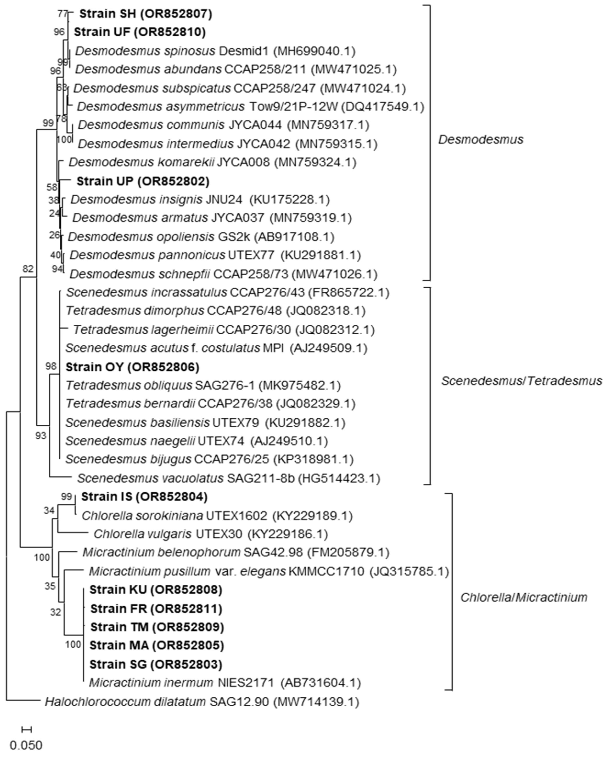
Figure 1. Phylogenetic tree of the algal isolates with known relatives based on ITS sequences constructed using the maximum-likelihood method. Bootstrap values (percentage value of 1000 samplings) are shown at the internodes. The scale bar represents an evolutionary distance (Knuc) of 0.05. Accession numbers for each DNA are indicated in parentheses.
Fermentation 10 00134 g001
Figure 2. Optical micrographs of the algal isolates. Scale bar = 10 μm.
Fermentation 10 00134 g002
Figure 3. Growth of the algal isolates under different pH conditions. The data are presented as means ± standard deviation of independent triplicates.
Fermentation 10 00134 g003
Figure 4. Growth of the algal isolates under different CO2 concentrations with pH fixed to the optimal value indicated next to the strain names for each isolate. The data are presented as means ± standard deviation of independent triplicates.
Fermentation 10 00134 g004
Figure 5. Growth of the algal isolates in glass vials tested for the biogas upgrading assay. Algal cell density in the vials in which a small test tube dispensed with DABYS microflora and digested sludge (a) or excess sludge (b) is shown. The data are presented as means ± standard deviation of independent triplicates. Asterisks on the columns indicate significant differences at p < 0.05 (Student’s t-test) between cell density for algal strains in 0- and 7-day cultures.
Fermentation 10 00134 g005
Figure 6. Biogas yields in the glass vials of the biogas upgrading assay. The data for assay using digested sludge (a) and excess sludge (b) as a substrate are shown. The data are presented as means ± standard deviation of independent triplicates. The letters on the columns shown in blue, orange, and gray indicate significant differences at p < 0.05 (Student’s t-test) for hydrogen, methane, and CO2 gasses, respectively.
Fermentation 10 00134 g006
Figure 7. Cellular content of carotenoids in the algal isolates. The data are presented as means ± standard deviation of independent triplicates. Letters on the columns indicate significant differences at p < 0.05 (Student’s t-test) between the isolates.
Fermentation 10 00134 g007
Figure 8. Cellular content of protein in the algal isolates. The data are presented as means ± standard deviation of independent triplicates. Letters on the columns indicate significant differences at p < 0.05 (Student’s t-test) between the isolates.
Fermentation 10 00134 g008
Table 1. List of sampling locations and isolated algal strains used in this study.
Sample Nos.Sampling LocationIsolated Algal Strain
1Ooyori, the midstream of the Kanna river, Gumma Pref. (36°13′ N, 138°94′ E)OY
2Furuta, the midstream of the Kanna river, Gumma Pref. (36°10′ N, 138°87′ E)FR
3Ueno, the upstream of the Kanna river, Gumma Pref. (36°08′ N, 138°79′ E)KU
4Shimokubo, the downstream of the Kanna river, Gumma Pref. (36°13′ N, 139°03′ E)
5Fountain pond in Kogakuin University, Hachioji city (35°68′ N, 139°32′ E)UF
6Ornamental pond in Kogakuin University, Hachioji city (35°68′ N, 139°32′ E)UP
7Ornamental pond in Shimizu park, Hachioji city (35°68′ N, 139°31′ E)SH
8Ornamental pond in Ishikawa-higashi park, Hachioji city (35°68′ N, 139°37′ E)IS
9Gotanda, the downstream of the Shiroyama river, Hachioji city (35°67′ N, 139°30′ E)SG
10Yokogawa, the downstream of the Minami-asakawa river, Hachioji city (35°67′ N, 139°31′ E)MA
11Akishima, the midstream of the Tama river, Hachioji city (35°69′ N, 139°37′ E)TM
Table 2. Change in the pH value of the strain culture under different CO2 concentrations. Obelisks indicate the optimal CO2 level for growth of each strain shown in Figure 4.
StrainInitial pH of the Culture MediumCO2 Concentration of Gas PhasepH of Grown CultureStrainInitial pH of the Culture MediumCO2 Concentration of Gas PhasepH of Grown Culture
AverageStdevAverageStdev
Strain OY12.0 5%7.34 0.02 Strain SH9.0 5% †10.85 0.58
10%10.16 1.58 10%10.53 0.70
20% †11.96 0.01 20%5.68 0.07
30%6.55 0.13 30%5.38 0.02
50%6.33 0.07 50%5.33 0.05
Strain FR9.0 5%11.20 0.05 Strain SG9.0 5%7.30 0.19
10% †11.25 0.04 10%6.64 0.14
20%6.26 0.13 20% †11.11 0.03
30%6.02 0.17 30%11.14 0.07
50%5.37 0.06 50%5.47 0.02
Strain KU8.0 5%6.90 0.18 Strain MA12.0 5%9.45 0.85
10%11.34 0.03 10%11.46 0.02
20% †11.22 0.10 20% †11.43 0.05
30%6.05 0.37 30%6.99 0.07
50%5.58 0.06 50%6.27 0.12
Strain UF9.0 5%6.77 0.22 Strain IS8.0 5%11.56 0.01
10% †6.66 0.03 10% †11.64 0.02
20%5.78 0.05 20%5.89 0.08
30%5.48 0.02 30%5.62 0.02
50%5.24 0.02 50%5.54 0.06
Strain UP10.0 5%11.12 0.02 Strain TM10.0 5%6.24 0.09
10%11.11 0.03 10%7.63 0.53
20% †10.22 0.78 20% †11.19 0.07
30%5.74 0.17 30%11.23 0.05
50%5.46 0.07 50%5.49 0.09
Table 3. Amino acid composition (mol%) of hydrolysates from microalgal strain proteins. Asterisks indicate essential amino acids for humans. Different letters next to the values indicate significant differences at p < 0.05 (Student’s t-test) in each amino acid content among the tested strains.
Amino AcidStrain OYStrain FRStrain KUStrain UFStrain UPStrain SHStrain SGStrain MAStrain ISStrain TM
AverageStdevAverageStdevAverageStdevAverageStdevAverageStdevAverageStdevAverageStdevAverageStdevAverageStdevAverageStdev
His *2.17 a0.06 1.83 be0.04 2.06 c0.04 1.87 b0.04 2.17 af0.03 2.63 d0.13 2.15 a0.01 1.73 e0.08 2.24 f0.03 2.21 f0.04
Ser3.70 a0.01 4.04 b0.04 5.03 c0.05 4.63 d0.03 3.38 e0.03 4.12 b0.09 4.62 d0.01 4.27 e0.06 3.54 f0.05 4.82 g0.07
Arg5.49 a0.06 4.99 b0.07 5.01 b0.08 6.48 c0.03 6.63 c0.07 12.20 d0.25 5.02 b0.01 5.06 b0.12 5.20 e0.04 5.05 b0.03
Gly13.72 a0.27 13.12 b0.09 12.67 c0.12 12.02 d0.14 12.74 c0.02 13.50 a0.18 13.07 b0.11 12.70 c0.31 13.63 a0.06 13.13 b0.07
Asp+Asn4.97 a0.18 5.33 b0.13 5.41 b0.09 5.75 c0.21 6.13 d0.09 4.64 e0.15 5.13 f0.07 5.79 c0.35 4.84 ae0.23 4.64 e0.17
Glu+Gln7.81 a0.29 8.38 b0.12 7.90 c0.11 12.14 e0.14 9.24 f0.14 9.45 f0.26 8.34 b0.12 9.21 f0.42 7.63 a0.23 7.83 a0.19
Thr *4.97 a0.01 4.65 b0.07 4.92 c0.06 4.91 c0.06 4.69 b0.04 5.16 d0.15 4.63 e0.03 4.77 b0.08 4.75 b0.10 4.94 ac0.10
Ala11.51 a0.25 11.04 b0.11 11.08 b0.12 10.97 bc0.15 10.80 c0.08 8.73 d0.33 11.02 b0.05 10.86 c0.39 9.84 e0.12 10.59 f0.05
Pro6.18 a0.01 6.27 b0.02 6.22 c0.04 5.84 d0.04 5.95 e0.08 5.12 f0.16 6.13 a0.07 6.01 e0.08 6.36 f0.08 6.23 g0.08
Cys0.25 a0.01 0.08 b0.00 0.10 c0.00 0.13 d0.00 0.20 e0.00 0.01 f0.03 0.08 b0.00 0.07 g0.00 0.08 b0.00 0.09 h0.00
Lys *4.15 ab0.36 3.91 a0.08 4.33 b0.11 4.14 ab0.19 4.48 b0.11 2.34 c0.36 4.25 ab0.08 4.46 b0.42 4.92 d0.18 4.05 a0.08
Tyr2.98 a0.14 2.87 b0.03 2.98 a0.03 2.35 c0.04 2.85 b0.01 2.96 a0.16 2.72 d0.05 2.89 a0.17 3.13 e0.08 2.85 b0.04
Met *2.93 a0.09 2.74 b0.02 2.69 c0.02 2.51 d0.02 2.73 b0.01 2.85 a0.10 2.78 e0.02 2.66 f0.10 2.70 e0.03 2.79 e0.01
Val *7.47 a0.02 7.98 b0.05 7.47 a0.03 7.17 c0.02 7.40 d0.08 6.75 e0.07 7.61 f0.01 7.62 f0.10 7.86 g0.09 7.65 f0.05
Ile *4.98 a0.03 5.16 b0.03 4.90 c0.01 4.46 d0.00 4.85 e0.05 4.30 f0.03 5.00 g0.03 5.00 g0.08 5.25 h0.05 5.06 g0.04
Leu *10.18 a0.12 11.12 b0.09 10.79 cd0.00 9.35 e0.02 9.83 f0.09 8.79 g0.08 10.93 h0.04 10.71 d0.13 10.99 ch0.12 11.14 b0.07
Phe *6.53 a0.32 6.51 a0.08 6.42 a0.08 5.28 b0.12 5.94 c0.05 6.46 a0.36 6.54 a0.16 6.18 ac0.42 7.05 d0.19 6.94 d0.12
Trp *<0.01 <0.01 <0.01 <0.01 <0.01 <0.01 <0.01 <0.01 <0.01 <0.01
Essential amino acids43.39 1.02 43.89 0.47 43.58 0.35 39.69 0.47 42.09 0.46 39.27 1.28 43.89 0.38 43.13 1.42 45.76 0.80 44.76 0.49
Table 4. Fatty acid composition (wt%) of the microalgal strains. Asterisks indicate essential fatty acids for humans. Different letters next to the values indicate significant differences at p < 0.05 (Student’s t-test) in each fatty acid content among the tested strains.
Fatty AcidStrain OYStrain FRStrain KUStrain UFStrain UPStrain SHStrain SGStrain MAStrain ISStrain TM
AverageStdevAverageStdevAverageStdevAverageStdevAverageStdevAverageStdevAverageStdevAverageStdevAverageStdevAverageStdev
Myristic acid
(C14:0)
<0.1 <0.1 <0.1 <0.1 <0.1 <0.1 <0.1 <0.1 <0.1 0.27 0.02
Palmitic acid
(C16:0)
28.62 a3.19 16.65 b0.13 15.48 c0.53 25.83 d2.67 26.46 d3.15 21.39 e6.12 17.31 f0.57 14.89 g0.13 22.63 e0.85 15.71 c0.09
Palmitoleic acid
(C16:1)
6.50 a0.53 17.33 b0.05 12.24 c2.86 9.45 d0.85 11.46 c1.79 12.15 c2.72 16.54 e0.50 16.83 e1.16 7.07 f0.24 15.76 g0.18
Hexadecaenoic
acid (C16:2)
5.72 a0.58 6.75 b0.19 6.38 c0.48 4.15 d0.23 3.66 d0.79 4.15 d1.43 7.06 be0.87 6.28 c0.42 7.17 be0.63 7.01 e0.12
Stearic acid
(C18:0)
18.83 a0.78 1.46 b0.03 0.92 be0.53 15.28 c0.34 10.09 d0.86 11.75 d9.36 1.36 b0.42 0.86 e0.07 4.03 f0.08 1.68 g0.02
Oleic acid
(C18:1)
4.47 a0.38 15.98 b0.09 24.72 c2.01 13.43 d0.50 9.31 e0.20 16.98 bdf4.78 18.02 f0.63 21.61 g0.50 30.92 h0.11 22.38 i0.27
Linoleic acid
(C18:2) *
30.34 a0.93 32.45 b0.19 20.41 c1.61 25.13 d1.19 32.97 e0.37 26.54 d2.40 28.28 f0.72 28.11 f0.68 9.11 g0.04 26.68 d0.25
Linolenic acid
(C18:3) *
<0.1 <0.1 <0.1 <0.1 <0.1 <0.1 <0.1 <0.1 <0.1 <0.1
Unidentified fatty acid5.52 9.38 19.48 6.37 6.05 7.03 11.43 11.42 19.06 10.50
Unsaturated fatty acids47.03 2.42 72.51 0.51 63.76 6.96 52.16 2.78 57.39 3.15 59.83 11.33 69.90 2.72 72.83 2.76 54.28 1.02 71.83 0.82
Disclaimer/Publisher’s Note: The statements, opinions and data contained in all publications are solely those of the individual author(s) and contributor(s) and not of MDPI and/or the editor(s). MDPI and/or the editor(s) disclaim responsibility for any injury to people or property resulting from any ideas, methods, instructions or products referred to in the content.