Article

Characterisation of a Biodegradable Electrode Substrate Based on Psyllium Husk–Carbon Nanoparticle Composites

1
School of Engineering, Ulster University, Belfast BT15 1ED, Northern Ireland, UK
2
School of Biomedical Sciences, Ulster University, Coleraine BT52 1SA, Northern Ireland, UK
3
School of Computing, Engineering and Intelligent Systems, Ulster University, Derry/Londonderry BT48 7JL, Northern Ireland, UK
*
Author to whom correspondence should be addressed.
Submission received: 10 June 2025 / Revised: 7 August 2025 / Accepted: 15 August 2025 / Published: 17 August 2025
(This article belongs to the Section Carbon Materials and Carbon Allotropes)

Abstract

Unrefined psyllium husk derived from Plantago ovata constitutes a complex mixture of water-soluble and insoluble polymeric chains that form an interpenetrating network capable of entrapping carbon nanoparticles. While the resulting composite was found to swell in aqueous electrolyte, it exhibited hydrogel-like properties where the electrochemical activity was retained and found to be stable upon repetitive voltammetric cycling. Planar film systems were characterized by electron microscopy, Raman spectroscopy, tensile testing, gravimetric analysis, contact angle and cyclic voltammetry. A key advantage of the composite lies in its ability to be cast in 3D geometric forms such as pyramidal microneedle arrays (700 μm high × 200 μm base × 500 μm pitch) that could serve as viable electrode sensors. In contrast to conventional composite electrode materials that rely on non-aqueous solvents, the psyllium mixture is processed entirely from an aqueous solution. This, along with its plant-based origins and simple processing requirements, provides a versatile matrix for the design of biodegradable electrode structures that can be manufactured from more sustainable sources.

Graphical Abstract

1. Introduction

Electrodes composed of composite structures have become commonplace in both energy and sensing applications where there is a need for functional material interfaces [1,2,3,4]. Traditionally, the goal has been the acquisition of high-performance substrates which are also amenable to scalable manufacturing. More recently, there has also been a drive for materials that are capable of biodegradation and ideally derived from sustainable sources, thereby reducing the potential for waste. Such requirements, however, have often been difficult to balance against the performance needs of the particular application such that one design factor is often sacrificed in the pursuit of addressing another [5]. Progress towards the ideal material, however, has been steady and systems based on cellulose acetate phthalate [6], polybutylene sebacate [7], Poly(hydroxybutyrate-co-hydroxyvalerate) [8], poly(lactic-co-glycolic acid) (PLGA) [9], polycaprolactone (PCL) [10,11] and silk [12,13] have emerged as potential candidates to address the challenge. While inherently non-conductive, the polymers have been successfully used as biodegradable binders for encapsulating conductive nanoparticles to provide the processable electrode substrates [6]. However, with the exception of silk, few could be considered to be sourced directly from renewable sources.
Psyllium husk is derived from the Plantago genus consisting of over 200 species worldwide [14,15] but is most common to India and Iran where it is used as a medicinal agent [15,16]. The material is readily available from different renewable sources and is generally recognised as being a low-cost source of non-starch polysaccharide when compared to other natural sources [15]. While PsH contains a variety of bioactive components such as flavonoids, alkaloids, terpenoids, and phenolic acid derivatives, non-starch polysaccharide (85%) is the principal component [15]. As a natural product, the structure of PsH is both complex and variable depending on the source; however, its core components are common to all. The solid, typically milled from the seeds of Plantago ovata, contains both insoluble and soluble fractions and is composed of a highly branched, xylan backbone consisting of linear β-D-(1 → 4)-linked xylopyranose (~22.6%) with α-L arabinofuranose (~74.6%) branching segments [15,16], as indicated in Figure 1.
Arabinoxylan branches (formed from galacturonic acid, glucuronic acid, rhamnose, galactose and glucose) constitute the bioactive component of PsH and are estimated to comprise 55–60% of the active gel-forming component of the polysaccharide [17,18,19,20]. Branching between the arabinoxylan and xylan backbone serve to create interpenetrating networks (IPN) that contribute to its gelling properties [20,21]. PsH has a long history of medicinal use, but the IPN nature of the material has enabled numerous applications as a stabilising and thickening additive within the food industry [21,22]. The potential of PsH to absorb large amounts of water while retaining a degree of structural integrity has also led to its more recent use in wound healing [23,24], tissue engineering [19] and drug delivery applications [20,25,26,27].
While there has been significant interest in the use of the soluble fraction of PsH as a gelling agent [21,22], its use would clearly be impractical as a binder for composite electrodes intended for sensing applications in aqueous solution. The combination of both insoluble and soluble components native to the unrefined PsH [15,18,20] could be expected to yield a more robust substrate which, if used to encapsulate carbon nanoparticles, could serve as a versatile hydrogel-like sensing platform. It could be expected that the hydrophilic nature of the backbone and inherent gelling properties would induce swelling of the material while the insoluble IPN component would prevent its dissolution.
The biocompatibility of the psyllium husk material is a key advantage when considering electrodes that may be in contact with the skin and especially when considering the fabrication of transdermal microneedles. While the biodegradability of psyllium husk is well established, it is significant that the material is not readily degraded by the body’s intrinsic enzyme collection, but rather is slowly fermented by gut bacteria [21,22,28]. The hyper branched nature and complexity of arabinoxylan chains is resistant to enzyme hydrolysis and, as such, it could be expected that the material would not readily degrade when exposed to biofluid. This would be important when considering the psyllium husk as a replacement for conventional screen-printed point-of-care (POC) sensing systems that could be in contact with fluids that are enzyme-rich (saliva, blood, etc.). Moreover, the material is known to be stable over a wide pH range [28,29] which could be critical when considering its applicability within a range of electrochemical applications.
While PsH is capable of addressing the natural product/biodegradability requirement, its electrochemical performance has yet to be explored. Therefore, the aim of this study is to investigate the use of psyllium husk (PsH), a sustainably sourced binder for the production of nanocarbon composite electrodes, assessing the physical properties of PsH–nanocarbon composite films and their potential viability for POC sensing applications.

2. Materials and Methods

All reagents were of the highest grade available, obtained from Merck unless otherwise specified, and were used without further purification. Carbon nanopowder (<100 nm, CAS: 7440-44-0) was from Sigma Aldrich. Electrochemical analysis was conducted at 22 °C ± 2 °C using a Zimmer Peacock Anapot potentiostat running PSTrace software (Version 5.9). Initial investigations employed a standard three-electrode configuration where the carbon composite film or microneedle served as the working electrode, platinum wire as the counter electrode and a commercial Ag|AgCl half cell (3 M NaCl) as the reference. Raman spectra of the carbon film samples were obtained using a Renishaw inVia™ confocal Raman microscope (New Mills, Gloucestershire, UK), with a 45 W, 532 nm laser operating at 1% power (10 s duration). Scanning electron microscopy (SEM) was performed with a Hitachi SU5000 FE-SEM (Hitachi UK, Maidenhead, UK). This allowed investigation of the surface characteristics of the electrodes during the various stages of modification. Tensile testing was conducted using an Instron 3344 system. Film thickness was estimated using a Bruker Dektak Surface profilometer (Brighton, UK). Contact angle measurements employed a Biolin Scientific Attention Theta Flex system using a sessile drop method (2 mL water droplet). Sheet resistance was assessed using an Ossila four-point probe.

2.1. Preparation of Carbon Composite Films

The films were prepared by measuring equal quantities of carbon nanoparticles and Psyllium husk binder by weight—typically 0.5 g aliquots, and then suspending in 20 mL of deionised water. The mixture was sonicated for 3 min to aid dispersion and was manually stirred upon its removal and prior to casting. This is to avoid coagulation of the psyllium gel. Once cast into a petri dish, the water was left to evaporate for 24 h at room temperature (RT).

2.2. Preparation of Psyllium Husk–Carbon Film Electrodes

Electrodes based on PsH–C films were prepared by attaching a thin adhesive copper tape to the edge of a 5 mm × 8 mm section of the film to serve as the electrical connection to the potentiostat. Both components (film and tape) were then thermally encapsulated within resin-backed polyester laminates (each 75 µm) with a copper tape tail exposed. The top laminate had been precut with a 3 mm × 3 mm window to expose the face of the film to the solution while taking care not to allow the latter to contact the copper tape. This has the effect of controlling the geometric area of the available electrode surface, and the overall approach is similar in format to that described by Casimero [30].

2.3. Preparation of Psyllium Husk–Carbon Microneedles

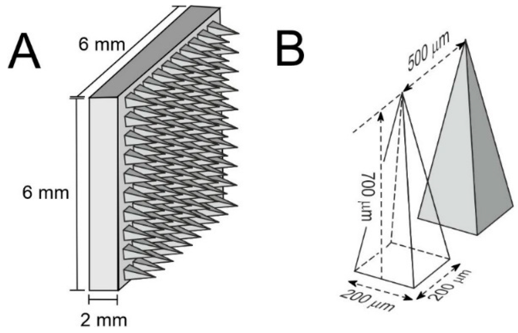
Carbon nanoparticles were combined with psyllium powder in a ratio of 1:1 by weight, dissolved in water as described in Section 2.1. Once a homogenous solution had been obtained, the mixture was cast into microneedle templates obtained from Technologies Pte Ltd. (Singapore), which were pyramidal in format with 200 µm (base) × 500 µm (pitch) × 700 µm (height) dimensions covering a 10 × 10 needle array. A summary of the dimensions and microneedle plate format is illustrated in Figure 2. The water was allowed to evaporate (typically 24 h) whereafter the mould was topped up with another aliquot of the PsH–C mixtures. Finally, a carbon fibre stub was placed into the base plate section to facilitate electrical connection. The process is similar to that reported previously for polystyrene-based microneedles and utilised the same templates [6]. Once dry, the baseplate and non-needle surfaces were coated with enamel (6 h drying period) to serve as a dielectric and thereby finalise the geometric electrode area. The total area of the exposed microneedle face was 0.603 cm2.

3. Results

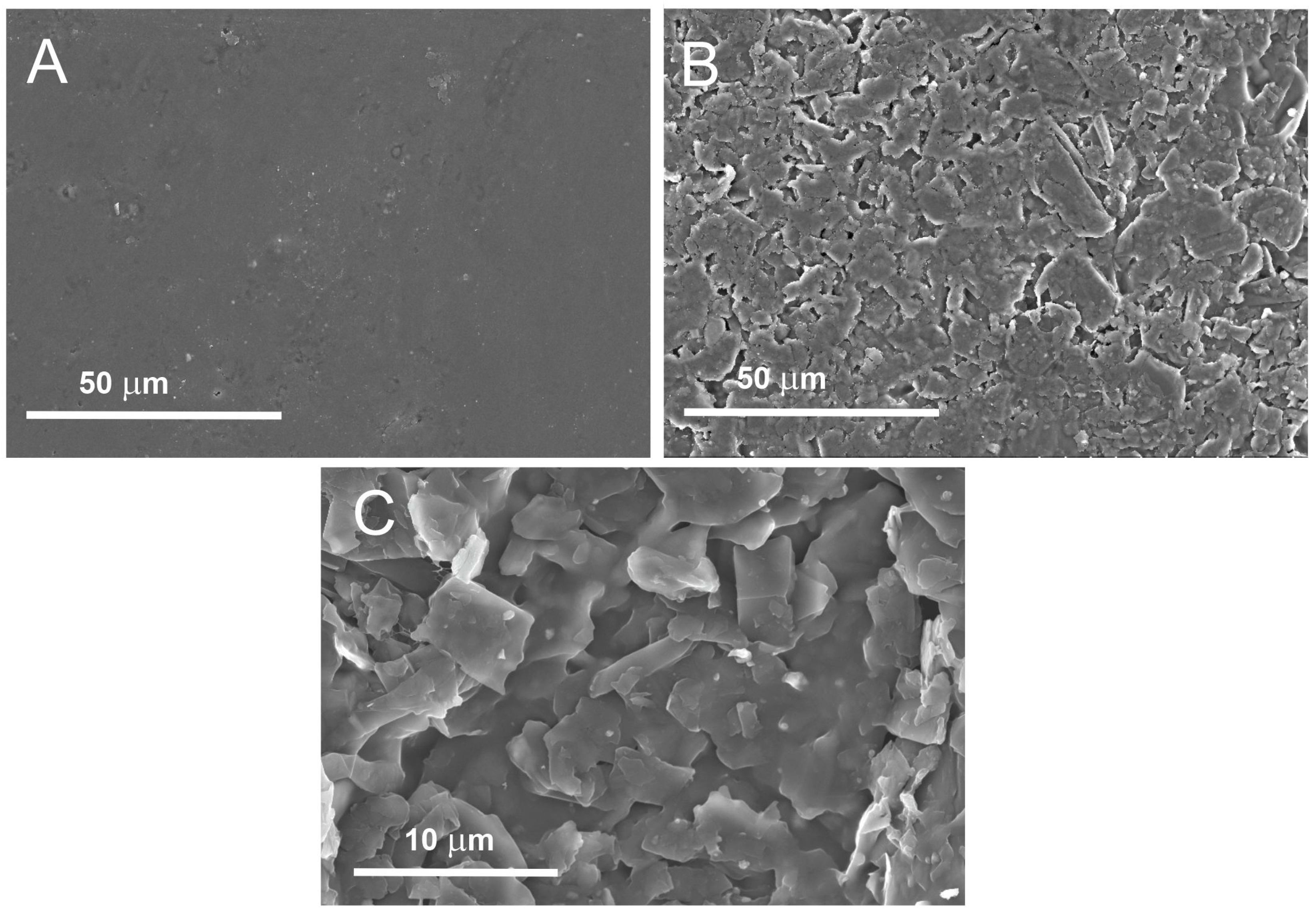
Scanning electron micrographs comparing the surface morphology of films composed of psyllium husk (PsH) and psyllium husk/carbon nanoparticle (PsH–C) composites are compared in Figure 3A and Figure 3B, respectively. While the PsH film is relatively featureless, the PsH–C film exhibits a platelet-like morphology which could be attributed to the aggregation of carbon within the film. This aspect is illustrated better in Figure 3C where increased magnification highlights the granular nature of the PsH–C composite A ratio of 50:50 PsH and carbon nanopowder was chosen as a compromise between conductivity and structural integrity. At 50:50, the gelling nature of the PsH is sufficient to bind the carbon, as evidenced by the platelet structure in Figure 3C. It has been found previously with carbon–polystyrene microneedles that increasing the ratio of carbon beyond this level exhausts the binding capacity of the polymer, increases the granularity and compromises the mechanical integrity of the electrode structure [6].
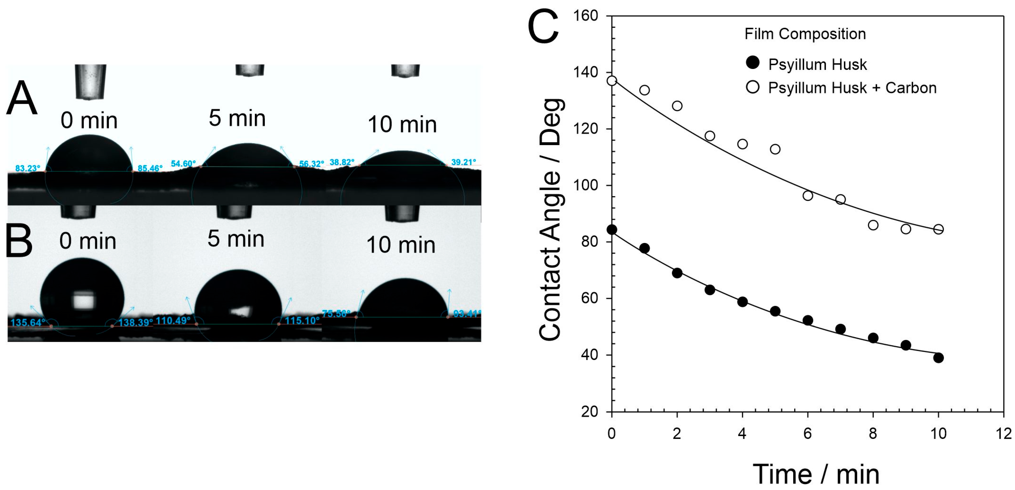
Both the PsH and PsH–C films were found to absorb water, and contact angle data comparing the respective interactions are detailed in Figure 4A (PsH) and Figure 4B (PsH–C). A more quantitative comparison over 10 min is detailed in Figure 4C. Both films readily absorb water at a similar rate, but the presence of the carbon nanoparticles clearly elicits a greater degree of hydrophobicity. The hydrophobicity could be useful from both electroanalytical and electrocatalytic (tri-phasic) perspectives where adsorption of analytes to the carbon surface can be critical [31,32,33].
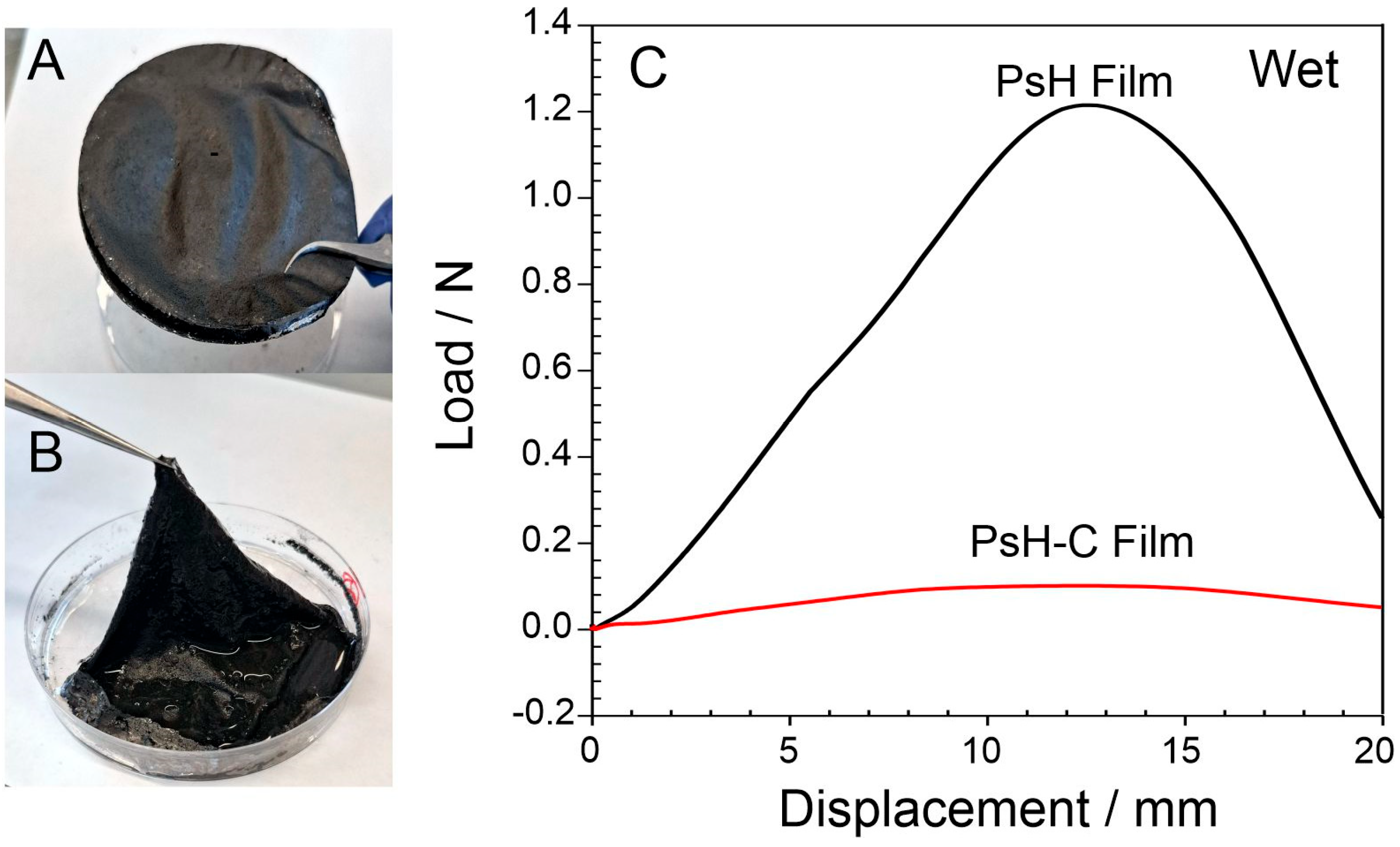
Psyllium husk, in powder form, has long been recognized as a gelling agent [21,22] but, when in a film format, its interaction with water, as indicated in Figure 4, is much slower. While it absorbs water, it does not readily lose its integrity but rather becomes more like a structured hydrogel. This feature is highlighted in Figure 5 where the initial rigid PsH–C film (A), once saturated with water, can be transformed into a mouldable cloth-like form (B). The ability of psyllium husk to sequester water, both in its pure form and as a carbon composite, was examined by gravimetric analysis by exposing the material to an excess of water for 30 min. The saturated material was then removed and gently pressed on an absorbent towel to remove surface water with the weight recorded before and after, allowing the swelling capacity (g of water per g of film) to be estimated. The swelling capacity for the PsH film alone was found to be 11.58 ± 1.15 g g−1, whereas that of the PsH–C was lower at 7.83 ± 1.04 g g−1 (with the measurements each based on five replicate samples). Given that the carbon nanoparticles have no intrinsic swelling characteristics, it could be expected that substitution of the PsH for the carbon would likewise decrease the swelling capacity.
A preliminary examination of the tensile properties of the PsH–C material was also considered. Average film thicknesses were measured using a Dektak profiler, varying between 260–320 mm across batches. The latter were subsequently used to estimate the Young’s modulus of both the PsH and PsH–C in the dry state. Both were found to be brittle, with a Young’s modulus of 1.17 ± 0.35 GPa and 0.47 ± 0.08 GPa, respectively. When wet, the materials exhibit a more elastic response (Figure 5C) and this could be expected given the transformation from brittle to a more gel-like consistency. In this case, the presence of the carbon at 50:50 wt% clearly compromises the gelling capacity of the PsH–C material.
Raman spectroscopy of the PsH–C film and carbon nanoparticles was conducted and typical spectra are presented in Figure 6. The PsH–C film (Figure 5) primarily shows the intense Raman active D (1349 cm−1) and G (1578 cm−1) bands, as well as the 2D (2689 cm−1) band arising from the carbon nanoparticles [34,35,36]. The D band is known to arise from the stretching vibration of sp3 hybridized carbons typically at the edge of graphene sheets and is an indicator of disorder in graphitic structures [35]. The ratio of the intensities of the D and G peak (ID/IG) of PsH–C film is substantially reduced (0.4) compared to that of carbon nanoparticles (1.33). Moreover, the 2D peak of PsH–C film, resulting from two-phonon, second-order intervalley scatter, is shifted to higher wavenumbers (~2715 cm−1) compared to that of carbon nanoparticles (2687 cm−1). The reduction in ID/IG ratio and blue shift of 2D band observed in PsH–C film reveal that the carbon nanoparticles are under strain when introduced in the PsH matrix [36].
The PsH–C film exhibits considerable conductivity in the dry state with a sheet resistance of 213 ± 26 W/square (N = 3). This is however still significantly higher than that observed with other carbon substrates, such as laser-induced graphene where <35 W/square is typical [37], and can be attributed to percolation transfer from particle to particle within the composite film. It could have been anticipated that swelling of the psyllium binder could be problematic in terms of providing a reliable voltammetric signature for sensing purposes where physical displacement of the carbon could occur and thereby reduce electron percolation through the network. Similar issues have been observed with composite systems based on carbohydrate chains such as cellulose acetate phthalate (CAP) where the polymer is found to be stable in acid but becomes soluble in neutral or alkaline conditions. The loss of the CAP binder in such neutral solutions also results in a loss of carbon particles, thereby reducing the electrode area and current magnitude [6]. Cyclic voltammograms detailing the response of the psyllium husk–carbon electrode to ferrocyanide are shown in Figure 7A. The PsH–C film (Figure 7A) was found to be relatively stable in the pH 7 buffer with little overall change in the voltammetric profile upon repetitive scanning. Both oxidation and reduction peaks are clearly observed (ΔEp = 204 mV) but the response is clearly more capacitive and can be attributed to the swelling of the psyllium gel binder. Notably, there is little change in the profile with increasing scan number. Alkaline conditions are routinely used in the processing and extraction of psyllium husk and therefore, it could be expected that were the PsH–C electrodes subjected to such conditions, the electrode response would be affected. Cyclic voltammograms detailing the response in 0.1 M NaOH (pH 13.6) are shown in Figure 7B. There is an increase in the capacitive background but ferrocyanide peaks are clearly visible and the profile is akin to that observed in pH 7 buffer. While it is inevitable that the strong alkaline conditions would eventually degrade the structure of the film, it is notable that the electrode is viable and that it is unlikely that such conditions would be encountered in a physiological sample. The electrode response to ferrocyanide under less aggressive conditions (pH 3–9) exhibited similar profiles to those observed in Figure 7A, and these are included in the supplementary data (Figure S1).
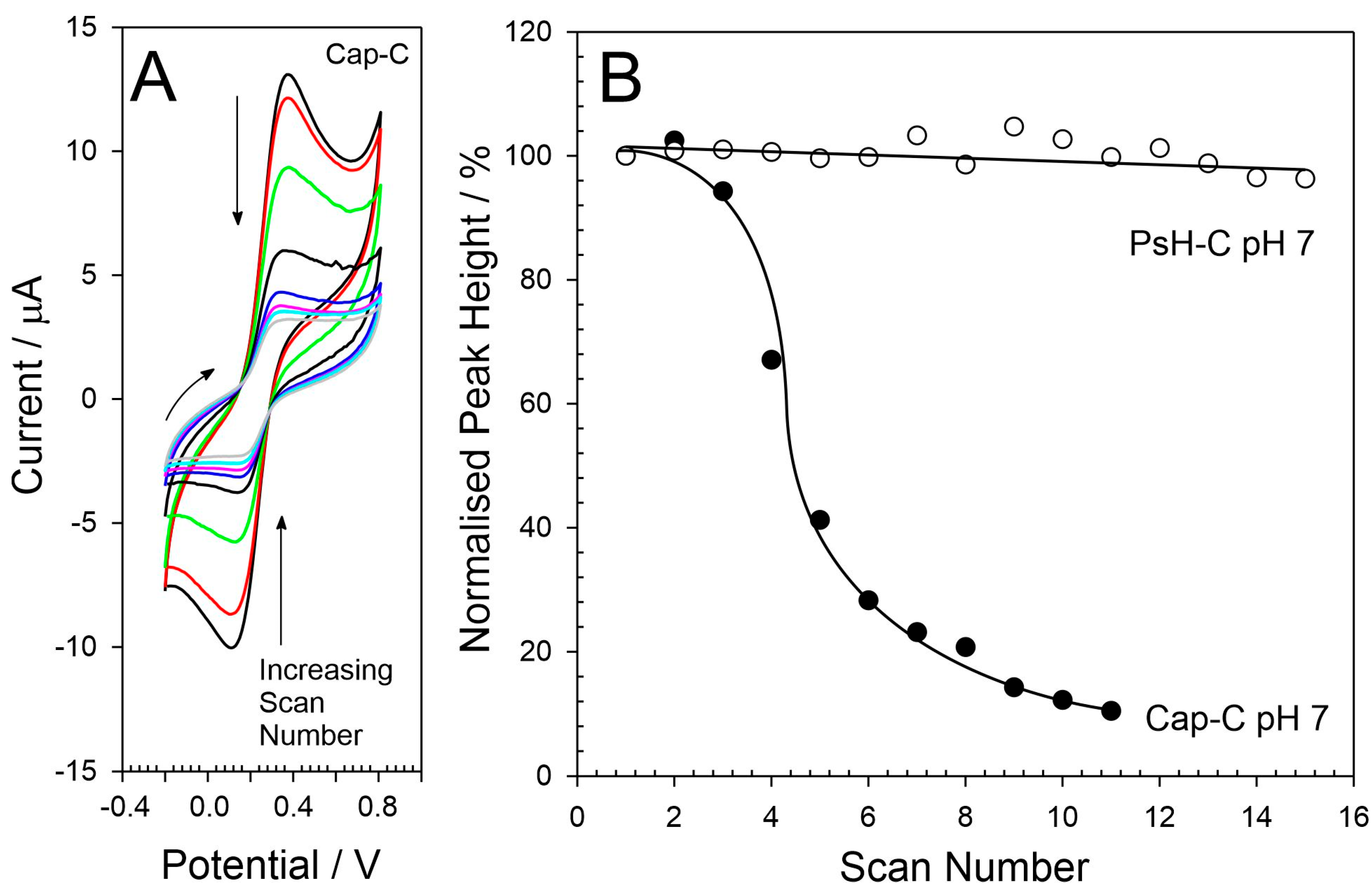
In contrast, the CAP-C electrode is much less stable in even moderate (pH 7) conditions. It was found to provide well-defined peak profiles on the first few scans (Figure 8A), but rather than simply swelling (as observed with the PsH–C in 0.1 M NaOH), dissolution of the electrode occurs, resulting in a sustained loss of active electrode area and a diminishing peak magnitude. The dissolution process slows after a few scans, but this arises as an artefact of the thin film electrode design. While the loss of the electrode material within the exposed window occurs relatively quickly, there will still be CAP–C composite physically entrapped under the lamination sheet which retains some electrochemical activity (the majority of the electrode area having fallen away). As such, the response does not fall to zero but rather slowly dissipates, and this is indicated in Figure 8B where a quantitative comparison of the ferrocyanide oxidation peak magnitude with scan number is detailed. As previously reported, the CAP–C electrode is stable in acid solution [6]. The stability in pH 3 solution is consistent with its use as an enteric coating in pharmaceutical preparations where it serves to protect the active pharmaceutical ingredients from the acid environment of the stomach before dissolving in the neutral/alkaline solution of the small intestine [38,39].
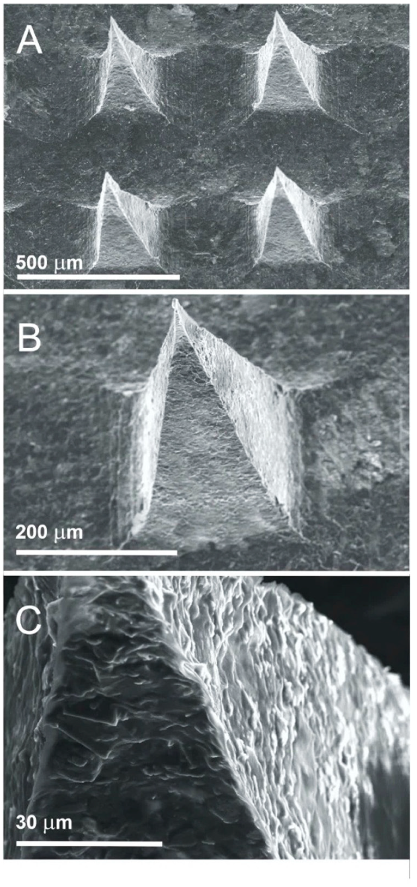
The use of psyllium husk as a binder within electrode substrates is not restricted to planar formats but can be moulded. The flexible nature of the hydrogel evidenced in Figure 5 clearly allows mechanical flexibility, but it can also be directly cast into specific geometric configurations. This is best exemplified using a PsH–C composite mixture to fabricate conductive microneedles. Rather than casting as a thin film (Figure 5A), it was decanted into silicone moulds (Figure 2). Once the water had evaporated to dryness, the microneedles could be released from the mould and revealed sharp needles of similar form to those produced using polycarbonate or polystyrene nanoparticle composites fabricated from non-aqueous solvents (i.e., cyclohexanone) [6,40,41]. Scanning electron micrographs of the resulting microneedles are shown in Figure 9A,B. The needles are well formed, though closer examination reveals a slight ripple texture on the side of the pyramidal structures (Figure 9C) which seems to be characteristic of the PsH component and may be attributed to the presence of insoluble fractions within the polymer mixture preventing the formation of a smooth interface. There are extensive reports in the literature on the use of carbohydrate-based MN systems for drug delivery [42,43,44], but this investigation is the first to report electrically conductive systems. While the authors have previously reported the fabrication of cellulose acetate phthalate- [6,41] and polystyrene [42]-based systems, both are dependent on the use of non-aqueous solvents for their processing. This is in stark contrast to the water-based system employed here, where the proposed system clearly offers a more environmentally sustainable route.
The electrochemical performance of the PsH–C MN was investigated by examining the response to 2 mM ferrocyanide in pH 7 Britton–Robinson buffer (cf. Figure 7A). Cyclic voltammograms detailing the response to the redox probe are shown in Figure 10A. The fresh MN electrode, while electrochemically active, shows little response to ferrocyanide with the oxidation and reduction processes being difficult to discern from the background current. When the electrode is rinsed, left to dry for 24 h and then retested, the response is substantially different with a dramatic improvement in the response profile (Figure 10A). The subsequent re-testing of the electrode reveals well defined peaks (ΔEp = 320 mV), albeit with the characteristic capacitive background current—also observed in the film electrodes (Figure 7A). The increased peak separation (compared to the 204 mV observed with the film electrodes) could be attributed to greater resistance caused by the increased thickness of the MN patch between the solution interface and the conductive carbon fibre stub. It is noteworthy that there was little indication of the incorporation of the ferrocyanide into the PsH component of the needle film itself (evidenced by a featureless response in fresh pH 7 buffer with no ferrocyanide present). Moreover, the relationship between peak current and the square root of scan rate was found to be linear (Ip [/μA] = 727.36 √scan rate [V/s] + 4.25; R2 = 0.996) which is consistent with a freely diffusing mediator rather than a surface-entrapped species.
The unresponsive nature of the first run was observed with other PsH–C MN batches, and it is only after they have been dried that the enhanced response, detailed in Figure 10A, is observed. This may be an artefact of the manufacturing process and it is possible to speculate that the initial poor response may be due to more soluble components of the PsH–C mixture obscuring the carbon interface from any substantive interaction with the ferrocyanide. While it must be acknowledged that the psyllium husk material is a natural product and will inevitably contain a variety of molecular species—some of which may be electroactive (i.e., polyphenolics) [45], the concentration is liable to be very low. No extraneous redox peaks were observed when investigating the blank scan responses in pH 7 buffer. However, given the trace level of the intrinsic antioxidants that may be present and the large capacitive background evident in Figure 7 and Figure 10 for the PsH–C electrode, resolution of these processes is compromised. It is possible that the initial swelling and subsequent drying may result in a partial reorganization of the interface with increased exposure of the carbon surface. From the perspective of sensor development, it could be expected that a presoak step could be introduced upon release from the MN mould followed by a drying step. At this point the sensors have been activated and ready for subsequent surface modification. The MN are not dependent on having to be presoaked and dried immediately before use at the POC.
It must also be noted that, as with the PsH–C film substrates, the MN electrodes were stable to repeated cycling and the responses were essentially the same as those observed with the film. This could be expected given that both are composed of the same material, but it provides a route through which prototyping could be done at the film stage (given the ease with which such electrodes can be made) before transferring the system to the MN format.

4. Discussion

The composite formulation involved the use of carbon nanopowder and was originally selected on the basis that it would provide a large surface area which could serve as a foundation for subsequent investigations should the psyllium husk prove to be a robust binder. The results obtained lend support to the latter assumption, and it could be envisaged that the use of psyllium husk could be expanded to other carbon nanomaterials. While the graphitic nature of the nanopowder can offer considerable chemical flexibility for the modification of the electrode interface, it does present some issues. Increasing the 50% wt/wt ratio of carbon to improve the conductivity is problematic in that the introduction of too much carbon compromises the structural integrity of the composite [6]. This is particularly important when considering the construction of microneedles where sufficient psyllium is required to enable preservation of the needle tips (Figure 9B).
Substitution of graphitic powder for graphene platelets could further enhance the voltammetric capabilities and potential peak resolution through the provision of greater edge planes. Bleiga and coworkers (2022) found that the conductivity within carbon–polymer composites can be significantly influenced by the aspect ratio and the 3D structure of the nanoparticles employed [46]. They found that the low packing factor characteristic of carbon black nanoparticles did little to aid the formation of a percolation network. In contrast, the use of carbon nanotubes and their inherent propensity for entanglements was found to be superior. It is possible that the use of CNTs here could similarly enhance the conductivity whilst also allowing the possibility of reducing the loading and could, as previously noted, improve the production process for MN manufacture.
Films of psyllium husk left to stand for three months were found to elicit responses similar to those observed shortly after the initial production. The voltammograms detailed in Figure 7A were recorded immediately after the films were cast while those detailed in Figure 7B were from the same material but recorded three months later. Electron microscopy and Raman spectroscopy, detailed in Figure S2 and Figure S3, respectively, revealed no significant changes to the film structure indicating that the core material has an appreciable shelf life. Moreover, no changes were observed in the electrode structure after they had been used. While the imposition of large anodic potentials can induce exfoliation of carbon surfaces [30], it could be expected that the potential range applied here (−0.2 V to +0.8 V vs. 3 M Ag|AgCl) would not induce any significant changes in the carbon material itself.
It is important to note that, in this case, the electrodes were in contact for a relatively short period of time (typically less than 1 h). It is conceivable that changes to the binder could occur were the electrode employed for longer periods (i.e., continuous monitoring). This will also depend on the nature of the solution. The original aim, however, was to consider the psyllium husk as a binder for point-of-care sensing—potentially as a replacement for conventional single-use screen-printed strips or as single-shot transdermal needles. In such cases, contact with the solution is likely to be relatively short.
The production of microneedles (as prepared here) is very much an artisanal approach and there will inevitably be substantial variation between electrodes and batches. This is due to both variations in the material composition (and carbon aggregation) and the manual nature of defining the working surface through enamelling the backplate and side of the MN patch. As such, the results considered here do not attempt to provide a rigorous analytical evaluation but rather seek to assess the mechanical properties of the psyllium as a binder. What is clear from the results obtained is that the PsH–C can indeed serve as an electrode material. It could be expected that greater automation of the film or needle production process would yield electrodes with appropriate batch reproducibility. As psyllium husk is a natural product it is also important to recognize that there will be variations in composition between batches.
The biodegradability of psyllium husk has long been recognized [28], but the degradation of the carbon nanoparticles can be more problematic. Carbon nanomaterials will degrade through a variety of processes (chemical, photochemical and biological) but the mechanisms are complex and will depend on the material itself and environment within which the spent PsH–C electrodes are disposed [47,48,49,50,51]. While the psyllium husk is degraded slowly by a range of microbial species (typically through the use of xylanoses), these same species can be inhibited by the carbon nanomaterials themselves (or the byproducts thereof) [48,51,52]. Nevertheless, the authors have demonstrated the biodegradability of CAP–C microneedles within soil [6] and it is evident that the polysaccharide-based binder remains a better alternative to the polystyrene or polycarbonate used previously.

5. Conclusions

The design of biodegradable electrode substrates has gathered pace as concerns over disposability mount and, while paper-based systems are often proffered as an alternative to conventional non-degradable substrates, they are invariably planar. The use of psyllium husk as the binding component addresses this issue through being mouldable, as evidenced by its use in the formation of microneedle arrays. The response to ferrocyanide, the standard redox probe, provided clear resolution of both oxidation and reduction process but the capacitive background could be an issue in electroanalytical applications. The ease with which the electrodes can be fabricated, the sustainability of the source material and the greater environmental acceptability of the aqueous processing method are major benefits. The latter provides a critical step forward when considering new technologies to address the burgeoning waste generated by disposable diagnostics.

Supplementary Materials

The following supporting information can be downloaded at: https://www.mdpi.com/article/10.3390/c11030064/s1, Figure S1: Repetitive cyclic voltammograms detailing the response of psyllium husk-carbon towards ferrocyanide (2 mM) in (A) pH 3, pH 5 and pH 9 Britton-Robinson buffer. Scan rate in all cases: 50 mV/s. Figure S2: Scanning electron micrograph of the psyllium husk – carbon nanoparticle electrode substrate after storage for 3 months and having being used to study the electrochemical response to ferrocyanide. Figure S3: Raman spectrum of the psyllium husk – carbon nanoparticle electrode substrate after storage for 3 months and having being used to study the electrochemical response to ferrocyanide.

Author Contributions

Conceptualization, J.D. and V.G.; methodology, J.D., C.M. and V.G.; validation, C.M., V.G. and R.M.; formal analysis, C.M., V.G. and R.M.; resources, J.D. and P.P.; data curation, C.M., J.D. and P.P.; writing—original draft preparation, J.D. and C.M.; writing—review and editing, J.D., C.I.R.G., K.M., P.P. and J.U.; supervision, J.D., P.P., C.I.R.G. and K.M.; project administration, J.D.; funding acquisition, J.D., C.I.R.G. and K.M. All authors have read and agreed to the published version of the manuscript.

Funding

This research was funded by the UK Medical Research Council (MR/W029561/1) and the Department for the Economy Northern Ireland.

Data Availability Statement

Raw data are available on request.

Acknowledgments

No GenAI has been used in the preparation of the manuscript or any of the data included therein.

Conflicts of Interest

The authors declare no conflicts of interest. The funders had no role in the design of the study; in the collection, analyses, or interpretation of data; in the writing of the manuscript; or in the decision to publish the results.

References

  1. Singh, P.; Singh, A.; Saini, R.; Kulriya, P.; Kumar, R. Advancements in graphene-based nanostructured conducting polymer hybrid composite electrodes for high-performance supercapacitors. J. Power Sources 2025, 630, 236176. [Google Scholar] [CrossRef]
  2. Islam, M.; Hossain, M.S.; Adak, B.; Rahman, M.M.; Moni, K.K.; Nur, A.S.M.; Hong, H.P.; Younes, H.; Mukhopadhyay, S. Recent advancements in carbon-based composite materials as electrodes for high-performance supercapacitors. J. Energy Sources 2025, 107, 114838. [Google Scholar] [CrossRef]
  3. Imran, M.; Afzal, A.M.; Iqbal, M.W.; Fouda, A.M.; Hegazy, H.H.; Mumtaz, S. Unlocking the potential of biodegradable and environment-friendly electrode materials for applications in energy storage devices. Ceram. Int. 2024, 50, 47529–47548. [Google Scholar] [CrossRef]
  4. Suganthi, S.; Ahmad, K.; Oh, T.H. Progress in Graphitic Carbon Nitride-Based Composite Materials for Electrochemical Sensing and Energy Storage Applications. ChemistrySelect 2025, 10, e202404670. [Google Scholar] [CrossRef]
  5. Lin, Y.Y.; Wu, A.B.; Zhang, Y.T.; Duan, H.Y.; Zhu, P.C.; Mao, Y.C. Recent progress of nanomaterials-based composite hydrogel sensors for human-machine interactions. Discov. Nano. 2025, 20, 60. [Google Scholar] [CrossRef]
  6. Devine, A.; Hegarty, E.; Hegarty, C.; Davis, J. Conductive Composite Microneedle Sensors Based on Cellulose Acetate Phthalate: Investigating Performance and Biodegradability. IEEE Sens. Lett. 2023, 7, 1–4. [Google Scholar] [CrossRef]
  7. Karami-Mosammam, M.; Danninger, D.; Schiller, D.; Kaltenbrunner, M. Stretchable and Biodegradable Batteries with High Energy and Power Density. Adv. Mater. 2022, 34, 2204457. [Google Scholar] [CrossRef] [PubMed]
  8. Nowak, A.P.; Trzcinski, K.; Zarach, Z.; Li, J.; Roda, D.; Szkoda, M. Poly(hydroxybutyrate-co-hydroxyvalerate) as a biodegradable binder in a negative electrode material for lithium-ion batteries. Appl. Surf. Sci. 2022, 606, 154933. [Google Scholar] [CrossRef]
  9. Chen, T.; Lau, K.; Singh, A.; Zhang, Y.X.; Salari, M.; Naguib, H.E.; Morshead, C.M. Biodegradable stimulating electrodes for resident neural stem cell activation in vivo. Biomaterials 2025, 315, 122957. [Google Scholar] [CrossRef] [PubMed]
  10. Fryn, P.; Jewłoszewicz, B.; Bogdanowicz, K.A.; Przybył, W.; Gonciarz, A.; Pich, R.; Marzec, M.; Iwan, A. Research of Binary and Ternary Composites Based on Selected Aliphatic or Aliphatic–Aromatic Polymers, 5CB or SWCN toward Biodegradable Electrodes. Materials 2020, 13, 2480. [Google Scholar] [CrossRef]
  11. Martin, C.; Dejardin, T.; Hart, A.; Riehle, M.O.; Cumming, D.R.S. Directed Nerve Regeneration Enabled by Wirelessly Powered Electrodes Printed on a Biodegradable Polymer. Adv. Healthc. Mater. 2014, 3, 1001–1006. [Google Scholar] [CrossRef]
  12. Meng, L.; Fu, Q.; Hao, S.; Xu, F.; Yang, J. Self-adhesive, biodegradable silk-based dry electrodes for epidermal electrophysiological monitoring. Chem. Eng. J. 2022, 427, 131999. [Google Scholar] [CrossRef]
  13. Molinnus, D.; Janus, K.A.; Fang, A.C.; Drinic, A.; Achtsnicht, S.; Köpf, M.; Keusgen, M.; Schöning, M.J. Thick-Film Carbon Electrode Deposited onto a Biodegradable Fibroin Substrate for Biosensing. Phys. Status Solidi A 2022, 219, 2200100. [Google Scholar] [CrossRef]
  14. Cui, S.W. Polysaccharide Gums from Agricultural Products; Taylor and Francis: Abingdon, UK, 2019; ISBN 9780367397999. [Google Scholar]
  15. Hussain, M.A.; Muhammad, G.; Jantan, I.; Bukhari, S.N.A. Psyllium Arabinoxylan: A Versatile Biomaterial for Potential Medicinal and Pharmaceutical Applications. Polym. Rev. 2016, 56, 1–30. [Google Scholar] [CrossRef]
  16. Akbar, S. Handbook of 200 Medicinal Plants: A Comprehensive Review of Their Traditional Medical Uses and Scientific Justifications; Springer International Publishing: Berlin/Heidelberg, Germany, 2020; ISBN 978-3-030-16806-3. [Google Scholar]
  17. Zhang, X.; Zhao, Y.; Li, Y.; Zhu, L.; Fang, Z.; Zhi, Q. Physicochemical, mechanical and structural properties of composite edible films based on whey protein isolate/psyllium seed gum. Int. J. Biol. Macromol. 2020, 153, 892–901. [Google Scholar] [CrossRef]
  18. Suresh, H.; Mikhael, M.; Ho, V.; Zhou, J. A HPLCESI-Q-ToF-MS Method for the Analysis of Monomer Constituents in PHGG, Gum Arabic And Psyllium Husk Prebiotic Dietary Fibre Supplements. Int. J. Food Prop. 2022, 25, 1650–1667. [Google Scholar] [CrossRef]
  19. Poddar, S.; Agarwal, P.S.; Sahi, A.K.; Varshney, N.; Vajanthri, K.Y.; Mahto, S.K. Fabrication and characterization of electrospun psyllium husk-based nanofibers for tissue regeneration. J. Appl. Polym. Sci. 2021, 138, e50569. [Google Scholar] [CrossRef]
  20. Ganguly, S.; Mondal, S.; Das, P.; Bhawal, P.; Maity, P.P.; Ghosh, S.; Dhara, S.; Das, N. Design of psyllium-g-poly(acrylic acid-co-sodium acrylate)/cloisite 10A semi-IPN nanocomposite hydrogel and its mechanical, rheological and controlled drug release behaviour. Int. J. Biol. Macromol. 2018, 111, 983–998. [Google Scholar] [CrossRef]
  21. Guo, Q.; Cui, S.W.; Wang, Q.; Goff, H.D.; Smith, A. Microstructure and rheological properties of psyllium polysaccharide gel. Food Hydrocoll. 2009, 23, 1542–1547. [Google Scholar] [CrossRef]
  22. Belorio, M.; Gomez, M.J. Psyllium: A useful functional ingredient in food systems. Crit. Rev. Food Nutr. 2022, 62, 527–538. [Google Scholar] [CrossRef]
  23. Fernandes, C.; Acharya, P.C.; Bhatt, S. Preparation of Lauroyl Grafted Alginate-Psyllium Husk Gel Composite Film with Enhanced Physicochemical, Mechanical and Antimicrobial Properties. Sci. Rep. 2018, 8, 17213. [Google Scholar] [CrossRef]
  24. Ahmad, N.; Tayyeb, D.; Ali, I.; Alruwaili, N.K.; Ahmad, W.; Rehman, A.; Khan, A.H.; Iqbal, M.S. Development and Characterization of Hemicellulose-Based Films for Antibacterial Wound-Dressing Application. Polymers 2020, 12, 548. [Google Scholar] [CrossRef] [PubMed]
  25. Bajpai, S.K.; Chand, N.; Agrawal, A. Carboxymethylpsyllium (CMPsy)/poly(acrylamide)(poly(AAm)) hydrogels for oral delivery of anti-diabetic drug Gliclazide. J. Macromol. Sci. Part A 2017, 54, 221–227. [Google Scholar] [CrossRef]
  26. Singh, B.; Kanwar, J.S.; Kumari, P. Modification of Dietary Fiber Psyllium with Poly(vinyl pyrrolidone) through Network Formation for Use in Slow Drug Delivery Application. Polym. Sci. Ser. B 2018, 60, 331–348. [Google Scholar] [CrossRef]
  27. Sharma, V.K.; Sharma, P.P.; Mazumder, B.; Bhatnagar, A.; Subramaniyan, V.; Fuloria, S.; Kumar Fuloria, N. Mucoadhesive microspheres of glutaraldehyde crosslinked mucilage of Isabgol husk for sustained release of gliclazide. J. Biomater. Sci. Polym. Ed. 2021, 32, 1420–1449. [Google Scholar] [CrossRef]
  28. Van Craeyveld, V.; Delcour, J.A.; Courtin, C.M. Extractability and chemical and enzymic degradation of psyllium (Plantago ovata Forsk) seed husk arabinoxylans. Food Chem. 2009, 112, 812–819. [Google Scholar] [CrossRef]
  29. Beer-Lech, K.; Skic, A.; Skic, K.; Stropek, Z.; Arczewska, M. Effect of Psyllium Husk Addition on the Structural and Physical Properties of Biodegradable Thermoplastic Starch Film. Materials 2022, 15, 4459. [Google Scholar] [CrossRef]
  30. Casimero, C.; Hegarty, C.; McGlynn, R.J.; Davis, J. Ultrasonic exfoliation of carbon fiber: Electroanalytical perspectives. J. Appl. Electrochem. 2020, 50, 383–394. [Google Scholar] [CrossRef]
  31. Lindner, E.; Guzinski, M.; Pendley, B.; Chaum, E. Plasticized PVC Membrane Modified Electrodes: Voltammetry of Highly Hydrophobic Compounds. Membranes 2020, 10, 202. [Google Scholar] [CrossRef] [PubMed]
  32. Chen, Y.; Su, D.; Chen, X.; Zhu, Z.; Li, W. Three-phase interface-assisted advanced electrochemistry-related applications. Cell Rep. Phys. Sci. 2021, 2, 100602. [Google Scholar] [CrossRef]
  33. Yang, Y.; Li, N.; Wang, J.; Zhao, W.; Wu, H.B. Antiflooding Gas Diffusion Electrodes Enabled by Liquid–Solid–Liquid Interfaces for Durable CO2 Electrolysis. ACS Appl. Energy Mater. 2024, 7, 9394–9401. [Google Scholar] [CrossRef]
  34. Ferrari, A.C.; Meyer, J.C.; Scardaci, V.; Casiraghi, C.; Lazzeri, M.; Mauri, F.; Piscanec, S.; Jiang, D.; Novoselov, K.S.; Roth, S.; et al. Raman Spectrum of Graphene and Graphene Layers. Phys. Rev. Lett. 2006, 97, 187401. [Google Scholar] [CrossRef]
  35. Gao, Z.; Zhu, J.; Rajabpour, S.; Joshi, K.; Kowalik, M.; Croom, B.; Schwab, Y.; Zhang, L.; Bumgardner, C.; Brown, K.R.; et al. Graphene Reinforced Carbon Fibers. Sci. Adv. 2020, 6, eaaz4191. [Google Scholar] [CrossRef]
  36. Li, Z.; Deng, L.; Kinloch, I.A.; Young, R.J. Raman spectroscopy of carbon materials and their composites: Graphene, nanotubes and fibres. Prog. Mater. Sci. 2023, 135, 101089. [Google Scholar] [CrossRef]
  37. Lin, J.; Peng, Z.; Liu, Y.; Ruiz-Zepeda, F.; Ye, R.; Samuel, E.L.G.; Yacaman, M.J.; Yakobson, B.I.; Tour, J.M. Laser-induced porous graphene films from commercial polymers. Nat. Commun. 2014, 5, 5714. [Google Scholar] [CrossRef] [PubMed]
  38. Wong, C.Y.; Martinez, J.; Carnagarin, R.; Dass, C.R. In-Vitro Evaluation of Enteric Coated Insulin Tablets Containing Absorption Enhancer and Enzyme Inhibitor. J. Pharm. Pharmacol. 2017, 69, 285–294. [Google Scholar] [CrossRef]
  39. Hanafi, A.; Nograles, N.; Abdullah, S.; Shamsudin, M.N.; Rosli, R. Cellulose Acetate Phthalate Microencapsulation and Delivery of Plasmid DNA to the Intestines. J. Pharm. Sci. 2013, 102, 617–626. [Google Scholar] [CrossRef]
  40. Anderson, A.; Hegarty, C.; Casimero, C.; Davis, J. Electrochemically Controlled Dissolution of Nanocarbon–Cellulose Acetate Phthalate Microneedle Arrays. ACS Appl. Mater. Interfaces 2019, 11, 35540–35547. [Google Scholar] [CrossRef] [PubMed]
  41. Hegarty, C.; McConville, A.; McGlynn, R.J.; Mariotti, D.; Davis, J. Design of composite microneedle sensor systems for the measurement of transdermal pH. Mater. Chem. Phys. 2019, 227, 340–346. [Google Scholar] [CrossRef]
  42. Abbas, M.F.; Karim, D.K.; Kareem, H.R.; Kamil, M.M.; Al-Musawi, M.H.; Asker, M.H.; Ghanamid, M.; Shahriari-Khalajid, M.; Sattare, M.; Mirhajf, M.; et al. Fucoidan and its derivatives: From extraction to cutting-edge biomedical applications. Carbohydr. Polym. 2025, 357, 123468. [Google Scholar] [CrossRef] [PubMed]
  43. Wang, X.; Chen, Y.; Yue, J. Dissolving microneedles: A transdermal drug delivery system for the treatment of rheumatoid arthritis. Int. J. Pharm. 2025, 671, 125206. [Google Scholar] [CrossRef]
  44. Senobari, F.; Abolmaali, S.S.; Farahavr, G.; Tamaddon, A.M. Targeting inflammation with hyaluronic acid-based micro- and nanotechnology: A disease-oriented review. Int. J. Biol. Macromol. 2024, 280, 135923. [Google Scholar] [CrossRef] [PubMed]
  45. Waleed, M.; Saeed, F.; Afzaal, M.; Niaz, B.; Raza, M.A.; Hussain, M.; Tufail, T.; Rasheed, A.; Ateeq, H.; Al Jbawi, E. Structural and nutritional properties of psyllium husk arabinoxylans with special reference to their antioxidant potential. Int. J. Food Prop. 2022, 25, 2505–2513. [Google Scholar] [CrossRef]
  46. Bleija, M.; Platnieks, O.; Macutkevic, J.; Starkova, O.; Gaidukovs, S. Comparison of Carbon-Nanoparticle-Filled Poly(Butylene Succinate-co-Adipate) Nanocomposites for Electromagnetic Applications. Nanomaterials 2022, 12, 3671. [Google Scholar] [CrossRef]
  47. Pikula, K.; Johari, S.A.; Golokhvast, K. Colloidal Behavior and Biodegradation of Engineered Carbon-Based Nanomaterials in Aquatic Environment. Nanomaterials 2022, 12, 4149. [Google Scholar] [CrossRef] [PubMed]
  48. Zhang, W.; Zeng, Z.; Liu, Z.; Huang, J.; Xiao, R.; Shao, B.; Liu, Y.; Liu, Y.; Tang, W.; Zeng, G.; et al. Effects of carbon nanotubes on biodegradation of pollutants: Positive or negative. Ecotoxicol. Environ. Saf. 2020, 189, 109914. [Google Scholar] [CrossRef] [PubMed]
  49. Takahashi, S.; Taguchi, F.; Hori, K. Contribution of the Fenton reaction to the degradation of carbon nanotubes by enzymes. Front. Environ. Sci. 2023, 11, 1184257. [Google Scholar] [CrossRef]
  50. Takahashi, S.; Hori, K. Long-term continuous degradation of carbon nanotubes by a bacteria-driven Fenton reaction. Front. Microbiol. 2023, 14, 1298323. [Google Scholar] [CrossRef]
  51. Mokhtari-Farsani, A.; Hasany, M.; Lynch, I.; Mehrali, M. Biodegradation of Carbon-Based Nanomaterials: The Importance of “Biomolecular Corona” Consideration. Adv. Funct. Mater. 2022, 32, 2105649. [Google Scholar] [CrossRef]
  52. Al-Jumaili, A.; Alancherry, S.; Bazaka, K.; Jacob, M.V. Review on the Antimicrobial Properties of Carbon Nanostructures. Materials 2017, 10, 1066. [Google Scholar] [CrossRef]
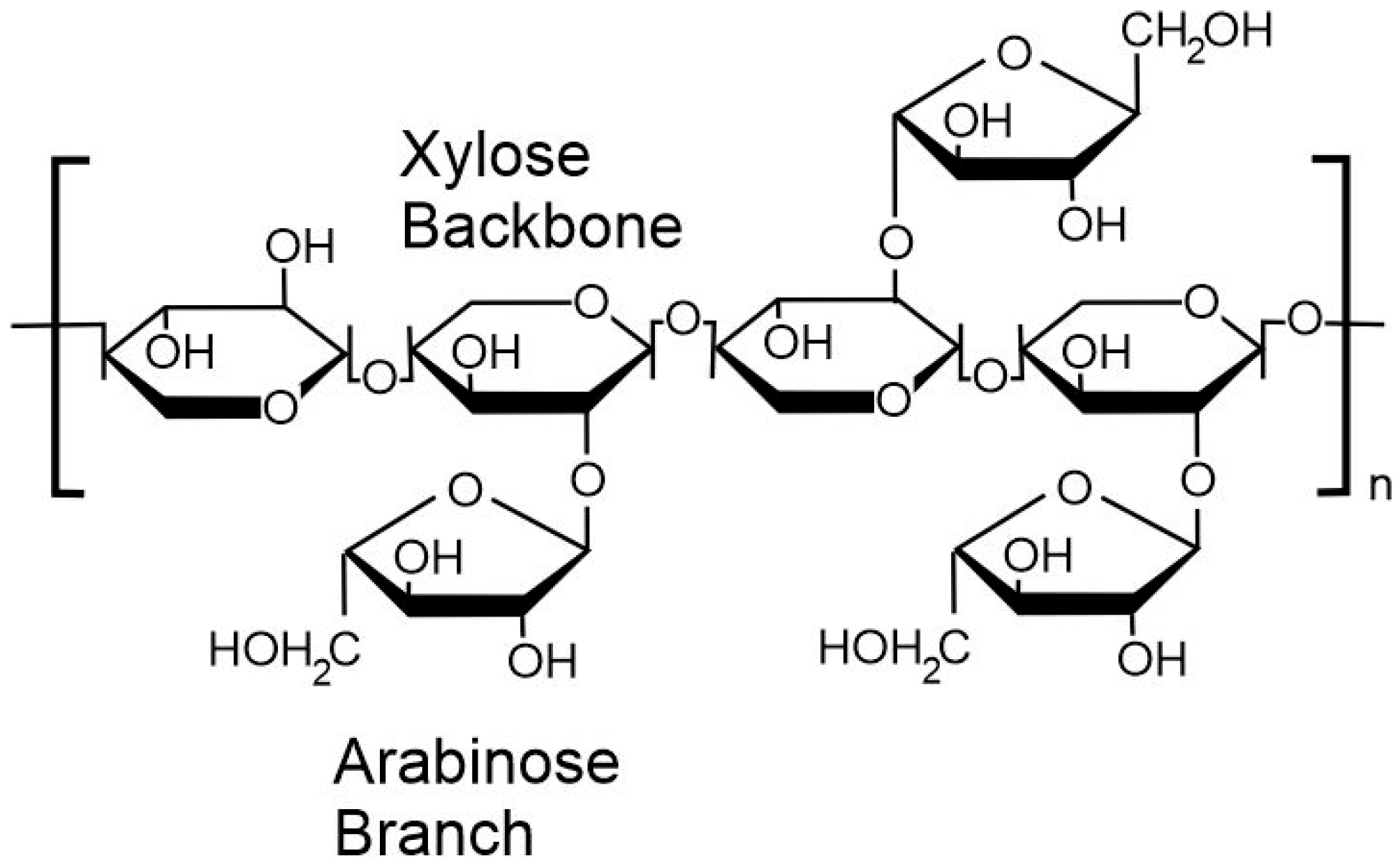
Figure 1. Xylan backbone characteristic of psyllium husk polysaccharides.
Carbon 11 00064 g001
Figure 2. Dimensions of the microneedle patch (A) and individual needle (B).
Carbon 11 00064 g002
Figure 3. Electron micrographs detailing the surface morphology of films of (A) psyllium husk and (B,C) psyllium husk–carbon nanoparticle composite.
Carbon 11 00064 g003
Figure 4. Contact angle images detailing the effect of time on the surface interaction with a water droplet on (A) psyllium husk and (B) psyllium husk–carbon nanoparticle composite. (C) A detailed comparison of the change in angle (averaged for both left and right) with time.
Carbon 11 00064 g004
Figure 5. Images of the psyllium husk–carbon nanoparticle composite before (A) and after (B) saturation with water. (C) Tensile properties of the psyllium husk and psyllium husk–carbon nanoparticle composite after exposure to water.
Carbon 11 00064 g005
Figure 6. Raman spectroscopy of a psyllium husk–carbon nanoparticle film.
Carbon 11 00064 g006
Figure 7. (A) Repetitive cyclic voltammograms detailing the response of psyllium husk–carbon towards ferrocyanide (2 mM) in (A) pH 7 Britton–Robinson buffer and (B) 0.1 M NaOH. Scan rate in all cases: 50 mV/s.
Carbon 11 00064 g007
Figure 8. (A) Cyclic voltammograms detailing the response of the cellulose acetate phthalate–carbon (CAP–C) film to ferrocyanide (2 mM, pH 7 Britton–Robinson buffer). (B) Change in oxidation peak height associated with repetitive scanning for the psyllium husk–carbon (PsH–C) and CAP–C film in pH 7 Britton–Robinson buffer solution.
Carbon 11 00064 g008
Figure 9. (A,B) Scanning electron micrographs of microneedle arrays constructed from equal parts psyllium husk and carbon nanoparticles. (C) Ripple-like contouring of the pyramidal needle structures. Needle dimensions: 700 mm (height), 200 mm (base) and 500 mm (pitch).
Carbon 11 00064 g009
Figure 10. (A) Cyclic voltammograms comparing the response of a psyllium husk–carbon (PsH–C) microneedle electrode to ferrocyanide (2 mM, pH 7, 50 mV/s) initially and after being rinsed, dried and then retested after a period of 24 h. (B) Influence of scan rate variations (5–500 mV/s) on the response of the PsH–C microneedle to ferrocyanide.
Carbon 11 00064 g010
Disclaimer/Publisher’s Note: The statements, opinions and data contained in all publications are solely those of the individual author(s) and contributor(s) and not of MDPI and/or the editor(s). MDPI and/or the editor(s) disclaim responsibility for any injury to people or property resulting from any ideas, methods, instructions or products referred to in the content.