Review

CARINH, an Interferon-Induced LncRNA in Cancer and Inflammation

by
Morgane Gourvest
and
Coen van Solingen
*
Department of Medicine, Cardiovascular Research Center, New York University Grossman School of Medicine, New York, NY 10016, USA
*
Author to whom correspondence should be addressed.
Non-Coding RNA 2025, 11(6), 79; https://doi.org/10.3390/ncrna11060079
Submission received: 8 October 2025 / Revised: 14 November 2025 / Accepted: 19 November 2025 / Published: 21 November 2025

Abstract

CARINH is an intriguing long noncoding RNA whose unique regulatory functions intersect the seemingly distinct processes of innate immunity and cancer development. Notably, CARINH is conserved across species, offering powerful experimental models for uncovering its mechanistic roles and physiological functions across diverse biological contexts. Stimulated by interferons and viral infections, CARINH stands out as a key player in the body’s antiviral defense mechanisms. Additionally, its dysregulation has been implicated in autoimmune disorders such as psoriasis, asthma, and inflammatory bowel disease, underscoring its broader role in maintaining immune homeostasis. Furthermore, alterations in CARINH expression have been connected to cancer progression, highlighting its dual role in immune response and tumor suppression. In this review, we delve into CARINH’s pivotal function in modulating interferon responses and influencing cancer development, with a focus on the molecular pathways that regulate its expression and contribute to its diverse roles. Understanding these pathways is crucial for evaluating CARINH’s significance as a biomarker and therapeutic target, potentially leading to groundbreaking advancements in medical research and treatment strategies.

1. Introduction

In recent years, the identification and characterization of noncoding RNAs (ncRNAs) have drastically transformed our perception of genetic function, revealing the intricate ways in which genomic elements can impact cellular processes. This newfound understanding of the genome’s so-called “junk DNA” has unveiled a variety of RNA classes that are essential for modulating protein-coding genes and organizing the genome’s structural framework [1]. Within the broad spectrum of ncRNAs discovered over the last few decades, long noncoding RNAs (lncRNAs) have been particularly instrumental in illuminating RNA’s significant role in gene regulatory mechanisms [2]. Like conventional messenger RNAs (mRNAs), ncRNAs are transcribed from DNA, yet they do not encode proteins. Their synthesis is primarily mediated by RNA polymerase II or, less commonly, by RNA polymerase I and III for certain ncRNA subclasses. The initiation of ncRNA transcription is tightly regulated by promoters, enhancers, and epigenetic modifications, while transcription factors and chromatin remodeling complexes modulate ncRNA loci chromatin accessibility. Following transcription, primary ncRNA transcripts undergo similar processing steps to those of mRNAs, including capping, splicing, and 3′ polyadenylation, reflecting their shared biogenesis pathways, although many ncRNAs also exhibit alternative splicing patterns, variable polyadenylation, or incomplete processing. The stability and turnover of ncRNAs are further controlled by RNA-binding proteins, RNA modification pathways (such as m6A), and RNA degradation mechanisms, ensuring appropriate expression levels within the cell. Moreover, ncRNA expression is highly dynamic and responsive to physiological and pathological cues. External stimuli such as stress, infection, and metabolic alterations, as well as diverse intracellular signaling pathways, can rapidly modulate ncRNA transcription and processing, allowing cells to rapidly adapt transcriptional responses to environmental cues. Collectively, these multilayered regulatory processes integrate lncRNA biogenesis, processing, and context-dependent expression within broader transcriptional and post-transcriptional control networks [3,4].
LncRNAs, a diverse group of transcripts, are distinguished by their length of over 200 nucleotides. They exhibit limited evolutionary conservation across species, which has posed challenges to deciphering their functional complexities [5,6,7]. These molecules are versatile, residing either in the nucleus or the cytoplasm, and they fulfill distinct tasks within these cellular compartments [8,9]. In the nucleus, lncRNAs are known for their multifaceted functions and can assemble into ribonucleoprotein complexes that serve as guides or decoys, influence nuclear organization, and contribute to the formation of higher-order chromosomal structures [10,11]. Additionally, they play roles in scaffolding effector proteins, enabling the modulation—either reduction or enhancement—of gene regulatory activities [12]. Furthermore, lncRNAs operate in both cis and trans regulatory modes; they regulate gene expressions either near their transcription sites or at distal genomic locations [13,14]. Within the cytoplasm, lncRNAs act as competitive inhibitors to microRNAs (miRNAs), where miRNAs typically bind to the 3′ untranslated regions of target mRNAs to suppress their translation. By sponging miRNAs, lncRNAs can mitigate this inhibitory effect. Besides their interaction with miRNAs, cytoplasmic lncRNAs are involved in post-transcriptional regulation by affecting the stability and translation of mRNA directly [15,16,17].
Despite significant advancements in lncRNA research, our understanding of their impacts on health and disease remains incomplete. One of the principal challenges stems from the relatively poor conservation of human lncRNAs across species. This is thought to arise because regulatory sequences, including lncRNAs, evolve at a faster pace than protein-coding sequences, likely due to the more relaxed structure–function constraints these molecules face [2]. This evolutionary flexibility positions lncRNAs as intriguing components of genomic innovation. Interestingly, while many lncRNAs exhibit rapid sequence divergence, several notable examples, such as MALAT1, demonstrate strong conservation across vertebrates [18]. This implies that while most lncRNAs may tolerate substantial sequence variation, a subset of them retains critical functional or structural features under stronger evolutionary pressure. Research into conserved lncRNA families is still at an early stage, and further studies are needed to define how conservation relates to biological activity and physiological relevance [2,19]. The limited conversation of many human lncRNAs has restricted the predictive power of preclinical animal models, resulting in an underestimation of their contributions to disease progression and a slowdown in efforts to harness their therapeutic potential. In this context, this review is focused on the lncRNA Colitis Associated IRF1 antisense Regulator of Intestinal Homeostasis (CARINH), a lncRNA conserved in mice that plays important regulatory roles in interferon-driven innate immunity [20,21,22,23,24] and various cancers [25,26,27,28,29]. Remarkably, the presence of a mouse ortholog to CARINH has offered unique opportunities to investigate its functions in preclinical models using Carinh knockout mice [22,23]. By investigating CARINH in these in vivo models as well complementary in vitro models, researchers have gained critical insights into its biological functions and potential therapeutic applications, which we have highlighted in this review.

2. CARINH: Nomenclature, Genomic Location, and Synteny

The GENCODE consortium and other groups have made substantial efforts to establish a unified system for naming lncRNAs, drawing on criteria such as their genomic location relative to protein-coding genes, their broader genomic context, or predicted structural and functional features [30]. Regardless of these efforts, uniformity has yet to be fully achieved. As a result, many lncRNAs continue to be annotated under several different names across distinct databases, creating redundancy and confusion. LncRNA CARINH further illustrates this, as it is associated with multiple, sometimes inconsistent aliases. Currently, The Human Genome Organisation (HUGO) Gene Nomenclature Committee (HGNC) [31] lists CARINH as the approved short form for the full gene name ‘Colitis Associated Interferon Response Factor 1 [IRF1] antisense Regulator of Intestinal Homeostasis’, with previous gene names being ‘IRF1 Antisense RNA 1′ (IRF1-AS, IRF1-AS1) and ‘Chromosome 5 Open Reading Frame 56′ (C5ORF56). CARINH was originally identified in human hepatic Huh7 cells exposed to varying concentrations of interferon (IFN)α for up to 72 h. Within this experimental setting, researchers detected a collection of lncRNAs that were either induced (IFN-Stimulated lncRNAs) or repressed (IFN-Downregulated lncRNAs) by IFN signaling. Notably, three of these transcripts displayed expression profiles closely paralleling those of their neighboring protein-coding genes. Among these pairs was the coding gene IRF1 and the adjacent noncoding transcript AC116366.6 or ENST00000461203, which was subsequently named IFN-Stimulated lncRNA 8 (ISR8) [21]. Of note, ISR8 represents one of the splice variants for CARINH currently annotated in the Ensembl database.
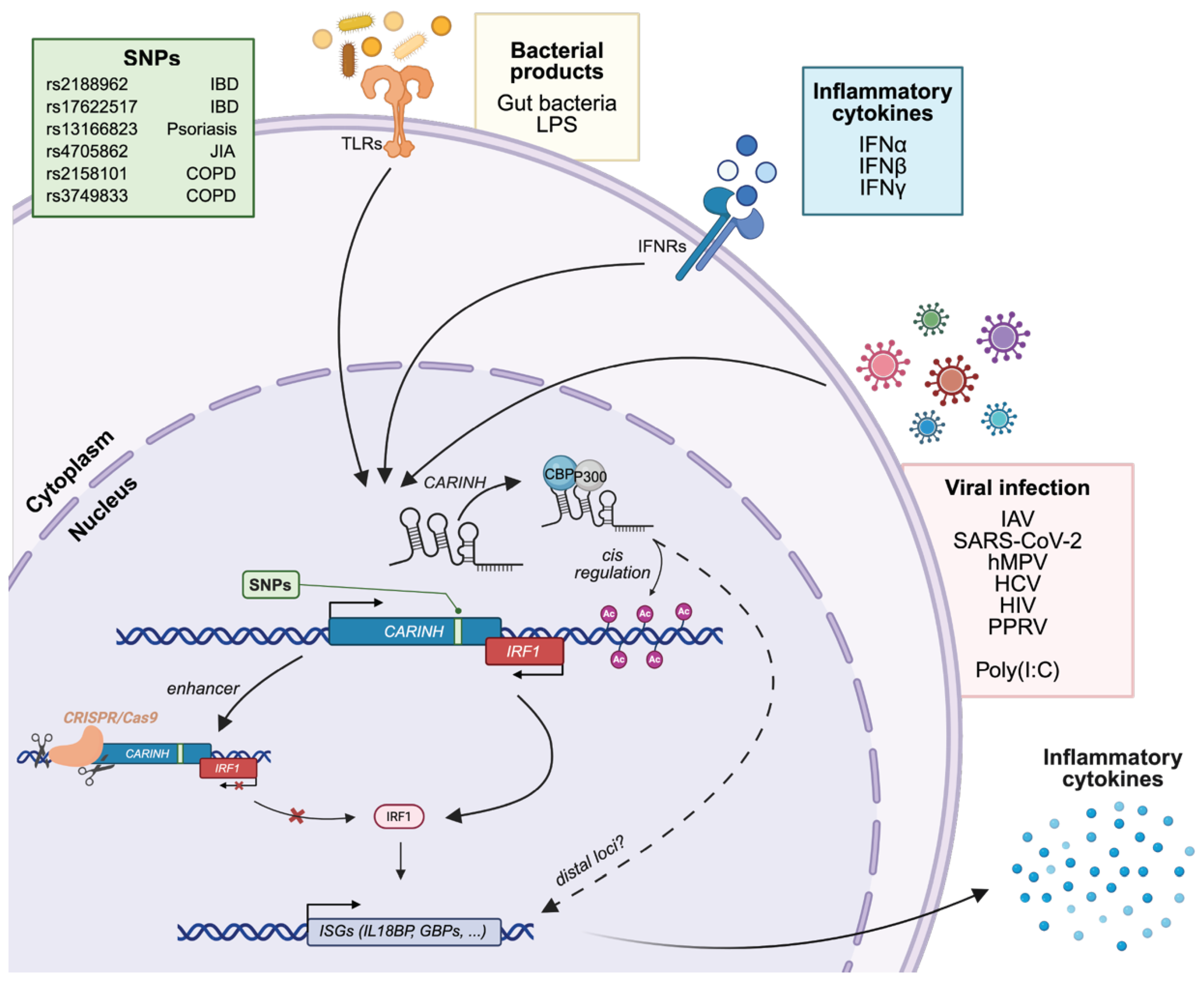
The CARINH gene, also designated by identifiers including ENSG00000197536 (Ensembl v115), NR_161242 (GenBank), AC116366 (GENCODE v48), and 441108 (NCBI), is located on human chromosome 5. It comprises three splice variants (ENST00000612967.2 [CARINH-V1], ENST00000337752.6 [CARINH-V2], and ENST00000407797.6 [CARINH-V3]) currently documented in GENCODE, all of which have the same first three exons but vary in their 3′ end sequences [22]. CARINH is positioned antisense to the IRF1 gene; one of these splice variants, ENST00000612967.2, partially overlaps with the coding sequence of IRF1 [22]. Besides IRF1, the genetic locus in which CARINH is situated contains several genes that are imporant in inflammatory processes such as interleukin(IL)-4, IL-13 and IL-5 [32,33], as well as genes involved in the processing of DNA double-stranded breaks (RAD50) [34] and cellular uptake of carnitine (SLC22A5)31 (Figure 1a).
Synteny analysis comparing human and mouse genomes has identified that CARINH is one of the rare human lncRNAs that has a mouse ortholog, known as Carinh—also referred to as Gm12216—located on mouse chromosome 11. This mouse ortholog is similarly positioned antisense to the mouse Irf1 gene and in genomic vicinity to Il-4, Il-13, Il-15, Rad50, and Slc22a5 (Figure 1b) [22,23]. The presence of a mouse ortholog to CARINH presents a unique opportunity to explore its functions in vivo using Carinh knock-out mice, as demonstrated in inflammatory bowel disease and viral infection [22,23]. A deeper exploration of the regulatory functions of this specific lncRNA and its mouse ortholog, situated in a locus encompassing crucial genes for inflammatory signaling, may reveal essential mechanisms underpinning cellular responses and potentially influence clinical outcomes.

3. CARINH and Innate Immunity

Interferons (IFNs) are critical components of the innate immune system, primarily responsible for the initial defense against infections. These cytokines are produced and released by host cells upon sensing pathogens such as viruses, bacteria, and parasites. By binding to their specific receptors at the surface of cells, IFNs activate signaling pathways that induce the expression of IFN-stimulated genes (ISGs). These ISGs encode antiviral, antimicrobial, and immunoregulatory proteins that collectively restrict pathogen replication, promote antigen presentation, and shape downstream immune responses, enhancing the detection and elimination of pathogens. Additionally, IFNs play a role in activating and recruiting effector immune cells, including natural killer cells and macrophages, thereby orchestrating a coordinated immune response [35,36,37].

3.1. CARINH in Viral Infection

Initial evidence implicating CARINH in IFN signaling and antiviral immunity emerged from a study profiling lncRNA expression in IFNα-stimulated hepatoma cells. This work identified a signature of interferon-stimulated lncRNAs (ISRs), among which ISR8 (a splice variant of CARINH) was particularly robustly induced. CARINH expression was found to be co-regulated with the nearby interferon-stimulated gene IRF1, with its induction consistently observed across various cell types in response to both IFNα and IFNβ. Additionally, CARINH levels were elevated in liver samples from patients with hepatitis C virus and in the peripheral blood of individuals infected with human immunodeficiency virus, demonstrating its in vivo relevance [21]. Further mechanistic studies by the same group employed CRISPR/Cas9 to edit the promoter and second exon on a genomic level. These studies revealed that such alterations impaired the induction of CARINH, IRF1, GBP1, and other ISGs following IFNα stimulation. However, direct manipulation of the mature RNA form of CARINH through its depletion or overexpression did not affect ISG activation, highlighting that not only the RNA transcript but also the chromatin context of the CARINH locus may be crucial for activating IFN-responsive promoters [20].
A study examining the role of CARINH in virally infected macrophages provided further mechanistic insights into its function. This research discovered a cis-regulatory mechanism through which CARINH directly influences IRF1 transcription. Initially, it was demonstrated that CARINH and IRF1 are co-upregulated in the circulation of patients infected with influenza A (IAV), severe acute respiratory syndrome coronavirus 2 (SARS-CoV-2), or human metapneumovirus (hMPV). A similar co-upregulation was also observed in human macrophages exposed to IAV, the viral mimic polyinosinic–polycytidylic acid (poly[I:C]), as well as IFNβ. Functionally, the genomic deletion of Carinh in mice resulted in reduction of Irf1 levels and its downstream antiviral transcriptional program, as evidenced by impaired cytokine induction and increased viral burden in mice challenged with a sublethal dose of IAV. Chromatin Isolation by RNA Purification followed by DNA sequencing (ChIRP-seq) revealed that CARINH binds directly to the IRF1 locus, indicative of cis-regulatory function (Figure 2) [22].
Intriguingly, viral-induced expression of CARINH was also observed in non-model species. In goat endometrial epithelial cells, infection with the peste des petits ruminants virus (PPRV) led to a boost of ISGs through IRF3 activation, driven by enhanced CARINH levels [38]. The findings highlight that CARINH serves as a conserved antiviral regulator across different species and functions as a powerful amplifier of IFN signaling.

3.2. CARINH in Autoimmunity and Chronic Inflammatory Diseases

Currently, the role of CARINH has been implicated in several immune-driven conditions, including inflammatory bowel disease (IBD) [23,24], psoriasis [39,40], asthma, and Chronic Obstructive Pulmonary Disease (COPD) [41,42] as well as cardiometabolic disorders [43,44,45]. Additionally, there is a broader connection between CARINH and loci with autoimmune diseases, such as juvenile idiopathic arthritis [46] (Figure 2).

3.2.1. Inflammatory Bowel Disease

Among these areas of research, studies on IBD have provided the most detailed understanding of CARINH’s role in disease. Analysis of RNA sequencing data from 1821 mucosal biopsies from the Mount Sinai Crohn’s and Colitis Registry identified CARINH as a hub gene within a module linked to inflammation and disease severity. Further functional studies revealed that CARINH enhances inflammatory cytokine production in LPS-stimulated primary human myeloid cells. Interestingly, these experiments showed that IRF1 expression was minimally affected by this treatment, suggesting that CARINH might promote cytokine production independently of IRF1 in this context [24]. Mechanistic studies in Carinh—/— mice revealed that although these mice appeared phenotypically normal under steady-state conditions, they experienced significantly more severe colitis compared to their wildtype counterparts when subjected to dextran sulfate sodium (DSS) or trinitrobenzene sulfonic acid (TNBS). This increased severity was characterized by greater weight loss, colon shortening, bleeding, inflammatory cell infiltration, and crypt damage, highlighting Carinh’s protective role against colitis. At the molecular level, RNA-sequencing analysis of bone marrow-derived macrophages (BMDMs) from Carinh—/— mice revealed marked downregulation of Irf1 and ISGs, including Il18bp and Gbp. Combination of RNA and chromatin immunoprecipitation assays showed that Carinh physically interacts with the histone acetyltransferases p300/CBP and mediates the deposition of H3k27 acetylation marks at the Irf1 locus, driving Irf1 transcription and subsequent downstream activation of ISGs (Figure 2). These data were strengthened by the identification of eight IBD-associated single nucleotide polymorphisms (SNPs) in the 5q31.1 locus, with rs2188962 emerging as the top candidate causal SNP (C > T), increasing the carrier’s risk of IBD by approximately 7.5% [23]. Interestingly, dysregulated crosstalk between the gut immune system and microbiota is a key driver of IBD. In this context, Carinh, Irf1, and Il18bp are microbiota-dependent, with their expression sustained by intestinal commensals in myeloid cells and reduced in germ-free or antibiotic-treated mice. Carinh deficiency disrupts microbial balance, enriching pro-inflammatory taxa like Prevotellaceae, and its microbiota transfer confers heightened colitis susceptibility, linking depletion of Carinh to defective antimicrobial defense.

3.2.2. Cardiometabolic Disorders

Autoimmune disorders are known to elevate the risk of cardiovascular diseases [47]; however, the shared genetic and biological mechanisms underlying these associations remain largely elusive. Over the past decades, Genome Wide Association Studies (GWASs) have identified that most risk-related SNPs are in noncoding regions of the genome [48]. This observation highlights the significant role of noncoding regulatory elements, such as promoters and enhancers, as well as transcribed noncoding RNAs, including lncRNAs, in disease susceptibility. Notably, a comprehensive integration of large-scale GWAS datasets—including over 10,000 psoriasis cases and approximately 400,000 controls—alongside UK Biobank cardiovascular cohorts, revealed significant positive genetic correlations between psoriasis and various cardiovascular conditions, such as hypertension, coronary heart disease, coronary atherosclerosis, and heart failure. Among 13 loci encompassing 653 SNPs shared between psoriasis and cardiovascular traits, the 5q31.1 locus, which includes CARINH, emerged as particularly significant in these associations [39].
The analysis of genotype data from over 3000 metabolic dysfunction-associated fatty liver disease (MAFLD) cases and more than 10,000 controls, sourced from the Korean Genome and Epidemiology Study, revealed strong associations with established risk loci, such as PNPLA3, SAMM50, PARVB, TM6SF2, and GCKR. However, by employing new methodologies to annotate and prioritize variants and genes from these association results, including Multivariate Analysis of Genomic Annotation (MAGMA) [49] and Functional Mapping and Annotation (FUMA)’s SNP2Gene and GENE2FUNC tools [50], several novel loci were identified. Among these, CARINH showed significant association (p = 4.4 × 10⁻⁵), with 232 variants reported [43]. This enrichment suggests that variants at the CARINH locus may influence MAFLD susceptibility and underscores the idea that CARINH acts as a genetic hub connecting immune dysregulation, metabolic stress, and chronic inflammation across tissues.
Using weighted gene co-expression network analysis (WGCNA) on publicly available datasets [51,52], ten lncRNAs, including CARINH, were identified that strongly correlated with Tetralogy of Fallot (TOF), a congenital heart disease characterized by a combination of ventricular septal defect, overriding aorta, pulmonary stenosis, and right ventricular hypertrophy. The data reported suggested that CARINH may contribute to TOF pathogenesis through disruption of splicing machinery. While mechanistically distinct from the described inflammatory circuits of CARINH, these findings reinforce a broader principle: lncRNAs act as central disease mediators in cardiovascular disease [45].

3.2.3. CARINH Variants as Determinants of Inflammatory Disease Risk and Therapeutic Response

GWASs and subsequent meta-analyses have linked CARINH to a wide array of autoimmune-related diseases. In the context of psoriasis, CARINH has been associated with therapeutic responses. This connection was established through two independent cohorts of psoriatic patients treated with a biosimilar of Etanercept, a TNFα inhibitor, which identified 148 good responders and 29 poor responders. A meta-analysis of 3.4 million SNPs identified 7 loci with potential associations to treatment outcomes, among which rs13166823 within the CARINH locus exhibited one of the strongest signals. The protective minor allele was significantly more prevalent among good responders in both cohorts, suggesting that CARINH expression might influence clinical responses to TNFα blockade [40].
In juvenile idiopathic arthritis (JIA), genetic associations previously reported [53] were further validated by an independent replication study [46]. Using the Australian CLARITY cohort, which includes 404 JIA patients and 676 healthy controls, 19 candidate SNPs were genotyped. Subsequent regression analyses were performed, adjusting for sex and ancestry. This analysis revealed seven loci replicated with consistent direction of effect, including the 5q31.1 region encompassing IRF1 and CARINH. The lead SNP at this locus, rs4705862, showed a significant association with JIA (odds ratio = ~1.27, p = 1.1 × 10⁻⁵), providing independent confirmation of prior findings. This replication study further establishes robust genetic evidence that variation at the CARINH locus contributes to JIA risk [46], and, considering studies demonstrating its role in IFN signaling and intestinal immune homeostasis, highlights a functional lncRNA as a mechanistic link between JIA and broader autoimmune pathophysiology.
Two large GWASs aimed at uncovering genetic risk factors for asthma and COPD identified two distinct SNPs associated with CARINH. The SNP rs2158101 was identified as the strongest male-specific variant linked to COPD, while rs3749833 was primarily associated with susceptibility to fixed airflow obstruction. Further functional annotation suggested these SNPs may exert regulatory effects on the expression of nearby genes that play roles in immune responses and inflammation. Subgroup analyses confirmed that these associations were independent of smoking status and age at asthma diagnosis. Additionally, phenome-wide association studies demonstrated that this locus was strongly associated with traits related to eosinophil counts, atopy, and asthma, highlighting its relevance in respiratory disease mechanisms [41,42].
More broadly, the molecular mechanisms underlying the autoimmune-associated variant rs17622517, located in an intron of the CARINH gene, were investigated. To this end, primary human monocytes from 134 donors stimulated with LPS were studied, and they found that rs17622517 influenced the expression of IRF1 and subsequent function. Furthermore, perturbation of the SNP locus resulted in a decrease in IRF1, suggesting that this region has enhancer functions [54], confirming work by others [20]. In a transcriptomic study utilizing LPS-stimulated human HMC3 microglial cells, five upregulated lncRNAs, including CARINH, were identified. The induction of CARINH was completely inhibited by JQ1, a small-molecule inhibitor that prevents epigenetic readers from binding to acetylated histones. Correlation analysis showed that CARINH expression was linked to 57 protein-coding genes, which included ISGs such as BATF2, RSAD2, IFIH1, IFIT3, TLR3, TRANK1, and GBP5. Pathway enrichment analysis revealed associations with terms related to type I IFN signaling, antiviral defense, chemokine-mediated signaling, and the negative regulation of metabolic processes. It also pointed to the activation of cell death pathways like TRAIL signaling and necroptosis. These findings suggest that CARINH acts as an LPS-responsive lncRNA with roles in coordinating inflammatory responses in human monocytes and microglial cells [55].

4. CARINH and Cancer

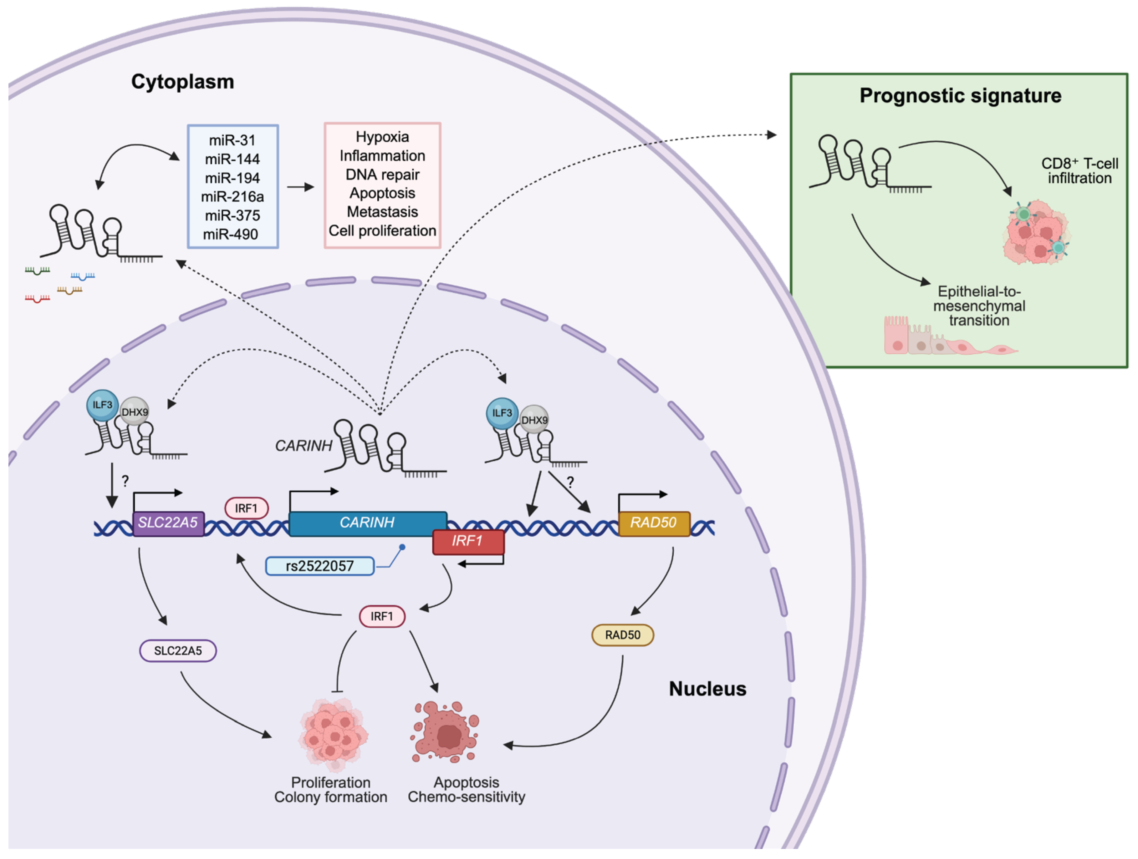
IFNs are crucial mediators of antitumor immunity, exerting effects directly on cancer cells and indirectly via the immune system. When IFNs bind to their receptors, they activate the Janus kinase-signal transducer and activator of transcription (JAK-STAT) pathway. This activation triggers the transcription of hundreds of ISGs that control various cellular processes, including cancer cell proliferation, apoptosis, differentiation, survival, and migration. Additionally, IFNs orchestrate immune responses by modulating the activity of nearly all immune cell subsets, making them pivotal in tumor-immune interactions [56,57]. As expected, given its role in regulating IRF1 expression and the IFN response, CARINH is implicated in various cancers (Figure 3) [25,26,27,28,29].

4.1. Esophageal Squamous Cell Carcinoma

The first direct evidence linking CARINH to cancer was observed in esophageal cancer, specifically within esophageal squamous cell carcinoma (ESCC). A transcriptomic analysis, along with the integration of lncRNA expression changes in three ESCC cell lines (KYSE30, KYSE180, and KYSE45) treated with IFNβ, identified nearly 1000 lncRNAs with altered expression levels (499 being upregulated, and 495 downregulated). Notably, among the identified lncRNA, CARINH was most significantly induced upon IFN stimulation. Moreover, CARINH levels were positively correlated with IRF1 expression in these ESCC cell lines. Expression studies in ESCC cell lines further confirmed that CARINH is a genuine ISG, induced by both type I (IFNβ) and type II (IFNγ) IFNs via the canonical JAK-STAT pathway. This was demonstrated by the effect of treatment with the JAK inhibitor Ruxolitinib, which inhibited CARINH expression in ESCC cell lines. Further support came from publicly available ENCODE Chromatin Immunoprecipitation (ChIP) sequencing data, which showed binding of STAT proteins at the CARINH promoter. Subsequent ChIP-qPCR validation confirmed STAT1 occupancy at the promoter region, further reinforcing the role of CARINH in the IFN pathway [25].
Mechanistically, CARINH associates with the transcriptional co-activators Interleukin Enhancer Binding Factor 3 (ILF3) and DExH-Box Helicase 9 (DHX9), acting as a nuclear scaffold to augment binding to the IRF1 promoter, driving its transcription and subsequent IFN signaling. In turn, IRF1 binds the CARINH promoter to boost its transcription, suggesting a positive feed-forward regulatory loop between CARINH and IRF1. Loss-of-function experiments using short hairpin RNAs (shRNAs) revealed that knocking down CARINH promoted colony formation of KYSE cells, reduced cisplatin-induced apoptosis in vitro, and led to increased tumor growth in xenografts of subcutaneously injected KYSE30 cells. This growth was accompanied by elevated levels of proliferation factors such as Antigen Kiel 67 (Ki67) and Proliferating Cell Nuclear Antigen (PCNA) and a reduction in apoptotic markers. Conversely, CARINH overexpression suppressed proliferation, impaired colony formation, and promoted apoptosis in vitro, while tumor weights and volumes of in vivo xenografts were reduced when compared to controls [25].
Clinically, CARINH expression was significantly reduced in ESCC tumors compared to healthy esophageal tissues, and low expression correlated with poor clinical outcomes, consistent with a tumor-suppressive function [25]. Interestingly, analyses of The Atlas of Noncoding RNAs in Cancer (TANRIC [58]) datasets revealed positive correlations between CARINH and IRF1 expression across multiple cancer types, including lung, breast, and head and neck squamous cell carcinoma [25], suggesting that this regulatory axis may extend beyond ESCC.

4.2. Bladder Cancer

Building on the broader association with cancer, a study of bladder cancer investigated CARINH as part of an epithelial-to-mesenchymal transition (EMT)-related lncRNA signature linked to cancer prognosis and disease progression. By analyzing RNA-sequencing data and paired clinical information from 402 bladder cancer patients sourced from The Cancer Genome Atlas (TCGA) [59], 525 lncRNAs were found to be correlated with a curated set of 200 EMT genes [27], derived from the gene set ‘Hallmark Epithelial-to-Mesenchymal Transition’ in The Molecular Signatures Database [60]. Further analysis using univariate and multivariate Cox regression, alongside Gene Set Enrichment Analysis (GSEA), refined this to a prognostic signature comprising 14 lncRNAs. Interestingly, each of these lncRNA serves as an independent prognostic factor in bladder cancer patients, and CARINH was among the five lncRNAs associated with unfavorable outcome [27]. Of note, the classification of these lncRNAs as “EMT-related” is based on co-expression patterns with EMT hallmark genes, without supporting functional assays or further mechanistic insights. This leaves it unclear whether CARINH has a direct causal role in bladder cancer progression. Consequently, the clinical significance of CARINH in relation to altering EMT in bladder cancer remains preliminary and necessitates direct experimental investigation.

4.3. Acute Lymphoblastic Leukemia

A third study extended the scope of CARINH to hematologic malignancies, specifically acute lymphoblastic leukemia (ALL). LncRNAs are increasingly recognized as central players in hematological cancers [61,62,63], and dysregulated lncRNA expression has been reported in ALL as well [64,65,66]. Elevated expression levels of CARINH were found in blood samples collected from 33 patients—subdivided in three groups: Philadelphia positive ALL, Philadelphia negative ALL, and T-cell ALL—when compared to a control group consisting of 15 healthy donors [26]. Analysis of publicly available microarray data [67] from 381 bone marrow samples, collected both at diagnosis and after eight days of remission-induction therapy, identified 52 differentially expressed lncRNAs altered in ALL, including CARINH. CARINH expression levels significantly reduced after 8 days of remission-induction therapy [26]. Notably, CARINH was connected through an extensive competing endogenous (ce)RNA network comprising 6 lncRNA, 17 miRNAs, and nearly 200 mRNAs, evaluated using miRcode, Targetscan, miRTarBase, and Cytoscope software [68,69]. This network is associated with inflammation via TNFα, as well as hypoxia, DNA repair, apoptosis, metastasis, and proliferation [26]. Within a larger ceRNA network, CARINH levels were associated with, miR-31, miR-144, miR-194, miR-216a, miR-375, miR-490, and their individual mRNA targets [26].
Albeit exploratory, this study lays the groundwork for understanding the potential role of CARINH in ALL. However, the contribution of CARINH to hallmark mechanisms of leukemogenesis, such as differentiation blockade, stemness, or therapy resistance, were not directly investigated, nor were clinical correlations with patient outcomes assessed. The findings highlight CARINH as a therapeutic candidate of interest across ALL subgroups. The predicted ceRNA interactions also suggest that CARINH intersects with pathways known to be deregulated in ALL, warranting further validation in leukemic cells.

4.4. Breast Cancer

A cross-ancestry GWAS meta-analysis conducted with data from the Breast Cancer Association Consortium, which included women of African (9241 cases and 10,193 controls) and European ancestry (122,977 cases and 105,974 controls), pinpointed four loci linked to overall breast cancer risk. Among these, the SNP rs2522057, located at the 5q31.3 locus, was strongly associated with the expression of IRF1. This SNP maps approximately 15 kb downstream of IRF1, within an intron of CARINH. Functional annotation using the 25-state chromatin model indicated that rs2522057 aligns with active enhancer and promoter regions, characterized by H3K4me1 and H3K27ac histone modifications [28]. These findings suggest that rs2522057 and its associated gene CARINH could be key mediators of breast cancer susceptibility by influencing the expression of IRF1 as well as other nearby genes such as RAD50 and SLC22A5, with all three genes implicated in breast cancer [70,71,72,73]. The presence of rs2522057 as a risk locus in both African and European populations and its proximity to oncogenic genes bolsters its candidacy as a causal variant driving cancer progression. Further in vitro and in vivo studies are necessary to confirm this causal role and to uncover the mechanisms through which rs2522057 may regulate IRF1, RAD50, and SCL22A5 expression via CARINH, ultimately affecting cancer development.
In another study, a comprehensive transcriptomic analysis was conducted to identify immune-related lncRNAs associated with breast cancer prognosis and immune infiltration. Using a publicly available microarray dataset of 327 breast cancer cases [74], researchers correlated lncRNA expression with immune-related genes and applied multivariate Cox regression to establish a six-lncRNA prognostic signature. This signature, which included CARINH, effectively stratified patients into high- and low-risk groups that demonstrated significantly different outcomes. The prognostic accuracy of the model was further enhanced when combined with clinical variables. Patients with higher expression levels of CARINH tended to fall into the low-risk group, which correlated with better survival rates and increased infiltration of CD8+ T-cells. This finding suggests that CARINH could serve as a favorable prognostic biomarker in breast cancer by modulating antitumor immune responses [29]. While the observations in these two patient studies point to a putative regulatory role for CARINH in breast cancer, further in vitro and in vivo functional studies are essential to validate its causal influence and to uncover the mechanisms through which CARINH impacts carcinogenesis.

5. Perspective

5.1. Expression of CARINH in Other Cells and Cell Types

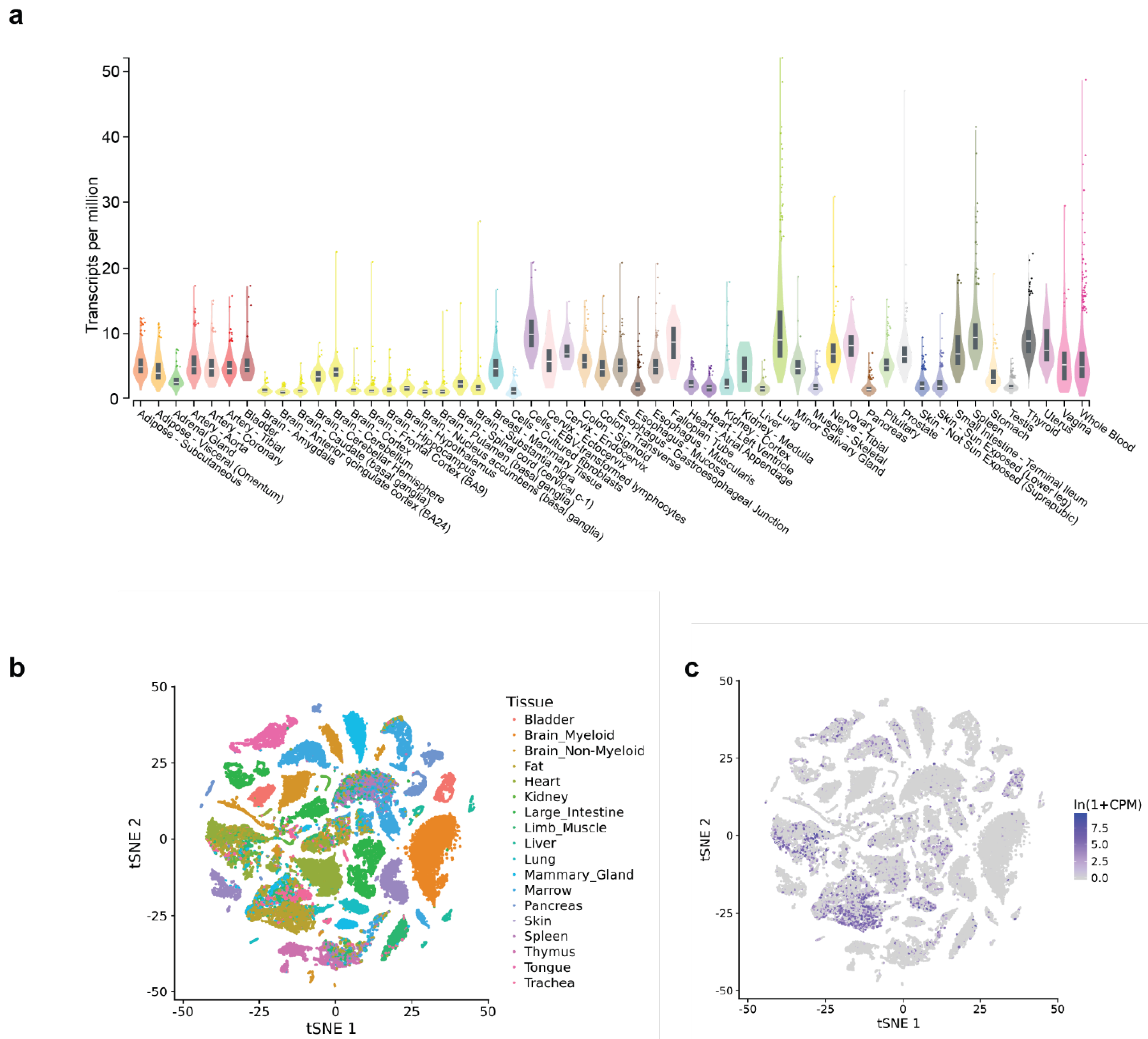
CARINH exhibits broad expression across human tissues and cell types, as evidenced by publicly available data from the NIH Genotype-Tissue Expression (GTEx) Project (V8, August 2019; 17,382 RNA-sequencing samples from 948 donors spanning 54 tissues) (Figure 4a) [75]. Elevated expression is particularly evident in the lung, spleen, colon, and whole blood, consistent with the reported role of CARINH in antiviral responses to respiratory infections and in the pathogenesis of inflammatory bowel disease [20,21,22,23,24]. It is plausible that specific cell populations, particularly inflammatory subtypes within these organs, exhibit differential CARINH expression in response to environmental cues; however, tissue-wide analyses of this cellular heterogeneity and its functional implications have not yet been undertaken on this scale.
In parallel with the GTEx initiative, large-scale single-cell transcriptomic resources have been generated for the mouse, comprising nearly 100,000 cells profiled across 20 different organs and tissues. This compendium, called Tabula Muris [76], enables high-resolution, controlled comparisons of gene expression across shared cell types, such as immune populations residing in distinct anatomical compartments, thereby providing a powerful framework for dissecting tissue-specific and context-dependent transcriptional programs. Like its human counterpart, analysis of the Tabula Muris dataset demonstrates that the mouse ortholog Carinh is broadly expressed across diverse tissues (Figure 4b,c). In contrast to the bulk tissue profiles provided by GTEx, Tabula Muris offers single-cell resolution, readily accessible through its online platform (https://tabula-muris.sf.czbiohub.org/, accessed on 20 November 2025). This resource provides a unique opportunity to interrogate Carinh and other noncoding transcripts at the single-cell level across multiple organs of interest.

5.2. Subcellular Localization of CARINH

The subcellular localization patterns of lncRNA transcripts allow for the classification of these molecules into five distinct groups: (I) lncRNAs forming one or two prominent foci within the nucleus; (II) those with large nuclear foci accompanied by scattered single molecules throughout the nucleus; (III) mostly nuclear lncRNAs without forming any foci; (IV) lncRNAs present in both the cytoplasm and nucleus; and (V) lncRNAs primarily found in the cytoplasm. Notably, about 55% of lncRNAs are predominantly expressed in the nucleus, falling into categories I through III [77]. The subcellular localization of CARINH has not been extensively researched, yet functional studies have primarily indicated nuclear localization and chromatin-associated roles [22,23,25], implying that CARINH is likely localized in the nucleus, particularly in response to certain stimuli or in specific cell types. However, these studies do not fully rule out the presence of small cytoplasmic fractions or the possibility of cell type-specific redistribution. Additionally, in silico subcellular localization tools and databases like lncLocator [78] and RNALocate [79] suggest a more uniform distribution throughout the cell (Figure 5). Future research is expected to provide new insights into the subcellular localization of CARINH, which may change based on cell type, cellular activation, or cell cycle stage, reflecting the inherent dynamic nature of RNA, particularly lncRNAs.

5.3. RNA Modifications Driving CARINH Expression and Function

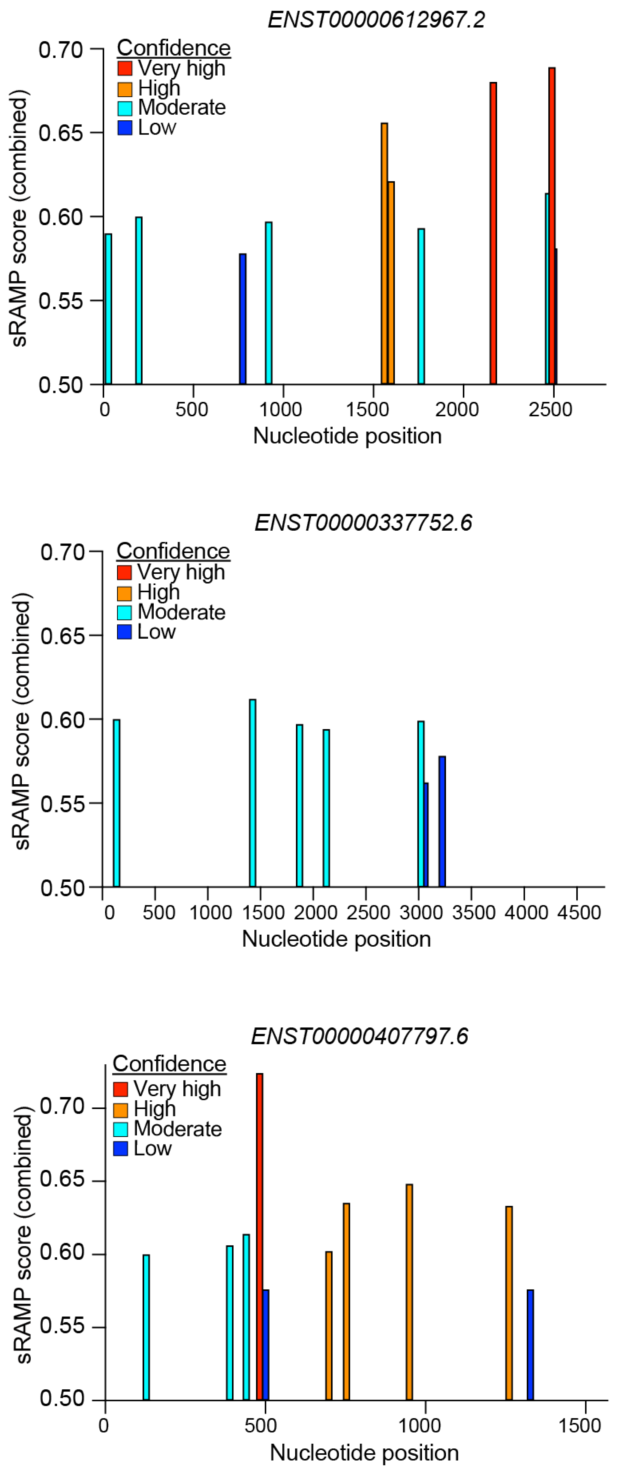
RNA modifications, such as N6-methyladenosine (m6A) methylation, are increasingly recognized as important regulators of RNA function. These modifications play a crucial role in modulating the accessibility of protein-binding motifs, which can influence various aspects of RNA biology. For example, m6A methylation can affect the export of RNA molecules to the cytoplasm and has implications for their targeting to ribosomes, thereby impacting translation efficiency. By altering these processes, m6A and similar modifications contribute to the dynamic regulation of gene expression and cellular function [80]. As our understanding of these modifications expands, it becomes clear that they are integral to the complex regulatory networks governing RNA behavior. To date, CARINH has not been described to undergo RNA methylation or to be linked to the expression of m6A-related genes. However, in silico prediction by sRAMP (a sequence-based RNA adenosine methylation site predictor) [81] for the three human splice variants of CARINH suggest distinct RNA modification profiles for each variant (Figure 6). This indicates the potential for splice variant-specific roles of CARINH driven by differences in RNA methylation. Future experimental studies are necessary to confirm these in silico predictions and explore the potential functional consequences.

5.4. Coding Potential of CARINH

Despite traditionally being considered noncoding, recent studies have identified thousands of functional micropeptides produced from small open reading frames (smORFs) hidden within lncRNA transcripts [82,83,84,85,86]. Recent high-throughput advancements for evaluating the translational capacity of these smORFs have uncovered that these micropeptides may contribute significantly to most, if not all, biological pathways [87,88].
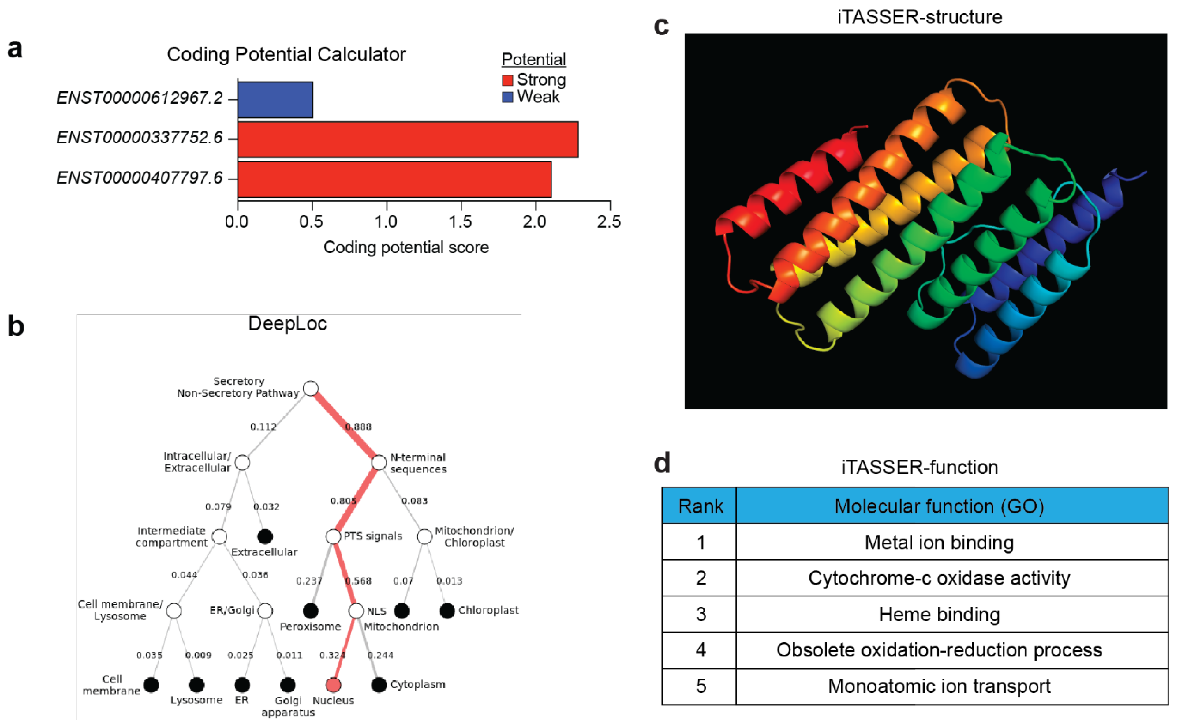
Analysis by the NIH’s ORF finder (https://www.ncbi.nlm.nih.gov/orffinder/) of the three CARINH splice sequences identified over 50 different putative ORFs, producing a range of amino acid lengths. Further assessment of protein-coding potential using the Coding Potential Calculator (CPC) [89] revealed that two out of the tree splice variants have a coding potential score indicating the presence of one or more translatable ORFs (Figure 7a). Among these is a putative peptide consisting of 188 amino acids, found in variant ENST00000337752.6, which has the highest reliability score determined by CPC. For conceptual purposes, we analyzed this putative peptide using I-TASSER [90] to predict protein structure and function, as well as DeepLoc [91] to predict subcellular localization. These analyses suggest likely nuclear localization (Figure 7b), a peptide with seven alpha helices (Figure 7c), and a potential function in metal ion binding (Figure 7d). Though this is primarily an academic exercise, these analyses offer an in silico pipeline for exploring novel ncRNA-derived peptides. However, a more in-depth analysis and functional evaluation of other putative peptides in CARINH are needed to uncover any further biological insights. It is important to note that previous BLAST analysis of a 149-amino-acid peptide revealed only a single low-homology hit, and codon substitution frequency scores were negative [25]. Meanwhile, experiments using a mini-circle reporter system showed that neither the human nor the murine CARINH locus had any ribosome binding activity, a finding confirmed by Ribo-seq analysis [23]. These experiments provide compelling evidence confirming CARINH as a noncoding transcript, overriding any in silico predictions to the contrary.

5.5. Chromatin- and Protein-Binding Abilities of CARINH

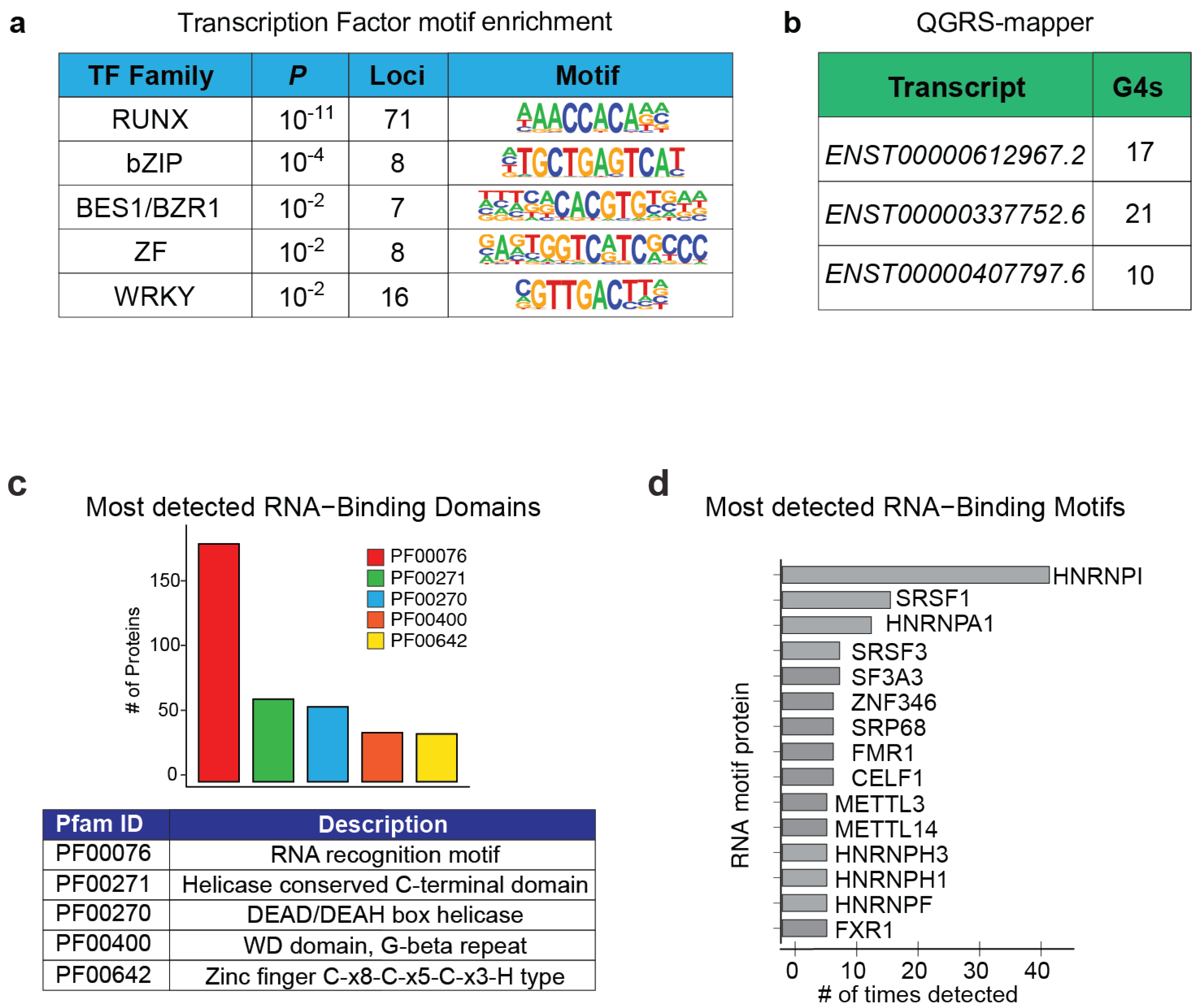
A combination of RNA pull-down assays with mass spectrometry, DNA sequencing, and targeted immunoprecipitation has yielded detailed insights into the molecular mechanisms through which CARINH regulates IRF1 expression, including evidence for a potential feed-forward loop involving both molecules [22,23,25]. Interestingly, disruption of the CARINH locus in HeLa cells demonstrated that transcriptional activity at this site alone may be sufficient to promote ISG expression, highlighting a potential enhancer-like role for CARINH [20]. On a chromatin level, CARINH binding is shown to associate with the DNA at approximately 232 loci, with the highest enrichment observed at the IRF1 locus [22]. This includes its binding to IL18BP, a gene previously shown to be regulated by CARINH through IRF1 [23], hinting at the potential for direct control by CARINH as well. Furthermore, transcription factor motif enrichment analysis using HOMER [92] of these 232 loci revealed that CARINH may binds to 71 loci in conjunction with DNA-binding motifs associated with the RUNX transcription family (Figure 8a). The RUNX family, known for various functions, can alter type I IFN signaling [93,94]. This suggests that CARINH might mediate target gene expression by competing or cooperating with RUNX family members.
Physical interactions between CARINH and several transcriptional co-activators, such as ILF3, DHX9, and p300/CBP, have been identified [23,25]. However, the specific mechanisms through which these RNA–protein interactions occur on the molecular level remain unclear. One important structural feature that supports RNA–protein interactions is tandem G-rich sequences, which form G-quadruplexes. These G-quadruplexes are secondary RNA structures that can serve as docking sites for proteins [95]. Analysis of CARINH sequences using the Quadruplex forming G-Rich Sequences (QGRS) mapper [96], an algorithm designed to identify and provide details about the composition and distribution of potential QGRS in nucleotide sequences, revealed between 10 and 20 unique QGRS (Figure 8b), unveiling a potential molecular mechanism, as has been described for other lncRNAs [97,98]. Alternatively, CatRAPID [99] can be utilized to explore potential RNA-binding domains within CARINH. This method identified predicted binding domains containing general RNA recognition motifs as well as motifs for DEAD/DEAH box helicases, a family that includes DHX9, known to interact with CARINH [25] (Figure 8c). In turn, linking these identified motifs in CARINH back to proteins pointed to several members of the heterogeneous nuclear ribonucleoproteins (hnRNPs), a family of functionally diverse proteins involved in mRNA stabilization, as well as transcriptional and translational regulation [100]. Notably, hnRNPs have been reported to interact with lncRNAs that function as transcriptional mediators for immune response genes [101,102] (Figure 8d). To date, experimental evidence has identified molecular mechanisms centered on the regulatory control of IRF1 expression by CARINH through association with several transcriptional co-activators [23,25]. However, an examination of CARINH’s chromatin-binding loci suggests the possibility of alternative mechanisms for gene regulation by CARINH [22]. These mechanisms are likely to be uncovered through further in silico analysis as well as functional experimentation.

5.6. CARINH as a Competing Endogenous RNA

The lncRNA–miRNA–mRNA axis is a critical regulatory network controlling gene expression at multiple levels. LncRNAs often sequester miRNAs, and through these interactions, lncRNAs modulate miRNA-mediated post-transcriptional repression of mRNAs, creating a dynamic interplay. This axis plays an essential role in various biological processes such as development, differentiation, immune modulation, and disease progression. Dysregulation of the lncRNA–miRNA–mRNA network has been implicated in cancers, cardiovascular diseases, and inflammatory conditions [15,17,103]. Despite its potential, understanding this axis remains challenging due to tissue- and cell-specificity and intricate interactions beyond direct RNA–RNA competition.
Studies in leukemia suggest that CARINH interacts with several miRNAs, including miR-31, miR-144, miR-194, miR-216a, miR-375, and miR-490, thereby influencing the translation of their downstream targets. However, when using the prediction algorithm miR-Target2 (miRDB [104]), which identifies interactions based on shared features of miRNA binding and target suppression, only a potential interaction with miR-194, a miRNA known to promote anti-tumor immunity in pancreatic cancer [105], was detected (Supplementary Table S1). This points to a possible secondary layer of lncRNA–miRNA crosstalk within the ceRNA networks investigated in ALL. Interestingly, among the miRDB-predicted miRNA interactions with CARINH were several miRNAs that were linked to innate immunity and cancer biology, such as miR-342-5p [106,107], miR-423-5p [108,109], miR-766-3p [110,111], and miR-4516 [112], among many others (Supplementary Table S1). These potential interactions should be interpreted with caution, as CARINH is predominantly localized to the nucleus and associated with nuclear functions [22,23,25]. This raises questions about its capacity to act as a ceRNA. Consequently, any proposed CARINH–miRNA interactions must be validated through rigorous approaches, such as luciferase reporter assays, site-directed mutagenesis, and functional perturbation studies, to definitively establish their biological relevance and impact on downstream targets [15,16].

6. Conclusions

In this review, we have highlighted CARINH, a conserved long noncoding RNA that is upregulated upon viral infection, as well as upon stimulation with IFNs [20,21,22,25,38]. CARINH has been shown to control the expression of its neighboring gene IRF1 in a cis-regulatory manner, likely through binding of p300/CBP, driving the activation of ISGs and subsequent immune response [22,23]. Due to its effects on the IFN-driven immunity, CARINH has been implicated viral infections [20,21,22,38], immune and auto-immune diseases [23,24,39,40,41,42], and cardiometabolic disorders [43,44,45]. Interestingly, many SNPs have been detected at the CARINH locus that were associated with disease, and although direct correlation between expression of CARINH and these SNPS have not been established, these data underline the importance of this locus in linking genetic variation to immune regulation.
In cancer research, CARINH has been found to interact with transcriptional co-activators DHX9, ILF3 to regulate the expression of IRF1, with its loss contributing to tumor progression [23,25]. Additionally, CARINH is part of an extensive ceRNA network that is involved in various cancer-related pathways, such as metastasis and cell proliferation [26]. Patients’ studies further suggest that CARINH is included in several lncRNA signatures that may be predictive of clinical outcomes [27,28,29], highlighting its potential as a future biomarker in cancer. However, the precise link between elevated levels of CARINH and either poor or favorable prognosis remains unclear and is likely cancer dependent. This will be a focal point of future scientific exploration within this field.
As lncRNAs continue to be recognized as crucial orchestrators of genome architecture and gene expression regulation, the scientific community is increasingly optimistic about targeting RNA for therapeutic purposes. Previously deemed undruggable, RNA has now become the focus of a growing class of pharmacological interventions, notably including antisense oligonucleotides (ASOs), short polymers comprising 15 to 21 nucleotides [113]. To date, at least thirteen ASO-based treatments have received clinical approval for conditions like familial hypercholesterolemia and chylomicronemi [114], underscoring their promise in the development of therapies targeting lncRNAs. ncRNAs have thus become significant targets for therapeutic innovation, with substantial efforts directed at the development of oligonucleotide-based treatments. Currently, progress in programs targeting miRNAs is more advanced than those focusing on lncRNAs; however, analogous methodologies utilizing synthetically modified ASOs are being harnessed to address these differences [115,116]. GapmeRs, which are short DNA ASOs flanked by RNA mimics, and small interfering RNAs (siRNA) have been effectively employed to target specific lncRNAs such as CARINH [20,22,23,25]. GapmeRs, in particular, are noteworthy for their strong affinity to target RNAs, enabling them to bind efficiently before being degraded by RNAseH1 [117], thereby minimizing off-target effects [118,119]. As such, gaining a deeper understanding of the molecular mechanisms underlying CARINH’s functions, along with the capability to effectively target this lncRNA, will be crucial for unlocking its potential in both therapeutic and diagnostic applications.

Supplementary Materials

The following supporting information can be downloaded at: https://www.mdpi.com/article/10.3390/ncrna11060079/s1, Supplementary Table S1. Predicted miRNA interactions with CARINH variants.

Author Contributions

M.G. and C.v.S. drafted the manuscript and figures. All authors have read and agreed to the published version of the manuscript.

Funding

This work was supported by grants from the National Institutes of Health (T32HL098129 to MG) and the American Heart Association (23POST1029885 to MG and 23SCEFIA1153739 to CvS).

Institutional Review Board Statement

Not applicable.

Informed Consent Statement

Not applicable.

Data Availability Statement

Not applicable.

Conflicts of Interest

The authors declare no conflicts of interest.

References

  1. Chen, L.L.; Kim, V.N. Small and Long Non-Coding Rnas: Past, Present, and Future. Cell 2024, 187, 6451–6485. [Google Scholar] [CrossRef] [PubMed]
  2. Mattick, J.S.; Amaral, P.P.; Carninci, P.; Carpenter, S.; Chang, H.Y.; Chen, L.L.; Chen, R.; Dean, C.; Dinger, M.E.; Fitzgerald, K.A.; et al. Long Non-Coding Rnas: Definitions, Functions, Challenges and Recommendations. Nat. Rev. Mol. Cell Biol. 2023, 24, 430–447. [Google Scholar] [CrossRef]
  3. Spurlock, C.F., 3rd; Crooke, P.S., 3rd; Aune, T.M. Biogenesis and Transcriptional Regulation of Long Noncoding Rnas in the Human Immune System. J. Immunol. 2016, 197, 4509–4517. [Google Scholar] [CrossRef] [PubMed]
  4. Nojima, T.; Proudfoot, N.J. Mechanisms of Lncrna Biogenesis as Revealed by Nascent Transcriptomics. Nat. Rev. Mol. Cell Biol. 2022, 23, 389–406. [Google Scholar] [CrossRef]
  5. Rinn, J.L.; Chang, H.Y. Genome Regulation by Long Noncoding Rnas. Annu. Rev. Biochem. 2012, 81, 145–166. [Google Scholar] [CrossRef]
  6. Palazzo, A.F.; Lee, E.S. Non-Coding Rna: What Is Functional and What Is Junk? Front. Genet. 2015, 6, 2. [Google Scholar] [CrossRef]
  7. Schmitz, S.U.; Grote, P.; Herrmann, B.G. Mechanisms of Long Noncoding Rna Function in Development and Disease. Cell Mol. Life Sci. 2016, 73, 2491–2509. [Google Scholar] [CrossRef] [PubMed]
  8. Bridges, M.C.; Daulagala, A.C.; Kourtidis, A. Lnccation: Lncrna Localization and Function. J. Cell Biol. 2021, 220, e202009045. [Google Scholar] [CrossRef]
  9. Krause, H.M. New and Prospective Roles for Lncrnas in Organelle Formation and Function. Trends Genet. 2018, 34, 736–745. [Google Scholar] [CrossRef]
  10. Goff, L.A.; Rinn, J.L. Linking Rna Biology to Lncrnas. Genome Res. 2015, 25, 1456–1465. [Google Scholar] [CrossRef]
  11. Satpathy, A.T.; Chang, H.Y. Long Noncoding Rna in Hematopoiesis and Immunity. Immunity 2015, 42, 792–804. [Google Scholar] [CrossRef]
  12. Wang, K.C.; Chang, H.Y. Molecular Mechanisms of Long Noncoding Rnas. Mol. Cell 2011, 43, 904–914. [Google Scholar] [CrossRef] [PubMed]
  13. Guil, S.; Esteller, M. Cis-Acting Noncoding Rnas: Friends and Foes. Nat. Struct. Mol. Biol. 2012, 19, 1068–1075. [Google Scholar] [CrossRef] [PubMed]
  14. Joung, J.; Engreitz, J.M.; Konermann, S.; Abudayyeh, O.O.; Verdine, V.K.; Aguet, F.; Gootenberg, J.S.; Sanjana, N.E.; Wright, J.B.; Fulco, C.P.; et al. Genome-Scale Activation Screen Identifies a Lncrna Locus Regulating a Gene Neighbourhood. Nature 2017, 548, 343–346. [Google Scholar] [CrossRef]
  15. Hennessy, E.J.; van Solingen, C.; Scacalossi, K.R.; Ouimet, M.; Afonso, M.S.; Prins, J.; Koelwyn, G.J.; Sharma, M.; Ramkhelawon, B.; Carpenter, S.; et al. The Long Noncoding Rna Chrome Regulates Cholesterol Homeostasis in Primate. Nat. Metab. 2019, 1, 98–110. [Google Scholar] [CrossRef]
  16. Ala, U. Competing Endogenous Rnas, Non-Coding Rnas and Diseases: An Intertwined Story. Cells 2020, 9, 1574. [Google Scholar] [CrossRef]
  17. Liu, X.H.; Sun, M.; Nie, F.Q.; Ge, Y.B.; Zhang, E.B.; Yin, D.D.; Kong, R.; Xia, R.; Lu, K.H.; Li, J.H.; et al. Lnc Rna Hotair Functions as a Competing Endogenous Rna to Regulate Her2 Expression by Sponging Mir-331-3p in Gastric Cancer. Mol. Cancer 2014, 13, 92. [Google Scholar] [CrossRef]
  18. Monroy-Eklund, A.; Taylor, C.; Weidmann, C.A.; Burch, C.; Laederach, A. Structural Analysis of Malat1 Long Noncoding Rna in Cells and in Evolution. RNA 2023, 29, 691–704. [Google Scholar] [CrossRef]
  19. Quinn, J.J.; Zhang, Q.C.; Georgiev, P.; Ilik, I.A.; Akhtar, A.; Chang, H.Y. Rapid Evolutionary Turnover Underlies Conserved Lncrna-Genome Interactions. Genes. Dev. 2016, 30, 191–207. [Google Scholar] [CrossRef] [PubMed]
  20. Barriocanal, M.; Prats-Mari, L.; Razquin, N.; Prior, C.; Unfried, J.P.; Fortes, P. Isr8/Irf1-As1 Is Relevant for Ifnalpha and Nf-Kappab Responses. Front. Immunol. 2022, 13, 829335. [Google Scholar] [CrossRef]
  21. Carnero, E.; Barriocanal, M.; Segura, V.; Guruceaga, E.; Prior, C.; Borner, K.; Grimm, D.; Fortes, P. Type I Interferon Regulates the Expression of Long Non-Coding Rnas. Front. Immunol. 2014, 5, 548. [Google Scholar] [CrossRef]
  22. Cyr, Y.; Gourvest, M.; Ciabattoni, G.O.; Zhang, T.; Newman, A.A.; Zahr, T.; Delbare, S.; Schlamp, F.; Dittmann, M.; Moore, K.J.; et al. Lncrna Carinh Regulates Expression and Function of Innate Immune Transcription Factor Irf1 in Macrophages. Life Sci. Alliance 2025, 8, e2024003021. [Google Scholar] [CrossRef] [PubMed]
  23. Ma, H.; Hu, T.; Tao, W.; Tong, J.; Han, Z.; Herndler-Brandstetter, D.; Wei, Z.; Liu, R.; Zhou, T.; Liu, Q.; et al. A Lncrna from an Inflammatory Bowel Disease Risk Locus Maintains Intestinal Host-Commensal Homeostasis. Cell Res. 2023, 33, 372–388. [Google Scholar] [CrossRef]
  24. Johnson, J.L.; Sargsyan, D.; Neiman, E.M.; Hart, A.; Stojmirovic, A.; Kosoy, R.; Irizar, H.; Suarez-Farinas, M.; Song, W.M.; Argmann, C.; et al. Gene Coexpression Networks Reveal a Broad Role for Lncrnas in Inflammatory Bowel Disease. JCI Insight 2024, 9, e168988. [Google Scholar] [CrossRef] [PubMed]
  25. Huang, J.; Li, J.; Li, Y.; Lu, Z.; Che, Y.; Mao, S.; Lei, Y.; Zang, R.; Zheng, S.; Liu, C.; et al. Interferon-Inducible Lncrna Irf1-as Represses Esophageal Squamous Cell Carcinoma by Promoting Interferon Response. Cancer Lett. 2019, 459, 86–99. [Google Scholar] [CrossRef]
  26. Nekoeian, S.; Rostami, T.; Norouzy, A.; Hussein, S.; Tavoosidana, G.; Chahardouli, B.; Rostami, S.; Asgari, Y.; Azizi, Z. Identification of Lncrnas Associated with the Progression of Acute Lymphoblastic Leukemia Using a Competing Endogenous Rnas Network. Oncol. Res. 2022, 30, 259–268. [Google Scholar] [CrossRef]
  27. Tong, H.; Li, T.; Gao, S.; Yin, H.; Cao, H.; He, W. An Epithelial-Mesenchymal Transition-Related Long Noncoding Rna Signature Correlates with the Prognosis and Progression in Patients with Bladder Cancer. Biosci. Rep. 2021, 41, BSR20203944. [Google Scholar] [CrossRef] [PubMed]
  28. Adedokun, B.; Du, Z.; Gao, G.; Ahearn, T.U.; Lunetta, K.L.; Zirpoli, G.; Figueroa, J.; John, E.M.; Bernstein, L.; Zheng, W.; et al. Cross-Ancestry Gwas Meta-Analysis Identifies Six Breast Cancer Loci in African and European Ancestry Women. Nat. Commun. 2021, 12, 4198. [Google Scholar] [CrossRef]
  29. Li, Z.; Li, Y.; Wang, X.; Yang, Q. Identification of a Six-Immune-Related Long Non-Coding Rna Signature for Predicting Survival and Immune Infiltrating Status in Breast Cancer. Front. Genet. 2020, 11, 680. [Google Scholar] [CrossRef]
  30. Seal, R.L.; Tweedie, S.; Bruford, E.A. A Standardised Nomenclature for Long Non-Coding Rnas. IUBMB Life 2023, 75, 380–389. [Google Scholar] [CrossRef]
  31. Wright, M.W. A Short Guide to Long Non-Coding Rna Gene Nomenclature. Hum. Genom. 2014, 8, 7. [Google Scholar] [CrossRef]
  32. Weinstock, A.; Rahman, K.; Yaacov, O.; Nishi, H.; Menon, P.; Nikain, C.A.; Garabedian, M.L.; Pena, S.; Akbar, N.; Sansbury, B.E.; et al. Wnt Signaling Enhances Macrophage Responses to Il-4 and Promotes Resolution of Atherosclerosis. Elife 2021, 10, e67932. [Google Scholar] [CrossRef]
  33. Zhao, W.; Lei, T.; Li, H.; Sun, D.; Mo, X.; Wang, Z.; Zhang, K.; Ou, H. Macrophage-Specific Overexpression of Interleukin-5 Attenuates Atherosclerosis in Ldl Receptor-Deficient Mice. Gene Ther. 2015, 22, 645–652. [Google Scholar] [CrossRef]
  34. Kissling, V.M.; Reginato, G.; Bianco, E.; Kasaciunaite, K.; Tilma, J.; Cereghetti, G.; Schindler, N.; Lee, S.S.; Guerois, R.; Luke, B.; et al. Mre11-Rad50 Oligomerization Promotes DNA Double-Strand Break Repair. Nat. Commun. 2022, 13, 2374. [Google Scholar] [CrossRef] [PubMed]
  35. Trinchieri, G. Type I Interferon: Friend or Foe? J. Exp. Med. 2010, 207, 2053–2063. [Google Scholar] [CrossRef] [PubMed]
  36. Gonzalez-Navajas, J.M.; Lee, J.; David, M.; Raz, E. Immunomodulatory Functions of Type I Interferons. Nat. Rev. Immunol. 2012, 12, 125–135. [Google Scholar] [CrossRef]
  37. Kopitar-Jerala, N. The Role of Interferons in Inflammation and Inflammasome Activation. Front. Immunol. 2017, 8, 873. [Google Scholar] [CrossRef]
  38. Wen, B.; Qi, X.; Lv, D.; Yang, L.; Tang, P.; Chang, W.; Han, S.; Yu, S.; Wei, S.; Xue, Q.; et al. Long Noncoding Rna Irf1-as Is Associated with Peste Des Petits Ruminants Infection. Vet. Res. 2022, 53, 89. [Google Scholar] [CrossRef] [PubMed]
  39. Li, X.; Yan, Z.; Lan, H.; Wu, Y.; Chen, S.; Qiu, G.; Wu, Y. Genetic Comorbidity of Psoriasis and Four Cardiovascular Diseases: Uncovering Shared Mechanisms and Potential Therapeutic Targets. Exp. Dermatol. 2025, 34, e70158. [Google Scholar] [CrossRef]
  40. Ren, Y.; Wang, L.; Dai, H.; Qiu, G.; Liu, J.; Yu, D.; Liu, J.; Lyu, C.Z.; Liu, L.; Zheng, M. Genome-Wide Association Analysis of Anti-Tnf-Alpha Treatment Response in Chinese Patients with Psoriasis. Front. Pharmacol. 2022, 13, 968935. [Google Scholar] [CrossRef]
  41. John, C.; Guyatt, A.L.; Shrine, N.; Packer, R.; Olafsdottir, T.A.; Liu, J.; Hayden, L.P.; Chu, S.H.; Koskela, J.T.; Luan, J.; et al. Genetic Associations and Architecture of Asthma-Copd Overlap. Chest 2022, 161, 1155–1166. [Google Scholar] [CrossRef]
  42. Joo, J.; Himes, B. Gene-Based Analysis Reveals Sex-Specific Genetic Risk Factors of Copd. AMIA Annu. Symp. Proc. 2021, 2021, 601–610. [Google Scholar] [PubMed]
  43. Park, J.H.; Park, K.J. Genetic Variants Associated with Metabolic Dysfunction-Associated Fatty Liver Diseases in a Korean Population. Eur. J. Med. Res. 2025, 30, 318. [Google Scholar] [CrossRef] [PubMed]
  44. Yang, H.; Zhang, Z.; Peng, R.; Zhang, L.; Liu, H.; Wang, X.; Tian, Y.; Sun, Y. Rna-Seq Analysis Reveals Critical Transcriptome Changes Caused by Sodium Butyrate in Dn Mouse Models. Biosci. Rep. 2021, 41, BSR20203005. [Google Scholar] [CrossRef] [PubMed]
  45. Ding, Y.; Zhu, C. Identification of Hub Lncrnas Correlated with Tetralogy of Fallot Based on Weighted Gene Co-Expression Network Analysis. Biochem. Biophys. Rep. 2024, 39, 101786. [Google Scholar] [CrossRef]
  46. Chiaroni-Clarke, R.C.; Munro, J.E.; Chavez, R.A.; Pezic, A.; Allen, R.C.; Akikusa, J.D.; Piper, S.E.; Saffery, R.; Ponsonby, A.L.; Ellis, J.A. Independent Confirmation of Juvenile Idiopathic Arthritis Genetic Risk Loci Previously Identified by Immunochip Array Analysis. Pediatr. Rheumatol. Online J. 2014, 12, 53. [Google Scholar] [CrossRef]
  47. Weber, B.; Merola, J.F.; Husni, M.E.; Di Carli, M.; Berger, J.S.; Garshick, M.S. Psoriasis and Cardiovascular Disease: Novel Mechanisms and Evolving Therapeutics. Curr. Atheroscler. Rep. 2021, 23, 67. [Google Scholar] [CrossRef]
  48. Kumar, V.; Westra, H.J.; Karjalainen, J.; Zhernakova, D.V.; Esko, T.; Hrdlickova, B.; Almeida, R.; Zhernakova, A.; Reinmaa, E.; Vosa, U.; et al. Human Disease-Associated Genetic Variation Impacts Large Intergenic Non-Coding Rna Expression. PLoS Genet. 2013, 9, e1003201. [Google Scholar] [CrossRef]
  49. de Leeuw, C.A.; Mooij, J.M.; Heskes, T.; Posthuma, D. Magma: Generalized Gene-Set Analysis of Gwas Data. PLoS Comput. Biol. 2015, 11, e1004219. [Google Scholar] [CrossRef] [PubMed]
  50. Watanabe, K.; Taskesen, E.; van Bochoven, A.; Posthuma, D. Functional Mapping and Annotation of Genetic Associations with Fuma. Nat. Commun. 2017, 8, 1826. [Google Scholar] [CrossRef]
  51. Grunert, M.; Dorn, C.; Schueler, M.; Dunkel, I.; Schlesinger, J.; Mebus, S.; Alexi-Meskishvili, V.; Perrot, A.; Wassilew, K.; Timmermann, B.; et al. Rare and Private Variations in Neural Crest, Apoptosis and Sarcomere Genes Define the Polygenic Background of Isolated Tetralogy of Fallot. Hum. Mol. Genet. 2014, 23, 3115–3128. [Google Scholar] [CrossRef] [PubMed]
  52. Grunert, M.; Dorn, C.; Cui, H.; Dunkel, I.; Schulz, K.; Schoenhals, S.; Sun, W.; Berger, F.; Chen, W.; Sperling, S.R. Comparative DNA Methylation and Gene Expression Analysis Identifies Novel Genes for Structural Congenital Heart Diseases. Cardiovasc. Res. 2016, 112, 464–477. [Google Scholar] [CrossRef] [PubMed]
  53. Hinks, A.; Cobb, J.; Marion, M.C.; Prahalad, S.; Sudman, M.; Bowes, J.; Martin, P.; Comeau, M.E.; Sajuthi, S.; Andrews, R.; et al. Dense Genotyping of Immune-Related Disease Regions Identifies 14 New Susceptibility Loci for Juvenile Idiopathic Arthritis. Nat. Genet. 2013, 45, 664–669. [Google Scholar] [CrossRef] [PubMed]
  54. Brandt, M.; Kim-Hellmuth, S.; Ziosi, M.; Gokden, A.; Wolman, A.; Lam, N.; Recinos, Y.; Daniloski, Z.; Morris, J.A.; Hornung, V.; et al. An Autoimmune Disease Risk Variant: A Trans Master Regulatory Effect Mediated by Irf1 under Immune Stimulation? PLoS Genet. 2021, 17, e1009684. [Google Scholar] [CrossRef]
  55. Baek, M.; Chai, J.C.; Choi, H.I.; Yoo, E.; Binas, B.; Lee, Y.S.; Jung, K.H.; Chai, Y.G. Analysis of Differentially Expressed Long Non-Coding Rnas in Lps-Induced Human Hmc3 Microglial Cells. BMC Genom. 2022, 23, 853. [Google Scholar] [CrossRef]
  56. Parker, B.S.; Rautela, J.; Hertzog, P.J. Antitumour Actions of Interferons: Implications for Cancer Therapy. Nat. Rev. Cancer 2016, 16, 131–144. [Google Scholar] [CrossRef]
  57. Schneider, W.M.; Chevillotte, M.D.; Rice, C.M. Interferon-Stimulated Genes: A Complex Web of Host Defenses. Annu. Rev. Immunol. 2014, 32, 513–545. [Google Scholar] [CrossRef]
  58. Li, J.; Han, L.; Roebuck, P.; Diao, L.; Liu, L.; Yuan, Y.; Weinstein, J.N.; Liang, H. Tanric: An Interactive Open Platform to Explore the Function of Lncrnas in Cancer. Cancer Res. 2015, 75, 3728–3737. [Google Scholar] [CrossRef]
  59. The Cancer Genome Atlas Research Network; Weinstein, J.N.; Collisson, E.A.; Mills, G.B.; Shaw, K.R.; Ozenberger, B.A.; Ellrott, K.; Shmulevich, I.; Sander, C.; Stuart, J.M. The Cancer Genome Atlas Pan-Cancer Analysis Project. Nat. Genet. 2013, 45, 1113–1120. [Google Scholar] [CrossRef]
  60. Subramanian, A.; Tamayo, P.; Mootha, V.K.; Mukherjee, S.; Ebert, B.L.; Gillette, M.A.; Paulovich, A.; Pomeroy, S.L.; Golub, T.R.; Lander, E.S.; et al. Gene Set Enrichment Analysis: A Knowledge-Based Approach for Interpreting Genome-Wide Expression Profiles. Proc. Natl. Acad. Sci. USA 2005, 102, 15545–15550. [Google Scholar] [CrossRef]
  61. De Clara, E.; Gourvest, M.; Ma, H.; Vergez, F.; Tosolini, M.; Dejean, S.; Demur, C.; Delabesse, E.; Recher, C.; Touriol, C.; et al. Long Non-Coding Rna Expression Profile in Cytogenetically Normal Acute Myeloid Leukemia Identifies a Distinct Signature and a New Biomarker in Npm1-Mutated Patients. Haematologica 2017, 102, 1718–1726. [Google Scholar] [CrossRef] [PubMed]
  62. Gourvest, M.; Brousset, P.; Bousquet, M. Long Noncoding Rnas in Acute Myeloid Leukemia: Functional Characterization and Clinical Relevance. Cancers 2019, 11, 1638. [Google Scholar] [CrossRef]
  63. Gourvest, M.; De Clara, E.; Wu, H.C.; Touriol, C.; Meggetto, F.; De The, H.; Pyronnet, S.; Brousset, P.; Bousquet, M. A Novel Leukemic Route of Mutant Npm1 through Nuclear Import of the Overexpressed Long Noncoding Rna Lona. Leukemia 2021, 35, 2784–2798. [Google Scholar] [CrossRef]
  64. Outhwaite, J.E.; McGuire, V.; Simmons, D.G. Genetic Ablation of Placental Sinusoidal Trophoblast Giant Cells Causes Fetal Growth Restriction and Embryonic Lethality. Placenta 2015, 36, 951–955. [Google Scholar] [CrossRef]
  65. Chaim, I.A.; Nagel, Z.D.; Jordan, J.J.; Mazzucato, P.; Ngo, L.P.; Samson, L.D. In Vivo Measurements of Interindividual Differences in DNA Glycosylases and Ape1 Activities. Proc. Natl. Acad. Sci. USA 2017, 114, E10379–E10388. [Google Scholar] [CrossRef]
  66. Cruz-Miranda, G.M.; Hidalgo-Miranda, A.; Barcenas-Lopez, D.A.; Nunez-Enriquez, J.C.; Ramirez-Bello, J.; Mejia-Arangure, J.M.; Jimenez-Morales, S. Long Non-Coding Rna and Acute Leukemia. Int. J. Mol. Sci. 2019, 20, 735. [Google Scholar] [CrossRef]
  67. Yeoh, A.E.; Li, Z.; Dong, D.; Lu, Y.; Jiang, N.; Trka, J.; Tan, A.M.; Lin, H.P.; Quah, T.C.; Ariffin, H.; et al. Effective Response Metric: A Novel Tool to Predict Relapse in Childhood Acute Lymphoblastic Leukaemia Using Time-Series Gene Expression Profiling. Br. J. Haematol. 2018, 181, 653–663. [Google Scholar] [CrossRef]
  68. Shannon, P.; Markiel, A.; Ozier, O.; Baliga, N.S.; Wang, J.T.; Ramage, D.; Amin, N.; Schwikowski, B.; Ideker, T. Cytoscape: A Software Environment for Integrated Models of Biomolecular Interaction Networks. Genome Res. 2003, 13, 2498–2504. [Google Scholar] [CrossRef]
  69. Jeggari, A.; Marks, D.S.; Larsson, E. Mircode: A Map of Putative Microrna Target Sites in the Long Non-Coding Transcriptome. Bioinformatics 2012, 28, 2062–2063. [Google Scholar] [CrossRef] [PubMed]
  70. Wang, C.; Uray, I.P.; Mazumdar, A.; Mayer, J.A.; Brown, P.H. Slc22a5/Octn2 Expression in Breast Cancer Is Induced by Estrogen Via a Novel Intronic Estrogen-Response Element (Ere). Breast Cancer Res. Treat. 2012, 134, 101–115. [Google Scholar] [CrossRef] [PubMed]
  71. Zhong, Q.; Chen, C.F.; Li, S.; Chen, Y.; Wang, C.C.; Xiao, J.; Chen, P.L.; Sharp, Z.D.; Lee, W.H. Association of Brca1 with the Hrad50-Hmre11-P95 Complex and the DNA Damage Response. Science 1999, 285, 747–750. [Google Scholar] [CrossRef]
  72. Armstrong, M.J.; Stang, M.T.; Liu, Y.; Yan, J.; Pizzoferrato, E.; Yim, J.H. Irf-1 Inhibits Nf-Kappab Activity, Suppresses Traf2 and Ciap1 and Induces Breast Cancer Cell Specific Growth Inhibition. Cancer Biol. Ther. 2015, 16, 1029–1041. [Google Scholar] [CrossRef]
  73. Bouker, K.B.; Skaar, T.C.; Riggins, R.B.; Harburger, D.S.; Fernandez, D.R.; Zwart, A.; Wang, A.; Clarke, R. Interferon Regulatory Factor-1 (Irf-1) Exhibits Tumor Suppressor Activities in Breast Cancer Associated with Caspase Activation and Induction of Apoptosis. Carcinogenesis 2005, 26, 1527–1535. [Google Scholar] [CrossRef]
  74. Kao, K.J.; Chang, K.M.; Hsu, H.C.; Huang, A.T. Correlation of Microarray-Based Breast Cancer Molecular Subtypes and Clinical Outcomes: Implications for Treatment Optimization. BMC Cancer 2011, 11, 143. [Google Scholar] [CrossRef]
  75. Consortium, G. TEx. The Genotype-Tissue Expression (Gtex) Project. Nat. Genet. 2013, 45, 580–585. [Google Scholar]
  76. Tabula Muris Consortium; Overall coordination; Logistical coordination; Organ collection and processing; Library preparation and sequencing; Computational data analysis; Cell type annotation; Writing group; Supplemental text writing group; Principal investigators. Single-Cell Transcriptomics of 20 Mouse Organs Creates a Tabula Muris. Nature 2018, 562, 367–372. [Google Scholar] [CrossRef]
  77. Cabili, M.N.; Dunagin, M.C.; McClanahan, P.D.; Biaesch, A.; Padovan-Merhar, O.; Regev, A.; Rinn, J.L.; Raj, A. Localization and Abundance Analysis of Human Lncrnas at Single-Cell and Single-Molecule Resolution. Genome Biol. 2015, 16, 20. [Google Scholar] [CrossRef] [PubMed]
  78. Cao, Z.; Pan, X.; Yang, Y.; Huang, Y.; Shen, H.B. The Lnclocator: A Subcellular Localization Predictor for Long Non-Coding Rnas Based on a Stacked Ensemble Classifier. Bioinformatics 2018, 34, 2185–2194. [Google Scholar] [CrossRef] [PubMed]
  79. Wu, L.; Wang, L.; Hu, S.; Tang, G.; Chen, J.; Yi, Y.; Xie, H.; Lin, J.; Wang, M.; Wang, D.; et al. Rnalocate V3.0: Advancing the Repository of Rna Subcellular Localization with Dynamic Analysis and Prediction. Nucleic Acids Res. 2025, 53, D284–D292. [Google Scholar] [CrossRef]
  80. Yang, Y.; Hsu, P.J.; Chen, Y.S.; Yang, Y.G. Dynamic Transcriptomic M(6)a Decoration: Writers, Erasers, Readers and Functions in Rna Metabolism. Cell Res. 2018, 28, 616–624. [Google Scholar] [CrossRef] [PubMed]
  81. Zhou, Y.; Zeng, P.; Li, Y.H.; Zhang, Z.; Cui, Q. Sramp: Prediction of Mammalian N6-Methyladenosine (M6a) Sites Based on Sequence-Derived Features. Nucleic Acids Res. 2016, 44, e91. [Google Scholar] [CrossRef]
  82. Andrews, S.J.; Rothnagel, J.A. Emerging Evidence for Functional Peptides Encoded by Short Open Reading Frames. Nat. Rev. Genet. 2014, 15, 193–204. [Google Scholar] [CrossRef] [PubMed]
  83. Bazzini, A.A.; Johnstone, T.G.; Christiano, R.; Mackowiak, S.D.; Obermayer, B.; Fleming, E.S.; Vejnar, C.E.; Lee, M.T.; Rajewsky, N.; Walther, T.C.; et al. Identification of Small Orfs in Vertebrates Using Ribosome Footprinting and Evolutionary Conservation. EMBO J. 2014, 33, 981–993. [Google Scholar] [CrossRef] [PubMed]
  84. Hofman, D.A.; Ruiz-Orera, J.; Yannuzzi, I.; Murugesan, R.; Brown, A.; Clauser, K.R.; Condurat, A.L.; van Dinter, J.T.; Engels, S.A.G.; Goodale, A.; et al. Translation of Non-Canonical Open Reading Frames as a Cancer Cell Survival Mechanism in Childhood Medulloblastoma. bioRxiv 2023. [Google Scholar] [CrossRef]
  85. Sandmann, C.L.; Schulz, J.F.; Ruiz-Orera, J.; Kirchner, M.; Ziehm, M.; Adami, E.; Marczenke, M.; Christ, A.; Liebe, N.; Greiner, J.; et al. Evolutionary Origins and Interactomes of Human, Young Microproteins and Small Peptides Translated from Short Open Reading Frames. Mol. Cell 2023, 83, 994–1011.e18. [Google Scholar] [CrossRef]
  86. Prensner, J.R.; Enache, O.M.; Luria, V.; Krug, K.; Clauser, K.R.; Dempster, J.M.; Karger, A.; Wang, L.; Stumbraite, K.; Wang, V.M.; et al. Noncanonical Open Reading Frames Encode Functional Proteins Essential for Cancer Cell Survival. Nat. Biotechnol. 2021, 39, 697–704. [Google Scholar] [CrossRef]
  87. van Heesch, S.; Witte, F.; Schneider-Lunitz, V.; Schulz, J.F.; Adami, E.; Faber, A.B.; Kirchner, M.; Maatz, H.; Blachut, S.; Sandmann, C.L.; et al. The Translational Landscape of the Human Heart. Cell 2019, 178, 242–260.e29. [Google Scholar] [CrossRef] [PubMed]
  88. Prensner, J.R.; Abelin, J.G.; Kok, L.W.; Clauser, K.R.; Mudge, J.M.; Ruiz-Orera, J.; Bassani-Sternberg, M.; Moritz, R.L.; Deutsch, E.W.; van Heesch, S. What Can Ribo-Seq, Immunopeptidomics, and Proteomics Tell Us About the Noncanonical Proteome? Mol. Cell Proteom. 2023, 22, 100631. [Google Scholar] [CrossRef]
  89. Kong, L.; Zhang, Y.; Ye, Z.Q.; Liu, X.Q.; Zhao, S.Q.; Wei, L.; Gao, G. Cpc: Assess the Protein-Coding Potential of Transcripts Using Sequence Features and Support Vector Machine. Nucleic Acids Res. 2007, 35, W345–W349. [Google Scholar] [CrossRef]
  90. Roy, A.; Kucukural, A.; Zhang, Y. I-Tasser: A Unified Platform for Automated Protein Structure and Function Prediction. Nat. Protoc. 2010, 5, 725–738. [Google Scholar] [CrossRef]
  91. Almagro Armenteros, J.J.; Sonderby, C.K.; Sonderby, S.K.; Nielsen, H.; Winther, O. Deeploc: Prediction of Protein Subcellular Localization Using Deep Learning. Bioinformatics 2017, 33, 3387–3395. [Google Scholar] [CrossRef]
  92. Heinz, S.; Benner, C.; Spann, N.; Bertolino, E.; Lin, Y.C.; Laslo, P.; Cheng, J.X.; Murre, C.; Singh, H.; Glass, C.K. Simple Combinations of Lineage-Determining Transcription Factors Prime Cis-Regulatory Elements Required for Macrophage and B Cell Identities. Mol. Cell 2010, 38, 576–589. [Google Scholar] [CrossRef]
  93. Bencze, D.; Fekete, T.; Pazmandi, K. Type I Interferon Production of Plasmacytoid Dendritic Cells under Control. Int. J. Mol. Sci. 2021, 22, 4190. [Google Scholar] [CrossRef]
  94. Hu, Y.; Pan, Q.; Zhou, K.; Ling, Y.; Wang, H.; Li, Y. Runx1 Inhibits the Antiviral Immune Response against Influenza a Virus through Attenuating Type I Interferon Signaling. Virol. J. 2022, 19, 39. [Google Scholar] [CrossRef] [PubMed]
  95. Varshney, D.; Spiegel, J.; Zyner, K.; Tannahill, D.; Balasubramanian, S. The Regulation and Functions of DNA and Rna G-Quadruplexes. Nat. Rev. Mol. Cell Biol. 2020, 21, 459–474. [Google Scholar] [CrossRef] [PubMed]
  96. Kikin, O.; D’Antonio, L.; Bagga, P.S. Qgrs Mapper: A Web-Based Server for Predicting G-Quadruplexes in Nucleotide Sequences. Nucleic Acids Res. 2006, 34, W676–W682. [Google Scholar] [CrossRef] [PubMed]
  97. van Solingen, C.; Cyr, Y.; Scacalossi, K.R.; de Vries, M.; Barrett, T.J.; de Jong, A.; Gourvest, M.; Zhang, T.; Peled, D.; Kher, R.; et al. Long Noncoding Rna Chromr Regulates Antiviral Immunity in Humans. Proc. Natl. Acad. Sci. USA 2022, 119, e2210321119. [Google Scholar] [CrossRef]
  98. Shukla, C.; Datta, B. G-Quadruplexes in Long Non-Coding Rnas and Their Interactions with Proteins. Int. J. Biol. Macromol. 2024, 278, 134946. [Google Scholar] [CrossRef]
  99. Armaos, A.; Colantoni, A.; Proietti, G.; Rupert, J.; Tartaglia, G.G. Catrapid Omics V2.0: Going Deeper and Wider in the Prediction of Protein-Rna Interactions. Nucleic Acids Res. 2021, 49, W72–W79. [Google Scholar] [CrossRef]
  100. Geuens, T.; Bouhy, D.; Timmerman, V. The Hnrnp Family: Insights into Their Role in Health and Disease. Hum. Genet. 2016, 135, 851–867. [Google Scholar] [CrossRef]
  101. Atianand, M.K.; Hu, W.; Satpathy, A.T.; Shen, Y.; Ricci, E.P.; Alvarez-Dominguez, J.R.; Bhatta, A.; Schattgen, S.A.; McGowan, J.D.; Blin, J.; et al. A Long Noncoding Rna Lincrna-Eps Acts as a Transcriptional Brake to Restrain Inflammation. Cell 2016, 165, 1672–1685. [Google Scholar] [CrossRef]
  102. Carpenter, S.; Aiello, D.; Atianand, M.K.; Ricci, E.P.; Gandhi, P.; Hall, L.L.; Byron, M.; Monks, B.; Henry-Bezy, M.; Lawrence, J.B.; et al. A Long Noncoding Rna Mediates Both Activation and Repression of Immune Response Genes. Science 2013, 341, 789–792. [Google Scholar] [CrossRef] [PubMed]
  103. Huang, Y. The Novel Regulatory Role of Lncrna-Mirna-Mrna Axis in Cardiovascular Diseases. J. Cell Mol. Med. 2018, 22, 5768–5775. [Google Scholar] [CrossRef] [PubMed]
  104. Liu, W.; Wang, X. Prediction of Functional Microrna Targets by Integrative Modeling of Microrna Binding and Target Expression Data. Genome Biol. 2019, 20, 18. [Google Scholar] [CrossRef]
  105. Wang, C.; Li, X.; Zhang, L.; Chen, Y.; Dong, R.; Zhang, J.; Zhao, J.; Guo, X.; Yang, G.; Li, Y.; et al. Mir-194-5p down-Regulates Tumor Cell Pd-L1 Expression and Promotes Anti-Tumor Immunity in Pancreatic Cancer. Int. Immunopharmacol. 2021, 97, 107822. [Google Scholar] [CrossRef] [PubMed]
  106. Li, C.; Sun, X.; Yang, X.; Zhang, R.; Chen, J.; Wang, X. Mirna Sequencing Identifies Immune-Associated Mirnas and Highlights the Role of Mir-193b-5p in Sepsis and Septic Shock Progression. Sci. Rep. 2025, 15, 5323. [Google Scholar] [CrossRef]
  107. Wu, Y.Y.; Lai, H.F.; Huang, T.C.; Chen, Y.G.; Ye, R.H.; Chang, P.Y.; Lai, S.W.; Chen, Y.C.; Lee, C.H.; Liu, W.N.; et al. Aberrantly Reduced Expression of Mir-342-5p Contributes to Ccnd1-Associated Chronic Myeloid Leukemia Progression and Imatinib Resistance. Cell Death Dis. 2021, 12, 908. [Google Scholar] [CrossRef] [PubMed]
  108. Xu, H.; Ji, L.; Yu, C.; Chen, Q.; Ge, Q.; Lu, Y. Mir-423-5p Regulates Cells Apoptosis and Extracellular Matrix Degradation Via Nucleotide-Binding, Leucine-Rich Repeat Containing X1 (Nlrx1) in Interleukin 1 Beta (Il-1beta)-Induced Human Nucleus Pulposus Cells. Med. Sci. Monit. 2020, 26, e922497. [Google Scholar] [CrossRef]
  109. Tang, X.; Zeng, X.; Huang, Y.; Chen, S.; Lin, F.; Yang, G.; Yang, N. Mir-423-5p Serves as a Diagnostic Indicator and Inhibits the Proliferation and Invasion of Ovarian Cancer. Exp. Ther. Med. 2018, 15, 4723–4730. [Google Scholar] [CrossRef]
  110. Hayakawa, K.; Kawasaki, M.; Hirai, T.; Yoshida, Y.; Tsushima, H.; Fujishiro, M.; Ikeda, K.; Morimoto, S.; Takamori, K.; Sekigawa, I. Microrna-766-3p Contributes to Anti-Inflammatory Responses through the Indirect Inhibition of Nf-Kappab Signaling. Int. J. Mol. Sci. 2019, 20, 809. [Google Scholar] [CrossRef]
  111. Gao, J.; Fei, L.; Wu, X.; Li, H. Mir-766-3p Suppresses Malignant Behaviors and Stimulates Apoptosis of Colon Cancer Cells Via Targeting Tgfbi. Can. J. Gastroenterol. Hepatol. 2022, 2022, 7234704. [Google Scholar] [CrossRef] [PubMed]
  112. Chowdhari, S.; Saini, N. Hsa-Mir-4516 Mediated Downregulation of Stat3/Cdk6/Ube2n Plays a Role in Puva Induced Apoptosis in Keratinocytes. J. Cell Physiol. 2014, 229, 1630–1638. [Google Scholar] [CrossRef]
  113. Zhu, Y.; Zhu, L.; Wang, X.; Jin, H. Rna-Based Therapeutics: An Overview and Prospectus. Cell Death Dis. 2022, 13, 644. [Google Scholar] [CrossRef]
  114. Collotta, D.; Bertocchi, I.; Chiapello, E.; Collino, M. Antisense Oligonucleotides: A Novel Frontier in Pharmacological Strategy. Front. Pharmacol. 2023, 14, 1304342. [Google Scholar] [CrossRef]
  115. Scharner, J.; Aznarez, I. Clinical Applications of Single-Stranded Oligonucleotides: Current Landscape of Approved and in-Development Therapeutics. Mol. Ther. 2021, 29, 540–554. [Google Scholar] [CrossRef]
  116. Kulkarni, J.A.; Witzigmann, D.; Thomson, S.B.; Chen, S.; Leavitt, B.R.; Cullis, P.R.; van der Meel, R. The Current Landscape of Nucleic Acid Therapeutics. Nat. Nanotechnol. 2021, 16, 630–643. [Google Scholar] [CrossRef]
  117. Kasuya, T.; Hori, S.; Watanabe, A.; Nakajima, M.; Gahara, Y.; Rokushima, M.; Yanagimoto, T.; Kugimiya, A. Ribonuclease H1-Dependent Hepatotoxicity Caused by Locked Nucleic Acid-Modified Gapmer Antisense Oligonucleotides. Sci. Rep. 2016, 6, 30377. [Google Scholar] [CrossRef]
  118. Crooke, S.T.; Baker, B.F.; Crooke, R.M.; Liang, X.H. Antisense Technology: An Overview and Prospectus. Nat. Rev. Drug Discov. 2021, 20, 427–453. [Google Scholar] [CrossRef] [PubMed]
  119. Dhuri, K.; Bechtold, C.; Quijano, E.; Pham, H.; Gupta, A.; Vikram, A.; Bahal, R. Antisense Oligonucleotides: An Emerging Area in Drug Discovery and Development. J. Clin. Med. 2020, 9, 2004. [Google Scholar] [CrossRef] [PubMed]
Figure 1. (a,b) Schematic representation of human (a) and mouse (b) IRF1/Irf1 loci (green) showing lncRNAs CARINH and Carinh (red). Arrowheads indicate direction of transcription.
Ncrna 11 00079 g001
Figure 2. CARINH is a central regulator of innate and autoimmune inflammatory responses. CARINH expression is triggered by diverse environmental and cellular stimuli, including inflammatory cytokines, bacterial and viral products, and viral infections. CARINH acts as a cis-regulatory lncRNA that promotes IRF1 transcription through interaction with chromatin regulators such as CBP/p300. Elevated IRF1 levels drive the expression of ISGs contributing to antiviral and inflammatory responses. Genetic variants (SNPs) within the CARINH locus are associated with several immune-mediated disorders, including inflammatory bowel disease (IBD), psoriasis, juvenile idiopathic arthritis (JIA), and chronic obstructive pulmonary disease (COPD), highlighting its relevance in immune homeostasis. CRISPR/Cas9-mediated disruption of the CARINH locus impairs IRF1 transcription and downstream ISG induction, underscoring the enhancer-like regulatory function of CARINH in controlling innate immune gene expression. Solid arrows indicate established mechanisms, dashed lines indicate mechanisms that are postulated based on published data.
Ncrna 11 00079 g002
Figure 3. CARINH modulates cancer-associated pathways, thereby attenuating tumor development. CARINH expression is found in a variety of cancers, such as esophageal cancer, bladder cancer, leukemia, and breast cancer. CARINH can exert control within its locus, such as on IRF1, RAD50, and SLC22A5, which in turn control key pathways in cancer growth. In the cytoplasm, CARINH acts as a competing endogenous RNA on a set of microRNAs associated with pathways linked to tumor cell proliferation and metastasis. CARINH has been shown to be part of the prognostic signature driving epithelial-to-mesenchymal transition and CD8+ T-cell infiltration. Solid arrows indicate established mechanisms, dashed lines indicate mechanisms that are postulated based on pulished data.
Ncrna 11 00079 g003
Figure 4. (a) Violin plot detailing the expression in transcripts per million of CARINH in 54 different human tissues. Data is provided by the National Institutes of Health Adult Genotype Tissue Expression Project. Data is derived from RNA-sequencing of 17,382 samples, 948 donors. (b) t-Stochastic neighbor embedding (t-SNE) visualization of all cells collected by Tabula Maris, colored by organ. (c) t-SNE visualization from (b) overlaid with dots showing level of Carinh expression (color scale) within each organ. Data is derived from Tabular Maris. This data includes 53,760 cells from 20 tissues from 8 mice.
Ncrna 11 00079 g004
Figure 5. LncLocator prediction of subcellular localization of the three human variants of CARINH. Colors indicate different cell compartments.
Ncrna 11 00079 g005
Figure 6. sRAMP prediction of methylation sites within the three human variants of CARINH. Colors indicate confidence scores.
Ncrna 11 00079 g006
Figure 7. (a) Bar graph representing coding potential of three CARINH splice variants, as calculated by Coding Potential Calculator. Colors indicate strong/weak potential. (b) Hierarchical tree of predicted subcellular localization of a putative peptide present in CARINH as predicted by DeepLoc. (c) iTASSER predicted structure of a putative peptide present in CARINH. (d) iTASSER predicted molecular function of a putative peptide present in CARINH.
Ncrna 11 00079 g007
Figure 8. (a) HOMER analysis of regions bound by CARINH, showing transcription factor families with lowest p-value, number of loci, and transcription factor binding motif. p-values calculated using a binomial test. (b) Table denoting number of unique G-quadruplexes (G4s) found using QGRS mapper in each of the CARINH splice variants. (c) Identified RNA-binding domains in proteins predicted to bind CARINH predicted by catRAPID. (d) List of proteins containing most RNA-binding motifs that are predicted to bind CARINH detected by catRAPID.
Ncrna 11 00079 g008
Disclaimer/Publisher’s Note: The statements, opinions and data contained in all publications are solely those of the individual author(s) and contributor(s) and not of MDPI and/or the editor(s). MDPI and/or the editor(s) disclaim responsibility for any injury to people or property resulting from any ideas, methods, instructions or products referred to in the content.