Review

Chromatin-Associated RNAs Regulate Gene Expression and Chromatin Structure

Laboratory of Cellular and Developmental Biology, National Institute of Diabetes and Digestive and Kidney, Diseases, National Institutes of Health, Bethesda, MD 20892, USA
*
Authors to whom correspondence should be addressed.
Non-Coding RNA 2025, 11(5), 68; https://doi.org/10.3390/ncrna11050068
Submission received: 25 June 2025 / Revised: 7 September 2025 / Accepted: 9 September 2025 / Published: 12 September 2025

Abstract

Inside the eukaryotic nucleus, various RNAs are associated with chromatin. These include protein-coding pre-mRNA and different types of non-coding RNAs that are referred to as chromatin-associated RNAs (caRNAs). Recent studies have revealed the important roles of these caRNAs in regulating gene expression and chromatin interactions. In this review, we discuss the recent advances in understanding caRNAs. We first focus on their mode of action, then we summarize the methods used to detect caRNAs and categorize them into three classes: RNA-centric, DNA-centric and protein-centric. Finally, we turn to the proteins that mediate their functions.

1. Introduction

Inside of the nucleus the genome is widely transcribed, as more than 75% of the human genome has active transcription to produce protein-coding RNAs and various types of non-coding RNAs [1,2,3]. However, less than 1.5% of the genome encodes for protein-coding genes [4]. Non-coding RNAs inside the nucleus can be divided into two classes based on their length: (1) those shorter than 200 nt are short non-coding RNAs, which include miRNAs, piRNA, snRNAs, snoRNAs and tRNAs; (2) those longer than 200 nt are long non-coding RNAs (lncRNAs), such as Xist and HOTAIR.
A portion of the nuclear RNAs is associated with chromatin, so they are called chromatin-associated RNAs (caRNAs). caRNAs are a class of regulatory RNAs with important functions. caRNAs play various roles, including regulating gene expression and chromatin structure. There are several excellent reviews that comprehensively summarize the functions and mechanisms of caRNAs [1,3,5,6,7]. In this review, we further update the progress in caRNA studies, especially focusing on the non-coding function of pre-mRNA, combinatorial cis and trans action of caRNAs and advances in the techniques used to study RNA and chromatin interactions.

2. Chromatin-Associated RNA Species

2.1. Chromatin-Associated Protein-Coding RNAs

In addition to their canonical role in encoding proteins, some protein-coding RNAs also have non-coding functions [8,9,10]. Some protein-coding RNAs have architectural roles in chromatin organization. Chromatin-interlinking RNAs (ciRNAs), a class of long non-polyadenylated coding transcripts, maintain active chromatin architecture, as their depletion disrupts actively transcribed chromatin domains and triggers chromatin condensation [11]. A widespread caRNA is nascent mRNA (pre-mRNA). Beyond studies using perturbation, such as RNase A treatment or RNA polymerase II inhibition, to deplete nascent transcripts globally, individual nascent mRNAs such as Cpox pre-mRNA have been shown to regulate chromatin structure and gene expression through non-coding mechanisms [12]. The pre-mRNA transcribed from the Cpox locus participates in repressive loop formation by bridging the chromatin interactions between its gene body (containing a silencer-like element) with target neighboring genes and an enhancer locus, exerting a repressive effect on their expression. Knockdown of pre-mRNA of Cpox leads to accumulation of PRC2 and H3K27me3 on its genomic locus, which in turn perturbs the chromatin interaction of the Cpox gene body genomic locus with target loci, thus activating their expression. However, knockdown of mature Cpox mRNA fails to replicate the phenotype, and overexpression of a Cpox ORF plasmid (with shRNA-resistant mutations) in Cpox-depleted cells does not rescue the defect. This confirms that the observed effect is due to the non-coding function of Cpox pre-mRNA rather than its protein-coding role [12]. Emerging evidence from our previous work suggests that the non-coding regulatory function of pre-mRNAs may be preferentially associated with genes located at head-to-tail TAD boundaries [12]. While Cpox (a TAD boundary gene) pre-mRNA demonstrates this capacity through chromatin loop formation, it remains unknown whether non-boundary genes can similarly utilize their pre-mRNAs for chromatin regulation. This open question highlights the need for systematic screening of pre-mRNAs across different genomic contexts.

2.2. Chromatin-Associated ncRNAs

Chromatin-associated ncRNAs can be further divided into several classes, including lncRNAs (with length longer than 200 nt), snRNAs, snoRNAs, regulatory-element-derived ncRNAs (for example, enhancer RNAs (eRNAs) and promoter-associated RNAs (paRNAs)) and repetitive-element-derived ncRNAs [13] (Figure 1).

2.2.1. Chromatin-Associated lncRNAs

lncRNAs are a class of non-coding transcripts with length longer than 200 nt. Some of the lncRNAs interact with chromatin, influencing gene expression and chromatin structure [15,16]. These lncRNAs function either in cis (acting near their transcription sites) or in trans (regulating distant genomic loci), or via a combination of both modes.
For Cis-regulatory functions, they can perform the following functions: (1) Modulate chromatin structure: lncRNAs can interact directly with chromatin, altering its structure to activate or repress gene expression [17]. (2) Recruit chromatin-modifying proteins: Some lncRNAs are localized to specific genomic regions where they can bind and recruit chromatin-modifying proteins (e.g., histones or DNA methyltransferases), thereby regulating gene expression activation or silencing [3,6]. (3) Mediate long-range chromatin interactions: lncRNAs can contribute to the three-dimensional organization of the genome by mediating interactions between distant chromatin regions [18,19]. (4) Perform scaffolding and anchoring: lncRNAs can act as scaffolds or anchors to bring together chromatin-modifying complexes, facilitating their action on chromatin [20].
The Trans-regulatory functions include the following: (1) Interacting with other lncRNAs: lncRNAs can interact with other lncRNAs, forming networks that regulate gene expression [21].
(2) Interacting with and regulating genes at distal loci or other chromosomes: lncRNAs can interact with mRNA or protein-coding genes in distal loci or other chromosomes, either directly or indirectly, to regulate their expression and stability [22,23,24]. (3) Affecting other aspects of chromatin biology: lncRNAs can influence DNA replication, DNA repair and the response to DNA damage, as well as other aspects of chromatin biology [25,26].
The in cis and in trans actions of nuclear RNA are not mutually exclusive, and some caRNAs have been shown to operate in both modes depending on context [6]. A well-known example of a nuclear RNA that acts both in cis and in trans is Xist. The ncRNAs transcribed from the Xist locus interact in cis with the inactive X chromosome (Xi), and this is followed by in trans action to spread the repressive histone mark H3K27me3 across the entire Xi [27,28,29].
Collectively, lncRNAs regulate gene expression and chromatin organization through diverse cis-acting and trans-acting mechanisms, with some employing both modes. While their functional versatility is established, unanswered questions include the sequence/structure determinants of their regulatory specificity, the prevalence of functional lncRNAs versus transcriptional noise and how their cis/trans activities are dynamically coordinated.

2.2.2. ncRNAs Derived from Chromatin-Associated Regulatory Elements

Regulatory elements such as enhancers and promoters can be transcribed into non-coding RNAs [30,31,32,33]. The RNA molecules transcribed from enhancers are called enhancer RNAs (eRNAs). eRNAs are a class of important regulatory non-coding RNAs which have variable lengths ranging from 50 to 2000 nt, and most of them are bidirectionally transcribed and non-polyadenylated [31,34,35,36,37,38]. The majority of eRNAs are rapidly degraded by the RNA exosome upon transcription; however, some of them have relatively longer half-lives and have been reported to play roles as RNA molecules [39]. Disruption of the RNA degradation pathway was reported to result in accumulation of eRNAs in the cytoplasm, which indicates that the instability of eRNAs underlies their chromatin enrichment [40,41,42]. The transcription of enhancers and eRNA production is a hallmark of active enhancers, and the expression level of eRNA is positively correlated with enhancer activity [31,36,43,44,45,46,47]. Functionally, eRNAs were reported to mediate chromatin looping between an enhancer locus and target gene locus [48,49] and to promote transcription by establishing chromatin accessibility [50,51]; in addition, eRNAs can also act in trans by migrating to a genomic locus on another chromosome [24,48,52,53,54].
A common feature of protein-coding genes is the production of short promoter-associated RNAs (paRNAs) [55,56,57,58,59,60]. These include promoter antisense RNAs (PAS RNAs), which are often expressed in concert with their corresponding sense mRNA transcripts, particularly during differentiation or cellular stress responses [61]. Using a CRISPR-Cas13a- based RNA knockdown strategy and a CRISPR-dCas9- based PAS RNA tethering strategy, PAS RNAs were shown to be functional entities in gene regulation [62]. Mechanistically, PAS RNAs can stabilize the binding of H3K9me3 demethylases KDM4B and KDM4C to ensure removal of the H3K9me3 mark and disrupt the assembly of HP1-7SK-NELFA, thereby facilitating transcription of sense protein-coding RNAs [62].
eRNAs and paRNAs represent functionally important classes of regulatory non-coding RNAs that mediate chromatin interactions, transcriptional activation and epigenetic modifications. While eRNAs contribute to enhancer–promoter communication and chromatin accessibility, paRNAs regulate local transcription through epigenetic complex recruitment. However, key questions remain regarding the structural features determining eRNA stability versus degradation, the mechanisms enabling some eRNAs to act in trans and whether paRNA-mediated regulation represents a widespread or gene-specific phenomenon.

2.2.3. ncRNAs Transcribed from Repetitive DNA Elements

Approximately half of the genome consists of repetitive sequences, like LINE, SINE, LTR, satellites, simple repeats and others. The RNAs transcribed from repeat sequences have important roles in regulating gene expression and chromatin structures. For example, ncRNA transcribed from the human endogenous retrovirus subfamily H (HERVH) interacts with OCT4 and other transcriptional regulators to enhance the expression of neighboring genes and maintain the pluripotency of ESC [63]. Repeat-derived ncRNAs also play roles in facilitating heterochromatin formation. The major satellite repeat RNAs interact with H3K9me3 methyltransferase SUV39H and the nuclear matrix protein SAFB to stabilize heterochromatin architecture [64,65]. Furthermore, repeat-derived RNAs also regulate chromatin compartments, as LINE1 transcript depletion disrupts A/B compartmentalization [66]. LINE1 RNAs and C0t-1 RNAs (a heterogeneous group of repetitive RNA transcripts that are enriched in RNA in situ hybridization when using Cot-1 DNA probes (which target highly repetitive genomic sequences) [67]), maintain the supercoiled chromatin structure through regulating the oligomerization of HNRNPU (SAF-A) [68].
Telomeric-repeat-containing RNA (TERRA) is a long non-coding RNA transcribed from telomeric repeat regions, representing a subclass of satellite RNAs that plays essential roles in telomere function and genome stability. TERRA regulates telomeric chromatin structure, participates in the DNA damage response at telomeres and modulates telomere maintenance by both telomerase and homology-directed repair (HDR). It localizes to chromosome ends via RNA–DNA hybrid (R-loop) formation, a process mediated by RAD51 and RAD51AP1 [69]. These telomeric R-loops can stall replication forks and promote a switch from conventional DNA replication to HDR—a mechanism exploited by telomerase-negative cancer cells that use the alternative lengthening of telomeres (ALT) pathway. In aging cells, TERRA promotes HDR at short telomeres, delaying senescence. Furthermore, during carcinogenesis, elevated TERRA levels can trigger autophagic cell death in response to telomere crisis, highlighting its role as a signaling molecule in telomere-based genome surveillance [70].
Pericentromeric satellite RNAs, transcribed from highly repetitive satellite DNA sequences within constitutive heterochromatin, have emerged as important regulators of nuclear structure and gene expression. Although historically linked to transcriptional silencing, these RNAs are dynamically expressed under specific biological or stress conditions and play diverse roles [71,72,73]. For example, HSat3 lncRNAs serve as scaffolds for nuclear stress bodies (nSBs) during heat stress, sequestering RNA-binding proteins and regulating cellular responses like alternative splicing [71]. Importantly, Pericentromeric satellite RNAs also contribute to the maintenance and repression of pericentromeric heterochromatin via feedback mechanisms [73]. In senescent cells, pericentromeric RNAs can disrupt CTCF binding, leading to increased chromatin accessibility and aberrant activation of senescence-associated secretory phenotype (SASP) genes, potentially driving inflammation and malignant transformation [72]. These findings highlight the context-dependent regulatory potential of pericentromeric RNAs in both normal and pathological states.
caRNAs further drive heterochromatin relaxation. Degradation of heterochromatic transcripts by the Ccr4-Not complex and RNAi pathways is crucial for maintaining heterochromatic silencing. Accumulation of these RNAs on chromatin can lead to the formation of DNA:RNA hybrids, which can negatively regulate heterochromatin assembly and potentially lead to its relaxation [74]. During preimplantation development, pericentromeric-derived caRNAs directly inhibit SUV39H2 histone methyltransferase activity, suppressing H3K9me3 deposition to destabilize heterochromatin condensation [75].
While these studies collectively demonstrate that repetitive sequences and their transcripts play crucial roles in genome architecture and gene expression regulation, fundamental unresolved questions persist regarding (1) how their repetitive nature confers functional specificity, (2) the mechanistic balance between their chromatin-stabilizing and -destabilizing effects.

2.2.4. Chromatin-Associated snRNAs and snoRNAs

Small nuclear RNAs (snRNAs) and small nucleolar RNAs (snoRNAs) are short, functional and the most abundant chromatin-associated ncRNAs in the nucleus. Both snRNAs and snoRNAs mainly function in trans due to their longer half-lives and greater mobility [3].
The function of snRNAs is mainly involved in pre-mRNA transcription and processing. For example, 7SK RNA regulates PolII pause–release by controlling the release of p-TEFb [76,77]. U1, U2, U4, U5 and U6 snRNA are components of the spliceosome. U1 snRNP also antagonizes the polyadenylation machinery to prevent premature transcription termination [78,79,80,81].
snoRNAs mainly localize in the nucleolus and participate in the chemical modifications of rRNAs [82]. A subset of snoRNAs was found located in other subnuclear regions, and some of such snoRNAs in mammalian cells that lack modification targets are called orphan snoRNAs. Under DNA damage stress, they show dynamic chromatin binding and function in regulating genome stability and the differentiation of malignant myeloid cells [82,83,84,85].

3. Chromosome-Associated RNAs During Mitosis and Meiosis

Mitosis and meiosis represent cellular states characterized by dramatic chromatin and nuclear structural reorganization during which condensed chromosomes exhibit globally reduced transcription. Despite this transcriptional silencing, numerous RNAs remain persistently associated with mitotic/meiotic chromosomes [86,87,88,89,90,91,92]. Quantitative analyses across three human cell lines, a monkey cell line and a mouse cell line revealed on average 20,000 transcripts per cell type maintain mitotic chromosome association, with 5000 demonstrating specific chromosomal enrichment compared to cytosolic levels. Notably, only 821 mitotic chromosome-associated RNAs (mCARs) are conserved across three human cell types, suggesting substantial cell-type specificity that may contribute to cellular identity maintenance. The majority of these mCARs comprise non-coding RNAs, particularly snoRNAs [87]. Shen W. et al. found that highly abundant chromatin-enriched RNAs (cheRNAs), which are predominantly small non-coding RNAs, remain largely associated with mitotic chromatin and represent a major component of chromatin-bound RNA across the cell cycle [86]. The preservation of interphase DNA targeting patterns by mitotic mCARs has led to speculation that these molecules may function as epigenetic memory carriers regulating gene expression [86].
While the precise functions of these retained RNAs remain incompletely understood, several mechanistic insights have emerged. Xist RNA maintains stable transcription and chromatin association throughout mitosis, persistently marking the inactive X chromosome [93]. In Drosophila, SAT III RNA binds kinetochore component CENP-C to ensure proper centromere assembly [94], while mammalian α-satellite RNAs recruit SAF-A during mitotic exit to preserve chromosomal stability [95]. The Ki-67 protein anchors rRNAs to mitotic chromosomes via its FHA domain, facilitating proper chromosomal dispersion during prometaphase [96]. Meiotic examples of retained RNAs include lncRNA R53, which contains a SINE-B1F motif essential for spermatogenesis [90], and S. pombe meiRNA that is attached to its genomic locus and mediates chromosomal pairing during meiosis [91,92]. These conserved mechanisms suggest that chromosome-retained RNAs represent a fundamental strategy for cellular memory transmission across cell divisions.

4. Methods to Study Chromatin and RNA Interactions

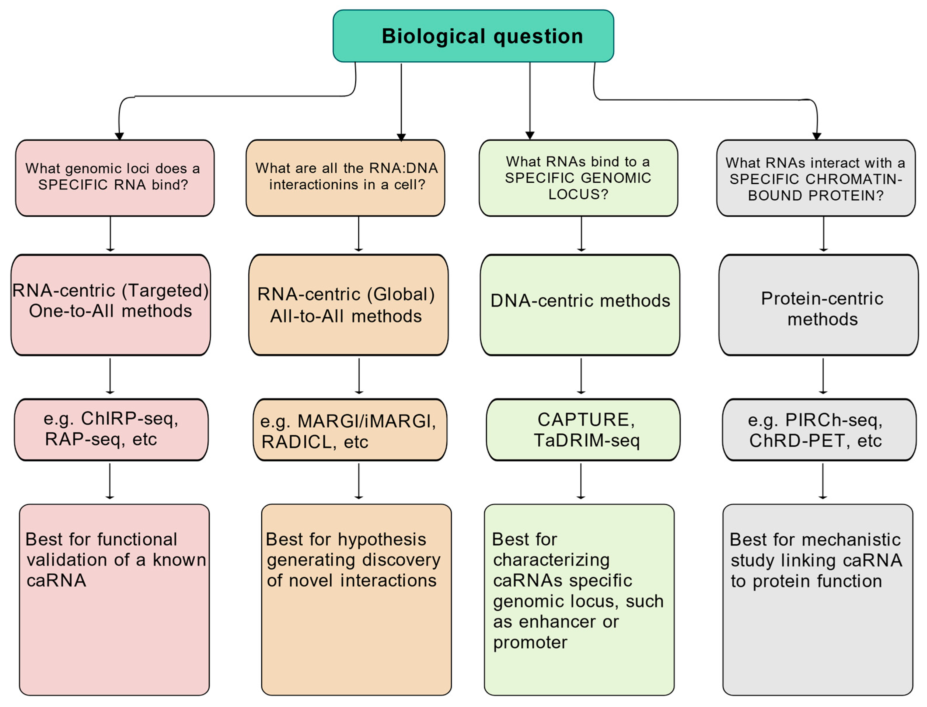
Since chromatin and RNA interactions often involve proteins, there are generally three ways to study RNA and chromatin association, i.e., RNA-centric, DNA-centric and chromatin-bound protein-centric (Table 1, Figure 2). For the RNA-centric methods, they can be further divided into one-to-all methods and all-to-all methods based on whether the method is focused on a specific RNA or aims to profile caRNAs genome-wide.

4.1. RNA-Centric Methods

4.1.1. One-to-All Methods

The one-to-all methods use capture-based approaches with biotin-labeled DNA or RNA probes designed to hybridize with a specific RNA of interest. After pull-down of RNA chromatin complexes and high-throughput sequencing of the associated DNA, the genomic loci interacting with the target RNA can be identified. This kind of one-to-all method can be used to study caRNAs regardless of their expression level [6]. However, because of the intrinsic background noise associated with hybridization-based assays, it is essential to include proper controls, such as RNA knockdown or knockout samples in parallel experiments.
Chromatin Isolated by RNA Purification (ChIRP)
ChIRP [97] is one of the earliest and most widely used methods for identifying genomic binding sites of specific RNAs. It uses complementary biotinylated DNA probes to capture target RNAs and their associated chromatin. This method enables high-resolution mapping of RNA occupancy on chromatin. One of its strengths is the ability to distinguish specific interactions by using independently designed probe sets (e.g., odd and even pools), as well as non-targeting control probes. However, ChIRP requires efficient crosslinking and can be sensitive to probe design and RNA abundance. It is more suitable for relatively stable and abundant nuclear RNAs. A limitation is potential nonspecific capture of DNA fragments due to strong crosslinking or hybridization artifacts. ChIRP has been extended to 3D chromatin conformation studies in the HiChIRP method, which combines RNA pull-down with proximity ligation [98].
Capture Hybridization Analysis of RNA Targets (CHART)
CHART [99] is another hybridization-based method used to map RNA-associated chromatin regions. Unlike ChIRP, CHART incorporates an RNase H-based step to empirically identify accessible regions on the target RNA for probe hybridization [99], which helps improve specificity and reduce background noise. CHART has been successfully applied to study lncRNAs across various species, including flies [99], lizard [100], mouse [101] and human [102,103]. A key advantage of CHART is its rational probe design based on in situ RNA structure, which minimizes off-target effects. However, CHART may be limited by the availability and efficiency of accessible probe-binding sites, and it generally performs best with well-characterized RNAs with known structures.
RNA Antisense Purification (RAP)
RAP [104] is a hybridization-based approach designed to identify genomic binding sites of specific RNAs. It uses long, overlapping probes tiled across the full length of the target RNA, enabling efficient capture even when the RNA is highly structured, partially degraded or bound by proteins. This strategy offers high specificity and comprehensive coverage, making RAP particularly suitable for long and structured RNAs. Compared to ChIRP and CHART, RAP utilizes longer probes, which improves hybridization efficiency but requires more extensive probe synthesis and optimization.

4.1.2. All-to-All Methods

Unlike one-to-all methods, which require prior knowledge of the RNA of interest, all-to-all methods are designed to identify novel caRNAs in an unbiased manner. These genome-wide approaches enable the detection of RNA-DNA interactions without targeting a specific RNA, and several techniques have been developed in recent years.
Mapping RNA–Genome Interactions (MARGI) and iMARGI
Sridhar et al. reported a method called MARGI (mapping RNA–genome interactions) [84] which can massively detect native RNA–chromatin interactions from unperturbed cells, enabling detection of genome-wide RNA-binding sites without prior knowledge. This method is based on connecting caRNAs with their target genomic sequences by proximity ligation. Ligation steps are performed on streptavidin beads. The resulting RNA-DNA chimeric sequences were then converted into a sequencing library for paired-end sequencing. Later the same group reported an updated version called iMARGI [105], which differs from the previous version MARGI in that the ligation steps are performed in situ instead of on streptavidin beads. This in situ ligation preserves spatial context and improves interaction detection fidelity. These methods are well suited for investigating the global architecture of RNA–chromatin interactions, although they may have limited resolution and require extensive sequencing depth due to high background.
Global RNA Interaction with DNA Sequencing (GRID-seq)
GRID-seq [106] uses a biotinylated bivalent linker to ligate RNA to DNA in situ in formaldehyde and disuccinimidyl glutarate-fixed nuclei. There are several differences in the GRID-seq protocol compared with other techniques: (1) In contrast to the bivalent linker used for MARGI, the linker in GRID-seq is single-stranded RNA with the 5′ end pre-adenylated instead of DNA. (2) The linkers that GRID-seq uses have two restriction sites for a type IIS restriction enzyme (MmeI), thus allowing subsequent digestion to generate size-specific products. Other techniques like ChAR-seq use sonication to achieve fragmentation of ligated products. The advantage of the restriction-based approach in the GRID-seq protocol is that all reads contain RNA and DNA sequences, while in other techniques like MARGI and ChAR-seq, this is not certain. The disadvantage is the decreased mapping power of short reads with GRID-seq [106,107].
Chromatin-Associated RNA Sequencing (ChAR-seq)
ChAR-seq [108] is also a proximity-ligation-based method that detects RNA–chromatin interactions. The fragmentation step is performed with sonication, not the restriction enzyme digestion used in GRID-seq [106]. ChAR-seq was first developed for Drosophila cells, then applied to human cells and Xenopus embryos. The disadvantage of ChAR-seq is that it requires more sequencing depths in mammalian cells since random ligation may be more common in mammalian cells than in Drosophila [107].
RNA and DNA Interacting Complexes Ligated and Sequenced (RADICL-seq)
RADICL-seq [109] is a proximity-ligation-based technique designed to reduce biases associated with nascent transcription and improve genomic coverage and unique mapping rates. By incorporating transcriptional inhibition with actinomycin D treatment and RNase H treatment prior to crosslinking, RADICL-seq distinguishes more stable RNA–chromatin interactions from transient ones and reduces the bias toward nascent transcription. This method is particularly valuable for studying how transcription shapes chromatin organization, although the additional steps to suppress nascent transcription may reduce the capture of dynamic interactions.
RNA Ends on DNA Capture (Red-C)
Red-C [110] identifies RNA–chromatin interactions by capturing the 3′ ends of RNA ligated to fragmented DNA in formaldehyde fixed cells. This method enables the global detection of both coding and non-coding RNA interactions with chromatin. Notably, Red-C revealed that non-coding RNAs associate with both active and repressed chromatin regions, suggesting their broad role in transcriptional regulation and genome organization. However, the reliance on RNA 3′ ends may limit detection of certain RNA species or interaction types.

4.2. DNA-Centric Methods

DNA-centric methods aim to identify RNAs associated with specific genomic loci. These methods are especially valuable when investigating low-abundance caRNAs that may not be efficiently detected by genome-wide (all-to-all) approaches.

4.2.1. CRISPR Affinity Purification in Situ of Regulatory Elements (CAPTURE) Approach

To better understand regulation at one genomic locus, one needs to obtain the whole picture of what kinds of RNAs and proteins are bound there. In the case of low-abundance caRNAs, all-to-all methods are less sensitive in detecting them; therefore, all-to-all methods are not suitable for obtaining the whole picture of RNAs bound at a specific genomic locus. To overcome this drawback, Liu X. et al. developed a novel approach called CRISPR affinity purification in situ of regulatory elements (CAPTURE) [111]. This method uses guide RNAs coupled with biotinylated nuclease-dead cas9 (dCas9) to detect locus-specific interactions. After streptavidin affinity purification, RNA-seq was used to detect caRNAs, 3C-seq was used to detect long-range DNA interactions and proteomics was used to detect the trans-regulatory factors. Applying this method to the β-globin LCR established evidence for a composition-based hierarchical organization. Spatial features that causally control gene transcription were identified by using this method for unbiased analysis of chromatin interactions at disease-associated cis-elements and developmentally regulated super-enhancers. This method is particularly useful for studying low-abundance RNAs at specific loci, which are often missed by global methods. However, it requires careful gRNA design and may be less suited for genome-wide screening.

4.2.2. Targeted DNA-Associated RNA and RNA-RNA Interaction Mapping by Sequencing (TaDRIM-seq)

TaDRIM-seq [112] is a novel technique that combines Protein G (PG)-Tn5-targeted DNA element technology with in situ proximity ligation to capture nearby RNA molecules, thus allowing researchers to simultaneously map both DNA-RNA interactions and RNA-RNA interactions within cells, providing a comprehensive view of the RNA interactome at specific DNA loci. This approach improves upon traditional methods for mapping RNAs associated with specific DNA loci, like ChRD-PET and RedChIP (see below), which are time-consuming, need significant resources and struggle with in vitro ligation efficiency and RNA-RNA interaction detection. TaDRIM-seq is efficient, high-resolution and powerful, while also reducing the need for large cell numbers and time. It can be used in both animal and plant systems.

4.3. Protein-Centric Methods

Protein-centric approaches focus on identifying caRNAs that interact with specific chromatin-binding proteins or histone modifications. These methods typically involve immunoprecipitation and allow targeted detection of RNA:DNA interactions mediated or stabilized by protein complexes.

4.3.1. Chromatin-Associated RNA Immunoprecipitation Followed by Next-Generation Sequencing (CARIP-seq)

CARIP-seq [113,114] is a method used to identify and analyze RNAs associated with specific chromatin marks. It modifies ChIP and RIP protocols, basically performing crosslinking with high-concentration (1%) formaldehyde, similar to ChIP, but with a 1 min treatment to avoid over-crosslinking. A reduced number of sonication cycles is used to preserve RNA–protein interactions at condensed heterochromatin regions. Then an antibody targeted to a specific protein of interest is used for immunoprecipitation to pull down RNA and DNA associated with this protein, followed by sequencing. This method is especially useful for exploring RNA associations with repressive chromatin states.

4.3.2. Profiling Interacting RNAs on Chromatin Followed by Deep Sequencing (PIRCh-seq)

PIRCh-seq [115] enables the identification of caRNAs in a histone-modification-specific manner. This method cannot provide information about the exact binding sites of the chromatin-associated lncRNA; thus, it cannot tell whether each lncRNA functions in cis or in trans. PIRCh-seq can better capture RNA–chromatin association by using 1% glutaraldehyde, which forms stronger crosslinks between chromatin and associated RNAs. Compared to other methods, PIRCh-seq reduces contamination from nascent transcripts due to stronger glutaraldehyde crosslinking. However, similar to RIP-seq and CLIP-seq, it may still pull down abundant mRNAs that nonspecifically associate with chromatin or immunoprecipitated complexes. Therefore, to find the bona fide caRNAs, it is necessary to perform further experiments and analysis.

4.3.3. Chromatin-Associated RNA–DNA Interactions, Followed by Paired-End-Tag Sequencing (ChRD-PET)

ChRD-PET (chromatin-associated RNA–DNA interactions followed by paired-end-tag sequencing) [116] combines chromatin immunoprecipitation (ChIP), RNA-DNA proximity ligation and paired-end-tag sequencing (PET) to identify and characterize the specific locations where RNA and DNA interact in the nucleus. Formaldehyde is used to fix the RNA-DNA interactions. The specific RNA-DNA interactions are enriched by immunoprecipitation with an antibody for a target protein or histone modification associated with the RNA or DNA, for example, H3K4me3. Then the DNA fragment and RNA with proximity are ligated and subject to paired-end-tag sequencing to map their interaction sites. ChRD-PET provides paired information and allows identification of interaction loci between RNAs and chromatin with spatial context, thus offering insights into chromatin looping and 3D organization involving RNA.

4.3.4. RedChIP

RedChIP sequencing [117] is a technique used to identify non-coding RNAs associated with specific genomic locations. It combines the principles of ChIP-Seq (chromatin immunoprecipitation sequencing) with RNA-DNA chimera formation to identify and sequence RNA-DNA complexes. Basically, cells are fixed with formaldehyde and DNA fragmentation is achieved by NlaIII digestion. A biotinylated bridge adapter is ligated to RNA 3′ ends, and then the opposite end of the bridges is ligated to the DNA ends in spatial proximity. The ligated complexes are sonicated and immunoprecipitated with an antibody for the target protein. The pull-down complex is subject to reverse transcription, and the cDNA and DNA ends are then used for library preparation and sequencing.

4.3.5. RT & Tag

RT & Tag [118] uses an antibody to capture RNA associated with the chromatin epitope, and then localized reverse transcription generates RNA/cDNA hybrids that are subsequently tagmented by Tn5 transposases. This method enables the mapping of RNA–chromatin interactions, RNA–protein interactions and RNA modifications.

4.3.6. RNA and DNA Interacting Complexes Ligated and Sequenced (RADICL-seq) with Immunoprecipitation (RADIP)

RADIP [119] extends RADICL-seq by combining it with immunoprecipitation to specifically enrich RNA-DNA complexes associated with target proteins; thus, this technique can characterize protein-mediated RNA–chromatin interactions, enhancing its resolution and specificity.

4.3.7. Chromatin Sequencing (Chrom-seq)

Chrom-seq [120] uses a crosslinking-free and antibody-free approach to identify RNA associated with chromatin marks in living cells. This technique combines specific chromatin mark readers with APEX2 enzyme, which catalyzes the oxidation of biotin-aniline to label and isolate the adjacent RNAs by streptavidin-coated beads, allowing the systemic mapping of caRNAs that play regulatory roles in epigenetic events.
Table 1. Methods to study chromatin and RNA interactions.
TypeMethodPrincipleStrengthsLimitationsSuitable forReference
RNA-centric One to allChIRPHybridization+ pull-downHigh specificity with probe design;
compatible with sequencing
Requires strong crosslinking; may miss transient interactionsGeneral Individual RNA-
chromatin mapping
[97]
CHARTHybridization (guided by RNase H)Rational probe design reduces
background; cross-species applicability
Requires known RNA
accessibility; less sensitive for poorly characterized RNAs
Structured RNAs with known access sites[99]
RAPLong, tiled probesHigh specificity and coverage;
tolerant to RNA structure
Requires complex probe synthesislong, structured, or
degraded RNAs
[104]
All to allMARGI/
iMARGI
Proximity ligationGenome wide and unbiased;
no need for RNA specific probes
High background;
needs deep sequencing
Global profiling of RNA-
chromatin interactions
[84,105]
GRID-seqProximity ligation with linkerAll reads contain RNA and DNA;
high interaction confidence
short reads may limit resolutionHigh confidence global
interaction mapping
[106]
ChAR-seqproximity ligation with
sonication
Compatible with complex
genomes; longer chimeric
fragments
Higher background in
mammalian cells
Comparative studies
across species; flexible
ligation fragment sizes
[108]
RADICL-seqProximity ligation with
transcription inhibition
Reduces nascent transcription
bias; high mapping efficiency
May miss transient or
dynamic interactions
investigating stable RNA-chromatin contacts;
transcription-dependent chromatin regulation
[109]
Red-CLigation of RNA 3′ends to
fragmented DNA
Capture diverse coding and non-coding RNA interactionsBiased toward RNA 3′end;
limited resolution
Mapping broad RNA-
chromatin interactions across chromatin states
[110]
DNA-centric CAPTURELocus specific targeting
with dCas9-biotin complex
High sensitivity for specific loci;
identifies RNA, DNA, and protein
interactions
Requires gRNA design;
not genome-wide
Detailed analysis of
chromatin regulation at
specific loci
[111]
TaDRIM-seqPG-Tn5 tethering+
in situ proximity ligation
Simultaneous detection of
DNA-RNA and RNA-RNA
interactions; efficient; low input
Requires targeting design;
not genome-wide
Mapping complex RNA
networks at specific DNA loci in animal or plant
systems
[112]
Chromatin-bound
protein-centric
CARIP-seqChIP-RIP hybrid: Crosslink
+IP of chromatin proteins
Captures RNAs bound
to specific chromatin proteins;
good for heterochromatin
Limited to protein-specific
interactions; resolution depends on antibody quality
Profiling RNAs associated
with repressive chromatin marks
[113,114]
PIRCh-seqCrosslinking+IP+
caRNA profiling
Histone modification specific
caRNA identification; reduced nascent RNA contamination
Cannot identify binding
sites; may still co-purify mRNAs
Global profiling of caRNAs[115]
ChRD-PETChIP+promximity ligation
+PET sequencing
Maps specific RNA-DNA
interactions with spatial context; reveals RNA-mediated chromatin looping
Technically complex;
resolution depends on proximity
ligation efficiency
Studying RNA’s role in 3D
genome organization
[116]
RedChIPChIP+RNA-DNA chimera
formation+sequencing
Identifies ncRNAs associated with
protein defined chromatin regions
Resolution limited by proximity
ligation and ChIP antibody quality
Studying ncRNAs at specific chromatin environments[117]
RT&TagAntibody targeting+
in situ RT+Tn5 tagmentation
High resolution, efficient, detects RNA-chromatin/protein/modificationDepends on antibody specificity and reverse transcription efficiencyMapping RNA-chromatin/
protein/modification in situ
[118]
RADIPCrosslinking+RNA-DNA
ligation+IP of target protein
High specificity for protein
mediated RNA-DNA interactions
Requires good antibody, may miss transient or weak interactionsStudying protein specific RNA-chromatin interactions[119]
Chrom-seqChromatin mark reader+
APEX2 labeling without
crosslinking or antibodies
Live cell, label free mapping of RNAs near specific chromatin marksRequires efficient expression of fusion proteins; limited by APEX2 labeling
radius
In vivo detection of caRNAs at specific epigenetic states[120]
Note: Blue color shows the RNA-centric methods, red color shows the DNA-centric methods, and green color shows the chromatin-bound-protein-centric methods.

5. Non-Canonical Structures in caRNAs

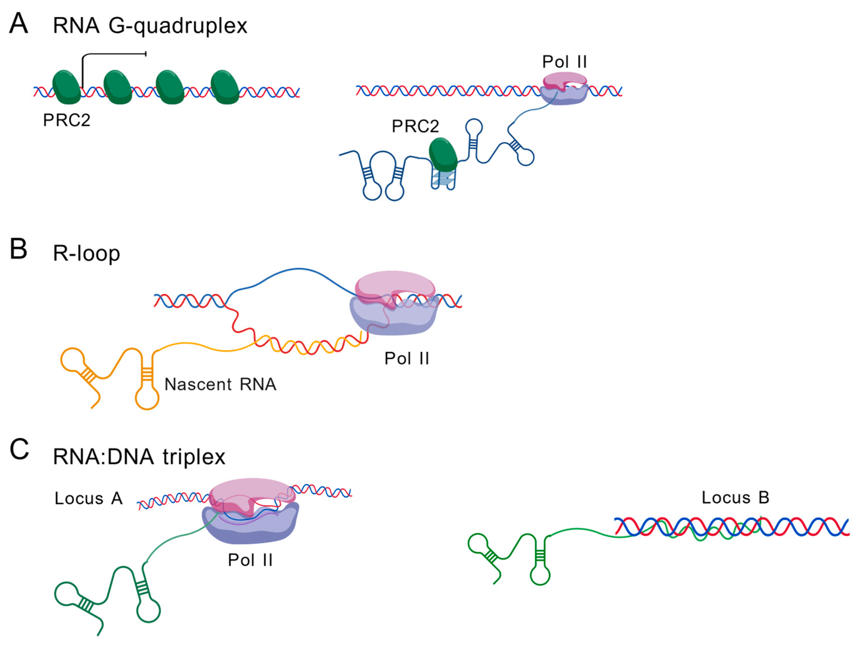
Beyond canonical RNA functions, caRNAs utilize non-canonical structural conformations to mediate precise genomic targeting and epigenetic regulation. These specialized architectures—including G-quadruplexes (G4s), R-loops and RNA:DNA triplexes—enable caRNAs to act as dynamic scaffolds, recruiting effector complexes to specific genomic loci.

5.1. RNA G-Quadruplexes (G4s)

RNA G-quadruplexes (G4s) represent a key class of non-canonical RNA structures that plays multifaceted roles in chromatin organization and gene regulation. These stable four-stranded structures form through Hoogsteen base-pairing of guanine-rich (G-tracts) sequences in RNA [121,122]. RNA G4s in caRNAs serve as critical structural and functional elements that regulate chromatin organization, gene expression and RNA processing. At telomeres, the long non-coding RNA TERRA forms G4 structures that act as scaffolds for protein recruitment, facilitating the binding of telomere-protective factors (e.g., TRF2, FUS, EWS) and histone methyltransferases to maintain heterochromatin stability [123,124,125]. Beyond telomeres, RNA G4s influence alternative splicing by recruiting splicing regulators (e.g., hnRNPH/F) to pre-mRNAs, ensuring proper intron removal in transcripts such as p53, PAX9 and BACE1 [126,127,128,129,130]. Additionally, RNA G4 structures function as recognition platforms for chromatin-modifying complexes, particularly PRC2, which exhibits preferential binding to G4-containing RNAs [131]. This PRC2-G4 RNA interaction sequesters PRC2 away from transcriptionally active genes, effectively depleting it from activated loci [132] (Figure 3A). RNA G4s also regulate microRNA biogenesis by inhibiting Dicer processing when G4 structures compete with canonical stem-loops [133,134]. Furthermore, G4s in retrotransposon-derived RNAs may promote transposition, linking them to genome evolution [135]. Collectively, RNA G4s emerge as versatile regulators that integrate RNA structure with chromatin dynamics, impacting transcription, splicing and epigenetic silencing.

5.2. R-Loop

R-loops, formed by nascent RNA:DNA hybrids and displaced single-stranded DNA, serve as key mediators of caRNA–chromatin interactions (Figure 3B) [3,6,7]. These structures preferentially arise in GC-skewed regions, repetitive sequences (e.g., telomeres) and highly transcribed loci, where they modulate transcription pausing, termination and antisense RNA synthesis. Notably, R-loops at gene promoters often induce transcriptional pausing, whereas those at the 3′ end can disrupt termination, with transcripts harboring R-loops generally exhibiting higher expression levels [3,7].
Beyond transcriptional regulation, R-loops contribute to chromatin remodeling and epigenetic reprogramming. For example, lncRNAs such as TARID form R-loops at promoter regions (e.g., TCF21), recruiting DNA demethylases (TET1 via GADD45A) to activate gene expression [136]. Similarly, TERRA lncRNA invades telomeric DNA via R-loop formation, mediated by RAD51, to maintain telomere structure—though excessive accumulation leads to fragility [69]. R-loops also facilitate long-range chromatin interactions, as enhancer-derived RNAs (eRNAs) form trans-acting R-loops or triplex structures to bridge enhancer–promoter contacts [137].
Additionally, R-loops collaborate with chromatin remodelers (e.g., BRG1 of the SWI/SNF complex) to resolve transcription–replication conflicts [138], highlighting their role in maintaining genomic stability. The interplay between R-loops and G-quadruplexes (G4s) further amplifies their regulatory impact, as G4 structures on displaced DNA strands stabilize R-loops, linking RNA secondary structure to transcriptional outcomes [139]. Taken together, R-loops serve as dynamic platforms through which caRNAs coordinate gene expression, chromatin accessibility and higher-order genome organization, balancing functional roles with the need to prevent DNA damage.

5.3. RNA:DNA Triplex

Although the triplex structure is also an RNA:DNA hybrid structure, triplexes are distinct from R-loops. In the triplex formation process, an RNA molecule establishes sequence-specific interactions by forming Hoogsteen bonds with purine bases in the DNA major groove (Figure 3C) [3,6]. Many caRNAs use this triplex mechanism to act in trans to regulate distal genes or genes on other chromosomes. For example, Khps1 targets the promoter of SPHK1 through a triplex structure [17]. Meg3 forms an RNA:DNA triplex with thousands of target loci across the genome [140]. However, due to lack of techniques to map RNA:DNA triplexes genome-wide, further studies are needed to uncover whether additional RNAs use this mechanism [3].

6. caRNA-Interacting Proteins

Studies suggest that caRNAs may facilitate long-range chromatin architecture by interacting with proteins, DNA or other RNAs [3,6,7,21]. RNA is normally present in the cell in the form of ribonucleoprotein (RNP) complexes, and its associated proteins are crucial for both cis- and trans-regulatory functions. In this section of the review, we primarily focus on the mechanisms of caRNA–protein–chromatin interactions, and therefore do not elaborate in depth on direct RNA–RNA or RNA–DNA interaction modes. Many RNA-binding proteins and non-canonical RBPs have been shown to interact with caRNAs, supporting their localization, stability and function, particularly in trans. In this section, we focus on three key chromatin-associated proteins—CTCF, cohesin and PRC2—and discuss their roles in working with RNA to regulate chromatin architecture. Multiple lines of evidence demonstrate that both CTCF and PRC2 can bind RNA. For CTCF, the lncRNA Jpx interacts with CTCF to displace it from the Xist promoter during the onset of X chromosome inactivation [22]. The zinc finger domain of CTCF mediates both its self-clustering and RNA recruitment [141,142,143]. Mutating this RNA-binding domain disrupts around 50% of chromatin loops in mESCs and causes gene dysregulation, demonstrating the functional significance of CTCF-RNA interactions [141,142]. For PRC2, while PRC2 exhibits promiscuous RNA-binding capacity [144,145,146], it shows selectivity for specific RNAs such as lncRNA RepA [145] and high affinity for G-quadruplex-containing RNAs [147,148]. However, whether CTCF/PRC2 interact with RNA is under debate. Guo et al. [149] used a denaturing method to purify in vivo crosslinked RNA–protein complexes and concluded that CTCF/PRC2 binding to RNA was an experimental artifact. However, this conclusion may arise from data misanalysis, as the pipeline Guo et al. used inflated background reads and suppressed mappable signals [150].

6.1. CTCF

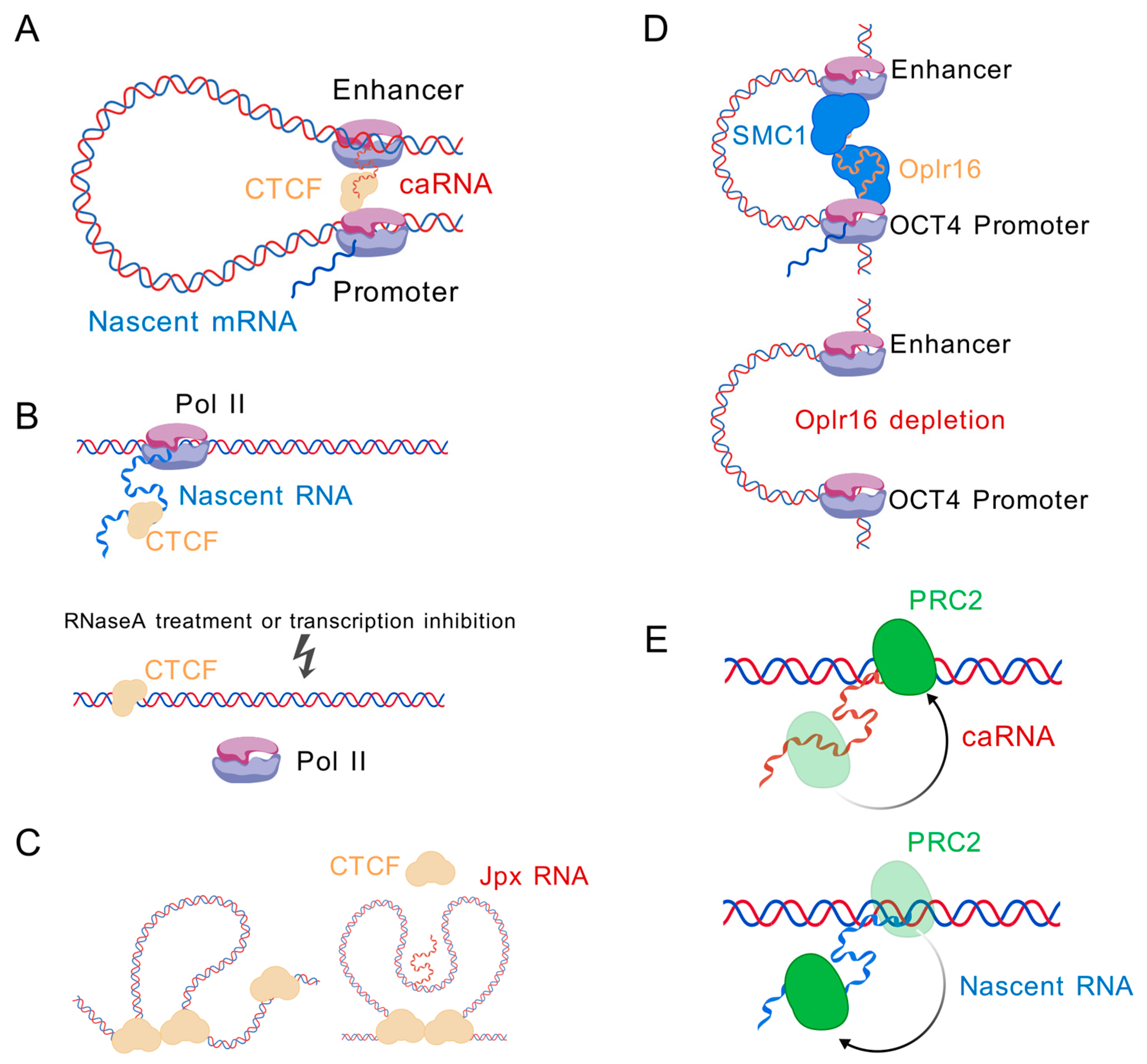
CTCF is the important architectural protein localized at the anchors of chromatin loops and TAD boundaries [16]. Recent studies have shown that some ncRNAs can facilitate CTCF binding to chromatin to regulate chromatin looping and transcription (Figure 4A) [23,151,152,153,154]. lncRNA MYCNOS promotes CTCF recruitment to the MYCN promoter, driving chromatin remodeling that upregulates MYCN expression and modulates downstream transcriptional programs [153]. lncRNA HOTTIP mediated recruitment of CTCF/cohesin, and R-loop regulators initiated boundary-associated R-loop formation, which in turn stabilized chromatin loops and promoted TAD organization [23]. Boundary-associated RNAs promote the recruitment and spatial clustering of CTCF proteins at TAD borders. This CTCF enrichment subsequently strengthens TAD insulation, modulates enhancer–promoter communication within TADs and fine-tunes domain-wide gene expression patterns [154]. The lncRNA CCAT-L, located upstream of Myc, regulates its expression by facilitating enhancer–promoter looping through two distinct mechanisms. First, it recruits CTCF to modulate the chromatin architecture between the Myc promoter and enhancer [155]. Second, CCAT-L interacts with HNRNPK—often in conjunction with promoter-derived uaRNAs—to promote chromatin looping via HNRNPK oligomerization [21]. An RNA-binding domain mutation of CTCF was reported to disrupt about half of all chromatin loops [141,142,156]. The disrupted loops can be subdivided into two classes depending on whether the CTCF and cohesin binding were lost. The most disrupted loops are mainly enriched in the B compartment, with lost binding of CTCF and cohesin, while the unaffected loops are generally associated with active genes in the A compartment without changes in CTCF and cohesin occupancy [141]. These findings indicate that the interaction between CTCF and RNA is important for chromatin looping.
In contrast to RNAs that recruit CTCF and facilitate its binding to chromatin, global depletion of nascent RNA by transcription inhibition or RNase treatment was reported to increase CTCF binding to chromatin, suggesting that nascent RNA can antagonize CTCF–chromatin interactions (Figure 4B) [157,158]. However, the transcription inhibition and RNA degradation results could not distinguish whether the effect was from loss of pre-mRNA or other types of RNA. Jpx is a non-coding RNA which was initially found to activate Xist expression by evicting CTCF from the Xist promoter [22]. Jpx functions as a CTCF release factor and selectively evicts CTCF bound to low-affinity, developmentally sensitive sites genome-wide (Figure 4C) [159]. Our previous study identified a novel eRNA, CpoxeRNA [48], which binds to CTCF/cohesin and antagonize cohesin binding to chromatin. Knockdown of the CpoxeRNA leads to accumulation of Rad21 at CTCF-binding sites (CBSs) located at its promoter and interaction targets. However, the chromatin loops anchored by CTCF between the CpoxeRNA genomic locus and target loci appear to rely on the eRNA itself, as chromatin looping decreased after CpoxeRNA knockdown, although CTCF binding increased.

6.2. Cohesin

lncRNAs play roles in facilitating chromatin loops formed by the cohesin complex. For example, Oplr16 (Oct4 promoter-interacting long non-coding RNA 16)—a pluripotency-associated lncRNA—binds specifically to the Oct4 promoter. Depletion of Oplr16 disrupts the chromatin loop between the Oct4 promoter and enhancer, indicating its essential role in maintaining this interaction (Figure 4D). Mechanistically, Oplr16 interacts with cohesin subunit SMC1 via at least four binding sites in its 3′ fragment, thereby recruiting SMC1 to stabilize the Oct4 enhancer–promoter loop and sustain pluripotency [160].

6.3. PRC2

PRC2 (polycomb repressive complex 2) is a key chromatin-modifying complex that mediates transcriptional repression by trimethylating histone H3 on lysine 27 (H3K27me3). RNA has been reported to interact with PRC2 and modulate its function, such as regulating its chromatin recruitment, enzymatic activity and target specificity (Figure 4E). One well-known example is the lncRNA Xist, which directly binds to PRC2 and facilitates its recruitment to the inactive X chromosome during X chromosome inactivation [144,145,146,161]. Another example is HOTAIR, an lncRNA transcribed from the HOXC locus, which serves as a scaffold to recruit PRC2 to the targets loci and promote transcriptional silencing [162,163,164]. In addition, nascent RNAs transcribed from active genes have been shown to inhibit PRC2 binding to chromatin, suggesting that RNA binding may prevent PRC2 from accessing active chromatin regions [144,145].
However, recent transcriptome-wide analyses, such as iCLiP, have shown that PRC2 predominantly interacts with nascent, unspliced transcripts from actively transcribed genes [144]. These interactions are largely co-transcriptional and widespread, suggesting a more general role for nascent RNA in modulating PRC2 activity. Nascent RNAs compete with chromatin to bind to PRC2, thus preventing active genes from inappropriate silencing by PRC2. This nascent RNA-PRC2 interaction has an indirect role in regulating neighboring genes and eRNAs, as a consequence of H3K27me3 accumulation at the nascent RNA gene locus upon loss of nascent RNA [12].

7. Perspective

Over the past decade, our understanding of caRNAs has expanded from isolated examples of nuclear-retained transcripts to a rich and complex network of RNA species—including nascent mRNAs, diverse classes of non-coding RNAs, regulatory-element-derived transcripts, repeat element RNAs and small nuclear RNAs—that collectively shape genome organization and function. caRNAs play dynamic roles across the cell cycle, with select RNAs like Xist and satellite transcripts persistently remaining bound to mitotic/meiotic chromosomes [93,94,95]. These RNAs maintain cellular memory and ensure chromosome stability and proper dispersion, yet key questions persist about their retention mechanisms during chromatin condensation and their role in epigenetic inheritance. The ability of caRNAs to form non-canonical structures such as G-quadruplexes, R-loops and RNA:DNA triplexes, as well as to engage architectural proteins (e.g., CTCF, cohesin) and chromatin-modifying complexes (e.g., PRC2), underscores their central role in shaping genome architecture and gene regulation. Beyond classical RNA-binding proteins (RBPs) containing canonical RNA-binding domains, accumulating evidence reveals that many DNA/chromatin-associated proteins (e.g., transcription factors CTCF, histone modifiers PRC2) can also bind RNAs [6]. The role of these non-canonical RBPs needs to be further studied to fully understand the mechanims of caRNAs.
Recent technological advances—spanning RNA-centric (e.g., ChIRP [97], RAP [104], RADICL-seq [109]), DNA-centric (e.g., CAPTURE [111], TaDRIM-seq [112]) and protein-centric (e.g., RedChIP [117], RT & Tag [118]) approaches—have enabled increasingly fine-grained maps of RNA–chromatin interactions. However, these methods often operate in isolation, limiting our ability to fully resolve the tripartite RNA–DNA–protein interactome. The integration of these platforms, particularly with single-molecule and single-cell resolution, while capturing structural, positional and temporal dynamics, will be crucial to dissecting functional relationships from mere spatial proximity.
Several pressing challenges define the next phase of caRNA research. First, there is an urgent need for standardized annotation frameworks to distinguish functional caRNAs from pervasive transcriptional noise, especially for repetitive-element-derived species that may be overlooked or misclassified. Second, decoding the structural plasticity of caRNAs—particularly the dynamics of G4s, triplexes and R-loops—in native nuclear contexts may uncover a new regulatory layer in chromatin biology. Third, moving beyond static maps to dynamic, temporal analyses will be essential for understanding how caRNA-mediated interactions respond to developmental cues, stress signals and pathological states.
Looking forward, we envision the convergence of single-cell multi-omics, live-cell imaging and structure-resolved interactome mapping to illuminate the causal links between caRNA localization, structure and transcriptional outcomes. Coupled with computational models that integrate evolutionary conservation, structural motifs and interaction profiles, these approaches could enable the prediction—and even therapeutic targeting—of functional caRNA elements. Such advances will transform caRNAs from enigmatic nuclear byproducts into actionable regulators of chromatin state, with broad implications for developmental biology, genome stability and disease intervention.

Author Contributions

Conceptualization, B.X. and A.D.; writing—original draft preparation, B.X.; writing—review and editing, A.D. All authors have read and agreed to the published version of the manuscript.

Funding

This work was funded by the Intramural Program of National Institute of the Diabetes and Digestive and Kidney Diseases, NIH (DK 075033 to A.D.).

Conflicts of Interest

The authors declare no conflicts of interest.

Abbreviations

The following abbreviations are used in this manuscript:
caRNAsChromatin-associated RNAs
lncRNAsLong non-coding RNAs
eRNAsEnhancer RNAs
paRNAsPromoter-associated RNAs
circRNAsCircular RNAs
snRNAsSmall nuclear RNAs
snoRNAsSmall nucleolar RNAs
asRNAsAntisense RNAs
HERVHHuman endogenous retrovirus subfamily H
ChIRPChromatin isolated by RNA purification
CHARTCapture hybridization analysis of RNA targets
RAPRNA antisense purification
CARIP-seqChromatin-associated RNA immunoprecipitation followed by next-generation sequencing
PIRCh-seqProfiling interacting RNAs on chromatin followed by deep sequencing
MARGIMapping RNA–genome interactions
GRID-seqGlobal RNA interaction with DNA sequencing
ChAR-seqChromatin-associated RNA sequencing
RADICL-seqRNA and DNA interacting complexes ligated and sequenced
PAS RNAsPromoter antisense RNAs
Red-CRNA ends on DNA capture
CAPTURECRISPR affinity purification in situ of regulatory elements
TaDRIM-seqTargeted DNA-associated RNA and RNA-RNA interaction mapping by sequencing
ChRD-PETChromatin-associated RNA–DNA interactions, followed by paired-end-tag sequencing
RADIPRNA and DNA interacting complexes ligated and sequenced (RADICL-seq) with immunoprecipitation
Chrom-seqChromatin sequencing
mCARsMitotic chromosome-associated RNAs

References

  1. Palihati, M.; Saitoh, N. RNA in chromatin organization and nuclear architecture. Curr. Opin. Genet. Dev. 2024, 86, 102176. [Google Scholar] [CrossRef]
  2. Consortium, E.P. An integrated encyclopedia of DNA elements in the human genome. Nature 2012, 489, 57–74. [Google Scholar] [CrossRef]
  3. Yin, Y.; Shen, X. Noncoding RNA-chromatin association: Functions and mechanisms. Fundam. Res. 2023, 3, 665–675. [Google Scholar] [CrossRef]
  4. Zhao, L.; Wang, J.; Li, Y.; Song, T.; Wu, Y.; Fang, S.; Bu, D.; Li, H.; Sun, L.; Pei, D.; et al. NONCODEV6: An updated database dedicated to long non-coding RNA annotation in both animals and plants. Nucleic Acids Res. 2021, 49, D165–D171. [Google Scholar] [CrossRef] [PubMed]
  5. Razin, S.V.; Gavrilov, A.A. Non-coding RNAs in chromatin folding and nuclear organization. Cell Mol. Life Sci. 2021, 78, 5489–5504. [Google Scholar] [CrossRef]
  6. Li, X.; Fu, X.D. Chromatin-associated RNAs as facilitators of functional genomic interactions. Nat. Rev. Genet. 2019, 20, 503–519. [Google Scholar] [CrossRef] [PubMed]
  7. Tang, J.; Wang, X.; Xiao, D.; Liu, S.; Tao, Y. The chromatin-associated RNAs in gene regulation and cancer. Mol. Cancer 2023, 22, 27. [Google Scholar] [CrossRef] [PubMed]
  8. Sampath, K.; Ephrussi, A. CncRNAs: RNAs with both coding and non-coding roles in development. Development 2016, 143, 1234–1241. [Google Scholar] [CrossRef]
  9. Dinger, M.E.; Pang, K.C.; Mercer, T.R.; Mattick, J.S. Differentiating protein-coding and noncoding RNA: Challenges and ambiguities. PLoS Comput. Biol. 2008, 4, e1000176. [Google Scholar] [CrossRef]
  10. Ulveling, D.; Francastel, C.; Hube, F. When one is better than two: RNA with dual functions. Biochimie 2011, 93, 633–644. [Google Scholar] [CrossRef]
  11. Caudron-Herger, M.; Müller-Ott, K.; Mallm, J.-P.; Marth, C.; Schmidt, U.; Fejes-Tóth, K.; Rippe, K. Coding RNAs with a non-coding function: Maintenance of open chromatin structure. Nucleus 2014, 2, 410–424. [Google Scholar] [CrossRef]
  12. Xie, B.; Dean, A. Noncoding function of super enhancer derived Cpox pre-mRNA in modulating neighbouring gene expression and chromatin interactions. RNA Biol. 2025, 22, 1–17. [Google Scholar] [CrossRef] [PubMed]
  13. Maldonado, R.; Langst, G. The chromatin-triple helix connection. Biol. Chem. 2023, 404, 1037–1049. [Google Scholar] [CrossRef]
  14. Jiang, S.; Li, H.; Zhang, L.; Mu, W.; Zhang, Y.; Chen, T.; Wu, J.; Tang, H.; Zheng, S.; Liu, Y.; et al. Generic Diagramming Platform (GDP): A comprehensive database of high-quality biomedical graphics. Nucleic Acids Res. 2025, 53, D1670–D1676. [Google Scholar] [CrossRef]
  15. Mishra, K.; Kanduri, C. Understanding Long Noncoding RNA and Chromatin Interactions: What We Know So Far. Noncoding RNA 2019, 5, 54. [Google Scholar] [CrossRef]
  16. Thakur, J.; Henikoff, S. Architectural RNA in chromatin organization. Biochem. Soc. Trans. 2020, 48, 1967–1978. [Google Scholar] [CrossRef]
  17. Postepska-Igielska, A.; Giwojna, A.; Gasri-Plotnitsky, L.; Schmitt, N.; Dold, A.; Ginsberg, D.; Grummt, I. LncRNA Khps1 Regulates Expression of the Proto-oncogene SPHK1 via Triplex-Mediated Changes in Chromatin Structure. Mol. Cell 2015, 60, 626–636. [Google Scholar] [CrossRef]
  18. Deng, C.; Li, Y.; Zhou, L.; Cho, J.; Patel, B.; Terada, N.; Li, Y.; Bungert, J.; Qiu, Y.; Huang, S. HoxBlinc RNA Recruits Set1/MLL Complexes to Activate Hox Gene Expression Patterns and Mesoderm Lineage Development. Cell Rep. 2016, 14, 103–114. [Google Scholar] [CrossRef] [PubMed]
  19. Wang, K.C.; Yang, Y.W.; Liu, B.; Sanyal, A.; Corces-Zimmerman, R.; Chen, Y.; Lajoie, B.R.; Protacio, A.; Flynn, R.A.; Gupta, R.A.; et al. A long noncoding RNA maintains active chromatin to coordinate homeotic gene expression. Nature 2011, 472, 120–124. [Google Scholar] [CrossRef]
  20. Mondal, T.; Rasmussen, M.; Pandey, G.K.; Isaksson, A.; Kanduri, C. Characterization of the RNA content of chromatin. Genome Res. 2010, 20, 899–907. [Google Scholar] [CrossRef] [PubMed]
  21. Cai, Z.; Cao, C.; Ji, L.; Ye, R.; Wang, D.; Xia, C.; Wang, S.; Du, Z.; Hu, N.; Yu, X.; et al. RIC-seq for global in situ profiling of RNA-RNA spatial interactions. Nature 2020, 582, 432–437. [Google Scholar] [CrossRef] [PubMed]
  22. Sun, S.; Del Rosario, B.C.; Szanto, A.; Ogawa, Y.; Jeon, Y.; Lee, J.T. Jpx RNA activates Xist by evicting CTCF. Cell 2013, 153, 1537–1551. [Google Scholar] [CrossRef]
  23. Luo, H.; Zhu, G.; Eshelman, M.A.; Fung, T.K.; Lai, Q.; Wang, F.; Zeisig, B.B.; Lesperance, J.; Ma, X.; Chen, S.; et al. HOTTIP-dependent R-loop formation regulates CTCF boundary activity and TAD integrity in leukemia. Mol. Cell 2022, 82, 833–851.e11. [Google Scholar] [CrossRef]
  24. Deforzh, E.; Kharel, P.; Zhang, Y.; Karelin, A.; El Khayari, A.; Ivanov, P.; Krichevsky, A.M. HOXDeRNA activates a cancerous transcription program and super enhancers via genome-wide binding. Mol. Cell 2024, 84, 3950–3966.e6. [Google Scholar] [CrossRef]
  25. Statello, L.; Guo, C.J.; Chen, L.L.; Huarte, M. Gene regulation by long non-coding RNAs and its biological functions. Nat. Rev. Mol. Cell Biol. 2021, 22, 96–118. [Google Scholar] [CrossRef]
  26. Guiducci, G.; Stojic, L. Long Noncoding RNAs at the Crossroads of Cell Cycle and Genome Integrity. Trends Genet. 2021, 37, 528–546. [Google Scholar] [CrossRef]
  27. Jegu, T.; Aeby, E.; Lee, J.T. The X chromosome in space. Nat. Rev. Genet. 2017, 18, 377–389. [Google Scholar] [CrossRef]
  28. Hasegawa, Y.; Brockdorff, N.; Kawano, S.; Tsutui, K.; Tsutui, K.; Nakagawa, S. The matrix protein hnRNP U is required for chromosomal localization of Xist RNA. Dev. Cell 2010, 19, 469–476. [Google Scholar] [CrossRef] [PubMed]
  29. Pullirsch, D.; Hartel, R.; Kishimoto, H.; Leeb, M.; Steiner, G.; Wutz, A. The Trithorax group protein Ash2l and Saf-A are recruited to the inactive X chromosome at the onset of stable X inactivation. Development 2010, 137, 935–943. [Google Scholar] [CrossRef]
  30. Henninger, J.E.; Young, R.A. An RNA-centric view of transcription and genome organization. Mol. Cell 2024, 84, 3627–3643. [Google Scholar] [CrossRef] [PubMed]
  31. Andersson, R.; Gebhard, C.; Miguel-Escalada, I.; Hoof, I.; Bornholdt, J.; Boyd, M.; Chen, Y.; Zhao, X.; Schmidl, C.; Suzuki, T.; et al. An atlas of active enhancers across human cell types and tissues. Nature 2014, 507, 455–461. [Google Scholar] [CrossRef]
  32. Lorenzi, L.; Chiu, H.S.; Avila Cobos, F.; Gross, S.; Volders, P.J.; Cannoodt, R.; Nuytens, J.; Vanderheyden, K.; Anckaert, J.; Lefever, S.; et al. The RNA Atlas expands the catalog of human non-coding RNAs. Nat. Biotechnol. 2021, 39, 1453–1465. [Google Scholar] [CrossRef]
  33. Sartorelli, V.; Lauberth, S.M. Enhancer RNAs are an important regulatory layer of the epigenome. Nat. Struct. Mol. Biol. 2020, 27, 521–528. [Google Scholar] [CrossRef]
  34. Schwalb, B.; Michel, M.; Zacher, B.; Frühauf, K.; Demel, C.; Tresch, A.; Gagneur, J.; Cramer, P. TT-seq maps the human transient transcriptome. Science 2016, 352, 1225–1228. [Google Scholar] [CrossRef]
  35. Kristjansdottir, K.; Dziubek, A.; Kang, H.M.; Kwak, H. Population-scale study of eRNA transcription reveals bipartite functional enhancer architecture. Nat. Commun. 2020, 11, 5963. [Google Scholar] [CrossRef]
  36. Kim, T.K.; Hemberg, M.; Gray, J.M.; Costa, A.M.; Bear, D.M.; Wu, J.; Harmin, D.A.; Laptewicz, M.; Barbara-Haley, K.; Kuersten, S.; et al. Widespread transcription at neuronal activity-regulated enhancers. Nature 2010, 465, 182–187. [Google Scholar] [CrossRef] [PubMed]
  37. De Santa, F.; Barozzi, I.; Mietton, F.; Ghisletti, S.; Polletti, S.; Tusi, B.K.; Muller, H.; Ragoussis, J.; Wei, C.L.; Natoli, G. A large fraction of extragenic RNA pol II transcription sites overlap enhancers. PLoS Biol. 2010, 8, e1000384. [Google Scholar] [CrossRef]
  38. Harrison, L.J.; Bose, D. Enhancer RNAs step forward: New insights into enhancer function. Development 2022, 149, dev.200398. [Google Scholar] [CrossRef] [PubMed]
  39. Li, Q.; Liu, X.; Wen, J.; Chen, X.; Xie, B.; Zhao, Y. Enhancer RNAs: Mechanisms in transcriptional regulation and functions in diseases. Cell Commun. Signal 2023, 21, 191. [Google Scholar] [CrossRef]
  40. Schlackow, M.; Nojima, T.; Gomes, T.; Dhir, A.; Carmo-Fonseca, M.; Proudfoot, N.J. Distinctive Patterns of Transcription and RNA Processing for Human lincRNAs. Mol. Cell 2017, 65, 25–38. [Google Scholar] [CrossRef]
  41. Pefanis, E.; Wang, J.; Rothschild, G.; Lim, J.; Kazadi, D.; Sun, J.; Federation, A.; Chao, J.; Elliott, O.; Liu, Z.P.; et al. RNA exosome-regulated long non-coding RNA transcription controls super-enhancer activity. Cell 2015, 161, 774–789. [Google Scholar] [CrossRef] [PubMed]
  42. Yin, Y.; Lu, J.Y.; Zhang, X.; Shao, W.; Xu, Y.; Li, P.; Hong, Y.; Cui, L.; Shan, G.; Tian, B.; et al. U1 snRNP regulates chromatin retention of noncoding RNAs. Nature 2020, 580, 147–150. [Google Scholar] [CrossRef]
  43. Mikhaylichenko, O.; Bondarenko, V.; Harnett, D.; Schor, I.E.; Males, M.; Viales, R.R.; Furlong, E.E.M. The degree of enhancer or promoter activity is reflected by the levels and directionality of eRNA transcription. Genes Dev. 2018, 32, 42–57. [Google Scholar] [CrossRef] [PubMed]
  44. Ding, M.; Liu, Y.; Liao, X.; Zhan, H.; Liu, Y.; Huang, W. Enhancer RNAs (eRNAs): New Insights into Gene Transcription and Disease Treatment. J. Cancer 2018, 9, 2334–2340. [Google Scholar] [CrossRef] [PubMed]
  45. Li, W.; Notani, D.; Ma, Q.; Tanasa, B.; Nunez, E.; Chen, A.Y.; Merkurjev, D.; Zhang, J.; Ohgi, K.; Song, X.; et al. Functional roles of enhancer RNAs for oestrogen-dependent transcriptional activation. Nature 2013, 498, 516–520. [Google Scholar] [CrossRef]
  46. Hah, N.; Murakami, S.; Nagari, A.; Danko, C.G.; Kraus, W.L. Enhancer transcripts mark active estrogen receptor binding sites. Genome Res. 2013, 23, 1210–1223. [Google Scholar] [CrossRef]
  47. Arner, E.; Daub, C.O.; Vitting-Seerup, K.; Andersson, R.; Lilje, B.; Drabløs, F.; Lennartsson, A.; Rönnerblad, M.; Hrydziuszko, O.; Vitezic, M.; et al. Transcribed enhancers lead waves of coordinated transcription in transitioning mammalian cells. Science 2015, 347, 1010–1014. [Google Scholar] [CrossRef]
  48. Xie, B.; Dean, A. A Super Enhancer-Derived Enhancer RNA Acts Together with CTCF/Cohesin in Trans to Regulate Erythropoiesis. Genes 2025, 16, 389. [Google Scholar] [CrossRef]
  49. Ivaldi, M.S.; Diaz, L.F.; Chakalova, L.; Lee, J.; Krivega, I.; Dean, A. Fetal gamma-globin genes are regulated by the BGLT3 long noncoding RNA locus. Blood 2018, 132, 1963–1973. [Google Scholar] [CrossRef]
  50. Mousavi, K.; Zare, H.; Dell’orso, S.; Grontved, L.; Gutierrez-Cruz, G.; Derfoul, A.; Hager, G.L.; Sartorelli, V. eRNAs promote transcription by establishing chromatin accessibility at defined genomic loci. Mol. Cell 2013, 51, 606–617. [Google Scholar] [CrossRef]
  51. Kim, J.; Diaz, L.F.; Miller, M.J.; Leadem, B.; Krivega, I.; Dean, A. An enhancer RNA recruits KMT2A to regulate transcription of Myb. Cell Rep. 2024, 43, 114378. [Google Scholar] [CrossRef]
  52. Tsai, P.F.; Dell’orso, S.; Rodriguez, J.; Vivanco, K.O.; Ko, K.D.; Jiang, K.; Juan, A.H.; Sarshad, A.A.; Vian, L.; Tran, M.; et al. A Muscle-Specific Enhancer RNA Mediates Cohesin Recruitment and Regulates Transcription In trans. Mol. Cell 2018, 71, 129–141. [Google Scholar] [CrossRef]
  53. Alvarez-Dominguez, J.R.; Knoll, M.; Gromatzky, A.A.; Lodish, H.F. The Super-Enhancer-Derived alncRNA-EC7/Bloodlinc Potentiates Red Blood Cell Development in trans. Cell Rep. 2017, 19, 2503–2514. [Google Scholar] [CrossRef]
  54. Liu, G.; Kim, J.; Nguyen, N.; Zhou, L.; Dean, A. Long noncoding RNA GATA2AS influences human erythropoiesis by transcription factor and chromatin landscape modulation. Blood 2024, 143, 2300–2313. [Google Scholar] [CrossRef]
  55. Core, L.J.; Waterfall, J.J.; Lis, J.T. Nascent RNA sequencing reveals widespread pausing and divergent initiation at human promoters. Science 2008, 322, 1845–1848. [Google Scholar] [CrossRef]
  56. Kapranov, P.; Cheng, J.; Dike, S.; Nix, D.A.; Duttagupta, R.; Willingham, A.T.; Stadler, P.F.; Hertel, J.; Hackermüller, J.; Hofacker, I.L.; et al. RNA maps reveal new RNA classes and a possible function for pervasive transcription. Science 2007, 316, 1484–1488. [Google Scholar] [CrossRef]
  57. Neil, H.; Malabat, C.; d’Aubenton-Carafa, Y.; Xu, Z.; Steinmetz, L.M.; Jacquier, A. Widespread bidirectional promoters are the major source of cryptic transcripts in yeast. Nature 2009, 457, 1038–1042. [Google Scholar] [CrossRef] [PubMed]
  58. Preker, P.; Nielsen, J.; Kammler, S.; Lykke-Andersen, S.; Christensen, M.S.; Mapendano, C.K.; Schierup, M.H.; Jensen, T.H. RNA exosome depletion reveals transcription upstream of active human promoters. Science 2008, 322, 1851–1854. [Google Scholar] [CrossRef] [PubMed]
  59. Seila, A.C.; Calabrese, J.M.; Levine, S.S.; Yeo, G.W.; Rahl, P.B.; Flynn, R.A.; Young, R.A.; Sharp, P.A. Divergent transcription from active promoters. Science 2008, 322, 1849–1851. [Google Scholar] [CrossRef]
  60. Taft, R.J.; Glazov, E.A.; Cloonan, N.; Simons, C.; Stephen, S.; Faulkner, G.J.; Lassmann, T.; Forrest, A.R.; Grimmond, S.M.; Schroder, K.; et al. Tiny RNAs associated with transcription start sites in animals. Nat. Genet. 2009, 41, 572–578. [Google Scholar] [CrossRef] [PubMed]
  61. Sigova, A.A.; Mullen, A.C.; Molinie, B.; Gupta, S.; Orlando, D.A.; Guenther, M.G.; Almada, A.E.; Lin, C.; Sharp, P.A.; Giallourakis, C.C.; et al. Divergent transcription of long noncoding RNA/mRNA gene pairs in embryonic stem cells. Proc. Natl. Acad. Sci. USA 2013, 110, 2876–2881. [Google Scholar] [CrossRef]
  62. Yang, F. Promoter antisense RNAs: Beyond transcription by-products of active promoters. RNA Biol. 2022, 19, 533–540. [Google Scholar] [CrossRef]
  63. Lu, X.; Sachs, F.; Ramsay, L.; Jacques, P.E.; Goke, J.; Bourque, G.; Ng, H.H. The retrovirus HERVH is a long noncoding RNA required for human embryonic stem cell identity. Nat. Struct. Mol. Biol. 2014, 21, 423–425. [Google Scholar] [CrossRef]
  64. Huo, X.; Ji, L.; Zhang, Y.; Lv, P.; Cao, X.; Wang, Q.; Yan, Z.; Dong, S.; Du, D.; Zhang, F.; et al. The Nuclear Matrix Protein SAFB Cooperates with Major Satellite RNAs to Stabilize Heterochromatin Architecture Partially through Phase Separation. Mol. Cell 2020, 77, 368–383.e7. [Google Scholar] [CrossRef] [PubMed]
  65. Velazquez Camacho, O.; Galan, C.; Swist-Rosowska, K.; Ching, R.; Gamalinda, M.; Karabiber, F.; De La Rosa-Velazquez, I.; Engist, B.; Koschorz, B.; Shukeir, N.; et al. Major satellite repeat RNA stabilize heterochromatin retention of Suv39h enzymes by RNA-nucleosome association and RNA:DNA hybrid formation. Elife 2017, 6, e25293. [Google Scholar] [CrossRef]
  66. Lu, J.Y.; Chang, L.; Li, T.; Wang, T.; Yin, Y.; Zhan, G.; Han, X.; Zhang, K.; Tao, Y.; Percharde, M.; et al. Homotypic clustering of L1 and B1/Alu repeats compartmentalizes the 3D genome. Cell Res. 2021, 31, 613–630. [Google Scholar] [CrossRef]
  67. Hall, L.L.; Carone, D.M.; Gomez, A.V.; Kolpa, H.J.; Byron, M.; Mehta, N.; Fackelmayer, F.O.; Lawrence, J.B. Stable C0T-1 Repeat RNA Is Abundant and Is Associated with Euchromatic Interphase Chromosomes. Cell 2014, 156, 907–919. [Google Scholar] [CrossRef] [PubMed]
  68. Nozawa, R.S.; Boteva, L.; Soares, D.C.; Naughton, C.; Dun, A.R.; Buckle, A.; Ramsahoye, B.; Bruton, P.C.; Saleeb, R.S.; Arnedo, M.; et al. SAF-A Regulates Interphase Chromosome Structure through Oligomerization with Chromatin-Associated RNAs. Cell 2017, 169, 1214–1227.e18. [Google Scholar] [CrossRef]
  69. Feretzaki, M.; Pospisilova, M.; Valador Fernandes, R.; Lunardi, T.; Krejci, L.; Lingner, J. RAD51-dependent recruitment of TERRA lncRNA to telomeres through R-loops. Nature 2020, 587, 303–308. [Google Scholar] [CrossRef] [PubMed]
  70. Kyriacou, E.; Lingner, J. TERRA long noncoding RNA: At the interphase of telomere damage, rescue and signaling. Curr. Opin. Cell Biol. 2024, 91, 102437. [Google Scholar] [CrossRef] [PubMed]
  71. Ninomiya, K.; Yamazaki, T.; Hirose, T. Satellite RNAs: Emerging players in subnuclear architecture and gene regulation. EMBO J. 2023, 42, e114331. [Google Scholar] [CrossRef]
  72. Miyata, K.; Takahashi, A. Pericentromeric repetitive ncRNA regulates chromatin interaction and inflammatory gene expression. Nucleus 2022, 13, 74–78. [Google Scholar] [CrossRef]
  73. Lopes, M.; Louzada, S.; Gama-Carvalho, M.; Chaves, R. Pericentromeric satellite RNAs as flexible protein partners in the regulation of nuclear structure. Wiley Interdiscip. Rev. RNA 2024, 15, e1868. [Google Scholar] [CrossRef]
  74. Brönner, C.; Salvi, L.; Zocco, M.; Ugolini, I.; Halic, M. Accumulation of RNA on chromatin disrupts heterochromatic silencing. Genome Res. 2017, 27, 1174–1183. [Google Scholar] [CrossRef]
  75. Burton, A.; Brochard, V.; Galan, C.; Ruiz-Morales, E.R.; Rovira, Q.; Rodriguez-Terrones, D.; Kruse, K.; Le Gras, S.; Udayakumar, V.S.; Chin, H.G.; et al. Heterochromatin establishment during early mammalian development is regulated by pericentromeric RNA and characterized by non-repressive H3K9me3. Nat. Cell Biol. 2020, 22, 767–778. [Google Scholar] [CrossRef]
  76. Chen, F.X.; Xie, P.; Collings, C.K.; Cao, K.; Aoi, Y.; Marshall, S.A.; Rendleman, E.J.; Ugarenko, M.; Ozark, P.A.; Zhang, A.; et al. PAF1 regulation of promoter-proximal pause release via enhancer activation. Science 2017, 357, 1294–1298. [Google Scholar] [CrossRef]
  77. Flynn, R.A.; Do, B.T.; Rubin, A.J.; Calo, E.; Lee, B.; Kuchelmeister, H.; Rale, M.; Chu, C.; Kool, E.T.; Wysocka, J.; et al. 7SK-BAF axis controls pervasive transcription at enhancers. Nat. Struct. Mol. Biol. 2016, 23, 231–238. [Google Scholar] [CrossRef]
  78. Berg, M.G.; Singh, L.N.; Younis, I.; Liu, Q.; Pinto, A.M.; Kaida, D.; Zhang, Z.; Cho, S.; Sherrill-Mix, S.; Wan, L.; et al. U1 snRNP determines mRNA length and regulates isoform expression. Cell 2012, 150, 53–64. [Google Scholar] [CrossRef]
  79. Kaida, D.; Berg, M.G.; Younis, I.; Kasim, M.; Singh, L.N.; Wan, L.; Dreyfuss, G. U1 snRNP protects pre-mRNAs from premature cleavage and polyadenylation. Nature 2010, 468, 664–668. [Google Scholar] [CrossRef] [PubMed]
  80. Will, C.L.; Luhrmann, R. Spliceosome structure and function. Cold Spring Harb. Perspect. Biol. 2011, 3, a003707. [Google Scholar] [CrossRef] [PubMed]
  81. Tilgner, H.; Knowles, D.G.; Johnson, R.; Davis, C.A.; Chakrabortty, S.; Djebali, S.; Curado, J.; Snyder, M.; Gingeras, T.R.; Guigo, R. Deep sequencing of subcellular RNA fractions shows splicing to be predominantly co-transcriptional in the human genome but inefficient for lncRNAs. Genome Res. 2012, 22, 1616–1625. [Google Scholar] [CrossRef] [PubMed]
  82. Matera, A.G.; Terns, R.M.; Terns, M.P. Non-coding RNAs: Lessons from the small nuclear and small nucleolar RNAs. Nat. Rev. Mol. Cell Biol. 2007, 8, 209–220. [Google Scholar] [CrossRef] [PubMed]
  83. Han, C.; Sun, L.Y.; Luo, X.Q.; Pan, Q.; Sun, Y.M.; Zeng, Z.C.; Chen, T.Q.; Huang, W.; Fang, K.; Wang, W.T.; et al. Chromatin-associated orphan snoRNA regulates DNA damage-mediated differentiation via a non-canonical complex. Cell Rep. 2022, 38, 110421. [Google Scholar] [CrossRef]
  84. Sridhar, B.; Rivas-Astroza, M.; Nguyen, T.C.; Chen, W.; Yan, Z.; Cao, X.; Hebert, L.; Zhong, S. Systematic Mapping of RNA-Chromatin Interactions In Vivo. Curr. Biol. 2017, 27, 602–609. [Google Scholar] [CrossRef]
  85. Schubert, T.; Pusch, M.C.; Diermeier, S.; Benes, V.; Kremmer, E.; Imhof, A.; Langst, G. Df31 protein and snoRNAs maintain accessible higher-order structures of chromatin. Mol. Cell 2012, 48, 434–444. [Google Scholar] [CrossRef] [PubMed]
  86. Shen, W.; Zhang, Y.; Shi, M.; Ye, B.; Yin, M.; Li, P.; Shi, S.; Jin, Y.; Zhang, Z.; Zhang, M.Q.; et al. Profiling and characterization of constitutive chromatin-enriched RNAs. iScience 2022, 25, 105349. [Google Scholar] [CrossRef]
  87. Zhang, L.; Hu, C.; Xu, Z.; Li, H.; Ye, B.; Li, X.; Czajkowsky, D.M.; Shao, Z. Quantitative catalogue of mammalian mitotic chromosome-associated RNAs. Sci. Data 2024, 11, 43. [Google Scholar] [CrossRef]
  88. Meng, Y.; Yi, X.; Li, X.; Hu, C.; Wang, J.; Bai, L.; Czajkowsky, D.M.; Shao, Z. The non-coding RNA composition of the mitotic chromosome by 5′-tag sequencing. Nucleic Acids Res. 2016, 44, 4934–4946. [Google Scholar] [CrossRef]
  89. Ding, D.-Q.; Okamasa, K.; Katou, Y.; Oya, E.; Nakayama, J.-i.; Chikashige, Y.; Shirahige, K.; Haraguchi, T.; Hiraoka, Y. Chromosome-associated RNA–protein complexes promote pairing of homologous chromosomes during meiosis in Schizosaccharomyces pombe. Nat. Commun. 2019, 10, 5598. [Google Scholar] [CrossRef]
  90. Nakajima, R.; Sato, T.; Ogawa, T.; Okano, H.; Noce, T. A noncoding RNA containing a SINE-B1 motif associates with meiotic metaphase chromatin and has an indispensable function during spermatogenesis. PLoS ONE 2017, 12, e0179585. [Google Scholar] [CrossRef]
  91. Ding, D.-Q.; Okamasa, K.; Yamane, M.; Tsutsumi, C.; Haraguchi, T.; Yamamoto, M.; Hiraoka, Y. Meiosis-Specific Noncoding RNA Mediates Robust Pairing of Homologous Chromosomes in Meiosis. Science 2012, 336, 732–736. [Google Scholar] [CrossRef]
  92. Shichino, Y.; Yamashita, A.; Yamamoto, M. Meiotic long non-coding meiRNA accumulates as a dot at its genetic locus facilitated by Mmi1 and plays as a decoy to lure Mmi1. Open Biol. 2014, 4, 140022. [Google Scholar] [CrossRef]
  93. Jonkers, I.; Monkhorst, K.; Rentmeester, E.; Grootegoed, J.A.; Grosveld, F.; Gribnau, J. Xist RNA is confined to the nuclear territory of the silenced X chromosome throughout the cell cycle. Mol. Cell Biol. 2008, 28, 5583–5594. [Google Scholar] [CrossRef][Green Version]
  94. Rošić, S.; Köhler, F.; Erhardt, S. Repetitive centromeric satellite RNA is essential for kinetochore formation and cell division. J. Cell Biol. 2014, 207, 335–349. [Google Scholar] [CrossRef]
  95. Ren, B.; Zhong, Y.; Yang, Y.; Chang, S.; Li, Y.; You, M.; Shan, G.; Wang, X.; Chen, E. Chromatin-associated α-satellite RNA maintains chromosome stability by reestablishing SAF-A in the mitotic cell cycle. Nucleic Acids Res. 2025, 53, gkaf294. [Google Scholar] [CrossRef] [PubMed]
  96. Ma, K.; Luo, M.; Xie, G.; Wang, X.; Li, Q.; Gao, L.; Yu, H.; Yu, X. Ribosomal RNA regulates chromosome clustering during mitosis. Cell Discov. 2022, 8, 51. [Google Scholar] [CrossRef]
  97. Chu, C.; Quinn, J.; Chang, H.Y. Chromatin isolation by RNA purification (ChIRP). J. Vis. Exp. 2012, 25, 3912. [Google Scholar] [CrossRef]
  98. Mumbach, M.R.; Granja, J.M.; Flynn, R.A.; Roake, C.M.; Satpathy, A.T.; Rubin, A.J.; Qi, Y.; Jiang, Z.; Shams, S.; Louie, B.H.; et al. HiChIRP reveals RNA-associated chromosome conformation. Nat. Methods 2019, 16, 489–492. [Google Scholar] [CrossRef] [PubMed]
  99. Simon, M.D.; Wang, C.I.; Kharchenko, P.V.; West, J.A.; Chapman, B.A.; Alekseyenko, A.A.; Borowsky, M.L.; Kuroda, M.I.; Kingston, R.E. The genomic binding sites of a noncoding RNA. Proc. Natl. Acad. Sci. USA 2011, 108, 20497–20502. [Google Scholar] [CrossRef]
  100. Tenorio, M.; Cruz-Ruiz, S.; Encarnación-Guevara, S.; Hernández, M.; Corona-Gomez, J.A.; Sheccid-Santiago, F.; Serwatowska, J.; López-Perdomo, S.; Flores-Aguirre, C.D.; Arenas-Moreno, D.M.; et al. MAYEX is an old long noncoding RNA recruited for X chromosome dosage compensation in a reptile. Science 2024, 385, 1347–1354. [Google Scholar] [CrossRef]
  101. Bujisic, B.; Lee, H.G.; Xu, L.; Weissbein, U.; Rivera, C.; Topisirovic, I.; Lee, J.T. 7SL RNA and signal recognition particle orchestrate a global cellular response to acute thermal stress. Nat. Commun. 2025, 16, 1630. [Google Scholar] [CrossRef]
  102. Simon, M.D.; Pinter, S.F.; Fang, R.; Sarma, K.; Rutenberg-Schoenberg, M.; Bowman, S.K.; Kesner, B.A.; Maier, V.K.; Kingston, R.E.; Lee, J.T. High-resolution Xist binding maps reveal two-step spreading during X-chromosome inactivation. Nature 2013, 504, 465–469. [Google Scholar] [CrossRef]
  103. Ito, S.; Ueno, A.; Ueda, T.; Ogura, R.; Sako, S.; Gabata, Y.; Murashita, J.; Takahashi, H.; Ukimura, O. A testis-specific lncRNA functions as a post-transcriptional regulator of MDM2 and stimulates apoptosis of testicular germ cell tumor cells. Cell Death Discov. 2024, 10, 348. [Google Scholar] [CrossRef] [PubMed]
  104. Engreitz, J.M.; Pandya-Jones, A.; McDonel, P.; Shishkin, A.; Sirokman, K.; Surka, C.; Kadri, S.; Xing, J.; Goren, A.; Lander, E.S.; et al. The Xist lncRNA exploits three-dimensional genome architecture to spread across the X chromosome. Science 2013, 341, 1237973. [Google Scholar] [CrossRef]
  105. Yan, Z.; Huang, N.; Wu, W.; Chen, W.; Jiang, Y.; Chen, J.; Huang, X.; Wen, X.; Xu, J.; Jin, Q.; et al. Genome-wide colocalization of RNA-DNA interactions and fusion RNA pairs. Proc. Natl. Acad. Sci. USA 2019, 116, 3328–3337. [Google Scholar] [CrossRef]
  106. Li, X.; Zhou, B.; Chen, L.; Gou, L.T.; Li, H.; Fu, X.D. GRID-seq reveals the global RNA-chromatin interactome. Nat. Biotechnol. 2017, 35, 940–950. [Google Scholar] [CrossRef]
  107. Kato, M.; Carninci, P. Genome-Wide Technologies to Study RNA-Chromatin Interactions. Noncoding RNA 2020, 6, 20. [Google Scholar] [CrossRef]
  108. Bell, J.C.; Jukam, D.; Teran, N.A.; Risca, V.I.; Smith, O.K.; Johnson, W.L.; Skotheim, J.M.; Greenleaf, W.J.; Straight, A.F. Chromatin-associated RNA sequencing (ChAR-seq) maps genome-wide RNA-to-DNA contacts. Elife 2018, 7, e27024. [Google Scholar] [CrossRef] [PubMed]
  109. Bonetti, A.; Agostini, F.; Suzuki, A.M.; Hashimoto, K.; Pascarella, G.; Gimenez, J.; Roos, L.; Nash, A.J.; Ghilotti, M.; Cameron, C.J.F.; et al. RADICL-seq identifies general and cell type-specific principles of genome-wide RNA-chromatin interactions. Nat. Commun. 2020, 11, 1018. [Google Scholar] [CrossRef] [PubMed]
  110. Gavrilov, A.A.; Zharikova, A.A.; Galitsyna, A.A.; Luzhin, A.V.; Rubanova, N.M.; Golov, A.K.; Petrova, N.V.; Logacheva, M.D.; Kantidze, O.L.; Ulianov, S.V.; et al. Studying RNA-DNA interactome by Red-C identifies noncoding RNAs associated with various chromatin types and reveals transcription dynamics. Nucleic Acids Res. 2020, 48, 6699–6714. [Google Scholar] [CrossRef]
  111. Liu, X.; Zhang, Y.; Chen, Y.; Li, M.; Zhou, F.; Li, K.; Cao, H.; Ni, M.; Liu, Y.; Gu, Z.; et al. In Situ Capture of Chromatin Interactions by Biotinylated dCas9. Cell 2017, 170, 1028–1043.e19. [Google Scholar] [CrossRef]
  112. Ding, C.; Chen, G.; Luan, S.; Gao, R.; Fan, Y.; Zhang, Y.; Wang, X.; Li, G.; Foda, M.F.; Yan, J.; et al. Simultaneous profiling of chromatin-associated RNA at targeted DNA loci and RNA-RNA Interactions through TaDRIM-seq. Nat. Commun. 2025, 16, 1500. [Google Scholar] [CrossRef] [PubMed]
  113. Kidder, B.L. CARIP-Seq and ChIP-Seq: Methods to Identify Chromatin-Associated RNAs and Protein-DNA Interactions in Embryonic Stem Cells. J. Vis. Exp. 2018, 135, e57481. [Google Scholar] [CrossRef]
  114. Kurup, J.T.; Kidder, B.L. Identification of H4K20me3- and H3K4me3-associated RNAs using CARIP-Seq expands the transcriptional and epigenetic networks of embryonic stem cells. J. Biol. Chem. 2018, 293, 15120–15135. [Google Scholar] [CrossRef]
  115. Fang, J.; Ma, Q.; Chu, C.; Huang, B.; Li, L.; Cai, P.; Batista, P.J.; Tolentino, K.E.M.; Xu, J.; Li, R.; et al. PIRCh-seq: Functional classification of non-coding RNAs associated with distinct histone modifications. Genome Biol. 2019, 20, 292. [Google Scholar] [CrossRef]
  116. Xiao, Q.; Huang, X.; Zhang, Y.; Xu, W.; Yang, Y.; Zhang, Q.; Hu, Z.; Xing, F.; Sun, Q.; Li, G.; et al. The landscape of promoter-centred RNA-DNA interactions in rice. Nat. Plants 2022, 8, 157–170. [Google Scholar] [CrossRef]
  117. Gavrilov, A.A.; Sultanov, R.I.; Magnitov, M.D.; Galitsyna, A.A.; Dashinimaev, E.B.; Lieberman Aiden, E.; Razin, S.V. RedChIP identifies noncoding RNAs associated with genomic sites occupied by Polycomb and CTCF proteins. Proc. Natl. Acad. Sci. USA 2022, 119, e2116222119. [Google Scholar] [CrossRef] [PubMed]
  118. Khyzha, N.; Henikoff, S.; Ahmad, K. Profiling RNA at chromatin targets in situ by antibody-targeted tagmentation. Nat. Methods 2022, 19, 1383–1392. [Google Scholar] [CrossRef]
  119. Shu, X.; Kato, M.; Takizawa, S.; Suzuki, Y.; Carninci, P. RADIP technology comprehensively identifies H3K27me3-associated RNA-chromatin interactions. Nucleic Acids Res. 2024, 52, e104. [Google Scholar] [CrossRef] [PubMed]
  120. Fan, L.; Sun, W.; Lyu, Y.; Ju, F.; Sun, W.; Chen, J.; Ma, H.; Yang, S.; Zhou, X.; Wu, N.; et al. Chrom-seq identifies RNAs at chromatin marks. Sci. Adv. 2024, 10, eadn1397. [Google Scholar] [CrossRef]
  121. Dumas, L.; Herviou, P.; Dassi, E.; Cammas, A.; Millevoi, S. G-Quadruplexes in RNA Biology: Recent Advances and Future Directions. Trends Biochem. Sci. 2021, 46, 270–283. [Google Scholar] [CrossRef]
  122. Varshney, D.; Spiegel, J.; Zyner, K.; Tannahill, D.; Balasubramanian, S. The regulation and functions of DNA and RNA G-quadruplexes. Nat. Rev. Mol. Cell Biol. 2020, 21, 459–474. [Google Scholar] [CrossRef]
  123. Biffi, G.; Tannahill, D.; Balasubramanian, S. An intramolecular G-quadruplex structure is required for binding of telomeric repeat-containing RNA to the telomeric protein TRF2. J. Am. Chem. Soc. 2012, 134, 11974–11976. [Google Scholar] [CrossRef]
  124. Takahama, K.; Takada, A.; Tada, S.; Shimizu, M.; Sayama, K.; Kurokawa, R.; Oyoshi, T. Regulation of telomere length by G-quadruplex telomere DNA- and TERRA-binding protein TLS/FUS. Chem. Biol. 2013, 20, 341–350. [Google Scholar] [CrossRef] [PubMed]
  125. Takahama, K.; Kino, K.; Arai, S.; Kurokawa, R.; Oyoshi, T. Identification of Ewing’s sarcoma protein as a G-quadruplex DNA- and RNA-binding protein. FEBS J. 2011, 278, 988–998. [Google Scholar] [CrossRef]
  126. Conlon, E.G.; Lu, L.; Sharma, A.; Yamazaki, T.; Tang, T.; Shneider, N.A.; Manley, J.L. The C9ORF72 GGGGCC expansion forms RNA G-quadruplex inclusions and sequesters hnRNP H to disrupt splicing in ALS brains. Elife 2016, 5, 17820. [Google Scholar] [CrossRef]
  127. Huang, H.; Zhang, J.; Harvey, S.E.; Hu, X.; Cheng, C. RNA G-quadruplex secondary structure promotes alternative splicing via the RNA-binding protein hnRNPF. Genes Dev. 2017, 31, 2296–2309. [Google Scholar] [CrossRef] [PubMed]
  128. Marcel, V.; Tran, P.L.; Sagne, C.; Martel-Planche, G.; Vaslin, L.; Teulade-Fichou, M.P.; Hall, J.; Mergny, J.L.; Hainaut, P.; Van Dyck, E. G-quadruplex structures in TP53 intron 3: Role in alternative splicing and in production of p53 mRNA isoforms. Carcinogenesis 2011, 32, 271–278. [Google Scholar] [CrossRef] [PubMed]
  129. Ribeiro, M.M.; Teixeira, G.S.; Martins, L.; Marques, M.R.; de Souza, A.P.; Line, S.R. G-quadruplex formation enhances splicing efficiency of PAX9 intron 1. Hum. Genet. 2015, 134, 37–44. [Google Scholar] [CrossRef]
  130. Fisette, J.F.; Montagna, D.R.; Mihailescu, M.R.; Wolfe, M.S. A G-rich element forms a G-quadruplex and regulates BACE1 mRNA alternative splicing. J. Neurochem. 2012, 121, 763–773. [Google Scholar] [CrossRef]
  131. Wang, X.; Goodrich, K.J.; Gooding, A.R.; Naeem, H.; Archer, S.; Paucek, R.D.; Youmans, D.T.; Cech, T.R.; Davidovich, C. Targeting of Polycomb Repressive Complex 2 to RNA by Short Repeats of Consecutive Guanines. Mol. Cell 2017, 65, 1056–1067.e5. [Google Scholar] [CrossRef] [PubMed]
  132. Beltran, M.; Tavares, M.; Justin, N.; Khandelwal, G.; Ambrose, J.; Foster, B.M.; Worlock, K.B.; Tvardovskiy, A.; Kunzelmann, S.; Herrero, J.; et al. G-tract RNA removes Polycomb repressive complex 2 from genes. Nat. Struct. Mol. Biol. 2019, 26, 899–909. [Google Scholar] [CrossRef] [PubMed]
  133. Kwok, C.K.; Sahakyan, A.B.; Balasubramanian, S. Structural Analysis using SHALiPE to Reveal RNA G-Quadruplex Formation in Human Precursor MicroRNA. Angew. Chem. Int. Ed. Engl. 2016, 55, 8958–8961. [Google Scholar] [CrossRef]
  134. Mirihana Arachchilage, G.; Dassanayake, A.C.; Basu, S. A potassium ion-dependent RNA structural switch regulates human pre-miRNA 92b maturation. Chem. Biol. 2015, 22, 262–272. [Google Scholar] [CrossRef]
  135. Sahakyan, A.B.; Murat, P.; Mayer, C.; Balasubramanian, S. G-quadruplex structures within the 3′ UTR of LINE-1 elements stimulate retrotransposition. Nat. Struct. Mol. Biol. 2017, 24, 243–247. [Google Scholar] [CrossRef] [PubMed]
  136. Arab, K.; Karaulanov, E.; Musheev, M.; Trnka, P.; Schäfer, A.; Grummt, I.; Niehrs, C. GADD45A binds R-loops and recruits TET1 to CpG island promoters. Nat. Genet. 2019, 51, 217–223. [Google Scholar] [CrossRef]
  137. Kuznetsov, V.A.; Bondarenko, V.; Wongsurawat, T.; Yenamandra, S.P.; Jenjaroenpun, P. Toward predictive R-loop computational biology: Genome-scale prediction of R-loops reveals their association with complex promoter structures, G-quadruplexes and transcriptionally active enhancers. Nucleic Acids Res. 2018, 46, 7566–7585. [Google Scholar] [CrossRef]
  138. Bayona-Feliu, A.; Barroso, S.; Muñoz, S.; Aguilera, A. The SWI/SNF chromatin remodeling complex helps resolve R-loop-mediated transcription-replication conflicts. Nat. Genet. 2021, 53, 1050–1063. [Google Scholar] [CrossRef]
  139. Lim, G.; Hohng, S. Single-molecule fluorescence studies on cotranscriptional G-quadruplex formation coupled with R-loop formation. Nucleic Acids Res. 2020, 48, 9195–9203. [Google Scholar] [CrossRef]
  140. Mondal, T.; Subhash, S.; Vaid, R.; Enroth, S.; Uday, S.; Reinius, B.; Mitra, S.; Mohammed, A.; James, A.R.; Hoberg, E.; et al. MEG3 long noncoding RNA regulates the TGF-beta pathway genes through formation of RNA-DNA triplex structures. Nat. Commun. 2015, 6, 7743. [Google Scholar] [CrossRef]
  141. Hansen, A.S.; Hsieh, T.S.; Cattoglio, C.; Pustova, I.; Saldana-Meyer, R.; Reinberg, D.; Darzacq, X.; Tjian, R. Distinct Classes of Chromatin Loops Revealed by Deletion of an RNA-Binding Region in CTCF. Mol. Cell 2019, 76, 395–411.e13. [Google Scholar] [CrossRef]
  142. Saldana-Meyer, R.; Rodriguez-Hernaez, J.; Escobar, T.; Nishana, M.; Jacome-Lopez, K.; Nora, E.P.; Bruneau, B.G.; Tsirigos, A.; Furlan-Magaril, M.; Skok, J.; et al. RNA Interactions Are Essential for CTCF-Mediated Genome Organization. Mol. Cell 2019, 76, 412–422.e5. [Google Scholar] [CrossRef] [PubMed]
  143. Huber, J.; Tanasie, N.-L.; Zernia, S.; Stigler, J. Single-molecule imaging reveals a direct role of CTCF’s zinc fingers in SA interaction and cluster-dependent RNA recruitment. Nucleic Acids Res. 2024, 52, 6490–6506. [Google Scholar] [CrossRef]
  144. Beltran, M.; Yates, C.M.; Skalska, L.; Dawson, M.; Reis, F.P.; Viiri, K.; Fisher, C.L.; Sibley, C.R.; Foster, B.M.; Bartke, T.; et al. The interaction of PRC2 with RNA or chromatin is mutually antagonistic. Genome Res. 2016, 26, 896–907. [Google Scholar] [CrossRef]
  145. Davidovich, C.; Wang, X.; Cifuentes-Rojas, C.; Goodrich, K.J.; Gooding, A.R.; Lee, J.T.; Cech, T.R. Toward a consensus on the binding specificity and promiscuity of PRC2 for RNA. Mol. Cell 2015, 57, 552–558. [Google Scholar] [CrossRef]
  146. Davidovich, C.; Zheng, L.; Goodrich, K.J.; Cech, T.R. Promiscuous RNA binding by Polycomb repressive complex 2. Nat. Struct. Mol. Biol. 2013, 20, 1250–1257. [Google Scholar] [CrossRef]
  147. Hemphill, W.O.; Fenske, R.; Gooding, A.R.; Cech, T.R. PRC2 direct transfer from G-quadruplex RNA to dsDNA has implications for RNA-binding chromatin modifiers. Proc. Natl. Acad. Sci. USA 2023, 120, e2220528120. [Google Scholar] [CrossRef]
  148. Song, J.; Gooding, A.R.; Hemphill, W.O.; Love, B.D.; Robertson, A.; Yao, L.; Zon, L.I.; North, T.E.; Kasinath, V.; Cech, T.R. Structural basis for inactivation of PRC2 by G-quadruplex RNA. Science 2023, 381, 1331–1337. [Google Scholar] [CrossRef] [PubMed]
  149. Guo, J.K.; Blanco, M.R.; Walkup, W.G.t.; Bonesteele, G.; Urbinati, C.R.; Banerjee, A.K.; Chow, A.; Ettlin, O.; Strehle, M.; Peyda, P.; et al. Denaturing purifications demonstrate that PRC2 and other widely reported chromatin proteins do not appear to bind directly to RNA in vivo. Mol. Cell 2024, 84, 1271–1289.e12. [Google Scholar] [CrossRef] [PubMed]
  150. Lee, Y.; Blum, R.; Rosenberg, M.; Lee, J.T. Re-analysis of CLAP data affirms PRC2 as an RNA binding protein. bioRxiv 2024. [Google Scholar] [CrossRef]
  151. Kung, J.T.; Kesner, B.; An, J.Y.; Ahn, J.Y.; Cifuentes-Rojas, C.; Colognori, D.; Jeon, Y.; Szanto, A.; del Rosario, B.C.; Pinter, S.F.; et al. Locus-specific targeting to the X chromosome revealed by the RNA interactome of CTCF. Mol. Cell 2015, 57, 361–375. [Google Scholar] [CrossRef]
  152. Wulfridge, P.; Yan, Q.; Rell, N.; Doherty, J.; Jacobson, S.; Offley, S.; Deliard, S.; Feng, K.; Phillips-Cremins, J.E.; Gardini, A.; et al. G-quadruplexes associated with R-loops promote CTCF binding. Mol. Cell 2023, 83, 3064–3079.e5. [Google Scholar] [CrossRef]
  153. Zhao, X.; Li, D.; Pu, J.; Mei, H.; Yang, D.; Xiang, X.; Qu, H.; Huang, K.; Zheng, L.; Tong, Q. CTCF cooperates with noncoding RNA MYCNOS to promote neuroblastoma progression through facilitating MYCN expression. Oncogene 2016, 35, 3565–3576. [Google Scholar] [CrossRef] [PubMed]
  154. Islam, Z.; Saravanan, B.; Walavalkar, K.; Farooq, U.; Singh, A.K.; Radhakrishnan, S.; Thakur, J.; Pandit, A.; Henikoff, S.; Notani, D. Active enhancers strengthen insulation by RNA-mediated CTCF binding at chromatin domain boundaries. Genome Res. 2023, 33, 1–17. [Google Scholar] [CrossRef]
  155. Xiang, J.-F.; Yin, Q.-F.; Chen, T.; Zhang, Y.; Zhang, X.-O.; Wu, Z.; Zhang, S.; Wang, H.-B.; Ge, J.; Lu, X.; et al. Human colorectal cancer-specific CCAT1-L lncRNA regulates long-range chromatin interactions at the MYC locus. Cell Res. 2014, 24, 513–531. [Google Scholar] [CrossRef]
  156. Saldana-Meyer, R.; Gonzalez-Buendia, E.; Guerrero, G.; Narendra, V.; Bonasio, R.; Recillas-Targa, F.; Reinberg, D. CTCF regulates the human p53 gene through direct interaction with its natural antisense transcript, Wrap53. Genes Dev. 2014, 28, 723–734. [Google Scholar] [CrossRef] [PubMed]
  157. Skalska, L.; Begley, V.; Beltran, M.; Lukauskas, S.; Khandelwal, G.; Faull, P.; Bhamra, A.; Tavares, M.; Wellman, R.; Tvardovskiy, A.; et al. Nascent RNA antagonizes the interaction of a set of regulatory proteins with chromatin. Mol. Cell 2021, 81, 2944–2959.e10. [Google Scholar] [CrossRef]
  158. Hyder, U.; D’Orso, I. Nascent RNA: Friend or foe of the chromatin bound? Mol. Cell 2021, 81, 2871–2872. [Google Scholar] [CrossRef] [PubMed]
  159. Oh, H.J.; Aguilar, R.; Kesner, B.; Lee, H.G.; Kriz, A.J.; Chu, H.P.; Lee, J.T. Jpx RNA regulates CTCF anchor site selection and formation of chromosome loops. Cell 2021, 184, 6157–6173.e24. [Google Scholar] [CrossRef]
  160. Jia, L.; Wang, Y.; Wang, C.; Du, Z.; Zhang, S.; Wen, X.; Zhou, L.; Li, H.; Chen, H.; Li, D.; et al. Oplr16 serves as a novel chromatin factor to control stem cell fate by modulating pluripotency-specific chromosomal looping and TET2-mediated DNA demethylation. Nucleic Acids Res. 2020, 48, 3935–3948. [Google Scholar] [CrossRef]
  161. Skalska, L.; Beltran-Nebot, M.; Ule, J.; Jenner, R.G. Regulatory feedback from nascent RNA to chromatin and transcription. Nat. Rev. Mol. Cell Biol. 2017, 18, 331–337. [Google Scholar] [CrossRef] [PubMed]
  162. Raju, G.S.R.; Pavitra, E.; Bandaru, S.S.; Varaprasad, G.L.; Nagaraju, G.P.; Malla, R.R.; Huh, Y.S.; Han, Y.K. HOTAIR: A potential metastatic, drug-resistant and prognostic regulator of breast cancer. Mol. Cancer 2023, 22, 65. [Google Scholar] [CrossRef]
  163. Kuo, F.-C.; Neville, M.J.; Sabaratnam, R.; Wesolowska-Andersen, A.; Phillips, D.; Wittemans, L.B.L.; van Dam, A.D.; Loh, N.Y.; Todorčević, M.; Denton, N.; et al. HOTAIR interacts with PRC2 complex regulating the regional preadipocyte transcriptome and human fat distribution. Cell Rep. 2022, 40, 111136. [Google Scholar] [CrossRef] [PubMed]
  164. Gupta, R.A.; Shah, N.; Wang, K.C.; Kim, J.; Horlings, H.M.; Wong, D.J.; Tsai, M.-C.; Hung, T.; Argani, P.; Rinn, J.L.; et al. Long non-coding RNA HOTAIR reprograms chromatin state to promote cancer metastasis. Nature 2010, 464, 1071–1076. [Google Scholar] [CrossRef] [PubMed]
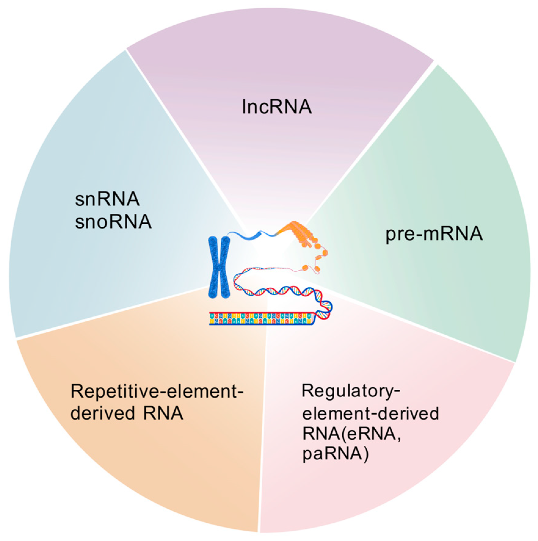
Figure 1. Different categories of chromatin/chromosome-associated RNAs. Figure created with BioGDP.com (accessed on 13 August 2025) [14].
Ncrna 11 00068 g001
Figure 2. Methods to study chromatin and RNA interactions. Figure created with BioGDP.com (accessed on 20 August 2025) [14].
Ncrna 11 00068 g002
Figure 3. Non-canonical structures of caRNAs. (A) RNA G-quadruplex. (B) R-loop. (C) RNA:DNA triplex. Figure created with BioGDP.com (accessed on 13 August 2025) [14].
Ncrna 11 00068 g003
Figure 4. Proteins interacting with caRNAs. (A) caRNAs interact with CTCF to facilitate promoter–enhancer loop formation and gene expression. (B) Jpx RNA evicts CTCF from chromatin. (C) Nascent RNA competes with chromatin for CTCF binding. (D) The lncRNA Oplr16 binds to the OCT4 promoter and recruits SMC1 to promote promoter–enhancer looping and OCT4 expression. (E) Dual functions of caRNA-PRC2 interaction: recruitment of PRC2 to chromatin (upper) and eviction of PRC2 from chromatin (lower). Figure created with BioGDP.com (accessed on 20 August 2025) [14].
Ncrna 11 00068 g004
Disclaimer/Publisher’s Note: The statements, opinions and data contained in all publications are solely those of the individual author(s) and contributor(s) and not of MDPI and/or the editor(s). MDPI and/or the editor(s) disclaim responsibility for any injury to people or property resulting from any ideas, methods, instructions or products referred to in the content.