Review

Applications of Hydrogels in Drug Delivery for Oral and Maxillofacial Diseases

1
State Key Laboratory of Oral Diseases, National Clinical Research Center for Oral Diseases, Department of Cariology and Endodontics, West China Hospital of Stomatology, Sichuan University, Chengdu 610041, China
2
Key Laboratory of Bio-Resource and Eco-Environment of Ministry of Education, College of Life Sciences, Sichuan University, Chengdu 610065, China
*
Author to whom correspondence should be addressed.
Gels 2023, 9(2), 146; https://doi.org/10.3390/gels9020146
Submission received: 19 December 2022 / Revised: 3 February 2023 / Accepted: 5 February 2023 / Published: 9 February 2023
(This article belongs to the Special Issue Gels: Applications in Drug Delivery and Tissue Engineering)

Abstract

:
Oral and maxillofacial diseases have an important impact on local function, facial appearance, and general health. As a multifunctional platform, hydrogels are widely used in the biomedical field due to their excellent physicochemical properties. In recent years, a large number of studies have been conducted to adapt hydrogels to the complex oral and maxillofacial environment by modulating their pore size, swelling, degradability, stimulus-response properties, etc. Meanwhile, many studies have attempted to use hydrogels as drug delivery carriers to load drugs, cytokines, and stem cells for antibacterial, anticancer, and tissue regeneration applications in oral and maxillofacial regions. This paper reviews the application and research progress of hydrogel-based drug delivery systems in the treatment of oral and maxillofacial diseases such as caries, endodontic diseases, periodontal diseases, maxillofacial bone diseases, mucosal diseases, oral cancer, etc. The characteristics and applications of hydrogels and drug-delivery systems employed for the treatment of different diseases are discussed in order to provide a reference for further research on hydrogel drug-delivery systems in the future.

1. Introduction

The oral and maxillofacial region bears various critical functions of the human body, such as mastication, swallowing, speech, and appearance. Oral health is crucial to patients’ overall health. No matter what anatomical structures suffer damage, e.g., pulpitis, periodontitis, alveolar osteitis, oral mucosal disease, and oral cancer, almost all lesions will do harm to patients’ physiological and mental health, and influence their social living. Hence, preventing and treating oral diseases is of vital significance to improve patients’ quality of life.
A suitable drug-delivery system (DDS) proves a powerful therapeutic tool for the thorough and efficient treatment of oral diseases, demanding biomaterials with excellent performance as drug-delivery platforms [1]. Conventional DDS mainly consists of oral gels, tablets, and lozenges. However, not only the complexity of oral and maxillofacial structures but also the specificity of the oral environment leads to the challenges of drug delivery to the lesion region. For example, there are many fascias and cavities in the maxillofacial region, which usually result in the spreading of infection and the difficulties of infection control. Only sustaining long-term and effective drug concentration in the infectious region can control the infection well [2]. What’s more, the elevated moistness of the oral environment and the movement of the tongue makes it more troublesome to control the therapeutic patch at the lesion site consistently, which brings problems to the prevention and treatment of oral mucosal diseases [3]. As a consequence, developing an on-demand and practical DDS becomes fairly essential and urgent.
Hydrogels have demonstrated unique properties of their structure and performance, which discriminate them from other biomaterials [4]. Hydrogel is a biological material by physical or chemical crosslinking reactions of monomers, forming a polymer network system [5]. The specific reactions are presented in Figure 1. For instance, the hydrogel, a methacrylate-based polymer, was prepared with the combination of N-vinylpyrrolidone (NVP), 2-hydroxyethyl methacrylate (HEMA), and glycerol monomethacrylate (GMMA) monomers with methacrylic acid (MA). These monomers could be crosslinked based on the radical polymerization initiated by thermic, ultraviolet (UV), or reductive-oxidative reactions [6]. Different crosslinking methods lead to different properties of hydrogels. Generally speaking, the physicochemical properties of chemical crosslinked hydrogels are relatively more tunable than those of physical crosslinked ones [7]. The network system provides a platform for various modules and cells and shows outstanding biocompatibility and biodegradability [8]. It is worth noting that the three-dimensional network structure of hydrogel shows great similarity with the extracellular matrix (ECM), which means it can provide a comparable living environment for cells and induces proliferation and differentiation [9].
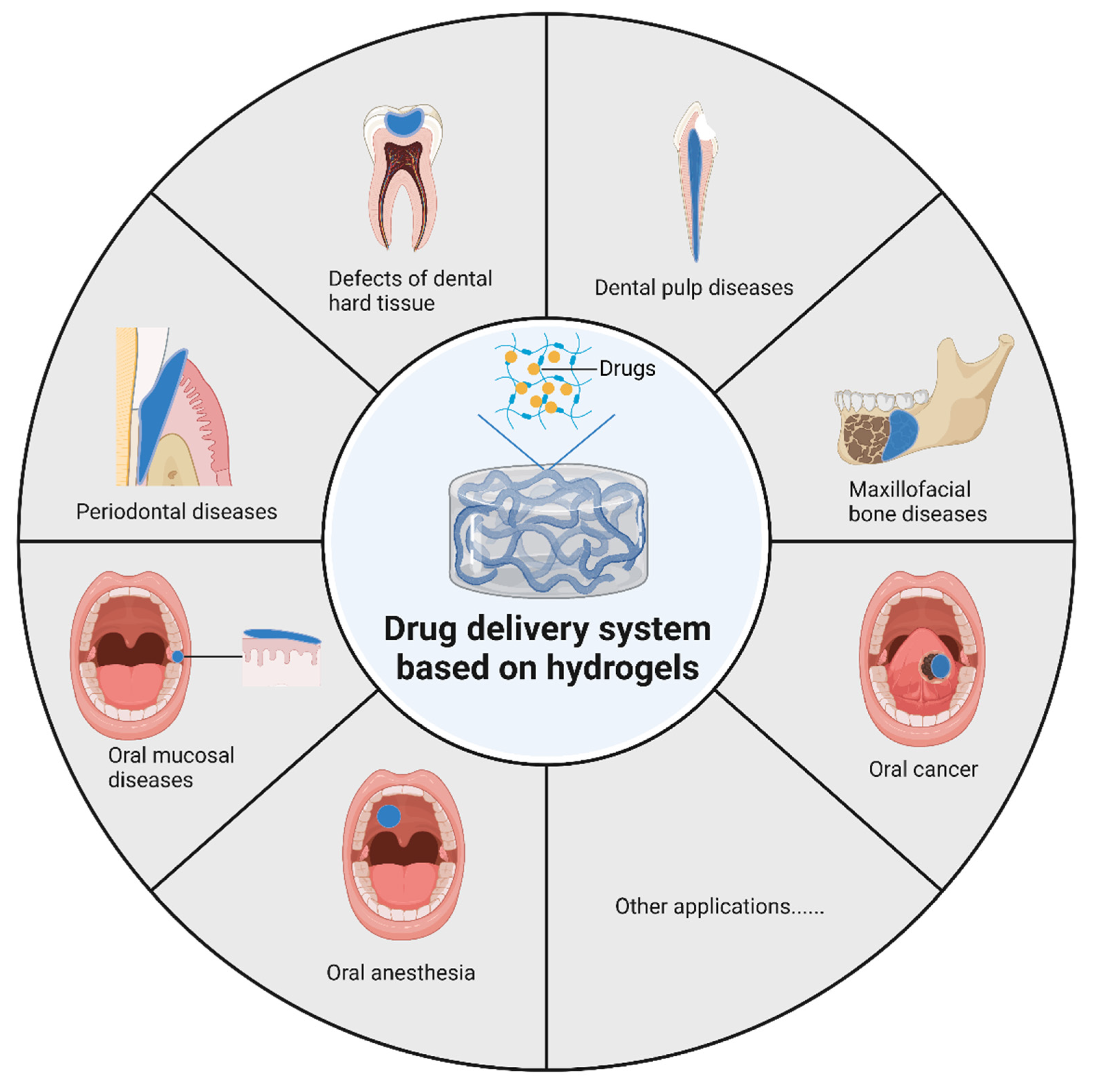
Above all, hydrogels have been put into application in massive fields of laboratory research and clinical treatment, covering hard and soft tissue regenerative engineering, local anesthesia, wound healing, and so on, revealing a promising prospect of DDS in the treatment of oral disease. This paper reviews the current status of hydrogel applications in oral and maxillofacial diseases, introduces the role of various hydrogels in oral drug delivery systems, and outlines future research directions. The applications of hydrogels in drug delivery for oral and maxillofacial diseases are presented in Figure 2.

2. Materials and Methods

In the literature search phase, the following databases were used: PubMed, Web of Science, Cochrane Library, and Embase. The literature search was first conducted using the keywords “Hydrogels”, “Drug Delivery Systems”, “Dentistry”, and their related keywords. Afterward, the search was conducted by combining different keywords for diseases with the keywords “Hydrogels” and “Drug Delivery Systems”, and the keywords for diseases used for the search included “Dental Caries”, “Dental Pulp Diseases”, “Periodontitis”, “Bisphosphonate Associated Osteonecrosis of the Jaw”, “Osteoradionecrosis”, “Mandibular Reconstructions”, “Dry Sockets”, “Peri-implant”, “Mucosal Diseases”, “Mouth Neoplasms”, “Anesthesia” and their related keywords. A total of 808 articles were found, 104 of which were approved for the writing phase, while the remaining articles were excluded due to duplication, irrelevance to drug delivery, or the absence of hydrogels in the system. Literature from 2017 to October 2022 was investigated.

3. Hydrogels for Oral and Maxillofacial Environment

Hydrogels, a hydrophilic polymer network, can be composed of natural or synthetic polymers. Natural polymers, such as gelatin, chitosan, sodium alginate, hyaluronic acid (HA), and so on, usually have outstanding biocompatibility and biodegradability, but their mechanical properties are often inferior. Synthetic polymers such as polycaprolactone (PCL), polyethylene glycol (PEG), Pluronic, and so on, usually have stronger mechanical properties, but often require surface modification to achieve better biological functionality [11]. Different natural or synthetic polymers can be chemically crosslinked or physically crosslinked to form hydrogels, including those with injectable, temperature-sensitive, adhesion, and other properties to achieve better local drug delivery. Chemical crosslinking connects different polymer molecules by forming covalent bonds between functional groups through certain reactions, including click chemistry, enzymatic reaction, Schiff base coupling, Diels-Alder linkage, etc., which usually provide stronger mechanical properties due to their high crosslinking density [12]. However, the toxicity of their crosslinkers needs to be considered. In contrast, physical crosslinking hydrogels are mainly gelled by non-covalent interactions such as hydrogen bonding, hydrophobic interaction, chain entanglement, van der Waals interactions, etc., which are easier to be synthesized because they do not require crosslinkers, but have relatively inferior physical properties [13].
To observe the micromorphology of hydrogels, Scanning Electronic Microscopy (SEM) inspection is used as an indispensable research tool, which requires the observation of samples in a high vacuum environment. However, the vacuum environment could not only contaminate the sample but also destroy the microstructure. Consequently, hydrogel samples are usually required to be dried before SEM inspection. The present drying means of hydrogels include freeze-drying and supercritical drying methods. The freeze-drying method is to solidify the product by freezing, and then evaporates the solvent by sublimation when heated [14]. While the supercritical drying method is to dry the hydrogel under supercritical fluid conditions, which avoids the collapse and contraction of the structure during the drying process and maintains the network framework structure of the hydrogel [15,16].
In the studies of topical drug delivery using hydrogels for oral and maxillofacial diseases, some of the studies used relatively simple hydrogels as carriers for drug delivery, either directly carrying the drug or carrying nanoparticles containing the drug to achieve continuous release of the drug. While another part of the studies further investigated hydrogel systems with different properties, such as injectability, adhesion, pH responsiveness, etc., to facilitate the topical application of hydrogel systems or to achieve controlled release of drugs under specific stimuli. The most commonly used hydrogel matrix for topical drug delivery studies in the oral and maxillofacial region is injectable hydrogels formed by chemical or physical crosslinking. Injectable hydrogels are widely used in the field of drug delivery because they are not limited by defect geometry [17] and are suitable for filling irregular root canals, periodontal pockets, bone defect areas, etc. Particularly, some injectable hydrogels exhibit a sol-gel transition in response to stimuli such as temperature, light, and pH. Among them, injectable thermosensitive hydrogels are one of the most commonly used hydrogel systems, as they usually can be injected at low temperatures and rapidly transform into gels at body temperature [18]. Thermosensitive hydrogels are temperature-sensitive materials and changes in environmental temperature can lead to changes in their sol-gel states [19]. In order to be an ideal thermosensitive hydrogel system for drug delivery, thermosensitive hydrogels should be free-flowing solutions at room temperature and transform into non-flowing gels at physiological temperatures (32–37 °C) [20]. The mussel-inspired mucoadhesive hydrogels are also often used in the field of transdermal drug delivery to the oral mucosa [21]. Compounds containing catechol structures, such as dopamine, can be used as biobased adhesives, allowing hydrogel materials to bind tightly to the oral mucosa in a moist oral environment, which contributes to the continuous targeted delivery of drugs [22]. Hydrogels with special properties for application in the oral environment are included in Table 1.

4. Applications in Different Oral and Maxillofacial Diseases

4.1. Defects of Dental Hard Tissue

Dental caries is one of the most common chronic diseases all over the world [74]. Caries can gradually destroy the hard tissues of the tooth, namely enamel, and dentin. Enamel is the hardest tissue in the human body, consisting mainly of hydroxyapatite, with 96% mineralization and high wear resistance, but it cannot be regenerated after destruction [75]. Unlike enamel, dentin consists of hydroxyapatite with a mineralization of about 70%, which can form a small amount of restorative dentin after external stimulation [76]. At present, caries lesions of enamel and dentin are usually restored clinically only by resin, silver amalgam, and other materials. The progression, cessation, or reversal of caries is related to the demineralization/remineralization process of dental hard tissue [77]. Many studies have been attempted in the remineralization of enamel and dentin using a biomimetic mineralization strategy [78]. As a drug delivery matrix with widely adjustable physical and chemical properties, hydrogels have been studied as carriers of drugs, peptides, stem cells, etc., in the restoration and remineralization of dental hard tissues.
Tooth enamel is a complex mineralized tissue [79] and the cells that make it are called ameloblasts, a type of highly specialized epithelial cell. It synthesizes and secretes amelogenin and transports calcium as well as phosphorus ions to the enamel matrix. Therefore, different studies have attempted to use calcium and phosphorus ions [80], amelogenin peptides [81], and even dental epithelial cells [32] to promote the biomimetic mineralization of tooth enamel. The positive charge of chitosan enables it to adhere to the negatively charged surface of demineralized enamel, supporting the use of chitosan for regenerating mineralized enamel tissue [81]. Therefore, a simple biomimetic mineralized hydrogel system can be prepared based on chitosan, using chitosan hydrogel loaded with inorganic ions (Ca2+ and PO43−) to repair the enamel surface [80]. One study used chitosan hydrogels loaded with smaller amelogenin peptides for enamel restoration and could restore hardness to 87% of normal enamel sections [81]. A few studies have tried to add dental stem cells into the hydrogel system to promote the regeneration of dental hard tissue. Through the Schiff base reaction, Mohabatpour et al. [32] use oxidized alginate and carboxymethyl chitosan to prepare injectable hydrogel, which can be used as a cell carrier for enamel regeneration. This hydrogel can maintain the shape and vitality of dental epithelial cells within 14 days.
Similar to enamel remineralization, the remineralization, and regeneration of dentin have also attracted many scholars’ attention, including hydrogel systems with different characteristics. To design scaffolds that resemble the characteristics of natural mineralized tissue for dentin regeneration, Campodoni et al. [82] used gelatin and chitosan as a hydrogel matrix, adding synthetic mineralized flakes similar to natural mineralized tissues, which were made of gelatin and magnesium-doped-hydroxyapatite (MgHA) nanocrystals. The mineral phase MgHA is very similar to the mineral components in the natural mineralized tissues, and the mineral phase is limited to the nanometer level through the interaction with gelatin, which is conducive to biomineralization. Alaohali et al. [43] prepared an injectable hydrogel with methacrylate (MA)–hyaluronic acid (HA) and di-thiol PEG and used eosin Y as the photoinitiator, which could be gelled in situ under blue light irradiation commonly used in dentistry. Meanwhile, GSK3 inhibitor NP928 can stimulate the formation of restorative dentin. Combining NP928 with injectable hydrogel makes it appropriate for clinical dental applications.
To increase the clinical application potential of materials, some studies have explored their antimicrobial or drug-carrying ability while maintaining their restorative or remineralizing role in dentistry. Ren et al. [83] combined QP5, an amelogenin-derived peptide with a remineralization effect, with antibacterial chitosan to form a hydrogel system that not only has good antibacterial ability but also shows an excellent remineralization effect. To investigate bioactive dental resins, Takeda et al. [84] tried to improve the biocompatibility of dental resins and developed a resin that releases growth factors to promote tissue regeneration. 2-hydroxyethyl methacrylate (HEMA) and the cross-linking monomer trimethylolpropane trimethacrylate (TMPT) were manufactured into a non-biodegradable hydrogel, and the loading and release characteristics of bovine serum albumin (BSA) and fiber last growth factor-2 (FGF-2) in hydrogels were studied.
Up to now, a large number of attempts have been made to remineralize and regenerate dental hard tissue using hydrogel-driven mineralization, protein/peptide-induced mineralization, and other methods. However, it is still incapable of fully restoring the complex structure of the natural dental hard tissues [78].

4.2. Dental Pulp Diseases

Deep caries, crown fracture, tooth dislocation, and other etiologies may lead to injury and inflammation of the dental pulp. In such cases, endodontic treatment becomes an important treatment option to preserve the affected teeth. Based on an accurate diagnosis of the pulp condition, root canal therapy, partial pulpotomy or pulp capping can be used as treatment options [85]. In recent years, pulp regeneration has also been applied in many studies in order to further preserve and reconstruct a vital pulp [86]. Since hydrogels with different compositions have widely adaptable properties, especially those with injectable properties that are suitable to act in the narrow space inside the root canal, many studies have chosen hydrogels as carriers for intracanal drug delivery and explored their applications in pulp capping, root canal disinfection, and pulp regeneration [87,88].

4.2.1. Pulp Capping

When the pulp is exposed due to caries or trauma, the exposed pulp can be covered by pulp capping agents to enhance tertiary dentin deposition, and researchers are also exploring how to achieve tooth regeneration using pulp capping agents [89]. Historically, calcium hydroxide has been the gold standard drug for pulp capping, but some studies have made other attempts at the choice of pulp capping materials [90]. In the study by Komabayashi et al. [44] on direct capping materials, calcium hydroxide was loaded into the light-cured PEGMC hydrogel and controlled calcium ion release was achieved, but the hydrogel had a cytotoxicity similar to that of the adhesive system. Some studies have explored hydrogel systems with better biocompatibility. Osmond et al. [33] used an injectable hydrogel of carboxymethyl-chitosan and diglycidyl ether with calcium phosphate nanoparticles to investigate its role in protecting exposed dental pulp. The results demonstrated that the hydrogel system is biocompatible and has a beneficial effect in inducing the proliferation and differentiation of residual dental pulp stem cells. In addition to calcium ions, growth factors were also added to the hydrogel capping material. Wu et al. [51] loaded vascular endothelial growth factor (VEGF) in chitosan/beta-glycerophosphate (CS/β-GP) to achieve continuous release of VEGF, promote odontogenic differentiation of dental pulp stem cells (DPSCs), and to explore its possible usefulness in pulp capping after pulpotomy.

4.2.2. Root Canal Disinfection

Root canal disinfection is one of the key factors in the success of endodontic treatment. To ensure decontamination of the infected root canal, mechanical preparation of the root canal is usually supplemented by chemical disinfection [91]. In some studies, antibacterial agents were added to the hydrogel system to achieve a good root canal disinfection effect. Chlorhexidine (CHX), an antibacterial drug, was added to the photocrosslinked gelatin methacrylate (GelMA) hydrogel, which showed a significant antibacterial effect and could maintain a high vitality of dental pulp stem cells. That is conducive to root canal disinfection in the treatment of pulp regeneration [39]. Haseeb et al. [92] encapsulated CHX in poly(ethylene glycol)-block-poly(l-lactide) (PEG-b-PLA) nanoparticles and loaded the nanoparticles in a hydroxyethyl cellulose hydrogel for root canal disinfection. Retarded release of CHX was observed over 21 days. Many antibiotics can be used for root canal disinfection [93] in addition to the clinically used CHX. For example, Bekhouche et al. [23] encapsulated clindamycin in polylactic acid (PLA) nanoparticles to improve the antibacterial properties of injectable fibrin hydrogels and similarly obtained hydrogel systems with superior antibacterial effect and biocompatibility. Likewise, Ribeiro et al. [94] loaded clindamycin or metronidazole on fibrous particles and used injectable GelMA hydrogel for ablation of endodontic infections, showing good results and low cytotoxicity.

4.2.3. Pulp Regeneration

The treatment of necrotic immature permanent teeth with open apical foramen is one of the challenges that dentists face in clinical work. Since the beginning of this century, a large number of clinical studies have reported the clinical application of pulp revascularization, which not only observed the healing of periapical lesions after surgery, but also observed the continuous development of the root, and even the restoration of pulp vitality [95,96,97].
Currently, different studies have attempted to achieve pulp tissue regeneration by two main methods: cell homing or cell transplantation [98]. The cell homing method, also known as a cell-free method, involves the use of biological signaling molecules to promote the migration, proliferation, and differentiation of endogenous stem cells [99]. In order to enable growth factors to work better in narrow root canals, many studies have chosen hydrogels with different properties to carry specific growth factors, allowing the growth factors to act continuously in the root canal. Carvalho et al. [100] used the cell homing strategy to load the secretion from stem cells of deciduous teeth into the chitosan hydrogel, so as to provide continuous and controlled release of several trophic factors. Similarly, some studies attempted to incorporate drugs to promote cell differentiation, migration, and regeneration. Soares et al. [101] incorporated simvastatin into chitosan hydrogels to achieve a controlled release of drugs and increase the chemotaxis and regenerative capacity of human dental pulp stem cells (hDPSCs). Another drug, iloprost, has also been added to the injectable thermosensitive hydrogel PF127 for inducing pulp regeneration due to its ability to increase the expression of VEGF [46].
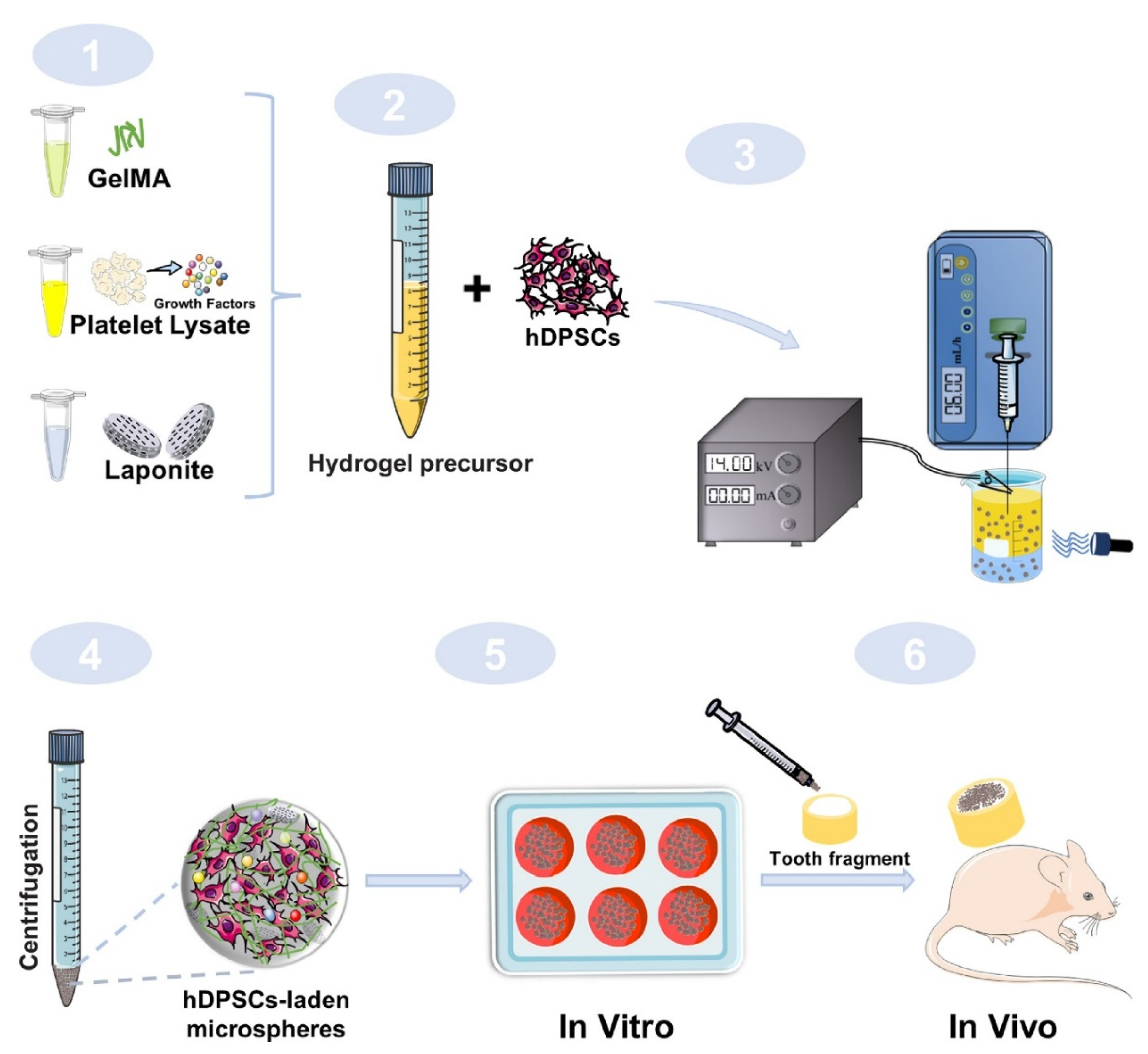
The cell transplantation method mainly uses stem cells from a host or allogeneic source for isolation and in vitro expansion, implanted in a scaffold, and transplanted into the root canal for regeneration [40,102]. Growth factors also play an important role in cell transplantation, so many studies have incorporated growth factors in hydrogel scaffolds to promote the growth and differentiation of transplanted hDPSCs. Zhang et al. [40] used GelMA to fabricate a microsphere for the delivery of pro-angiogenic growth factors and improved the drug delivery ability of the microsphere by modifying it with the nanoclay Laponite (Figure 3). Silva et al. [24] added platelet lysate (PL), which releases platelet-derived growth factor (PDGF) and VEGF, to injectable HA hydrogels, and the stability of the material was enhanced by fibrin nanocrystals. This injectable hydrogel could not only continuously release growth factors to promote stem cell recruitment and vascular growth, but also exhibit support for the growth of hDPSCs. Park et al. [41] worked on promoting the differentiation of hDPSCs. They coupled BMP-mimetic peptide into GelMA hydrogels and used 3D printing to fabricate the material, which promoted the odontogenic differentiation of hDPSCs. In practical applications, the effects of the hydrogel system on the adhesion, chemotaxis, proliferation, and differentiation of pulp cells are all very important. Anitua et al. [103] investigated the effects of adding either hydroxyapatite or plasma rich in growth factor (PRGF) to gelatin and alginate on human pulp cells. Cell adhesion and chemotaxis were significantly increased when PRGF was added, while the combination of hydroxyapatite and PRGF promoted cell proliferation and stimulated osteogenic differentiation.
Up to now, although many studies have shown that the addition of different biologic factors or drugs in hydrogels promotes the proliferation and differentiation of DPSCs, there are still no studies that have achieved real pulp regeneration. Pulp regeneration still has a long way to go.

4.3. Periodontal Disease

Periodontitis is an inflammatory process that occurs in the tissues surrounding the teeth due to plaque accumulation on the teeth [104] and is mainly associated with anaerobic bacteria such as Porphyromonas gingivalis and Treponema denticola [105]. Periodontitis is treated by mechanical scaling, combined with topical antibacterial medication and occasional systemic antibacterial therapy [106]. Bacterial control in periodontal pockets and regeneration of alveolar bone are two important aspects of periodontal treatment. Thus, drugs with anti-inflammatory, antibacterial, or osteogenic effects can be selected for the topical treatment of periodontitis [107,108].
As a highly hydrated polymer network, hydrogels are commonly used in periodontal tissue formation and drug delivery [109]. For convenient administration in periodontal pockets, injectable hydrogels are widely used for drug delivery in periodontal disease [31,34,62,110]. Usually, loading drugs into hydrogel systems is designed to prolong the action or release of the drug. For example, Wang et al. [36] incorporated minocycline into injectable in situ hydrogels composed of poly(lactide-co-glycolide) (PLGA) and N-methylpyrrolidone (NMP) for periodontal antibacterial. Minocycline sustained release for more than 48 h after initial rapid release. In addition to the sustained release of drugs, the controlled release of drugs can also be achieved by special injectable drug delivery systems. Wang et al. [2,38] loaded doxycycline and lipoxin into PLGA microspheres and dispersed them in polyisocyanopeptide (PIC) hydrogels. The hydrogel system is injectable and structurally stable, and the release rate of the drug can be adjusted by adjusting the loading ratio of acid-terminated and ester-capped PLGA microspheres. For the treatment of chronic periodontitis combined with diabetes mellitus, Zhao et al. [35] developed a drug delivery system for ROS-triggered drug release in which oxidized dextran and phenylboronic acid-functionalized poly (ethylene imine) form an injectable hydrogel via a Schiff-base reaction, and the anti-inflammatory drug doxycycline and the osteogenic drug metformin were bound in the hydrogel network through B−N coordination. The release of the drug is triggered by excessive ROS due to hyperglycemia to achieve synergistic therapeutic effects of anti-inflammatory, antibacterial, and osteogenic effects.
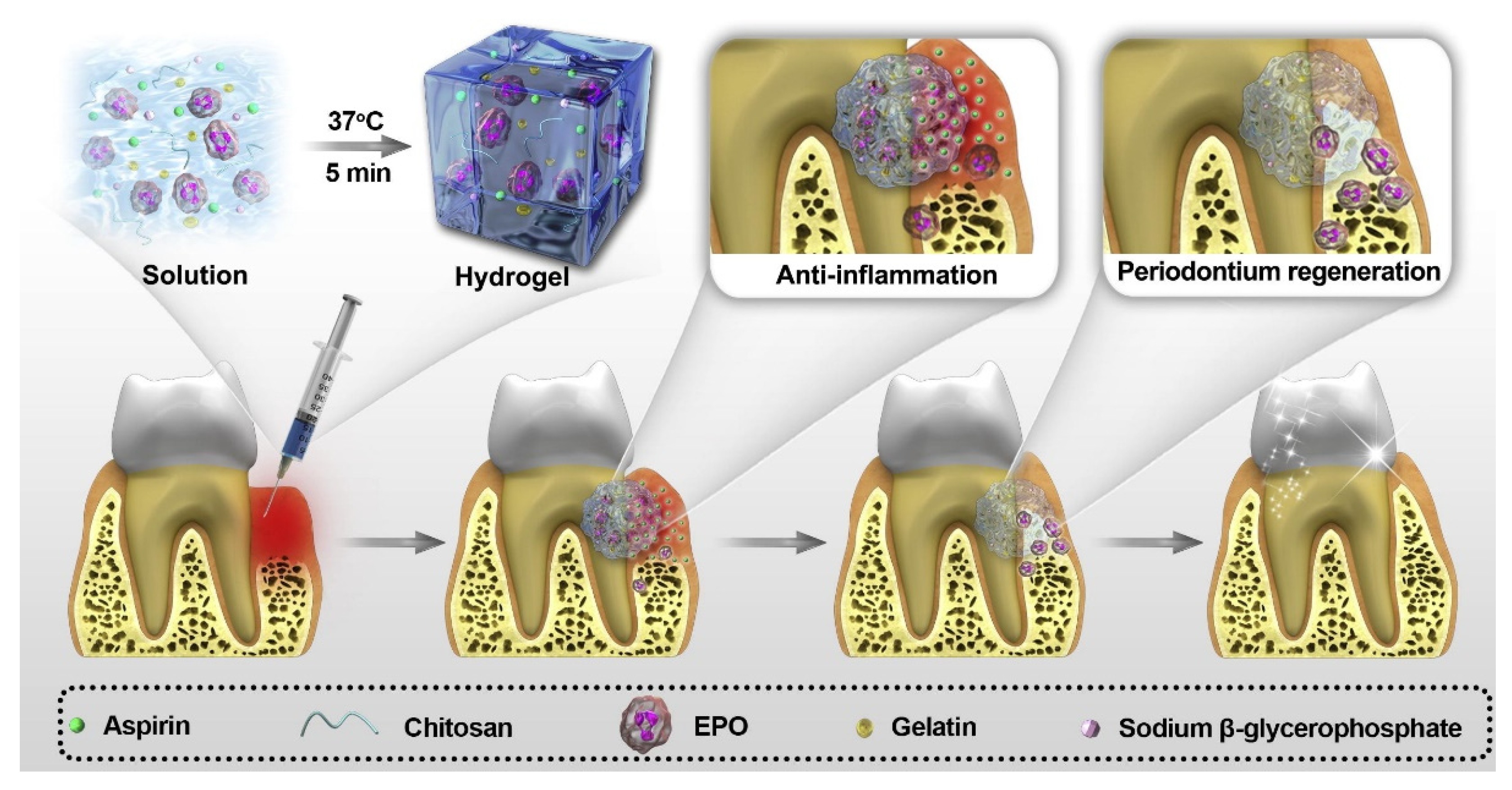
Based on injectable hydrogels, some studies have used injectable thermosensitive hydrogels, which are able to form hydrogels in situ at body temperature after injection into the periodontal pocket, making the administration of the hydrogel in the pocket more convenient and effective [49,54,111]. Hydrogels prepared with CS and β-GP are one of the most commonly used injectable thermosensitive hydrogels for the treatment of periodontitis. Zang et al. [53] added ornidazole and BMP-7 simultaneously to hydrogels formed from CS and β-GP for the treatment of bone loss and inflammation that typically exist together in patients with severe periodontitis. The results showed that the hydrogel system could release the drug stably and consistently. Some studies introduced gelatin into the crosslinking of CS and β-GP to reduce the gelation time by electrostatic interactions [112]. Xu et al. [112] prepared a thermosensitive hydrogel for the sustained release of aspirin and erythropoietin (EPO) using CS, β-GP, and gelatin, and drug release could last for at least 21 days (Figure 4). Poloxamer 407 (Pluronic F127) is also one of the most commonly used thermosensitive polymers. Chen et al. [48] used injectable thermosensitive hydrogel based on Pluronic F127 to deliver simvastatin locally to treat periodontitis and reduce periodontal bone loss. On the contrary, a study used a hydrogel system that maintains the gel shape at low temperatures and transforms it into liquid at body temperature [55]. They added BMP-6 to an injectable thermosensitive chitosan/gelatin/glycerol phosphate hydrogel, which was able to reduce inflammatory progression and promote periodontal regeneration.
Inflammation leads to a decrease in pH, and a slightly acidic environment of pH 5.0 to 7.0 is conducive to the growth of many periodontal pathogenic bacteria [113]. When pH decreases, the pH-responsive hydrogel drug release system is able to achieve natural control of inflammation [114]. Based on the pH changes at the inflammation site, some studies have attempted to modulate the local drug release by pH for on-demand and controlled drug delivery, especially in the slightly acidic inflammatory environment. Bako et al. [45,115] made a visible light polymerized hydrogel for the co-release of metronidazole and CHX, which allows rapid gel formation under blue light commonly used in dentistry. The methacryloil-group modified polymer and methacrylated-poly-γ-glutamic acid nanoparticles can work together as a pH-sensitive drug delivery system, with lower pH resulting in greater drug release. Chang et al. [113] incorporated naringin into a pH-responsive hydrogel with thermogelling properties. This hydrogel system composed of carboxymethyl hexanoyl chitosan (CHC) and β-GP was able to significantly reduce periodontal bone loss, and the release of naringin is faster at lower pH conditions. In addition, lower pH can also regulate drug delivery by modulating the drug carrier structure. Li et al. [116] attempted to load dexamethasone into a pH-responsive host-guest nanoparticle, assembled from cyclodextrins and multivalent hydrophilic guest macromolecules. This nanoparticle is able to convert into a hydrogel in the pH conditions of periodontitis, which facilitates drug release and local treatment.
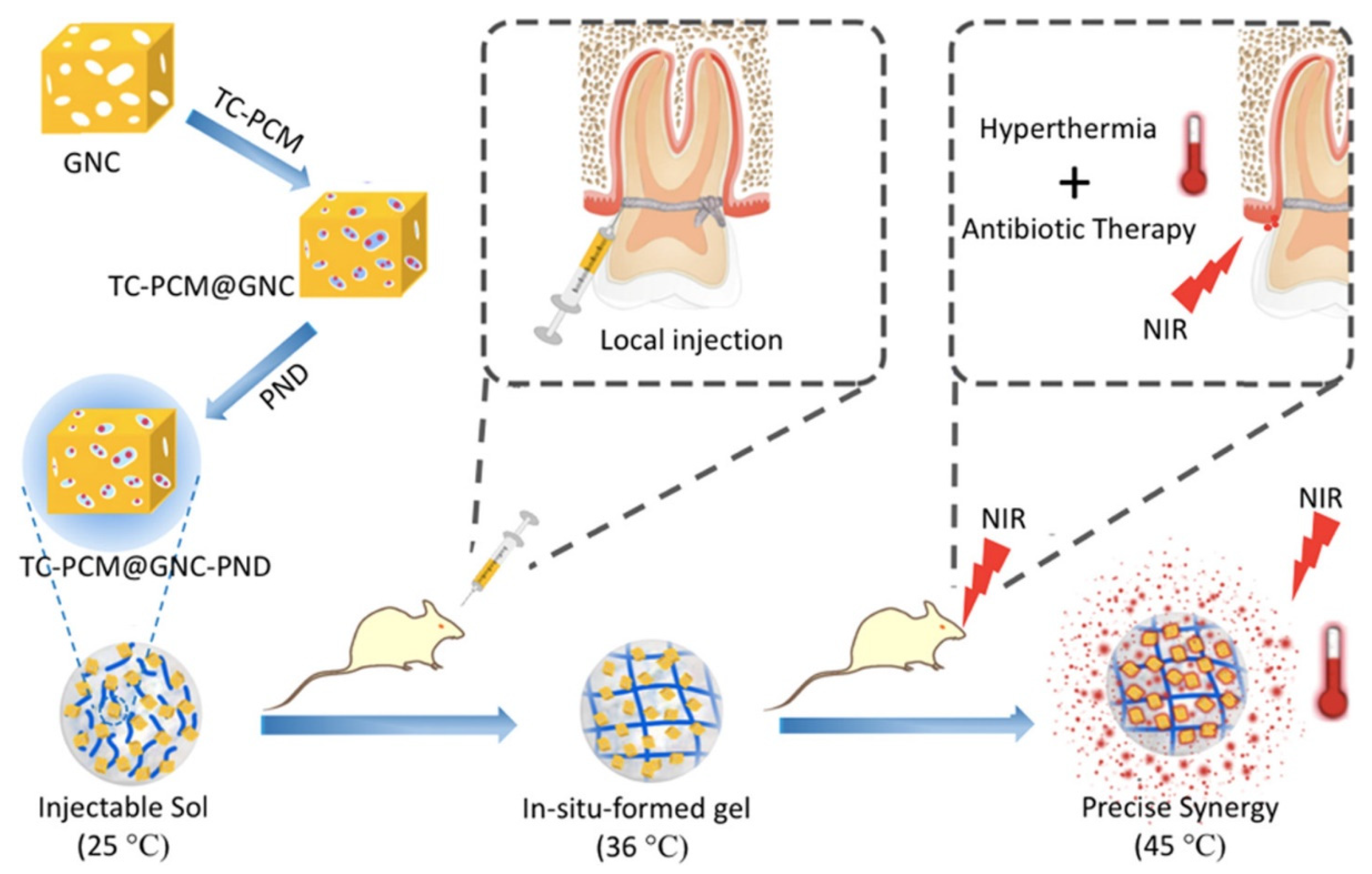
Some hydrogel systems use the photothermal effect to control the release of drugs, combining the antibacterial effect of the photothermal effect with antibacterial drugs to achieve a better antibacterial effect. Zhang et al. [61] fabricated hydrogel platforms for the on-demand release of antibiotics under near-infrared light irradiation. Poly (N-isopropylacrylamide-co-diethylaminoethyl methacrylate) (PND) formed hydrogels in situ at body temperature, which triggered the solid-liquid phase transition of the phase change material through the photothermal effect, resulting in controlled release of tetracycline (Figure 5). Lin et al. [117] similarly used the photothermal effect for periodontal antimicrobial therapy by loading minocycline in GelMA-Au NBPs@SiO2 hybrid hydrogel, where near-infrared light irradiation at 808 nm increased the release rate of the drug and controlled bacterial proliferation in periodontal pockets.
In general, hydrogels have been widely used for drug delivery in periodontal disease to achieve antibacterial and periodontal soft and hard tissue regeneration in periodontal pockets. Among them, injectable and temperature-sensitive hydrogels are more frequently chosen in various studies because their physicochemical properties are more suitable for the periodontal pocket microenvironment. To achieve better local targeted drug delivery, specific stimulus-responsive drug release hydrogel systems are also increasingly being used in the treatment of periodontitis.

4.4. Maxillofacial Bone Diseases

Maxillofacial bone defect generally emerges after dental removal, implantation, and maxillofacial surgery, which can result in different degrees of damage. The defect resulting from dental extraction and implantation is usually small-scale and faster-healing relatively. However, the defect of mandibular surgery is trickier most of the time.

4.4.1. Mandibular Reconstruction

Mandibular surgery is generally performed in patients with tumor ablation, traumatic infection, and osteonecrosis debridement [118], all of which will carry both great physical sickness and psychological pain to patients. At the moment, the considerable challenges in the clinic include various osteomyelitis, delayed wound healing, mandibular dysfunction, and temporomandibular joint disorder. To overcome these problems, recovering maxillofacial bone defects and promoting bone reconstruction is the key to treatment. Due to its physical structure, great biocompatibility, and controllable bio-degradability [4], the hydrogel has been attempted in mandibular reconstruction and shows enormous potential. To match the complex anatomy and physiology of the mandible, Zhang et al. [119] implemented 3D-printed composite scaffolds combined with dual small molecules, consisting of resveratrol and strontium ranelate. This biological material showed great advantages in promoting angiogenesis and inhibiting osteoclasts. Meanwhile, the application of dual-drug molecules collaboratively promoted mesenchymal stem cell (MSCs) osteogenic differentiation. Aiming at stimulating stem cell differentiation, Lei, L., et al. [60] also developed an injectable hydrogel, which could instantly provide microRNA-222 and aspirin (ASP) at local sites. MicroRNA-222 was proved to be beneficial for the translation of MSCs into neural-like cells by specific signaling. Moreover, ASP played a role in enhancing bone formation as well. The injectable hydrogel incorporated with MSCs and ASP was regarded as promising for innervated bone tissue engineering. What’s more, collagen/nano-hydroxyapatite/alginate (Col/nHA/Alg) hydrogel carrying nerve growth factor (NGF) was developed for increasing new bone formation [120], as well as the hydrogel material impregnated with transforming growth factor-β1 (TGF-β1) and insulin-like growth factor-1 (IGF-1) [121].
BMP-2 proved beneficial in supporting bone formation and was widely applied in various biocompatible scaffolds and matrices [122,123]. A number of studies on mandibular reconstruction fabricated BMP-2 in biological materials. Jung, S.W., et al. [29] developed in situ gelling ALG/HA hydrogels that could release BMP-2 sustainably. The experiments in vitro and in vivo showed that the material has the potential to promote osteogenic differentiation of human bone marrow stem cells (hBMSCs) and the regeneration of mandibular bone. Based on the function of BMP-2, Kim, J., et al. [25] explored the application of electrical stimulation on the bone regeneration of mandibular defect. After electrical stimulation, recombinant human bone morphogenic protein-2 (rhBMP-2) was injected into the scaffold combined with hBMSCs and hydrogels, which proved the combined treatment an effective method. However, not all systems have the function to enhance the effect of BMP-2. The combination of PEG hydrogel and hydroxyapatite/β-tricalcium phosphate (HA/TCP) resulted in the inhibition of BMP-2-induced bone formation compared with the structure in which BMP-2 was loaded with HA/TCP [124]. This study demonstrated that different hydrogels might still exist limitations, which required the scientist’s deeper exploration.

4.4.2. Peri-Implant Diseases

Dental implants have developed rapidly over the years. The correct implantation procedure and postoperative peri-implant osteogenesis are equally significant. Adequate osteogenesis around dental implants inhibits the appearance of peri-implant diseases and makes sure for a healthy post-implant recovery environment [125]. With the number of dental implants increasing, dental practitioners should master the procedures and get a better application of enhancing bone regeneration. Hyaluronic acid hydrogel has been widely used to carry and release BMP-2 to increase peri-implant osteogenesis [26]. An injectable carrying BMP-2 hydrogel system was also prepared, which could sustainably release BMP-2 and showed higher osseointegration levels [37]. Not only the type of materials but also the rate of degradation of the biological material could affect osteogenesis. Meanwhile, one clinic study explored the difference between the application of BMP-2 gel and bisphosphonate gel on the stability of dental implants and marginal bone level, and the consequence showed no significance among the study groups [126]. Akagawa, Y., et al. [127] analyzed the effectiveness of both the fast and slow degradation-type basic fibroblast growth factor (bFGF)-gelatin hydrogel system on peri-implant new bone formation, only to find the latter one reached the highest level of the height of new bone. Beside the BMP-2 and bFGF, the CHX is also applied in the hydrogel to treat peri-implantitis. T. Asbi, et al. [128] reached the conclusion that the application of CHX gel could effectively diminish gingival inflammation during osseointegration in a short-term clinical study. Considering that soft tissue inflammation might lead to peri-implant bone loss in the future, a long-term study should be carried out on the anti-inflammatory effect of CHX gel.

4.4.3. Alveolar Osteitis (Dry Socket)

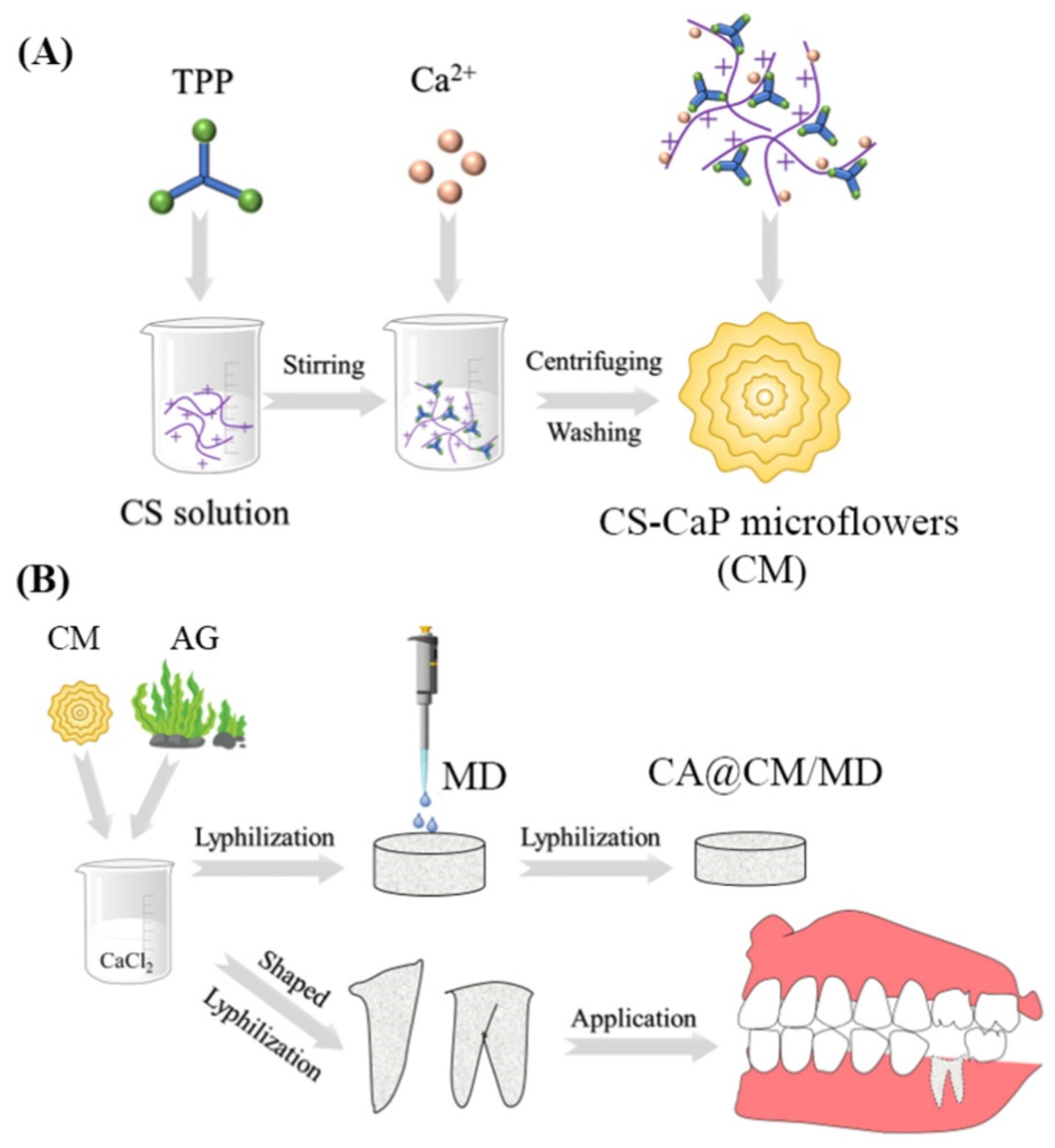
Alveolar osteitis (dry socket) is one of the most common complications following dental extraction, which has an incidence of approximately 3% for routine treatment, and even more than 30% for impacted mandibular third molars [129]. The etiology of alveolar osteitis is still under exploration, and the mainstream believes that the leading causes are the absence of blood clots and the inflammation of the socket [130]. Hence, the patients are generally requested to take metronidazole orally for 7~14 days after the operation to decrease inflammation. However, long-term antibiotics administration may cause severe side effects, such as vomiting, diarrhea, and constipation. Plenty of materials to prevent alveolar osteitis have been developed. Based on the antioxidant and anti-inflammatory properties of silibinin [131], Xu et al. [42] engineered silibinin into GelMA hydrogels to synthesize the Sil-GelMA. The controlled release of silibinin from the system exhibited an effect of anti-inflammatory and promoted vascularization by inducing the polarization of M2-type macrophage and regulating the secretion of anti-inflammatory factors and VEGF. Calcium alginate (CA) sponges loading with chitosan-CaP microflowers (CM) and metronidazole (MD) (Figure 6) were designed in the shape of the tooth root [132]. It developed the function of anti-bacterial, hemostatic, and osteogenesis, and provided a promising choice for preventing alveolar osteitis.

4.4.4. Osteonecrosis of the Jaw

Osteonecrosis of the jaw (ONJ) has a high incidence in tumor patients, who have to intake high-dose antineoplastic drugs or undergo long-term radiotherapy. According to etiology, ONJ is classified into medication-related ONJ (MRONJ) [133] and osteoradionecrosis (ORN) [134].
MRONJ generally refers to the ONJ resulting from bisphosphonates (BPs) administration which could inhibit the function of osteoclasts and treat benign and malignant bone diseases [135]. It has been proved that BPs have a negative and direct effect on bone cells and vascular tissue, causing necrosis of tissue eventually [136]. Based on the pathogenesis, researchers have come up with plenty of strategies to prevent MRONJ and enhance bone healing. VEGF, which plays a vital part in wound healing, was encapsulated in hydrogels, and then inserted into the maxillary extraction defect [30]. The ultimate outcome showed that the hydrogels combined with VEGF were beneficial to assist bone healing. VEGF demonstrated a pro-angiogenic and immunomodulatory function to prevent the occurrence of MRONJ as expected. Meanwhile, Brierly et al. [137] evaluated the function of another delivery system, which consisted of a poly (ethylene glycol)-heparin hydrogel core and encapsulated molecules including arginyl glycylaspartic acid (RGD) and rhBMP-2. This delivery system was also proved as a potentially useful tool to enhance the formation of bone cells and prevent MRONJ development. Besides VEGF and rhBMP-2, bFGF [138] is also put into an application, which promotes bone formation and inhibits the development of MRONJ.
Long-term exposure to high-dose radiation could lead to terrible adverse effects on the hard and soft tissues of the maxillofacial area. At present, the exact pathogenesis of ORN is uncertain and may be related to reduced wound healing and secondary infection [139]. Consequently, the treatment of ORN is still based mainly on symptomatic therapy, such as the surgical resection of the lesion area and debridement [140]. However, the outcome of conventional therapy is generally unsatisfactory, because the ORN always gets recurrence. Based on this problem, the researchers came up with MSCs and growth factors, which might have the capacity to promote the regeneration of the lesion tissue with delayed wound healing [141]. Combined hydrogels with rat MSCs and BMP-2, to investigate the effect on the osseous healing of ORN, which proved that the application of both MSCs and BMP-2 led to the best effects on enhancing osseous healing [27].

4.5. Oral Mucosa Diseases

Oral mucosal diseases are a large group of benign and malignant diseases common in oral lesions and the etiology is complicated and mostly uncertain [142]. Oral mucosal diseases often result in patients’ long-term pain. Hence, clinical doctors should pay more attention to the diagnosis and treatment of oral mucosal diseases.
Due to the complexity and specificity of the oral environment, topical drug administration of oral mucosal diseases has met plenty of limitations and problems. The high moistness and the presence of multiple proteins and mucins prevent robust, stable adhesion of drugs and biomaterials to the buccal mucosa [3]. Therefore, it’s of vital significance to develop a type of biomaterial displaying superior adhesion capability. To promote the properties, Janus patches with a function of wet adhesive were developed to treat the oral ulcer [143]. The researchers were inspired by the components of barnacle and hybrid adhesive mechanism and then combined modified glycine with hydroxyapatite nanoparticles by bridge bonding polymer chains to create a robust adhesive layer. The patch got over the shortcomings of commercial products and achieved analogous instant wet adhesion finally. Meanwhile, Ryu et al. [144] similarly developed chitosan oral patches called Chitoral from the inspiration of mussel adhesion. As well as Janus, Chitoral provided an adhesive layer in oral wet environments. When Chitoral has contact with a mucous layer and saliva, it instantly dissolves into compounds that interact with mucins and form a tough and insoluble adhesion layer between Chitoral and mucous. In the end, Chitoral would get a transformation into adhesive hydrogels under the physical and chemical reactions. Not limited to promoting the property of mucoadhesion, the researchers also endeavor to enhance other functions of biomaterial hydrogels. Ding et al. [145] fabricated a supramolecular hydrogel with mucosal adhesion and anti-oral leukoplakia function under the interaction of isoguanosine-tannic acid. Both isoguanosine and tannic acid have the anti-oral leukoplakia function, and this hydrogel was shown to be a potential platform for inhibiting the deterioration of oral leukoplakia.
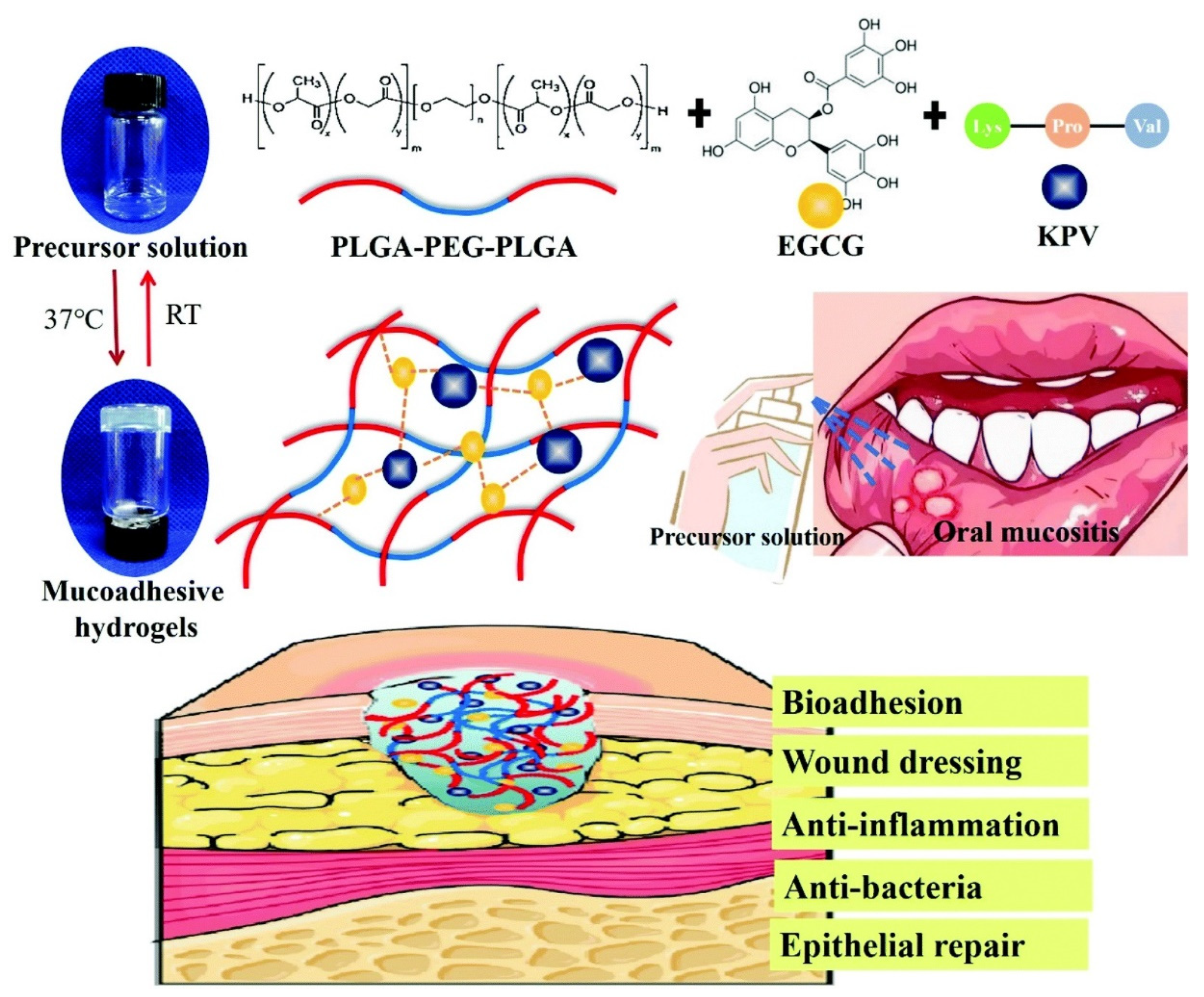
As mentioned above, oral mucosal diseases are a group of diseases with complex etiology. One of the most common lesions is an infectious disease caused by fungi or bacteria. However, more and more studies reported drug resistance during the treatment, making it crucial to prepare an advanced anti-fungi/bacteria biomaterial drug at the moment. Candida spp. is one of the most prevalent opportunistic pathogens which causes oral candidiasis [146]. Aiming at Candida spp., hydrogels fabricated with amino acid substituted histatin-5 (Hst-5) were designed to present the function of anti-fungi and promote wound healing at the same time [147]. Meanwhile, methylcellulose hydrogels loaded with Melissa officinalis oil produced a critical reduction in C. albicans [65]. In addition to anti-fungal, Shao et al. exploited temperature-sensitive PLGA-PEG-PLGA as a matrix, adding epigallocatechin-3-gallate (EGCG) as a compound of anti-bacterial and enhancing adhesion, and obtained a promising anti-inflammation application of treating chemotherapy-induced oral mucositis (Figure 7) [58].

4.6. Oral Cancer

Currently, oral cancer is the eleventh most common cancer worldwide, most commonly occurring in middle age and older adults. Squamous cell carcinoma is the most common oral malignant tumor, accounting for 90% of oral malignant tumors [148]. In the current treatment of oral cancer, chemotherapeutic drugs such as doxorubicin, cisplatin, and 5-fluorouracil are often used alone or in combination [149,150,151]. The low solubility and low bioavailability of these anticancer drugs are the limiting factors for their use [152]. Therefore, it is necessary to develop different drug delivery systems applied to oral cancer.
In the application of hydrogel systems for oral cancer drug delivery, hydrogels with adhesive or injectable properties are often used to load chemotherapeutic drugs. For instance, Shtenberg et al. [153] mixed alginate and liposomes in different ratios to obtain a hybrid hydrogel with adhesive properties and attempted to use this drug delivery system for sustained release of the anticancer drug doxorubicin (DOX) over a long period of time. Tan et al. [57] mixed a metal-organic framework (MOFs) with thermosensitive hydrogel PLGA-PEG-PLGA into an injectable system to load DOX and celecoxib into the system for local treatment of oral cancer and to achieve stable delivery of two drugs.
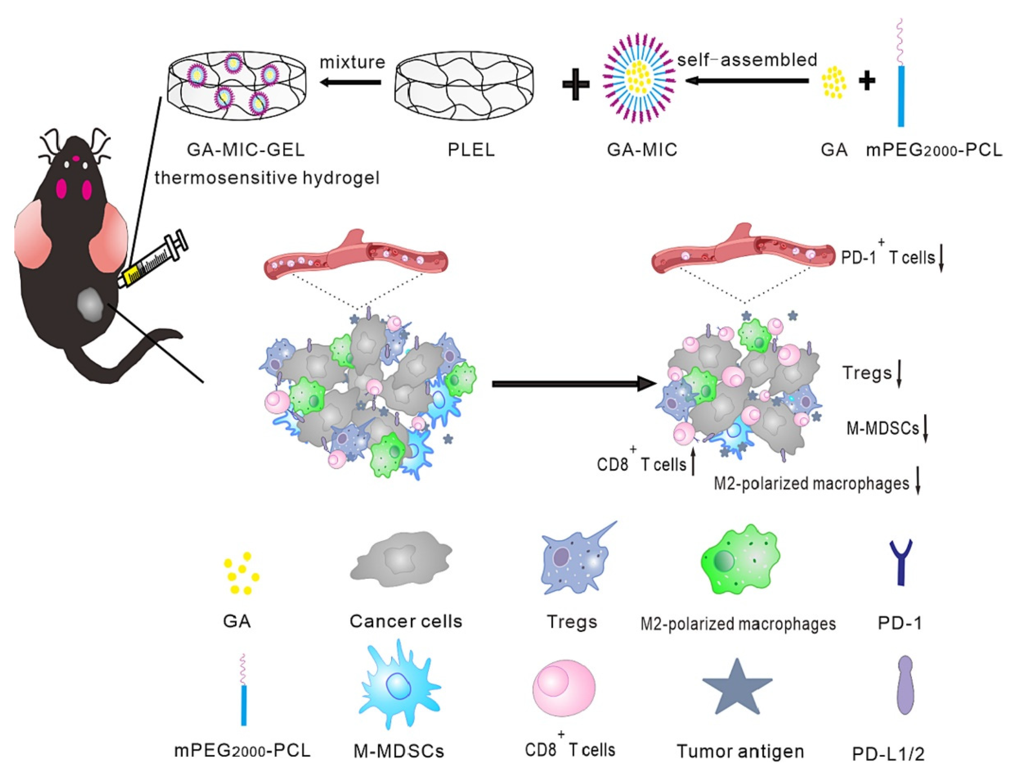
Immunotherapy is a recently developed treatment approach that treats disease by strengthening the patient’s immune defenses [154]. Several immune checkpoint blockade treatments have been approved by the FDA, including anti-PD1/PD-L1 and anti-CTLA-4 antibodies [155]. Some studies have explored the local delivery of immunotherapeutic agents in hydrogels. Shi et al. [156] used multidomain peptide (MDP) hydrogels to mimic the natural extracellular matrix and achieved the prolonged release of PD-1 immune-checkpoint inhibitor. Chen et al. [56] loaded gambogic acid (GA) into mPEG200 PCL micelles (MIC), after which GA-MIC was mixed with thermosensitive hydrogels to form injectable hydrogels. The anti-tumor immunity of the tumor-bearing mice was enhanced by down-regulating the expression of PD-1 through the action of GA (Figure 8). At the same time, it’s interesting that both the drug and hydrogel used by Leach et al. [157] had immunotherapeutic effects on tumors. They loaded the immunotherapeutic drug cyclic dinucleotide (CDN) into inducible nitric oxide synthase (iNOS)-inhibited L-nil-MDP hydrogel, which effectively prolonged the median survival of tumor-bearing mice. Besides, it has been found that Peptostreptococcus could activate the immune response in oral cancer patients. Zheng et al. [70] used silver nanoparticles combined with an adhesive hydrogel and exogenous bacteria to inhibit the proliferation of bacteria other than Peptostreptococcus. This hydrogel system enhanced the anti-tumor immune response by modulating the oral microbiota.
The effect of monotherapy on tumor inhibition is relatively limited, and some studies have combined chemotherapy, radiotherapy, immunotherapy, photothermal therapy, and other therapies to synergistically improve local tumor control and overall survival. Bollareddy et al. [6] combined chemotherapy and immunotherapy. In their study, they applied the chemotherapeutic drug 5-FU and the cyclooxygenase-2 inhibitor Etodolac in combination in transfersomes. These transfersomes prepared from phospholipids and edge activators were loaded in hydroxypropyl methylcellulose (HPMC) hydrogel for local drug delivery in oral cancer treatment. Wu et al. [64] combined chemotherapy and photothermal therapy. They applied near-infrared light (NIR) light-responsive hydrogels to oral cancer treatment by loading DOX into light-responsive mesoporous silica nanoparticles (MSNs), which was loaded into an injectable hydrogel made of methylcellulose together with IR820 as a photothermal agent. The degradation of MSNs was triggered by near-infrared light, which led to the controlled release of DOX. To treat drug-resistant tumors, Alamzadeh et al. [158] combined chemotherapy, radiotherapy, and photothermal therapy by co-loading cisplatin and gold nanoparticles into alginate hydrogel with 532 nm laser and X-ray irradiation. The combination of the three therapies was able to achieve better anti-cancer effects than either monotherapy or the combination of the two therapies.
There have been many attempts at various drug delivery strategies for oral cancer, and currently, it is available to load anti-cancer drugs through different nanoparticles, hydrogels, liposomes, and other carriers for locally targeted drug delivery. At present, most studies are still focused on in vitro or in vivo studies, and there are still few relevant clinical studies. How to apply the existing drug delivery systems in the clinic to achieve lower costs and higher patient survival rates is still a problem that needs to be studied.

4.7. Oral Anesthesia

Topical anesthesia is a commonly used method of anesthesia in dentistry to promote a comfortable experience of dental treatment. Effective dental topical anesthesia has many applications in clinical practice, such as relief of postoperative pain, analgesia for oral ulcers, initial periodontal therapy, placement of rubber barrier clip, soft tissue biopsy, etc [159].
At present, the biggest challenge of oral topical anesthesia is how to make the drug reach the target tissue through the oral mucosa epithelium. At present, the main disadvantages of topical anesthetics commonly used in clinical practice are short contact time with the target tissue, limited drug release, and poor anesthetic effect. At the same time, how to safely and effectively prolong the analgesic time of drugs during surgery is also an important issue [160].
Due to their adhesive properties and good biocompatibility, hydrogels have attracted some attention in surface anesthesia [161]. Muniz et al. [69] encapsulated 2.5% lidocaine and prilocaine in poly(ε-caprolactone) nanocapsules and loaded them into CARBOPOL hydrogel. The hydrogel system has satisfactory adhesion ability, and its anesthetic effect is better than that of commercial products. Cubayachi et al. [66] loaded the anesthetics prilocaine hydrochloride and lidocaine hydrochloride into HPMC hydrogels with good adhesive properties, and iontophoresis was used to enhance the penetration and retention of the anesthetics in the mucosa. In addition, by combining microneedles with an adhesive hydrogel, the anesthetic drug can more effectively break through the epithelial barrier for sustained release. Zhang et al. [68] used microneedles and adhesive PAM-PDA-AuNP hydrogels with near-infrared light response in transdermal drug delivery, which reduced pain during anesthesia and could trigger the release of anesthetic drugs by near-infrared light.
Usually, the anesthetic is loaded into the hydrogel to speed up or slow down the release of the drug and to regulate the duration of the drug. Sometimes, rapid drug release is necessary to achieve good anesthesia more quickly. Mihalache et al. [162] prepared interpenetrated/interconnected hydrogels with porous structure and excellent biocompatibility by an amidation reaction between CS and poly[(maleic anhydride)-alt-(vinyl acetate)], which can rapidly release bupivacaine within 15 min, causing a rapid anesthetic effect. On the other hand, by slowing down the drug release and controlling the effective amount of the drug, the anesthetic effect can be effectively prolonged. Ribeiro et al. [163] used xanthan as a hydrogel matrix, employing nanostructured lipid carriers for the delivery of lidocaine-prilocaine for transbuccal pre-anesthesia. The anesthesia time was four times longer compared to commercial products (8 h).
In order to achieve locally controlled delivery of anesthetic and prolong the anesthesia time, temperature-sensitive hydrogels have also been used in some studies. Recently, the thermosensitive injectable hydrogels CS/β-GP were used to controllably release bupivacaine hydrochloride (BH), and graphene oxide (GO) was incorporated into the hydrogel system to alter its chemical and physical properties. Ultimately, the duration of anesthesia was prolonged by 6.5 times in the in vivo study [50]. A hydrogel based on the temperature-sensitive hydrogel, poloxamer 407, has also been used in local anesthesia by incorporating a nanostructured lipid carrier containing bupivacaine into the hydrogel. This hydrogel can transition from liquid to gel at specific concentrations and temperatures and adhere to the mucous to prolong drug retention time. Ultimately, the use of this hydrogel system slowed the penetration of anesthetic agents and extended the duration of analgesia up to three times [160].

4.8. Other Oral Diseases

In addition to the oral and maxillofacial diseases mentioned above, a small number of studies have also applied the drug delivery of hydrogels to areas that have received less attention, such as orthodontic tooth movement, temporomandibular joint disorders, Sjögren’s syndrome, etc., with equally excellent therapeutic results.
Orthodontic tooth movement is a complex biological process that involves remodeling the alveolar bone and periodontal ligaments in response to orthodontic force stimulation. How to improve orthodontic tooth movement by regulating the process of bone remodeling is an important issue. Some studies have made attempts through hydrogel drug delivery systems to solve this issue. Xing et al. [164] used a nanofibrous self-assembled peptide hydrogel for the sustained and controlled release of RANKL protein to induce local osteoclastogenesis and facilitate the bone remodeling process of orthodontic tooth movement. Lu et al. [59] used PEG-PCL-PEG temperature-sensitive hydrogel for the controlled release of parathyroid hormone (PTH) or parathyroid hormone-related protein (PTHrP), which was able to enhance orthodontic tooth movement in rats by modulating bone remodeling.
Temporomandibular joint disorders (TMD) are a group of diseases including masticatory muscle disorders, structural disorders of the temporomandibular relationship, inflammatory diseases, and osteoarthropathies [165]. Intra-articular injection of hyaluronic acid is often applied in the treatment of TMD [166]. Chitosan-based thermosensitive hydrogels can be used to control the release of hyaluronic acid within the TMJ, slowing the clearance of hyaluronic acid from the injected portion and thus reducing the number of injections [52].
Sjögren’s syndrome is a chronic autoimmune disease with the involvement of the exocrine glands. Involvement of the salivary and lacrimal glands usually causes dryness of the mouth and eyes [167]. Pilocarpine hydrochloride as the drug of choice for the treatment of dry mouth or dry eyes caused by Sjögren’s syndrome [168] has also been attempted in studies using hydrogels for drug delivery. Gelatin can be used to load the highly soluble drug pilocarpine hydrochloride, which was able to effectively reduce the rate of drug release and had a significantly prolonged effect on tear formation [169].
The sustained antimicrobial effect in the complex bacterial or fungal environment of the oral cavity is significant. In the oral environment, sustained release of antimicrobial drugs is essential for effective bacterial inhibition. Ribeiro et al. [170] loaded chlorhexidine in nanotubes and used cytocompatible GelMA hydrogel to provide sustained release of CHX. In another study, the releasing period of cetylpyridinium chloride (CPC) can be extended by loading CPC into HEMA/TMPT hydrogels to achieve sustained antibacterial activity [73]. Loading ciprofloxacin with fibrin hydrogel allows the gradual release of ciprofloxacin over 168 h. In contrast, alginate hydrogel released all ciprofloxacin within 1 h [171].
To date, for diseases that have received less attention, hydrogels have been used as drug delivery vehicles in a small number of studies, and the application of hydrogels in them deserves more in-depth study.

5. Perspective and Conclusions

It is necessary to acknowledge that this review can hardly avoid bias and errors to some extent, as there is no uniform evaluation of the scientific validity of the design, methodology, and results of the original literature such as a systematic review. However, it is still informative to learn about the application and research progress of hydrogels in maxillofacial and oral drug delivery. Compared with other materials such as nanoparticles, nanofibers, and thin films, many hydrogels have biocompatibility and unique stimulus-responsive properties that make them suitable as carriers or platforms for transporting drugs, cells, and others to target locations, which have unique advantages in local therapies and are therefore well suited for topical applications in the oral environment. This paper describes the application and research progress of hydrogels in maxillofacial and oral drug delivery. Different types of hydrogels have a wide range of applications in oral soft and hard tissue regeneration, antibacterial, and local drug targeting delivery due to their injectability, temperature sensitivity, pH sensitivity, biodegradability, and other properties. Many hydrogels do not have the function of repairing tissue defects themselves, but can be used as carriers for different drugs, growth factors, and even stem cells to achieve higher concentrations maintained over a long period of time. Some hydrogel systems are able to respond to stimuli such as temperature, pH, and light irradiation, which can modulate the drug release from the hydrogel on demand.
In the future, how to obtain hydrogel systems with greater biocompatibility through smaller cost and simpler synthesis methods to achieve superior sustained local drug delivery efficacy are still important issues for researchers to focus on. There are many hydrogel drug delivery systems made with different materials and synthesis methods for complex oral and maxillofacial environments, but the high costs, complex synthesis steps, and toxic biodegradation by-products may be the key challenges affecting their further clinical applications. Meanwhile, in vivo studies and animal studies are relatively scarce, and the exploration of relevant hydrogels for clinical applications is even rarer. The stability of topical drug delivery and the biocompatibility of hydrogels need to be further investigated, and their use in the clinic requires more rigorous testing. It is expected that more clinical studies will be conducted in the future to verify the therapeutic effects of hydrogels as drug release platforms in different oral diseases, especially in the treatment of periodontal diseases, which currently have a relatively large number of clinical studies.

Author Contributions

Conceptualization, J.Y.; methodology, M.C., M.L. and L.P.; writing—original draft preparation, L.L. and D.W.; writing—review and editing, J.Y. and M.C.; visualization, H.T.; supervision, J.Y.; funding acquisition, J.Y. All authors have read and agreed to the published version of the manuscript.

Funding

This research was funded by the Research and Develop Program, West China Hospital of Stomatology Sichuan University (NO. LCYJ2020-YJ-3).

Institutional Review Board Statement

Not applicable.

Informed Consent Statement

Not applicable.

Data Availability Statement

Not applicable.

Conflicts of Interest

The authors declare no conflict of interest.

References

  1. Bako, J.; Szepesi, M.; Marton, I.; Borbely, J.; Hegedus, C. Synthesis of nanoparticles for dental drug delivery systems. Fogorv. Szle. 2007, 100, 109–113. [Google Scholar]
  2. Wang, B.; Booij-Vrieling, H.E.; Bronkhorst, E.M.; Shao, J.; Kouwer, P.H.J.; Jansen, J.A.; Walboomers, X.F.; Yang, F. Antimicrobial and anti-inflammatory thermo-reversible hydrogel for periodontal delivery. Acta Biomater. 2020, 116, 259–267. [Google Scholar] [CrossRef] [PubMed]
  3. Barber, C.; Powell, R.; Ellis, A.; Hewett, J. Comparing pain control and ability to eat and drink with standard therapy vs Gelclair: A preliminary, double centre, randomised controlled trial on patients with radiotherapy-induced oral mucositis. Support. Care Cancer 2007, 15, 427–440. [Google Scholar]
  4. Oliva, N.; Conde, J.; Wang, K.; Artzi, N. Designing Hydrogels for On-Demand Therapy. Acc. Chem. Res. 2017, 50, 669–679. [Google Scholar] [CrossRef] [PubMed]
  5. Salar Amoli, M.; EzEldeen, M.; Jacobs, R.; Bloemen, V. Materials for Dentoalveolar Bioprinting: Current State of the Art. Biomedicines 2021, 10, 71. [Google Scholar] [CrossRef]
  6. Bollareddy, S.R.; Krishna, V.; Roy, G.; Dasari, D.; Dhar, A.; Venuganti, V.V.K. Transfersome Hydrogel Containing 5-Fluorouracil and Etodolac Combination for Synergistic Oral Cancer Treatment. Aaps. Pharmscitech. 2022, 23, 70. [Google Scholar] [CrossRef]
  7. Chuang, C.H.; Lin, R.Z.; Melero-Martin, J.M.; Chen, Y.C. Comparison of covalently and physically cross-linked collagen hydrogels on mediating vascular network formation for engineering adipose tissue. Artif. Cells Nanomed. Biotechnol. 2018, 46, S434–S447. [Google Scholar] [CrossRef]
  8. Ahmed, E.M. Hydrogel: Preparation, characterization, and applications: A review. J. Adv. Res. 2015, 6, 105–121. [Google Scholar]
  9. Buwalda, S.J.; Boere, K.W.M.; Dijkstra, P.J.; Feijen, J.; Vermonden, T.; Hennink, W.E. Hydrogels in a historical perspective: From simple networks to smart materials. J. Control. Release Off. J. Control. Release Soc. 2014, 190, 254–273. [Google Scholar] [CrossRef]
  10. Mushtaq, F.; Raza, Z.A.; Batool, S.R.; Zahid, M.; Onder, O.C.; Rafique, A.; Nazeer, M.A. Preparation, properties, and applications of gelatin-based hydrogels (GHs) in the environmental, technological, and biomedical sectors. Int. J. Biol. Macromol. 2022, 218, 601–633. [Google Scholar] [CrossRef]
  11. Dimatteo, R.; Darling, N.J.; Segura, T. In situ forming injectable hydrogels for drug delivery and wound repair. Adv. Drug Deliv. Rev. 2018, 127, 167–184. [Google Scholar] [CrossRef] [PubMed]
  12. Norouzi, M.; Nazari, B.; Miller, D.W. Injectable hydrogel-based drug delivery systems for local cancer therapy. Drug Discov. Today 2016, 21, 1835–1849. [Google Scholar] [CrossRef] [PubMed]
  13. Huang, M.; Huang, Y.; Liu, H.; Tang, Z.; Chen, Y.; Huang, Z.; Xu, S.; Du, J.; Jia, B. Hydrogels for the treatment of oral and maxillofacial diseases: Current research, challenges, and future directions. Biomater. Sci. 2022, 10, 6413–6446. [Google Scholar] [CrossRef]
  14. Shahini, A.; Yazdimamaghani, M.; Walker, K.J.; Eastman, M.A.; Hatami-Marbini, H.; Smith, B.J.; Ricci, J.L.; Madihally, S.V.; Vashaee, D.; Tayebi, L. 3D conductive nanocomposite scaffold for bone tissue engineering. Int. J. Nanomed. 2014, 9, 167–181. [Google Scholar] [CrossRef]
  15. Baldino, L.; Zuppolini, S.; Cardea, S.; Diodato, L.; Borriello, A.; Reverchon, E.; Nicolais, L. Production of biodegradable superabsorbent aerogels using a supercritical CO2 assisted drying. J. Supercrit. Fluids 2020, 156, 104681. [Google Scholar] [CrossRef]
  16. Gorshkova, N.; Brovko, O.; Palamarchuk, I.; Bogolitsyn, K.; Ivakhnov, A. Preparation of bioactive aerogel material based on sodium alginate and chitosan for controlled release of levomycetin. Polym. Adv. Technol. 2021, 32, 3474–3482. [Google Scholar] [CrossRef]
  17. Kondiah, P.J.; Choonara, Y.E.; Kondiah, P.P.; Marimuthu, T.; Kumar, P.; du Toit, L.C.; Pillay, V. A Review of Injectable Polymeric Hydrogel Systems for Application in Bone Tissue Engineering. Molecules 2016, 21, 1580. [Google Scholar] [CrossRef]
  18. Xiao, Y.; Gu, Y.; Qin, L.; Chen, L.; Chen, X.; Cui, W.; Li, F.; Xiang, N.; He, X. Injectable thermosensitive hydrogel-based drug delivery system for local cancer therapy. Colloids Surf. B Biointerfaces 2021, 200, 111581. [Google Scholar] [CrossRef]
  19. Fan, R.; Cheng, Y.; Wang, R.; Zhang, T.; Zhang, H.; Li, J.; Song, S.; Zheng, A. Thermosensitive Hydrogels and Advances in Their Application in Disease Therapy. Polymers 2022, 14, 2379. [Google Scholar] [CrossRef]
  20. Wang, Q.; Zuo, Z.; Cheung, C.K.C.; Leung, S.S.Y. Updates on thermosensitive hydrogel for nasal, ocular and cutaneous delivery. Int. J. Pharm. 2019, 559, 86–101. [Google Scholar] [CrossRef]
  21. Chen, J.; Wang, D.; Wang, L.H.; Liu, W.; Chiu, A.; Shariati, K.; Liu, Q.; Wang, X.; Zhong, Z.; Webb, J.; et al. An Adhesive Hydrogel with “Load-Sharing” Effect as Tissue Bandages for Drug and Cell Delivery. Adv. Mater. 2020, 32, 2001628. [Google Scholar] [CrossRef] [PubMed]
  22. Abebe, M.W.; Appiah-Ntiamoah, R.; Kim, H. Gallic acid modified alginate self-adhesive hydrogel for strain responsive transdermal delivery. Int. J. Biol. Macromol. 2020, 163, 147–155. [Google Scholar] [CrossRef] [PubMed]
  23. Bekhouche, M.; Bolon, M.; Charriaud, F.; Lamrayah, M.; Da Costa, D.; Primard, C.; Costantini, A.; Pasdeloup, M.; Gobert, S.; Mallein-Gerin, F.; et al. Development of an antibacterial nanocomposite hydrogel for human dental pulp engineering. J. Mater. Chem. B 2020, 8, 8422–8432. [Google Scholar] [CrossRef]
  24. Silva, C.R.; Babo, P.S.; Gulino, M.; Costa, L.; Oliveira, J.M.; Silva-Correia, J.; Domingues, R.M.A.; Reis, R.L.; Gomes, M.E. Injectable and tunable hyaluronic acid hydrogels releasing chemotactic and angiogenic growth factors for endodontic regeneration. Acta Biomater. 2018, 77, 155–171. [Google Scholar] [CrossRef]
  25. Kim, J.; Yang, H.J.; Cho, T.H.; Lee, S.E.; Park, Y.D.; Kim, H.M.; Kim, I.S.; Seo, Y.K.; Hwang, S.J.; Kim, S.J. Enhanced regeneration of rabbit mandibular defects through a combined treatment of electrical stimulation and rhBMP-2 application. Med. Biol. Eng. Comput. 2013, 51, 1339–1348. [Google Scholar] [CrossRef] [PubMed]
  26. Pan, H.; Han, J.J.; Park, Y.D.; Cho, T.H.; Hwang, S.J. Effect of sustained release of rhBMP-2 from dried and wet hyaluronic acid hydrogel carriers compared with direct dip coating of rhBMP-2 on peri-implant osteogenesis of dental implants in canine mandibles. J. Cranio-Maxill. Surg. 2016, 44, 116–125. [Google Scholar] [CrossRef]
  27. Jin, I.G.; Kim, J.H.; Wu, H.G.; Kim, S.K.; Park, Y.; Hwang, S.J. Effect of bone marrow-derived stem cells and bone morphogenetic protein-2 on treatment of osteoradionecrosis in a rat model. J. Cranio-Maxillo-Facial. Surg. 2015, 43, 1478–1486. [Google Scholar]
  28. Xun, X.; Qiu, J.; Zhang, J.; Wang, H.; Han, F.; Xu, X.; Yuan, R. Triple-functional injectable liposome-hydrogel composite enhances bacteriostasis and osteo/angio-genesis for advanced maxillary sinus floor augmentation. Colloids Surf. B Biointerfaces 2022, 217, 112706. [Google Scholar] [CrossRef]
  29. Jung, S.W.; Byun, J.H.; Oh, S.H.; Kim, T.H.; Park, J.S.; Rho, G.J.; Lee, J.H. Multivalent ion-based in situ gelling polysaccharide hydrogel as an injectable bone graft. Carbohydr. Polym. 2018, 180, 216–225. [Google Scholar] [CrossRef]
  30. Sharma, D.; Hamlet, S.; Vaquette, C.; Petcu, E.B.; Ramamurthy, P.; Ivanovski, S. Local delivery of hydrogel encapsulated vascular endothelial growth factor for the prevention of medication-related osteonecrosis of the jaw. Sci. Rep. 2021, 11, 23371. [Google Scholar] [CrossRef]
  31. Guo, H.; Huang, S.; Yang, X.; Wu, J.; Kirk, T.B.; Xu, J.; Xu, A.; Xue, W. Injectable and Self-Healing Hydrogels with Double-Dynamic Bond Tunable Mechanical, Gel-Sol Transition and Drug Delivery Properties for Promoting Periodontium Regeneration in Periodontitis. Acs Appl. Mater. Interfaces 2021, 13, 61638–61652. [Google Scholar] [CrossRef] [PubMed]
  32. Mohabatpour, F.; Yazdanpanah, Z.; Papagerakis, S.; Chen, X.; Papagerakis, P. Self-Crosslinkable Oxidized Alginate-Carboxymethyl Chitosan Hydrogels as an Injectable Cell Carrier for In Vitro. J. Funct. Biomater. 2022, 13, 71. [Google Scholar] [CrossRef] [PubMed]
  33. Osmond, M.J.; Krebs, M.D. Tunable chitosan-calcium phosphate composites as cell-instructive dental pulp capping agents. J. Biomater. Sci.-Polym. Ed. 2021, 32, 1450–1465. [Google Scholar] [CrossRef] [PubMed]
  34. Abboud, A.R.; Ali, A.M.; Youssef, T. Preparation and characterization of insulin-loaded injectable hydrogels as potential adjunctive periodontal treatment. Dent. Med. Probl. 2020, 57, 377–384. [Google Scholar] [CrossRef]
  35. Zhao, X.; Yang, Y.; Yu, J.; Ding, R.; Pei, D.; Zhang, Y.; He, G.; Cheng, Y.; Li, A. Injectable hydrogels with high drug loading through B-N coordination and ROS-triggered drug release for efficient treatment of chronic periodontitis in diabetic rats. Biomaterials 2022, 282, 121387. [Google Scholar] [CrossRef] [PubMed]
  36. Wang, X.; Ma, J.; Zhu, X.; Wang, F.; Zhou, L. Minocycline-loaded In situ Hydrogel for Periodontitis Treatment. Curr. Drug Deliv. 2018, 15, 664–671. [Google Scholar] [CrossRef]
  37. Seo, B.B.; Chang, H.I.; Choi, H.; Koh, J.T.; Yun, K.D.; Lee, J.Y.; Song, S.C. New approach for vertical bone regeneration using in situ gelling and sustained BMP-2 releasing poly(phosphazene) hydrogel system on peri-implant site with critical defect in a canine model. J. Biomed. Mater. Res. B Appl. Biomater. 2018, 106, 751–759. [Google Scholar] [CrossRef]
  38. Wang, B.; Wang, J.; Shao, J.; Kouwer, P.H.J.; Bronkhorst, E.M.; Jansen, J.A.; Walboomers, X.F.; Yang, F. A tunable and injectable local drug delivery system for personalized periodontal application. J. Control. Release 2020, 324, 134–145. [Google Scholar] [CrossRef]
  39. Ribeiro, J.S.; Sanz, C.K.; Münchow, E.A.; Kalra, N.; Dubey, N.; Suárez, C.E.C.; Fenno, J.C.; Lund, R.G.; Bottino, M.C. Photocrosslinkable methacrylated gelatin hydrogel as a cell-friendly injectable delivery system for chlorhexidine in regenerative endodontics. Dent. Mater 2022, 38, 1507–1517. [Google Scholar] [CrossRef]
  40. Zhang, Q.; Yang, T.; Zhang, R.; Liang, X.; Wang, G.; Tian, Y.; Xie, L.; Tian, W. Platelet lysate functionalized gelatin methacrylate microspheres for improving angiogenesis in endodontic regeneration. Acta Biomater. 2021, 136, 441–455. [Google Scholar] [CrossRef]
  41. Park, J.H.; Gillispie, G.J.; Copus, J.S.; Zhang, W.; Atala, A.; Yoo, J.J.; Yelick, P.C.; Lee, S.J. The effect of BMP-mimetic peptide tethering bioinks on the differentiation of dental pulp stem cells (DPSCs) in 3D bioprinted dental constructs. Biofabrication 2020, 12, 035029. [Google Scholar] [CrossRef] [PubMed]
  42. Xu, W.; Sun, Y.; Wang, J.; Wang, B.; Xu, F.; Xie, Z.; Wang, Y. Controlled release of silibinin in GelMA hydrogels inhibits inflammation by inducing M2-type macrophage polarization and promotes vascularization in vitro. RSC Adv. 2020, 12, 13192–13202. [Google Scholar] [CrossRef] [PubMed]
  43. Alaohali, A.; Salzlechner, C.; Zaugg, L.K.; Suzano, F.; Martinez, A.; Gentleman, E.; Sharpe, P.T. GSK3 Inhibitor-Induced Dentinogenesis Using a Hydrogel. J. Dent. Res. 2022, 101, 46–53. [Google Scholar] [CrossRef] [PubMed]
  44. Komabayashi, T.; Wadajkar, A.; Santimano, S.; Ahn, C.; Zhu, Q.; Opperman, L.A.; Bellinger, L.L.; Yang, J.; Nguyen, K.T. Preliminary study of light-cured hydrogel for endodontic drug delivery vehicle. J. Investig. Clin. Dent. 2016, 7, 87–92. [Google Scholar] [CrossRef] [PubMed] [Green Version]
  45. Bako, J.; Toth, F.; Gall, J.; Kovacs, R.; Csík, A.; Varga, I.; Sculean, A.; Zelko, R.; Hegedus, C. Combined Release of Antiseptic and Antibiotic Drugs from Visible Light Polymerized Biodegradable Nanocomposite Hydrogels for Periodontitis Treatment. Pharmaceutics 2022, 14, 957. [Google Scholar] [CrossRef]
  46. Limjeerajarus, C.N.; Kreua-Ongarjnukool, N.; Seang, S.; Pavasant, P.; Niyomthai, S.T. Characterization of a Thermo-Sensitive Injectable Hydrogel as an Iloprost Delivery System for Dental Use. Key Eng. Mater. 2020, 856, 391–398. [Google Scholar] [CrossRef]
  47. Pham, D.T.; Phewchan, P.; Navesit, K.; Chokamonsirikun, A.; Khemwong, T.; Tiyaboonchai, W. Development of Metronidazole-loaded In situ Thermosensitive Hydrogel for Periodontitis Treatment. Turk. J. Pharm. Sci. 2021, 18, 510–516. [Google Scholar] [CrossRef]
  48. Chen, N.; Ren, R.; Wei, X.; Mukundan, R.; Li, G.; Xu, X.; Zhao, G.; Zhao, Z.; Lele, S.M.; Reinhardt, R.A.; et al. Thermoresponsive Hydrogel-Based Local Delivery of Simvastatin for the Treatment of Periodontitis. Mol. Pharm. 2021, 18, 1992–2003. [Google Scholar] [CrossRef]
  49. Almoshari, Y.; Ren, R.; Zhang, H.; Jia, Z.; Wei, X.; Chen, N.; Li, G.; Ryu, S.; Lele, S.M.; Reinhardt, R.A.; et al. GSK3 inhibitor-loaded osteotropic Pluronic hydrogel effectively mitigates periodontal tissue damage associated with experimental periodontitis. Biomaterials 2020, 261, 120293. [Google Scholar] [CrossRef] [PubMed]
  50. Al Homsi, R.; Eltahir, S.; Jagal, J.; Ali Abdelkareem, M.; Ghoneim, M.M.; Rawas-Qalaji, M.M.; Greish, K.; Haider, M. Thermosensitive injectable graphene oxide/chitosan-based nanocomposite hydrogels for controlling the in vivo release of bupivacaine hydrochloride. Int. J. Pharm. 2022, 621, 121786. [Google Scholar] [CrossRef] [PubMed]
  51. Wu, S.; Zhou, Y.; Yu, Y.; Zhou, X.; Du, W.; Wan, M.; Fan, Y.; Zhou, X.; Xu, X.; Zheng, L. Evaluation of Chitosan Hydrogel for Sustained Delivery of VEGF for Odontogenic Differentiation of Dental Pulp Stem Cells. Stem. Cells Int. 2019, 2019, 1515040. [Google Scholar] [CrossRef]
  52. Talaat, W.M.; Haider, M.; Kawas, S.A.; Kandil, N.G.; Harding, D.R. Chitosan-Based Thermosensitive Hydrogel for Controlled Drug Delivery to the Temporomandibular Joint. J. Craniofac. Surg. 2016, 27, 735–740. [Google Scholar] [CrossRef]
  53. Zang, S.; Mu, R.; Chen, F.; Wei, X.; Zhu, L.; Han, B.; Yu, H.; Bi, B.; Chen, B.; Wang, Q.; et al. Injectable chitosan/beta-glycerophosphate hydrogels with sustained release of BMP-7 and ornidazole in periodontal wound healing of class III furcation defects. Mater. Sci. Eng. C 2019, 99, 919–928. [Google Scholar] [CrossRef]
  54. Arpornmaeklong, P.; Sareethammanuwat, M.; Apinyauppatham, K.; Boonyuen, S. Characteristics and biologic effects of thermosensitive quercetin-chitosan/collagen hydrogel on human periodontal ligament stem cells. J. Biomed. Mater. Res. B Appl Biomater. 2021, 109, 1656–1670. [Google Scholar] [CrossRef] [PubMed]
  55. Chien, K.H.; Chang, Y.L.; Wang, M.L.; Chuang, J.H.; Yang, Y.C.; Tai, M.C.; Wang, C.Y.; Liu, Y.Y.; Li, H.Y.; Chen, J.T.; et al. Promoting Induced Pluripotent Stem Cell-driven Biomineralization and Periodontal Regeneration in Rats with Maxillary-Molar Defects using Injectable BMP-6 Hydrogel. Sci. Rep. 2018, 8, 114. [Google Scholar] [CrossRef]
  56. Chen, X.; Chen, D.-R.; Liu, H.; Yang, L.; Zhang, Y.; Bu, L.-L.; Sun, Z.-J.; Cai, L. Local delivery of gambogic acid to improve anti-tumor immunity against oral squamous cell carcinoma. J. Control. Release 2022, 351, 381–393. [Google Scholar] [CrossRef]
  57. Tan, G.; Zhong, Y.; Yang, L.; Jiang, Y.; Liu, J.; Ren, F. A multifunctional MOF-based nanohybrid as injectable implant platform for drug synergistic oral cancer therapy. Chem. Eng. J. 2020, 390, 124446. [Google Scholar] [CrossRef]
  58. Shao, W.; Chen, R.; Lin, G.; Ran, K.; Zhang, Y.; Yang, J.; Pan, H.; Shangguan, J.; Zhao, Y.; Xu, H. In situ mucoadhesive hydrogel capturing tripeptide KPV: The anti-inflammatory, antibacterial and repairing effect on chemotherapy-induced oral mucositis. Biomater. Sci. 2021, 10, 227–242. [Google Scholar] [CrossRef]
  59. Lu, W.; Li, X.; Yang, Y.; Yi, J.; Xie, L.; Zhao, Z.; Li, Y. PTH/PTHrP in controlled release hydrogel enhances orthodontic tooth movement by regulating periodontal bone remodaling. J. Periodontal. Res. 2021, 56, 885–896. [Google Scholar] [CrossRef]
  60. Lei, L.; Liu, Z.; Yuan, P.; Jin, R.; Wang, X.; Jiang, T.; Chen, X. Injectable colloidal hydrogel with mesoporous silica nanoparticles for sustained co-release of microRNA-222 and aspirin to achieve innervated bone regeneration in rat mandibular defects. J. Mater. Chemistry. B 2019, 7, 2722–2735. [Google Scholar]
  61. Zhang, L.; Wang, Y.; Wang, C.; He, M.; Wan, J.; Wei, Y.; Zhang, J.; Yang, X.; Zhao, Y.; Zhang, Y. Light-Activable On-Demand Release of Nano-Antibiotic Platforms for Precise Synergy of Thermochemotherapy on Periodontitis. ACS Appl. Mater. Interfaces 2020, 12, 3354–3362. [Google Scholar] [CrossRef]
  62. Koch, F.; Ekat, K.; Kilian, D.; Hettich, T.; Germershaus, O.; Lang, H.; Peters, K.; Kreikemeyer, B. A Versatile Biocompatible Antibiotic Delivery System Based on Self-Assembling Peptides with Antimicrobial and Regenerative Potential. Adv. Healthc. Mater. 2019, 8, 1900167. [Google Scholar] [CrossRef]
  63. Liu, S.; Wang, Y.N.; Ma, B.; Shao, J.; Liu, H.; Ge, S. Gingipain-Responsive Thermosensitive Hydrogel Loaded with SDF-1 Facilitates In Situ Periodontal Tissue Regeneration. ACS Appl. Mater. Interfaces 2021, 13, 36880–36893. [Google Scholar] [CrossRef]
  64. Wu, Y.; Chen, F.; Huang, N.; Li, J.; Wu, C.; Tan, B.; Liu, Y.; Li, L.; Yang, C.; Shao, D.; et al. Near-infrared light-responsive hybrid hydrogels for the synergistic chemo-photothermal therapy of oral cancer. Nanoscale 2021, 13, 17168–17182. [Google Scholar] [CrossRef]
  65. Serra, E.; Saubade, F.; Ligorio, C.; Whitehead, K.; Sloan, A.; Williams, D.W.; Hidalgo-Bastida, A.; Verran, J.; Malic, S. Methylcellulose Hydrogel with Melissa officinalis Essential Oil as a Potential Treatment for Oral Candidiasis. Microorganisms 2020, 8, 215. [Google Scholar] [CrossRef] [Green Version]
  66. Cubayachi, C.; Couto, R.O.; de Gaitani, C.M.; Pedrazzi, V.; Freitas, O.; Lopez, R.F. Needle-free buccal anesthesia using iontophoresis and amino amide salts combined in a mucoadhesive formulation. Colloids Surf. B Biointerfaces 2015, 136, 1193–1201. [Google Scholar] [CrossRef]
  67. Wang, Z.; Zhou, Z.; Fan, J.; Zhang, L.; Zhang, Z.; Wu, Z.; Shi, Y.; Zheng, H.; Zhang, Z.; Tang, R.; et al. Hydroxypropylmethylcellulose as a film and hydrogel carrier for ACP nanoprecursors to deliver biomimetic mineralization. J. Nanobiotechnol. 2021, 19, 385. [Google Scholar] [CrossRef]
  68. Zhang, F.; Bao, W.; Li, R.; Zhao, S.; Liu, Y.; Xu, Y.; Liao, L.; Wang, X. Microneedles combined with a sticky and heatable hydrogel for local painless anesthesia. Biomater. Sci. 2019, 7, 4503–4507. [Google Scholar] [CrossRef]
  69. Muniz, B.V.; Baratelli, D.; Di Carla, S.; Serpe, L.; da Silva, C.B.; Guilherme, V.A.; Ribeiro, L.N.M.; Cereda, C.M.S.; de Paula, E.; Volpato, M.C.; et al. Hybrid Hydrogel Composed of Polymeric Nanocapsules Co-Loading Lidocaine and Prilocaine for Topical Intraoral Anesthesia. Sci. Rep. 2018, 8, 17972. [Google Scholar] [CrossRef] [PubMed]
  70. Zheng, D.-W.; Deng, W.-W.; Song, W.-F.; Wu, C.-C.; Liu, J.; Hong, S.; Zhuang, Z.-N.; Cheng, H.; Sun, Z.-J.; Zhang, X.-Z. Biomaterial-mediated modulation of oral microbiota synergizes with PD-1 blockade in mice with oral squamous cell carcinoma. Nat. Biomed. Eng. 2022, 6, 32–43. [Google Scholar] [CrossRef]
  71. Raafat, A.I.; Mahmoud, G.A.; Ali, A.E.-H.; Badawy, N.A.; Elshahawy, M.F. In vitro evaluation of mucoadhesive and self-disinfection efficiency of (acrylic acid/polyethylene glycol)-silver nanocomposites for buccal drug delivery. J. Bioact. Compat. Polym. 2018, 33, 95–115. [Google Scholar] [CrossRef]
  72. Hrib, J.; Sirc, J.; Lesny, P.; Hobzova, R.; Duskova-Smrckova, M.; Michalek, J.; Smucler, R. Hydrogel tissue expanders for stomatology. Part I. Methacrylate-based polymers. J. Mater. Sci.-Mater. Med. 2017, 28, 12. [Google Scholar] [CrossRef]
  73. Kitagawa, H.; Takeda, K.; Kitagawa, R.; Izutani, N.; Miki, S.; Hirose, N.; Hayashi, M.; Imazato, S. Development of sustained antimicrobial-release systems using poly(2-hydroxyethyl methacrylate)/trimethylolpropane trimethacrylate hydrogels. Acta Biomater. 2014, 10, 4285–4295. [Google Scholar] [CrossRef] [PubMed]
  74. Gibbons, R.J.; van Houte, J. Dental caries. Annu. Rev. Med. 1975, 26, 121–136. [Google Scholar] [CrossRef]
  75. Guentsch, A.; Fahmy, M.D.; Wehrle, C.; Nietzsche, S.; Popp, J.; Watts, D.C.; Kranz, S.; Krafft, C.; Sigusch, B.W. Effect of biomimetic mineralization on enamel and dentin: A Raman and EDX analysis. Dent. Mater. 2019, 35, 1300–1307. [Google Scholar] [CrossRef]
  76. Bertassoni, L.E. Dentin on the nanoscale: Hierarchical organization, mechanical behavior and bioinspired engineering. Dent. Mater. 2017, 33, 637–649. [Google Scholar] [CrossRef]
  77. Selwitz, R.H.; Ismail, A.I.; Pitts, N.B. Dental caries. Lancet 2007, 369, 51–59. [Google Scholar] [CrossRef]
  78. Shao, C.; Jin, B.; Mu, Z.; Lu, H.; Zhao, Y.; Wu, Z.; Yan, L.; Zhang, Z.; Zhou, Y.; Pan, H.; et al. Repair of tooth enamel by a biomimetic mineralization frontier ensuring epitaxial growth. Sci. Adv. 2019, 5, eaaw9569. [Google Scholar] [CrossRef]
  79. Pandya, M.; Diekwisch, T.G.H. Enamel biomimetics-fiction or future of dentistry. Int. J. Oral. Sci. 2019, 11, 8. [Google Scholar] [CrossRef]
  80. Hanafy, R.A.; Mostafa, D.; Abd El-Fattah, A.; Kandil, S. Biomimetic chitosan against bioinspired nanohydroxyapatite for repairing enamel surfaces. Bioinspired Biomim. Nanobiomater. 2020, 9, 85–94. [Google Scholar] [CrossRef]
  81. Mukherjee, K.; Ruan, Q.C.; Liberman, D.; White, S.N.; Moradian-Oldak, J. Repairing human tooth enamel with leucine-rich amelogenin peptide-chitosan hydrogel. J. Mater. Res. 2016, 31, 556–563. [Google Scholar] [CrossRef]
  82. Campodoni, E.; Dozio, S.M.; Panseri, S.; Montesi, M.; Tampieri, A.; Sandri, M. Mimicking Natural Microenvironments: Design of 3D-Aligned Hybrid Scaffold for Dentin Regeneration. Front. Bioeng. Biotechnol. 2020, 8, 836. [Google Scholar] [CrossRef] [PubMed]
  83. Ren, Q.; Ding, L.; Li, Z.; Wang, X.; Wang, K.; Han, S.; Li, W.; Zhou, X.; Zhang, L. Chitosan hydrogel containing amelogenin-derived peptide: Inhibition of cariogenic bacteria and promotion of remineralization of initial caries lesions. Arch. Oral. Biol. 2019, 100, 42–48. [Google Scholar] [CrossRef]
  84. Takeda, K.; Kitagawa, H.; Tsuboi, R.; Kiba, W.; Sasaki, J.; Hayashi, M.; Imazato, S. Effectiveness of non-biodegradable poly(2-hydroxyethyl methacrylate)-based hydrogel particles as a fibroblast growth factor-2 releasing carrier. Dent. Mater. 2015, 31, 1406–1414. [Google Scholar] [CrossRef] [PubMed]
  85. Theyse, L.F. Endodontic treatment. Vet. Q. 1998, 20 (Suppl. 1), S32. [Google Scholar] [CrossRef]
  86. Schmalz, G.; Widbiller, M.; Galler, K.M. Clinical Perspectives of Pulp Regeneration. J. Endod. 2020, 46, S161–S174. [Google Scholar] [CrossRef]
  87. Matoug-Elwerfelli, M.; Nazzal, H.; Duggal, M.; El-Gendy, R. What the future holds for regenerative endodontics: Novel antimicrobials and regenerative strategies. Eur. Cells Mater. 2021, 41, 811–833. [Google Scholar] [CrossRef]
  88. Samiei, M.; Fathi, M.; Barar, J.; Fathi, N.; Amiryaghoubi, N.; Omidi, Y. Bioactive hydrogel-based scaffolds for the regeneration of dental pulp tissue. J. Drug Deliv. Sci. Technol. 2021, 64, 102600. [Google Scholar] [CrossRef]
  89. Whitehouse, L.L.; Thomson, N.H.; Do, T.; Feichtinger, G.A. Bioactive molecules for regenerative pulp capping. Eur. Cell Mater. 2021, 42, 415–437. [Google Scholar] [CrossRef] [PubMed]
  90. Brizuela, C.; Ormeño, A.; Cabrera, C.; Cabezas, R.; Silva, C.I.; Ramírez, V.; Mercade, M. Direct Pulp Capping with Calcium Hydroxide, Mineral Trioxide Aggregate, and Biodentine in Permanent Young Teeth with Caries: A Randomized Clinical Trial. J. Endod. 2017, 43, 1776–1780. [Google Scholar] [CrossRef]
  91. Meire, M.A.; van der Waal, S.V. A critical analysis of research methods and experimental models to study intracanal medicaments. Int. Endod. J. 2022, 55 (Suppl. 2), 330–345. [Google Scholar] [CrossRef] [PubMed]
  92. Haseeb, R.; Lau, M.; Sheah, M.; Montagner, F.; Quiram, G.; Palmer, K.; Stefan, M.C.; Rodrigues, D.C. Synthesis and Characterization of New Chlorhexidine-Containing Nanoparticles for Root Canal Disinfection. Materials 2016, 9, 452. [Google Scholar] [CrossRef]
  93. AlSaeed, T.; Nosrat, A.; Melo, M.A.; Wang, P.; Romberg, E.; Xu, H.; Fouad, A.E. Antibacterial Efficacy and Discoloration Potential of Endodontic Topical Antibiotics. J. Endod. 2018, 44, 1110–1114. [Google Scholar] [CrossRef]
  94. Ribeiro, J.S.; Münchow, E.A.; Bordini, E.A.F.; Rodrigues, N.S.; Dubey, N.; Sasaki, H.; Fenno, J.C.; Schwendeman, S.; Bottino, M.C. Engineering of Injectable Antibiotic-laden Fibrous Microparticles Gelatin Methacryloyl Hydrogel for Endodontic Infection Ablation. Int. J. Mol. Sci. 2022, 23, 971. [Google Scholar] [CrossRef]
  95. Diogenes, A.; Ruparel, N.B. Regenerative Endodontic Procedures: Clinical Outcomes. Dent. Clin. North Am. 2017, 61, 111–125. [Google Scholar] [CrossRef] [PubMed]
  96. Iwaya, S.I.; Ikawa, M.; Kubota, M. Revascularization of an immature permanent tooth with apical periodontitis and sinus tract. Dent. Traumatol. 2001, 17, 185–187. [Google Scholar] [CrossRef]
  97. Banchs, F.; Trope, M. Revascularization of immature permanent teeth with apical periodontitis: New treatment protocol? J. Endod. 2004, 30, 196–200. [Google Scholar] [CrossRef]
  98. Zaky, S.H.; Shehabeldin, M.; Ray, H.; Sfeir, C. The role of inflammation modulation in dental pulp regeneration. Eur. Cells Mater. 2021, 41, 184–193. [Google Scholar] [CrossRef]
  99. Yang, J.; Yuan, G.; Chen, Z. Pulp Regeneration: Current Approaches and Future Challenges. Front. Physiol. 2016, 7, 58. [Google Scholar] [CrossRef] [PubMed]
  100. Carvalho, G.L.; Sarra, G.; Schroter, G.T.; Gomes Silva, L.S.R.; Kubo Ariga, S.K.; Goncalves, F.; Caballero-Flores, H.V.; Moreira, M.S. Pro-angiogenic potential of a functionalized hydrogel scaffold as a secretome delivery platform: An innovative strategy for cell homing-based dental pulp tissue engineering. J. Tissue Eng. Regen. Med. 2022, 16, 472–483. [Google Scholar] [CrossRef] [PubMed]
  101. Soares, D.G.; Anovazzi, G.; Bordini, E.A.F.; Zuta, U.O.; Silva Leite, M.L.A.; Basso, F.G.; Hebling, J.; de Souza Costa, C.A. Biological Analysis of Simvastatin-releasing Chitosan Scaffold as a Cell-free System for Pulp-dentin Regeneration. J. Endod. 2018, 44, 971–976. [Google Scholar] [CrossRef] [PubMed]
  102. Pulyodan, M.K.; Paramel Mohan, S.; Valsan, D.; Divakar, N.; Moyin, S.; Thayyil, S. Regenerative Endodontics: A Paradigm Shift in Clinical Endodontics. J. Pharm. Bioallied. Sci. 2020, 12, S20–S26. [Google Scholar] [CrossRef]
  103. Anitua, E.; Zalduendo, M.; Troya, M.; Erezuma, I.; Lukin, I.; Hernaez-Moya, R.; Orive, G. Composite alginate-gelatin hydrogels incorporating PRGF enhance human dental pulp cell adhesion, chemotaxis and proliferation. Int. J. Pharm. 2022, 617, 121631. [Google Scholar] [CrossRef] [PubMed]
  104. Loesche, W.J.; Grossman, N.S. Periodontal disease as a specific, albeit chronic, infection: Diagnosis and treatment. Clin. Microbiol. Rev. 2001, 14, 727–752. [Google Scholar] [CrossRef]
  105. Kinane, D.F. Causation and pathogenesis of periodontal disease. Periodontol 2000 2001, 25, 8–20. [Google Scholar] [CrossRef]
  106. Slots, J. Periodontitis: Facts, fallacies and the future. Periodontol 2000 2017, 75, 7–23. [Google Scholar] [CrossRef] [PubMed]
  107. Azari, G.; Aghayan, S.; Seyedjafari, E. Sustained Release of Risedronate from PLGA Microparticles Embedded in Alginate Hydrogel for Treatment of Bony Lesions. Iran. Biomed. J. 2022, 26, 124–131. [Google Scholar] [CrossRef] [PubMed]
  108. Aminu, N.; Chan, S.-Y.; Yam, M.-F.; Toh, S.-M. A dual-action chitosan-based nanogel system of triclosan and flurbiprofen for localised treatment of periodontitis. Int. J. Pharm. 2019, 570, 118659. [Google Scholar] [CrossRef]
  109. Isik, G.; Hasirci, N.; Tezcaner, A.; Kiziltay, A. Multifunctional periodontal membrane for treatment and regeneration purposes. J. Bioact. Compat. Polym. 2020, 35, 117–138. [Google Scholar] [CrossRef]
  110. Nagai, K.; Ideguchi, H.; Kajikawa, T.; Li, X.; Chavakis, T.; Cheng, J.; Messersmith, P.B.; Heber-Katz, E.; Hajishengallis, G. An injectable hydrogel-formulated inhibitor of prolyl-4-hydroxylase promotes T regulatory cell recruitment and enhances alveolar bone regeneration during resolution of experimental periodontitis. Faseb. J. 2020, 34, 13726–13740. [Google Scholar] [CrossRef]
  111. Pakzad, Y.; Ganji, F. Thermosensitive hydrogel for periodontal application: In vitro drug release, antibacterial activity and toxicity evaluation. J. Biomater. Appl. 2016, 30, 919–929. [Google Scholar] [CrossRef]
  112. Xu, X.; Gu, Z.; Chen, X.; Shi, C.; Liu, C.; Liu, M.; Wang, L.; Sun, M.; Zhang, K.; Liu, Q.; et al. An injectable and thermosensitive hydrogel: Promoting periodontal regeneration by controlled-release of aspirin and erythropoietin. Acta Biomater. 2019, 86, 235–246. [Google Scholar] [CrossRef]
  113. Chang, P.C.; Chao, Y.C.; Hsiao, M.H.; Chou, H.S.; Jheng, Y.H.; Yu, X.H.; Lee, N.; Yang, C.; Liu, D.M. Inhibition of Periodontitis Induction Using a Stimuli-Responsive Hydrogel Carrying Naringin. J. Periodontol. 2017, 88, 190–196. [Google Scholar] [CrossRef]
  114. Yu, M.C.; Chang, C.Y.; Chao, Y.C.; Jheng, Y.H.; Yang, C.; Lee, N.; Yu, S.H.; Yu, X.H.; Liu, D.M.; Chang, P.C. pH-Responsive Hydrogel With an Anti-Glycation Agent for Modulating Experimental Periodontitis. J. Periodontol. 2016, 87, 742–748. [Google Scholar] [CrossRef] [PubMed]
  115. Bakó, J.; Vecsernyés, M.; Ujhelyi, Z.; Kovácsné, I.B.; Borbíró, I.; Bíró, T.; Borbély, J.; Hegedűs, C. Composition and characterization of in situ usable light cured dental drug delivery hydrogel system. J. Mater. Sci. Mater. Med. 2013, 24, 659–666. [Google Scholar] [CrossRef]
  116. Li, Z.; Li, G.; Xu, J.; Li, C.; Han, S.; Zhang, C.; Wu, P.; Lin, Y.; Wang, C.; Zhang, J.; et al. Hydrogel Transformed from Nanoparticles for Prevention of Tissue Injury and Treatment of Inflammatory Diseases. Adv. Mater. 2022, 34, 2109178. [Google Scholar] [CrossRef]
  117. Lin, J.; He, Z.; Liu, F.; Feng, J.; Huang, C.; Sun, X.; Deng, H. Hybrid Hydrogels for Synergistic Periodontal Antibacterial Treatment with Sustained Drug Release and NIR-Responsive Photothermal Effect. Int. J. Nanomed. 2020, 15, 5377–5387. [Google Scholar] [CrossRef]
  118. Hayden, R.E.; Mullin, D.P.; Patel, A.K. Reconstruction of the segmental mandibular defect: Current state of the art. Curr. Opin. Otolaryngol. Head. Neck. Surg. 2012, 20, 231–236. [Google Scholar] [CrossRef]
  119. Zhang, W.; Shi, W.; Wu, S.; Kuss, M.; Jiang, X.; Untrauer, J.B.; Reid, S.P.; Duan, B. 3D printed composite scaffolds with dual small molecule delivery for mandibular bone regeneration. Biofabrication 2020, 12, 035020. [Google Scholar]
  120. Cao, J.; Wang, L.; Lei, D.L.; Liu, Y.P.; Du, Z.J.; Cui, F.Z. Local injection of nerve growth factor via a hydrogel enhances bone formation during mandibular distraction osteogenesis. Oral. Surg. Oral. Med. Oral. Pathol. Oral. Radiol. 2012, 113, 48–53. [Google Scholar] [CrossRef]
  121. Srouji, S.; Rachmiel, A.; Blumenfeld, I.; Livne, E. Mandibular defect repair by TGF-beta and IGF-1 released from a biodegradable osteoconductive hydrogel. J. Cranio-maxillofac. Surg. 2005, 33, 79–84. [Google Scholar] [CrossRef]
  122. Tatakis, D.N.; Koh, A.; Jin, L.; Wozney, J.M.; Rohrer, M.D.; Wikesjo, U.M. Peri-implant bone regeneration using recombinant human bone morphogenetic protein-2 in a canine model: A dose-response study. J. Periodontal. Res. 2002, 37, 93–100. [Google Scholar] [CrossRef] [PubMed]
  123. Wen, B.; Kuhn, L.; Charles, L.; Pendrys, D.; Shafer, D.; Freilich, M. Comparison of bone morphogenetic protein-2 delivery systems to induce supracrestal bone guided by titanium implants in the rabbit mandible. Clin. Oral. Implants Res. 2016, 27, 676–685. [Google Scholar] [CrossRef]
  124. Gruber, R.M.; Krohn, S.; Mauth, C.; Dard, M.; Molenberg, A.; Lange, K.; Perske, C.; Schliephake, H. Mandibular reconstruction using a calcium phosphate/polyethylene glycol hydrogel carrier with BMP-2. J. Clin. Periodontol. 2014, 41, 820–826. [Google Scholar] [CrossRef]
  125. Sculean, A.; Gruber, R.; Bosshardt, D.D. Soft tissue wound healing around teeth and dental implants. J. Clin. Periodontol. 2014, 41 (Suppl. 15), S6–S22. [Google Scholar] [CrossRef]
  126. Al-Assaf, D.A.; Bede, S.Y. The Effects of Local Alendronate With or Without Recombinant Human Bone Morphogenetic Protein 2 on Dental Implant Stability and Marginal Bone Level: A Randomized Controlled Study. J. Craniofac. Surg. 2022, 33, 1003–1007. [Google Scholar] [CrossRef]
  127. Akagawa, Y.; Kubo, T.; Koretake, K.; Hayashi, K.; Doi, K.; Matsuura, A.; Morita, K.; Takeshita, R.; Yuan, Q.; Tabata, Y. Initial bone regeneration around fenestrated implants in Beagle dogs using basic fibroblast growth factor-gelatin hydrogel complex with varying biodegradation rates. J. Prosthodont. Res. 2009, 53, 41–47. [Google Scholar] [CrossRef]
  128. Asbi, T.; Hussein, H.A.; Horwitz, J.; Gabay, E.; Machtei, E.E.; Giladi, H.Z. A single application of chlorhexidine gel reduces gingival inflammation and interleukin 1-beta following one-stage implant placement: A randomized controlled study. Clin. Implant. Dent. Relat. Res. 2021, 23, 726–734. [Google Scholar] [CrossRef]
  129. Bowe, D.D.C. The management of dry socket/alveolar osteitis. J. Ir. Dent. Assoc. 2011, 57, 305–310. [Google Scholar] [PubMed]
  130. Lone, P.A.; Ahmed, S.w.; Prasad, V.; Ahmed, B. Role of turmeric in management of alveolar osteitis (dry socket): A randomised clinical study. J. Oral Biol. Craniofacial Res. 2018, 8, 44–47. [Google Scholar] [CrossRef]
  131. Davis-Searles, P.R.; Nakanishi, Y.; Kim, N.-C.; Graf, T.N.; Oberlies, N.H.; Wani, M.C.; Wall, M.E.; Agarwal, R.; Kroll, D.J. Milk thistle and prostate cancer: Differential effects of pure flavonolignans from Silybum marianum on antiproliferative end points in human prostate carcinoma cells. Cancer Res. 2005, 65, 4448–4457. [Google Scholar] [CrossRef] [PubMed]
  132. Yan, M.; Pan, Y.; Lu, S.; Li, X.; Wang, D.; Shao, T.; Wu, Z.; Zhou, Q. Chitosan-CaP microflowers and metronidazole loaded calcium alginate sponges with enhanced antibacterial, hemostatic and osteogenic properties for the prevention of dry socket after tooth removal. Int. J. Biol. Macromol. 2022, 212, 134–145. [Google Scholar]
  133. Otto, S.; Pautke, C.; Van den Wyngaert, T.; Niepel, D.; Schiødt, M. Medication-related osteonecrosis of the jaw: Prevention, diagnosis and management in patients with cancer and bone metastases. Cancer Treat. Rev. 2018, 69, 177–187. [Google Scholar] [CrossRef] [PubMed]
  134. Frankart, A.J.; Frankart, M.J.; Cervenka, B.; Tang, A.L.; Krishnan, D.G.; Takiar, V. Osteoradionecrosis: Exposing the Evidence Not the Bone. Int. J. Radiat. Oncol. Biol. Phys. 2021, 109, 1206–1218. [Google Scholar] [CrossRef]
  135. Malmgren, B.; Aström, E.; Söderhäll, S. No osteonecrosis in jaws of young patients with osteogenesis imperfecta treated with bisphosphonates. J. Oral. Pathol. Med. Off. Publ. Int. Assoc. Oral. Pathol. Am. Acad. Oral. Pathol. 2008, 37, 196–200. [Google Scholar] [CrossRef]
  136. Khosla, S.; Burr, D.; Cauley, J.; Dempster, D.W.; Ebeling, P.R.; Felsenberg, D.; Gagel, R.F.; Gilsanz, V.; Guise, T.; Koka, S.; et al. Bisphosphonate-associated osteonecrosis of the jaw: Report of a task force of the American Society for Bone and Mineral Research. J. Bone. Miner. Res. Off. J. Am. Soc. Bone. Miner. Res. 2007, 22, 1479–1491. [Google Scholar] [CrossRef]
  137. Brierly, G.I.; Ren, J.; Baldwin, J.; Saifzadeh, S.; Theodoropoulos, C.; Tsurkan, M.V.; Lynham, A.; Hsu, E.; Nikolarakos, D.; Werner, C.; et al. Investigation of Sustained BMP Delivery in the Prevention of Medication-Related Osteonecrosis of the Jaw (MRONJ) in a Rat Model. Macromol. Biosci. 2019, 19, e1900226. [Google Scholar] [CrossRef]
  138. Imada, M.; Yagyuu, T.; Ueyama, Y.; Maeda, M.; Yamamoto, K.; Kurokawa, S.; Jo, J.I.; Tabata, Y.; Tanaka, Y.; Kirita, T. Prevention of tooth extraction-triggered bisphosphonate-related osteonecrosis of the jaws with basic fibroblast growth factor: An experimental study in rats. PLoS ONE 2019, 14, e0211928. [Google Scholar] [CrossRef]
  139. Chrcanovic, B.R.; Reher, P.; Sousa, A.A.; Harris, M. Osteoradionecrosis of the jaws—A current overview—Part 1: Physiopathology and risk and predisposing factors. Oral. Maxillofac. Surg. 2010, 14, 3–16. [Google Scholar] [CrossRef]
  140. O’Dell, K.; Sinha, U. Osteoradionecrosis. Oral. Maxillofac. Surg. Clin. North Am. 2011, 23, 455–464. [Google Scholar]
  141. Springer, I.N.G.; Niehoff, P.; Açil, Y.; Marget, M.; Lange, A.; Warnke, P.H.; Pielenz, H.; Roldán, J.C.; Wiltfang, J. BMP-2 and bFGF in an irradiated bone model. J. Cranio-Maxillofac. Surg. 2008, 36, 210–217. [Google Scholar] [CrossRef]
  142. Lin, D.; Yang, L.; Wen, L.; Lu, H.; Chen, Q.; Wang, Z. Crosstalk between the oral microbiota, mucosal immunity, and the epithelial barrier regulates oral mucosal disease pathogenesis. Mucosal. Immunol. 2021, 14, 1247–1258. [Google Scholar] [CrossRef]
  143. Xing, J.; Ding, Y.; Zheng, X.; Yu, P.; Qin, M.; Qiu, R.; Li, Y.; Shang, S.; Xie, J.; Li, J. Barnacle-Inspired robust and aesthetic Janus patch with instinctive wet adhesive for oral ulcer treatment. Chem. Eng. J. 2022, 444, 136580. [Google Scholar] [CrossRef]
  144. Ryu, J.H.; Choi, J.S.; Park, E.; Eom, M.R.; Jo, S.; Lee, M.S.; Kwon, S.K.; Lee, H. Chitosan oral patches inspired by mussel adhesion. J. Control. Release Off. J. Control. Release Soc. 2020, 317, 57–66. [Google Scholar]
  145. Ding, T.; Zou, J.; Qi, J.; Dan, H.; Tang, F.; Zhao, H.; Chen, Q. Mucoadhesive Nucleoside-Based Hydrogel Delays Oral Leukoplakia Canceration. J. Dent. Res. 2022, 101, 921–930. [Google Scholar] [CrossRef]
  146. Singh, S.; Fatima, Z.; Hameed, S. Predisposing factors endorsing Candida infections. Infez. Med. 2015, 23, 211–223. [Google Scholar]
  147. Sultan, A.S.; Vila, T.; Hefni, E.; Karlsson, A.J.; Jabra-Rizk, M.A. Evaluation of the Antifungal and Wound-Healing Properties of a Novel Peptide-Based Bioadhesive Hydrogel Formulation. Antimicrob. Agents Chemother. 2019, 63, e00888-19. [Google Scholar] [CrossRef]
  148. D’Souza, S.; Addepalli, V. Preventive measures in oral cancer: An overview. Biomed. Pharmacother. 2018, 107, 72–80. [Google Scholar] [CrossRef]
  149. Baselga, J.; Trigo, J.M.; Bourhis, J.; Tortochaux, J.; Cortés-Funes, H.; Hitt, R.; Gascón, P.; Amellal, N.; Harstrick, A.; Eckardt, A. Phase II multicenter study of the antiepidermal growth factor receptor monoclonal antibody cetuximab in combination with platinum-based chemotherapy in patients with platinum-refractory metastatic and/or recurrent squamous cell carcinoma of the head and neck. J. Clin. Oncol. 2005, 23, 5568–5577. [Google Scholar] [CrossRef]
  150. Abbasi, M.M.; Jahanban-Esfahlan, R.; Monfaredan, A.; Seidi, K.; Hamishehkar, H.; Khiavi, M.M. Oral and IV dosages of doxorubicin-methotrexate loaded-nanoparticles inhibit progression of oral cancer by down-regulation of matrix Methaloproteinase 2 expression in vivo. Asian Pac. J. Cancer Prev. 2014, 15, 10705–10711. [Google Scholar] [CrossRef] [Green Version]
  151. Szturz, P.; Vermorken, J.B. Management of recurrent and metastatic oral cavity cancer: Raising the bar a step higher. Oral. Oncol. 2020, 101, 104492. [Google Scholar] [CrossRef] [PubMed]
  152. Zhang, M.; Liang, J.; Yang, Y.; Liang, H.; Jia, H.; Li, D. Current Trends of Targeted Drug Delivery for Oral Cancer Therapy. Front. Bioeng. Biotechnol. 2020, 8, 618931. [Google Scholar] [CrossRef] [PubMed]
  153. Shtenberg, Y.; Goldfeder, M.; Prinz, H.; Shainsky, J.; Ghantous, Y.; Abu El-Naaj, I.; Schroeder, A.; Bianco-Peled, H. Mucoadhesive alginate pastes with embedded liposomes for local oral drug delivery. Int. J. Biol. Macromol. 2018, 111, 62–69. [Google Scholar] [CrossRef] [PubMed]
  154. Li, H.; Zhang, Y.; Xu, M.; Yang, D. Current trends of targeted therapy for oral squamous cell carcinoma. J. Cancer Res. Clin. Oncol. 2022, 148, 2169–2186. [Google Scholar] [CrossRef]
  155. Cheng, C.T.; Castro, G.; Liu, C.H.; Lau, P. Advanced nanotechnology: An arsenal to enhance immunotherapy in fighting cancer. Clin. Chim. Acta 2019, 492, 12–19. [Google Scholar] [CrossRef]
  156. Shi, Y.; Xie, T.X.; Leach, D.G.; Wang, B.; Young, S.; Osman, A.A.; Sikora, A.G.; Ren, X.; Hartgerink, J.D.; Myers, J.N.; et al. Local Anti-PD-1 Delivery Prevents Progression of Premalignant Lesions in a 4NQO-Oral Carcinogenesis Mouse Model. Cancer Prev. Res. 2021, 14, 767–778. [Google Scholar] [CrossRef]
  157. Leach, D.G.; Dharmaraj, N.; Lopez-Silva, T.L.; Venzor, J.R.; Pogostin, B.H.; Sikora, A.G.; Hartgerink, J.D.; Young, S. Biomaterial-Facilitated Immunotherapy for Established Oral Cancers. ACS Biomater. Sci. Eng. 2021, 7, 415–421. [Google Scholar] [CrossRef]
  158. Alamzadeh, Z.; Beik, J.; Mirrahimi, M.; Shakeri-Zadeh, A.; Ebrahimi, F.; Komeili, A.; Ghalandari, B.; Ghaznavi, H.; Kamrava, S.K.; Moustakis, C. Gold nanoparticles promote a multimodal synergistic cancer therapy strategy by co-delivery of thermo-chemo-radio therapy. Eur. J. Pharm. Sci. 2020, 145, 105235. [Google Scholar] [CrossRef]
  159. Franz-Montan, M.; de Morais Ribeiro, L.N.; Volpato, M.C.; Saia Cereda, C.M.; Groppo, F.C.; Tofoli, G.R.; de Araujo, D.R.; Santi, P.; Padula, C.; de Paula, E. Recent advances and perspectives in topical oral anesthesia. Expert. Opin. Drug Deliv. 2017, 14, 673–684. [Google Scholar] [CrossRef]
  160. Calixto, G.M.F.; Muniz, B.V.; Castro, S.R.; de Araujo, J.S.M.; de Souza Amorim, K.; Ribeiro, L.N.M.; Ferreira, L.E.N.; de Araujo, D.R.; de Paula, E.; Franz-Montan, M. Mucoadhesive, thermoreversible hydrogel, containing tetracaine-loaded nanostructured lipid carriers for topical, intranasal needle-free anesthesia. Pharmaceutics 2021, 13, 1760. [Google Scholar]
  161. Nguyen, S.; Hiorth, M. Advanced drug delivery systems for local treatment of the oral cavity. Ther. Deliv. 2015, 6, 595–608. [Google Scholar] [CrossRef] [PubMed] [Green Version]
  162. Mihalache, C.; Rata, D.M.; Cadinoiu, A.N.; Patras, X.; Sindilar, E.V.; Bacaita, S.E.; Popa, M.; Atanase, L.I.; Daraba, O.M. Bupivacaine-loaded chitosan hydrogels for topical anesthesia in dentistry. Polym. Int. 2020, 69, 1152–1160. [Google Scholar] [CrossRef]
  163. Ribeiro, L.N.M.; Franz-Montan, M.; Breitkreitz, M.C.; Rodrigues da Silva, G.H.; de Castro, S.R.; Guilherme, V.A.; de Araújo, D.R.; de Paula, E. Nanohybrid hydrogels designed for transbuccal anesthesia. Int. J. Nanomed. 2018, 13, 6453–6463. [Google Scholar] [CrossRef]
  164. Xing, J.Z.; Lu, L.; Unsworth, L.D.; Major, P.W.; Doschak, M.R.; Kaipatur, N.R. RANKL release from self-assembling nanofiber hydrogels for inducing osteoclastogenesis in vitro. Acta Biomater. 2017, 49, 306–315. [Google Scholar] [CrossRef]
  165. Gauer, R.L.; Semidey, M.J. Diagnosis and treatment of temporomandibular disorders. Am. Fam. Physician 2015, 91, 378–386. [Google Scholar]
  166. Mountziaris, P.M.; Kramer, P.R.; Mikos, A.G. Emerging intra-articular drug delivery systems for the temporomandibular joint. Methods 2009, 47, 134–140. [Google Scholar] [CrossRef]
  167. Fox, R.I. Sjögren’s syndrome. Lancet 2005, 366, 321–331. [Google Scholar] [CrossRef]
  168. Al Hamad, A.; Lodi, G.; Porter, S.; Fedele, S.; Mercadante, V. Interventions for dry mouth and hyposalivation in Sjögren’s syndrome: A systematic review and meta-analysis. Oral. Dis. 2019, 25, 1027–1047. [Google Scholar] [CrossRef]
  169. Cha, S.; Kim, H.K.; Kho, H.S.; Park, Y.S. The Sustained Effects on Tear Volume of Pilocarpine Hydrochloride in Gelatin by Hydrogel Administered by An Implant-mediated Drug Delivery System. Curr. Drug Deliv. 2017, 14, 581–586. [Google Scholar] [CrossRef]
  170. Ribeiro, J.S.; Bordini, E.A.F.; Ferreira, J.A.; Mei, L.; Dubey, N.; Fenno, J.C.; Piva, E.; Lund, R.G.; Schwendeman, A.; Bottino, M.C. Injectable MMP-Responsive Nanotube-Modified Gelatin Hydrogel for Dental Infection Ablation. ACS Appl. Mater. Interfaces 2020, 12, 16006–16017. [Google Scholar] [CrossRef]
  171. Chotitumnavee, J.; Parakaw, T.; Srisatjaluk, R.L.; Pruksaniyom, C.; Pisitpipattana, S.; Thanathipanont, C.; Amarasingh, T.; Tiankhum, N.; Chimchawee, N.; Ruangsawasdi, N. In vitro evaluation of local antibiotic delivery via fibrin hydrogel. J. Dent. Sci. 2019, 14, 7–14. [Google Scholar] [CrossRef] [PubMed]
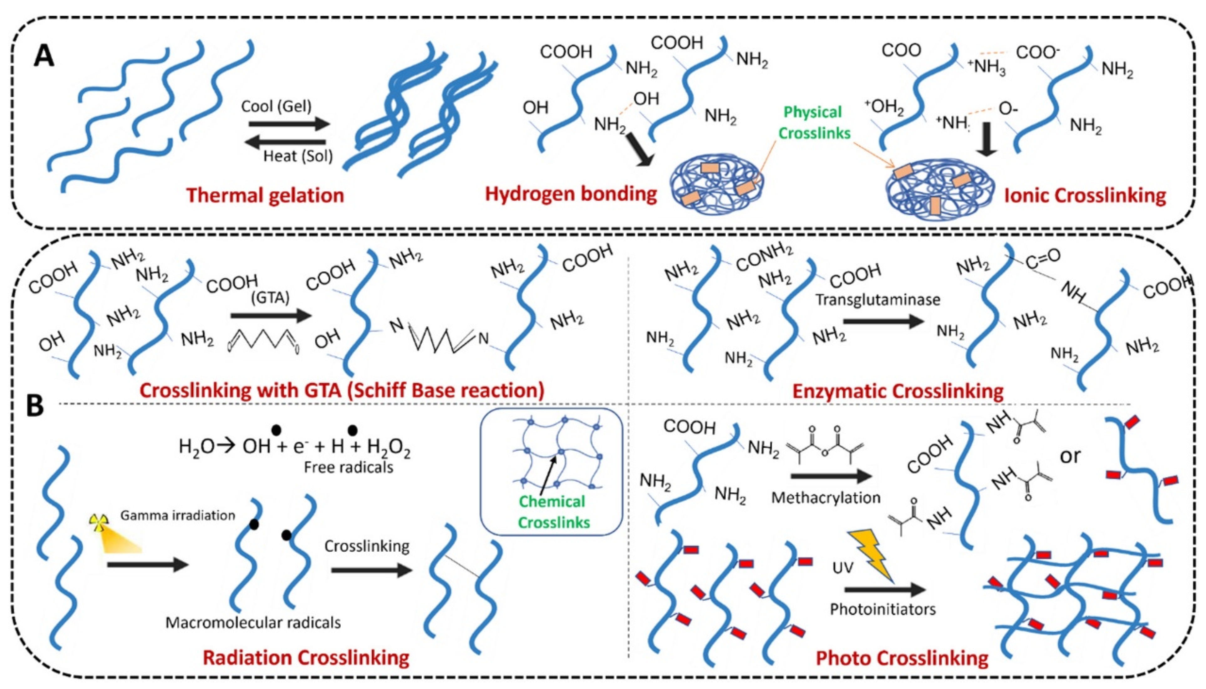
Figure 1. (A) physical and (B) chemical crosslinking techniques employed for gelatin hydrogel formation. Reprinted with permission from Ref. [10] Copyright 2022 Elsevier.
Gels 09 00146 g001
Figure 2. Applications of hydrogels in drug delivery for oral and maxillofacial diseases.
Gels 09 00146 g002
Figure 3. Schematic diagram of fabrication of GelMA/PL/Laponite microspheres and its application for endodontic regeneration. (1) GelMA, Platelet Lysate, and Laponite formed a hydrogel precursor. (2) Hydrogel precursors mixed with hDPSCs to make microspheres. (3) Preparation of microspheres by electrostatic microdroplet method. (4) Microspheres were collected after centrifugation and observed under the microscope. (5) In vitro biological experiments including the viability, spreading, proliferation, and differentiation behaviors of hDPSCs encapsulated in the microspheres, as well as the effects of released PL-derived GFs on tube-formation of HUVECs and hDPSCs migration behavior were systematically carried out. (6) In vivo studies were done to further evaluate whether this hybrid microsphere system could facilitate angiogenesis and histogenesis when subcutaneously implanted in immune deficiency mouse model. Reprinted with permission from Ref. [40] Copyright 2021 Elsevier.
Gels 09 00146 g003
Figure 4. Schematic illustration of the preparation and application of the CS/β-GP/gelatin hydrogels. Reprinted with permission from Ref. [112] Copyright 2019 Elsevier.
Gels 09 00146 g004
Figure 5. Schematic Illustration of TC-PCM@GNC-PND as a Combined Platform for Hyperthermia and Antibiotics with an Obvious Synergistic Antibacterial Effect. Reprinted with permission from Ref. [61] Copyright 2020 American Chemical Society.
Gels 09 00146 g005
Figure 6. Schematic illustration of the preparation of (A) CM and (B) CA@CM/MD composite sponges, as well as the application for the prevention of dry sockets after tooth removal. The CM with the porous structure were prepared by combining ionotropic gelation with biomimetic mineralization. After Ca2+ crosslinking, lyophilization, and electrostatic interaction, CA@CM/MD composite sponges were fabricated and shaped to the root-like shape for better suitability and applicability. Reprinted with permission from Ref. [132] Copyright 2022 Elsevier.
Gels 09 00146 g006
Figure 7. Scheme of mucoadhesive KPV@PPP_E hydrogel for chemotherapy-induced oral mucositis. Reprinted with permission from Ref. [58] Copyright 2022 Royal Society of Chemistry.
Gels 09 00146 g007
Figure 8. Schematic diagram of the in situ thermosensitive hydrogel containing GA micelles for improving anti-tumor immunity against OSCC. The GA micelle-encapsulated PLEL sol was locally injected into the tumor, formed hydrogel at the body temperature, and continually released GA in situ, thus exerting the chemotherapeutic effect and anti-tumor immune activation. The mice treated with GA-MIC-GEL showed increased cytotoxic T cells as well as reduced immunosuppressive cells at the tumor sites, suggesting the T cell activation and reversal of the tumor immune microenvironment. Besides, the systemic expression of PD-1 in GA-MIC-GEL treated mice also decreased. Reprinted with permission from Ref. [56] Copyright 2022 Elsevier.
Gels 09 00146 g008
Table 1. The characteristics of the studies using hydrogels with special properties in this review.
ClassificationHydrogelsCrosslinking MethodDrug-Loading WaysActive IngredientsApplicationCharacteristicReferences
InjectableFibrinPhysical crosslinkingPoly (d,l) Lactic Acid nanoparticlesClindamycinRegenerative endodontics and antibacterialExcellent cytocompatibility; physiological degradation kinetics; non-toxicity of degradation products; replacement with cell-derived ECM within a few days.[23]
Hyaluronic acid (HA)Chemical crosslinking/rhBMP-2; rMSCs and BMP-2; platelet lysate, chemotactic and pro-angiogenic growth factors (PDGF and VEGF)Regenerative endodontics; Mandibular reconstruction; Peri-implant osteogenesisControlled release of drugs; supportive matrix for cell culture, recruitment, and revascularization induction.[24,25,26,27]
AlginateChemical crosslinking/Vancomycin/deferoxamine/dexamethasone (Van/DFO/Dex)Maxillofacial bone regenerationLocally sustained release property; prominent biological functions.[28]
Alginate (ALG)/hyaluronic acid (HA)Chemical crosslinking/BMP-2Mandibular reconstructionIn situ gelling hydrogel with a controllable gelation rate using CaSO4 as a crosslinking agent and Na2HPO4 as a crosslinking retardation agent.[29]
Gelatin-hyaluronic acid hydrogelChemical crosslinking/Vascular endothelial growth factor (VEGF)medication-related ONJ (MRONJ)Assists bone healing and prevents MRONJ via a pro-angiogenic and immunomodulatory mechanism.[30]
−CHO inaldehyde-modified hyaluronic acid (HA-CHO) and −NH2 in glycol chitosan (GC), Fe3+Chemical crosslinking/Ginsenoside Rg1 and amelogeninPeriodontal diseaseInjectable and self-healing hydrogels with double-dynamic bond tunable mechanical, gel-sol transition, and drug delivery properties.[31]
Oxidized alginate/carboxymethyl chitosanChemical crosslinking/A dental epithelial cell line, HAT-7Dental Enamel RegenerationThe Self-Crosslinkable hydrogels could be used as an injectable cell carrier for dental enamel tissue engineering applications.[32]
Carboxymethyl-chitosan and a diglycidyl etherChemical Crosslinking/Calcium phosphate nanoparticlesPulp cappingThese composites have moduli up to 3 MPa, and support the culture of dental pulp stem cells for more than 3 weeks.[33]
The chitosan hydrogel as well as blends of polyvinyl pyrrolidone (PVP), polyvinyl alcohol (PVA) and PEGChemical crosslinking/InsulinPeriodontal bone regenerationA linear hydrogel that is injectable into periodontal pockets, and is able to carry a small insulin load through physical bonds and provide sustained release.[34]
Oxidized dextran (OD) and phenylboronic acid-functionalized poly (ethylene imine) (PBA-PEI)Chemical crosslinking/Doxycycline and metformin Periodontal antibacterial, anti-inflammatory and bone regenerationSimultaneously improving drug loading efficiency (doxycycline and metformin) through B−N coordination and achieve ROS triggered drug release locally.[35]
poly(lactide-co-glycolide) (PLGA) and N-methylpyrrolidone (NMP)Chemical crosslinking/Minocycline (MCL)Periodontal antibacterialExhibited the characteristic of Newton fluid with acceptable syringeability. Drug release could last for more than 48 h with an acceptable “burst release”.[36]
poly(phosphazene)Physical crosslinking/BMP-2Peri-implant osteogenesisVertical bone regeneration and higher osseointegration levels.[37]
polyisocyanopeptide (PIC)Physical crosslinkingPLGA microspheresDoxycycline and lipoxinPeriodontal anti-inflammatory and antibacterialAppropriate injectability; long-term structural stability; the release profiles of drugs could be manipulated by adjusting the loaded mass ratio of acid- and ester- terminated PLGA microspheres in the PIC gels.[2,38]
Gelatin methacrylate (GelMA)Chemical crosslinking (Photocrosslinking)(Au NBPs@SiO (2)) or noneChlorhexidine (CHX); pro-angiogenic growth factors (GFs); BMP-mimetic peptide; minocycline; silibininRegenerative endodontics; Periodontal antibacterial; Prevention of dry socketsCytocompatible; biodegradable; provides sustained release of drugs.[39,40,41,42]
MA-HA/di-thiol PEGChemical crosslinking (Photocrosslinking)/A novel, small-molecule noncompetitive adenosine triphosphate (ATP) drug: NP928, belongs to the thiadiazolidinone (TDZD) familyDentin RegenerationBiodegradable; gelling in situ upon dental blue light exposure.[43]
Polyethylene glycol-maleate-citrate (PEGMC)Chemical crosslinking (Photocrosslinking)/Calcium hydroxideDirect pulp cappingThe light-curing time for hydrogel is comparable to composite resin. Controlled Ca2+ release was obtained.[44]
Methacrylated-poly-γ-glutamic acid (MPGA) polymerChemical crosslinking (Photocrosslinking)Methacrylated-poly-γ-glutamic acid nanoparticles (PGA-MNP)Metronidazole and CHXPeriodontal antibacterialIt is a pH-sensitive drug delivery system which used blue-light photopolymerization for preparation.[45]
Injectable and thermosensitivePluronic F127 (PF127)Chemical crosslinking (thermal crosslinking)/Iloprost; Simvastatin; glycogen synthase kinase 3 beta inhibitor (BIO); MetronidazoleRegenerative endodontics; Periodontal anti-inflammatory and osteogenicControlled drug release; could adhere to hard tissue and gradually release.[46,47,48,49]
chitosan (CS)/β-glycerophosphate (GP)Physical crosslinkingGraphene oxide (GO) nanosheets or noneBupivacaine hydrochloride (BH); VEGF; hyaluronic acid; bone morphogenetic protein-7 (BMP-7) and ornidazole (ORN); Naringin; aspirin and erythropoietin (EPO); quercetinTopical anesthesia; Pulp capping; Temporomandibular disorders; Periodontal anti-inflammatory and tissue regenerationProlonged drug release time; a stable and sustained drug release system.[50,51,52,53,54]
Chitosan/gelatin/glycerol phosphateChemical crosslinking/BMP-6Periodontal tissue regenerationProvide a 3D environment for transplanted stem cells and to enhance stem cell delivery and engraftment.[55]
poly(D, L-lactide)-poly(ethylene glycol)-poly(D, L-lactide) (PLEL)Chemical crosslinkingmPEG2000-PCL micellesGambogic acid (GA)Oral cancerThe thermosensitive GA-MIC-GEL with sensitive sol-gel transition characteristics could form hydrogel at 37 °C within 24 s, facilitating the local delivery and sustained GA release.[56]
poly(D,L-lactide-coglycolide)-poly(ethy-lene glycol)-poly(D,L-lactide-coglycolide) triblock copolymers (PLGA-PEG-PLGA) Chemical crosslinking/Doxorubicin (DOX) and celecoxibOral cancerpH-responsiveness; biocompatibility; simultaneously release hydrophobic and hydrophilic drugs at the oral tumor site.[57]
PLGA-PEG-PLGA (PPP)/epigallocatechin-3-gallate (EGCG)Chemical crosslinking/Tripeptide KPVOral mucosal diseaseIn situ mucoadhesive; anti-inflammatory, antibacterial and repairing effect on chemotherapy-induced oral mucositis.[58]
poly(ethylene glycol)-poly(ε- caprolactone)-poly(ethylene glycol) (PEG-PCL-PEG, PECE)Chemical crosslinking/Parathyroid hormone (PTH) or parathyroid hormone-related protein (PTHrP)Orthodontic tooth movementAqueous solution of PECE copolymers changed from the “sol” phase to the “gel” phase with the increase in temperature.[59]
poly(ethylene glycol)-6-poly(lactic-co-glycolic acid)-6-po(y(N-isopropy!acrylamide) (PEG-PLGA-PNIPAM) hydrogelChemical crosslinkingMesoporous silica nanoparticle (MSN)-embedded core-shell structureMicroRNA-222 and ASPMandibular reconstructionInjectable colloidal hydrogel with mesoporous silica nanoparticles for sustained co-release of microRNA-222 and aspirin[60]
poly(N-isopropylacrylamide-co-diethylaminoethyl methacrylate) (PND)Chemical crosslinkingGold nanocages (GNC)Tetracycline(TC)Periodontal antibacterialNear infrared light (NIR) light controlling drug release through the dual thermosensitive interaction of liquid-solid transition of PCM and coil-granule transition of PND.[61]
Self-assembling peptides (SAP) hydrogel (P11-4 and P11-28/29)Physical crosslinking/Tetracycline, ciprofloxacin, and doxycyclinePeriodontal tissue regeneration and antibacterialBiocompatibility; cargo-loading capacity; tunable physicochemical and mechanical properties.[62]
Polyethylene glycol diacrylate (PEG-DA) based scaffolds, dithiothreitol (DTT), and a novel designed functional peptide module (FPM)Chemical crosslinking/Stromal cell derived factor-1 (SDF-1)Periodontal tissue regeneration and antibacterialPEGPD@SDF-1 hydrogel exhibited preferable biocompatibility and could promote the proliferation, migration, osteogenic differentiation of periodontal ligament stem cells (PDLSCs) and inhibit the growth of Porphyromonas gingivalis.[63]
AdhesiveMethylcellulosePhysical crosslinkingMesoporous silica nanoparticles (MSNs) or noneDOX; Melissa officinalis oilOral cancer; Oral mucosal diseaseBiocompatibility; controllable mechanical performance;
thermosensitive and injectable characteristics.
[64,65]
Hydroxypropyl methylcellulose (HPMC)Chemical crosslinkingTransfersomes or nonePrilocaine hydrochloride and lidocaine hydrochloride; 5-Fluorouracil and Etodolac; polyaspartic acid-stabilized amorphous calcium phosphate (PAsp-ACP) nanoparticlesTopical anesthesia; Oral cancer; Biomimetic mineralizationHPMC can be desiccated to form a dry film. In a moist environment, this film gradually changes into a gel.[6,66,67]
PAM-PDAPhysical crosslinkingAuNPsMedical anestheticTopical anesthesiaThis hydrogel with microneedle resulted in reduced pain, higher anesthetic accuracy and faster recovery.[68]
CarbopolPhysical crosslinkingPoly(ε-caprolactone) nanocapsulesLidocaine and prilocaineTopical anesthesiaNon-Newtonian pseudoplastic flows; satisfactory mucoadhesive strength; non-cytotoxicity; slow permeation across oral mucosa.[69]
Polyaldehyde dextran and chitosanChemical crosslinking/Silver nanoparticlesOral cancerAntitumor responses were enhanced by the subcutaneous delivery of an adhesive hydrogel incorporating silver nanoparticles (which inhibited the growth of bacteria competing with Peptostreptococcus).[70]
Acrylic acid/polyethylene glycolChemical crosslinking/Silver nanoparticles and propranolol HClAntibacterialThe nanocomposites show a promising self-disinfection property and mucoadhesive strength.[71]
ExpansiveN-vinylpyrrolidone(NVP), 2-hydroxyethyl methacrylate (HEMA), and glycerolmonomethacrylate (GMMA) monomers with methacrylicacid (MA).Chemical crosslinking/BenzocaineAnesthesia of tissue expandersTissue expanders based on the controlled rate expansive hydrogels. Most of the drug (90%) was released within 48 h.[72]
Nondegradable2-hydroxyethyl methacrylate (HEMA) and trimethylolpropane trimethacrylate (TMPT)Chemical crosslinking/Cetylpyridinium chloride (CPC)AntibacterialApplying a non-biodegradable hydrogel to resin-based materials as a reservoir for water-soluble antimicrobials[73]
Disclaimer/Publisher’s Note: The statements, opinions and data contained in all publications are solely those of the individual author(s) and contributor(s) and not of MDPI and/or the editor(s). MDPI and/or the editor(s) disclaim responsibility for any injury to people or property resulting from any ideas, methods, instructions or products referred to in the content.