Article

Calcium-Polyphosphate Submicroparticles (CaPP) Improvement Effect of the Experimental Bleaching Gels’ Chemical and Cellular-Viability Properties

by
Mariángela Ivette Guanipa Ortiz
1,
Juliana Jarussi dos Santos
2,
Jonny Burga Sánchez
3,
Ubirajara Pereira Rodrigues-Filho
2,
Flávio Henrique Baggio Aguiar
1,
Klaus Rischka
1,4,* and
Débora Alves Nunes Leite Lima
1
1
Department of Restorative Dentistry, Piracicaba Dental School, University of Campinas—UNICAMP, Piracicaba 13414-903, Brazil
2
Group of Chemistry of Hybrid and Inorganic Materials (GQMATHI), São Carlos Institute of Chemistry, University of São Paulo (USP), São Carlos 13563-120, Brazil
3
Department of Physiological Science, Piracicaba Dental School, University of Campinas—UNICAMP, Piracicaba 13414-903, Brazil
4
Fraunhofer Institute for Manufacturing Technology and Advanced Materials IFAM, Wiener Straße 12, 28359 Bremen, Germany
*
Author to whom correspondence should be addressed.
Submission received: 1 December 2022 / Revised: 28 December 2022 / Accepted: 30 December 2022 / Published: 4 January 2023

Abstract

:
The aim of this research was to develop and characterize the chemical and cellular-viability properties of an experimental high-concentration bleaching gel (35 wt%-H2O2) containing calcium-polyphosphate particles (CaPP) at two concentrations (0.5 wt% and 1.5 wt%). The CaPP submicroparticles were synthesized by coprecipitation, keeping a Ca:P ratio of 2:1. The CaPP morphology, size, and chemical and crystal profiles were characterized through scanning and transmission electron microscopy, energy-dispersive X-ray analysis, and X-ray diffraction, respectively. The assessed bleaching gels were experimental (without CaPP); 0.5% CaPP; 1.5% CaPP; and commercial. The gels’ pH values and H2O2 concentrations (iodometric titration) were determined. The odontoblast-like cell viability after a gel’s exposure was assessed by the MTT assay. The pH and H2O2 concentration were compared through a repeated-measures analysis of variance (ANOVA) and a Tukey’s test and the cell viability through a one-way ANOVA and a Tukey’s test using a GraphPad Prism (α < 0.05). The CaPP particles were spherical (with Ca and P, 135.7 ± 80.95 nm size) and amorphous. The H2O2 concentration decreased in all groups after mixing (p < 0.001). The 0.5% CaPP resulted in more-stable pH levels and higher viability levels than the experimental one (p < 0.05). The successful incorporation of CaPP had a positive impact on the bleaching gel’s chemical and cellular-viability properties when compared to the experimental gel without these particles.

Graphical Abstract

1. Introduction

Given the high levels of dissatisfaction (30%) with teeth darkening, bleaching gels based on hydrogen peroxide have been extensively introduced into the market [1]. Their chief mechanism of action is the oxidation of the organic molecules, which stain teeth, into smaller and lighter compounds [2]. Along with their bleaching effect, both the H2O2 and its reactive oxygen species (ROS) are responsible for the “nonspecific” oxidative action on the organic and inorganic structural components of enamel [3,4,5,6,7] and dentin [8], generating topographic and mineral changes in these structures, even applying more-neutral or -alkaline gels [9,10,11].
Although enamel is a biologically inert biomaterial, remineralizing components that dissolve through a chemical process can generate local precipitation of biosimilar Ca/PO43− on the surface. This must be considered for the development of dental materials with remineralizing properties, such as bleaching gels, that can achieve an adequate bleaching effect while reducing the intrinsic adverse effects of the therapy [3,12,13,14].
Therefore, the demand for experimental bleaching agents that can ensure maximum benefit with minimum harm has grown in recent years [5,14,15,16,17]. Those that incorporate a source of Ca/PO43− compounds still report changes in the tooth mineral structure [18], and some of the positive evidence could be attributed not only to the compound effect but also to pH changes [14,17] or the decrease in peroxide concentration [1]. For this, it can be inferred that there is still no gold standard Ca/PO43− compound for bleaching-gel preparation.
One promising material is polyphosphate (PolyP), a natural compound of living cells and a source of ortho-phosphate (PO43−), which is required for hard tissue formation [19,20]. More recently, the synthesis of calcium-polyphosphate (CaPP) submicroparticles by a defined Ca:P molar ratio of 2:1 has been described [21]. CaPP is an amorphous source of Ca and PO43− that stimulates mineral deposition in both enamel and dentin [19,22]. The interaction of CaPP with these tissues can be partially explained by the formation of Ca2+ bonds between the mineral matrix of dental hydroxyapatite and CaPP [19,23].
In view of bleaching treatment–induced mineral alterations and given CaPP’s possible remineralizing effects, the incorporation of CaPP in experimental bleaching gels is an innovative and promising alternative for treatment. Thus, the aim of this research was to develop and characterize the chemical and cellular-viability properties of an experimental high-concentration bleaching gel (H2O2/HP—35 wt%) containing calcium-polyphosphate (HP-CaPP) particles in two concentrations (0.5 wt% and 1.5 wt%). For this study, the null hypotheses were as follows: (1) the H2O2 concentration and pH levels of the HP-CaPP bleaching agent will not differ from those of a commercial bleaching agent and an experimental bleaching gel without CaPP, and (2) the cytotoxic potential of the HP-CaPP bleaching agent will not differ from that of a commercial bleaching agent or that of an experimental bleaching gel without CaPP.

2. Results

2.1. Scanning Electron Microscopy (SEM) and Energy-Dispersive X-ray Analysis (EDX)

SEM analysis showed that the particles had a spherical morphology (Figure 1a). The presence of Ca and P was detected by using the EDX system. Although certain levels of Na and Cl persisted, the amounts were minimal. The particles presented a Ca:P atomic ratio of 1.11, as shown in Figure 1b.

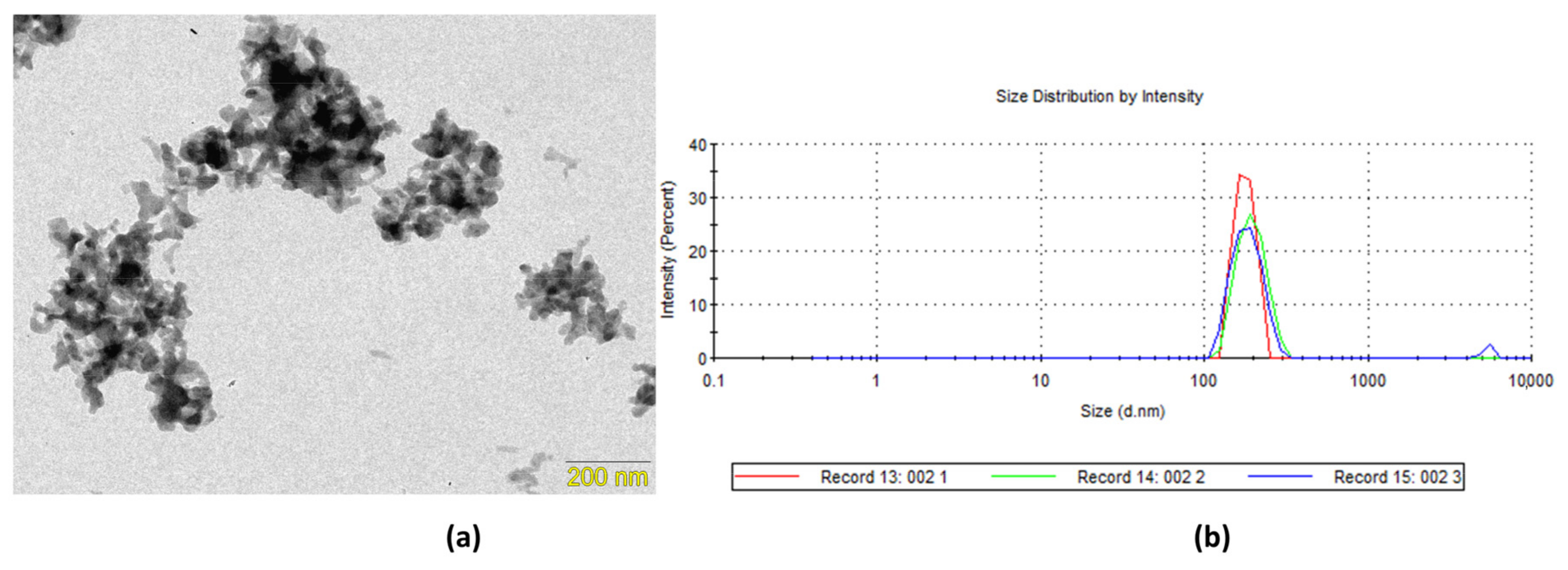
2.2. Transmission Electron Microscopy (TEM) and Dynamic Light Scattering (DLS)

According to the TEM images, the CaPP particles showed a submicro particle size (135.7 ± 80.95 nm; Figure 2a). The hydrodynamic radius of the CaPPs showed a distribution of 257.1 nm (±26.52 nm), with a polydispersity index of 0.43 (±0.05) and a correlation of 0.90 (Figure 2b).

2.3. X-ray Diffraction (XRD)

XRD analysis demonstrated the amorphous character of the synthesized CaPP particles given that the characteristic peaks of crystalline materials were not observed in the diffractogram (Figure 3).

2.4. Visual and pH Evaluation Results

The visual aspect of the experimental gels (Figure S1) denoted an opaque appearance with increasing CaPP concentrations. The pH values (without dilution) varied among the periods of time (ANOVA; p < 0.0001) and among the bleaching gels (ANOVA; p < 0.0001). The gel with 0.5% CaPP had the highest pH values when compared to the others. After 15 min of mixing, all gels maintained pH values above 5.1. The 1.5% CaPP and commercial gels (Tukey; p < 0.05) had the lowest pH values in this assessment period. After 45 min of mixing, the experimental bleaching gel had the lowest pH value (Tukey; p ≤ 0.007) (Figure 4).

2.5. Hydrogen Peroxide Concentration of the HP-CaPP—Titrimetric Analysis (Iodometric)

The H2O2 concentration decreased with time in all groups (ANOVA; p < 0.0001). There were no significant differences among the groups for H2O2 concentration after 15, 30, and 45 min of gel mixing. After 90 min, the experimental bleaching gel presented the lowest H2O2 concentration (Tukey; p < 0.005); however, after 150 min, the 0.5% and 1.5% CaPP gels presented the highest H2O2 levels (Tukey; p < 0.005) (Table 1).

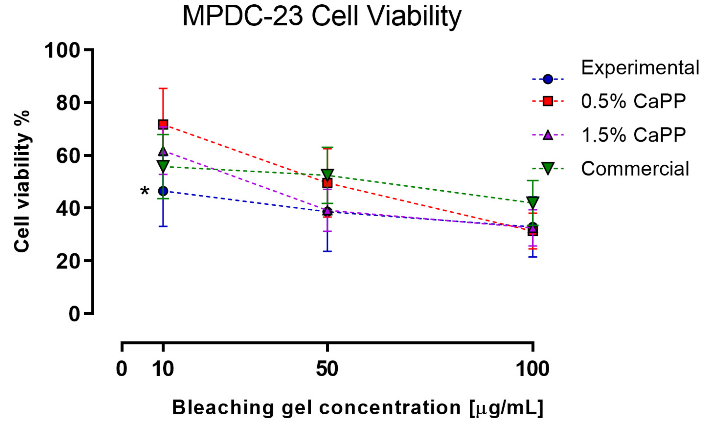
2.6. Odontoblasts-like Cells (MDPC-23) Viability after Exposure to the HP-CaPP Bleaching Gel

The cell viability results are displayed in Figure 5. For the 10 µg/mL concentration, the experimental gel was significantly different from the other gels (p < 0.05; Tukey’s post hoc ANOVA test). The IC50 of this experimental gel (9.81) was significantly different from the 0.5% CaPP (35.92), 1.5% CaPP (22.65), and commercial (26) gels. Additionally, the IC50 of the 0.5% CaPP was significantly different from the 1.5% CaPP (p < 0.05).

3. Discussion

An experimental bleaching gel based on H2O2-35 wt% with calcium-polyphosphate submicroparticles (HP-CaPP) was developed, and its chemical (pH and H2O2 concentration) and biological (cellular-viability) properties were assessed. According to the obtained results, the first null hypothesis was rejected, as the HP-CaPP bleaching gels (CaPP—0.5 and 1.5 wt%) had higher pH levels compared to the experimental gel without CaPP and similar values to the commercial bleaching gel. The H2O2 concentration was similar among the bleaching gels. The second hypothesis was also rejected, as the cell viability was higher for the HP-CaPP groups (CaPP—0.5 and 1.5 wt%) than for the experimental gel without CaPP but was similar to the commercial bleaching gel.
Following the coprecipitation method [21], CaPPs were synthetized and characterized for later incorporation into an experimental bleaching gel (HP-CaPP). The SEM images display particles with spherical morphology. Additionally, the EDX spectra display marginal Na levels but significant levels of P and Ca, as previously reported [21,22,24].
Concerning the CaPP size, the TEM analysis indicated submicrometric particles (135.7 ± 80.95 nm). The starting Ca:P molar ratio was 2:1, which allowed the formation of smaller particles [22]. Although the DLS results display a higher hydrodynamic radius (257.1 ± 26.52 nm), this analysis considers the particles’ hydrodynamic diameters (257.1 ± 26.52 nm). Another factor that may influence DLS analysis and contribute to a higher hydrodynamic radius reading is the particle agglomeration that usually occurs throughout the reading [25,26]. As determined by XRD analysis, the CaPPs retained their amorphous character. The amorphous state is relevant for increasing the remineralizing potential of the particles. This is related to the fact that when dental tissues are exposed to highly supersaturated phosphate solutions (e.g., CaPP), an amorphous calcium phosphate precipitate can be formed and later transformed into an organized crystal apatite structure [22,23,27].
The initial pH values of the experimental formulations were intended to remain close to those of the undiluted commercial bleaching gel to determine the effect of the CaPP particles within a similar pH range [28]. The smallest CaPP concentration (0.5 wt%) seemed to have a beneficial effect on pH value stability. Initially, the medium protons, thanks to their high electronegativity, replace the Ca2+ ions on the CaPP structure, leading to an increase in pH, as observed after 10 min in the 0.5% CaPP gel. Moreover, phosphoric acid is also produced during the hydrolytic cleavage of the CaPP chain, which decreases the gel’s pH [29]. When we tripled the CaPP concentration (1.5 wt%), this latter effect was predominant, as after 15 min, this gel had a lower pH than the experimental and 0.5% CaPP gels. The commercial gel showed a pH decrease 15 min after mixing (5.17), which followed the trend of previous assessments [15,30].
After the longest assessment time, 45 min after the preparation of the experimental gel without CaPP, the lowest pH values were reached. The lower pH values have been related to higher enamel alterations (e.g., lower microhardness, higher superficial roughness) and to reduced bleaching efficacy [5,7]. Thus, the incorporation of CaPP into an experimental bleaching gel could be regarded as positive for the pH of the bleaching gels and therefore positive for the enamel properties in future in vitro/in vivo applications.
The H2O2 concentration was lower than 35 wt% 15 min after mixing the gels. However, given the H2O2 instability, the exact concentration is difficult to determine by titration [31], which can be considered a limitation of the current study. After gel preparation, the H2O2 present in the bleaching gel starts to decompose into free radicals and H2O as time passes [31]. Nevertheless, in the experimental gel containing CaPP, this reduction was not as accentuated as in the experimental gel without CaPP and the commercial gel. This could occur because of the H2O2 stabilization promoted by the phosphate molecules released from the CaPP chain [31,32,33,34].
Given that CaPP is a biocompatible compound, its presence in bleaching gels, as expected, did not increase their cytotoxic potential [21,35,36]. Interestingly, lower amounts of CaPP (0.5 wt%) were better at sustaining MDPC-23 cells’ viability compared to the experimental gel without CaPP. CaPP chain cleavage not only provides ortho-phosphate units that aid in the remineralization process but also generates “metabolic fuel” by supporting ATP production [21]. This intracellular burst of energy could increase cell proliferation [37,38], which could explain why this group presented the best results for cell viability at a concentration of 10 µg/mL.
However, a higher polyphosphate concentration within the bleaching gel (1.5 wt%) did not result in an increase in cell viability. The hydrolytic cleavage of the CaPP chain led to a more acidic gel, as seen in our pH assessments. Under these conditions, cell growth, proliferation, and differentiation were retarded [39]. It could also be speculated that H2O2 decomposition into free radicals at this pH level is reduced; therefore, the free remaining H2O2 cytotoxic potential could have affected cell viability [40,41], surpassing the CaPP protective effect displayed at a lower CaPP concentration and a higher pH level (0.5% CaPP).
As previously described, CaPP, as a long-chain bioinorganic polymer, is a promising bioactive compound in bioremineralization [19,23,42]. This characteristic behavior would be useful to reduce the enamel mineral loss associated with high-concentration bleaching gels, while the presence of the ions would have a potential effect on reducing dental sensitivity, which remains the most common clinical symptom of the bleaching treatment [1,5]. Although the successful incorporation of CaPP did not negatively alter the experimental gels’ chemical and biological properties compared to commercial and experimental bleaching gels based on 35% H2O2 without these particles, further studies should be conducted to determine the effect of CaPP on the bleaching efficacy and the enamel chemical and mechanical properties of this type of experimental bleaching gel when compared to commercially established treatments.

4. Conclusions

In the present study, calcium-polyphosphate fine particles were successfully synthesized through the coprecipitation method [21] and incorporated into bleaching gels based on 35 wt% H2O2. The incorporation of CaPP, even in smaller concentrations (0.5% w/t), stabilized the pH, reduced the cytotoxic potential, and retained similar hydrogen peroxide concentration values compared to the experimental bleaching gel without CaPP after 45 min of mixture in vitro. Because the CaPP gel’s retained similar chemical and biological properties to the commercial bleaching gel without CaPP, their remineralizing potential turns them into a promising clinical alternative to conventional bleaching gels.

5. Materials and Methods

5.1. Synthesis of Calcium-Polyphosphate Particles (CaPP) and Characterization

To obtain CaPP particles, a coprecipitation technique was used [21]. Based on our own pilot synthesis and previous works [19,21], a sodium polyphosphate (NaPP), with an average chain length of ~35 phosphate units (Chemische Fabrik; Budenheim, Germany), was used for CaPP synthesis (Table 2).
The pH of the NaPP solution (5 g in 250 mL H2O) was adjusted to 10 using an aqueous 1 M NaOH solution. Then, the CaCl2·2H2O (Sigma-Aldrich, Taufkirchen, Germany) solution (14 g in 250 mL H2O) was added to the NaPP solution at a controlled rate, 1 mL/min, using a peristaltic pump (P-1; Pharmacia Biotech, Uppsala, Sweden) to obtain a Ca:P ratio of 2:1. During the addition, the pH was maintained (10) at room temperature using 1 M NaOH solution. After 4 h of stirring, the final solution was washed for 10 min and centrifuged (3500 rpm × 20 min) twice with distilled water and at last twice with absolute ethanol. The final slurry was kept in an oven at 60 °C (14 h) [21,22]. The dried material was crushed with a pestle in a mortar to obtain a fine CaPP powder (Figure 6).

5.1.1. Scanning Electron Microscopy (SEM) and Energy-Dispersive X-ray Analysis (EDX)

A thin gold layer was deposited on the CaPP surface (BAL-TEC SCD 050; Capovani Brothers Inc., NY, USA) to assess CaPP morphology. Images at ×5000 magnification were obtained on an SEM (JSM-5600, JEOL; Tokyo, Japan) with a 15 kV accelerating voltage, 13 mm Z, and 15 mm WD.
The powder was coated with carbon by vapor deposition (Delton vacuum Desk II, Moorestown, NJ, USA) to characterize the chemical elements of the CaPP particles. Elemental analysis was performed using an EDX detector (Vantage, Acquisition Engine Company, Tokyo, Japan) connected to an SEM (JSM-5600, JEOL, Tokyo, Japan). The EDX system was operated at 15 kV with a collection time of 100 s, 30° incidence, Z = 20 mm, and WD = 20 mm. Three areas of approximately 10 µm2 were evaluated.

5.1.2. Transmission Electron Microscopy (TEM) and Dynamic Light Scattering (DLS)

Morphological and size examinations of CaPP were performed using a transmission electron microscope (Phillips CM 200, Phillips, Amsterdam, the Netherlands) at 200 kV with a LaB6 filament. The sample was prepared, and the obtained images were analyzed to estimate the particle size on the basis of an average diameter of 60 particles (ImageJ; public domain image software).
To determine the particle hydrodynamic radius, CaPP was diluted in distilled water (1 mg/mL), stirred for 15 min (50 °C), and centrifuged for 10 min (10,000 rpm). The supernatant was analyzed using a laser diffraction particle analyzer (Zetasizer Nano ZS90; Malvern Instruments Ltd., Worcestershire, UK) with the following parameters: 10 runs of 10 s, at 25 °C, with 60 s of stabilization, and a 90° scattering angle.

5.1.3. X-ray Diffraction (XRD)

The CaPP powder crystallinity pattern was recorded on a diffractometer (D8 Advance; Bruker, MA, USA) with Cu-Kα radiation (40 kV, 30 mA) and an equipment geometry of 2θ. Continuous readings were performed with a step of 0.02° and an accumulation time of 0.3 s (interval of 4°/min).

5.2. Experimental Bleaching Gel Containing Calcium-Polyphosphate Particles (HP-CaPP)

Formulations of high-concentration H2O2-based bleaching gel (H2O2—35 wt%) with two separate components were proposed: A—H2O2 (pH ≈ 1.8); B—thickener and calcium-polyphosphate particles (CaPP) (pH ≈ 12) [5]. Three types of experimental bleaching gels were manipulated: without CaPP (experimental) and containing 0.5 and 1.5 wt% CaPP (CaPP 0.5% and CaPP 1.5%). In addition, Whiteness HP Maxx (FGM Dental Products, SC, Brazil) was used as a commercially available control.
The reagents of component A were stirred for 60 min (≈10 °C) in the dark to prevent H2O2 decomposition [31,43]. For the gels containing CaPP in component B, CaPP was first diluted in distilled water and stirred in a water bath for 15 min (50 °C). After cooling down, the remaining reagents of component B were homogenized (Speed Mixer DAC 150.1; FlackTek, Inc., SC, USA) at 2000–2500 rpm for 10 min (Table 3).
For all gel analyses, experimental components A and B were mixed in a 3:1 weight proportion, while the commercial control bleaching gel was prepared according to the manufacturer’s instructions. All gels were kept under refrigeration (8 ± 1 °C) before the experiment and were brought to room temperature 30 min before mixing.

5.2.1. Evaluation of pH

The pH values of 4 g of each bleaching gel were recorded using a benchtop digital pH meter (mPA210; MS TECNOPON, Piracicaba, SP, Brazil), which was previously calibrated with buffered solutions (pH 4, 7, and 10) at 25 °C. The pH values of the gels were recorded in triplicate 5, 10, 15, 30, and 45 min after the mixture of both components. The mean of the three measurements was considered the final value for each assessment time.

5.2.2. Hydrogen Peroxide Concentration of the HP-CaPP—Titrimetric Analysis (Iodometric)

The gels’ respective H2O2 concentrations were determined by employing iodometric titration with a hydrogen peroxide test kit (Hanna Instruments; Barueri, SP, Brazil) and a standardized sodium thiosulfate solution (Na2S2O3; 0.1 N). In brief, iodide ions (I) can be oxidized by H2O2 to iodine (I2), which in the presence of starch forms a blue charge-transfer complex by the imprisonment of polyiodide species within the helix structure of amylose. I2 can be reduced by titration with a standard Na2S2O3 solution to I, which is colorless [31,44]. The H2O2 concentration can be calculated by the volume of the standard Na2S2O3 solution required to change the color of the bleaching solution using Equation (1) [45].
  C =   [ ( 0.1 M × V ) / 2 ] × 34.0147 g / m o l V f      
where C = final concentration; V = volume of the Na2S2O3 (0.1 N) solution used; and Vf = final volume of the diluted H2O2 solution used to conduct the titration.
All bleaching gels were diluted in distilled water (1 mg/mL of H2O2) and remained at room temperature in static and dark conditions. At defined times, samples were collected from the upper area of the samples (15, 30, 45, 90, and 150 min after mixing) and further diluted to obtain 0.2 mg/mL H2O2 solutions in three 25 mL glass vials [31]. The titration procedure was performed on the three samples, and the mean was considered the final value of each sample/time assessed.

5.2.3. Odontoblasts-like Cells (MDPC-23) Viability in the HP-CaPP Bleaching Gel

The MTT reduction method was performed to determine the viability of MDPC-23 cells after exposure to the bleaching gels. The MDPC-23 cells were cultured in Dulbecco’s modified Eagle’s medium (DMEM, Sigma-Aldrich, St. Louis, MO, USA) supplemented with antibiotics, 100 IU/mL penicillin and 100 μg/mL streptomycin (Vitrocell Embriolife, Campinas, Brazil), 2 mmol/L glutamine, and 10% fetal bovine serum (FBS—GIBCO; Thermo Fisher Scientific, Waltham, MA, USA) at 37 °C in a 5% CO2 atmosphere [39,46].
After reaching 80% confluence, the MDPC-23 cells were washed with 0.25% trypsin/EDTA (GIBCO; Thermo Fisher Scientific, Waltham, MA, USA) to separate them from the plate. The separated cells were centrifuged at 3000 rpm for 5 min at 4°C. A Neuberger chamber was used to count the cells. The supernatant was discarded; the cells were passed to a new medium (DMEM), transferred to 96-well cell culture plates (Corning Costar Corp., Cambridge, MA, USA) at a concentration of 5 × 10⁴ cells/mL, and subsequently incubated in a 5% CO2 atmosphere at 37 °C for 24 h.
After the incubation period, the cells were exposed to diluted bleaching gels (10, 50, and 100 µg/mL) with DMEM for 45 min. The wells were then washed twice with phosphate buffered saline (PBS—pH = 7.4), and 200 μL of MTT diluted in DMEM medium at 0.3 mg/mL (Invitrogen; Thermo Fisher Scientific, Waltham, MA, USA) was added. After 3 h of incubation in a 5% CO2 atmosphere at 37 °C, the wells were washed twice with PBS and filled with 200 μL of ethanol. Finally, the absorbance values were obtained using a microspectrophotometer (ASYS UVM340; Biochrome Ltd., Cambridge, England) at 570 nm [39,47].

5.3. Statistical Analyses

Shapiro–Wilk and Levene tests were used to verify the normality and homoscedasticity of variances of the cell viability, pH, and concentration data. The cell-viability data were compared through a one-way analysis of variance (ANOVA) and a post hoc Tukey’s test. The IC50 values were determined by logistic regression analysis. The respective pH values and concentrations were assessed through a repeated-measures ANOVA and a post hoc Tukey’s test (significance level 5%) using GraphPad Prism 8.0.2 software for Windows (GraphPad Software, San Diego, CA, USA).

Supplementary Materials

The following supporting information can be downloaded at https://www.mdpi.com/article/10.3390/gels9010042/s1, Figure S1: Bleaching gels appearance after manipulation from left to right—experimental; 0.5% CaPP; 1.5% CaPP; commercial.

Author Contributions

Conceptualization, M.I.G.O. and D.A.N.L.L.; methodology, M.I.G.O., J.J.d.S. and J.B.S.; investigation, M.I.G.O., J.J.d.S. and J.B.S.; writing—original draft, M.I.G.O.; writing—review and editing, M.I.G.O., J.J.d.S., J.B.S., U.P.R.-F., F.H.B.A., K.R. and D.A.N.L.L.; validation, J.J.d.S.; resources, U.P.R.-F., F.H.B.A., K.R. and D.A.N.L.L.; supervision U.P.R.-F., F.H.B.A., K.R. and D.A.N.L.L.; project administration, K.R. and D.A.N.L.L.; funding acquisition, K.R. and D.A.N.L.L. All authors have read and agreed to the published version of the manuscript.

Funding

This research was partially funded by the Coordination for the Improvement of Higher Education Personnel (CAPES)—code 001, the São Paulo Research Foundation (FAPESP) grant #2019/20272-1 and #2021/10175-9, and the National Council for Scientific and Technological Development (CNPq) grant #141320/2020-6. The founding agencies were not involved in the study design, data collection, and analysis, decision to publish, or the manuscript preparation.

Institutional Review Board Statement

Not applicable.

Informed Consent Statement

Not applicable.

Data Availability Statement

The data that support the findings of this study are available from the corresponding author upon reasonable request.

Acknowledgments

The authors thank the Chemische Fabrik Budenheim KG for the donation of the sodium polyphosphate used in the calcium-polyphosphate synthesis.

Conflicts of Interest

The authors declare no conflict of interest.

References

  1. Barbosa, J.G.; Benetti, F.; Gallinari, M.D.O.; Carminatti, M.; da Silva, A.B.D.; Lopes, I.N.I.; Briso, A.L.F.; Cintra, L.T.A. Bleaching gel mixed with MI Paste Plus reduces penetration of H2O2 and damage to pulp tissue and maintains bleaching effectiveness. Clin. Oral Investig. 2019, 24, 1299–1309. [Google Scholar] [CrossRef]
  2. Tan, L.; Zhang, P.; Zhao, X.; Fan, C.; Zhang, C.; Yan, Y.; Von Gadow, K. Analysing species abundance distribution patterns across sampling scales in three natural forests in Northeastern China. Iforest Biogeosci. For. 2020, 13, 482. [Google Scholar] [CrossRef]
  3. Gomes, M.N.; Rodrigues, F.P.; Silikas, N.; Francci, C.E. Micro-CT and FE-SEM enamel analyses of calcium-based agent application after bleaching. Clin. Oral Investig. 2017, 22, 961–970. [Google Scholar] [CrossRef] [PubMed]
  4. Vieira-Junior, W.; Ferraz, L.; Pini, N.; Ambrosano, G.; Aguiar, F.; Tabchoury, C.; Lima, D. Effect of Toothpaste Use Against Mineral Loss Promoted by Dental Bleaching. Oper. Dent. 2018, 43, 190–200. [Google Scholar] [CrossRef] [PubMed]
  5. Torres, C.; Zanatta, R.; Silva, T.; Borges, A. Effect of Calcium and Fluoride Addition to Hydrogen Peroxide Bleaching Gel On Tooth Diffusion, Color, and Microhardness. Oper. Dent. 2019, 44, 424–432. [Google Scholar] [CrossRef] [PubMed]
  6. Vieira-Junior, W.; Ferraz, L.; Giorgi, M.; Ambrosano, G.; Aguiar, F.; Lima, D. Effect of Mouth Rinse Treatments on Bleached Enamel Properties, Surface Morphology, and Tooth Color. Oper. Dent. 2019, 44, 178–187. [Google Scholar] [CrossRef] [PubMed]
  7. Wijetunga, C.L.; Otsuki, M.; Hiraishi, N.; Luong, M.N.; Tagami, J. Effect of pH of bleaching agent on tooth bleaching action in vitro. Dent. Mater. J. 2021, 40, 566–572. [Google Scholar] [CrossRef] [PubMed]
  8. Jiang, T.; Guo, Y.; Feng, X.; Sa, Y.; Yang, X.; Wang, M.; Li, P.; Wang, Y. Hydrogen Peroxide Might Bleach Natural Dentin by Oxidizing Phosphoprotein. J. Dent. Res. 2018, 97, 1339–1345. [Google Scholar] [CrossRef] [PubMed]
  9. Xu, B.; Li, Q.; Wang, Y. Effects of pH Values of Hydrogen Peroxide Bleaching Agents on Enamel Surface Properties. Oper. Dent. 2011, 36, 554–562. [Google Scholar] [CrossRef]
  10. Elfallah, H.M.; Bertassoni, L.E.; Charadram, N.; Rathsam, C.; Swain, M.V. Effect of tooth bleaching agents on protein content and mechanical properties of dental enamel. Acta Biomater. 2015, 20, 120–128. [Google Scholar] [CrossRef]
  11. Vilhena, K.F.B.; Nogueira, B.C.L.; Fagundes, N.C.F.; Loretto, S.C.; Angelica, R.S.; Lima, R.R.; E Souza, M.H.S. Dental enamel bleached for a prolonged and excessive time: Morphological changes. PLoS ONE 2019, 14, e0214948. [Google Scholar] [CrossRef] [PubMed] [Green Version]
  12. Kwon, S.; Pallavi, F.; Shi, Y.; Oyoyo, U.; Mohraz, A.; Li, Y. Effect of Bleaching Gel Viscosity on Tooth Whitening Efficacy and Pulp Chamber Penetration: An In Vitro Study. Oper. Dent. 2018, 43, 326–334. [Google Scholar] [CrossRef] [PubMed]
  13. Pfefferkorn, F.; Ilie, N.; Blunck, U. Northern Lights 2018: Konsensus. 2018. Available online: https://www.zmk-aktuell.de/fachgebiete/allgemeine-zahnheilkunde/story/northern-lights-2018-konsensus__7162.html (accessed on 1 December 2022).
  14. Yang, S.-Y.; Choi, J.-W.; Kim, K.-M.; Kwon, J.-S. Effects of 35% hydrogen peroxide solution containing hydrated calcium silicate on enamel surface. Clin. Oral Investig. 2021, 26, 2133–2142. [Google Scholar] [CrossRef]
  15. Pinto, A.; Bridi, E.; Amaral, F.; França, F.; Turssi, C.; Pérez, C.; Martinez, E.; Flório, F.; Basting, R. Enamel Mineral Content Changes After Bleaching With High and Low Hydrogen Peroxide Concentrations: Colorimetric Spectrophotometry and Total Reflection X-ray Fluorescence Analyses. Oper. Dent. 2017, 42, 308–318. [Google Scholar] [CrossRef] [PubMed]
  16. Pini, N.I.P.; Piccelli, M.R.; Vieira-Junior, W.F.; Ferraz, L.N.; Aguiar, F.H.B.; Lima, D.A.N.L. In-office tooth bleaching with chitosan-enriched hydrogen peroxide gels: In vitro results. Clin. Oral Investig. 2021, 26, 471–479. [Google Scholar] [CrossRef]
  17. Yang, S.-Y.; Han, A.R.; Kim, K.-M.; Kwon, J.-S. Effects of incorporating 45S5 bioactive glass into 30% hydrogen peroxide solution on whitening efficacy and enamel surface properties. Clin. Oral Investig. 2022, 26, 5301–5312. [Google Scholar] [CrossRef]
  18. Orilisi, G.; Tosco, V.; Monterubbianesi, R.; Notarstefano, V.; Özcan, M.; Putignano, A.; Orsini, G. ATR-FTIR, EDS and SEM evaluations of enamel structure after treatment with hydrogen peroxide bleaching agents loaded with nano-hydroxyapatite particles. Peerj 2021, 9, e10606. [Google Scholar] [CrossRef]
  19. Müller, W.E.; Neufurth, M.; Tolba, E.; Wang, S.; Geurtsen, W.; Feng, Q.; Schröder, H.C.; Wang, X. A biomimetic approach to ameliorate dental hypersensitivity by amorphous polyphosphate microparticles. Dent. Mater. 2016, 32, 775–783. [Google Scholar] [CrossRef]
  20. Tolba, E.; Wang, X.; Wang, S.; Neufurth, M.; Ackermann, M.; Schröder, H.C.; Müller, W.E.G. Amorphous Polyphosphate and Ca-Carbonate Nanoparticles Improve the Self-Healing Properties of both Technical and Medical Cements. Biotechnol. J. 2020, 15, e2000101. [Google Scholar] [CrossRef]
  21. Müller, W.E.; Tolba, E.; Schröder, H.C.; Wang, S.; Glaßer, G.; Muñoz-Espí, R.; Link, T.; Wang, X. A new polyphosphate calcium material with morphogenetic activity. Mater. Lett. 2015, 148, 163–166. [Google Scholar] [CrossRef]
  22. Müller, W.E.; Neufurth, M.; Ackermann, M.; Tolba, E.; Korzhev, M.; Wang, S.; Feng, Q.; Schröder, H.C.; Wang, X. Bifunctional dentifrice: Amorphous polyphosphate a regeneratively active sealant with potent anti- Streptococcus mutans activity. Dent. Mater. 2017, 33, 753–764. [Google Scholar] [CrossRef] [PubMed]
  23. Ackermann, M.; Tolba, E.; Neufurth, M.; Wang, S.; Schröder, H.C.; Wang, X.; Müller, W.E. Biomimetic transformation of polyphosphate microparticles during restoration of damaged teeth. Dent. Mater. 2019, 35, 244–256. [Google Scholar] [CrossRef] [PubMed]
  24. Müller, W.E.G.; Wang, S.; Tolba, E.; Neufurth, M.; Ackermann, M.; Muñoz-Espí, R.; Lieberwirth, I.; Glasser, G.; Schröder, H.C.; Wang, X. Transformation of Amorphous Polyphosphate Nanoparticles into Coacervate Complexes: An Approach for the Encapsulation of Mesenchymal Stem Cells. Small 2018, 14, e1801170. [Google Scholar] [CrossRef] [PubMed]
  25. Čadež, V.; Erceg, I.; Selmani, A.; Jurašin, D.D.; Šegota, S.; Lyons, D.M.; Kralj, D.; Sikirić, M.D. Amorphous Calcium Phosphate Formation and Aggregation Process Revealed by Light Scattering Techniques. Crystals 2018, 8, 254. [Google Scholar] [CrossRef] [Green Version]
  26. Mellgren, T.; Qin, T.; Öhman-Mägi, C.; Zhang, Y.; Wu, B.; Xia, W.; Engqvist, H. Calcium Phosphate Microspheres as a Delivery Vehicle for Tooth-Bleaching Agents. J. Dent. Res. 2017, 97, 283–288. [Google Scholar] [CrossRef]
  27. Ionescu, A.C.; Degli Esposti, L.; Iafisco, M.; Brambilla, E. Dental tissue remineralization by bioactive calcium phosphate nanoparticles formulations. Sci. Rep. 2022, 12, 5994. [Google Scholar] [CrossRef]
  28. Borges, A.; Zanatta, R.; Barros, A.; Silva, L.; Pucci, C.; Torres, C. Effect of Hydrogen Peroxide Concentration on Enamel Color and Microhardness. Oper. Dent. 2015, 40, 96–101. [Google Scholar] [CrossRef] [Green Version]
  29. Wang, K.; Chen, F.; Liu, C.; Rüssel, C. The effect of polymeric chain-like structure on the degradation and cellular biocompatibility of calcium polyphosphate. Mater. Sci. Eng. C 2008, 28, 1572–1578. [Google Scholar] [CrossRef]
  30. Balladares, L.; Alegría-Acevedo, L.; Montenegro-Arana, A.; Arana-Gordillo, L.; Pulido, C.; Salazar-Gracez, M.; Reis, A.; Loguercio, A. Effects of pH and Application Technique of In-office Bleaching Gels on Hydrogen Peroxide Penetration into the Pulp Chamber. Oper. Dent. 2019, 44, 659–667. [Google Scholar] [CrossRef]
  31. Qin, T.; Mellgren, T.; Jefferies, S.; Xia, W.; Engqvist, H. A Study for Tooth Bleaching via Carbamide Peroxide-Loaded Hollow Calcium Phosphate Spheres. Dent. J. 2016, 5, 3. [Google Scholar] [CrossRef]
  32. Jones, P.; Tobe, M.L.; Wynne-Jones, W.F.K. Hydrogen peroxide + water mixtures. Part 5.—Stabilization against the iron perchlorate catalyzed decomposition of hydrogen peroxide. Trans. Faraday Soc. 1959, 55, 91–97. [Google Scholar] [CrossRef]
  33. Vandersmissen, K.; De Smedt, F.; Vinckier, C. The Impact of Traces of Hydrogen Peroxide and Phosphate on the Ozone Decomposition Rate in “Pure Water”. Ozone Sci. Eng. 2008, 30, 300–309. [Google Scholar] [CrossRef]
  34. Jang, D.; Kwon, S.; Jo, S. Effect of Phosphate Stabilizers in Hydrogen Peroxide Decomposition on Manganese-Based Catalysts. J. Propuls. Power 2015, 31, 904–911. [Google Scholar] [CrossRef]
  35. Wang, X.; Ackermann, M.; Wang, S.; Tolba, E.; Neufurth, M.; Feng, Q.; Schröder, H.C.; Müller, W.E.G. Amorphous polyphosphate/amorphous calcium carbonate implant material with enhanced bone healing efficacy in a critical-size defect in rats. Biomed. Mater. 2016, 11, 035005. [Google Scholar] [CrossRef] [PubMed]
  36. Wang, X.; Schröder, H.C.; Müller, W.E.G. Amorphous polyphosphate, a smart bioinspired nano-/bio-material for bone and cartilage regeneration: Towards a new paradigm in tissue engineering. J. Mater. Chem. B 2018, 6, 2385–2412. [Google Scholar] [CrossRef] [PubMed] [Green Version]
  37. Dhivya, S.; Narayan, A.K.; Kumar, R.L.; Chandran, S.V.; Vairamani, M.; Selvamurugan, N. Proliferation and differentiation of mesenchymal stem cells on scaffolds containing chitosan, calcium polyphosphate and pigeonite for bone tissue engineering. Cell Prolif. 2017, 51, e12408. [Google Scholar] [CrossRef] [Green Version]
  38. Darvishnia, F.; Rabiee, S.M.; Sabour, D. Bioactivity evaluation of printable calcium polyphosphate/alginoplast cement for bone tissue engineering; In vitro study. Bioprinting 2022, 27, e00210. [Google Scholar] [CrossRef]
  39. Públio, J.D.C.; Zeczkowski, M.; Burga-Sánchez, J.; Ambrosano, G.M.B.; Groppo, F.C.; Aguiar, F.H.B.; Lima, D.A.N.L. Influence of different thickeners in at-home tooth bleaching: A randomized clinical trial study. Clin. Oral Investig. 2018, 23, 2187–2198. [Google Scholar] [CrossRef]
  40. Acuña, E.D.; Parreiras, S.O.; Dds, M.W.F.; Cruz, G.P.; Gomes, A.; Dds, C.P.F.B.; Loguercio, A.D.; Reis, A. In-office bleaching with a commercial 40% hydrogen peroxide gel modified to have different pHs: Color change, surface morphology, and penetration of hydrogen peroxide into the pulp chamber. J. Esthet. Restor. Dent. 2019, 34, 322–327. [Google Scholar] [CrossRef]
  41. Soares, D.G.; Marcomini, N.; Duque, C.C.D.O.; Bordini, E.A.F.; Zuta, U.O.; Basso, F.G.; Hebling, J.; Costa, C.A.D.S. Increased whitening efficacy and reduced cytotoxicity are achieved by the chemical activation of a highly concentrated hydrogen peroxide bleaching gel. J. Appl. Oral Sci. 2019, 27, e20180453. [Google Scholar] [CrossRef]
  42. Desbord, M.; Soulié, J.; Rey, C.; Combes, C. Tunable Behavior in Solution of Amorphous Calcium Ortho/Pyrophosphate Materials: An Acellular In Vitro Study. ACS Biomater. Sci. Eng. 2022, 8, 2363–2374. [Google Scholar] [CrossRef] [PubMed]
  43. Cavalli, V.; Da Rosa, D.A.; Da Silva, D.P.; Kury, M.; Liporoni, P.C.S.; Soares, L.E.S.; Martins, A.A. Effects of experimental bleaching agents on the mineral content of sound and demineralized enamels. J. Appl. Oral Sci. 2018, 26, e20170589. [Google Scholar] [CrossRef] [PubMed]
  44. Marson, F.; Gonçalves, R.; Silva, C.; Cintra, L.; Pascotto, R.; dos Santos, P.; Briso, A. Penetration of Hydrogen Peroxide and Degradation Rate of Different Bleaching Products. Oper. Dent. 2015, 40, 72–79. [Google Scholar] [CrossRef] [PubMed] [Green Version]
  45. Baccan, N.; de Andrade, J.C.; Godinho, O.E.; Barone, J.S. Química Analítica Quantitativa Elementar, 3rd ed.; Edgard Blucher Ltda: Sao Paulo, Brazil, 2001. [Google Scholar]
  46. Mazilu, A.; Sarosi, C.; Moldovan, M.; Miuta, F.; Prodan, D.; Antoniac, A.; Prejmerean, C.; Dumitrescu, L.S.; Popescu, V.; Raiciu, A.D.; et al. Preparation and Characterization of Natural Bleaching Gels Used in Cosmetic Dentistry. Materials 2019, 12, 2106. [Google Scholar] [CrossRef] [Green Version]
  47. Soares, D.G.; Basso, F.G.; Hebling, J.; Costa, C.A.D.S. Concentrations of and application protocols for hydrogen peroxide bleaching gels: Effects on pulp cell viability and whitening efficacy. J. Dent. 2014, 42, 185–198. [Google Scholar] [CrossRef]
Figure 1. Morphological and chemical characterization of the synthesized calcium-polyphosphate particles: (a) scanning electron microscopy (SEM) images (×5000); (b) energy-dispersive X-ray analysis (EDX).
Gels 09 00042 g001
Figure 2. Sized and size distribution of the synthesized calcium-polyphosphate particles: (a) transmission electron microscopy (TEM); (b) dynamic light scattering (DLS).
Gels 09 00042 g002
Figure 3. Results of X-ray diffraction analysis (XRD) of the calcium-polyphosphate particles.
Gels 09 00042 g003
Figure 4. The pH mean values of the used bleaching gels in the different periods of assessment. Different letters in the bars indicate statistically significant differences among the groups (p ≤ 0.05). The results were obtained through triplicate analyses.
Gels 09 00042 g004
Figure 5. Cell viability (%) of the MDPC-23 cells after exposure to different concentrations of the bleaching gels (10, 50, and 100 µg/mL). * Statistically different from the rest of the groups (p < 0.05).
Gels 09 00042 g005
Figure 6. Graphical summary of the synthesis procedure of calcium-polyphosphate (CaPP) particles employing the coprecipitation method: (a) flow pump; (b) controlled addition of CaCl2⋅2H2O solution (1 mL/min; pH = 10) into the NaPP solution; (c) After 4 h of stirring, distribution of the crude product in falcon tubes; (d) washing of CaPP (twice with water and twice with ethanol); (e) Centrifugation after every wash; (f) CaPP slurry obtained; (g) CaPP dried fine powder.
Gels 09 00042 g006
Table 1. Hydrogen peroxide concentration of the bleaching gels determined through the iodometric method.
Gel15 min30 min45 min90 min150 min
Hydrogen Peroxide Concentration Mean (wt%) *
Experimental26.74 a25.28 a21.39 a17.01 b14.10 c
0.5% CaPP 25.16 a24.67 a23.19 a22.69 a23.68 a
1.5% CaPP 24.91 a24.91 a23.25 a23.50 a21.85 a
Commercial29.86 a23.08 a22.10 a22.10 a18.79 b
Different letters in the columns indicate statistically significant differences among the groups (p ≤ 0.05). * The results were obtained through triplicate analyses.
Table 2. Main reagents used for the synthesis of calcium-polyphosphate (CaPP).
ReagentBrandLot
CaCl2⋅2H2O (C3306-1000g)Sigma#SLBZ8395
Na-PolyP (DP-PCB/O-2018-001)Chemische Fabrik BudenheimMV58/371
NaOH (Pearls)LabsynthH2000.01.AH
Table 3. Composition of the experimental bleaching gels (components A and B).
Component AComponent B
H2O2 (50% sol)H2O distilled
Carbopol 940CaPP (0.5 or 1.5 wt%)
GlycerolCarbopol 940
Propylene glycolGlycerol
Citric acidPropylene glycol
-NaPP
NaOH to adjust the pH ≈ 1.8NaOH to adjust the pH ≈ 12
Disclaimer/Publisher’s Note: The statements, opinions and data contained in all publications are solely those of the individual author(s) and contributor(s) and not of MDPI and/or the editor(s). MDPI and/or the editor(s) disclaim responsibility for any injury to people or property resulting from any ideas, methods, instructions or products referred to in the content.

