
Chemically Crosslinked Methylcellulose Substrates for Cell Sheet Engineering


Abstract
:
1. Introduction
2. Results and Discussion
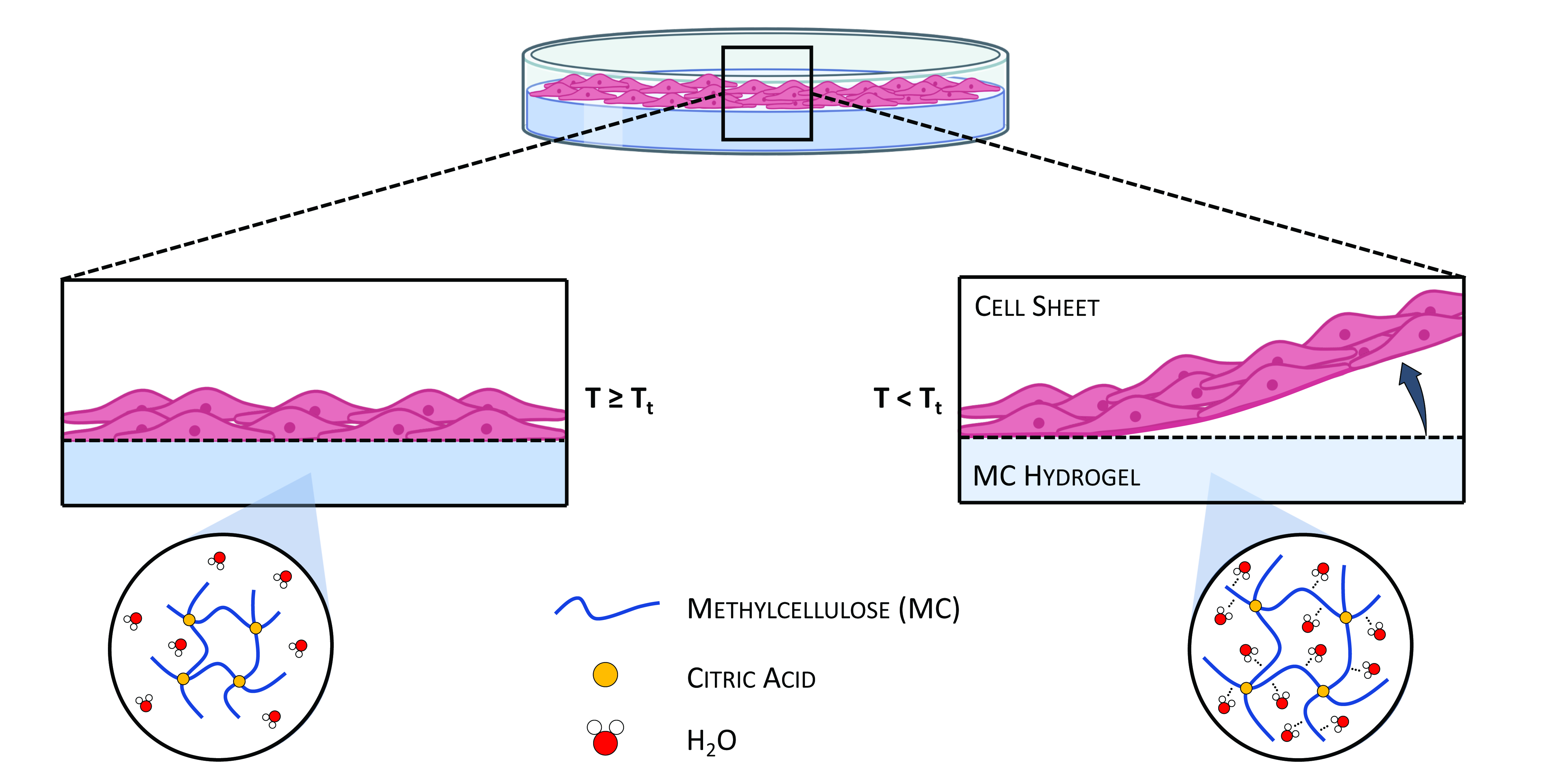
2.1. MC Hydrogels
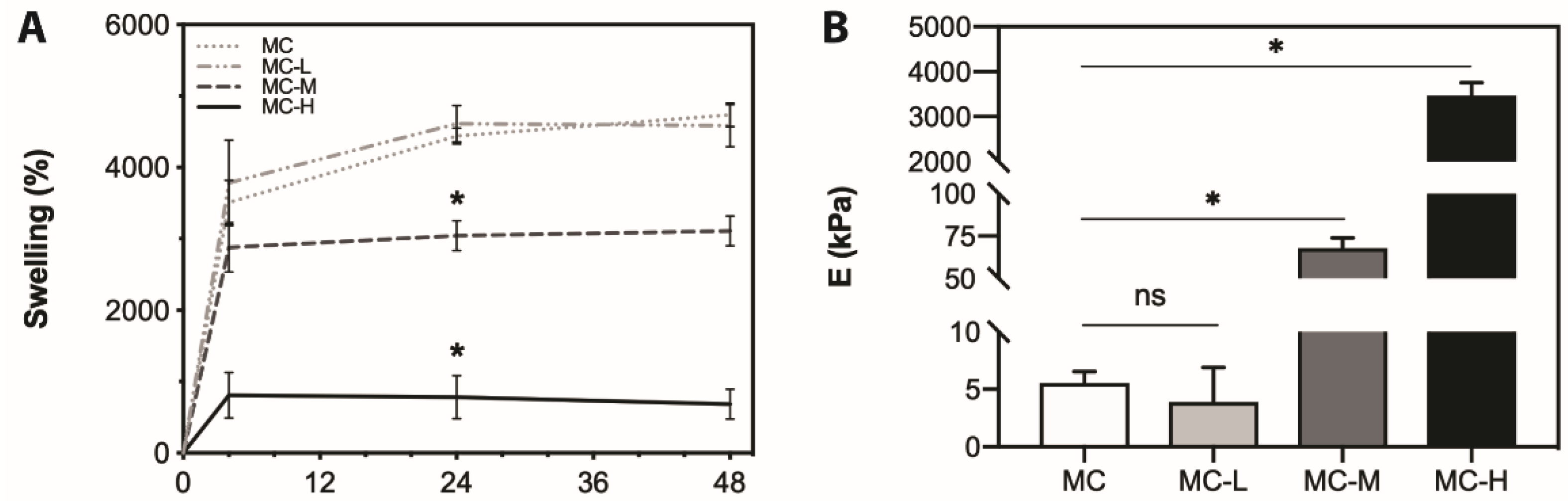
2.2. Swelling Tests
2.3. Tensile Tests
2.4. In Vitro Biological Tests
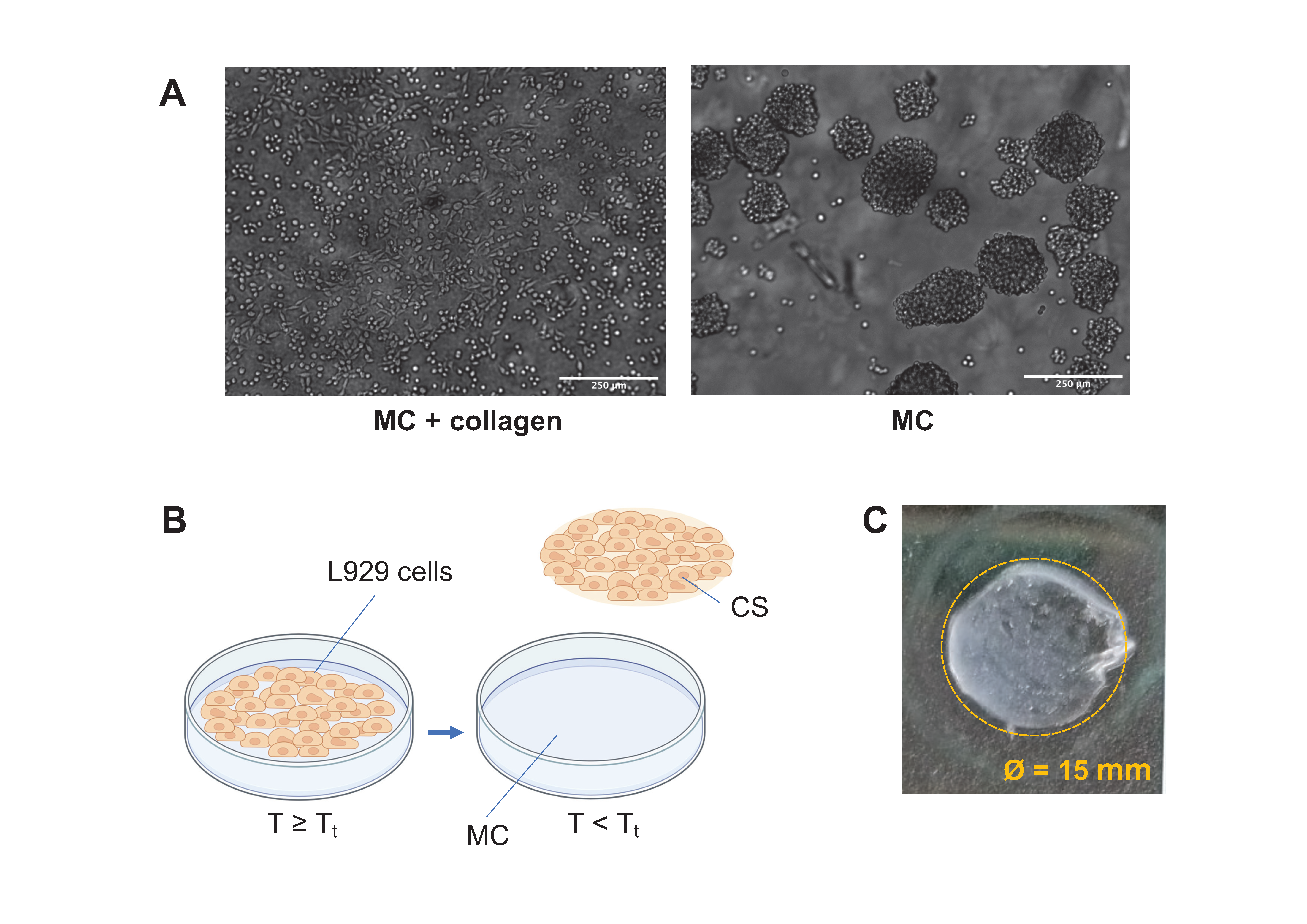
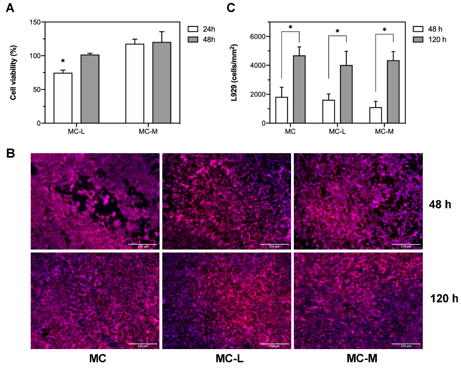
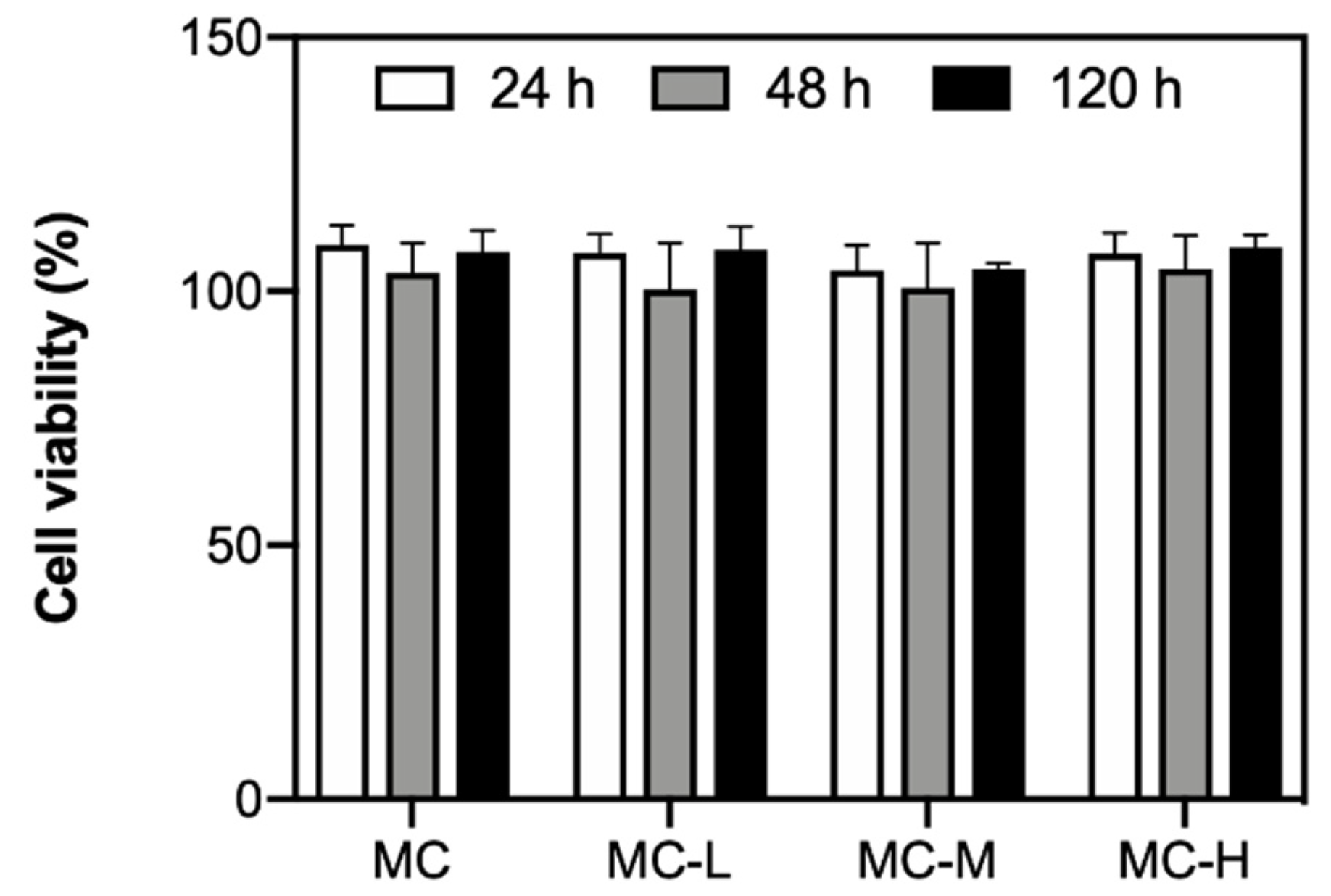
2.4.1. Indirect Cytotoxicity
2.4.2. Cell Sheets Harvesting
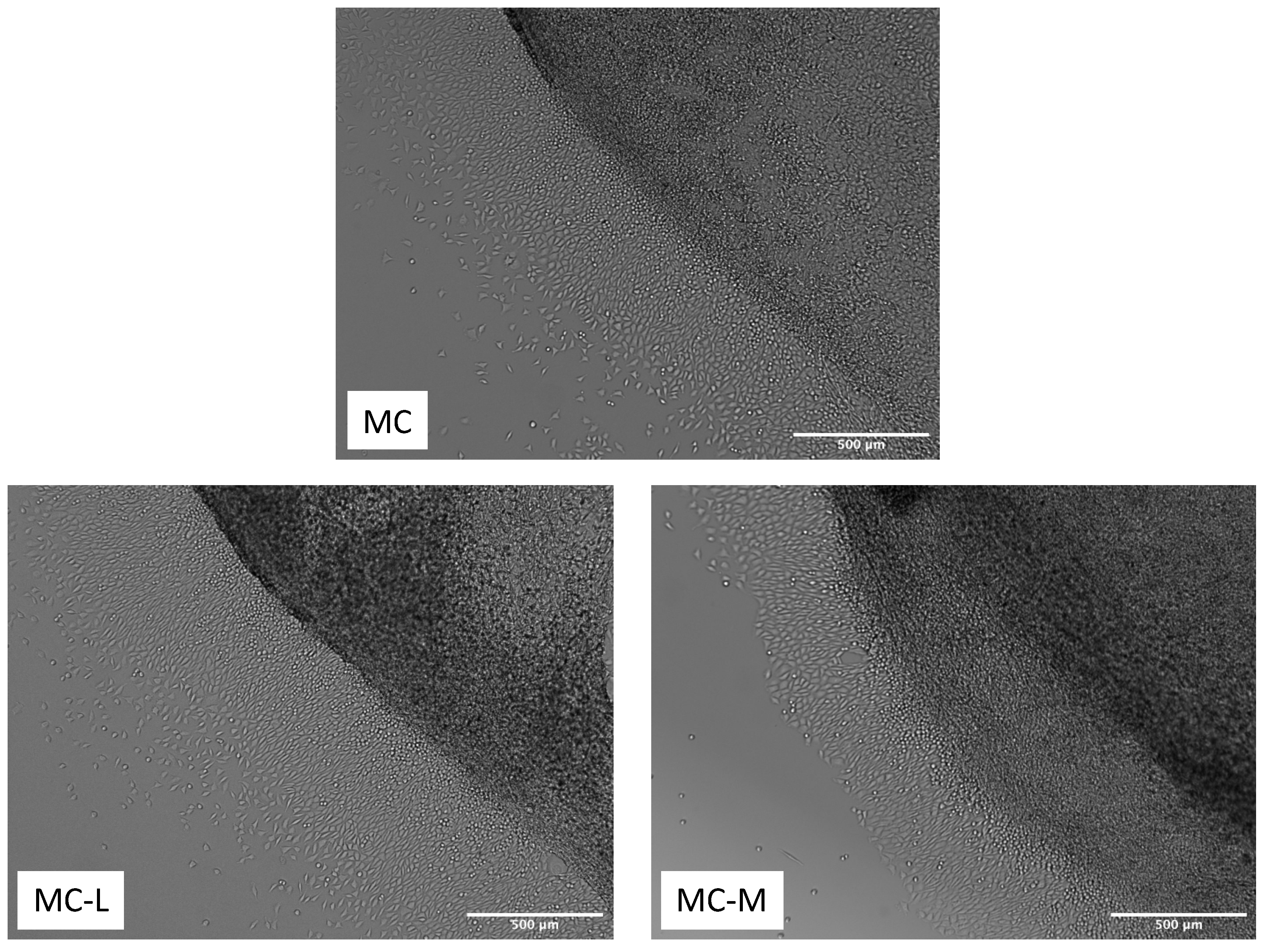
2.4.3. Cell Sheets Characterization
3. Conclusions
4. Materials and Methods
4.1. MC Hydrogels Preparation
4.2. Swelling Tests
4.3. Mechanical Tensile Tests
4.4. In Vitro Biological Tests
4.4.1. Indirect Cytotoxicity
4.4.2. Cell Culture
CSs Harvesting
Resazurin Assay
Immunofluorescence Staining
Adhesion and Proliferation on a New Substrate
4.5. Statistical Data Analysis
Supplementary Materials
Author Contributions
Funding
Institutional Review Board Statement
Informed Consent Statement
Acknowledgments
Conflicts of Interest
References
- Owaki, T.; Shimizu, T.; Yamato, M.; Okano, T. Cell sheet engineering for regenerative medicine: Current challenges and strategies. Biotechnol. J. 2014, 9, 904–914. [Google Scholar] [CrossRef]
- Kobayashi, J.; Kikuchi, A.; Aoyagi, T.; Okano, T. Cell sheet tissue engineering: Cell sheet preparation, harvesting/manipulation, and transplantation. J. Biomed. Mater. Res. Part A 2019, 107, 955–967. [Google Scholar] [CrossRef] [PubMed]
- Devireddy, R.V. Cell sheets for tissue engineering applications. In Cell Engineering and Regeneration; Springer International Publishing: Cham, Switzerland, 2019; pp. 1–20. [Google Scholar]
- Moschouris, K.; Firoozi, N.; Kang, Y. The application of cell sheet engineering in the vascularization of tissue regeneration. Regen. Med. 2016, 11, 559–570. [Google Scholar] [CrossRef] [Green Version]
- Akiyama, Y. Design of temperature-responsive cell culture surfaces for cell sheet engineering. Cyborg Bionic Syst. 2021, 2021, 1–15. [Google Scholar]
- Yang, J.; Yamato, M.; Nishida, K.; Ohki, T.; Kanzaki, M.; Sekine, H.; Shimizu, T.; Okano, T. Cell delivery in regenerative medicine: The cell sheet engineering approach. J. Control. Release 2006, 116, 193–203. [Google Scholar] [CrossRef] [PubMed]
- Li, M.; Ma, J.; Gao, Y.; Yang, L. Cell sheet technology: A promising strategy in regenerative medicine. Cytotherapy 2019, 21, 3–16. [Google Scholar] [CrossRef] [PubMed]
- Thirumala, S.; Gimble, J.; Devireddy, R. Methylcellulose based thermally reversible hydrogel system for tissue engineering applications. Cells 2013, 2, 460–475. [Google Scholar] [CrossRef] [Green Version]
- Cochis, A.; Bonetti, L.; Sorrentino, R.; Contessi Negrini, N.; Grassi, F.; Leigheb, M.; Rimondini, L.; Farè, S. 3D printing of thermo-responsive methylcellulose hydrogels for cell-sheet engineering. Materials 2018, 11, 579. [Google Scholar] [CrossRef] [Green Version]
- Taylor, M.; Tomlins, P.; Sahota, T. Thermoresponsive gels. Gels 2017, 3, 4. [Google Scholar] [CrossRef]
- Mokhtarinia, K.; Masaeli, E. Transiently thermally responsive surfaces: Concepts for cell sheet engineering. Eur. Polym. J. 2020, 141, 110076. [Google Scholar] [CrossRef]
- Bonetti, L.; De Nardo, L.; Farè, S. Thermo-responsive methylcellulose hydrogels: From design to applications as smart biomaterials. Tissue Eng. Part B Rev. 2020. [Google Scholar] [CrossRef]
- Chen, C.-H.; Tsai, C.-C.; Chen, W.; Mi, F.-L.; Liang, H.-F.; Chen, S.-C.; Sung, H.-W. Novel living cell sheet harvest system composed of thermoreversible methylcellulose hydrogels. Biomacromolecules 2006, 7, 736–743. [Google Scholar] [CrossRef]
- Bonetti, L.; De Nardo, L.; Variola, F.; Fare’, S. Evaluation of the subtle trade-off between physical stability and thermo-responsiveness in crosslinked methylcellulose hydrogels. Soft Matter 2020, 16, 5577–5587. [Google Scholar] [CrossRef] [PubMed]
- Bonetti, L.; De Nardo, L.; Variola, F.; Farè, S. In-situ Raman spectroscopy: An effective technique for the quantification of LCST transition of methylcellulose hydrogels. Mater. Lett. 2020, 274, 128011. [Google Scholar] [CrossRef]
- Altomare, L.; Cochis, A.; Carletta, A.; Rimondini, L.; Farè, S. Thermo-responsive methylcellulose hydrogels as temporary substrate for cell sheet biofabrication. J. Mater. Sci. Mater. Med. 2016, 27, 95. [Google Scholar] [CrossRef] [PubMed]
- Bloom, A.B.; Zaman, M.H. Influence of the microenvironment on cell fate determination and migration. Physiol. Genomics 2014, 46, 309–314. [Google Scholar] [CrossRef] [Green Version]
- Morozova, S.; Coughlin, M.L.; Early, J.T.; Ertem, S.P.; Reineke, T.M.; Bates, F.S.; Lodge, T.P. Properties of chemically cross-linked methylcellulose gels. Macromolecules 2019, 52, 7740–7748. [Google Scholar] [CrossRef]
- Stalling, S.S.; Akintoye, S.O.; Nicoll, S.B. Development of photocrosslinked methylcellulose hydrogels for soft tissue reconstruction. Acta Biomater. 2009, 5, 1911–1918. [Google Scholar] [CrossRef]
- Shin, J.Y.; Yeo, Y.H.; Jeong, J.E.; Park, S.A.; Park, W.H. Dual-crosslinked methylcellulose hydrogels for 3D bioprinting applications. Carbohydr. Polym. 2020, 238, 116192. [Google Scholar] [CrossRef]
- Rimdusit, S.; Somsaeng, K.; Kewsuwan, P.; Jubsilp, C.; Tiptipakorn, S. Comparison of gamma radiation crosslinking and chemical crosslinking on properties of methylcellulose hydrogel. Eng. J. 2012, 16, 15–28. [Google Scholar] [CrossRef] [Green Version]
- Wach, R.A.; Mitomo, H.; Nagasawa, N.; Yoshii, F. Radiation crosslinking of methylcellulose and hydroxyethylcellulose in concentrated aqueous solutions. Nucl. Instrum. Methods Phys. Res. Sect. B Beam Interact. Mater. At. 2003, 211, 533–544. [Google Scholar] [CrossRef]
- Park, J.-S.; Park, J.-W.; Ruckenstein, E. Thermal and dynamic mechanical analysis of PVA/MC blend hydrogels. Polymer 2001, 42, 4271–4280. [Google Scholar] [CrossRef]
- Harsh, D.C.; Gehrke, S.H. Controlling the swelling characteristics of temperature-sensitive cellulose ether hydrogels. J. Control. Release 1991, 17, 175–185. [Google Scholar] [CrossRef]
- Tao, X.; Nonaka, H. Wet extrusion molding of wood powder with hydroxy-propylmethyl cellulose and with citric acid as a crosslinking agent. BioResources 2021, 16, 2314–2325. [Google Scholar] [CrossRef]
- Xu, Y.; Li, L.; Zheng, P.; Lam, Y.C.; Hu, X. Controllable gelation of methylcellulose by a salt mixture. Langmuir 2004, 20, 6134–6138. [Google Scholar] [CrossRef]
- Flory, P.J.; Rehner, J. Statistical mechanics of cross-linked polymer networks II. Swelling. J. Chem. Phys. 1943, 11, 521–526. [Google Scholar] [CrossRef]
- Baier Leach, J.; Bivens, K.A.; Patrick, C.W., Jr.; Schmidt, C.E. Photocrosslinked hyaluronic acid hydrogels: Natural, biodegradable tissue engineering scaffolds. Biotechnol. Bioeng. 2003, 82, 578–589. [Google Scholar] [CrossRef]
- Gold, G.T.; Varma, D.M.; Taub, P.J.; Nicoll, S.B. Development of crosslinked methylcellulose hydrogels for soft tissue augmentation using an ammonium persulfate-ascorbic acid redox system. Carbohydr. Polym. 2015, 134, 497–507. [Google Scholar] [CrossRef] [Green Version]
- Gold, G.T.; Varma, D.M.; Harbottle, D.; Gupta, M.S.; Stalling, S.S.; Taub, P.J.; Nicoll, S.B. Injectable redox-polymerized methylcellulose hydrogels as potential soft tissue filler materials. J. Biomed. Mater. Res. Part A 2014, 102, 4536–4544. [Google Scholar] [CrossRef]
- D’Angelo, M.; Benedetti, E.; Tupone, M.G.; Catanesi, M.; Castelli, V.; Antonosante, A.; Cimini, A. The role of stiffness in cell reprogramming: A potential role for biomaterials in inducing tissue regeneration. Cells 2019, 8, 1036. [Google Scholar] [CrossRef] [Green Version]
- Wells, R.G. The role of matrix stiffness in regulating cell behavior. Hepatology 2008, 47, 1394–1400. [Google Scholar] [CrossRef]
- Munoz-Robles, B.G.; Kopyeva, I.; DeForest, C.A. Surface patterning of hydrogel biomaterials to probe and direct cell–matrix interactions. Adv. Mater. Interfaces 2020, 7, 2001198. [Google Scholar] [CrossRef]
- Yamato, M.; Okano, T. Cell sheet engineering. Mater. Today 2004, 7, 42–47. [Google Scholar] [CrossRef]
- Nagase, K.; Yamato, M.; Kanazawa, H.; Okano, T. Poly(N-isopropylacrylamide)-based thermoresponsive surfaces provide new types of biomedical applications. Biomaterials 2018, 153, 27–48. [Google Scholar] [CrossRef]
- Shi, R.; Bi, J.; Zhang, Z.; Zhu, A.; Chen, D.; Zhou, X.; Zhang, L.; Tian, W. The effect of citric acid on the structural properties and cytotoxicity of the polyvinyl alcohol/starch films when molding at high temperature. Carbohydr. Polym. 2008, 74, 763–770. [Google Scholar] [CrossRef]
- Jiang, Q.; Reddy, N.; Zhang, S.; Roscioli, N.; Yang, Y. Water-stable electrospun collagen fibers from a non-toxic solvent and crosslinking system. J. Biomed. Mater. Res. Part A 2013, 101A, 1237–1247. [Google Scholar] [CrossRef]
- Oryan, A.; Kamali, A.; Moshiri, A.; Baharvand, H.; Daemi, H. Chemical crosslinking of biopolymeric scaffolds: Current knowledge and future directions of crosslinked engineered bone scaffolds. Int. J. Biol. Macromol. 2018, 107, 678–688. [Google Scholar] [CrossRef]
- Stabenfeldt, S.E.; García, A.J.; LaPlaca, M.C. Thermoreversible laminin-functionalized hydrogel for neural tissue engineering. J. Biomed. Mater. Res. Part A 2006, 77A, 718–725. [Google Scholar] [CrossRef] [PubMed]
- Martin, B.C.; Minner, E.J.; Wiseman, S.L.; Klank, R.L.; Gilbert, R.J. Agarose and methylcellulose hydrogel blends for nerve regeneration applications. J. Neural Eng. 2008, 5, 221–231. [Google Scholar] [CrossRef] [Green Version]
- Payne, C.; Dolan, E.B.; O’Sullivan, J.; Cryan, S.-A.; Kelly, H.M. A methylcellulose and collagen based temperature responsive hydrogel promotes encapsulated stem cell viability and proliferation in vitro. Drug Deliv. Transl. Res. 2017, 7, 132–146. [Google Scholar] [CrossRef] [PubMed]
- Forghani, A.; Kriegh, L.; Hogan, K.; Chen, C.; Brewer, G.; Tighe, T.B.; Devireddy, R.; Hayes, D. Fabrication and characterization of cell sheets using methylcellulose and PNIPAAm thermoresponsive polymers: A comparison Study. J. Biomed. Mater. Res. Part A 2017, 105, 1346–1354. [Google Scholar] [CrossRef] [Green Version]
- Bucur, M.; Constantin, C.; Neagu, M.; Zurac, S.; Dinca, O.; Vladan, C.; Cioplea, M.; Popp, C.; Nichita, L.; Ionescu, E. Alveolar blood clots and platelet-rich fibrin induce in vitro fibroblast proliferation and migration. Exp. Ther. Med. 2018, 17, 982–989. [Google Scholar] [CrossRef]
- Imashiro, C.; Shimizu, T. Fundamental technologies and recent advances of cell-sheet-based tissue engineering. Int. J. Mol. Sci. 2021, 22, 425. [Google Scholar] [CrossRef] [PubMed]
- Kim, H.; Kim, Y.; Park, J.; Hwang, N.; Lee, Y.; Hwang, Y. Recent advances in engineered stem cell-derived cell sheets for tissue regeneration. Polymers 2019, 11, 209. [Google Scholar] [CrossRef] [Green Version]
- Yang, J.; Yamato, M.; Kohno, C.; Nishimoto, A.; Sekine, H.; Fukai, F.; Okano, T. Cell sheet engineering: Recreating tissues without biodegradable scaffolds. Biomaterials 2005, 26, 6415–6422. [Google Scholar] [CrossRef] [PubMed] [Green Version]
- Wang, H.-B.; Dembo, M.; Wang, Y.-L. Substrate flexibility regulates growth and apoptosis of normal but not transformed cells. Am. J. Physiol. Physiol. 2000, 279, C1345–C1350. [Google Scholar] [CrossRef] [Green Version]
- Tilghman, R.W.; Cowan, C.R.; Mih, J.D.; Koryakina, Y.; Gioeli, D.; Slack-Davis, J.K.; Blackman, B.R.; Tschumperlin, D.J.; Parsons, J.T. Matrix rigidity regulates cancer cell growth and cellular phenotype. PLoS ONE 2010, 5, e12905. [Google Scholar] [CrossRef] [Green Version]
- Mih, J.D.; Sharif, A.S.; Liu, F.; Marinkovic, A.; Symer, M.M.; Tschumperlin, D.J. A multiwell platform for studying stiffness-dependent cell biology. PLoS ONE 2011, 6, e19929. [Google Scholar] [CrossRef] [PubMed]
- Liu, Y.; Li, L.; Chen, X.; Wang, Y.; Liu, M.-N.; Yan, J.; Cao, L.; Wang, L.; Wang, Z.-B. Atomic force acoustic microscopy reveals the influence of substrate stiffness and topography on cell behavior. Beilstein J. Nanotechnol. 2019, 10, 2329–2337. [Google Scholar] [CrossRef]


| Sample | Crosslinking Degree | [CA] (% wCA/wMC) | -COOH (mmol/100 g) | wester * (%) |
|---|---|---|---|---|
| MC | - | 0 | 0 | 0 |
| MC-L | Low | 1 | 15.6 | 0.6 |
| MC-M | Medium | 3 | 46.9 | 51.3 |
| MC-H | High | 5 | 78.1 | 77.6 |
| Sample | |||
|---|---|---|---|
| MC | 1.36 × 10⁴ ± 5.33 × 10² | 0.20 ± 0.01 | 59.98 ± 1.64 |
| MC-L | 1.45 × 10⁴ ± 1.25 × 10³ | 0.19 ± 0.02 | 62.53 ± 3.77 |
| MC-M | 7.72 × 10³ ± 7.70 × 10² * | 0.36 ± 0.04 # | 40.21 ± 2.84 † |
| MC-H | 1.28 × 10³ ± 5.77 × 10² * | 2.42 ± 0.87 # | 11.29 ± 3.51 † |
| Sample | [CA] (% wCA/wMC) | Temperature (°C) | Time (min) |
|---|---|---|---|
| MC | - | - | - |
| MC-L | 1 | 165 | 1 |
| MC-M | 3 | 177.5 | 8 |
| MC-H | 5 | 190 | 15 |
Publisher’s Note: MDPI stays neutral with regard to jurisdictional claims in published maps and institutional affiliations. |
© 2021 by the authors. Licensee MDPI, Basel, Switzerland. This article is an open access article distributed under the terms and conditions of the Creative Commons Attribution (CC BY) license (https://creativecommons.org/licenses/by/4.0/).
Share and Cite
Bonetti, L.; De Nardo, L.; Farè, S. Chemically Crosslinked Methylcellulose Substrates for Cell Sheet Engineering. Gels 2021, 7, 141. https://doi.org/10.3390/gels7030141
Bonetti L, De Nardo L, Farè S. Chemically Crosslinked Methylcellulose Substrates for Cell Sheet Engineering. Gels. 2021; 7(3):141. https://doi.org/10.3390/gels7030141
Chicago/Turabian StyleBonetti, Lorenzo, Luigi De Nardo, and Silvia Farè. 2021. "Chemically Crosslinked Methylcellulose Substrates for Cell Sheet Engineering" Gels 7, no. 3: 141. https://doi.org/10.3390/gels7030141
APA StyleBonetti, L., De Nardo, L., & Farè, S. (2021). Chemically Crosslinked Methylcellulose Substrates for Cell Sheet Engineering. Gels, 7(3), 141. https://doi.org/10.3390/gels7030141