Review

In Situ Forming Poloxamer-Based Thermo-Sensitive Hydrogels for Ocular Application: A Focus on the Derivatives 407 and 188

1
Department of Health Sciences, University “Magna Græcia” of Catanzaro, Campus Universitario “S Venuta”, I-88100 Catanzaro, Italy
2
Apotiga Laboratory, Farmacia Europea, Via Milano, 24/A, I-88100 Catanzaro, Italy
3
”AGreenFood” Research Center, University “Magna Græcia” of Catanzaro, Campus Universitario “S Venuta”, I-88100 Catanzaro, Italy
*
Authors to whom correspondence should be addressed.
Gels 2025, 11(9), 752; https://doi.org/10.3390/gels11090752
Submission received: 25 August 2025 / Revised: 14 September 2025 / Accepted: 15 September 2025 / Published: 17 September 2025
(This article belongs to the Special Issue Innovative Gels: Structure, Properties, and Emerging Applications)

Abstract

In ophthalmology, developing effective drug delivery systems is crucial to overcome anatomical and physiological barriers, such as rapid tear turnover and blinking, which limit the efficacy of conventional formulations like eye drops. Poloxamers, especially the derivatives 407 (P407) and 188, are amphiphilic triblock copolymers characterized by an intriguing thermo-reversible behavior, making them ideal candidates for the development of in situ hydrogels for ocular applications. Various thermo-sensitive poloxamer-based hydrogels were designed to be easily instilled as liquids at room temperature, gelling promptly upon contact with the corneal surface. These systems promoted a controlled release of active compounds, significantly improving their adhesion to the ocular surface. This review discusses the most relevant scientific literature on the topic, with particular attention to studies published in recent years. The results demonstrated that poloxamer formulations are capable of overcoming typical ocular barriers, thereby increasing drug bioavailability. The intrinsic biocompatibility of poloxamers contributes to the safety and tolerability of the system. Furthermore, P407 showed additional wound healing features. The combination of biocompatibility and thermo-reversible behavior makes poloxamer-based hydrogels a promising platform for the development of innovative ocular drug delivery systems able to enhance therapeutic efficacy and patient comfort.

Graphical Abstract

1. Introduction

The most commonly used pharmaceutical form for ophthalmic drug administration is eye drops, typically formulated as aqueous solutions or suspensions. These are widely employed for delivering drugs intended to act on the ocular or intraocular surface. While eye drops offer certain advantages, including low cost and ease of administration, they also present significant drawbacks. These include a very short residence time on the ocular surface, poor bioavailability due to rapid elimination via the nasolacrimal pathway and, often, instability of the drug in conventional formulations [1,2]. Consequently, although eye drops are convenient, they are also among the least effective forms of ocular drug delivery. This is due to several anatomical and physiological features of the eye that limit both the efficacy and retention of the drug at the site of instillation, including tear turnover, blinking and the presence of corneal, conjunctival and blood–ocular barriers, all of which reduce drug residence time and absorption despite the availability of potent therapeutic agents [1]. The protective mechanisms of the eye, including conjunctival blood capillaries and lymphatics, nasolacrimal drainage, protein binding, systemic absorption and enzymatic degradation, further reduce drug absorption and result in only brief therapeutic effects [3]. In addition, certain physicochemical properties of the drug, such as lipophilicity, solubility, molecular weight, charge, and degree of ionization, affect both the route and the rate of corneal permeation [4]. These challenges underscore the need to develop innovative drug delivery formulations to enhance therapeutic outcomes. In this context, triblock copolymers have emerged as highly promising candidates. Their ability to self-assemble into micelles or form in situ hydrogels improves drug solubility, stability and controlled release, while enhancing retention on the ocular surface and facilitating penetration across ocular barriers [5]. Triblock polymers can be obtained by arranging hydrophilic and hydrophobic segments in different sequences, allowing the design of nanocarriers with tailored physico-chemical properties. Their chemical structure provides peculiar amphiphilic architectures that enable self-assembly [6]. In ocular delivery, such structural versatility offers various advantages: triblock copolymers protect drugs from enzymatic or oxidative degradation in the tear film, enhance retention on the ocular surface through interactions with mucin and respond to local physico-chemical conditions such as temperature, pH or ionic strength, enabling in situ gelation and controlled drug release [7]. Critical features such as gelation temperature, mechanical strength and release kinetics can be fine-tuned by adjusting the ratio of hydrophilic and hydrophobic blocks,, making them superior to other copolymers for both anterior and posterior segment therapies [8,9]. While this review will specifically focus on poloxamer-based triblock copolymers due to their extensive application in ophthalmology, it is important to note that other systems, such as PLGA-PEI-PEG (poly-lactic-co-glycolic acid- poly-ethyleneimine- polyethylene glycol) have also shown promising results in ocular applications, including antifungal therapy as was true in the case of natamycin-loaded thermosensitive nanoparticle–hydrogel composites proposed for the treatment of Fusarium solani [10].

2. Approaches to Ophthalmic Treatment

2.1. Physiological Limitations Affecting Therapy

Ocular therapy presents a significant challenge for doctors and pharmacologists due to the various anatomical barriers within the eye. Structurally, the eye is divided into two main parts: the anterior segment and the posterior segment, each containing multiple layers that act as biobarriers, limiting drug penetration. The anterior segment accounts for about one-third of the eye and consists of the cornea, aqueous humor, iris, lens and ciliary body. The pupil is also part of this region and it acts as a window into the integrity of a complex network of ocular structures through its multifaceted functions [11]. The posterior segment, which accounts for the remaining two-thirds, consists of the vitreous humor, retina, choroid, macula, optic nerve and sclera, forming the bulk of the ocular anatomy [12]. Structures like the conjunctiva, cornea, sclera and retina, along with the blood–retina barrier, serve as static barriers to drug delivery, while dynamic barriers include factors such as tear dilution, blinking, nasolacrimal drainage, blood flow and lymphatic clearance [5]. Various parameters and challenges must be taken into account depending on the route of administration of active compounds. This review will focus specifically on the topical application of drugs (Figure 1).
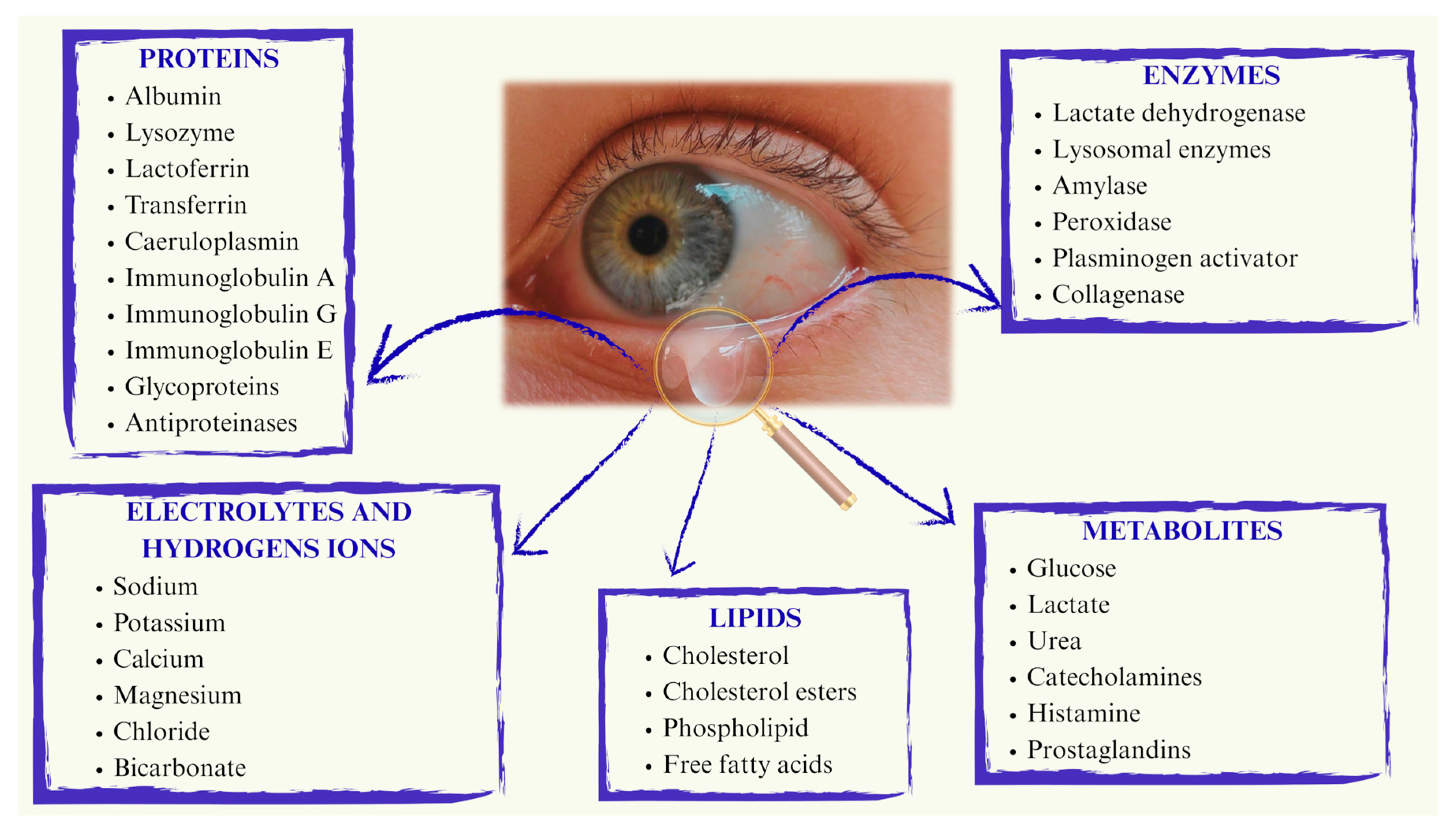
Following topical instillation, drug absorption occurs through either the corneal or non-corneal route, depending on the molecule’s chemical properties. The cornea, composed of the epithelium, stroma and endothelium, plays a critical role in protecting the eye under physiological conditions by acting as a barrier against pathogens and exogenous agents [13]. Each layer acts as a barrier to molecules with different origins and chemical properties, with the epithelium and stroma posing greater obstacles than the endothelium [14]. The epithelium is largely impermeable to topically administered hydrophilic drugs due to its lipophilic nature. Additionally, the tight junctions between corneal epithelial cells act as a selective barrier, allowing the permeation of small molecules while preventing the diffusion of macromolecules through the paracellular pathway [15]. In contrast, the stroma, which is highly hydrated due to its lamellar arrangement of fibrils and collagen, is permeable to hydrophilic drugs and it hinders the diffusion of lipophilic molecules reinforcing the lipophilic–hydrophilic dual barrier nature of cornea [1]. The conjunctiva is a vascularized mucous membrane that lines the inner surface of the eyelids and extends over the anterior part of the sclera up to the cornea. Both the conjunctival and corneal surfaces are coated with mucin, which helps to moisturize, cleanse and protect the eye from pathogens. Ocular bioavailability is further reduced by drug loss into the systemic circulation, facilitated by the conjunctival blood and lymphatic vessels [16,17]. With respect to the sclera, trans-scleral administration is more straightforward due to its high hydration and permeability to water-soluble molecules. In fact, the sclera is generally more permeable than both the cornea and conjunctiva [18]. However, molecules with a molecular weight of 150 kDa or higher face greater challenges in crossing the sclera and exhibit slower clearance compared to low molecular weight compounds [19]. Additionally, systemic drug administration to ocular tissues can occur via intravenous or oral routes. However, oral administration is limited by drug passage through the gastrointestinal tract. The retina, meanwhile, acts as a physical barrier that affects the distribution of active compounds in both the anterior ocular tissue and the posterior segment of the eye, due to the presence of the inner blood–retinal barrier (BRB) and the retinal pigment barrier (RPB) [20]. In the context of ocular physiology, ocular surface temperature and tear film sensitivity are crucial and closely interconnected parameters. In healthy eyes, ocular surface temperature values, measured immediately after blinking are typically 34–35 °C, and then progressively decrease in the interblink interval. Cyclosporine has been shown to display retrograde solubility in aqueous media and solid-state transformations that are accelerated at higher temperatures, while prostaglandin analogues such as bimatoprost, latanoprost and travoprost demonstrate notable thermal instability under conditions simulating daily use [21,22]. Taken together, these findings suggest that the ocular surface temperature range may critically influence both solubility and stability of topically applied drugs, underscoring the need to consider local thermal conditions in ophthalmic formulation design. Ocular temperature is influenced by a variety of factors, including environmental and body temperature, inflammation and blinking itself, but it is also significantly related to tear film parameters [23]. After topical administration, the drug initially mixes with the tear film, which consists of ions, lipids and proteins (Figure 2).
Despite the development of effective therapies, the drug’s contact time with ocular tissues remains a significant challenge. The continuous production of tear fluid and its rapid drainage through the upper and lower canaliculi drastically limit how long a drug can exert its function on the ocular surface [24]. For this reason the tear film viscosity plays a key role in the mean residence time of a drug in the eye. Its composition directly influences the stability and permanence of active compounds in the administration site. Although it was previously believed that mucins were solely responsible for this viscosity due to their ability to form a hydrogel network, more recent studies revealed that viscosity is related not only to a single component but rather to complex molecular interactions between secretory and membrane-associated mucins, lipids and several proteins. This multifaceted and intricate nature of the tear film represents a criticism to be evaluated when novel formulations are developed; in fact, altering viscosity with the aim of prolonging the drug contact time without compromising ocular function is a challenge [25].
Lacrimation and blinking are effective protective mechanisms that help keep the eye clear of external substances but also hinder efficient ocular drug delivery [3]. Additionally, the volume of the conjunctival cul-de-sac, which typically holds around 7 μL of tear fluid, is much smaller than the volume of a standard eye drop (30–40 μL). As a result, the blink reflex immediately expels the excess fluid that the eye cannot retain, leading to a rapid decrease in drug concentration within just a few minutes [26,27]. Moreover, the tear turnover is approximately 1 μL/min, which contributes to ongoing drug loss from the ocular surface [28]. As a result, a significant portion of topically applied drugs is immediately diluted in the tear film, with excess fluid spilling over the lid margin, while the remainder is rapidly drained into the nasolacrimal duct. Notably, it is estimated that less than 5% of the administered dose reaches the target tissue via topical application [1].

2.2. Ocular Drug Delivery Systems

Ocular drug formulations are categorized into liquids (solutions, suspensions, and emulsions), semi-liquids (ointments and gels) and solids (ocular inserts, contact lenses, etc.) [29,30]. The primary route of ocular drug administration is topical; however, periocular and intraocular administration, involving the use of injections and implants, is also employed [29]. The topical route is generally preferred due to its non-invasive nature, the relatively low incidence of side effects and improved patient compliance. Topical ophthalmic solutions are utilized in the treatment of eye injuries, but these formulations typically require multiple daily applications to maintain a therapeutic drug concentration at the site of administration [31]. Various ocular diseases can affect both the anterior and posterior segments of the eye. While anterior-segment conditions are more common and directly exposed to the external environment, posterior-segment diseases, such as age-related macular degeneration (AMD) and diabetic retinopathy, pose a greater treatment challenge when using topical therapies [32]. The high sensitivity of eye, along with its biological barriers, has attracted significant research interest, as these factors can compromise the therapeutic effectiveness of drugs [16].
Given the aforementioned challenges, a sufficient therapeutic concentration can only be achieved through frequent instillations of conventional eye drops. However, this approach may increase the risk of side effects due to the absorption of the drug through blood vessels in the conjunctival stroma or the nasal mucosa [33]. To address these limitations, various ophthalmic drug delivery systems, such as hydrogels, nanoparticles, liposomes, in situ gels and micelles have been developed to optimize the localization of active compounds at the target tissue in the eye. All conventional pharmaceutical formulations have inherent limitations in ophthalmic therapy [31]. For these reasons, in the last decade, scientific research has focused on evaluating technologies that encapsulate drugs within biocompatible systems. These systems aim to increase drug residence time, minimize clearance mechanisms in the eye and reduce the side effects associated with conventional eye drops [34]. The design of new pharmaceutical forms must, of course, adhere to the standard characteristics of conventional eye drops, such as isotonicity, appropriate pH, adequate viscosity and surface tension. Additionally, attention must be given to bioadhesion, particularly to polymers such as cellulose derivatives, polyvinyl alcohol, polyacrylic acid, chitosan and hyaluronic acid, which can effectively interact with mucin [35].
The advantages of ocular drug delivery systems can be summarized as follows:
  • Patients can self-administer the treatment, leading to improved compliance;
  • Enhanced stability of the drug encapsulated within the formulation;
  • Reduced drug elimination due to increased residence time on the ocular or intraocular surface or in the conjunctival sac, as well as improved absorption through the corneal cells;
  • The ability to modulate drug release, reducing the frequency of instillations and minimizing drug loss from the precorneal area;
  • The potential for polymer manipulation to achieve specific tissue targeting [36].
It is crucial to emphasize that the topical application of drugs is not limited to treating conditions affecting the anterior structures of the eye, such as inflammation and infections [9]. In fact, hydrogels, made of different materials, have also been developed for the treatment of glaucoma [37,38], a pathology characterized by high intraocular pressure (IOP) that induces mechanical stress on various structures of the anterior and posterior chambers [39]. An example is the nifedipine-containing hydrogel, made up of P407, P188 and hydroxypropyl methylcellulose K4M, which in a preclinical study demonstrated a significant reduction in IOP when compared to commercially available conventional eye drops [40].
In consideration of the aforementioned aspects, this review is focused on the most recent developments in drug delivery systems intended for topical ophthalmic therapy, with particular attention paid to hydrogels formulated from polymers, such as poloxamers, characterized by thermo-responsive properties.
Relevant keywords such as in situ hydrogels, ocular application, poloxamer 407, poloxamer 188 and their association, etc., have been used in order to collect the research and review articles mainly published between 2020 to 2025 and available on Google Scholar, PubMed and Scopus.
Hydrogels employed in ocular drug delivery applications have exhibited promising features, facilitating the sustained and controlled release of therapeutic agents. Moreover, their high water content closely resembles the natural ocular environment, thereby ensuring biocompatibility and underscoring their potential utility in the treatment of ocular diseases [41].

3. Hydrogels

3.1. General Properties of Hydrogels

In 1960, Wichterle and Lim pioneered the development of a synthetic hydrogel composed of poly-2-hydroxyethyl methacrylate (PHEMA), which was later adopted as a filler following eye enucleation and in the fabrication of contact lenses [42]. Hydrogels constitute three-dimensional polymeric matrices comprising natural or synthetic polymers (Table 1), which assemble via physical or chemical cross-linking and are characterized by diverse properties including flexibility, biodegradability and biocompatibility [26]. Within their polymeric network, hydrogels exhibit a variety of functional groups, such as -NH2, -COOH and -OH, which facilitate the absorption of substantial quantities of water and regulate the swelling behaviour of the system. Notwithstanding their significant hydrophilicity, hydrogels undergo swelling but retain their structural integrity in the medium due to the presence of cross-links, encompassing covalent, hydrogen, Van der Waals interactions and ionic interactions, the latter being particularly relevant for polyelectrolyte hydrogels [43].
These chemical interactions, augmented by physical entanglement, permit the entrapment of a range of active compounds [44,45].
The encapsulation of drugs within hydrogels can be achieved through two principal methods:
  • Post-loading: this involves the entrapment of the drug molecule within the established three-dimensional network of the hydrogel matrix;
  • In situ loading: in this approach, the drug forms interactions with the solution or the polymer before the hydrogel structure is created [46].
The development of hydrogels can utilize both natural and synthetic polymers. Nevertheless, the use of natural polymers can present complexities owing to their inherent limitations in stability and mechanical resistance. Indeed, synthetic polymers typically possess greater mechanical and chemical strength than natural polymers [47]. Such resistance, attributed to the robust bonds within their matrix, prevents the premature degradation of the systems. As a result, the utilization of synthetic polymers is often favored due to their enhanced susceptibility to chemical and physical manipulation [44].
Biodegradable and hydrophobic synthetic polymers, characterized by low toxicity and minimal side effects, represent the most commonly employed materials in the development of smart hydrogels for drug delivery applications. This facilitates the localized, sustained and prolonged release of the therapeutic agent, thereby diminishing the requisite number of administrations [45,46]. This review will focus on the development of synthetic hydrogels, obtained through the polymerization of monomers such as PEO and PPO, for ocular applications.
Interest in hydrogels has increased significantly in the last decade due to their potential smart properties [48]. Indeed, depending on their polymer composition, hydrogels can exhibit a response as a function of a specific stimulus. These stimuli-responsive properties can enable the development of formulations useful for drug targeting and the controlled release of bioactives [49]. Polymer-based gels are categorized into two groups: bioadhesive gels and in situ forming systems. These formulations serve to enhance bioavailability and mitigate side effects associated with the systemic absorption of topically administered ophthalmic drugs. Bioadhesive gels present as viscous solutions prior to ocular application and are commonly employed in the treatment of dry eye syndrome as a substitute for natural tears. These ophthalmic solutions do not undergo any alterations following their administration [50,51].
Bioadhesive ophthalmic solutions commonly exhibit several limitations, including a lack of consistent accuracy and reproducibility in administration. Moreover, they may cause patient discomfort, transient blurred vision and reflex tearing, which can further compromise the vision. As a result, their application is frequently restricted to the treatment of dry eye disease, serving as artificial tears [52]. The polymers incorporated into these formulations are predominantly cellulose derivatives, such as hydroxypropyl methylcellulose (HPMC), which serve to enhance viscosity, mucoadhesion and the stability of the formulation. Furthermore, polymers such as hyaluronic acid (HA) are employed to exploit their pseudoplastic characteristics, thereby ensuring effective diffusion of the formulation across the ocular surface [53]. In situ gelling systems are liquid formulations applied to the eye in drop form and subsequently undergo a sol-to-gel phase transition within the conjunctival cul-de-sac [54].

3.2. In Situ Hydrogels

Contemporary research has increasingly concentrated on in situ hydrogel formation as a means of mitigating the limitations inherent in conventional ophthalmic solutions. In situ hydrogels are developed as polymeric solutions capable of being administered in liquid form and subsequently undergoing a phase transition to a semisolid gel upon exposure to physiological environments. The in situ gelation system constitutes a particularly interesting strategy and a promising approach for prolonging the residence time of drugs on the ocular surface and improving their bioavailability [55]. In situ forming hydrogels are capable of increasing viscosity in response to alterations in pH or temperature within the precorneal region, thereby resulting in decreased naso-lacrimal drainage, extended ocular absorption time and enhanced bioavailability. A notable advantage of this formulation type is its liquid state at ambient temperature, permitting simple instillation as an ophthalmic drop, with subsequent gelation occurring as a function of a specific physiological parameter. This behavior allows the formulation to resist shear forces within the conjunctival cul-de-sac and to impede excessive dilution by lacrimal fluid [56]. The preparation of in situ hydrogels involves the dissolution of a suitable polymer in an appropriate solvent. The selection of the solvent is contingent not only upon the solubility of the polymer but also upon the biocompatibility and overall stability of the delivery system. Moreover, these systems are characterized by a facile preparation procedure, typically consisting of a single step of mixing and solubilization of the constituent components and cost-effective. This ensures the uniformity and reproducibility of the resultant product [57]. The polymers employed in the development of in situ hydrogels exhibit reversible phase transition characteristics and pseudoplastic behaviour, a property useful for minimizing interference with the natural act of eye blinking. The extended ocular residence time of these systems promotes a sustained release of the active compounds, resulting in enhanced bioavailability and a decrease in the number of administrations required, thereby leading to improved patient compliance and comfort, without inducing blurred vision [54]. The instillation of an in situ hydrogel can favour a decrease of the drug concentration used and administration times, consequently reducing the potential side effects and toxicity of several active compounds [58]. However, hydrogels present certain drawbacks that can hinder their use; among them, for example for hydrophobic drugs, the low loading capacity and distribution in the polymeric matrix of the molecules can compromise the administration of various active compounds. Furthermore, stability criticisms and the microbial contamination are other problems to be properly investigated [59].

3.3. Thermo-Sensitive In Situ Hydrogels

Within the spectrum of pharmaceutical formulations, thermo-sensitive hydrogels have been shown to be systems capable of promoting a sustained release of the active compounds over an extended period [60]. Temperature-sensitive polymers, characterized by the presence of both hydrophilic and hydrophobic domains within their structure, exhibit a phase transition that is dependent on temperature fluctuations and modulates their solubility.
In particular, changes in temperature affect the interplay between the hydrophilic and hydrophobic residues of the polymer and water molecules, thereby altering the solubility of the cross-linked network and inducing the sol–gel phase transition. Temperature-sensitive hydrogels can demonstrate either a positive response, wherein the gel material swells upon an increase in temperature, or a negative behavior, wherein the material undergoes shrinkage as the temperature rises [61]. Thermo-sensitive hydrogels demonstrate volume phase transitions or sol–gel phase transitions at critical temperatures, specifically the lower critical solution temperature (LCST) or the upper critical solution temperature (UCST). Polymers exhibiting LCST undergo a transition from swelling to shrinking (or sol to gel) upon an increase in temperature, whereas UCST systems undergo the inverse transition [62]. Polymers that exhibit LCST phase separation phenomena are more prevalent and advantageous compared to polymers characterized by UCST properties. Thermo-responsive systems at temperatures below the lower critical solution temperature (LCST) exist as transparent, homogeneous, free-flowing polymer solutions and undergo a transition to a cloudy state upon reaching the LCST [63]. The necessity of high temperatures for mixing UCST polymers and drugs can, in fact, prove detrimental to the activity of thermo-sensitive compounds. Conversely, hydrogels exhibiting LCST behaviour, particularly those with a sol–gel transition temperature of approximately 37 °C, have been extensively investigated as carriers for cells, drugs and biomolecules, as they permit their incorporation under mild conditions. A representative example of such biocompatible thermo-reversible LCST hydrogels is composed of poloxamers [64].
The application of these considerations to ophthalmic formulations leads to varying outcomes. The limited therapeutic response of conventional ophthalmic solutions, often due to precorneal drug elimination, can be overcome by administering in situ hydrogels. These hydrogels are prepared from polymers that exhibit reversible phase transitions and pseudoplastic behaviour to minimize interference with blinking [29]. Subject to specific conditions, the formulation undergoes an in situ phase transition, resulting in the formation of a gel that exhibits resistance to shear forces within the conjunctival cul-de-sac, thereby promoting drug release under physiological conditions. In situ gelation systems demonstrate an increase in viscosity as a consequence of the temperature within the precorneal region [56]. Subsequent to the instillation of the aqueous solution containing polymers responsive to external stimuli, a viscous and mucoadhesive hydrogel is formed on the surface of the eye, leading to an increase in ocular retention time and the bioavailability of the incorporated drug [35].
Thermo-responsive polymer-based systems exhibit a liquid state at ambient temperature (20–25 °C) and undergo gelation at approximately 35 °C, which corresponds to the temperature of the ocular surface (Figure 3). This characteristic facilitates their easy instillation as ophthalmic drops without inducing irritation or blurred vision, with subsequent gelation occurring upon interaction with the eye [65]. In accordance with these principles, a thermosensitive hydrogel composed of 20 wt% P407 was formulated for the delivery of progranulin (PGRN). This macromolecule is a protein exhibiting anti-inflammatory, regenerative and re-epithelializing characteristics relevant to corneal tissue. Yan et al. [66] demonstrated that P407 is a thermo-responsive polymer of the LCST type in solution, forming micelles at low material concentrations and hydrogels at elevated temperatures (≥LCST). At a temperature of 4 °C, the hydrogel exists in the form of a solution, thereby allowing for its admixture with the protein. This physical state was preserved at ambient temperature and the formulation was instilled into the eye as ophthalmic drops, undergoing a transition to a hydrogel at 34.5 °C. This physical transition of the system favoured the slow and sustained release of PGRN, while the formulation also functioned as a protective barrier for the cornea.

4. Poloxamers

4.1. General Properties of Poloxamers

Poloxamers, by virtue of their thermosensitive properties, have been the focus of research for numerous years, with a particular emphasis on drug delivery applications. Furthermore, they are also employed as non-ionic surfactants and cross-linking agents [27]. Poloxamers, trade name Pluronic®, are triblock polymeric structures composed of two hydrophilic blocks, such as PEO, and a central hydrophobic block, such as PPO. Owing to this amphiphilic nature, poloxamers exhibit surfactant properties and the capacity to interact with hydrophobic surfaces and biological membranes. In aqueous solutions, the amphiphilic character of these copolymers leads to self-aggregation of the macromolecules into micelles upon reaching the CMC (critical micellar concentration). PPO portions aggregate to form hydrophobic micellar cores surrounded by hydrophilic PEO coronas able to interact with water-soluble molecules. This mechanism leads to thermo-responsive gelation and the formation of a three-dimensional micellar network [67]. The obtained system absorbs water and swells upon contact with tear fluid, inducing a variation of the pore size and diffusional pathways, thereby modulating the release of the entrapped drugs. The relative balance between hydrophilic and hydrophobic domains determines gel density, erosion behaviour and release kinetics. This interplay of micellization, swelling and erosion provides a robust rate-controlling mechanism that sustains therapeutic levels compared with conventional eye drops [68,69]. The thermo-sensitivity of poloxamers is determined by their solubility in water. While PEO is water-soluble even at low temperatures, PPO is not. Consequently, the PEO/PPO chain ratio dictates the thermo-sensitivity. The distinctive properties of poloxamers have been discussed in numerous review articles and will not be detailed in this manuscript [60,70]. Polymer concentration represents an additional parameter capable of influencing the phase transition of poloxamer-based systems. Indeed, poloxamers at physiological temperature typically undergo gelation at concentrations greater than 15%. Conversely, formulations characterized by an excessive polymer content can elicit undesirable effects. For example, intraperitoneal administration of P407 has been shown to induce significant hypertriglyceridemia and hypercholesterolemia; this effect is dose-dependent and does not occur at lower doses [71].
Gugleva et al. [72] developed an in situ poloxamer hydrogel with the capacity to retain niosomal doxycycline. They exploited the interaction between the PEO/PPO residues and the corneal surface to augment the residence time of the bioactive agent. Additionally, it was demonstrated that the mucoadhesion and phase transition temperature could be modulated through the incorporation of HPMC. The optimal formulation, characterized by a favourable phase transition temperature of 34 °C, was determined to be composed of 15% P407 and 1.5% HPMC. The temperature also exerts an influence on this process; specifically, upon reaching the critical micelle temperature (CMT), the dehydration of the PPO blocks commences, leading to the formation of multichain spherical micelles [72].
Natural antioxidant agents, such as resveratrol, exert a scavenging activity against free radicals and various derivatives are characterized by antibacterial properties [73,74]. Nevertheless, these molecules may exhibit poor aqueous solubility often leads to low permeation and rapid degradation. To address these limitations, poloxamer 407-based micelles and poloxamer 407/β-casein nanoaggregates have been developed [75]. Mixed micelles may possess a low CMC and promote significant stability against both physical and chemical agents. Both types of micelles demonstrated an excellent capacity for resveratrol retention, enhancing its apparent solubility (by more than 50 times) and preserving its antioxidant capacity [76]. However, mixed micelles exhibited slightly increased toxicity, and therefore, P407 micelles were proposed as useful systems for the ocular application of resveratrol [75].
Sangitra et Pujala [77] demonstrated that poloxamers, when exceeding their CMC and at temperatures above their CMT, are capable of forming a denser gel network, characterized by increased resistance to deformation as a consequence of aggregation phenomena.
In a separate experimental investigation, artemisinin-loaded micelles with a size of approximately 50 nm, composed of 8% polyvinylpyrrolidone (PVP K90) and 4% P407, were developed. These micelles enhanced both the solubility and corneal permeation of artemisinin and have been proposed as a therapeutic strategy for age-related macular degeneration (AMD) [78].
Padaga et al. [79] described an innovative system composed of a novel synthesized copolymer. Specifically, the FCOL derivative was obtained by carboxylation of P407 with succinic anhydride, followed by the reaction with the amino groups of chitosan oligosaccharide lactate (COL). Chitosan was selected for its antibacterial activity and mucoadhesive nature, attributed to the positive charges of the polysaccharide that can interact with the negative charges on the corneal surface. This new copolymer was used to create a gel containing gatifloxacin.
Furthermore, mixed micelles can be formed utilizing two distinct poloxamers, as exemplified by systems proposed for the entrapment of fexofenadine, a drug with limited aqueous solubility employed in the treatment of allergic conjunctivitis. The drug was encapsulated within nanoaggregates composed of Pluronic® 127 and 123, and enriched with Phospholipon 90G with the aim of augmenting their thermodynamic stability as a consequence of strong hydrophobic interactions with the PPO blocks. The optimized formulation demonstrated a high entrapment efficiency and a sustained release profile of the active compound retained within the inner core of the micelles [80].
Polymeric micelles can also be utilized to address certain critical issues associated with contact lenses, including water content, wettability and optical properties. Experimental investigation revealed that micelles composed of P407 and Tween® 80, incorporating cyclosporine, were capable of increasing the water content, optical transmission and wettability of contact lenses [81]. Furthermore, they promoted a sustained release of cyclosporine, lasting approximately 120 h and enhanced its ocular absorption [81].
Poloxamers 188 and 407 represent the most widely utilized members of the United States (U.S.) FDA-approved Pluronics® material classes for human use [61].

4.2. Poloxamer 407 Hydrogels for the Treatment of Ocular Diseases

Poloxamer 407 (P407), commercially known as Pluronic® F-127, comprises 70% PEO blocks, which contribute to its hydrophilic characteristics. P407 is a member of a family encompassing more than 30 copolymers characterized by an ABA block structure, wherein a hydrophobic PPO block is positioned between two hydrophilic PEO residues. This non-toxic copolymer possesses a molecular weight of 12,000 Da, exhibits low viscosity and demonstrates the ability to form gels at physiological temperatures [82]. P407 demonstrates superior solubility in cold water compared to hot water, a phenomenon attributed to the increased formation of hydrogen bonds at lower temperatures and exhibits high stability [83]. Despite the potential benefits, poloxamers face significant challenges that hinder their widespread use. These include non-ideal mechanical properties, reduced viscosity and gel strength, accelerated erosion and insufficient bio-adhesion that require the association with other polymers such as chitosan, HPMC, carbopol, or other poloxamers such as P188 [84,85].
Li et al. [86] developed a thermo-responsive nanosuspension hydrogel for the treatment of uveitis. Carboxymethyl chitosan was employed to stabilize clobetasol propionate obtaining a nanosuspension that was integrated into a hydrogel made up of P407 and sodium alginate [86].
P407 forms an optically clear and transparent gel at the physiological temperature of the ocular cavity and its self-assembling characteristics facilitate the administration of both hydrophilic and lipophilic drugs, favoring a controlled release profile over time. Nevertheless, P407 exhibits limited structural stability at low temperatures and concentrations where micellar structures are unstable. Consequently, it is typically combined with other excipient [87].
Hydrogels composed of P407 have been proposed as drug carriers for the treatment of various diseases, including dry eye, a pathology associated with inflammation and induced by oxidative stress [3,88].
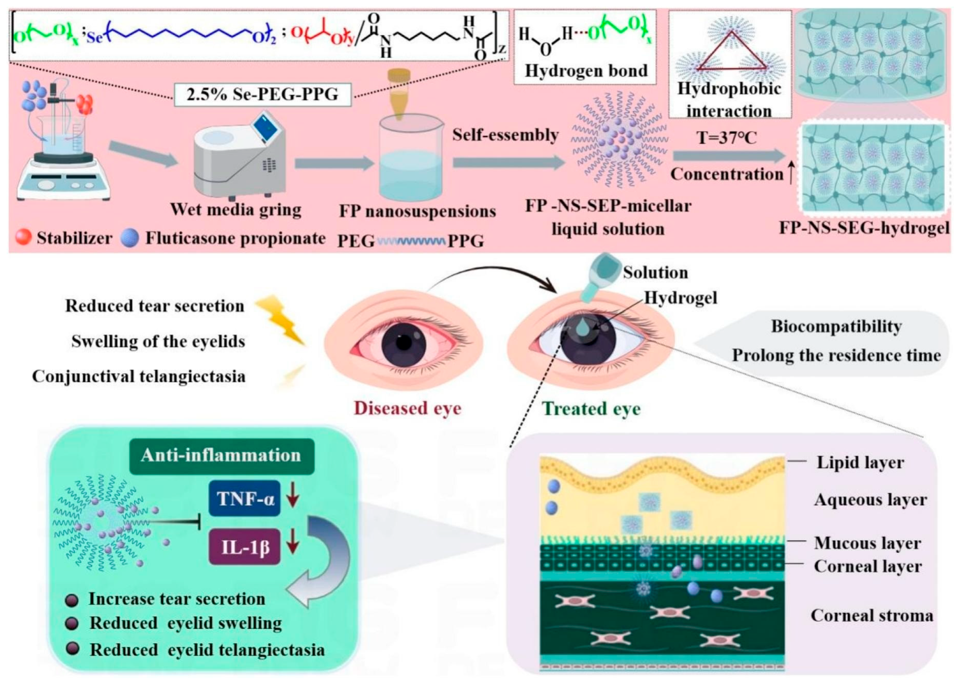
Inflammatory eyelid conditions could also benefit from the treatment with in situ hydrogels due to the opportunity to overcome the limited retention of conventional eye drops provided by these formulations. In this regard, a fluticasone propionate nanosuspension modified with carboxymethyl chitosan was developed and encapsulated within a synthesized P407-based gel, named SEP (Figure 4), synthesized from selenol, PEO and PPO. Considering that selenium is a vital trace element that exerts a protective influence on the cells of ocular surface, it was hypothesized that the use of selenium could alter the intermolecular forces, allowing for gelation even at low concentrations. The proposed formulation offers several advantages, such as a more practical and effective therapeutic option for blepharitis [89].
A novel formulation combining uniform, ultrasmall (approximately 3 nm in diameter) antioxidant copper-selenide (Cu2-xSe) nanoparticles and aldehyde-modified Pluronic®, (AF127) hydrogels was used as eye drops and tested in an in vivo mouse model of dry eye disease (DED) [90]. These nanoparticles exhibited the capacity to neutralize ROS, thereby exerting antioxidant and antiapoptotic effects through a mechanism that mimics the activity of superoxide dismutase and glutathione peroxidase. Additionally, aldehyde residues were incorporated into F127 to facilitate the formation of Schiff bases with mucin constituents on the ocular surface, which is intended to enhance the bioavailability of the nanosystems. The formulation comprising 20% AF127 with incorporated Cu2-xSe nanoparticles presented a gel-forming temperature of 36.8 °C [90].
Tacrolimus is another drug used to treat dry eye syndrome. However, it is an immunosuppressive compound that can also be employed for the treatment of other significant conditions, such as corneal allograft rejection, immunogenic inflammatory diseases of the ocular surface and allergic conjunctivitis [91,92]. To enhance its delivery, an in situ gelling formulation composed of P407 and chitosan was developed, demonstrating increased drug retention on the ocular surface [93].
Nonetheless, P407 has limited mucosal adhesion and, when used at high concentrations, may induce hypertriglyceridemia in the eye. To mitigate these drawbacks, it is combined with HPMC, a polymer commonly used to improve eye hydration (particularly in glaucomatous dry eye patients), increase viscosity and enhance the comfort of eye drops [33,94].
A similar formulation, consisting of 10% w/v P407 and 0.725% w/v HPMC, incorporating chloramphenicol, was also developed [95]. More recently, however, chloramphenicol was encapsulated within chitosan nanoparticles embedded in a thermo-sensitive in situ gel system composed of 20.5% w/v P407 and 5% w/v P188. The resulting formulation demonstrated good stability, caused no ocular irritation and promoted prolonged drug release for up to 18 h, along with precorneal retention lasting up to 40 min [96]. HPMC can also be used to enhance gel strength.
The findings of Jiang and co-workers [97] showed that berberine hydrochloride, a poorly water-soluble active compound, lowered the gelation temperature of P407 hydrogels and improved corneal adhesion, further reducing the gel erosion rate while modulating drug release [97]. The addition of HPMC enhanced gel strength due to its strong interaction with P407.
Paul et al. evaluated nine in situ gel formulations to identify the best option for enhancing the ocular delivery of ganciclovir. The optimized formulation, made up of 15% w/v P407 and 1% w/v HPMC E-50 LV, notably improved the eye permeation of the drug and prolonged its ocular retention time. Moreover, it enhanced the therapeutic efficacy of ganciclovir and it represents a promising alternative to conventional eye drops, offering a better patient compliance and effectiveness [98].
Chandler et al. [99] investigated a novel biodegradable, in situ thermo-responsive hydrogel proposed for the ocular surface healing. This formulation, made up of P407 (18.0 w/v%), P188 (5.0 w/v%) and HPMC (1.0 w/v%), was used to encapsulate human recombinant (rh)MG53 protein, a macromolecule already known for its capacity to promote corneal healing. The daily application of this system significantly improved the pharmacological activity of the protein and the related-healing phenomena [99].
In a promising new formulation proposed for wound healing, P407 was combined with alginate and foetal bovine serum to deliver the repurposed drug raloxifene. The system was investigated for its ability to influence cell proliferation, thereby facilitating rapid wound closure within 14 days. In-depth studies revealed that this formulation maximizes the proliferative potential of raloxifene. Further in vitro and in vivo analyses confirmed the efficacy and non-toxic nature of the formulation [100]. Histopathological studies revealed that P407 hydrogels promote collagen deposition, epithelial regeneration and neovascularization [101]. This evidence suggests that the poloxamer may act not only as a simple excipient, but as an active component characterized by wound healing features [102].
Poloxamers are generally valued in drug delivery formulations for antibiotics, particularly in the treatment of bacterial keratitis, an acute corneal infection caused by microorganisms such as Staphylococcus aureus. Conventional antibiotic formulations must be administered frequently to maintain effective antibacterial activity [20]. Suboptimal concentrations of these drugs may result in treatment failure or the emergence of bacterial resistance [103]. P407 is capable of inhibiting bacterial adhesion, including that of P. aeruginosa, through the formation of a hydrated layer on the surface of the microorganism [104].
In another in vivo study, an infected group of albino rabbits showed improved clinical outcomes after being treated with a specific bacteriophage, phage vB_Pa_ZCPS1, administered by an in situ gel. The formulation made up of 14% w/v P407 and 1.5% w/v HPMC K4M demonstrated a gelling time of 30 ± 1 s at a temperature of 33 °C ± 1 and useful muco-adhesive features [105].
A hybrid hydrogel was designed to effectively address ocular inflammation and infection through the localized co-delivery of antibiotics and corticosteroids. The system was obtained combining gellan gum (GG) (0.1% w/v) with various concentrations of P407 (16.5, 17 and 18% w/v). The formulation was enriched with polymyxin B sulphate, neomycin sulfate dexamethasone-loaded chitosan nanoparticles. The analyses revealed that GG positively influenced the system decreasing the gelation temperature and increasing the viscosity of P407 hydrogels. These effects contributed to obtain a sustained release of entrapped drugs enhanced their bioavailability, promoting potentially useful clinical outcomes [106].
Flurbiprofen is an anti-inflammatory drug also used to prevent miosis during eye surgery and to treat post-operative ocular inflammation. However, its poor water solubility presents a challenge [107]. To address this issue, nanoparticles composed of HPMC were fabricated using a spray-drying technique [108]. These nanoparticles were then dispersed in aqueous solutions containing 14% w/w P407 and Carbopol 934, leading to the development of innovative nanogels [109].
Owing to their gelling properties and extensive surface area, nanogels can interact with the ocular mucosa, facilitating enhanced administration of ophthalmic drugs [110,111]. In general, polymeric nanosystems can provide mucoadhesive properties that help prevent drug removal from the conjunctiva [112].
Fungal eye infections are another group of diseases that can affect ocular health. The antifungal activity of an in situ gel formulation composed of hyaluronic acid and P407, containing voriconazole-loaded cubosomes, was compared to that of a dispersion of the free drug by measuring fungal growth inhibition zones using a modified disk-diffusion susceptibility assay. The gel formulation proved to be 3.89 times more effective than the free drug and enhanced the permeation of the active compound [113]. The polysaccharide helped reduce the gelation temperature, surface friction, ocular dryness, and keratoconjunctivitis. These effects contributed to enhanced cubosome permeation, demonstrating significant mucoadhesive and anti-inflammatory properties [113].
Significant antifungal activity against Candida albicans was achieved using a thermo-sensitive gel composed of three polymers: P407, P188 and HPMC, containing itraconazole nanocrystals. The underlying rationale is that P407 (at a concentration of 20% w/v) forms a gel at room temperature, while P188, due to its lower hydrophobic/hydrophilic ratio, modulates the in situ gelation temperature of the system and HPMC enhances mucoadhesive strength [114,115]. The resulting formulation has a gelation temperature of 35 °C, adequate adhesion strength, an appropriate pH, high drug content and optimal viscosity [115].

4.3. Poloxamer 188 Hydrogels for the Treatment of Ocular Diseases

Poloxamer 188 (P188), also known by the trade name Pluronic®, F-68, is composed of PEO-PPO-PEO triblocks with average lengths of 80-30-80, respectively. The hydrophobic and hydrophilic segments give P188 its non-ionic nature and surface-active properties. With a Hydrophilic-Lipophilic Balance (HLB) of 29, P188 contains fewer hydrophobic PPO units compared to P407, which has an HLB of 22. Studies have shown that poloxamers exhibit a decrease in the Zeta potential of nanosystems as the molecular weight increases (i.e., from P188 to P407) [67]. Moreover, the CMC of P188 is slightly higher than that of P407 [116]. P188 in water can form three distinct phases:
  • at concentrations lower than the CMC and at temperatures below the CMT, the polymer exists in solution as single unimers above CMC and CMT individual molecules aggregate to form micellar structures;
  • as the temperature increases further, the micellar structures alter their microstructure, forming a denser network due to a reduction in the hydrophilicity of the PPO residues compared to the PEO chains. Additionally, P188 solutions become soft solids at high temperatures. Therefore, a reversible liquid-to-solid transition occurs, representing the shift from a viscoelastic liquid to a soft solid [117].
One of the most important properties of P188 is its neuroprotective characteristic and its ability to restore the functions of various types of injured cells with permeable membranes [118]. In fact, the evaluation of the interaction between P188 and lipid monolayers and liposomal bilayers showed that the hydrophobic PPO blocks penetrate the hydrocarbon region of the lipid membrane, while the hydrophilic PEO blocks remain in the aqueous compartments [119]. In addition, it has been demonstrated that P188 inhibits P-glycoprotein efflux pumps in ocular tissues, promoting the residence time of the active compound in the eye and enhancing its efficacy [120,121].
In this regard, P188 was proposed to create a micellar solution of ferulic acid with the aim of improving its solubility, stability and corneal permeability [122].
In addition to its significant antioxidant activity, which helps counteract free radicals and promote corneal regeneration, ferulic acid has been shown to have good antimicrobial activity against both Gram-positive and Gram-negative bacteria [123].
Micelles of P188 released approximately 70% of ferulic acid in 4 h, while micelle-nanogel formulations, composed of hyaluronan and ε-polylysine, exhibited a leakage of about 50% of the bioactive compound in 6 h, with a prolonged release lasting up to 48 h [122]. Despite the physico-chemical differences between P188 and P407, both polymers are suitable for the development of thermo-sensitive hydrogels for in situ ophthalmic administration as a consequence of their favourable safety profile and inherent optical transparency.
A recent experimental study described the development of two hydrogels containing orthosiphon, proposed for ophthalmic application [124].
In particular, the two systems were composed of 21% P407 and P188 in amounts ranging from 4% to 5%. Although the formulations were homogeneous and clear, they had a brown coloration, but they exhibited a phase transition from a solution to a gel at eye temperature. Furthermore, both formulations demonstrated an optimal transition temperature, ranging from 33 °C to 37 °C, a prolonged release of the active compound for up to 12 h and an optimal pH for the ocular compartment (between 7.27 and 7.46) [124].
Another strategy used to improve the solubility of hydrophobic drugs and promote their transcorneal permeation is encapsulating them within nanostructured lipid carriers (NLCs) [125,126]. However, the criticism of this approach is related to the low corneal retention time [127].
Wu et al. [128] integrated NLCs containing flurbiprofen (FB) within a thermo-sensitive nanohydrogel (FB-NLC-Gel) composed of P188 and P407. The formulation demonstrated significant ophthalmic anti-inflammatory effects. In fact, in vivo studies showed that the system increased the bioavailability of FB while maintaining good concentrations in the aqueous humor after topical instillation [128].
A 332-fold increase in flurbiprofen solubility was also achieved through the direct solubilization of the drug within an in situ gel system composed of P407 and P188, containing a flurbiprofen solid dispersion [129].
Prednisolone (PRD) is a corticosteroid used to treat ocular inflammatory diseases caused by infection, allergy, immune-mediated inflammation, irritation, injury, or trauma of the eye. It is insoluble in water; therefore, a microgel composed of lipid nanosystems containing PRD embedded in thermo-responsive hydrogels of P188 and P407 was prepared [130]. The viscosity of in situ formulations is essential for easy instillation into the eye and rapid conversion into a gel, ensuring sustained drug release and appropriate residence time of the entrapped molecules on the ocular surface. These features are crucial for the treatment of bacterial and fungal infections, as they help maintain the MIC for the required duration.
For example, ketoconazole (KTZ) was complexed with sulfobutylether-β-cyclodextrins and embedded within in situ hydrogels composed of P407 (20% w/w), P188 (5% w/w) and Carbopol 934 (0.2% w/w). The formulation increased the corneal residence time of the active compound, preserving its pharmacological efficacy [131].
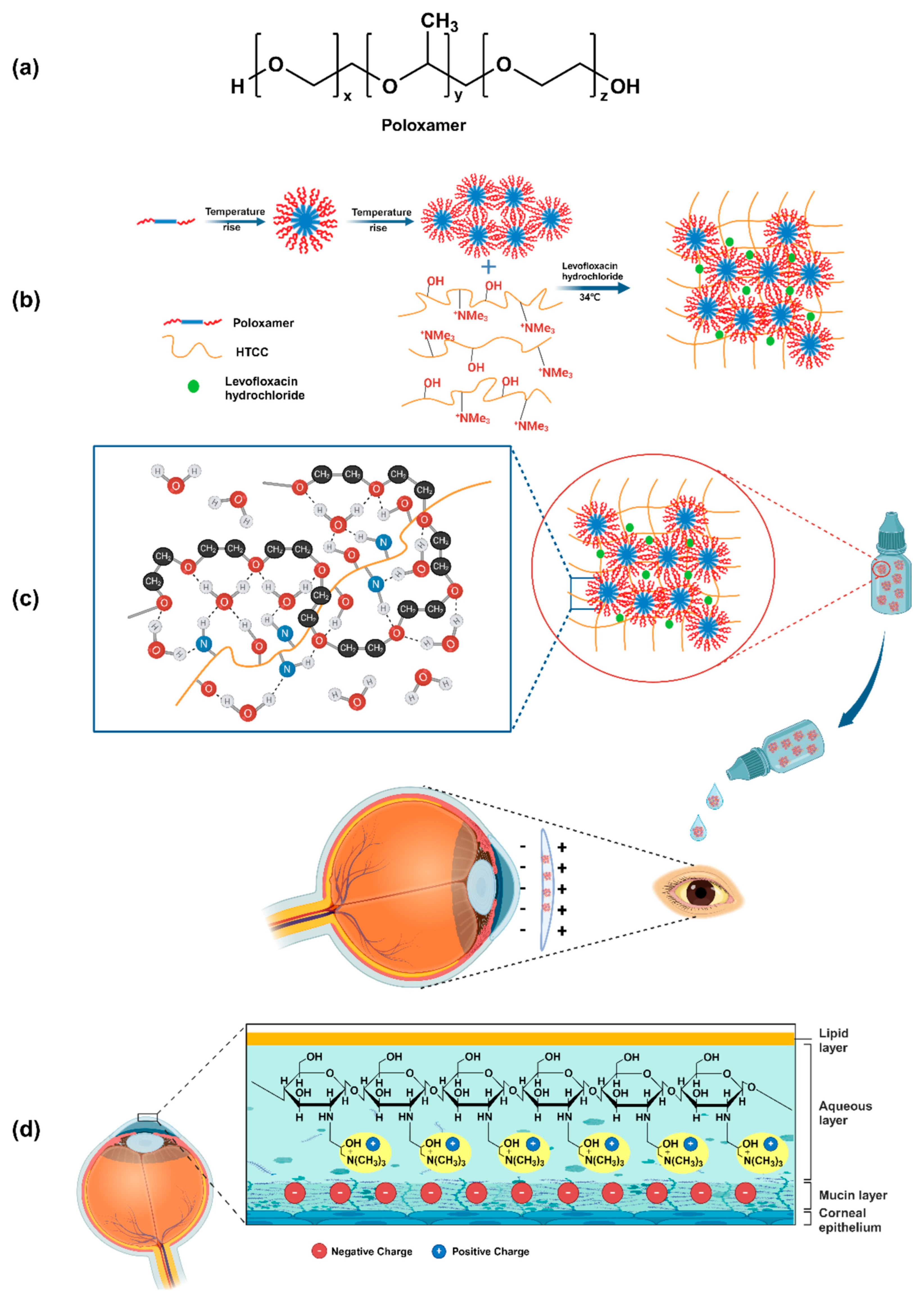
P407 and P188 were used to synthesize a poloxamer-quaternized chitosan (Figure 5) utilizing N-(2-hydroxy-3-trimethylammonium) propyl chitosan chloride (HTCC) used to obtain a thermo-sensitive hydrogel able to promote a prolonged release of levofloxacin hydrochloride up to 48 h at 34 °C [132].
Recently, another thermo-sensitive multifunctional eye drop made up of P407, P188 and tannic acid, has shown promising results. The addition of tannic acid to the hydrogel can increase its adhesive ability to bind collagen and it significantly reduces the sol–gel transition temperature. This eye drop is capable of maintaining corneal retention for at least 90 min, a time significantly higher than 15 min of commercial muco-adhesive eye drops and 30 min of conventional in situ gels. The encapsulation of amphotericin B within the obtained system and the daily administration of the formulation showed a pharmacological efficacy six times higher than conventional drops of the drug [133].
A summary of all the previously discussed studies is provided in Table 2.

5. Characterization and Analysis of Thermosensitive In Situ Hydrogels for Ocular Applications

A key characteristic for ophthalmic formulations is clarity and the absence of particles, to prevent vision blurring after application. For this reason, the prepared in situ gel formulations are visually inspected for appearance and their clarity, colour uniformity, and particle presence are evaluated [134]. In addition to the aforementioned parameters, other characteristics must be carefully evaluated during the development of these formulations (Table 3).
Along with the appropriate selection of preservatives, ensuring the sterility of the final solution is essential for achieving a high-quality preparation. Sterilization methods include moist heat under pressure (autoclaving), dry heat, filtration, gas sterilization and ionizing radiation [135,136]. Sterilization by autoclaving in the final container (terminal sterilization) is feasible only for products whose stability remains unaffected [116,137].
Balu et al. [138] prepared a sterile thermo-sensitive gel of ofloxacin using P188 and HPMC, following the procedure outlined by the United States Pharmacopeia [139] (autoclaving at 121 °C, 15 psi, for 20 min). The evaluation of the physicochemical parameters before and after the procedure (flowability, pH, viscosity and sol–gel transition temperature) showed that no changes occurred. Autoclaving is the preferred method for sterilizing most ophthalmic solutions due to its efficiency and convenience in large-scale production. However, certain products cannot be sterilized using this method, as it may compromise their stability [116]. Therefore, if stability issues arise, an ophthalmic formulation can be sterilized by filtration through a 0.22 μm filter [140]. Even in this case, certain critical aspects must be considered, including the choice of filter, which must remain intact before and after use, the potential release of fibers and the degree of sample absorption on the filter [141].
According to the European Pharmacopoeia (Ph. Eur.) sterility test, a sample should be inoculated into a medium consisting of a mixture of soybean-casein digest broth (20 mL) and thioglycolate broth (20 mL). These media are then incubated for 14 days at 20 °C and 25 °C to assess potential microbial growth in both aerobic and anaerobic cultures [142].
Another important parameter to evaluate is tonicity. Osmolarity can be measured using a cryoscopic osmometer. Ideally, an ophthalmic formulation should have an osmolarity similar to that of natural tears (approximately 310 mOsm/kg), although the recommended upper limit is 340 mOsm/kg [143], with a tonicity equivalent to a 0.9% sodium chloride solution [144].
An essential parameter for ocular formulations, to prevent irritation and side effects, is pH, which should ideally range from 6.5 to 7.6. Although the eye can tolerate a pH range of 4.5 to 11.5 due to the buffering action of tear fluid, the optimal pH is 7.2 [145].
The irritation potential of an ophthalmic formulation is typically assessed using the Draize test. Specifically, 0.1 mL of the sample is instilled into the conjunctival cul-de-sac or applied directly to the cornea of an albino rabbit’s eye [30]. This test has been criticized for its poor reproducibility, the anatomical differences between human and rabbit eyes, and primarily for ethical reasons.
Consequently, one alternative used to replace the Draize test was the hen egg chorioallantoic membrane test (HET-CAM) [146]. The CAM is a highly vascularized extraembryonic structure containing arteries, capillaries and veins. This was considered an adequate model to predict the effects of substances on the conjunctiva of the eye, as adverse effects on the CAM induced by a test substance would correlate with irritation and/or corrosion in vivo [146].
In 2024, a research team developed a 3D device called OphthalMimic that incorporates artificial tear flow, a cul-de-sac area, a movable eyelid and a surface that effectively interacts with ophthalmic formulations. This provides a faithful representation of human ocular conditions, aiming to reduce animal testing [146]. Currently, however, an important application of such a device would be for dissolution/release tests that closely mimic in vivo conditions [147].
Two parameters of gelation warrant further investigation. One crucial parameter is the sol–gel transition temperature, which is frequently assessed using the tube inversion method. This procedure involves the gradual heating of the copolymer solution, typically in a vial or test tube, periodically inverting it. The gelation temperature is defined as the specific temperature at which the solution stops to flow upon inversion, marking its transition from a liquid to a solid-like (gel) state [72]. Ideally, for ocular applications, the sol–gel transition should occur at a temperature close to the ocular surface temperature, which in healthy individuals is usually 35.06 ± 1.24 °C [148]. In addition, the influence of lacrimal dilution on the gelation of the system should also be investigated in order to define ideal concentration of poloxamer to be employed for the development of the formulation [149]. Therefore, a dilution with simulated tear fluid is typically performed (added at 7:30 ratio corresponding to the ratio between the volume of physiological tear fluid and the average on an eye drop) to better simulate the in vivo conditions, assessing the gelation temperature [150].
The second parameter is the gelling capacity, which is determined by evaluating the time required for gel formation and the time required for its dissolution. Ideally, the formulation would exhibit an immediate sol–gel transition and subsequently dissolve over an extended period of hours. This characteristic would result in prolonged contact time and, consequently, a sustained drug effect [151].
The viscosity and rheological properties represent other fundamental aspects requiring evaluation for an in situ forming drug delivery system [152]. In particular, based on dynamic properties such as elasticity, storage modulus (G′) and loss modulus (G″), the characteristics of a formulation at both room and physiological temperatures must be thoroughly assessed. Under optimal conditions, the formulation should exhibit a free-flowing liquid state upon application, allowing for facile instillation as eye drops. Consequently, G″ should be greater than G′ at the time of application. Conversely, following administration, gel formation is anticipated; therefore, G′ should be greater than G″ to withstand the shear forces exerted by the eye during blinking [153].
The viscosity of the ophthalmic formulation is an important factor in the determination of residence time of drug in the eye and it can be determined by using a rheometer. The viscosity of an ophthalmic formulation is a critical parameter influencing the residence time of a drug within the eye and can be quantified using a rheometer [154].
Clearly, other established tests focusing on the evaluation of the release profile of active compounds within the formulations, their physical stability over a period of time, and their safety profile are all mandatory considerations during the preformulation stage of a thermo-sensitive hydrogel intended for ocular application [155,156].
Table 3. Main parameters to be investigated for the characterization of thermo-sensitive hydrogels for ocular applications.
Parameter to Be EvaluatedObjective and Method of AnalysisIdeal Key OutcomeReferences
Appearance and ClarityVisual inspection for clarity, color uniformity and absence of particles to prevent vision blurring.Clear, uniform formulation with no particles.[134]
SterilityEvaluation of the absence of microbial contamination. Sterilization methods include autoclaving and filtration.
The European Pharmacopoeia test involves incubating the sample for 14 days in specific media.
The formulation remains sterile with no physical or chemical alterations.[138,139,142]
OsmolarityMeasurement of osmolarity using a cryoscopic osmometer. The goal is to prevent irritation.Osmolarity similar to that of natural tears (approximately 310 mOsm/kg), with a recommended upper limit of 340 mOsm/kg.[143,144]
pHpH measurement to prevent irritation and side effects.Ideal pH range between 6.5 and 7.6, with an optimal pH of 7.2.[145]
Sol–Gel TransitionEvaluation of the temperature at which the transition from liquid to gel occurs, often using the tube inversion method.Transition at ocular surface temperature after instillation.[72,148]
Gelling CapacityEvaluation of the time required for gel formation and dissolution.Rapid gel formation and prolonged dissolution over several hours, for extended contact time.[151]
Viscosity and RheologyMeasurement of viscosity and rheological properties (G′ and G″ moduli) with a rheometer to determine drug residence time.The formulation should be a free-flowing liquid (G″ > G′) at room temperature for easy application and transform into a gel (G′ > G″) at physiological temperature to withstand blinking forces.[152]
Physical Stability and Drug ReleaseEvaluation of the release profile of the active compound(s) and physical stability over time.Prolonged drug release and long-term stability of the formulation.[155,156]
Ocular IrritationEvaluation of irritation potential. The Draize test on albino rabbits is the traditional method.Low or no irritation potential.[30]

6. Conclusions

Ocular diseases present a significant therapeutic challenge due to the diverse anatomical biobarriers inherent to this bodily compartment. The rising global prevalence of these conditions, coupled with the severe risk of vision impairment or blindness in the absence of treatment, underscores the pressing need for innovative therapeutic strategies [157]. Furthermore, the prevalence of eye diseases imposes a substantial burden on healthcare systems; for instance, dry eye disease (DED) is estimated to incur costs of nearly $4 billion annually within the US healthcare system [158].
Academic research is fundamental for developing new therapeutic approaches and innovative formulations. However, translating proposed systems to a scale-up process can be challenging, as was the case with nanoparticles that often required complex and expensive manufacturing and specific equipment [159,160].
One advantage of poloxamer-based hydrogels is their quick and easy preparation using the cold method [161]. Ideally, the constituents of a hydrogel should be available in GMP quality at reasonable costs and possess properties such as biocompatibility, biodegradability and approval for human use [160]. Despite numerous experimental investigations, only a limited number of ophthalmic formulations incorporating P407 have obtained clinical approval from the FDA and EMA, as is the case with AzaSite Plus® (from Inspire Pharmaceuticals) and BromSite® (from Sun Pharmaceutical Industries) [85].
It is essential to emphasize that P407 is combined with other polymers, such as Polycarbophil, within these formulations to augment its inherent properties [24]. This synergy is intended to optimize the in situ gelation and mucoadhesive properties of the system, which are essential characteristics for the effective treatment of ocular inflammation and infections through the promotion of prolonged drug contact with the ocular surface [157].
Considering the aforementioned aspects and the experimental findings detailed in this document, thermo-responsive hydrogels composed of P407 and P188 present viable drug delivery systems for the treatment of conditions affecting the anterior segment of the eye.

Author Contributions

Conceptualization: E.L., M.V. and D.C.; Data curation, Investigation, software: E.L., E.G., A.G., V.G. and M.F. Writing—original draft: E.L.; Writing—review & editing: all authors. All authors have read and agreed to the published version of the manuscript.

Funding

This work was supported by the Italian Ministry of University and Research (PRIN2022_PNRR, prot. n. P202242HYK_001). Open access funding provided by “Università degli Studi Magna Graecia di Catanzaro” within the CRUI-CARE Agreement.

Data Availability Statement

No new data were created or analyzed in this study.

Conflicts of Interest

The authors declare no conflicts of interest.

References

  1. Ahmed, S.; Amin, M.M.; Sayed, S. Ocular Drug Delivery: A Comprehensive Review. AAPS PharmSciTech 2023, 24, 66. [Google Scholar] [CrossRef] [PubMed]
  2. Wu, Y.; Liu, Y.; Li, X.; Kebebe, D.; Zhang, B.; Ren, J.; Lu, J.; Li, J.; Du, S.; Liu, Z. Research Progress of In-Situ Gelling Ophthalmic Drug Delivery System. Asian J. Pharm. Sci. 2019, 14, 102342. [Google Scholar] [CrossRef]
  3. Grassiri, B.; Zambito, Y.; Bernkop-Schnürch, A. Strategies to Prolong the Residence Time of Drug Delivery Systems on Ocular Surface. Adv. Colloid Interface Sci. 2021, 288, 102342. [Google Scholar] [CrossRef]
  4. Smith, J.; Brown, L.; Zhang, Y. Corneal Permeability and Uptake of Twenty-Five Drugs: Species Comparison and Quantitative Structure–Permeability Relationships. Gels 2025, 11, 123–138. [Google Scholar]
  5. Awwad, S.; Mohamed Ahmed, A.H.A.; Sharma, G.; Heng, J.S.; Khaw, P.T.; Brocchini, S.; Lockwood, A. Principles of Pharmacology in the Eye. Br. J. Pharmacol. 2017, 174, 4205–4223. [Google Scholar] [CrossRef]
  6. Assiri, A.A.; Glover, K.; Mishra, D.; Waite, D.; Vora, L.K.; Thakur, R.R.S. Block Copolymer Micelles as Ocular Drug Delivery Systems. Drug Discov. Today 2024, 29, 104098. [Google Scholar] [CrossRef]
  7. Xu, Z.; Liu, S.; Kang, Y.; Wang, M. Glutathione-Responsive Polymeric Micelles Formed by a Biodegradable Amphiphilic Triblock Copolymer for Anticancer Drug Delivery and Controlled Release. ACS Biomater. Sci. Eng. 2015, 1, 585–592. [Google Scholar] [CrossRef]
  8. Rahmanian, M.; Oroojalian, F.; Kesharwani, P.; Sahebkar, A. Current Trends in Triblock Copolymer-Based Multifunctional Nanotheranostics for Cancer Treatment. J. Drug Deliv. Sci. Technol. 2024, 99, 105985. [Google Scholar] [CrossRef]
  9. Han, H.; Li, S.; Xu, M.; Zhong, Y.; Fan, W.; Xu, J.; Zhou, T.; Ji, J.; Ye, J.; Yao, K. Polymer- and Lipid-Based Nanocarriers for Ocular Drug Delivery: Current Status and Future Perspectives. Adv. Drug Deliv. Rev. 2023, 196, 114770. [Google Scholar] [CrossRef]
  10. Sha, X.; Chan, L.; Fan, X.; Guo, P.; Chen, T.; Liu, L.; Zhong, J. Thermosensitive Tri-Block Polymer Nanoparticle-Hydrogel Composites as Payloads of Natamycin for Antifungal Therapy Against Fusarium Solani. Int. J. Nanomed. 2022, 17, 1463–1478. [Google Scholar] [CrossRef] [PubMed]
  11. Sapino, S.; Chirio, D.; Peira, E.; Brunella, V.; Jadhav, S.A.; Abellán Rubio, E.; Chindamo, G.; Gallarate, M. Ocular Drug Delivery: A Special Focus on the Thermosensitive Approach. Gels 2019, 9, 884. [Google Scholar] [CrossRef]
  12. Jacob, S.; Nair, A.B.; Shah, J.; Gupta, S.; Boddu, S.H.S.; Sreeharsha, N.; Joseph, A.; Shinu, P.; Morsy, M.A. Lipid Nanoparticles as 800 a Promising Drug Delivery Carrier for Topical Ocular Therapy; An Overview on Recent Advances. Pharmaceutics 2022, 14, 533. [Google Scholar] [CrossRef]
  13. Onugwu, A.L.; Nwagwu, C.S.; Onugwu, O.S.; Echezona, A.C.; Agbo, C.P.; Ihim, S.A.; Emeh, P.; Nnamani, P.O.; Attama, A.A.; Khutoryanskiy, V.V. Nanotechnology Based Drug Delivery Systems for the Treatment of Anterior Segment Eye Diseases. J. Control. Release 2023, 354, 465–488. [Google Scholar] [CrossRef] [PubMed]
  14. Giri, B.R.; Jakka, D.; Sandoval, M.A.; Kulkarni, V.R.; Bao, Q. Advancements in Ocular Therapy: A Review of Emerging Drug Delivery Approaches and Pharmaceutical Technologies. Pharmaceutics 2024, 16, 1325. [Google Scholar] [CrossRef] [PubMed]
  15. Bachu, R.D.; Chowdhury, P.; Al-Saedi, Z.H.F.; Karla, P.K.; Boddu, S.H.S. Ocular Drug Delivery Barriers—Role of Nanocarriers in the Treatment of Anterior Segment Ocular Diseases. Pharmaceutics 2018, 10, 28. [Google Scholar] [CrossRef] [PubMed]
  16. Fathi-Karkan, S.; Amiri Ramsheh, N.; Arkaban, H.; Narooie-Noori, F.; Sargazi, S.; Mirinejad, S.; Roostaee, M.; Sargazi, S.; Barani, M.; Malahat Shadman, S.; et al. Nanosuspensions in Ophthalmology: Overcoming Challenges and Enhancing Drug Delivery for Eye Diseases. Int. J. Pharm. 2024, 658, 124226. [Google Scholar] [CrossRef]
  17. Mustafa, S. Ophthalmology: Navigating Ocular Barriers with Advanced Nanocarriers. Ger. J. Pharm. Biomater. 2024, 3, 34–58. [Google Scholar] [CrossRef]
  18. Hatami-Marbini, H. Effect of Hydration on Viscoelastic Tensile Properties of Sclera. Vision 2025, 9, 1. [Google Scholar] [CrossRef]
  19. Varela-Fernández, R.; Díaz-Tomé, V.; Luaces-Rodríguez, A.; Conde-Penedo, A.; García-Otero, X.; Luzardo-álvarez, A.; Fernández-Ferreiro, A.; Otero-Espinar, F.J. Drug Delivery to the Posterior Segment of the Eye: Biopharmaceutic and Pharmacokinetic Considerations. Pharmaceutics 2020, 12, 269. [Google Scholar] [CrossRef]
  20. Guidi, L.; Cascone, M.G.; Rosellini, E. Light-Responsive Polymeric Nanoparticles for Retinal Drug Delivery: Design Cues, Challenges and Future Perspectives. Heliyon 2024, 10, e26616. [Google Scholar] [CrossRef]
  21. Wu, K.; Gore, A.; Graham, R.; Meller, R. Solubilization of Cyclosporine in Topical Ophthalmic Formulations: Preformulation Risk Assessment on a New Solid Form. J. Pharm. Sci. 2019, 108, 3233–3239. [Google Scholar] [CrossRef]
  22. Johnson, T.V.; Gupta, P.K.; Vudathala, D.K.; Blair, I.A.; Tanna, A.P. Thermal Stability of Bimatoprost, Latanoprost, and Travoprost Under Simulated Daily Use. J. Ocul. Pharmacol. Ther. 2011, 27, 51–59. [Google Scholar] [CrossRef]
  23. Shah, A.M.; Galor, A. Impact of Ocular Surface Temperature on Tear Characteristics: Current Insights. Clin. Optom. 2021, 13, 51–62. [Google Scholar] [CrossRef]
  24. Destruel, P.L.; Zeng, N.; Maury, M.; Mignet, N.; Boudy, V. In Vitro and in Vivo Evaluation of in Situ Gelling Systems for Sustained Topical Ophthalmic Delivery: State of the Art and Beyond. Drug Discov. Today 2017, 22, 638–651. [Google Scholar] [CrossRef]
  25. Recchioni, A.; Mocciardini, E.; Ponzini, E.; Tavazzi, S. Viscoelastic Properties of the Human Tear Film. Exp Eye Res 2022, 219, 109083. [Google Scholar] [CrossRef]
  26. Han, J.; Shu, H.; Zhang, L.; Huang, S. Latest Advances in Hydrogel Therapy for Ocular Diseases. Polymer 2024, 306, 127207. [Google Scholar] [CrossRef]
  27. Wang, T.J.; Rethi, L.; Ku, M.Y.; Nguyen, H.T.; Chuang, A.E.Y. A Review on Revolutionizing Ophthalmic Therapy: Unveiling the Potential of Chitosan, Hyaluronic Acid, Cellulose, Cyclodextrin, and Poloxamer in Eye Disease Treatments. Int. J. Biol. Macromol. 2024, 273, 132700. [Google Scholar] [CrossRef] [PubMed]
  28. Jimenez, J.; Sakthivel, M.; Nischal, K.K.; Fedorchak, M.V. Drug Delivery Systems and Novel Formulations to Improve Treatment of Rare Corneal Disease. Drug Discov. Today 2019, 24, 1564–1574. [Google Scholar] [CrossRef]
  29. Akhter, M.H.; Ahmad, I.; Alshahrani, M.Y.; Al-Harbi, A.I.; Khalilullah, H.; Afzal, O.; Altamimi, A.S.A.; Najib Ullah, S.N.M.; Ojha, A.; Karim, S. Drug Delivery Challenges and Current Progress in Nanocarrier-Based Ocular Therapeutic System. Gels 2022, 8, 82. [Google Scholar] [CrossRef] [PubMed]
  30. Mazet, R.; Yaméogo, J.B.G.; Wouessidjewe, D.; Choisnard, L.; Gèze, A. Recent Advances in the Design of Topical Ophthalmic Delivery Systems in the Treatment of Ocular Surface Inflammation and Their Biopharmaceutical Evaluation. Pharmaceutics 2020, 12, 570. [Google Scholar] [CrossRef]
  31. Lynch, C.R.; Kondiah, P.P.D.; Choonara, Y.E.; du Toit, L.C.; Ally, N.; Pillay, V. Hydrogel Biomaterials for Application in Ocular Drug Delivery. Front. Bioeng. Biotechnol. 2020, 8, 228. [Google Scholar] [CrossRef]
  32. Qi, Q.; Wei, Y.; Zhang, X.; Guan, J.; Mao, S. Challenges and Strategies for Ocular Posterior Diseases Therapy via Non-Invasive Advanced Drug Delivery. J. Control. Release 2023, 361, 191–211. [Google Scholar] [CrossRef]
  33. Bisen, A.C.; Biswas, A.; Dubey, A.; Sanap, S.N.; Agrawal, S.; Yadav, K.S.; Singh, V.; Rawat, P.; Sagar, S.; Mugale, M.N.; et al. A Review on Polymers in Ocular Drug Delivery Systems. MedComm-Biomater. Appl. 2024, 3, e77. [Google Scholar] [CrossRef]
  34. Manjeri, A.; George, S.D. Hydrogel-Embedded Polydimethylsiloxane Contact Lens for Ocular Drug Delivery. ACS Appl. Bio. Mater. 2024, 7, 7324–7331. [Google Scholar] [CrossRef]
  35. Račić, A.; Krajišnik, D. Biopolymers in Mucoadhesive Eye Drops for Treatment of Dry Eye and Allergic Conditions: Application and Perspectives. Pharmaceutics 2023, 15, 470. [Google Scholar] [CrossRef]
  36. Yellepeddi, V.K.; Palakurthi, S. Recent Advances in Topical Ocular Drug Delivery. J. Ocul. Pharmacol. Ther. 2016, 32, 67–82. [Google Scholar] [CrossRef]
  37. Lai, J.Y.; Luo, L.J.; Nguyen, D.D. Multifunctional Glutathione-Dependent Hydrogel Eye Drops with Enhanced Drug Bioavailability for Glaucoma Therapy. Chem. Eng. J. 2020, 402, 126190. [Google Scholar] [CrossRef]
  38. Pakzad, Y.; Fathi, M.; Omidi, Y.; Mozafari, M.; Zamanian, A. Synthesis and Characterization of Timolol Maleate-Loaded Quaternized Chitosan-Based Thermosensitive Hydrogel: A Transparent Topical Ocular Delivery System for the Treatment of Glaucoma. Int. J. Biol. Macromol. 2020, 159, 117–128. [Google Scholar] [CrossRef] [PubMed]
  39. Roy, S. Emerging Strategies Targeting Genes and Cells in Glaucoma. Vision Res. 2025, 227, 108533. [Google Scholar] [CrossRef]
  40. El-Feky, Y.A.; Fares, A.R.; Zayed, G.; El-Telbany, R.F.A.; Ahmed, K.A.; El-Telbany, D.F.A. Repurposing of Nifedipine Loaded in Situ Ophthalmic Gel as a Novel Approach for Glaucoma Treatment. Biomed. Pharmacother. 2021, 142, 112008. [Google Scholar] [CrossRef]
  41. Torkashvand, A.; Izadian, A.; Hajrasouliha, A. Advances in Ophthalmic Therapeutic Delivery: A Comprehensive Overview of Present and Future Directions. Surv. Ophthalmol. 2024, 69, 967–983. [Google Scholar] [CrossRef]
  42. Haldon, R.A.; Leeb, B.E. Structure and Permeability of Porous Films of Poly(Hydroxy Ethyl Methacrylate). Br. Polym. J. 1972, 4, 491–501. [Google Scholar] [CrossRef]
  43. Fan, X.; Chen, Z.; Sun, H.; Zeng, S.; Liu, R.; Tian, Y. Polyelectrolyte-Based Conductive Hydrogels: From Theory to Applications. Soft Sci. 2022, 2, 10. [Google Scholar] [CrossRef]
  44. Gao, Y.; Peng, K.; Mitragotri, S. Covalently Crosslinked Hydrogels via Step-Growth Reactions: Crosslinking Chemistries, Polymers, and Clinical Impact. Adv. Mater. 2021, 33, 2006362. [Google Scholar] [CrossRef]
  45. Ho, T.C.; Chang, C.C.; Chan, H.P.; Chung, T.W.; Shu, C.W.; Chuang, K.P.; Duh, T.H.; Yang, M.H.; Tyan, Y.C. Hydrogels: Properties and Applications in Biomedicine. Molecules 2022, 27, 2902. [Google Scholar] [CrossRef] [PubMed]
  46. Hu, X.; Zhang, C.; Xiong, Y.; Ma, S.; Sun, C.; Xu, W. A Review of Recent Advances in Drug Loading, Mathematical Modeling and Applications of Hydrogel Drug Delivery Systems. J. Mater. Sci. 2024, 59, 15077–15116. [Google Scholar] [CrossRef]
  47. Satchanska, G.; Davidova, S.; Petrov, P.D. Natural and Synthetic Polymers for Biomedical and Environmental Applications. Polymers 2024, 16, 1159. [Google Scholar] [CrossRef]
  48. Delgado-Pujol, E.J.; Martínez, G.; Casado-Jurado, D.; Vázquez, J.; León-Barberena, J.; Rodríguez-Lucena, D.; Torres, Y.; Alcudia, A.; Begines, B. Hydrogels and Nanogels: Pioneering the Future of Advanced Drug Delivery Systems. Pharmaceutics 2025, 17, 215. [Google Scholar] [CrossRef]
  49. Fu, J.; Panhuis, M.I.H. Hydrogel Properties and Applications. J. Mater. Chem. B 2019, 7, 1523–1525. [Google Scholar] [CrossRef]
  50. Kabir, H.; Mahdavi, S.S.; Abdekhodaie, M.J.; Rafii, A.B.; Merati, M. Development of an In-Situ Forming Collagen-Based Hydrogel as a Regenerative Bioadhesive for Corneal Perforations. Int. J. Biol. Macromol. 2024, 278, 134761. [Google Scholar] [CrossRef] [PubMed]
  51. Sun, M.; Niu, J.; Zhang, Y.; Wang, M.; Shen, Y.; Chen, X.; Mao, Y.; Li, Q. Keratin Formed Bioadhesive Ophthalmic Gel for the Bacterial Conjunctivitis Treatment. AAPS PharmSciTech 2024, 25, 77. [Google Scholar] [CrossRef]
  52. Andrade del Olmo, J.; Sáez Martínez, V.; Martínez de Cestafe, N.; Alonso, J.M.; Olavarrieta, C.; Ucelay López de Heredia, M.; Benito Cid, S.; Pérez González, R. Effectiveness Evaluation of Hyaluronic Acid-Based Commercial Eye Drops to Treat Ophthalmic Dry Eye Disease. Carbohydr. Polym. Technol. Appl. 2024, 8, 100577. [Google Scholar] [CrossRef]
  53. Cheng, Y.H.; Fung, M.P.; Chen, Y.Q.; Chiu, Y.C. Development of Mucoadhesive Methacrylic Anhydride-Modified Hydroxypropyl Methylcellulose Hydrogels for Topical Ocular Drug Delivery. J. Drug Deliv. Sci. Technol. 2024, 93, 105450. [Google Scholar] [CrossRef]
  54. Garg, A.; Agrawal, R.; Singh Chauhan, C.; Deshmukh, R. In-Situ Gel: A Smart Carrier for Drug Delivery. Int. J. Pharm. 2024, 652, 123819. [Google Scholar] [CrossRef]
  55. Ezike, T.C.; Okpala, U.S.; Onoja, U.L.; Nwike, C.P.; Ezeako, E.C.; Okpara, O.J.; Okoroafor, C.C.; Eze, S.C.; Kalu, O.L.; Odoh, E.C.; et al. Advances in Drug Delivery Systems, Challenges and Future Directions. Heliyon 2023, 9, e17488. [Google Scholar] [CrossRef]
  56. Cassano, R.; Di Gioia, M.L.; Trombino, S. Gel-Based Materials for Ophthalmic Drug Delivery. Gels 2021, 7, 130. [Google Scholar] [CrossRef]
  57. Kanwar, N.; Sinha, V.R. In Situ Forming Depot as Sustained-Release Drug Delivery Systems. Crit. Rev. Ther. Drug Carr. Syst. 2019, 36, 93–136. [Google Scholar] [CrossRef]
  58. Das, B.; Chattopadhyay, D.; Rana, D. The Gamut of Perspectives, Challenges, and Recent Trends for: In Situ Hydrogels: A Smart Ophthalmic Drug Delivery Vehicle. Biomater. Sci. 2020, 8, 4665–4691. [Google Scholar] [CrossRef]
  59. Dahma, Z.; Álvarez-Álvarez, C.; de la Torre-Iglesias, P.M. A Mini-Review on Enhancing Solubility in Topical Hydrogel Formulations Using Solid Dispersion Technology for Poorly Water-Soluble Drugs. Colloids Interfaces 2025, 9, 17. [Google Scholar] [CrossRef]
  60. Giuliano, E.; Paolino, D.; Fresta, M.; Cosco, D. Drug-Loaded Biocompatible Nanocarriers Embedded in Poloxamer 407 Hydrogels as Therapeutic Formulations. Medicines 2018, 6, 7. [Google Scholar] [CrossRef]
  61. Russo, E.; Villa, C. Poloxamer Hydrogels for Biomedical Applications. Pharmaceutics 2019, 11, 671. [Google Scholar] [CrossRef] [PubMed]
  62. Wen, Y.; Ban, J.; Mo, Z.; Zhang, Y.; An, P.; Liu, L.; Xie, Q.; Du, Y.; Xie, B.; Zhan, X.; et al. A Potential Nanoparticle-Loaded in Situ Gel for Enhanced and Sustained Ophthalmic Delivery of Dexamethasone. Nanotechnology 2018, 29, 425101. [Google Scholar] [CrossRef]
  63. Mehrazin, M.; Asefnejad, A.; Mousavi, S.R.; Naeimi, F.; Khonakdar, H.A. Investigating the Rheological Behavior of Poloxamer–Chitosan Thermogel for in Situ Drug Delivery of Doxorubicin in Breast Cancer Treatment: Designed by Response Surface Method (RSM). Polym. Bull. 2024, 81, 15899–15930. [Google Scholar] [CrossRef]
  64. Rad, I.; Esmaeili, E.; Jahromi, B.B. Application of Thermo-Responsive Polymers as Smart Biomaterials in Wound Dressing. Polym. Bull. 2024, 81, 11399–11420. [Google Scholar] [CrossRef]
  65. Cook, M.T.; Haddow, P.; Kirton, S.B.; McAuley, W.J. Polymers Exhibiting Lower Critical Solution Temperatures as a Route to Thermoreversible Gelators for Healthcare. Adv. Funct. Mater. 2021, 31, 202008123. [Google Scholar] [CrossRef]
  66. Yan, D.; Ouyang, W.; Lin, J.; Liu, Z. Smart Coating by Thermo-Sensitive Pluronic F-127 for Enhanced Corneal Healing via Delivery of Biological Macromolecule Progranulin. Int. J. Biol. Macromol. 2023, 253, 127586. [Google Scholar] [CrossRef]
  67. Zarrintaj, P.; Ahmadi, Z.; Saeb, M.R.; Mozafari, M. Poloxamer-Based Stimuli-Responsive Biomaterials. Mater. Today Proc. 2018, 5, 15516–15523. [Google Scholar] [CrossRef]
  68. Villapiano, F.; Silvestri, T.; Lo Gatto, C.; Aleo, D.; Campani, V.; Graziano, S.F.; Giancola, C.; D’Aria, F.; De Rosa, G.; Biondi, M.; et al. Thermosensitive In Situ Gelling Poloxamers/Hyaluronic Acid Gels for Hydrocortisone Ocular Delivery. Gels 2024, 10, 193. [Google Scholar] [CrossRef]
  69. Abdeltawab, H.; Svirskis, D.; Hill, A.G.; Sharma, M. Increasing the Hydrophobic Component of Poloxamers and the Inclusion of Salt Extend the Release of Bupivacaine from Injectable In Situ Gels, While Common Polymer Additives Have Little Effect. Gels 2022, 8, 484. [Google Scholar] [CrossRef]
  70. Hirun, N.; Kraisit, P.; Santhan, S. Mixed Micellar Gel of Poloxamer Mixture for Improved Solubilization of Poorly Water-Soluble Ibuprofen and Use as Thermosensitive In Situ Gel. Pharmaceutics 2024, 16, 1055. [Google Scholar] [CrossRef]
  71. Dumortier, G.; Grossiord, J.L.; Agnely, F.; Chaumeil, J.C. A Review of Poloxamer 407 Pharmaceutical and Pharmacological Characteristics. Pharm. Res. 2006, 23, 2709–2728. [Google Scholar] [CrossRef]
  72. Gugleva, V.; Titeva, S.; Ermenlieva, N.; Tsibranska, S.; Tcholakova, S.; Rangelov, S.; Momekova, D. Development and Evaluation of Doxycycline Niosomal Thermoresponsive in Situ Gel for Ophthalmic Delivery. Int. J. Pharm. 2020, 591, 120010. [Google Scholar] [CrossRef]
  73. Liu, X.; Pei, J.; Li, J.; Zhu, H.; Zheng, X.; Zhang, X.; Ruan, B.; Chen, L. Recent Advances in Resveratrol Derivatives: Structural Modifications and Biological Activities. Molecules 2025, 30, 958. [Google Scholar] [CrossRef]
  74. Vestergaard, M.; Ingmer, H. Antibacterial and Antifungal Properties of Resveratrol. Int. J. Antimicrob. Agents 2019, 53, 716–723. [Google Scholar] [CrossRef]
  75. Vivero-Lopez, M.; Sparacino, C.; Quelle-Regaldie, A.; Sánchez, L.; Candal, E.; Barreiro-Iglesias, A.; Huete-Toral, F.; Carracedo, G.; Otero, A.; Concheiro, A.; et al. Pluronic®/Casein Micelles for Ophthalmic Delivery of Resveratrol: In Vitro, Ex Vivo, and in Vivo Tests. Int. J. Pharm. 2022, 628, 122281. [Google Scholar] [CrossRef] [PubMed]
  76. Delmas, D.; Cornebise, C.; Courtaut, F.; Xiao, J.; Aires, V. New Highlights of Resveratrol: A Review of Properties against Ocular Diseases. Int. J. Mol. Sci. 2021, 22, 1295. [Google Scholar] [CrossRef] [PubMed]
  77. Sangitra, S.N.; Pujala, R.K. Temperature-Dependent Yield Stress and Wall Slip Behaviour of Thermoresponsive Pluronic F127 Hydrogels. RSC Adv. 2024, 14, 23772–23784. [Google Scholar] [CrossRef] [PubMed]
  78. Ponnusamy, C.; Sugumaran, A.; Krishnaswami, V.; Palanichamy, R.; Velayutham, R.; Natesan, S. Development and Evaluation of Polyvinylpyrrolidone K90 and Poloxamer 407 Self-Assembled Nanomicelles: Enhanced Topical Ocular Delivery of Artemisinin. Polymers 2021, 13, 3038. [Google Scholar] [CrossRef]
  79. Padaga, S.G.; Ch, S.; Paul, M.; Wable, B.D.; Ghosh, B.; Biswas, S. Chitosan Oligosaccharide/Pluronic F127 Micelles Exhibiting Anti-Biofilm Effect to Treat Bacterial Keratitis. Carbohydr. Polym. 2024, 330, 121818. [Google Scholar] [CrossRef]
  80. El-Shahed, S.A.; Hassan, D.H.; El-Nabarawi, M.A.; El-Setouhy, D.A.; Abdellatif, M.M. Polymeric Mixed Micelle-Loaded Hydrogel for the Ocular Delivery of Fexofenadine for Treating Allergic Conjunctivitis. Polymers 2024, 16, 2240. [Google Scholar] [CrossRef]
  81. Maulvi, F.A.; Parmar, M.B.; Shetty, K.H.; Patel, A.R.; Desai, B.V.; Vyas, B.A.; Desai, D.T.; Kalaiselvan, P.; Masoudi, S.; Shah, D.O.; et al. Role of Micelle Dynamics in Enhancing Cyclosporine Uptake in Hyaluronic Acid-Contact Lenses for Improved Critical Lens Properties in Dry Eye Management. Colloids Surfaces A Physicochem. Eng. Asp. 2024, 688, 133550. [Google Scholar] [CrossRef]
  82. Fakhari, A.; Corcoran, M.; Schwarz, A.; Fakhari, A. Thermogelling Properties of Purified Poloxamer 407. Heliyon 2017, 3, e00390. [Google Scholar] [CrossRef] [PubMed]
  83. Castañeda Ruiz, A.J.; Shetab Boushehri, M.A.; Phan, T.; Carle, S.; Garidel, P.; Buske, J.; Lamprecht, A. Alternative Excipients for Protein Stabilization in Protein Therapeutics: Overcoming the Limitations of Polysorbates. Pharmaceutics 2022, 14, 2575. [Google Scholar] [CrossRef]
  84. Agrawal, M.; Saraf, S.; Saraf, S.; Dubey, S.K.; Puri, A.; Gupta, U.; Kesharwani, P.; Ravichandiran, V.; Kumar, P.; Naidu, V.G.M.; et al. Stimuli-Responsive In Situ Gelling System for Nose-to-Brain Drug Delivery. J. Control. Release 2020, 327, 235–265. [Google Scholar] [CrossRef]
  85. Soliman, K.A.; Ullah, K.; Shah, A.; Jones, D.S.; Singh, T.R.R. Poloxamer-Based in Situ Gelling Thermoresponsive Systems for Ocular Drug Delivery Applications. Drug Discov. Today 2019, 24, 1575–1586. [Google Scholar] [CrossRef] [PubMed]
  86. Li, B.; Shemkai, J.C.; Zhang, Q.; Qiao, F.; Yu, N.; Zhao, J.; Zuo, W.; Yang, J. Fabrication of an Alginate-Poloxamer 407 Thermosensitive Carboxymethyl Chitosan Stabilized Nanosuspension Hydrogel for Enhanced Ocular Delivery of Clobetasol Propionate in Uveitis Management. Int. J. Biol. Macromol. 2025, 319, 145356. [Google Scholar] [CrossRef]
  87. Li, X.; Park, E.; Hyun, K.; Oktavia, L.; Kwak, M. Rheological Analysis of Core-Stabilized Pluronic F127 by Semi-Interpenetrating Network (SIPN) in Aqueous Solution. J. Rheol. 2018, 62, 107–120. [Google Scholar] [CrossRef]
  88. Dewan, M.; Adhikari, A.; Jana, R.; Chattopadhyay, D. Development, Evaluation and Recent Progress of Ocular in Situ Gelling Drug Delivery Vehicle Based on Poloxamer 407. J. Drug Deliv. Sci. Technol. 2023, 88, 104885. [Google Scholar] [CrossRef]
  89. Bao, L.; Qiao, F.; Yu, N.; Zhao, Q.; Zuo, W.; Yang, J. Thermo-Responsive in Situ Gel of Fluticasone Propionate Nanosuspension Modified with Carboxymethyl Chitosan for Enhanced Blepharitis Therapy. Int. J. Biol. Macromol. 2025, 309, 143028. [Google Scholar] [CrossRef]
  90. Ou, L.; Wu, Z.; Hu, X.; Huang, J.; Yi, Z.; Gong, Z.; Li, H.; Peng, K.; Shu, C.; Koole, L.H. A Tissue-Adhesive F127 Hydrogel Delivers Antioxidative Copper-Selenide Nanoparticles for the Treatment of Dry Eye Disease. Acta Biomater. 2024, 175, 353–368. [Google Scholar] [CrossRef]
  91. Puente-Iglesias, M.; Cuartero-Martínez, A.; Touriño-Peralba, R.; Rodríguez-Ares, M.T.; Giráldez, M.J.; Yebra-Pimentel, E.; García-Quintanilla, L.; García-Otero, X.; González-Barcia, M.; Zarra-Ferro, I.; et al. Clinical Effectiveness, Safety, and Compliance of Two Compounded Formulations of Tacrolimus Eye Drops: An Open-Label, Sequential Prospective Study. Int. J. Mol. Sci. 2024, 25, 9847. [Google Scholar] [CrossRef] [PubMed]
  92. Zhai, J.; Gu, J.; Yuan, J.; Chen, J. Tacrolimus in the Treatment of Ocular Diseases. BioDrugs 2011, 25, 89–103. [Google Scholar] [CrossRef]
  93. Modi, D.; Mohammad; Warsi, M.H.; Garg, V.; Bhatia, M.; Kesharwani, P.; Jain, G.K. Formulation Development, Optimization, and in Vitro Assessment of Thermoresponsive Ophthalmic Pluronic F127-Chitosan in Situ Tacrolimus Gel. J. Biomater. Sci. Polym. Ed. 2021, 32, 1678–1702. [Google Scholar] [CrossRef]
  94. Khan, J.; Khan, A.; Khan, A.; Khan, D.; Shaikh, A.H.; Baig, M.S. In Situ Gel: A Promising Ocular Drug Delivery System. Int. J. Adv. Life Sci. Res. 2024, 7, 65–77. [Google Scholar] [CrossRef]
  95. Kurniawansyah, I.S.; Rusdiana, T.; Sopyan, I.; Ramoko, H.; Wahab, H.A.; Subarnas, A. In Situ Ophthalmic Gel Forming Systems of Poloxamer 407 and Hydroxypropyl Methyl Cellulose Mixtures for Sustained Ocular Delivery of Chloramphenicole: Optimization Study by Factorial Design. Heliyon 2020, 6, e05365. [Google Scholar] [CrossRef]
  96. Huang, C.; Peng, L.; Xu, X.; Lu, Y.; Wang, X.; Lan, Z.; Chen, J.; Zhou, Y. Preparation and Characteristics of a Thermosensitive in Situ Gel Loaded with Chitosan Nanoparticles for Optimal Ocular Delivery of Chloramphenicol. J. Drug Deliv. Sci. Technol. 2023, 89, 104962. [Google Scholar] [CrossRef]
  97. Jiang, Q.; Zhang, P.; Li, J. Elucidation of Colloid Performances of Thermosensitive In Situ–Forming Ophthalmic Gel Formed by Poloxamer 407 for Loading Drugs. J. Pharm. Sci. 2020, 109, 1703–1713. [Google Scholar] [CrossRef]
  98. Paul, S.; Majumdar, S.; Chakraborty, M.; Mukherjee, S.; Sarkar, N.; Prajapati, B.; Ali, N.; AlAsmari, A.F.; Nishat, S. In-Situ Gel Bases Ocular Delivery System of Ganciclovir, in-Vivo and in-Vitro Investigation. BMC Pharmacol. Toxicol. 2025, 26, 102. [Google Scholar] [CrossRef] [PubMed]
  99. Chandler, H.L.; Moradi, S.; Green, S.W.; Chen, P.; Madden, C.; Zhang, L.; Zhang, Z.; Park, K.H.; Ma, J.; Zhu, H.; et al. Development of an Ophthalmic Hydrogel to Deliver MG53 and Promote Corneal Wound Healing. Pharmaceutics 2025, 17, 526. [Google Scholar] [CrossRef]
  100. Kumar, S.; Verma, Y.K. Wound Healing by Enhancing Cell Proliferation: A Thermoreversible Formulation Containing Raloxifene. Cell Tissue Bank 2025, 26, 22. [Google Scholar] [CrossRef] [PubMed]
  101. Ganguly, R.; Kumar, S.; Kunwar, A.; Nath, S.; Sarma, H.D.; Tripathi, A.; Verma, G.; Chaudhari, D.P.; Aswal, V.K.; Melo, J.S. Structural and Therapeutic Properties of Curcumin Solubilized Pluronic F127 Micellar Solutions and Hydrogels. J. Mol. Liq. 2020, 314, 113591. [Google Scholar] [CrossRef]
  102. Faris Taufeq, F.Y.; Habideen, N.H.; Rao, L.N.; Podder, P.K.; Katas, H. Potential Hemostatic and Wound Healing Effects of Thermoresponsive Wound Dressing Gel Loaded with Lignosus Rhinocerotis and Punica Granatum Extracts. Gels 2023, 9, 48. [Google Scholar] [CrossRef]
  103. Qu, S.; Zheng, S.; Muhammad, S.; Huang, L.; Guo, B. An Exploration of the Ocular Mysteries Linking Nanoparticles to the Patho-Therapeutic Effects against Keratitis. J. Nanobiotechnol. 2025, 23, 184. [Google Scholar] [CrossRef] [PubMed]
  104. Muszanska, A.K.; Busscher, H.J.; Herrmann, A.; Van der Mei, H.C.; Norde, W. Pluronic-Lysozyme Conjugates as Anti-Adhesive and Antibacterial Bifunctional Polymers for Surface Coating. Biomaterials 2011, 32, 6333–6341. [Google Scholar] [CrossRef]
  105. Abdelrahman, F.; Makky, S.; Teba, H.E.; Agwa, M.M.; Abd ElAziz, M.M.; Awad, R.; Hassan, Y.Y.; Abdelsattar, A.S.; Connerton, I.F.; El-Shibiny, A. Potential of VB_Pa_ZCPS1 Phage Embedded in Situ Gelling Formulations as an Ocular Delivery System to Attenuate Pseudomonas Aeruginosa Keratitis in a Rabbit Model. J. Control. Release 2025, 380, 52–70. [Google Scholar] [CrossRef]
  106. Marandi, M.F.; Safary, A.; Abdolahinia, E.D.; Fathi, M.; Barar, J.; Omidi, Y. Poloxamer407/Gellan Gum-Based in Situ Hydrogels for Ocular Co-Delivery of Antibiotics and Corticosteroids. Eur. J. Pharm. Biopharm. 2025, 213, 114739. [Google Scholar] [CrossRef]
  107. Li, D.X.; Han, M.J.; Balakrishnan, P.; Yan, Y.D.; Oh, D.H.; Joe, J.H.; Seo, Y.; Kim, J.O.; Park, S.M.; Yong, C.S.; et al. Enhanced Oral Bioavailability of Flurbiprofen by Combined Use of Micelle Solution and Inclusion Compound. Arch. Pharm. Res. 2010, 33, 95–101. [Google Scholar] [CrossRef]
  108. Chaubal, M.V.; Popescu, C. Conversion of Nanosuspensions into Dry Powders by Spray Drying: A Case Study. Pharm. Res. 2008, 25, 2302–2308. [Google Scholar] [CrossRef]
  109. Maddiboyina, B.; Jhawat, V.; Desu, P.K.; Gandhi, S.; Nakkala, R.K.; Singh, S. Formulation and Evaluation of Thermosensitive Flurbiprofen in Situ Nano Gel for the Ocular Delivery. J. Biomater. Sci. Polym. Ed. 2021, 32, 1584–1597. [Google Scholar] [CrossRef]
  110. Wang, J.; Viola, M.; Migliorini, C.; Paoletti, L.; Arpicco, S.; Di Meo, C.; Matricardi, P. Polysaccharide-Based Nanogels to Overcome Mucus, Skin, Cornea, and Blood-Brain Barriers: A Review. Pharmaceutics 2023, 15, 2508. [Google Scholar] [CrossRef] [PubMed]
  111. Lee, H.; Noh, H. Advancements in Nanogels for Enhanced Ocular Drug Delivery: Cutting-Edge Strategies to Overcome Eye Barriers. Gels 2023, 9, 718. [Google Scholar] [CrossRef]
  112. Lynch, C.; Kondiah, P.P.D.; Choonara, Y.E.; du Toit, L.C.; Ally, N.; Pillay, V. Advances in Biodegradable Nano-Sized Polymer-Based Ocular Drug Delivery. Polymers 2019, 11, 1371. [Google Scholar] [CrossRef] [PubMed]
  113. Alhakamy, N.A.; Hosny, K.M.; Rizg, W.Y.; Eshmawi, B.A.; Badr, M.Y.; Safhi, A.Y.; Murshid, S.S.A. Development and Optimization of Hyaluronic Acid-Poloxamer In-Situ Gel Loaded with Voriconazole Cubosomes for Enhancement of Activity against Ocular Fungal Infection. Gels 2022, 8, 241. [Google Scholar] [CrossRef]
  114. Morsi, N.; Ghorab, D.; Refai, H.; Teba, H. Ketoroloac Tromethamine Loaded Nanodispersion Incorporated into Thermosensitive in Situ Gel for Prolonged Ocular Delivery. Int. J. Pharm. 2016, 506, 57–67. [Google Scholar] [CrossRef]
  115. Permana, A.D.; Utami, R.N.; Layadi, P.; Himawan, A.; Juniarti, N.; Anjani, Q.K.; Utomo, E.; Mardikasari, S.A.; Arjuna, A.; Donnelly, R.F. Thermosensitive and Mucoadhesive in Situ Ocular Gel for Effective Local Delivery and Antifungal Activity of Itraconazole Nanocrystal in the Treatment of Fungal Keratitis. Int. J. Pharm. 2021, 602, 120623. [Google Scholar] [CrossRef]
  116. Khaliq, N.U.; Lee, J.; Kim, S.; Sung, D.; Kim, H. Pluronic F-68 and F-127 Based Nanomedicines for Advancing Combination Cancer Therapy. Pharmaceutics 2023, 15, 2102. [Google Scholar] [CrossRef]
  117. Di Spirito, N.A.; Grizzuti, N.; Casalegno, M.; Castiglione, F.; Pasquino, R. Phase Transitions of Aqueous Solutions of Pluronic F68 in the Presence of Diclofenac Sodium. Int. J. Pharm. 2023, 644, 123353. [Google Scholar] [CrossRef] [PubMed]
  118. Inyang, E.; Abhyankar, V.; Chen, B.; Cho, M. Modulation of in Vitro Brain Endothelium by Mechanical Trauma: Structural and Functional Restoration by Poloxamer 188. Sci. Rep. 2020, 10, 3054. [Google Scholar] [CrossRef]
  119. Anosov, A.A.; Borisova, E.D.; Konstantinov, O.O.; Smirnova, E.Y.; Korepanova, E.A.; Kazamanov, V.A.; Derunets, A.S. The Changes in the Conductance of Bilayer Lipid Membranes Caused by Pluronics L61 and F68: Similarities and Differences. Russ. J. Electrochem. 2024, 60, 339–347. [Google Scholar] [CrossRef]
  120. Jiao, J. Polyoxyethylated Nonionic Surfactants and Their Applications in Topical Ocular Drug Delivery. Adv. Drug Deliv. Rev. 2008, 60, 1663–1673. [Google Scholar] [CrossRef] [PubMed]
  121. Darusman, F.; Rusdiana, T.; Sopyan, I.; Rahma, H.; Hanifa, M. Recent Progress in Pharmaceutical Excipients as P-Glycoprotein Inhibitors for Potential Improvement of Oral Drug Bioavailability: A Comprehensive Overview. Pharmacia 2025, 72, e140734. [Google Scholar] [CrossRef]
  122. Grimaudo, M.A.; Amato, G.; Carbone, C.; Diaz-Rodriguez, P.; Musumeci, T.; Concheiro, A.; Alvarez-Lorenzo, C.; Puglisi, G. Micelle-Nanogel Platform for Ferulic Acid Ocular Delivery. Int. J. Pharm. 2020, 576, 118986. [Google Scholar] [CrossRef]
  123. Chen, H.C.; Chen, Z.Y.; Wang, T.J.; Drew, V.J.; Tseng, C.L.; Fang, H.W.; Lin, F.H. Herbal Supplement in a Buffer for Dry Eye Syndrome Treatment. Int. J. Mol. Sci. 2017, 18, 1697. [Google Scholar] [CrossRef]
  124. Sheshala, R.; Wai, N.Z.; Said, I.D.; Ashraf, K.; Lim, S.M.; Ramasamy, K.; Zeeshan, F. Poloxamer and Chitosan-Based In Situ Gels Loaded with Orthosiphon Stamineus Benth. Extracts Containing Rosmarinic Acid for the Treatment of Ocular Infections. Turk. J. Pharm. Sci. 2022, 19, 671–680. [Google Scholar] [CrossRef] [PubMed]
  125. Gugleva, V.; Andonova, V. Recent Progress of Solid Lipid Nanoparticles and Nanostructured Lipid Carriers as Ocular Drug Delivery Platforms. Pharmaceuticals 2023, 16, 474. [Google Scholar] [CrossRef]
  126. Niamprem, P.; Srinivas, S.P.; Tiyaboonchai, W. Penetration of Nile Red-Loaded Nanostructured Lipid Carriers (NLCs) across the Porcine Cornea. Colloids Surf. B Biointerfaces 2019, 176, 371–378. [Google Scholar] [CrossRef]
  127. Wu, B.; Li, M.; Li, K.; Hong, W.; Lv, Q.; Li, Y.; Xie, S.; Han, J.; Tian, B. Cell Penetrating Peptide TAT-Functionalized Liposomes for Efficient Ophthalmic Delivery of Flurbiprofen: Penetration and Its Underlying Mechanism, Retention, Anti-Inflammation and Biocompatibility. Int. J. Pharm. 2021, 598, 120405. [Google Scholar] [CrossRef] [PubMed]
  128. Wu, B.; Feng, J.; Zeng, T.; Guo, Q.; Zhang, Z.; Ding, C.; Tian, B.; Sai, S. Flurbiprofen Loaded Thermosensitive Nanohydrogel for Ophthalmic Anti-Inflammatory Therapy. J. Drug Deliv. Sci. Technol. 2022, 70, 103253. [Google Scholar] [CrossRef]
  129. Adısanoğlu, P.; Özgüney, I. Development and Characterization of Thermosensitive and Bioadhesive Ophthalmic Formulations Containing Flurbiprofen Solid Dispersions. Gels 2024, 10, 267. [Google Scholar] [CrossRef]
  130. Hamed, R.; Abu Kwiak, A.D.; Al-Adhami, Y.; Hammad, A.M.; Obaidat, R.; Abusara, O.H.; Huwaij, R.A. Microemulsions as Lipid Nanosystems Loaded into Thermoresponsive In Situ Microgels for Local Ocular Delivery of Prednisolone. Pharmaceutics 2022, 14, 1975. [Google Scholar] [CrossRef]
  131. Chaudhari, P.; Naik, R.; Sruthi Mallela, L.; Roy, S.; Birangal, S.; Ghate, V.; Balladka Kunhanna, S.; Lewis, S.A. A Supramolecular Thermosensitive Gel of Ketoconazole for Ocular Applications: In Silico, in Vitro, and Ex Vivo Studies. Int. J. Pharm. 2022, 613, 121409. [Google Scholar] [CrossRef]
  132. Chen, Z.; Wang, A.; Qin, Y.; Chen, X.; Feng, X.; He, G.; Zhu, X.; Xiao, Y.; Yu, X.; Zhong, T.; et al. Preparation of a Thermosensitive and Antibacterial in Situ Gel Using Poloxamer-Quaternized Chitosan for Sustained Ocular Delivery of Levofloxacin Hydrochloride. Int. J. Biol. Macromol. 2024, 283, 137479. [Google Scholar] [CrossRef]
  133. Cai, M.; Zhong, H.; Wang, X.; Li, L.; Zhou, X.; Wang, Y.; Hua, X.; Guo, S.; Yuan, X. Pathology-Inspired Collagen-Binding Thermosensitive Micelle Drops Enable Prolonged and Efficient Treatment of Fungal Keratitis. Bioact. Mater. 2025, 50, 396–413. [Google Scholar] [CrossRef]
  134. Alsheikh, R.; Haimhoffer, Á.; Nemes, D.; Ujhelyi, Z.; Fehér, P.; Józsa, L.; Vasvári, G.; Pető, Á.; Kósa, D.; Nagy, L.; et al. Formulation of Thermo-Sensitive In Situ Gels Loaded with Dual Spectrum Antibiotics of Azithromycin and Ofloxacin. Polymers 2024, 16, 2954. [Google Scholar] [CrossRef]
  135. Bernal-Chávez, S.A.; Del Prado-Audelo, M.L.; Caballero-Florán, I.H.; Giraldo-Gomez, D.M.; Figueroa-Gonzalez, G.; Reyes-Hernandez, O.D.; González-Del Carmen, M.; González-Torres, M.; Cortés, H.; Leyva-Gómez, G. Insights into Terminal Sterilization Processes of Nanoparticles for Biomedical Applications. Molecules 2021, 26, 2068. [Google Scholar] [CrossRef]
  136. Gohil, U.; Chandarana, C.; Prajapati, P.; Prajapati, B. Polymeric Nanoparticles in Ophthalmology: A Comprehensive Review of Therapeutic Applications. Bionanoscience 2025, 15, 151. [Google Scholar] [CrossRef]
  137. Pišlová, M.; Kolářová, K.; Vokatá, B.; Brož, A.; Ulbrich, P.; Bačáková, L.; Kolská, Z.; Švorčík, V. A New Way to Prepare Gold Nanoparticles by Sputtering—Sterilization, Stability and Other Properties. Mater. Sci. Eng. C 2020, 115, 111087. [Google Scholar] [CrossRef] [PubMed]
  138. Balu, A.; Johnson, T.; Sundara, R.; Seetharaman, S. Optimization and Evaluation of Temperature Trig-Gered in Situ Gel Formulation Using Design of Experiments (DoE) and HET-CAM Test. J. Nanomed. 2020, 3, 1031. [Google Scholar]
  139. USP31/NF26; The United States Pharmacopeial Convention. Port City Press: Pikesville, MD, USA, 2007.
  140. Winter, C.; Springer, A.; Descamps, J.-L.; Hoefinghoff, J.; Mohammad-Sadegh, S.; Paudel, A.; Stankovic-Brandl, M. Unraveling the Effects of Filtration, Process Interruptions, and Post-Process Agitation on Protein Aggregation. AAPS PharmSciTech 2025, 26, 85. [Google Scholar] [CrossRef]
  141. Na, J.; Suh, D.; Cho, Y.H.; Baek, Y. Comparative Evaluation of the Performance of Sterile Filters for Bioburden Protection and Final Fill in Biopharmaceutical Processes. Membranes 2022, 12, 524. [Google Scholar] [CrossRef] [PubMed]
  142. Ordoyo-Pascual, J.; Ruiz-Alonso, S.; Gallego, I.; Saenz-del-Burgo, L.; Pedraz, J.L. Effects of Beta and Gamma Radiation Sterilization on Growth Factor-Loaded Nanoparticles: An Innovative Approach for Osteoarticular Disorders Treatment. Drug Deliv. Transl. Res. 2025, 15, 3716–3736. [Google Scholar] [CrossRef]
  143. Cruz-Cazarim, E.L.C.; Cazarim, M.S.; Ogunjimi, A.T.; Petrilli, R.; Rocha, E.M.; Lopez, R.F.V. Prospective Insulin-Based Ophthalmic Delivery Systems for the Treatment of Dry Eye Syndrome and Corneal Injuries. Eur. J. Pharm. Biopharm. 2019, 140, 1–10. [Google Scholar] [CrossRef]
  144. Acharya, A.; Goudanavar, P.; Chitti, R.; Dinnimath, B.M. Preparation of Gellan Gum and Chitosan Based In-Situ Gel of Timolol Maleate for Ophthalmic Drug Delivery and Evaluation of Physicochemical Properties and Drug Release Profile. Acta Sci. Pharm. Sci. 2019, 3, 68–78. [Google Scholar]
  145. Sanchez Armengol, E.; Grassiri, B.; Piras, A.M.; Zambito, Y.; Fabiano, A.; Laffleur, F. Ocular Antibacterial Chitosan-Maleic Acid Hydrogels: In Vitro and in Vivo Studies for a Promising Approach with Enhanced Mucoadhesion. Int. J. Biol. Macromol. 2024, 254, 127939. [Google Scholar] [CrossRef]
  146. Barbalho, G.N.; Falcão, M.A.; Amaral, V.A.; Contarato, J.L.; Gelfuso, G.M.; Cunha-Filho, M.; Gratieri, T. Hydrogel-Based Hybrid Membrane Enhances in Vitro Ophthalmic Drug Evaluation in the OphthalMimic Device. Methods 2024, 230, 21–31. [Google Scholar] [CrossRef]
  147. Barbalho, G.N.; Falcão, M.A.; Alves Amaral, V.; Contarato, J.L.A.; Barbalho, A.M.; Kaori Diógenes, G.; Mariana Gomes Silva, M.; Carvalho de Barros do Vale Rochelle, B.; Gelfuso, G.M.; Cunha-Filho, M.; et al. OphthalMimic: A New Alternative Apparatus without Animal Tissue for the Evaluation of Topical Ophthalmic Drug Products. Methods 2024, 228, 1–11. [Google Scholar] [CrossRef]
  148. Mi, B.; Mu, J.; Ding, X.; Guo, S.; Hua, X. Responsive Microneedles for Diagnostic and Therapeutic Applications of Ocular Diseases. Small Methods 2025, 9, e2402048. [Google Scholar] [CrossRef] [PubMed]
  149. Yang, L.; Li, X.; Zhang, Y.; Tian, J.; Ling, G.; Zhang, P. Construction of a Thermosensitive Gel Based on Hydroxypropyl-β-Cyclodextrin/Meloxicam Inclusion Complexes for Improving Meloxicam Solubility and Prolonging Drug Retention Time in the Cornea. Drug Deliv. Transl. Res. 2025, 15, 3185–3198. [Google Scholar] [CrossRef] [PubMed]
  150. Destruel, P.L.; Zeng, N.; Seguin, J.; Douat, S.; Rosa, F.; Brignole-Baudouin, F.; Dufaÿ, S.; Dufaÿ-Wojcicki, A.; Maury, M.; Mignet, N.; et al. Novel in Situ Gelling Ophthalmic Drug Delivery System Based on Gellan Gum and Hydroxyethylcellulose: Innovative Rheological Characterization, in Vitro and in Vivo Evidence of a Sustained Precorneal Retention Time. Int. J. Pharm. 2020, 574, 118734. [Google Scholar] [CrossRef] [PubMed]
  151. Elmotasem, H.; Awad, G.E.A. A Stepwise Optimization Strategy to Formulate in Situ Gelling Formulations Comprising Fluconazole-Hydroxypropyl-Beta-Cyclodextrin Complex Loaded Niosomal Vesicles and Eudragit Nanoparticles for Enhanced Antifungal Activity and Prolonged Ocular Delivery. Asian J. Pharm. Sci. 2020, 15, 617–636. [Google Scholar] [CrossRef]
  152. Giuliano, E.; Paolino, D.; Cristiano, M.C.; Fresta, M.; Cosco, D. Rutin-Loaded Poloxamer 407-Based Hydrogels for in Situ Administration: Stability Profiles and Rheological Properties. Nanomaterials 2020, 10, 1069. [Google Scholar] [CrossRef] [PubMed]
  153. Cardoso, C.O.; Ferreira-Nunes, R.; Cunha-Filho, M.; Gratieri, T.; Gelfuso, G.M. In Situ Gelling Microemulsion for Topical Ocular Delivery of Moxifloxacin and Betamethasone. J. Mol. Liq. 2022, 360, 119559. [Google Scholar] [CrossRef]
  154. Sathe, P.; Kailasam, V.; Nagarjuna, V.; Sharma, H.; Velpandian, T.; Garg, P.; Nirmal, J. Nanomicelles Empower Natamycin in Treating Fungal Keratitis: An in Vitro, Ex Vivo and in Vivo Study. Int. J. Pharm. 2024, 656, 124118. [Google Scholar] [CrossRef]
  155. Chomchalao, P.; Saelim, N.; Lamlertthon, S.; Sisopa, P.; Tiyaboonchai, W. Mucoadhesive Hybrid System of Silk Fibroin Nanoparticles and Thermosensitive In Situ Hydrogel for Amphotericin B Delivery: A Potential Option for Fungal Keratitis Treatment. Polymers 2024, 16, 148. [Google Scholar] [CrossRef]
  156. Kumbhar, S.T.; Salunke, M.A.; Mane, P.T.; Wakure, B.S. The Development of an Innovative Ophthalmic In-Situ Gel Containing Posaconazole. Int. J. Drug Deliv. Technol. 2024, 14, 913–918. [Google Scholar] [CrossRef]
  157. Kirchhof, S.; Goepferich, A.M.; Brandl, F.P. Hydrogels in Ophthalmic Applications. Eur. J. Pharm. Biopharm. 2015, 95, 227–238. [Google Scholar] [CrossRef]
  158. Ronderos, V.; Bollag, W.B. Therapeutic Benefits of Glycerol in Dry Eye Disease. Front. Med. 2024, 11, 1531670. [Google Scholar] [CrossRef]
  159. Gagliardi, A.; Voci, S.; Bonacci, S.; Iriti, G.; Procopio, A.; Fresta, M.; Cosco, D. SCLAREIN (SCLAREol Contained in ZeIN) Nanoparticles: Development and Characterization of an Innovative Natural Nanoformulation. Int. J. Biol. Macromol. 2021, 193, 713–720. [Google Scholar] [CrossRef] [PubMed]
  160. Ickenstein, L.M.; Garidel, P. Hydrogel Formulations for Biologicals: Current Spotlight from a Commercial Perspective. Ther. Deliv. 2018, 9, 221–230. [Google Scholar] [CrossRef] [PubMed]
  161. Giuliano, E.; Gagliardi, A.; Farhan, A.; Voci, S.; Costa, N.; Bulotta, S.; Mollace, V.; Palma, E.; Majid, M.; Cosco, D. Poloxamer 407-Based Hydrogels Containing Rutin Increase the In Vitro and In Vivo Wound Healing Phenomena. ACS Appl. Bio. Mater. 2025, 8, 1972–1983. [Google Scholar] [CrossRef]
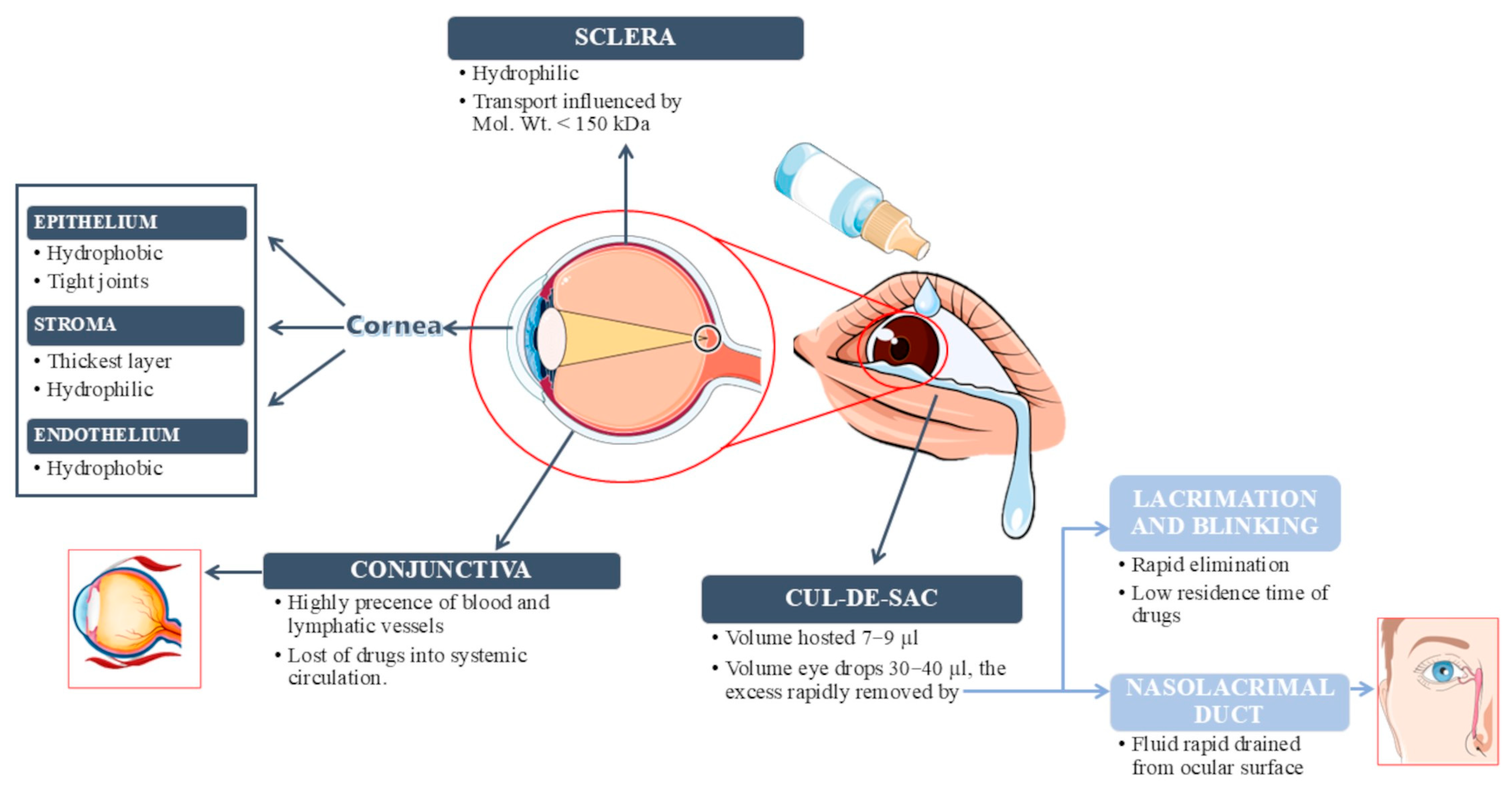
Figure 1. Schematic representation of the main ocular barriers to the topical application of drugs. The figure was created using images from Servier Medical Art (https://smart.servier.com/ accessed on 10 June 2025).
Gels 11 00752 g001
Figure 2. Schematic representation of tear composition.
Gels 11 00752 g002
Figure 3. Schematic representation of the in situ gelation mechanism of a P407 aqueous solution in the eye. Red arrow: increasing temperature; blue arrow: decreasing temperature.
Gels 11 00752 g003
Figure 4. Representation of the preparation of carboxymethyl chitosan-modified fluticasone propionate nanosuspensions loaded into a thermo-responsive in situ gel for blepharitis treatment. Reproduced with permission from [89]. Copyright (2025) Elsevier.
Gels 11 00752 g004
Figure 5. (a) Schematic representation of the poloxamer chemical structure, (b) mechanism of gelation for the in situ Poloxamer-HTCC system, (c) molecular structure of the in situ Poloxamer-HTCC gel and its utility in ocular drug delivery, (d) interaction with corneal mucins. Reproduced with permission from [132]. Copyright (2024) Elsevier.
Gels 11 00752 g005
Table 1. Parameters used for the classification of hydrogels.
SourceCompositionCrosslinkingConfigurationIonic ChargePropertyResponse
NaturalHomopolymerPhysicalAmorphousNon-ionicConventionalPhysical stimuli
SyntheticCopolymerChemicalCrystallineAnionicIntelligentChemical stimuli
Semi-syntheticSemi-IPN Semi-crystallineCationic
IPN Ampholytic
Abbreviations: IPN, interpenetrating polymer networks.
Table 2. Main characteristics of poloxamer-based in situ hydrogels for treating ocular diseases.
Type and Amount of Poloxamer UsedOther ComponentsType of HydrogelType of Embedded CarrierActive CompoundApplicationsReferences
P407 15% w/wHPMC 1.5% w/wENiosomesDoxycyclineAntibacterial activity[72]
P407 8.16% w/vHPMC 0.77% w/vG-ChloramphenicolAntimicrobial activity[95]
P407HPMCG-BerberineAntitumor activity;
Antibiotic property; Antioxidant;
Anti-inflammatory effects;
Gastroenteric discomfort;
Diabetes in clinic
[97]
P407 17.5–22.5% w/vChitosanG-TacrolimusAllograft corneal rejection;
Mooren’s ulcer;
Allergic conjunctivitis;
Immunogenic inflammatory ocular surface diseases;
Posterior uveitis;
Posterior blepharitis
[93]
P407
P188
HPMC K4MG-NifedipineGlaucoma[40]
P407 15–20% w/v
P188 0–7.50% w/v
HPMC 0.5–1.5% w/vENanocrystalsItraconazoleFungal infections[115]
P407 14% w/wCarbopol 934 0.3% w/wE, NHPMC nanoparticlesFlurbiprofenPrevention of miosis throughout ocular surgery;
Postoperative ocular inflammation
[109]
P7: P407 21% w/v
P188 4% w/v;
P8: P407 21% w/v
P188 5% w/v
F5: chitosan 1.5% w/v
β-glycerophosphate 45% w/v
G-Orthosiphon Stamineus BenthAntimicrobial activity[124]
F2: P407 15% w/w HA 0.2% w/w
F4: P407 10% w/w HA 0.4% w/w
-ECubosomesVoriconazoleFungal infections[113]
P407 27.36% w/v
P188 6.22% w/v
-E, NNanostructured lipid carriersFlurbiprofenAnti-inflammatory therapy[128]
P407 10% and 12% w/w
P407 12% w/w and P188 1–10% w/w
-E, MLipid nanosystems oil-in-water (O/W) microemulsionPrednisoloneOcular inflammatory disease[130]
P407 20% w/w
P188 5% w/w
Carbopol 0.2% w/wG Ketoconazole complexed with sulfobutylether-β-cyclodextrinFungal infections[131]
P407 20% wt-G-ProgranulinAnti-inflammatory action;
Regeneration and re-epithelialization of corneal tissue
[66]
P407 20.5% w/v
P188 5.0% w/v
-EChitosan nanoparticlesChloramphenicolAntimicrobial activity[96]
Aldehyde-functionalized P407 (AF127 different concentrations)-GPolyvinylpyrrolidone nanoparticles C u 2 x SeAntioxidant;
Dry eye disease
[90]
P407
P188
N-(2-hydroxy-3-trimethylammonium) propyl chitosan chlorideM-Levofloxacin hydrochlorideFungal keratitis[132]
P407AlginateE, NCarboxymethyl chitosan nanosuspensionClobetasol propionateUveitis treatment[86]
P407-based gel (SEP)SelenolMNanosuspensionFluticasone propionateBlepharitis[89]
P407 15% w/vHPMC E-50 LV 1% w/vG-GanciclovirCytomegalovirus retinitis;
Herpetic keratitis
[98]
P407 18.0 w/v%
P188 5.0 w/v%
HPMC (1.0 w/v%)G-(rh)MG53 proteinPromote corneal healing[99]
P407 14%HPMC K4M 1.5%EBacteriophagephage vB_Pa_ZCPS1Pseudomonas aeruginosa keratitis[105]
P407 16.5, 17, and 18% w/vGellan Gum 0.1% w/vE, MChitosan nanoparticlesDexamethasone
Polymyxin B sulfate
Neomycin sulfate
Inflammation and infection[106]
P407
P188
Tannic AcidG-Amphotericin BCorneal ulcers[133]
P407 10–20% w/vAlginateG-RaloxifeneWound healing[100]
Abbreviations: P407, poloxamer 407; P188, poloxamer 188; HPMC, hydroxypropyl methyl cellulose; HA, hyaluronic acid; E, embedded: thermo-responsive hydrogel containing nanosystems; G, thermo-responsive hydrogel; M, microhydrogel; N, nanohydrogel.
Disclaimer/Publisher’s Note: The statements, opinions and data contained in all publications are solely those of the individual author(s) and contributor(s) and not of MDPI and/or the editor(s). MDPI and/or the editor(s) disclaim responsibility for any injury to people or property resulting from any ideas, methods, instructions or products referred to in the content.