
Recent Progress of Biomaterial-Based Hydrogels for Wearable and Implantable Bioelectronics

 ,
, 


Abstract
1. Introduction
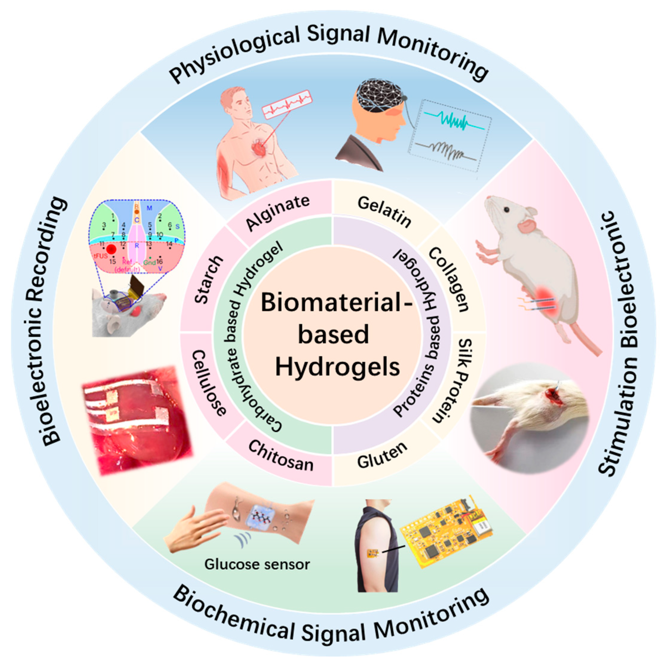
2. Biomaterial-Based Hydrogel
2.1. Protein-Based Hydrogels
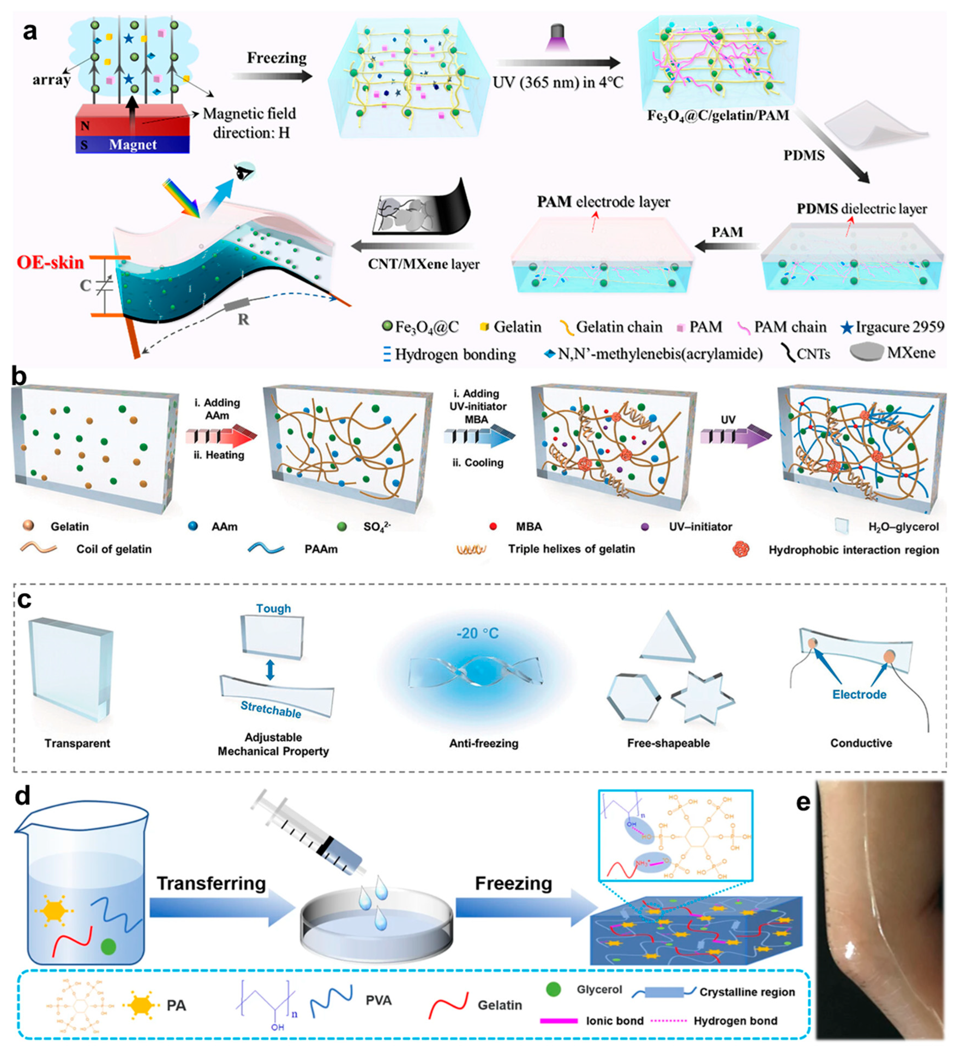
2.1.1. Gelatin
2.1.2. Collagen
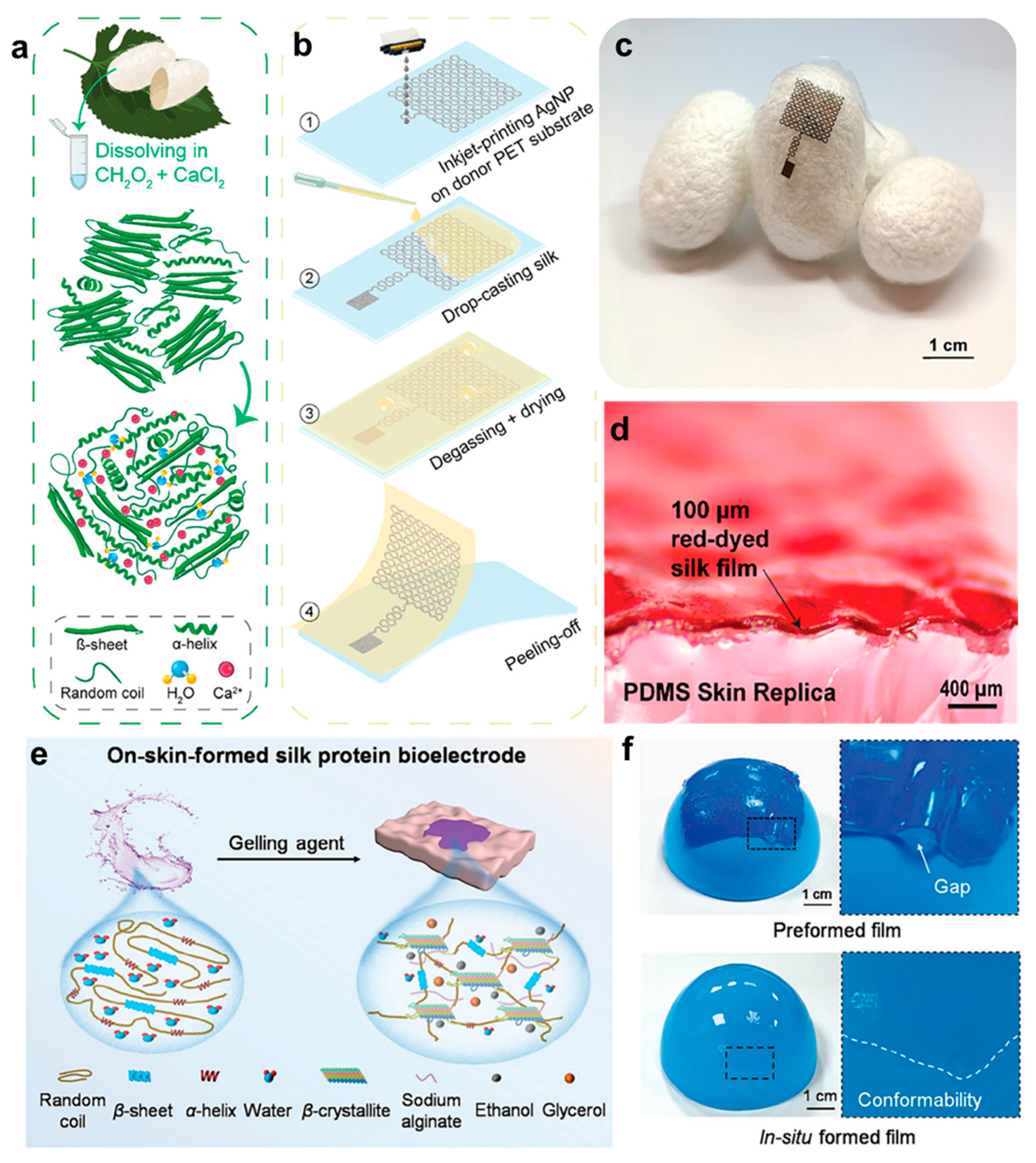
2.1.3. Silk Fibroin
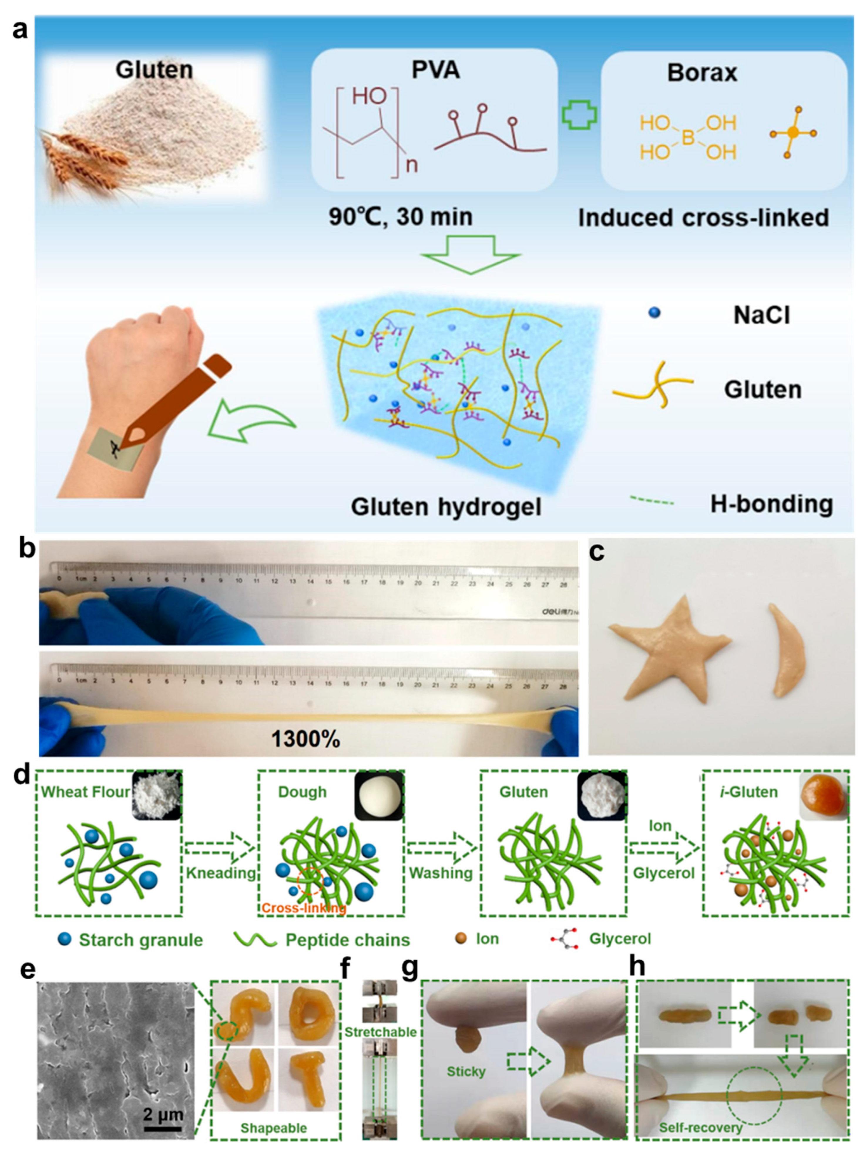
2.1.4. Gluten
2.2. Polysaccharide-Based Hydrogels
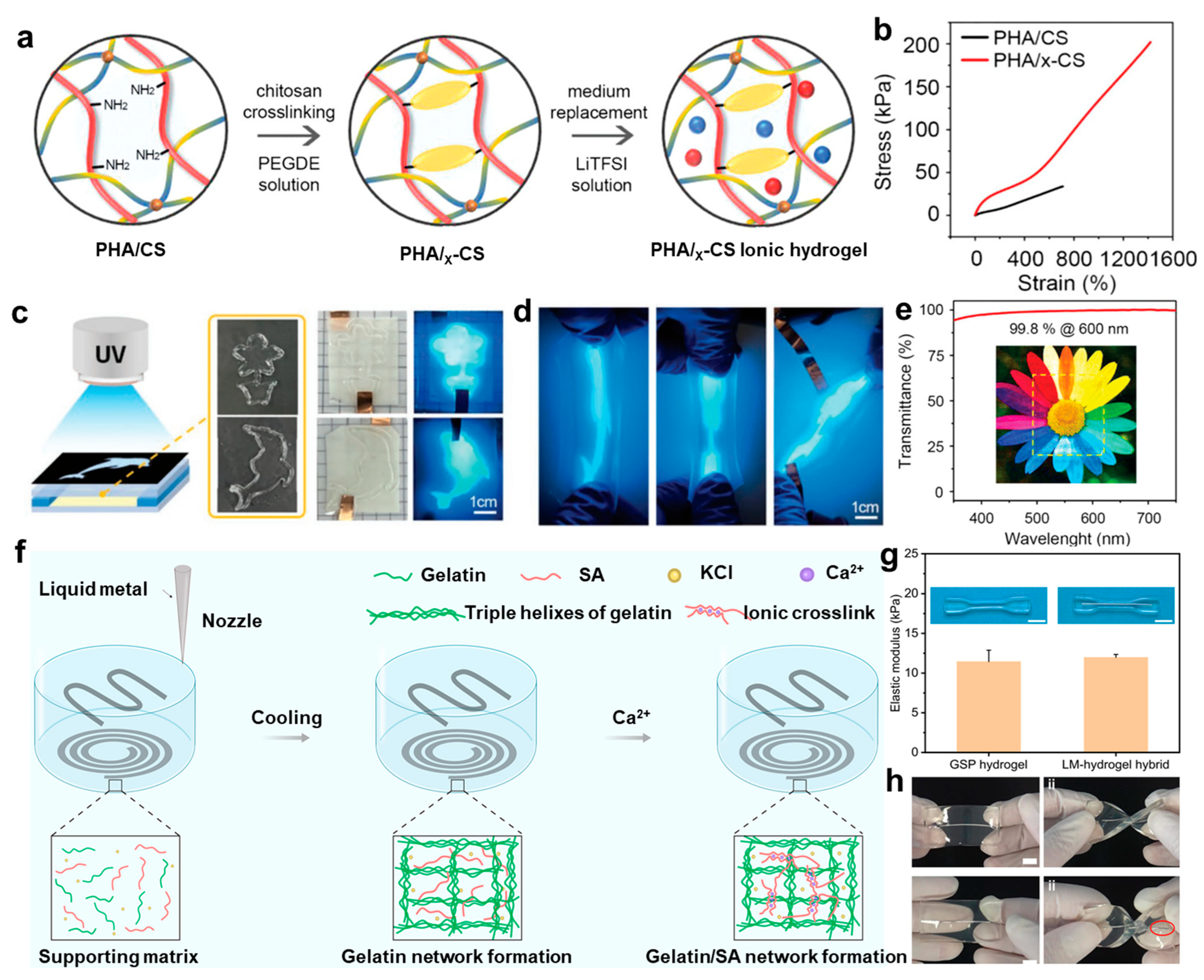
2.2.1. Chitosan
2.2.2. Cellulose
2.2.3. Alginate
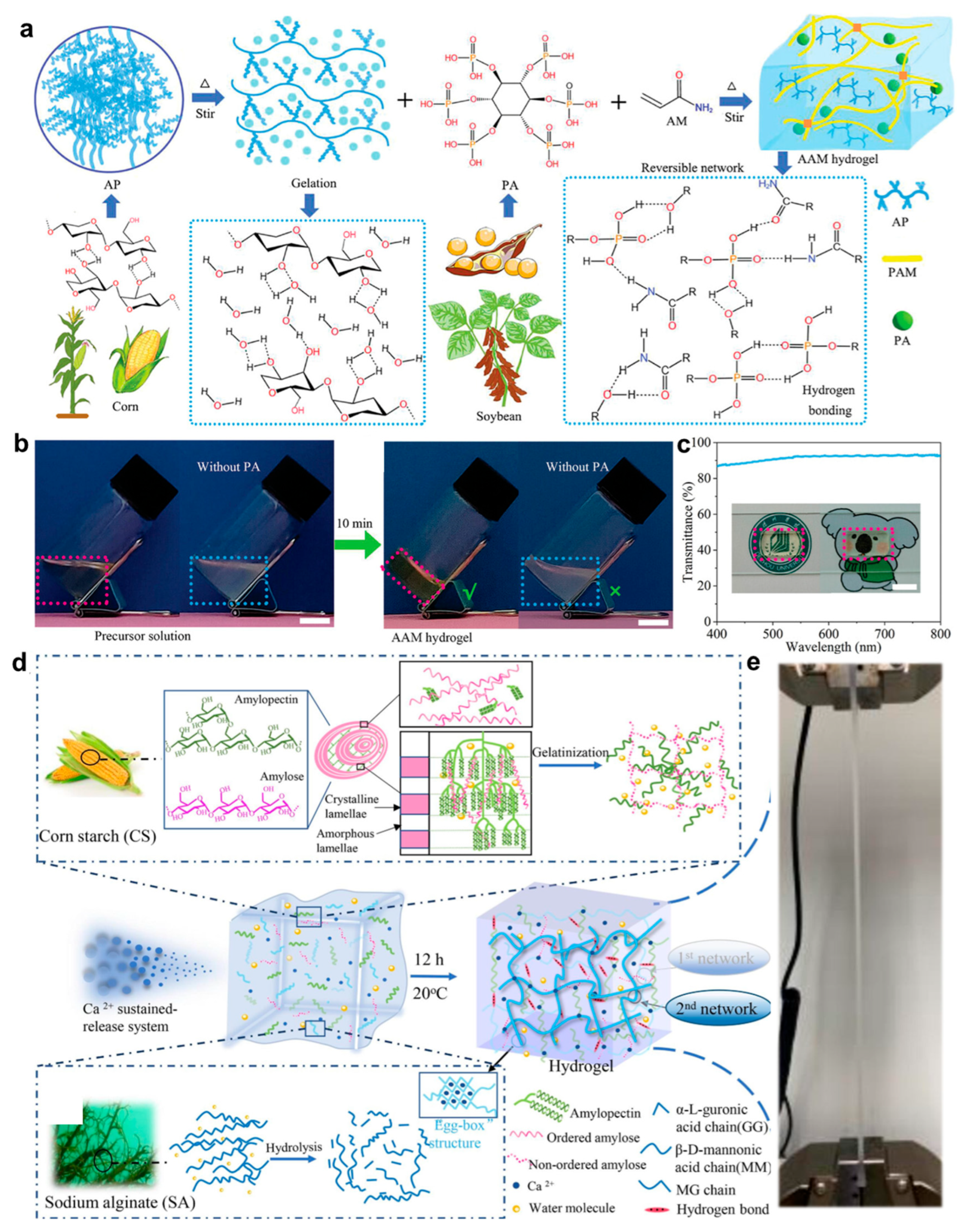
2.2.4. Starch
3. Application
3.1. Wearable Bioelectronics
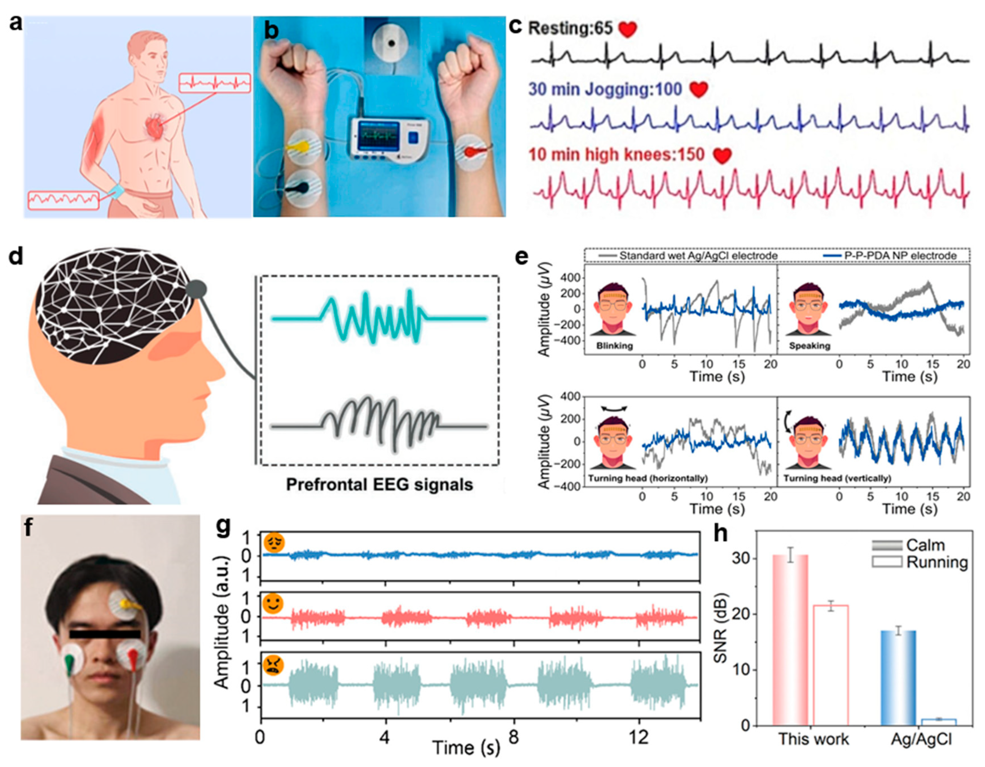
3.1.1. Electrophysiological Signal Monitoring
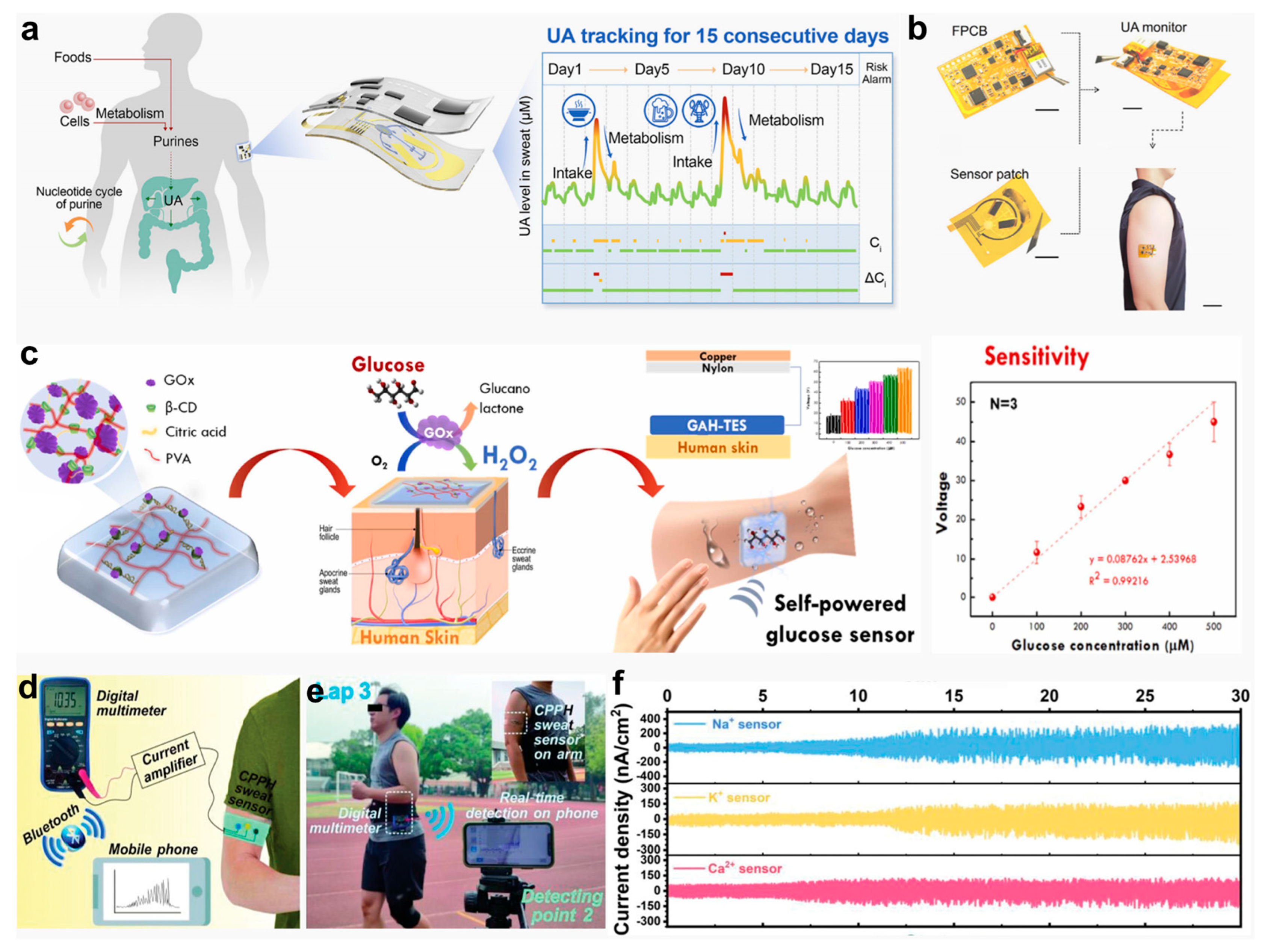
3.1.2. Biochemical Signal Monitoring
3.2. Implantable Bioelectronics
3.2.1. Bioelectric Recording
3.2.2. Bioelectrical Stimulation
4. Conclusions and Future Perspectives
Author Contributions
Funding
Institutional Review Board Statement
Informed Consent Statement
Data Availability Statement
Conflicts of Interest
References
- Koo, J.; MacEwan, M.R.; Kang, S.K.; Won, S.M.; Stephen, M.; Gamble, P.; Xie, Z.; Yan, Y.; Chen, Y.Y.; Shin, J.; et al. Wireless bioresorbable electronic system enables sustained nonpharmacological neuroregenerative therapy. Nat. Med. 2018, 24, 1830–1836. [Google Scholar] [CrossRef]
- Tringides, C.M.; Mooney, D.J. Materials for Implantable Surface Electrode Arrays: Current Status and Future Directions. Adv. Mater. 2022, 34, e2107207. [Google Scholar] [CrossRef] [PubMed]
- Khan, W.U.; Shen, Z.; Mugo, S.M.; Wang, H.; Zhang, Q. Implantable hydrogels as pioneering materials for next-generation brain-computer interfaces. Chem. Soc. Rev. 2025, 54, 2832–2880. [Google Scholar] [CrossRef] [PubMed]
- Chung, H.U.; Kim, B.H.; Lee, J.Y.; Lee, J.; Xie, Z.; Ibler, E.M.; Lee, K.; Banks, A.; Jeong, J.Y.; Kim, J.; et al. Binodal, wireless epidermal electronic systems with in-sensor analytics for neonatal intensive care. Science 2019, 363, eaau0780. [Google Scholar] [CrossRef]
- Lu, Y.; Yang, G.; Wang, S.; Zhang, Y.; Jian, Y.; He, L.; Yu, T.; Luo, H.; Kong, D.; Xianyu, Y.; et al. Stretchable graphene–hydrogel interfaces for wearable and implantable bioelectronics. Nat. Electron. 2023, 7, 51. [Google Scholar] [CrossRef]
- Duan, H.; Peng, S.; He, S.; Tang, S.Y.; Goda, K.; Wang, C.H.; Li, M. Wearable Electrochemical Biosensors for Advanced Healthcare Monitoring. Adv. Sci. 2025, 12, e2411433. [Google Scholar] [CrossRef]
- Li, J.; Carlos, C.; Zhou, H.; Sui, J.; Wang, Y.; Silva-Pedraza, Z.; Yang, F.; Dong, Y.; Zhang, Z.; Hacker, T.A.; et al. Stretchable piezoelectric biocrystal thin films. Nat. Commun. 2023, 14, 6562. [Google Scholar] [CrossRef]
- Jiao, F.; Lin, C.; Dong, L.; Mao, X.; Wu, Y.; Dong, F.; Zhang, Z.; Sun, J.; Li, S.; Yang, X.; et al. Silicon Vacancies Diamond/Silk/PVA Hierarchical Physical Unclonable Functions for Multi-Level Encryption. Adv. Sci. 2024, 11, e2308337. [Google Scholar] [CrossRef]
- Hou, Y.; Li, Z.; Wang, Z.; Zhang, X.; Li, Y.; Li, C.; Guo, H.; Yu, H. Programmable and Surface-Conformable Origami Design for Thermoelectric Devices. Adv. Sci. 2024, 11, e2309052. [Google Scholar] [CrossRef]
- Li, Y.; Li, N.; Liu, W.; Prominski, A.; Kang, S.; Dai, Y.; Liu, Y.; Hu, H.; Wai, S.; Dai, S.; et al. Achieving tissue-level softness on stretchable electronics through a generalizable soft interlayer design. Nat. Commun. 2023, 14, 4488. [Google Scholar] [CrossRef]
- Yi, J.; Zou, G.; Huang, J.; Ren, X.; Tian, Q.; Yu, Q.; Wang, P.; Yuan, Y.; Tang, W.; Wang, C.; et al. Water-responsive supercontractile polymer films for bioelectronic interfaces. Nature 2023, 624, 295–302. [Google Scholar] [CrossRef] [PubMed]
- Yu, C.; Shi, M.; He, S.; Yao, M.; Sun, H.; Yue, Z.; Qiu, Y.; Liu, B.; Liang, L.; Zhao, Z.; et al. Chronological adhesive cardiac patch for synchronous mechanophysiological monitoring and electrocoupling therapy. Nat. Commun. 2023, 14, 6226. [Google Scholar] [CrossRef] [PubMed]
- Zhang, Y.; Tao, T.H. Skin-Friendly Electronics for Acquiring Human Physiological Signatures. Adv. Mater. 2019, 31, 1905767. [Google Scholar] [CrossRef] [PubMed]
- Liu, Y.; Yang, T.; Zhang, Y.; Qu, G.; Wei, S.; Liu, Z.; Kong, T. Ultrastretchable and Wireless Bioelectronics Based on All-Hydrogel Microfluidics. Adv. Mater. 2019, 31, 1902783. [Google Scholar] [CrossRef] [PubMed]
- Lee, S.; Kum, J.; Kim, S.; Jung, H.; An, S.; Choi, S.J.; Choi, J.H.; Kim, J.; Yu, K.J.; Lee, W.; et al. A shape-morphing cortex-adhesive sensor for closed-loop transcranial ultrasound neurostimulation. Nat. Electron. 2024, 7, 800–814. [Google Scholar] [CrossRef]
- Chowdhury, I.F.; Shawon, M.T.A.; Alam, M.A.; Fatima, S.; Khan, A.A.; Yang, J.; Tang, Z.; Mondal, A.K. Ni2+-Rich Collagen/Lignin Composite Hydrogel: Transforming Industrial Waste Materials into Flexible Electronics. ACS Appl. Polym. Mater. 2024, 6, 15094–15104. [Google Scholar] [CrossRef]
- Cho, K.W.; Sunwoo, S.H.; Hong, Y.J.; Koo, J.H.; Kim, J.H.; Baik, S.; Hyeon, T.; Kim, D.H. Soft Bioelectronics Based on Nanomaterials. Chem. Rev. 2022, 122, 5068–5143. [Google Scholar] [CrossRef]
- Nguyen, T.K.; Yadav, S.; Truong, T.A.; Han, M.; Barton, M.; Leitch, M.; Guzman, P.; Dinh, T.; Ashok, A.; Vu, H.; et al. Integrated, Transparent Silicon Carbide Electronics and Sensors for Radio Frequency Biomedical Therapy. ACS Nano 2022, 16, 10890–10903. [Google Scholar] [CrossRef]
- Fan, H.; Gong, J.P. Bioinspired Underwater Adhesives. Adv. Mater. 2021, 33, e2102983. [Google Scholar] [CrossRef]
- Yuk, H.; Wu, J.; Zhao, X. Hydrogel interfaces for merging humans and machines. Nat. Rev. Mater. 2022, 7, 935–952. [Google Scholar] [CrossRef]
- Chen, C.-K.; Chen, P.-W.; Wang, H.-J.; Yeh, M.-Y. Alkyl Chain Length Effects of Imidazolium Ionic Liquids on Electrical and Mechanical Performances of Polyacrylamide/Alginate-Based Hydrogels. Gels 2021, 7, 164. [Google Scholar] [CrossRef] [PubMed]
- Niu, Y.; Liu, H.; He, R.; Luo, M.; Shu, M.; Xu, F. Environmentally Compatible Wearable Electronics Based on Ionically Conductive Organohydrogels for Health Monitoring with Thermal Compatibility, Anti-Dehydration, and Underwater Adhesion. Small 2021, 17, 2101151. [Google Scholar] [CrossRef]
- Ling, Q.; Fan, X.; Ling, M.; Liu, J.; Zhao, L.; Gu, H. Collagen-Based Organohydrogel Strain Sensor with Self-Healing and Adhesive Properties for Detecting Human Motion. ACS Appl. Mater. Interfaces 2023, 15, 12350–12362. [Google Scholar] [CrossRef]
- Li, S.; Liu, G.; Wen, H.; Liu, G.; Wang, H.; Ye, M.; Yang, Y.; Guo, W.; Liu, Y. A Skin-Like Pressure- and Vibration-Sensitive Tactile Sensor Based on Polyacrylamide/Silk Fibroin Elastomer. Adv. Funct. Mater. 2022, 32, 2111747. [Google Scholar] [CrossRef]
- Li, J.; Sun, Y.; Shi, W.; Li, Y.; Zou, Y.; Zhang, H. Fabrication, characterization, and in vitro digestion of gelatin/gluten oleogels from thermally crosslinked electrospun short fiber aerogel templates. Food. Chem. 2024, 454, 139804. [Google Scholar] [CrossRef] [PubMed]
- Lee, M.; Park, J.; Choe, G.; Lee, S.; Kang, B.G.; Jun, J.H.; Shin, Y.; Kim, M.C.; Kim, Y.S.; Ahn, Y.; et al. A Conductive and Adhesive Hydrogel Composed of MXene Nanoflakes as a Paintable Cardiac Patch for Infarcted Heart Repair. ACS Nano 2023, 17, 12290–12304. [Google Scholar] [CrossRef]
- Lan, M.; Zhang, J.; Zhou, J.; Gu, H. CQDs-Cross-Linked Conductive Collagen/PAA-Based Nanocomposite Organohydrogel Coupling Flexibility with Multifunctionality for Dual-Modal Sensing of Human Motions. ACS Appl. Mater. Interfaces 2024, 16, 23838–23854. [Google Scholar] [CrossRef] [PubMed]
- Meng, L.; Shao, C.; Cui, C.; Xu, F.; Lei, J.; Yang, J. Autonomous Self-Healing Silk Fibroin Injectable Hydrogels Formed via Surfactant-Free Hydrophobic Association. ACS Appl. Mater. Interfaces 2019, 12, 1628–1639. [Google Scholar] [CrossRef]
- Ohm, Y.; Pan, C.; Ford, M.J.; Huang, X.; Liao, J.; Majidi, C. An electrically conductive silver–polyacrylamide–alginate hydrogel composite for soft electronics. Nat. Electron. 2021, 4, 185–192. [Google Scholar] [CrossRef]
- Wang, C.; Xia, K.; Zhang, M.; Jian, M.; Zhang, Y. An All-Silk-Derived Dual-Mode E-skin for Simultaneous Temperature–Pressure Detection. ACS Appl. Mater. Interfaces 2017, 9, 39484–39492. [Google Scholar] [CrossRef]
- Zhu, M.; Gong, D.; Ji, Z.; Yang, J.; Wang, M.; Wang, Z.; Tao, S.; Wang, X.; Xu, M. Cellulose-reinforced poly(Ionic Liquids) composite hydrogel for infected wounds therapy and real-time reliable bioelectronic. Chem. Eng. J. 2023, 476, 146816. [Google Scholar] [CrossRef]
- Han, Q.; Zhang, C.; Guo, T.; Tian, Y.; Song, W.; Lei, J.; Li, Q.; Wang, A.; Zhang, M.; Bai, S.; et al. Hydrogel Nanoarchitectonics of a Flexible and Self-Adhesive Electrode for Long-Term Wireless Electroencephalogram Recording and High-Accuracy Sustained Attention Evaluation. Adv. Mater. 2023, 35, 2209606. [Google Scholar] [CrossRef]
- Jin, S.; Choi, H.; Seong, D.; You, C.-L.; Kang, J.-S.; Rho, S.; Lee, W.B.; Son, D.; Shin, M. Injectable tissue prosthesis for instantaneous closed-loop rehabilitation. Nature 2023, 623, 58–65. [Google Scholar] [CrossRef]
- Jiao, C.; Wang, Q.; Li, L.; Chen, W.; Liu, J.; Xu, Y.; Song, L.; Fu, S.; Hu, L. In situ 3D printing of liquid metal-hydrogel hybrid for multifunctional soft bioelectronics and devices. Cell Rep. Phys. Sci. 2023, 4, 101640. [Google Scholar] [CrossRef]
- Hu, Y.; Yang, Y.; Chu, H.; Shi, R.; Li, J.; Xu, G.; Huang, X.; Zhang, B.; Yiu, C.K.; Zhao, G.; et al. Prolonged monitoring and risk management of hyperuricemia using interference-resistant wearable bioelectronics. Device 2025, 3, 100753. [Google Scholar] [CrossRef]
- Kanokpaka, P.; Chang, Y.-H.; Chang, C.-C.; Rinawati, M.; Wang, P.-C.; Chang, L.-Y.; Yeh, M.-H. Enabling glucose adaptive self-healing hydrogel based triboelectric biosensor for tracking a human perspiration. Nano Energy 2023, 112, 108513. [Google Scholar] [CrossRef]
- Choi, H.; Kim, Y.; Kim, S.; Jung, H.; Lee, S.; Kim, K.; Han, H.-S.; Kim, J.Y.; Shin, M.; Son, D. Adhesive bioelectronics for sutureless epicardial interfacing. Nat. Electron. 2023, 6, 779–789. [Google Scholar] [CrossRef]
- Zhang, W.; Lou, Q.; Sun, J. Carbon nanodot-based flexible and self-powered white displays. Nano Res. 2025, 18, 94907117. [Google Scholar] [CrossRef]
- Qureshi, A.T.; Afrin, S.; Asim, S.; Rizwan, M. Imine Crosslinked, Injectable, and Self-Healing Fucoidan Hydrogel with Immunomodulatory Properties. Adv. Healthc. Mater. 2025, 14, e2405260. [Google Scholar] [CrossRef]
- Zhang, Y.; Ye, S.; Cao, L.; Lv, Z.; Ren, J.; Shao, Z.; Yao, Y.; Ling, S. Natural Silk Spinning-Inspired Meso-Assembly-Processing Engineering Strategy for Fabricating Soft Tissue-Mimicking Biomaterials. Adv. Funct. Mater. 2022, 32, 2200267. [Google Scholar] [CrossRef]
- Bi, S.; Wang, P.; Hu, S.; Li, S.; Pang, J.; Zhou, Z.; Sun, G.; Huang, L.; Cheng, X.; Xing, S.; et al. Construction of physical-crosslink chitosan/PVA double-network hydrogel with surface mineralization for bone repair. Carbohyd. Polym. 2019, 224, 115176. [Google Scholar] [CrossRef] [PubMed]
- Go, Y.; Park, H.Y.; Zhu, Y.; Yoo, K.; Kwak, J.; Jin, S.H.; Yoon, J. Optically Transparent and Mechanically Robust Ionic Hydrogel Electrodes for Bright Electroluminescent Devices Achieving High Stretchability Over 1400%. Adv. Funct. Mater. 2023, 33, 2215193. [Google Scholar] [CrossRef]
- Sun, J.-Y.; Zhao, X.; Illeperuma, W.R.K.; Chaudhuri, O.; Oh, K.H.; Mooney, D.J.; Vlassak, J.J.; Suo, Z. Highly stretchable and tough hydrogels. Nature 2012, 489, 133–136. [Google Scholar] [CrossRef]
- Wang, Z.; Wei, H.; Huang, Y.; Wei, Y.; Chen, J. Naturally sourced hydrogels: Emerging fundamental materials for next-generation healthcare sensing. Chem. Soc. Rev. 2023, 52, 2992–3034. [Google Scholar] [CrossRef]
- Feig, V.R.; Tran, H.; Lee, M.; Bao, Z. Mechanically tunable conductive interpenetrating network hydrogels that mimic the elastic moduli of biological tissue. Nat. Commun. 2018, 9, 2740. [Google Scholar] [CrossRef] [PubMed]
- Liu, X.; Chen, X.; Chi, X.; Feng, Z.; Yang, C.; Gao, R.; Li, S.; Zhang, C.; Chen, X.; Huang, P.; et al. Biomimetic integration of tough polymer elastomer with conductive hydrogel for highly stretchable, flexible electronic. Nano Energy 2022, 92, 106735. [Google Scholar] [CrossRef]
- Fu, Q.; Tang, J.; Wang, W.; Wang, R. Biocomposite Polyvinyl Alcohol/Ferritin Hydrogels with Enhanced Stretchability and Conductivity for Flexible Strain Sensors. Gels 2025, 11, 59. [Google Scholar] [CrossRef]
- Li, Y.; Tan, S.; Zhang, X.; Li, Z.; Cai, J.; Liu, Y. Design Strategies and Emerging Applications of Conductive Hydrogels in Wearable Sensing. Gels 2025, 11, 258. [Google Scholar] [CrossRef]
- Wang, R.; Jin, B.; Li, J.; Li, J.; Xie, J.; Zhang, P.; Fu, Z. Bio-Inspired Synthesis of Injectable, Self-Healing PAA-Zn-Silk Fibroin-MXene Hydrogel for Multifunctional Wearable Capacitive Strain Sensor. Gels 2025, 11, 377. [Google Scholar] [CrossRef]
- Tringides, C.M.; Vachicouras, N.; de Lázaro, I.; Wang, H.; Trouillet, A.; Seo, B.R.; Elosegui-Artola, A.; Fallegger, F.; Shin, Y.; Casiraghi, C.; et al. Viscoelastic surface electrode arrays to interface with viscoelastic tissues. Nat. Nanotechnol. 2021, 16, 1019–1029. [Google Scholar] [CrossRef]
- Wang, Q.; Ling, S.; Liang, X.; Wang, H.; Lu, H.; Zhang, Y. Self-Healable Multifunctional Electronic Tattoos Based on Silk and Graphene. Adv. Funct. Mater. 2019, 29, 1808695. [Google Scholar] [CrossRef]
- Yao, X.; Zhang, S.; Qian, L.; Wei, N.; Nica, V.; Coseri, S.; Han, F. Super Stretchable, Self-Healing, Adhesive Ionic Conductive Hydrogels Based on Tailor-Made Ionic Liquid for High-Performance Strain Sensors. Adv. Funct. Mater. 2022, 32, 2204565. [Google Scholar] [CrossRef]
- Park, B.; Shin, J.H.; Ok, J.; Park, S.; Jung, W.; Jeong, C.; Choy, S.; Jo, Y.J.; Kim, T.-i. Cuticular pad–inspired selective frequency damper for nearly dynamic noise–free bioelectronics. Science 2022, 376, 624–629. [Google Scholar] [CrossRef] [PubMed]
- Wang, Y.; Li, L.; Ji, Y.-E.; Wang, T.; Fu, Y.; Li, X.; Li, G.; Zheng, T.; Wu, L.; Han, Q.; et al. Silk-protein-based gradient hydrogels with multimode reprogrammable shape changes for biointegrated devices. Proc. Natl. Acad. Sci. USA 2023, 120, e2305704120. [Google Scholar] [CrossRef]
- Yang, Z.; Zhang, Z.; Zhou, T.; Yu, N.; Yu, R.; Du, X.; Bai, X.; Miao, Z.; Wu, L.; Pan, S.; et al. An On-Skin-Formed Silk Protein Bioelectrode for Conformable and Robust Electrophysiological Interface. Adv. Funct. Mater. 2024, 34, 2402608. [Google Scholar] [CrossRef]
- Sang, F.; Liu, C.; Yan, J.; Su, J.; Niu, S.; Wang, S.; Zhao, Y.; Dang, Q. Polysaccharide- and protein-based hydrogel dressings that enhance wound healing: A review. Int. J. Biol. Macromol. 2024, 280, 135482. [Google Scholar] [CrossRef]
- Song, X.; Wang, X.; Zhang, J.; Shen, S.; Yin, W.; Ye, G.; Wang, L.; Hou, H.; Qiu, X. A tunable self-healing ionic hydrogel with microscopic homogeneous conductivity as a cardiac patch for myocardial infarction repair. Biomaterials 2021, 273, 120811. [Google Scholar] [CrossRef]
- Qie, H.; Wang, Z.; Ren, J.; Lü, S.; Liu, M. A tough shape memory hydrogel strain sensor based on gelatin grafted polypyrrole. Polymer 2022, 263, 125524. [Google Scholar] [CrossRef]
- Wang, J.; Gao, C.; Hou, P.; Liu, Y.; Zhao, J.; Huo, P. All-bio-based, adhesive and low-temperature resistant hydrogel electrolytes for flexible supercapacitors. Chem. Eng. J. 2023, 455, 140952. [Google Scholar] [CrossRef]
- Jeong, J.; Kim, T.H.; Park, S.; Lee, J.; Chae, U.; Jeong, J.-Y.; Park, S.; Kim, S.; Cho, I.-J.; Jung, Y.; et al. Hybrid neural interfacing devices based on Au wires with nanogranular Au shell and hydrogel layer for anti-inflammatory and bi-directional neural communications. Chem. Eng. J. 2023, 465, 142966. [Google Scholar] [CrossRef]
- Zhang, Z.; Yang, J.; Wang, H.; Wang, C.; Gu, Y.; Xu, Y.; Lee, S.; Yokota, T.; Haick, H.; Someya, T.; et al. A 10-micrometer-thick nanomesh-reinforced gas-permeable hydrogel skin sensor for long-term electrophysiological monitoring. Sci. Adv. 2024, 10, eadj5389. [Google Scholar] [CrossRef] [PubMed]
- He, Z.; Yuan, W. Adhesive, Stretchable, and Transparent Organohydrogels for Antifreezing, Antidrying, and Sensitive Ionic Skins. ACS Appl. Mater. Interfaces 2021, 13, 1474–1485. [Google Scholar] [CrossRef]
- Yu, D.; Yi, J.; Zhu, S.; Tang, Y.; Huang, Y.; Lin, D.; Lin, Y. Hofmeister Effect-Assisted Facile One-Pot Fabrication of Double Network Organohydrogels with Exceptional Multi-Functions. Adv. Funct. Mater. 2024, 34, 2307566. [Google Scholar] [CrossRef]
- Li, T.; Qi, H.; Zhao, C.; Li, Z.; Zhou, W.; Li, G.; Zhuo, H.; Zhai, W. Robust skin-integrated conductive biogel for high-fidelity detection under mechanical stress. Nat. Commun. 2025, 16, 88. [Google Scholar] [CrossRef]
- Zhang, H.; Chen, H.; Lee, J.-H.; Kim, E.; Chan, K.-Y.; Venkatesan, H.; Shen, X.; Yang, J.; Kim, J.-K. Mechanochromic Optical/Electrical Skin for Ultrasensitive Dual-Signal Sensing. ACS Nano 2023, 17, 5921–5934. [Google Scholar] [CrossRef] [PubMed]
- Liu, W.; Xie, R.; Zhu, J.; Wu, J.; Hui, J.; Zheng, X.; Huo, F.; Fan, D. A temperature responsive adhesive hydrogel for fabrication of flexible electronic sensors. npj Flex. Electron. 2022, 6, 68. [Google Scholar] [CrossRef]
- Lin, K.; Zhang, D.; Macedo, M.H.; Cui, W.; Sarmento, B.; Shen, G. Advanced Collagen-Based Biomaterials for Regenerative Biomedicine. Adv. Funct. Mater. 2018, 29, 1804943. [Google Scholar] [CrossRef]
- Shi, X.; Lan, M.; Liu, J.; Zhou, J.; Gu, H. Highly robust, self-adhesive, self-healing, pH-responsive, cytocompatible and degradable collagen/PVA/tannin-based conductive hydrogel sensor for motion-monitoring. Polymer 2024, 308, 127365. [Google Scholar] [CrossRef]
- Song, X.; Zhang, J.; Shen, S.; Liu, D.; Zhang, J.; Yin, W.; Ye, G.; Wang, L.; Cai, L.; Hou, H.; et al. Cardiac-Adaptive Conductive Hydrogel Patch Enabling Construction of Mechanical–Electrical Anisotropic Microenvironment for Heart Repair. Research 2023, 6, 0161. [Google Scholar] [CrossRef]
- Zhang, M.; Yang, Q.; Liang, K.; Gao, L.; Lu, P.; Ding, C.; Dang, Y. Collagen/poly(acrylic acid)/MXene hydrogels with tissue-adhesive, biosensing, and photothermal antibacterial properties. Polym. Eng. Sci. 2023, 63, 3672–3683. [Google Scholar] [CrossRef]
- Roshanbinfar, K.; Schiffer, M.; Carls, E.; Angeloni, M.; Koleśnik-Gray, M.; Schruefer, S.; Schubert, D.W.; Ferrazzi, F.; Krstić, V.; Fleischmann, B.K.; et al. Electrically Conductive Collagen-PEDOT:PSS Hydrogel Prevents Post-Infarct Cardiac Arrhythmia and Supports hiPSC-Cardiomyocyte Function. Adv. Mater. 2024, 36, 2403642. [Google Scholar] [CrossRef] [PubMed]
- Li, T.; Zhou, Z.; Xie, Y.; Cai, W.; Zhu, X.; Jia, Y.; Zhang, Z.; Xu, F.; Huang, G. Engineering strong and tough collagen hydrogels and tissue constructs via twisting and crosslinking. Cell Rep. Phys. Sci. 2025, 6, 102454. [Google Scholar] [CrossRef]
- Mao, Z.; Bi, X.; Yu, C.; Chen, L.; Shen, J.; Huang, Y.; Wu, Z.; Qi, H.; Guan, J.; Shu, X.; et al. Mechanically robust and personalized silk fibroin-magnesium composite scaffolds with water-responsive shape-memory for irregular bone regeneration. Nat. Commun. 2024, 15, 4160. [Google Scholar] [CrossRef] [PubMed]
- Zhao, G.; Feng, Y.; Xue, L.; Cui, M.; Zhang, Q.; Xu, F.; Peng, N.; Jiang, Z.; Gao, D.; Zhang, X. Anisotropic conductive reduced graphene oxide/silk matrices promote post-infarction myocardial function by restoring electrical integrity. Acta Biomater. 2022, 139, 190–203. [Google Scholar] [CrossRef]
- Mirbakht, S.S.; Golparvar, A.; Umar, M.; Kuzubasoglu, B.A.; Irani, F.S.; Yapici, M.K. Highly Self-Adhesive and Biodegradable Silk Bioelectronics for All-In-One Imperceptible Long-Term Electrophysiological Biosignals Monitoring. Adv. Sci. 2025, 12, 2405988. [Google Scholar] [CrossRef]
- Chen, B.; Cao, Y.; Li, Q.; Yan, Z.; Liu, R.; Zhao, Y.; Zhang, X.; Wu, M.; Qin, Y.; Sun, C.; et al. Liquid metal-tailored gluten network for protein-based e-skin. Nat. Commun. 2022, 13, 1206. [Google Scholar] [CrossRef]
- Jaroenthai, N.; Srikhao, N.; Kasemsiri, P.; Okhawilai, M.; Theerakulpisut, S.; Uyama, H.; Chindaprasirt, P. Optimization of rapid self-healing and self-adhesive gluten/guar gum crosslinked gel for strain sensors and electronic devices. Int. J. Biol. Macromol. 2023, 253, 127401. [Google Scholar] [CrossRef]
- Peng, S.; Zhao, Y.; Liang, C.; Wang, H.; Zhou, H.; Yang, L. Conductive Ionically Cross-Linked Gluten/Poly(vinyl alcohol) Composite Organohydrogel with Freezing and Water Resistance for Wearable Sensors. ACS Appl. Polym. Mater. 2023, 5, 9515–9524. [Google Scholar] [CrossRef]
- Xiang, H.; Li, Z.; Bai, Z.; Wu, H.; Liu, G.; Zhou, H.; Liu, H. An E-skin for handwriting input at human-machine interface. Chem. Eng. J. 2025, 505, 158879. [Google Scholar] [CrossRef]
- Han, X.; Lu, W.; Yu, W.; Xu, H.; Bi, S.; Cai, H. Conductive and adhesive gluten ionic skin for eco-friendly strain sensor. J. Mater. Sci. 2020, 56, 3970–3980. [Google Scholar] [CrossRef]
- Wang, H.; Li, Z.; Zuo, M.; Zeng, X.; Tang, X.; Sun, Y.; Lin, L. Stretchable, freezing-tolerant conductive hydrogel for wearable electronics reinforced by cellulose nanocrystals toward multiple hydrogen bonding. Carbohydr. Polym. 2022, 280, 119018. [Google Scholar] [CrossRef] [PubMed]
- Li, J.; Wei, H.; Cui, S.; Hou, H.; Zhang, Y.; Zhang, Y.; Xu, B.B.; Chu, L.; El-Bahy, Z.M.; Melhi, S.; et al. Polyvinyl alcohol/sodium alginate-based conductive hydrogels with in situ formed bimetallic zeolitic imidazolate frameworks towards soft electronics. Carboh. Polym. 2024, 346, 122633. [Google Scholar] [CrossRef] [PubMed]
- Yan, L.; Zhou, T.; Han, L.; Zhu, M.; Cheng, Z.; Li, D.; Ren, F.; Wang, K.; Lu, X. Conductive Cellulose Bio-Nanosheets Assembled Biostable Hydrogel for Reliable Bioelectronics. Adv. Funct. Mater. 2021, 31, 2010465. [Google Scholar] [CrossRef]
- Yang, X.; Zhang, B.; Li, J.; Shen, M.; Liu, H.; Xu, X.; Shang, S. Self-healing, self-adhesive, and stretchable conductive hydrogel for multifunctional sensor prepared by catechol modified nanocellulose stabilized poly(α-thioctic acid). Carbohydr. Polym. 2023, 313, 120813. [Google Scholar] [CrossRef] [PubMed]
- Zheng, Y.; Huang, K.; You, X.; Huang, B.; Wu, J.; Gu, Z. Hybrid hydrogels with high strength and biocompatibility for bone regeneration. Int. J. Biol. Macromol. 2017, 104, 1143–1149. [Google Scholar] [CrossRef]
- Li, Y.; Wen, X.; Li, X.; Zahid, M.; Wang, H.; Zhang, J. Design of super stretchability, rapid self-healing, and self-adhesion hydrogel based on starch for wearable strain sensors. Carbohydr. Polym. 2025, 348, 122858. [Google Scholar] [CrossRef]
- Zhou, Q.; Abushammala, H.; Gao, D.; Xu, P.; Niu, D.; Yang, W.; Ma, P. Human soft tissues-like PVA/cellulose hydrogels with multifunctional properties towards flexible electronics applications. Carbohydr. Polym. 2025, 357, 123425. [Google Scholar] [CrossRef]
- Sun, X.; Yao, M.; He, S.; Dong, X.; Liang, L.; Yao, F.; Li, J. Antibacterial and UV-Blocking Bioelectronics Based on Transparent, Adhesive, and Strain-Sensitive Multifunctional Hydrogel. Adv. Mater. Technol. 2021, 7, 2101283. [Google Scholar] [CrossRef]
- Ryplida, B.; Lee, K.D.; In, I.; Park, S.Y. Light-Induced Swelling-Responsive Conductive, Adhesive, and Stretchable Wireless Film Hydrogel as Electronic Artificial Skin. Adv. Funct. Mater. 2019, 29, 1903209. [Google Scholar] [CrossRef]
- Yang, J.M.; Fan, C.-S.; Wang, N.-C.; Chang, Y.-H. Evaluation of membrane preparation method on the performance of alkaline polymer electrolyte: Comparison between poly(vinyl alcohol)/chitosan blended membrane and poly(vinyl alcohol)/chitosan electrospun nanofiber composite membranes. Electrochim. Acta 2018, 266, 332–340. [Google Scholar] [CrossRef]
- Pirahmadi, P.; Kokabi, M.; Alamdarnejad, G. Polyvinyl alcohol/chitosan/carbon nanotubes electroactive shape memory nanocomposite hydrogels. J. Appl. Polym. Sci. 2020, 138, e49995. [Google Scholar] [CrossRef]
- Guo, Z.; Xu, X.; Qiu, J.; Yu, W.; Zhang, S.; Li, J.; Zhu, Y.; Lu, J.; Gao, Q.; Nie, B.; et al. Fishing net-inspired PVA-chitosan-CNT hydrogels with high stretchability, sensitivity, and environmentally stability for textile strain sensors. Int. J. Biol. Macromol. 2024, 282, 137576. [Google Scholar] [CrossRef] [PubMed]
- Guo, S.; Yao, M.; Zhang, D.; He, Y.; Chang, R.; Ren, Y.; Guan, F. One-Step Synthesis of Multifunctional Chitosan Hydrogel for Full-Thickness Wound Closure and Healing. Adv. Healthc. Mater. 2021, 11, 2101808. [Google Scholar] [CrossRef] [PubMed]
- Kong, W.; Wang, C.; Jia, C.; Kuang, Y.; Pastel, G.; Chen, C.; Chen, G.; He, S.; Huang, H.; Zhang, J.; et al. Muscle-Inspired Highly Anisotropic, Strong, Ion-Conductive Hydrogels. Adv. Mater. 2018, 30, 1801934. [Google Scholar] [CrossRef]
- Zhu, P.; Wei, Y.; Kuang, Y.; Qian, Y.; Liu, Y.; Jiang, F.; Chen, G. Porous and conductive cellulose nanofiber/carbon nanotube foam as a humidity sensor with high sensitivity. Carbohydr. Polym. 2022, 292, 119684. [Google Scholar] [CrossRef]
- Yang, T.; Xu, C.; Liu, C.; Ye, Y.; Sun, Z.; Wang, B.; Luo, Z. Conductive polymer hydrogels crosslinked by electrostatic interaction with PEDOT:PSS dopant for bioelectronics application. Chem. Eng. J. 2022, 429, 132430. [Google Scholar] [CrossRef]
- Teng, Y.; Zhang, Z.; Cui, Y.; Su, Z.; Godwin, M.; Chung, T.; Zhou, Y.; Leontowich, A.F.G.; Islam, M.S.; Tam, K.C.; et al. High-Sensitivity and Flexible Motion Sensing Enabled by Robust, Self-Healing Wood-Based Anisotropic Hydrogel Composites. Small 2025, 21, 2500944. [Google Scholar] [CrossRef]
- Lei, T.; Pan, J.; Wang, N.; Xia, Z.; Zhang, Q.; Fan, J.; Tao, L.; Shou, W.; Gao, Y. Cold-resistant, highly stretchable ionic conductive hydrogels for intelligent motion recognition in winter sports. Mater. Horiz. 2024, 11, 1234–1250. [Google Scholar] [CrossRef]
- Wang, S.; Yu, L.; Wang, S.; Zhang, L.; Chen, L.; Xu, X.; Song, Z.; Liu, H.; Chen, C. Strong, tough, ionic conductive, and freezing-tolerant all-natural hydrogel enabled by cellulose-bentonite coordination interactions. Nat. Commun. 2022, 13, 3408. [Google Scholar] [CrossRef]
- Ye, Y.; Zhang, Y.; Chen, Y.; Han, X.; Jiang, F. Cellulose Nanofibrils Enhanced, Strong, Stretchable, Freezing-Tolerant Ionic Conductive Organohydrogel for Multi-Functional Sensors. Adv. Funct. Mater. 2020, 30, 2010465. [Google Scholar] [CrossRef]
- Ye, Y.; Oguzlu, H.; Zhu, J.; Zhu, P.; Yang, P.; Zhu, Y.; Wan, Z.; Rojas, O.J.; Jiang, F. Ultrastretchable Ionogel with Extreme Environmental Resilience through Controlled Hydration Interactions. Adv. Funct. Mater. 2022, 33, 2209787. [Google Scholar] [CrossRef]
- Sun, Z.; Hu, Y.; Wei, C.; Hao, R.; Hao, C.; Liu, W.; Liu, H.; Huang, M.; He, S.; Yang, M. Transparent, photothermal and stretchable alginate-based hydrogels for remote actuation and human motion sensing. Carbohydr. Polym. 2022, 293, 119727. [Google Scholar] [CrossRef] [PubMed]
- Cao, Q.; Shu, Z.; Zhang, T.; Ji, W.; Chen, J.; Wei, Y. Highly Elastic, Sensitive, Stretchable, and Skin-Inspired Conductive Sodium Alginate/Polyacrylamide/Gallium Composite Hydrogel with Toughness as a Flexible Strain Sensor. Biomacromolecules 2022, 23, 2603–2613. [Google Scholar] [CrossRef] [PubMed]
- Zhao, L.; Ren, Z.; Liu, X.; Ling, Q.; Li, Z.; Gu, H. A Multifunctional, Self-Healing, Self-Adhesive, and Conductive Sodium Alginate/Poly(vinyl alcohol) Composite Hydrogel as a Flexible Strain Sensor. ACS Appl. Mater. Interfaces 2021, 13, 11344–11355. [Google Scholar] [CrossRef]
- Wu, P.; Qin, Z.; Dassanayake, R.; Sun, Z.; Cao, M.; Fu, K.; Zhou, Y.; Liu, Y. Antimicrobial MXene-based conductive alginate hydrogels as flexible electronics. Chem. Eng. J. 2023, 455, 140546. [Google Scholar] [CrossRef]
- Cui, W.; Zheng, Y.; Zhu, R.; Mu, Q.; Wang, X.; Wang, Z.; Liu, S.; Li, M.; Ran, R. Strong Tough Conductive Hydrogels via the Synergy of Ion-Induced Cross-Linking and Salting-Out. Adv. Funct.Mater. 2022, 32, 2204823. [Google Scholar] [CrossRef]
- Rosciardi, V.; Baglioni, P. Role of amylose and amylopectin in PVA-starch hybrid cryo-gels networks formation from liquid-liquid phase separation. J. Colloid. Interf. Sci. 2023, 630, 415–425. [Google Scholar] [CrossRef]
- Su, C.; Li, D.; Wang, L.; Wang, Y. Green double crosslinked starch-alginate hydrogel regulated by sustained calcium ion-gluconolactone release for human motion monitoring. Chem. Eng. J. 2023, 455, 140653. [Google Scholar] [CrossRef]
- Li, X.; Zhang, S.; Li, X.; Lu, L.; Cui, B.; Yuan, C.; Guo, L.; Yu, B.; Chai, Q. Starch/polyvinyl alcohol with ionic liquid/graphene oxide enabled highly tough, conductive and freezing-resistance hydrogels for multimodal wearable sensors. Carbohydr. Polym. 2023, 320, 121262. [Google Scholar] [CrossRef]
- Li, J.; Deng, Y.; Fu, H.; Zhang, Y.; Zhang, Y.; Fu, L.; Xu, C.; Lin, B. Multifunctional Starch-Based Sensor with Non-Covalent Network to Achieve “3R” Circulation. Small 2023, 19, e2208116. [Google Scholar] [CrossRef]
- Chen, R.; Wang, L.; Ji, D.; Luo, M.; Zhang, Z.; Zhao, G.; Chang, X.; Zhu, Y. Highly stretchable, conductive, and self-adhesive starch-based hydrogel for high-performance flexible electronic devices. Carbohydr. Polym. 2025, 352, 123220. [Google Scholar] [CrossRef] [PubMed]
- Xu, Q.; Hou, M.; Wang, L.; Zhang, X.; Liu, L. Anti-bacterial, anti-freezing starch/ionic liquid/PVA ion-conductive hydrogel with high performance for multi-stimulation sensitive responsive sensors. Chem. Eng. J. 2023, 477, 147065. [Google Scholar] [CrossRef]
- Zeng, S.; Zhang, J.; Zu, G.; Huang, J. Transparent, flexible, and multifunctional starch-based double-network hydrogels as high-performance wearable electronics. Carbohydr. Polym. 2021, 267, 118198. [Google Scholar] [CrossRef] [PubMed]
- Zhao, J.; Chen, R.; Cheng, D.; Yang, X.; Zhang, H.; Zheng, J.; Hu, R. Extremely Ultrahigh Stretchable Starch-Based Hydrogels with Continuous Hydrogen Bonding. Adv. Funct. Mater. 2024, 35, 2415530. [Google Scholar] [CrossRef]
- Liu, Y.; Wong, T.H.; Huang, X.; Yiu, C.K.; Gao, Y.; Zhao, L.; Zhou, J.; Park, W.; Zhao, Z.; Yao, K.; et al. Skin-integrated, stretchable, transparent triboelectric nanogenerators based on ion-conducting hydrogel for energy harvesting and tactile sensing. Nano Energy 2022, 99, 107442. [Google Scholar] [CrossRef]
- Roshanbinfar, K.; Kolesnik-Gray, M.; Angeloni, M.; Schruefer, S.; Fiedler, M.; Schubert, D.W.; Ferrazzi, F.; Krstic, V.; Engel, F.B. Collagen Hydrogel Containing Polyethylenimine-Gold Nanoparticles for Drug Release and Enhanced Beating Properties of Engineered Cardiac Tissues. Adv. Healthc. Mater. 2023, 12, 2202408. [Google Scholar] [CrossRef]
- Yan, X.; Liu, Z.; Fu, Y.; Chen, X.; Gan, S.; Yang, W.; Chen, S.; Liu, L. Liquid Metal@Silk Fibroin Peptide Particles Initiated Hydrogels with High Toughness, Adhesion, and Conductivity for Portable and Continuous Electrophysiological Monitoring. Adv. Funct. Mater. 2025, 35, 2420240. [Google Scholar] [CrossRef]
- Zhu, W.; Zhang, Y.; Huang, S.; Geng, L.; Wu, J.; Mao, G.; Peng, X.; Cheng, Y. Self-assembly Polysaccharide Network Regulated Hydrogel Sensors with Toughness, Anti-freezing, Conductivity and Wide Working Conditions. Chem. Eng. J. 2024, 497, 154409. [Google Scholar] [CrossRef]
- Zhou, Y.; Wan, C.; Yang, Y.; Yang, H.; Wang, S.; Dai, Z.; Ji, K.; Jiang, H.; Chen, X.; Long, Y. Highly Stretchable, Elastic, and Ionic Conductive Hydrogel for Artificial Soft Electronics. Adv. Funct. Mater. 2018, 29, 1806220. [Google Scholar] [CrossRef]
- Ji, D.; Park, J.M.; Oh, M.S.; Nguyen, T.L.; Shin, H.; Kim, J.S.; Kim, D.; Park, H.S.; Kim, J. Superstrong, superstiff, and conductive alginate hydrogels. Nat. Commun. 2022, 13, 3019. [Google Scholar] [CrossRef]
- Lu, J.; Hu, O.; Hou, L.; Ye, D.; Weng, S.; Jiang, X. Highly tough and ionic conductive starch/poly(vinyl alcohol) hydrogels based on a universal soaking strategy. Int. J. Biol. Macromol. 2022, 221, 1002–1011. [Google Scholar] [CrossRef] [PubMed]
- Wang, Y.; Han, M.; Jing, L.; Jia, Q.; Lv, S.; Xu, Z.; Liu, J.; Cai, X. Enhanced neural activity detection with microelectrode arrays modified by drug-loaded calcium alginate/chitosan hydrogel. Biosens. Bioelectron. 2025, 267, 116837. [Google Scholar] [CrossRef]
- Gao, M.; Liu, W.; Chen, K. Piezoresistive Effect: A New Concept for Hearing Aids. Adv. Sci. 2025, 2501227. [Google Scholar] [CrossRef] [PubMed]
- Yang, Q.; Wei, T.; Yin, R.T.; Wu, M.; Xu, Y.; Koo, J.; Choi, Y.S.; Xie, Z.; Chen, S.W.; Kandela, I.; et al. Photocurable bioresorbable adhesives as functional interfaces between flexible bioelectronic devices and soft biological tissues. Nat. Mater. 2021, 20, 1559–1570. [Google Scholar] [CrossRef] [PubMed]
- Tang, H.; Yang, Y.; Liu, Z.; Li, W.; Zhang, Y.; Huang, Y.; Kang, T.; Yu, Y.; Li, N.; Tian, Y.; et al. Injectable ultrasonic sensor for wireless monitoring of intracranial signals. Nature 2024, 630, 84–90. [Google Scholar] [CrossRef] [PubMed]
- Bao, R.; Wang, C.; Dong, L.; Shen, C.; Zhao, K.; Pan, C. CdS nanorods/organic hybrid LED array and the piezo-phototronic effect of the device for pressure mapping. Nanoscale 2016, 8, 8078–8082. [Google Scholar] [CrossRef]
- Feng, T.; Ling, D.; Li, C.; Zheng, W.; Zhang, S.; Li, C.; Emel’yanov, A.; Pozdnyakov, A.S.; Lu, L.; Mao, Y. Stretchable on-skin touchless screen sensor enabled by ionic hydrogel. Nano Res. 2023, 17, 4462–4470. [Google Scholar] [CrossRef]
- Xu, Q.; Chu, N.; Wang, Y.; Wang, H.; Xu, T.; Li, X.; Huang, S.; Li, X.; Luo, Y.; Yang, H.Y.; et al. 3D Printed Low-Tortuosity and Ultra-Thick Hierarchical Porous Electrodes for High-Performance Wearable Quasi-Solid-State Zn-VOH Batteries. Adv. Sci. 2025, 12, 2401660. [Google Scholar] [CrossRef]
- Liu, H.; Qin, J.; Yang, X.; Lv, C.; Huang, W.; Li, F.; Zhang, C.; Wu, Y.; Dong, L.; Shan, C. Highly sensitive humidity sensors based on hexagonal boron nitride nanosheets for contactless sensing. Nano Res. 2023, 16, 10279–10286. [Google Scholar] [CrossRef]
- Ma, F.; Wu, Y.; Dai, S.; Lin, P.; Sun, J.; Dong, L. A soft-contact hybrid electromagnetic–triboelectric nanogenerator for self-powered water splitting towards hydrogen production. Nano Res. 2024, 17, 6567–6574. [Google Scholar] [CrossRef]
- Mao, Y.; Zhang, N.; Tang, Y.; Wang, M.; Chao, M.; Liang, E. A paper triboelectric nanogenerator for self-powered electronic systems. Nanoscale 2017, 9, 14499–14505. [Google Scholar] [CrossRef] [PubMed]
- Zhang, N.; Qin, C.; Feng, T.; Li, J.; Yang, Z.; Sun, X.; Liang, E.; Mao, Y.; Wang, X. Non-contact cylindrical rotating triboelectric nanogenerator for harvesting kinetic energy from hydraulics. Nano Res. 2020, 13, 1903–1907. [Google Scholar] [CrossRef]
- Li, G.; Chen, N.; Xu, T.; Zhang, Y. Flexible Fe3+-doped gelatin/poly(acrylate-co-acrylamide) conductive hydrogels for biopotential acquisition, salt recognition, and supercapacitors. Sens. Actuators A Phys. 2025, 387, 116425. [Google Scholar] [CrossRef]
- Jaiswal, A.K.; Kumar, V.; Jansson, E.; Huttunen, O.H.; Yamamoto, A.; Vikman, M.; Khakalo, A.; Hiltunen, J.; Behfar, M.H. Biodegradable Cellulose Nanocomposite Substrate for Recyclable Flexible Printed Electronics. Adv. Electron. Mater. 2023, 9, 2201094. [Google Scholar] [CrossRef]
- Wang, C.; Wang, Z.; Wei, W.; Zhang, Z.; Li, A.A.; Huang, G.; Li, X.; Ge, S.S.; Zhou, L.; Kong, H. High-precision flexible sweat self-collection sensor for mental stress evaluation. npj Flex. Electron. 2024, 8, 47. [Google Scholar] [CrossRef]
- Lu, L.; Hu, G.; Liu, J.; Yang, B. 5G NB-IoT System Integrated with High-Performance Fiber Sensor Inspired by Cirrus and Spider Structures. Adv. Sci. 2024, 11, 2309894. [Google Scholar] [CrossRef]
- Liu, D.; Zhu, P.; Zhang, F.; Li, P.; Huang, W.; Li, C.; Han, N.; Mu, S.; Zhou, H.; Mao, Y. Intrinsically stretchable polymer semiconductor based electronic skin for multiple perceptions of force, temperature, and visible light. Nano Res. 2022, 16, 1196–1204. [Google Scholar] [CrossRef]
- Miao, Y.; Xu, M.; Zhang, L. Electrochemistry-Induced Improvements of Mechanical Strength, Self-Healing, and Interfacial Adhesion of Hydrogels. Adv. Mater. 2021, 33, 2102308. [Google Scholar] [CrossRef]
- Campuzano, S.; Pedrero, M.; Barderas, R.; Pingarrón, J.M. Empowering Electrochemical Biosensing through Nanostructured or Multifunctional Nucleic Acid or Peptide Biomaterials. Adv. Mater. Technol. 2022, 7, 2200310. [Google Scholar] [CrossRef]
- Zhang, H.; Hu, H.; Li, Y.; Wang, J.; Ma, L. A ferrocene-based hydrogel as flexible electrochemical biosensor for oxidative stress detection and antioxidation treatment. Biosens. Bioelectron. 2024, 248, 115997. [Google Scholar] [CrossRef]
- Liu, C.; Wang, Y.; Shi, S.; Zheng, Y.; Ye, Z.; Liao, J.; Sun, Q.; Dang, B.; Shen, X. Myelin Sheath-Inspired Hydrogel Electrode for Artificial Skin and Physiological Monitoring. ACS Nano 2024, 18, 27420–27432. [Google Scholar] [CrossRef] [PubMed]
- Wang, C.; Wu, R.; Wang, L.; Wang, X. Multifunctional flexible graphene oxide/bacterial cellulose composite paper platforms for realtime monitoring sweat and strain in wearable devices. Chem. Eng. J. 2024, 481, 148390. [Google Scholar] [CrossRef]
- Wang, J.; Zhou, Q.; Wang, A.; Zhu, Z.; Moses, K.; Wang, T.; Li, P.; Huang, W. Sponge Inspired Flexible, Antibacterial Aerogel Electrode with Long-Term High-Quality Electrophysiological Signal Recording for Human-Machine Interface. Adv. Funct. Mater. 2024, 34, 2309704. [Google Scholar] [CrossRef]
- Jiang, L.; Gan, D.; Xu, C.; Zhang, T.; Gao, M.; Xie, C.; Zhang, D.; Lu, X. Polyphenol-Mediated Multifunctional Human-Machine Interface Hydrogel Electrodes in Bioelectronics. Small. Sci. 2025, 5, 2400362. [Google Scholar] [CrossRef] [PubMed]
- Huang, Y.; Yao, K.; Zhang, Q.; Huang, X.; Chen, Z.; Zhou, Y.; Yu, X. Bioelectronics for electrical stimulation: Materials, devices and biomedical applications. Chem. Soc. Rev. 2024, 53, 8632–8712. [Google Scholar] [CrossRef]
- Zhu, P.; Niu, M.; Liang, S.; Yang, W.; Zhang, Y.; Chen, K.; Pan, Z.; Mao, Y. Non-hand-worn, load-free VR hand rehabilitation system assisted by deep learning based on ionic hydrogel. Nano Res. 2025, 18, 94907301. [Google Scholar] [CrossRef]
- Yang, M.; Cheng, Y.; Yue, Y.; Chen, Y.; Gao, H.; Li, L.; Cai, B.; Liu, W.; Wang, Z.; Guo, H.; et al. High-Performance Flexible Pressure Sensor with a Self-Healing Function for Tactile Feedback. Adv. Sci. 2022, 9, 2200507. [Google Scholar] [CrossRef]
- Zhu, P.; Mu, S.; Huang, W.; Sun, Z.; Lin, Y.; Chen, K.; Pan, Z.; Haghighi, M.G.; Sedghi, R.; Wang, J.; et al. Soft multifunctional neurological electronic skin through intrinsically stretchable synaptic transistor. Nano Res. 2024, 17, 6550–6559. [Google Scholar] [CrossRef]
- Roubert Martinez, S.; Le Floch, P.; Liu, J.; Howe, R.D. Pure Conducting Polymer Hydrogels Increase Signal-to-Noise of Cutaneous Electrodes by Lowering Skin Interface Impedance. Adv. Healthc. Mater. 2023, 12, e2202661. [Google Scholar] [CrossRef]
- Li, N.; Wang, X.; Liu, Y.; Li, Y.; Li, J.; Qin, Z.; Jiao, T. Ultrastretchable, Self-Adhesive and conductive MXene nanocomposite hydrogel for body-surface temperature distinguishing and electrophysiological signal monitoring. Chem. Eng. J. 2024, 483, 149303. [Google Scholar] [CrossRef]
- Xia, X.; Liang, Q.; Sun, X.; Yu, D.; Huang, X.; Mugo, S.M.; Chen, W.; Wang, D.; Zhang, Q. Intrinsically Electron Conductive, Antibacterial, and Anti-swelling Hydrogels as Implantable Sensors for Bioelectronics. Adv. Funct. Mater. 2022, 32, 2208024. [Google Scholar] [CrossRef]
- Shen, G.; Zheng, K.; Jiang, C.; Shao, S.; Zhao, N.; Liu, J. A Gelatin-Based Hydrogel Electrode With High Moisturizing Ability for Wearable EEG Recording. IEEE Sens. J. 2023, 23, 25689–25697. [Google Scholar] [CrossRef]
- Liu, R.; Wang, T.; Li, G.; Fan, Z.; Zhou, Q.; Wang, K.; Li, P.; Huang, W. Self-Reinforced Hydrogel-Based Skin-Contactable Flexible Electronics for Multimodal Electrophysiological Signal Monitoring and Emergency Alarming System. Adv. Funct. Mater. 2023, 33, 2214917. [Google Scholar] [CrossRef]
- Liu, Y.; Wang, C.; Xue, J.; Huang, G.; Zheng, S.; Zhao, K.; Huang, J.; Wang, Y.; Zhang, Y.; Yin, T.; et al. Body Temperature Enhanced Adhesive, Antibacterial, and Recyclable Ionic Hydrogel for Epidermal Electrophysiological Monitoring. Adv. Healthc. Mater. 2022, 11, e2200653. [Google Scholar] [CrossRef]
- Huang, H.; Shen, J.; Wan, S.; Han, L.; Dou, G.; Sun, L. Wet-Adhesive Multifunctional Hydrogel with Anti-swelling and a Skin-Seamless Interface for Underwater Electrophysiological Monitoring and Communication. ACS Appl. Mater. Interfaces 2023, 15, 11549–11562. [Google Scholar] [CrossRef]
- Gong, R.; Dong, Y.; Ge, D.; Miao, Z.; Yu, H.-Y. Wet Spinning Fabrication of Robust and Uniform Intrinsically Conductive Cellulose Nanofibril/Silk Conductive Fibers as Bifunctional Strain/Humidity Sensor in Potential Smart Dressing. Adv. Fiber Mater. 2024, 6, 993–1007. [Google Scholar] [CrossRef]
- Li, Q.; He, C.; Wang, C.; Huang, Y.; Yu, J.; Wang, C.; Li, W.; Zhang, X.; Zhang, F.; Qing, G. Sustainable, Insoluble, and Photonic Cellulose Nanocrystal Patches for Calcium Ion Sensing in Sweat. Small 2023, 19, 2207932. [Google Scholar] [CrossRef]
- Siripongpreda, T.; Somchob, B.; Rodthongkum, N.; Hoven, V.P. Bacterial cellulose-based re-swellable hydrogel: Facile preparation and its potential application as colorimetric sensor of sweat pH and glucose. Carbohydr. Polym. 2021, 256, 117506. [Google Scholar] [CrossRef]
- Zou, J.; Lin, Z.; Yuan, Z.; Zhou, B.; Fu, X.; Ye, D. High-strength, high-toughness regenerated cellulose/graphene oxide nanofluidic membrane with highly oriented and charged nanochannels for wearable sweat-monitoring systems. Chem. Eng. J. 2023, 467, 143485. [Google Scholar] [CrossRef]
- Qin, Y.; Mo, J.; Liu, Y.; Zhang, S.; Wang, J.; Fu, Q.; Wang, S.; Nie, S. Stretchable Triboelectric Self-Powered Sweat Sensor Fabricated from Self-Healing Nanocellulose Hydrogels. Adv. Funct. Mater. 2022, 32, 2201846. [Google Scholar] [CrossRef]
- Liu, X.; Rao, S.; Chen, W.; Felix, K.; Ni, J.; Sahasrabudhe, A.; Lin, S.; Wang, Q.; Liu, Y.; He, Z.; et al. Fatigue-resistant hydrogel optical fibers enable peripheral nerve optogenetics during locomotion. Nat. Meth. 2023, 20, 1802–1809. [Google Scholar] [CrossRef] [PubMed]
- Lei, W.L.; Peng, C.W.; Chiu, S.C.; Lu, H.E.; Wu, C.W.; Cheng, T.Y.; Huang, W.C. All Biodisintegratable Hydrogel Biohybrid Neural Interfaces with Synergistic Performances of Microelectrode Array Technologies, Tissue Scaffolding, and Cell Therapy. Adv. Funct. Mater. 2023, 34, 2307365. [Google Scholar] [CrossRef]
- Park, S.; Yuk, H.; Zhao, R.; Yim, Y.S.; Woldeghebriel, E.W.; Kang, J.; Canales, A.; Fink, Y.; Choi, G.B.; Zhao, X.; et al. Adaptive and multifunctional hydrogel hybrid probes for long-term sensing and modulation of neural activity. Nat. Commun. 2021, 12, 3435. [Google Scholar] [CrossRef]
- Han, I.K.; Song, K.I.; Jung, S.M.; Jo, Y.; Kwon, J.; Chung, T.; Yoo, S.; Jang, J.; Kim, Y.T.; Hwang, D.S.; et al. Electroconductive, Adhesive, Non-Swelling, and Viscoelastic Hydrogels for Bioelectronics. Adv. Mater. 2022, 35, 2203431. [Google Scholar] [CrossRef]
- Zhou, T.; Yuk, H.; Hu, F.; Wu, J.; Tian, F.; Roh, H.; Shen, Z.; Gu, G.; Xu, J.; Lu, B.; et al. 3D printable high-performance conducting polymer hydrogel for all-hydrogel bioelectronic interfaces. Nat. Mater. 2023, 22, 895–902. [Google Scholar] [CrossRef] [PubMed]
- Liu, Y.; Feig, V.R.; Bao, Z. Conjugated Polymer for Implantable Electronics toward Clinical Application. Adv. Healthc. Mater. 2021, 10, 2001916. [Google Scholar] [CrossRef]
- Zhang, J.; Wang, L.; Xue, Y.; Lei, I.M.; Chen, X.; Zhang, P.; Cai, C.; Liang, X.; Lu, Y.; Liu, J. Engineering Electrodes with Robust Conducting Hydrogel Coating for Neural Recording and Modulation. Adv. Mater. 2023, 35, e2209324. [Google Scholar] [CrossRef]
- Xue, Y.; Zhang, J.; Chen, X.; Zhang, J.; Chen, G.; Zhang, K.; Lin, J.; Guo, C.; Liu, J. Trigger-Detachable Hydrogel Adhesives for Bioelectronic Interfaces. Adv. Funct. Mater. 2021, 31, 2106446. [Google Scholar] [CrossRef]
- Yuk, H.; Lu, B.; Zhao, X. Hydrogel bioelectronics. Chem. Soc. Rev. 2019, 48, 1642–1667. [Google Scholar] [CrossRef]
- Li, Z.; Lu, J.; Ji, T.; Xue, Y.; Zhao, L.; Zhao, K.; Jia, B.; Wang, B.; Wang, J.; Zhang, S.; et al. Self-Healing Hydrogel Bioelectronics. Adv. Mater. 2024, 36, e2306350. [Google Scholar] [CrossRef]
- Liu, G.; Lv, Z.; Batool, S.; Li, M.Z.; Zhao, P.; Guo, L.; Wang, Y.; Zhou, Y.; Han, S.T. Biocompatible Material-Based Flexible Biosensors: From Materials Design to Wearable/Implantable Devices and Integrated Sensing Systems. Small 2023, 19, e2207879. [Google Scholar] [CrossRef] [PubMed]
- Deng, J.; Yuk, H.; Wu, J.; Varela, C.E.; Chen, X.; Roche, E.T.; Guo, C.F.; Zhao, X. Electrical bioadhesive interface for bioelectronics. Nat. Mater. 2020, 20, 229–236. [Google Scholar] [CrossRef] [PubMed]
- Yuan, X.; Zhu, Z.; Xia, P.; Wang, Z.; Zhao, X.; Jiang, X.; Wang, T.; Gao, Q.; Xu, J.; Shan, D.; et al. Tough Gelatin Hydrogel for Tissue Engineering. Adv. Sci. 2023, 10, 2301665. [Google Scholar] [CrossRef] [PubMed]
- Zhu, Z.; Yan, Z.; Ni, S.; Yang, H.; Xie, Y.; Wang, X.; Zou, D.; Tao, C.; Jiang, W.; Jiang, J.; et al. Tissue/Organ Adaptable Bioelectronic Silk-Based Implants. Adv. Mater. 2024, 36, 2405892. [Google Scholar] [CrossRef]


| Hydrogel Materials | Conductivity (S m−1) | Young’s Modulus (kPa) | Biocompatibility | Refs. |
|---|---|---|---|---|
| Gelatin | 1.83 | 30.4 | Good | [26] |
| Gelatin | 1.2 | ~100 | Good | [115] |
| Collagen | ~4 | - | Good | [116] |
| Silk protein | 0.244 | 22.2 | Good | [117] |
| Gluten | ~3.2 | ~60 | Good | [79] |
| Chitosan | 0.256 | Skin-like | Good | [88] |
| Chitosan | 1.93 | 950 | - | [118] |
| Cellulose | 3.4 | 900 | Good | [119] |
| Alginate | 0.22 | 1,290,000 | Good | [120] |
| Alginate | ~164.1 | - | Excellent | [105] |
| Starch | 1.47 | 8850 | Good | [121] |
| Starch | 0.1753 | 7090 | Ideal | [110] |
Disclaimer/Publisher’s Note: The statements, opinions and data contained in all publications are solely those of the individual author(s) and contributor(s) and not of MDPI and/or the editor(s). MDPI and/or the editor(s) disclaim responsibility for any injury to people or property resulting from any ideas, methods, instructions or products referred to in the content. |
© 2025 by the authors. Licensee MDPI, Basel, Switzerland. This article is an open access article distributed under the terms and conditions of the Creative Commons Attribution (CC BY) license (https://creativecommons.org/licenses/by/4.0/).
Share and Cite
Chen, B.; Zhu, Y.; Yu, R.; Feng, Y.; Han, Z.; Liu, C.; Zhu, P.; Lu, L.; Mao, Y. Recent Progress of Biomaterial-Based Hydrogels for Wearable and Implantable Bioelectronics. Gels 2025, 11, 442. https://doi.org/10.3390/gels11060442
Chen B, Zhu Y, Yu R, Feng Y, Han Z, Liu C, Zhu P, Lu L, Mao Y. Recent Progress of Biomaterial-Based Hydrogels for Wearable and Implantable Bioelectronics. Gels. 2025; 11(6):442. https://doi.org/10.3390/gels11060442
Chicago/Turabian StyleChen, Baojin, Yan Zhu, Renjie Yu, Yunxiang Feng, Zhenpeng Han, Chang Liu, Pengcheng Zhu, Lijun Lu, and Yanchao Mao. 2025. "Recent Progress of Biomaterial-Based Hydrogels for Wearable and Implantable Bioelectronics" Gels 11, no. 6: 442. https://doi.org/10.3390/gels11060442
APA StyleChen, B., Zhu, Y., Yu, R., Feng, Y., Han, Z., Liu, C., Zhu, P., Lu, L., & Mao, Y. (2025). Recent Progress of Biomaterial-Based Hydrogels for Wearable and Implantable Bioelectronics. Gels, 11(6), 442. https://doi.org/10.3390/gels11060442