Article

Expanding the Microcolonial Black Fungi Aeminiaceae Family: Saxispiralis lemnorum gen. et sp. nov. (Mycosphaerellales), Isolated from Deteriorated Limestone in the Lemos Pantheon, Portugal

1
Centre for Functional Ecology (CFE)—Science for People & the Planet, Department of Life Sciences, University of Coimbra, Calçada Martim de Freitas, 3000-456 Coimbra, Portugal
2
FitoLab—Laboratory for Phytopathology, Instituto Pedro Nunes (IPN), Rua Pedro Nunes, 3030-199 Coimbra, Portugal
3
TERRA—Associate Laboratory for Sustainable Land Use and Ecosystem Services, Department of Life Sciences, University of Coimbra, Calçada Martim de Freitas, 3000-456 Coimbra, Portugal
*
Authors to whom correspondence should be addressed.
J. Fungi 2023, 9(9), 916; https://doi.org/10.3390/jof9090916
Submission received: 22 June 2023 / Revised: 4 September 2023 / Accepted: 8 September 2023 / Published: 10 September 2023
(This article belongs to the Section Environmental and Ecological Interactions of Fungi)

Abstract

:
With an impressive ability to survive in harsh environments, black fungi are an ecological group of melanized fungi that are widely recognized as a major contributor to the biodeterioration of stone cultural heritage materials. As part of the ongoing efforts to study the fungal diversity thriving in a deteriorated limestone funerary art piece at the Lemos Pantheon, a national monument located in Águeda, Portugal, two isolates of an unknown microcolonial black fungus were retrieved. These isolates were thoroughly studied through a comprehensive analysis based on a multi-locus phylogeny of a combined dataset of ITS rDNA, LSU, and rpb2, along with morphological, physiological, and ecological characteristics. Based on the data obtained from this integrative analysis, we propose a new genus, Saxispiralis gen. nov., and a new species, Saxispiralis lemnorum sp. nov., in the recently described Aeminiaceae family (order Mycosphaerellales). Prior to this discovery, this family only had one known genus and species, Aeminium ludgeri, also isolated from deteriorated limestone. Additionally, considering the isolation source of the fungus and to better understand its potential contribution to the overall stone monument biodeterioration, its in vitro biodeteriorative potential was also evaluated. This work represents a significant contribution to the understanding of the fungal diversity involved in the biodeterioration of limestone heritage.

1. Introduction

Our understanding of rocks as inert substrates devoid of life has been challenged over time with the discovery of lithobiontic organisms [1,2]. Lichens, cyanobacteria, chemoorganotrophic bacteria, and fungi are among the dwellers commonly found within these rock micro-communities [3]. These organisms inhabit the surface and interior of rocks (epi- and endolithic, respectively) and engage in interactions with the rock matrix, playing an active role in the alteration and transformation of rock substrates, which carry important ecological and biogeochemical consequences [4,5,6,7,8].
Fungi are one of the most ancient and diverse groups of organisms that can be found worldwide, successfully colonizing and flourishing in every biome, including extreme environments as far apart as the glacial valleys of Antarctica [9,10,11,12] and the hot and arid deserts [13,14], as well as habitats resulting from human activity such as acid mine waters, sewage and industrial effluents [15], areas contaminated with nuclear radiation [16], salterns [17], and many others. With their remarkable versatility and resilience, fungi can adopt various structural, morphological, and metabolic strategies to ensure their growth and survival on multiple substrates [18]. They are widely recognized as one of the most important stone colonizers [19,20], thriving in both natural and manmade rocky materials, and playing a pivotal role in the deterioration of these substrates, including stone cultural heritage artifacts, such as sculptures, monuments, and relics, causing aesthetic, chemical, physical, and mechanical alterations [19,20,21,22,23,24,25,26], ultimately resulting in economic, artistic, and historic losses.
Among fungal biodeteriogens, a specialized yet diverse group of melanized fungi are amidst the persistent settlers of rocks. Commonly known as black fungi (BF), among other names such as rock-inhabiting fungi (RIF) or microcolonial fungi (MCF) [27], these organisms represent a heterogeneous taxonomic group, encompassing several orders within the classes Dothideomycetes (e.g., order Mycosphaerellales) and Eurotiomycetes (e.g., order Chaetothyriales) in the phylum Ascomycota [28,29,30,31,32,33,34]. Despite their phylogenetic diversity, these fungi form an ecological group specialized in extremotolerance, exhibiting a set of convergent features that reflect their adaptation to harsh and extreme environments [34]. These morphophysiological traits related to stress tolerance include phenotypic plasticity, the ability to transition from a mycelial to a meristematic state, microcolonial growth, simple life cycles, and dispersal mechanisms reliant on vegetative fragmentation or poorly differentiated conidia-like cells [10,17,35,36,37,38,39,40]. Among these features, the most prevalent and distinctive is the presence of strongly melanized cell walls, with melanin being a major stress-protective compound/pigment [39]. Additionally, these fungi produce other protective molecules such as mycosporines and carotenoids [41,42]. They are a poikilotolerant group, with these remarkable adaptations enabling them to tolerate a wide range of stress factors, including extreme temperatures, high solar and UV radiation, oligotrophy, limited water availability, and variations in salinity and pH, commonly encountered on rock surfaces [35,36,37,38,39,40,43]. Multiple studies have consistently documented the ubiquitous presence of these fungi on stone monuments in the Mediterranean Basin, recognizing them as one of the most destructive microbial groups responsible for the deterioration and irreversible damage inflicted upon these assets [44,45,46,47,48,49,50,51,52,53,54,55,56,57,58]. When colonizing rocks, these fungi can induce chemical deterioration by secreting siderophore-like compounds [59]. However, the most significant damage is believed to result from their hyphal mechanical action, leading to exfoliation and biopitting [60,61]. Furthermore, they cause aesthetical damage as a consequence of melanin production [50,56]. The slow growth and weak competitive ability of these peculiar BF outside their extreme environment turn these unfavorable stone substrates into an ideal habitat for their colonization [58], potentially acting as a reservoir of undiscovered taxa with unknown implications for biodeterioration.
The Mediterranean region is renowned for its extensive cultural heritage, presenting a challenging task of preservation and conservation for the countries within this area [58]. Portugal, with its diverse and historically rich background, is not an exception, featuring numerous exceptional stone-built artworks and monuments throughout its territory. Knowledge of deteriorating agents is of utmost importance for developing effective conservation strategies and well-executed restoration initiatives, ensuring the long-term preservation of these invaluable cultural assets [20,23]. As part of the ongoing efforts to study the fungal diversity thriving in a deteriorated limestone funerary art piece at the Lemos Pantheon [62,63], a national monument located in Águeda, Portugal, two isolates of an unknown slow-growing microcolonial black fungus were obtained. Thus, the aim of this work was to determine the taxonomic relationships of these strains with close relatives through a multi-locus phylogenetic analysis (Internal Transcribed Spacer (ITS), 28S Large Subunit of ribosomal DNA (LSU), and the RNA Polymerase II Second Largest Subunit (rpb2)), coupled with morphological, physiological, and ecological examinations. This integrative analysis revealed that these strains were included within the Aeminiaceae family, in the Mycosphaerellales order. However, they did not cluster with the only known genus representative in this family. The Aeminiaceae family was recently established by Trovão et al. [57] to accommodate a novel monophyletic lineage, distinctly placed from other families in the Capnodiales but related to the families Extremaceae and Neodevriesiaceae (now part of Mycosphaerellales, Capnodiales s. lat. [33]). To date, this family has been represented by a single known genus and species, Aeminium ludgeri. However, based on the findings from our comprehensive analysis, we introduce and describe a new genus, Saxispiralis gen. nov., and a new species, Saxispiralis lemnorum sp. nov., within the Aeminiaceae family. Moreover, considering the isolation source of the strains and in an attempt to understand their contribution to the overall stone monument biodeterioration, their in vitro biodeteriorative potential was also evaluated. This assessment allowed us to verify their deteriorative ability, shedding light on the potential issues that the proliferation of this fungus could pose under specific conditions for the preservation of this monument.

2. Materials and Methods

2.1. Site Description, Sample Collection, and Fungal Isolation

The Church of São Salvador da Trofa is a Catholic temple located in Trofa do Vouga (Águeda county), in the district of Aveiro, Portugal. This historic Church houses the Lemos Pantheon, a 16th century burial place built in honor of the Lemos family, one of the most influential families in the region. Designated as a National Monument in 1992, it is an important local landmark and a shining example of Portuguese funerary art [64]. The pantheon comprises two groups of tombstones facing each other, which are carved from white Ançã limestone, a unique type of Portuguese limestone with a relatively high proportion of CaCO3 (>96.5%), known for its easy workability and intricate carvings [65,66]. Despite its beauty, the tomb complex shows clear visual signs of biological colonization and biodeterioration in certain areas, causing structural and aesthetic damage to the limestone. Four samples (L1, L2, L3, and L4) were collected indoors in July 2021. Temperature (T) and relative humidity (RH) were monitored at the beginning and end of the sampling procedure using a digital thermohygrometer, with median values of T 22 °C and RH 51%. Samples were collected from areas displaying clear signs of alteration and degradation, using both micro-invasive (scalpel scraping, 0.5 g/site) and non-invasive (nitrocellulose disc swabbing, Ø 5 cm, 2/site) sampling methods, from different types of biodeterioration observed in the tombs. Further details on the sampled areas, including pictures, and sampling procedures can be found in Paiva et al. [62]. Out of a total of 16 isolates obtained from the L4 sample, characterized by abundant salt damage (salt efflorescence), 2 were particularly distinctive and unique. Both isolates were obtained through the suspension of the retrieved sample in 2 mL of sterile 0.9% (w/v) NaCl solution, vortexed, and plated on Rose Bengal Agar Base (RB, DifcoTM, Sparks, MD, USA) supplemented with streptomycin (0.5 g L−1). Inoculated media plates were incubated in the dark at 25 ± 2 °C for 6 months, and the emerging colonies were transferred to axenic cultures in duplicate, onto Potato Dextrose Agar medium (PDA, DifcoTM, Sparks, MD, USA) and RB (the medium from which they were originally recovered).

2.2. DNA Extraction, PCR Amplification and Sequencing

Genomic DNA was extracted from PDA pure cultures of both isolates using the REDExtract-N-Amp™ Plant PCR Kit (Sigma Aldrich, St. Louis, MO, USA), with several modifications. About 1 mm3 of fungal biomass was collected from colonies, placed into 0.2 mL PCR microtubes with 20 μL of extraction solution, and incubated in a thermocycler using the following protocol: 94 °C for 10 min, followed by 60 °C for 13 min and 10 °C for 15 min. After incubation, 20 μL of dilution solution was added, and the resulting mixture was vortexed for 2 min. The obtained DNA was used for PCR amplification, using the primer pairs ITS1-F/ITS4 [67,68], LSU1fd/LR5 [69,70], and frpb2-5F/frpb2-414R [71,72] to amplify three nuclear regions, the Internal Transcribed Spacer (ITS), the 28S Large Subunit of ribosomal DNA (LSU), and a protein coding region, the RNA Polymerase II Second Largest Subunit (rpb2). Amplification reactions were performed in 50 μL final volumes and consisted of 25 μL of NZYTaq Green Master Mix (NZYTech™, Lisbon, Portugal), 2 μL of each primer (10 mM), 19 μL of ultra-pure water, and 2 μL of template DNA, using an ABI GeneAmp™ 9700 PCR System (Applied Biosystems, Waltham, MA, USA), with the following conditions: initial denaturation temperature of 96 °C for 2 min, followed by 40 cycles of denaturation temperature of 96 °C for 45 s, primer annealing at 54 °C (ITS), 52 °C (LSU), 49 °C (rpb2), primer extension at 72 °C for 90 s, and a final extension step at 72 °C for 2 min. The obtained amplicons were purified with the EXO/SAP Go PCR Purification Kit (GRISP, Porto, Portugal) according to the manufacturer’s protocol and sent for bidirectional Sanger sequencing, using an ABI 3730xl DNA Analyzer system (96 capillary instruments), at STABVIDA, Portugal.

2.3. Phylogenetic Analysis

Sequence reads were quality checked using Chromas v.2.6.6 (Technelysium, Southport, QLD, Australia) and aligned and assembled using BioEdit Sequence Alignment Editor© v.7.2.5 (https://bioedit.software.informer.com/download/ (accessed on 20 April 2023)). The obtained consensus sequences were deposited in the GenBank database with the accession numbers OR081767-OR081768 for ITS, OR081765-OR081766 for LSU, and OR074926-OR074927 rpb2. Similarity queries were performed using the obtained sequences against the National Center of Biotechnology Information (NCBI) nucleotide database using a BLASTn search algorithm [73]. Based on the results obtained from this initial assessment of all three regions, it was observed that they exhibited genetic similarity to taxa within the recently described Aeminiaceae family. The similarity was approximately 94% for ITS and 88% to 89% for rpb2, and the LSU region was the only gene providing a reasonable match of 99% in the BLAST analysis. The second closest matches for all sequences were Capnobotryella sp. (88%) for ITS, Neodevriesia hilliana (94%) for LSU, and Cystocoleus ebeneus (78%) for rpb2. Furthermore, environmental sequences obtained from deteriorated granite in the entrance hall of the Palace of Xelmírez in Spain, originating from a dark green biofilm studied by Vázquez-Nion and colleagues (2016) [74], also exhibited a significant ITS Blast result (92% to 93%) and were previously reported as being related to Aeminiaceae. To better understand the phylogenetics of the isolated strains, and based on these BLAST results, two datasets were created encompassing all representative sequences within the Aeminiaceae family, retrieved from GenBank, along with reference sequences from closely related families and other orders within Capnodiales, taking into account all three regions (LSU, ITS, and rpb2) (Table 1). For each gene, sequences were individually aligned using the online version of MAFFT v.7 [75] and manually adjusted using UGENE v.1.26.3 [76]. The individual alignments were then concatenated using SeaView v.4 [77]. Prior to the phylogenetic analysis, the model of nucleotide substitution was estimated under the Akaike Information Criterion (AIC) using MrModeltest v.2.3 [78] (dataset 1—LSU nst = 6 rates = propinv, ITS nst = 6 rates = invgamma, and rpb2 nst = 6 rates = gamma; dataset 2—LSU nst = 6 rates = invgamma, ITS nst = 6 rates = invgamma, and rpb2 nst = 6 rates = invgamma). Phylogenetic analysis was conducted considering both Maximum Likelihood (ML) and Bayesian (B) methods. The Maximum Likelihood analysis was conducted using RaxmlGUI v.2.0.0 with 1000 bootstrap replicates [79], while the Bayesian MCMC analysis was performed using MrBayes v.3.2.6 [80], with the following parameters: four runs, ten million generations, heated chain “temperature” of 0.15, trees being saved after every 100 generations, and a stop value of 0.01. Upon the analysis conclusion, Tracer v.1.5 [81] was used to ensure that convergence had been reached. The burn-in phase (25%) was discharged, and the remaining trees were used to calculate the Bayesian posterior probabilities in a 50% majority rule consensus tree that was then viewed and edited in FigTree v.1.2.2 [82]. The trees were rooted with Cladosporium ramotenellum (ATCC 36970) for dataset 1 and Parastagonospora nodorum (CBS 110109) for dataset 2. All the obtained alignments and phylogenetic trees were deposited in Figshare (https://figshare.com/s/c68a893bc9b8f9f3c5d7, (accessed on 21 June 2023)).

2.4. Morphological Characterization

For macro-morphological characterization, strains were cultured on PDA, Malt Extract Agar (DifcoTM, Sparks, MD, USA) supplemented with 10% NaCl (w/v) (MEA 10%), Leibniz Institute DSMZ—German Collection of Microorganisms and Cell Cultures 372-Halobacteria medium amended with 10% NaCl (w/v) (HM 10%), and Dichloran Glycerol Agar (DG 18, Oxoid, Basingstoke, UK) at 25 ± 2 °C for 2 to 6 months [57]. Morphological traits such as colony diameter, mycelium color, texture, and form, as well as other characteristics, were recorded via direct observation of the cultured media plates. For micro-morphological characterization, PDA (DifcoTM, Sparks, MD, USA) and synthetic low-nutrient agar (SNA), following the recipe by Nirenberg [83], were used and observed directly with a light microscope (Leica DM750 (Leica, Wetzlar, Germany)), as well as using the slide culture technique, and both were photographed with a Leica ICC50W digital camera (Leica, Wetzlar, Germany). At least 50 measurements per structure were considered. Both strains were deposited and preserved in Micoteca da Universidade do Minho (MUM), Braga, Portugal.

2.5. Physiological Characterization

Temperature preferences, NaCl tolerance, and pH tolerance were evaluated using protocols adapted from Sterflinger [84] and Selbmann et al. [9]. To determine strain preferences and tolerance ranges for these parameters, culture plates were divided into four sections. Inoculation was performed at the intersection point of both axes, at the center of Petri dishes, using small fragments (approximately 0.5 cm2 in size, collected using sterile scalpels) of fungal mycelia from fresh, fully matured pure cultures. Following each analysis, the colony diameter was measured along both axes of the sections (refer to Figure S1). Results from the average diameter measurements and standard errors were calculated and recorded for each temperature, NaCl concentration, and pH value using Microsoft 365® Excel® software version 2208. Data from the pH and NaCl concentration assays were subjected to one-way analysis of variance (ANOVA). Whenever significant differences were found (p ≤ 0.05), a post hoc Tukey’s Honestly Significant Difference (HSD) test was used to further elucidate differences among treatments, at a significance level α = 0.05. Data from the temperature assay was subjected to the non-parametric Kruskal–Wallis test. Whenever significant differences were found (p ≤ 0.05), Dunn’s post hoc analysis was conducted. All statistical analysis was performed using PAST software (v.4.09 https://past.en.lo4d.com/windows, accessed on 25 August 2023).
Tolerance to UV, high temperature, and cold stress was assessed following the methodology adapted from Rizk et al. [85] and Trovão et al. [57]. Inoculation was conducted in the same manner as previously described for each tested condition. Colonies displaying a diameter >2 mm were considered as positive, indicating their capability to recuperate after exposure to stress.

2.5.1. Temperature Preferences

To determine the optimal temperature growth range, the fungal mycelium was inoculated onto PDA (DifcoTM, Sparks, MD, USA) plates and incubated at four different temperatures (5 °C, 20 °C, 25 °C, and 29 °C) for two months. The colony diameter was then measured for each temperature, following the procedure outlined above. Each experiment was performed in triplicate.

2.5.2. NaCl Tolerance

To analyze the strain’s salinity preference and tolerance range, the fungal mycelium was inoculated onto DSMZ 372-Halobacteria medium containing increasing concentrations of NaCl, ranging from 0% to 30% in 5% increments. The plates were then incubated at 25 ± 2 °C for two months, after which the colony diameter was measured for each NaCl concentration as described above. Each experiment was performed in triplicate.

2.5.3. pH Tolerance

To access the strain’s ability to grow at different pH values, PDA (DifcoTM, Sparks, MD, USA) medium with pH values ranging from 5 to 11 in steps of 1 was used. The medium was adjusted to the different pH values using different buffer solutions, following the protocol described by Tiago et al. [86]. The plates were incubated at 25 ± 2 °C for two months, after which the colony diameter was measured for each pH as previously described. The experiment was performed in triplicate.

2.5.4. UV Tolerance

The ability of the strain to survive after exposure to UV-C radiation (253.7 nm) was tested under both wet and dry conditions. The fragments of fungal mycelium were placed in sterile Petri dishes with 1 mL of sterile 0.9% (w/v) NaCl solution (for wet conditions) and without the saline solution (for dry conditions). The Petri dishes were then positioned at ≈30 cm from the UV source (without lids). After 30 min of exposure, as well as at 1, 2, and 3 h, one treated mycelial piece was removed from each condition (wet and dry) and reinoculated in triplicate onto PDA (DifcoTM, Sparks, MD, USA) plates. The plates were then incubated at 25 ± 2 °C for one month to record the isolate regrowth ability.

2.5.5. Heat and Cold Stress Tolerance

To examine heat resistance, fragments of fungal mycelium were placed in 2 mL tubes containing 1 mL of sterile 0.9% (w/v) NaCl solution. These tubes were then placed into a shaking thermoblock and exposed to temperatures ranging from 60 °C to 70 °C in 5 °C increments. For each temperature, 3 tubes were utilized, each containing 3 fragments of ±0.5 cm2. After exposure periods of 15, 30, and 60 min at each temperature, one tube was collected, and the treated mycelia were subsequently reinoculated onto PDA (DifcoTM, Sparks, MD, USA) plates (1 fragment per plate, totaling 3 replicates). The plates were then incubated at 25 ± 2 °C for one month to record the fungus’ regrowth ability. The same experiment was repeated in dry conditions (without NaCl solution).
Cold resistance was assessed in a similar manner. Samples were prepared as described earlier, in both wet and dry conditions, and placed in a freezer at −20 °C. After 1 h, 2 h, and 24 h of cold exposure, the treated mycelia were reinoculated onto PDA (DifcoTM, Sparks, MD, USA). The plates were kept at 25 ± 2 °C for one month to assess the isolate regrowth ability.

2.6. Deteriorative Potential Analysis

As the fungal strains were originally isolated from deteriorated limestone, they were screened for their in vitro biodegradative abilities, specifically calcium carbonate (CaCO3) dissolution, coupled with the evaluation of media pH alteration (acidification of the culture medium), calcium oxalate crystal formation, and other mineralization development. To assess these abilities, the fungal strains were inoculated in triplicate at the center of Petri dishes (±0.5 cm2 agar blocks) on various culture media, as specified in Table 2. The effects were then evaluated after incubating for two months at 25 ± 2 °C. To rule out the possibility of spontaneous events, we conducted parallel incubations of non-inoculated Petri dishes using the same culture media utilized in all biodeterioration plate assays, and the occurrence of such phenomena was not observed. Additionally, for the specific pH variation test in Creatine Sucrose Agar (CREA), Penicillium brevicompactum (PL096 from Paica et al. [62]), a known acid producer, was simultaneously inoculated to ensure the medium’s sensitivity to pH alteration and its corresponding color change, serving as a secondary control.

3. Results and Discussion

3.1. Phylogenetic Analysis

Initial comparisons with the sequences deposited in the NCBI database revealed that the isolates exhibited the highest similarity to representatives of the recently described Aeminiaceae family within Capnodiales, in all three analyzed regions (ITS, LSU, and rpb2). Other BLASTn results showed only distant relationships, with a decrease in similarity ranging from 5% to 10% when comparing the best hits to the second-best result. These findings strongly suggested that the isolates were affiliated with the Aeminiaceae family.
The initial phylogenetic analysis was conducted using the aligned sequences of the three concatenated genes, totaling 1388 characters (700 for LSU, 449 for ITS, and 239 for rpb2, including alignment gaps). The dataset comprised all 10 representative sequences from the Aeminiaceae family, as well as selected representative sequences from the closely related Extremaceae and Neodevriesiaceae families (Figure 1). The trees generated from Maximum Likelihood and Bayesian analyses exhibited consistent topologies, and both were in accordance with the existing knowledge regarding this family [57]. Furthermore, the phylogenetic analysis demonstrated that the studied strains formed a well-supported monophyletic cluster (100% Bootstrap support and 1.00 Bayesian posterior probability), distinctly placed from Aeminium ludgeri, the sole genus and species, previously documented within this family. Based on these findings, we propose the establishment of a new genus, Saxispiralis gen. nov., and a new species, Saxispiralis lemnorum sp. nov., within the Aeminiaceae family to accommodate this fungus.
The traditional monophyletic concept of the Capnodiales order was recently redefined by Abdollahzadeh et al. [33] through a multigene phylogeny analysis and lifestyle types. This redefinition involved rearranging the taxa previously classified under Capnodiales s. lat., resulting in the redefinition of Capnodiales s. str., the revival of the Mycosphaerellales order, and the introduction of five new orders: Cladosporiales, Comminutisporales, Neophaeothecales, Phaeothecales, and Racodiales. The proposed Mycosphaerellales order comprised eight families, namely Cystocoleaceae, Dissoconiaceae, Extremaceae, Mycosphaerellaceae, Neodevriesiaceae, Phaeothecoidiellaceae, Schizothyriaceae, and Teratosphaeriaceae. However, the Aeminiaceae family was not included in the analysis and therefore it remained associated with the Capnodiales order [92]. Nevertheless, since Extremaceae and Neodevriesiaceae were included in Mycosphaerellales and considering their close relationship to Aeminiaceae, the latter should also be considered as part of the Mycosphaerellales order.
To further clarify the positioning of the Aeminiaceae family and the new genus and species proposed in this study, a second analysis was conducted using the aligned sequences of the three concatenated genes, resulting in a total of 1300 characters (681 for LSU, 407 for ITS, and 212 for rpb2, including alignment gaps). Following the proposed classification by Abdollahzadeh et al. [33], this second dataset included individuals analyzed in the first dataset, along with representatives of all families associated with the Mycosphaerellales order thus far, representatives of orders closely related to Mycosphaerellales in Capnodiales s. lat., and representatives of an order outside of Capnodiales (Myrangiales) (Figure 2). The trees generated from Maximum Likelihood and Bayesian phylogenetic analyses exhibited somewhat similar topologies. Furthermore, the obtained tree aligns with the aforementioned study, providing clear evidence of the placement of the Aeminiaceae family within the Mycosphaerellales order (100% Bootstrap support and 1.00 Bayesian posterior probability) within Capnodiales s. lat. Additionally, the studied strains remained a well-supported monophyletic cluster (100% Bootstrap support and 1.00 Bayesian posterior probability), clearly distinguished from Aeminium ludgeri within the Aeminiaceae family in the Mycosphaerellales order, within Capnodiales s. lat. Moreover, Crous et al. [93] introduced Xenodevriesiaceae as a new family within Capnodiales to accommodate Xenodevriesia strelitziicola, a species morphologically similar to Devriesia and Pseudocercospora (both belonging to the Teratosphaeriaceae family) but phylogenetically distinct from both genera. However, the Xenodevriesiaceae family was not included in the analysis conducted by Abdollahzadeh et al. [33], and like the Aeminiaceae family in the later study conducted by Wijayawardene et al. [92], it also remained associated with Capnodiales. Nonetheless, based on the data obtained in this study and in agreement with the available information for this family, our findings suggest not only an affiliation with the Teratosphaeriaceae family but also a close relationship with other families within Mycosphaerellales, indicating a probable placement of this family within the order. Based on the gathered information, it can be inferred that Mycosphaerellales may encompass not just 8, but rather 10 families.
To date, representatives of Aeminiaceae family have exclusively been found associated with different deterioration scenarios affecting stone monuments in the Iberian Peninsula, including Aeminium ludgeri isolated from limestone at the Old Cathedral of Coimbra [57], environmental samples obtained from granite at the Cathedral of Santiago de Compostela [74], and Saxispiralis lemnorum found in Ançã limestone at the Lemos Pantheon of Trofa do Vouga, Águeda. These findings provide important insights into the ecological aspects and potential geographical distribution of this family, suggesting that it may be endemic to the Iberian Peninsula and exclusively inhabit stone substrates.

3.2. Taxonomy and Morphological Characterization

Taxonomy
Aeminiaceae J. Trovão, I. Tiago and A. Portugal
Saxispiralis D.S. Paiva & A. Portugal, gen. nov. (Figure 3 and Figure 4).
MycoBank number: MB849259.
Etymology: «Saxi» (L. neut. n. saxum, genitive) derives from the Latin word for stone, reflecting the substratum from which the strain was isolated; «spiralis» (L. fem. adj.) is derived from the Latin word for spiral, alluding to the distinctive spiral shape of its hyphae.
Type species: Saxispiralis lemnorum D.S. Paiva & A. Portugal.
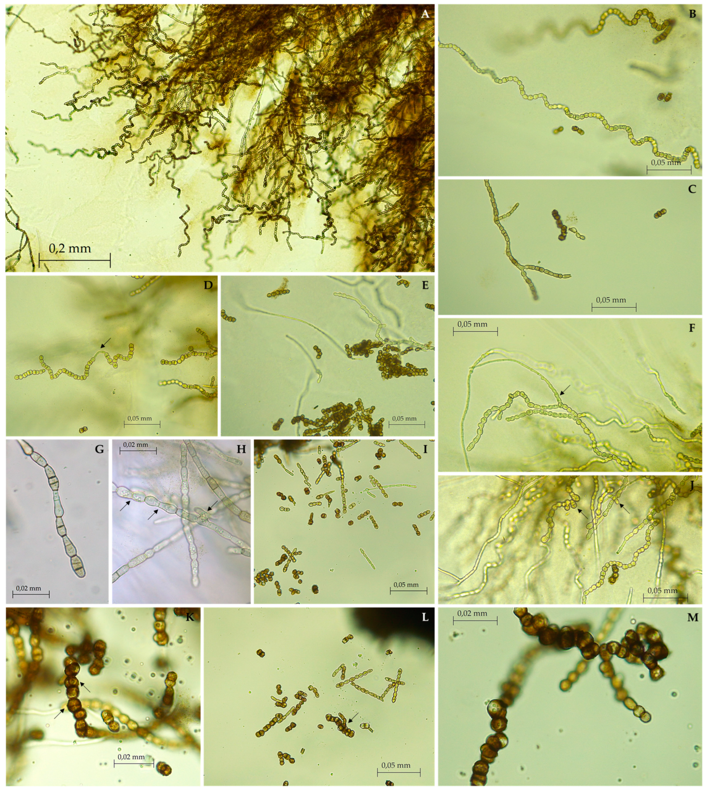
Description: Monotypic genus to accommodate a novel fungal species in the Aeminiaceae family, Mycosphaerellales order. Filamentous, slow-growing, anamorphic fungus. Mycelium consisting of cylindroid, pale brown, smooth, septate, and often branched hyphal cells. Prior to fragmentation into arthroconidia, the cells gradually become swollen (torulose), thick-walled, and darker and develop into long meristematic terminal chains of conidia. The conidia are dark brown, thick-walled, rugose, and globose. The arthric disarticulation of hyphae creates a spiral-like shape in the conidial chains, which is unique to this genus and is not reported in any phylogenetically close relatives. Chlamydospores were not observed in culture, and a recognizable sexual morph is absent.
Saxispiralis lemnorum D.S. Paiva & A. Portugal, sp. nov. (Figure 3 and Figure 4).
MycoBank number: MB849260.
Etymology: «Lemnorum» (derived Latinization of Portuguese “Lemos” to Latin “Lemnos”, genitive plural), in honor of the Lemos family, as the strain was found in the Lemos Pantheon.
Typification: PORTUGAL, Aveiro, Águeda (40°36.653′ N, 08°28.729′ W), isolated from a deteriorated funerary art piece carved in Ançã limestone with abundant salt damage, in the Lemos Pantheon, 8 June 2021, D.S. Paiva, (holotype MUM-H 23.14, dried specimen), ex-type culture MUM 23.14.
Description: Hyphae subhyaline to pale brown when young, consisting of cylindroid cells, guttulate, thin- and smooth-walled, branched, 2.91 ± 0.44 μm wide, occasionally terminating in a swollen ellipsoidal cell, with anastomoses often observed. Prior to maturation, hyphae gradually become swollen, dumbbell-shaped, constricted at the septum, thick-walled, and darker, and develop into long spirals of meristematic torulose conidial chains. Arthroconidia dark brown, thick-walled, verrucose or coarsely punctate, spherical, measuring 5.89 ± 0.52 µm, guttulate, resulting from the differentiation of terminal toruloid-like hyphal cells. Terminal and lateral blastic budding cells are frequently produced, developing longitudinal and oblique septa, and sometimes forming multicellular clumps. Occasionally, conidial secession is incomplete, leaving adjacent cells interconnected by narrow and pale connectives. Chlamydospores absent. Teleomorph unknown.
Colony characteristics: On PDA at 25 °C, colonies grew slowly, reaching 13.4 mm in diameter after 30 days and up to 24.8 mm after 2 months. Mycelium with fine velvety texture, dark greyish olive on the reverse (according to the ISCC-NBS Colour System No. 111) and strong orange-yellow color (ISCC-NBS No. 68) with a dark greyish olive margin (No. 111) on the obverse. After 2 months or longer, colonies reached full maturation and became completely melanized and black (ISCC-NBS No. 267) on both sides of the culture. Colonies raised centrally, cerebriform, flat near the periphery and partially immersed in the agar, compact, stiff, lobed, with well-defined regular margin.
Colonies on MEA 10% at 25 °C exhibited a similar morphology to those on PDA, with slight differences. No color alteration was observed; mycelium grew directly brownish black (ISCC-NBS No. 65) both on the reverse and obverse. Colonies were less folded, circular, velvety, with a slightly moist appearance, attaining a diameter of 12 mm in 30 days and up to 35 mm in two months.
On DG-18 agar, at 25 °C, colonies exhibited slow to moderate growth, reaching 21 mm in diameter after 30 days and attaining 32.7 mm after two months, circular, flat, margin entire and slight submerged, velvety, with a slightly moist appearance and covered by sparse whitish aerial mycelium, with a pale orange-yellow color (ISCC-NBS No. 73) on the obverse and reverse. After 2 months or longer, colonies reached full maturation and became completely melanized with a brownish-black color (ISCC-NBS No. 65) on both sides of the culture, becoming raised, rugose, with short aerial hyphae present, and undulate margin.
Colonies on HM 10% at 25 °C showed slow to moderate growth, reaching 26 mm in diameter after 30 days and up to 41 mm after two months, dark greyish olive in the reverse (ISCC-NBS No. 111) and dark olive (ISCC-NBS No. 108) on the obverse, becoming white (ISCC-NBS No. 263) near the periphery, circular, flat, with entire margin, producing thin whitish aerial mycelium.
Substrate: Ançã limestone.
Distribution: Portugal.
Additional specimens examined: PORTUGAL, Aveiro, Águeda (40°36.653′ N, 008°28.729′ W), isolated from a deteriorated funerary art piece carved in Ançã limestone with abundant salt damage, in the Lemos Pantheon, 8 June 2021, D.S. Paiva, MUM 23.15.
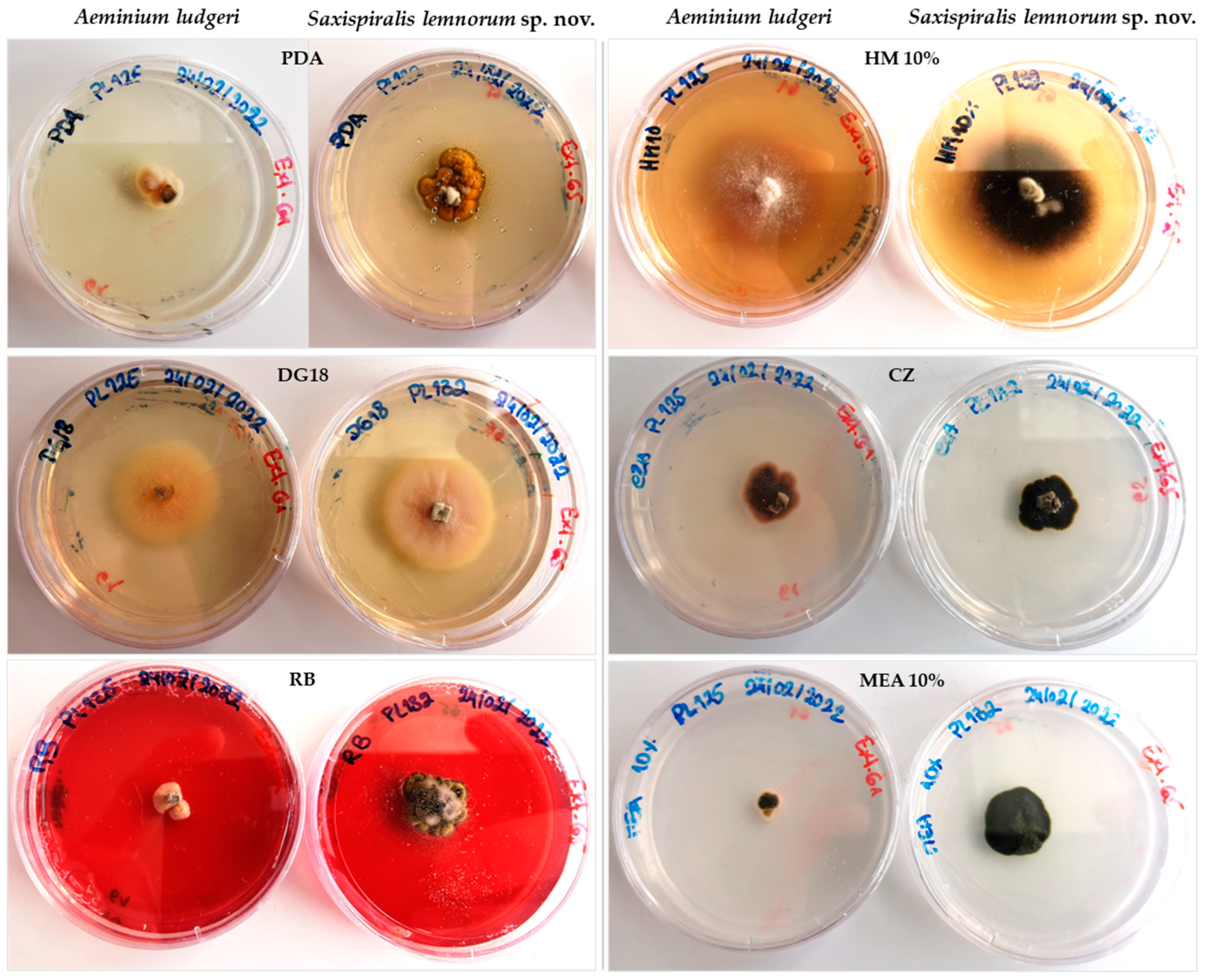
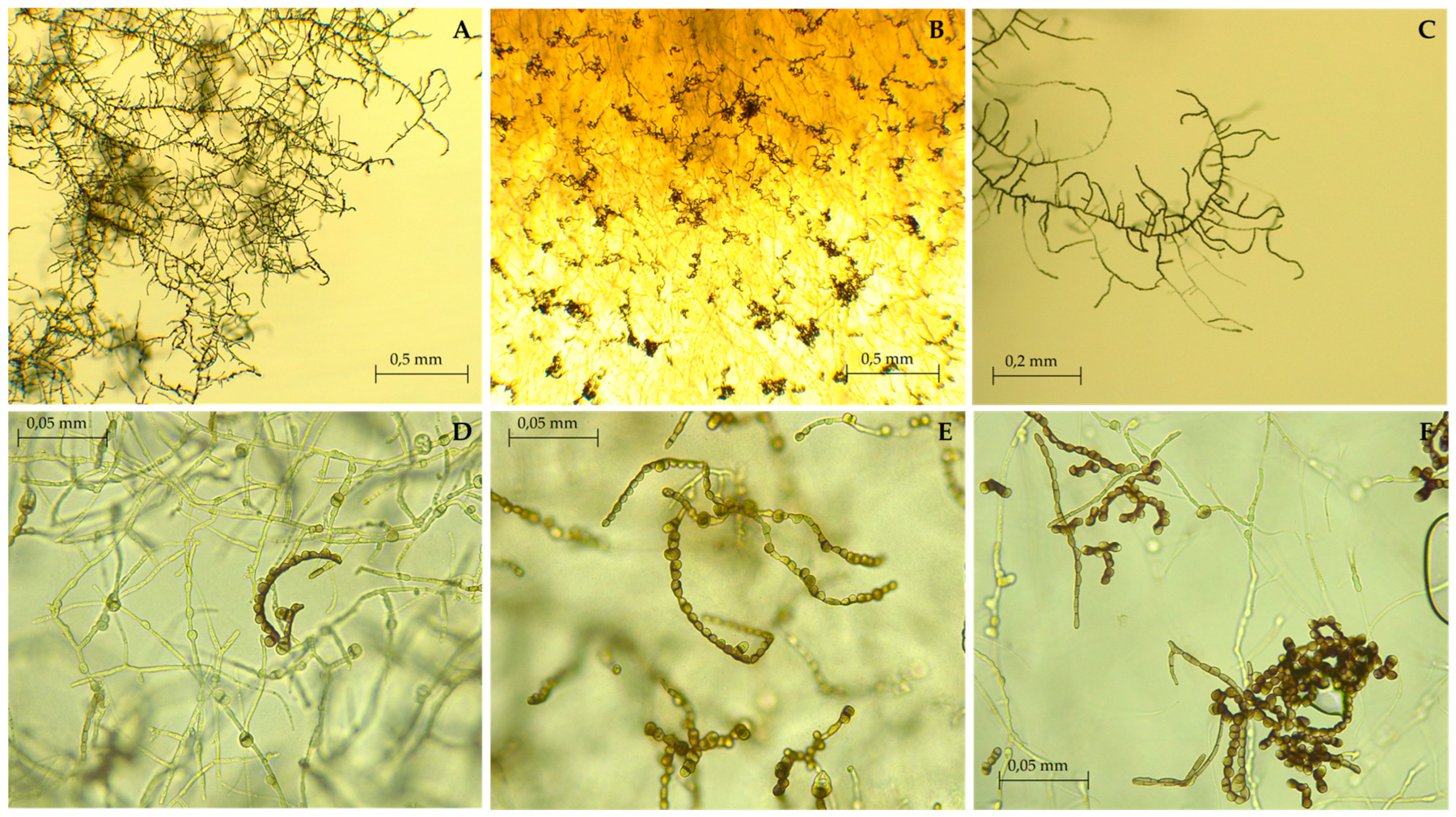
Notes: The newly identified species Saxispiralis lemnorum exhibits distinct phylogenetic and morphological characteristics that set it apart from other previously described members of the Aeminiaceae family. Phylogenetic analysis based on the concatenated ITS rDNA, LSU, and rpb2 dataset considered in the present study revealed that the retrieved strains form a distinct monophyletic lineage within the Aeminiaceae family, clearly separate from the previously known genus Aeminium, which, until now, represented the sole genus in this family. Phenotypically, it can be easily distinguished from A. ludgeri by its distinct colony color and hyphal characteristics in terms of shape and structure (Appendix A, Figure A1 and Figure A2), as well as its larger size with pronounced rough texture conidia, which are exclusive to this genus.

3.3. Physiological Characterization

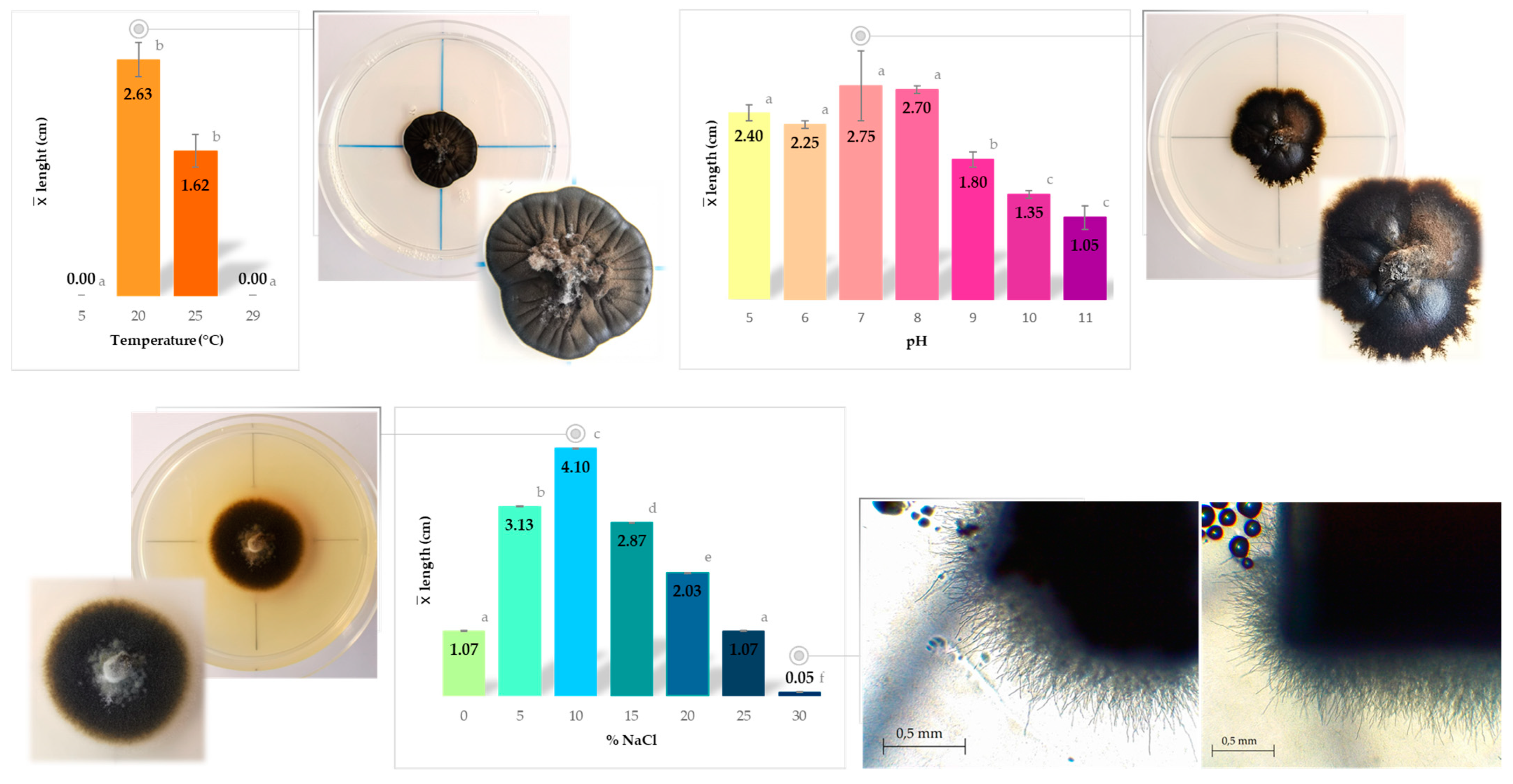
Physiological analyses were conducted on both isolates obtained in this study. However, no significant differences were observed among the isolates across the various tested conditions. The summary of the results from physiological characterization for temperature, pH, and salinity preference/tolerance is presented in Figure 5. The data obtained serve as a comprehensive overview of the strain’s responses to temperature, pH, and salinity, enabling a better understanding of their ecological preferences and adaptability.
Based on the observed growth patterns at the tested conditions, the strains exhibited optimal growth at moderate temperatures, particularly at 20 °C, where the colony diameter reached 2.63 cm after 2 months. At 25 °C, a slight decrease in growth was observed, and no growth was recorded at lower and higher temperatures. Considering these findings, can be inferred that the fungus displays a preference for mesophilic conditions.
Regarding the pH assessment, the strains displayed a wide range of tolerance, as they grew across a broad spectrum of pH levels, ranging from acidic (pH 5.0) to alkaline (pH 11.0). Optimal growth was observed between pH 7.0 and pH 8.0, while the lowest growth was recorded at pH 11.0. Although the optimum pH for growth was pH 7, there was only a slight decrease in growth at the more acidic pH values of 5 and 6, whereas a more significant decrease was observed at higher alkaline pH values (pH 11). These results indicate that the fungus can be classified as highly tolerant to pH fluctuations, showcasing the ability to thrive across a wide pH spectrum without experiencing severe growth impairments. This distinct characteristic sets it apart from Aeminium ludgeri, the other member in Aeminiaceae family, considered a facultative alkaliphile, with no growth being registered for pH levels below 6 and above 9 [57].
The isolates exhibited growth across the entire range of tested NaCl concentrations, from 0% up to 30%. Optimal growth was observed at a concentration of 10% NaCl. Growth was still observed at 30% NaCl, albeit at a slower rate (>2 mm diameter after 3-month post-inoculation). Among halotolerant fungi, Hortaea werneckii is currently considered the most halotolerant fungus known to date, as it can tolerate a wide range of NaCl concentrations from 0% to 32%, with an optimal growth range of 6% to 14% NaCl [17,94]. Additionally, a recently discovered new ecotype of Pseudotaeniolina globosa [85] has also demonstrated the ability to grow at up to 30% NaCl, placing Saxispiralis lemnorum alongside these notable “kings” of salinity. The fungus’ capacity to develop in diverse NaCl concentrations assumes particular significance when considering its original isolation from a sample collected in an environment characterized by pronounced salt damage. This environmental context aligns with its ability to thrive under high-salinity conditions. Based on this characteristic, the fungus can be classified as halotolerant.
After subjecting the fungus to thermal stress, no growth was observed for any of the tested conditions following the heat tolerance protocol. Consequently, the fungus was classified as non-heat tolerant. On the other hand, when exposed to low-temperature shock, across all tested conditions, the fungus consistently demonstrated recovery and growth, indicating its cold-tolerant nature or psychrotrophic adaptation. A similar pattern was observed when subjected to UV radiation stress, as it exhibited the ability to recover and grow under all tested conditions. Additionally, based on its successful growth on DG-18 culture medium, which was used for morphological characterization, the fungus can be considered as xerophilic.
The remarkable ability of this new species to tolerate a wide variety of physicochemical parameters and withstand different types of stress suggests an extremotolerant nature. Furthermore, the gathered data provide strong evidence that Saxispiralis lemnorum is well-suited for survival on stone surfaces, where high evaporation rates and low water availability lead to local increases in ion concentrations and fluctuations in pH, creating an environment to which it can successfully adapt.

3.4. Deteriorative Potential

When evaluating heritage items, it is imperative to not only assess the diversity and viability of microorganisms but also conduct complementary studies on physiological activities and their deteriorative potential. These additional studies are crucial for gaining a comprehensive understanding of the occurring biodeterioration processes and developing tailored and effective treatment strategies [88]. As the isolates were derived from deteriorated limestone, the evaluation of their deteriorative potential was determined by assessing their ability to induce pH alteration, CaCO3 dissolution, and mineral production. The overall results for the biodegradative plate assays considered in this study can be found in Table 3.
The presence of fungi on the stone surface can have a profound impact on the material properties due to the secretion of inorganic and organic acids as a byproduct of their metabolic processes [18,19,20,23,25,26]. Carbonate dissolution primarily occurs through the action of organic acids [18,20,23]. However, our results demonstrate that the fungal strains did not induce detectable pH alterations on CREA medium (Appendix A, Figure A3A) and exhibited no ability to dissolve CaCO3. In most cases, the production and release of acids into the substrate are particularly favored when fungi inhabit nutrient-rich substrates that promote rapid growth [95]. This intensive growth leads to the excessive production of organic acids beyond the necessary requirements for normal metabolism, which are subsequently excreted into the substrate, rendering them detectable [96]. Therefore, our findings are consistent with expectations, considering that Saxispiralis lemnorum is a microcolonial fungus characterized by slow growth and adaptation to harsh oligotrophic substrates. Furthermore, this is in line with current knowledge regarding MCF, as chemical deterioration of stone by acid substances has never been demonstrated for this group of fungi [20,26,97].
Fungal biological activities can release cations from minerals and lead to the formation of secondary minerals through a process called biomineralization [21,24,25,26,98]. This adaptation is particularly observed in fungi growing on calcium-rich substrates [99], and such mineralization events can contribute to various biodeterioration phenomena [18,100]. Biogenic crystal formation was observed in the B4 and HM 10% media with different configurations (Figure 6). In the B4 medium, spherical crystal formations were abundant around the fungal mycelium and surrounding areas (Figure 6C,D), while tetragonal forms were predominantly found at the edge of the Petri dish (Figure 6E). Irregularly shaped platy crystal aggregates were observed in the HM 10% medium, typically found beneath or near the fungal mycelium (Figure 6F–H). A possible explanation for the observed crystallization phenomenon in the B4 medium, but not in media containing calcium carbonate, could be attributed to fungal respiration activity, supplying the required CO2 for calcite precipitation. In the B4 medium, the release of calcium does not necessarily rely on the fungi producing organic acids, since calcium acetate is highly soluble, and calcium is readily available in the solution. However, in the MB4 medium (e.g.), which contains nearly insoluble calcium carbonate, the presence of organic acids becomes necessary to facilitate the release of calcium [96].
However, since crystals of different conformations exist in the same medium, further studies would be necessary to characterize their composition and understand the actual mechanisms involved in their formation. The same applies to the crystals present in HM 10%. However, for this latter case, it is important to note that although this medium was used with various salt concentrations in the NaCl tolerance assay, crystallization was only observed at a 10% NaCl concentration, which is the optimal growth concentration. Crystallization was never observed at lower or higher concentrations. Based on the obtained results, Saxispiralis lemnorum has demonstrated the ability for secondary mineral formation, suggesting its potential involvement in the promotion of significant stone biodeterioration. The formation of crystals on the stone matrix can result in pore and fissure expansion, efflorescence formation, as well as peeling and spalling of the materials [101], all of which are consistent with the characteristics observed in the sampled area. Therefore, the proliferation of this species, influenced by specific environmental factors, is likely to contribute to the observed deterioration phenomena in L4.
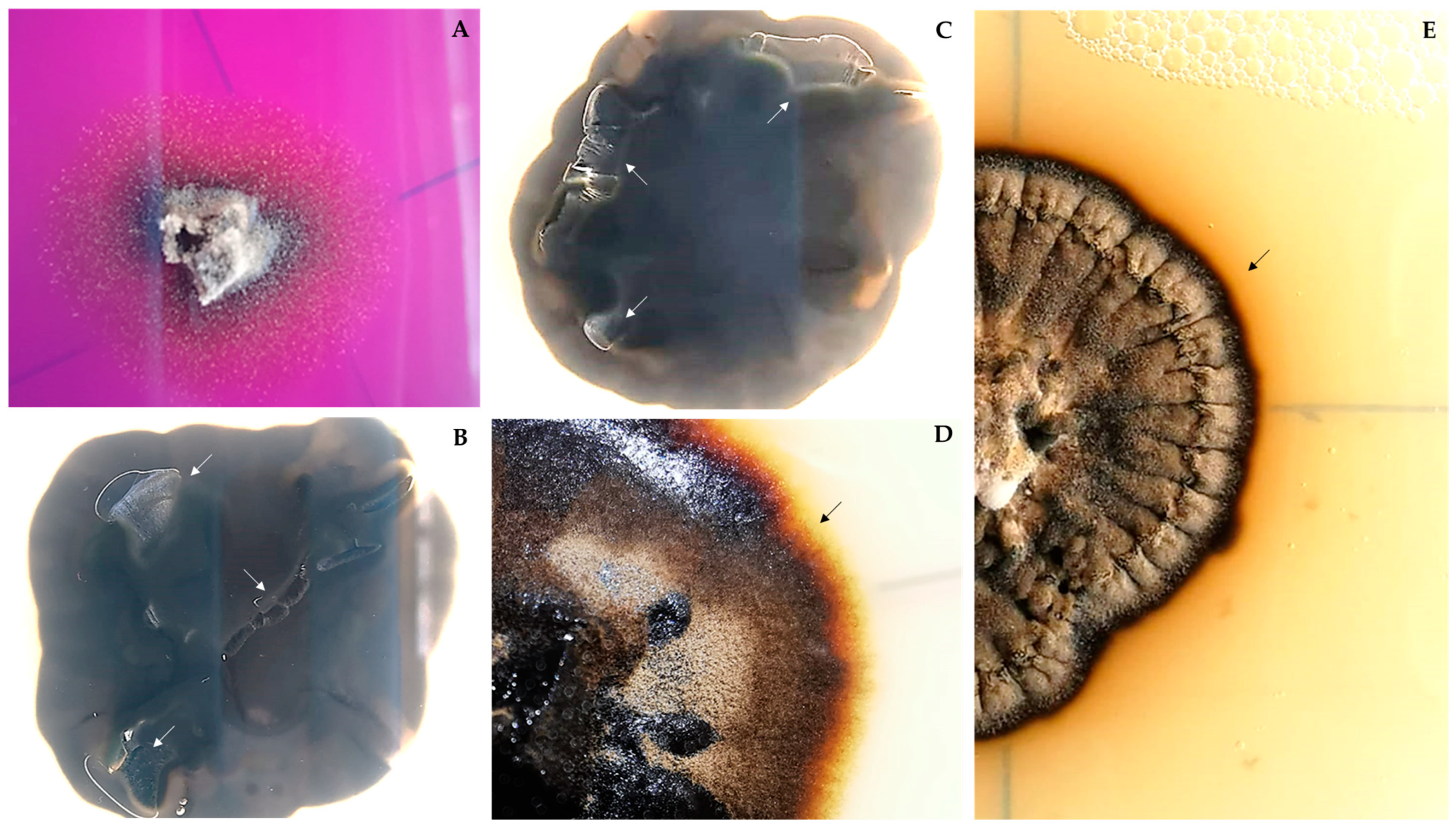
Another interesting observation made with some of the media used for characterizing the isolates, e.g., PDA, was the presence of fungal growth not only on the surface, but also hyphae growing within and through the medium. In many cases, this led to the medium cracking and detaching from the Petri dish (Appendix A, Figure A3B,C). This could be an indication of its potential to cause physical biodeterioration resulting from the mechanical forces generated by hyphal penetration, which led to the breakage of the medium. MCF have been consistently reported as capable of physically penetrating stone substrates through the combined action of mechanical forces, exerted by hyphal expansion during growth, and chemical processes involving metal chelating compounds [26,95,97,101]. This penetration process can lead to the loosening of the intercrystalline stone matrix due to internal pressure, ultimately resulting in detachment and material loss [96,101]. Taking this into consideration, along with the aforementioned ability for secondary mineral formation, this species is likely to actively contribute to the observed alterations in the sampled area.
Furthermore, in the B4 medium, as well as in the more alkaline media used in the pH tolerance assay (pH 9 to 11), the presence of a dark yellow pigment was observed (Appendix A, Figure A3D,E). Along with its highly melanized mycelium, which gives it a black color, these are indicators that these strains may cause chromatic alterations due to mycelial growth and pigment production.

4. Conclusions

Our study reports a newly discovered microcolonial black fungus belonging to the Aeminiaceae family, which had only one known representative until now. The fungal isolates here described represent a novel genus and species, found as part of a complex community colonizing deteriorated limestone in the Lemos Pantheon, highlighting the need for further investigation of fungal communities in similar environments. This work provides valuable molecular, morphological, and physiological data that enhance our understanding of fungi in this recently described family. Furthermore, the physiological and biodeteriorative potential assessment of this new species yielded data that demonstrate a significant deteriorative activity, indicating its potential to harm the substrate and cause various types of alterations. Further studies are currently underway to fully understand its nature and abilities. Understanding the microbial agents involved in the materials’ biodeterioration is crucial knowledge for appropriate safeguarding measures to be considered, discussed, and implemented for effective conservation. By comprehensively studying the microbial agents responsible for material deterioration, we can hope to preserve our cultural heritage for future generations.

Supplementary Materials

The following supporting information can be downloaded at https://www.mdpi.com/article/10.3390/jof9090916/s1, Figure S1: Visual representation outlining the measurement process in the temperature, NaCl, and pH assays. The pre-segmentation of the culture plate serves to establish a reliable reference line for measurements. This ensures that, particularly when colonies exhibit irregular shapes, diameter measurements remain impartial, mitigating any potential bias towards measuring the larger diameter, for instance.

Author Contributions

Conceptualization, D.S.P., N.M., I.T. and A.P.; Methodology, D.S.P. and L.F.; Software, D.S.P. and J.T.; Validation, D.S.P., N.M., I.T. and A.P.; Formal analysis, D.S.P.; Investigation, D.S.P.; Resources, N.M., I.T. and A.P.; Data Curation, D.S.P.; Writing—Original Draft Preparation, D.S.P.; Writing—Review and Editing, D.S.P., J.T., L.F., N.M., I.T. and A.P.; Visualization, D.S.P.; Supervision, N.M., I.T. and A.P.; Project Administration, A.P.; Funding Acquisition, A.P. All authors have read and agreed to the published version of the manuscript.

Funding

Diana Paiva was supported by a PhD research grant (UI/BD/150843/2021) awarded by the Centre for Functional Ecology—Science for People & the Planet (CFE) and co-funded by Fundação para a Ciência e Tecnologia, I.P. (FCT) through national funding by the Ministério da Ciência, Tecnologia e Ensino Superior (MCTES) from Fundo social Europeu (FSE). This work was carried out in the R&D Unit Centre for Functional Ecology—Science for People & the Planet (CFE), and Associate Laboratory TERRA, with references, respectively, UIDB/04004/2020 and LA/P/0092/2020, financed by FCT/MCTES through national funds (PIDDAC). The authors also gratefully acknowledge the funding of the PRR—Recovery and Resilience Plan and the NextGeneration EU European Funds.

Institutional Review Board Statement

Not applicable.

Informed Consent Statement

Not applicable.

Data Availability Statement

All relevant data are presented in the paper. The nucleotide sequences were deposited in the GenBank database under the accession numbers OR081765-OR081766 (LSU), OR081767-OR081768 (ITS), and OR074926-OR074926 (rpb2). The phylogenetic data can be accessed on Figshare via the following link: https://figshare.com/s/c68a893bc9b8f9f3c5d7 (accessed on 21 June 2023).

Acknowledgments

Sampling was conducted with permission from the Trofa’s Administration Parish Council and under the supervision of Father Pimenta. The authors express their gratitude to Father José Luís Pimenta, the Parish Priest of the Church of Trofa do Vouga, for warmly welcoming us and assistance during the sampling process. Additionally, his invaluable insights and knowledge about the monument’s history were greatly appreciated. The authors also wish to express their sincere gratitude to João Gouveia Monteiro of the University of Coimbra, for his unwavering support and guidance in ensuring the accurate use of Latin names for the description of the new species presented in this article. Figure S1 was generated using https://app.biorender.com/ (accessed on 25 August 2023).

Conflicts of Interest

The authors declare no conflict of interest.

Appendix A

Figure A1. Direct comparison of the macromorphology between Aeminium ludgeri (PL125 isolate, also obtained during the same study and from the same sample [61]) and Saxispiralis lemnorum is shown on different culture media after one month of inoculation (Ø 5 cm plates). PDA—Potato Dextrose Agar (PDA); DG 18—Dichloran-Glycerol Agar; RB—Rose Bengal Agar; HM 10%—DSMZ 372-Halobacteria medium supplemented with 10% NaCl (w/v); CZ*—Czapek Solution Agar; MEA 10%—Malt Extract Agar supplemented with 10% NaCl (w/v). * Culture medium used in Paiva et al. [61] for processing the initial samples.
Jof 09 00916 g0a1
Figure A2. Micromorphology of Aeminium ludgeri (PL125 isolate) shown as a reference for comparison with Saxispiralis lemnorum. (AC) Differentiated, toruloid-like hyphae and mature chains of arthroconidia. (DF) Micronematous intercalary and terminal conidial chains.
Jof 09 00916 g0a2
Figure A3. Observable alterations caused by Saxispiralis lemnorum in different culture media. (A) Poor growth and absence of acid reactions on CREA medium. (B,C) Hyphal intrusion resulting in the fragmentation of the PDA medium. (D,E) Noticeable production of dark yellow pigment in MEA media adjusted to pH 10 and B4.
Jof 09 00916 g0a3

References

  1. Beraldi-Campesi, H. Early life on land and the first terrestrial ecosystems. Ecol. Processes. 2013, 2, 1. [Google Scholar] [CrossRef]
  2. Gorbushina, A.A.; Krumbein, W.E.; Volkmann, M. Rock surfaces as life indicators: New ways to demonstrate life and traces of former life. Astrobiology 2002, 2, 203–213. [Google Scholar] [CrossRef] [PubMed]
  3. Gorbushina, A.A. Life on the rocks. Environ. Microbiol. 2007, 9, 1613–1631. [Google Scholar] [CrossRef] [PubMed]
  4. Hoppert, M.; Flies, C.; Pohl, W.; Gunzl, B.; Schneider, J. Colonization strategies of lithobiontic microorganisms on carbonate rocks. Environ. Geol. 2004, 46, 421–428. [Google Scholar] [CrossRef]
  5. Tiano, P. Biodegradation of cultural heritage: Decay mechanisms and control methods. In Seminar Article; New University of Lisbon, Department of Conservation and Restoration: Campolide, Lisbon, 2002; pp. 7–12. [Google Scholar]
  6. Garcia-Vallès, M.; Vendrell-Saz, M.; Molera, J.; Blazquez, F. Interaction of rock and atmosphere: Patinas on Mediterranean monuments. Environ. Geol. 1998, 36, 137–149. [Google Scholar] [CrossRef]
  7. Saiz-Jimenez, C. Deposition of anthropogenic compounds on monuments and their effect on airborne microorganisms. Aerobiologia 1995, 11, 161–175. [Google Scholar] [CrossRef]
  8. Golubic, S.; Perkins, R.D.; Lukas, K.J. Boring Microorganisms and Microborings in Carbonate Substrates. In The Study of Trace Fossils; Frey, R.W., Ed.; Springer: Berlin/Heidelberg, Germany, 1975; pp. 229–259. [Google Scholar] [CrossRef]
  9. Selbmann, L.; de Hoog, G.S.; Zucconi, L.; Isola, D.; Ruisi, S.; Gerrits van den Ende, A.H.G.; Ruibal, C.; De Leo, F.; Urzì, C.; Onofri, S. Drought meets acid: Three new genera in a dothidealean clade of extremotolerant fungi. Stud. Mycol. 2008, 61, 1–20. [Google Scholar] [CrossRef]
  10. Selbmann, L.; De Hoog, G.S.; Mazzaglia, A.; Friedmann, E.I.; Onofri, S. Fungi at the edge of life: Cryptoendolithic black fungi from Antarctic desert. Stud. Mycol. 2005, 51, 1–32. [Google Scholar]
  11. Hirsch, P.; Mevs, U.; Kroppenstedt, R.M.; Schumann, P.; Stackebrandt, E. Cryptoendolithic actinomycetes from Antarctic rock samples: Micromonospora endolithica sp. nov. and two isolates related to Micromonospora coerulea Jensen 1932. Syst. Appl. Microbiol. 2004, 27, 166–174. [Google Scholar] [CrossRef]
  12. Friedmann, E.I. Endolithic microorganisms in the Antarctic cold desert. Science 1982, 215, 1045–1053. [Google Scholar] [CrossRef]
  13. Staley, J.T.; Palmer, F.; Adams, J.B. Microcolonial fungi: Common inhabitants on desert rocks? Science 1982, 215, 1093–1095. [Google Scholar] [CrossRef]
  14. Krumbein, W.E.; Jens, K. Biogenic rock varnishes of the Negev desert (Israel) an ecological study of iron and manganese transformation by cyanobacteria and fungi. Oecologia 1981, 50, 25–38. [Google Scholar] [CrossRef] [PubMed]
  15. Selbmann, L.; Egidi, E.; Isola, D.; Onofri, S.; Zucconi, Z.; de Hoog, G.S.; Chinaglia, S.; Testa, L.; Tosi, S.; Balestrazzi, A.; et al. Biodiversity, evolution and adaptation of fungi in extreme environments. Plant Biosyst. 2013, 147, 237–246. [Google Scholar] [CrossRef]
  16. Yurlova, N.A.; de Hoog, G.S.; Fedorova, L.G. The influence of ortho- and para-diphenoloxidase substrates on pigment formation in black yeast-like fungi. Stud. Mycol. 2008, 61, 39–49. [Google Scholar] [CrossRef] [PubMed]
  17. Butinar, L.; Sonjak, S.; Zalar, P.; Plemenitaš, A.; Gunde-Cimerman, N. Melanized halophilic fungi are eukaryotic members of microbial communities in hypersaline waters of solar salterns. Bot. Mar. 2005, 48, 73–79. [Google Scholar] [CrossRef]
  18. Dakal, T.C.; Cameotra, S.S. Microbially induced deterioration of architectural heritages: Routes and mechanisms involved. Environ. Sci. Eur. 2012, 24, 36. [Google Scholar] [CrossRef]
  19. Scheerer, S.; Ortega-Morales, O.; Gaylarde, C. Chapter 5 Microbial Deterioration of Stone Monuments–An Updated Overview. Adv. Appl. Microbiol. 2009, 66, 97–139. [Google Scholar] [CrossRef]
  20. Sterflinger, K. Fungi as geologic agents. Geomicrobiol. J. 2000, 17, 97–124. [Google Scholar] [CrossRef]
  21. Gadd, G.M. Fungi, Rocks, and Minerals. Elements 2017, 3, 171–176. [Google Scholar] [CrossRef]
  22. Gadd, G.M. Geomycology: Biogeochemical transformations of rocks, minerals, metals and radionuclides by Fungi, Bioweathering and Bioremediation. Mycol. Res. 2007, 111, 3–49. [Google Scholar] [CrossRef]
  23. Warscheid, T.; Braams, J. Biodeterioration of stone: A review. Int. Biodeterior. Biodegrad. 2000, 46, 343–368. [Google Scholar] [CrossRef]
  24. Gadd, G.M. Geomicrobiology of the built environment. Nat. Microbiol. 2017, 2, 16275. [Google Scholar] [CrossRef] [PubMed]
  25. Sterflinger, K.; Piñar, G. Microbial deterioration of cultural heritage and works of ar –Tilting at windmills? Appl. Microbiol. Biotechnol. 2013, 97, 9637–9646. [Google Scholar] [CrossRef] [PubMed]
  26. Sterflinger, K. Fungi: Their role in deterioration of cultural heritage. Fungal Biol. Rev. 2010, 24, 47–55. [Google Scholar] [CrossRef]
  27. Liu, B.; Fu, R.; Wu, B.; Liu, X.; Xiang, M. Rock-inhabiting fungi: Terminology, diversity, evolution and adaptation mechanisms. Mycology 2021, 13, 1–31. [Google Scholar] [CrossRef]
  28. de Hoog, G.S.; McGinnis, M.R. Ascomycetous black yeasts. In The Expanding Realm of Yeast-Like Fungi; de Hoog, G.S., Smith, M.T., Weijman, A.C.M., Eds.; Elsevier: Amsterdam, The Netherlands, 1987; pp. 187–199. [Google Scholar]
  29. Gueidan, C.; Villasenor, C.R.; de Hoog, G.S.; Gorbushina, A.A.; Untereiner, W.A. A rock-inhabiting ancestor for mutualistic and pathogen-rich fungal lineages. Stud. Mycol. 2008, 61, 111–119. [Google Scholar] [CrossRef]
  30. Gueidan, C.; Ruibal, C.; de Hoog, G.S.; Schneider, H. Rock-inhabiting fungi originated during periods of dry climate in the late Devonian and middle Triassic. Fungal Biol. 2011, 115, 987–996. [Google Scholar] [CrossRef]
  31. Gueidan, C.; Aptroot, A.; Caceres, M.E.D.; Badali, H.; Stenroos, S. A reappraisal of orders and families within the subclass Chaetothyriomycetidae (Eurotiomycetes, Ascomycota). Mycol. Prog. 2014, 13, 1027–1039. [Google Scholar] [CrossRef]
  32. Ametrano, C.G.; Muggia, L.; Grube, M. Extremotolerant Black Fungi from Rocks and Lichens. In Fungi in Extreme Environments: Ecological Role and Biotechnological Significance; Grube, M., Ed.; Springer: Cham, Switzerland, 2019; pp. 119–143. [Google Scholar] [CrossRef]
  33. Abdollahzadeh, J.; Groenewald, J.Z.; Coetzee, M.P.A.; Wingfield, M.J.; Crous, P.W. Evolution of lifestyles in Capnodiales. Stud. Mycol. 2020, 95, 381–414. [Google Scholar] [CrossRef]
  34. De Leo, F.; Marchetta, A.; Urzì, C. Black Fungi on Stone-Built Heritage: Current Knowledge and Future Outlook. Appl. Sci. 2022, 12, 3969. [Google Scholar] [CrossRef]
  35. Palmer, F.E.; Emery, D.R.; Stemmler, J.; Staley, J.T. Survival and growth of microcolonial rock fungi as affected by temperature and humidity. New Phytol. 1987, 107, 155–162. [Google Scholar] [CrossRef]
  36. Urzì, C.; De Leo, F.; de Hoog, S.; Sterflinger, K. Recent advances in the molecular biology and ecophysiology of meristematic stone-inhabiting fungi. In Of Microbes and Art: The Role of Microbial Communities in the Degradation and Protection of Cultural Heritage; Ciferri, O., Tiano, P., Mastromei, G., Eds.; Springer: Boston, MA, USA, 2000; pp. 3–19. [Google Scholar] [CrossRef]
  37. Sterflinger, K. Black yeasts and meristematic fungi: Ecology, diversity and identification. In The Yeast Handbook, Biodiversity and Ecophysiology of Yeasts; Péter, G., Rosa, C., Eds.; Springer: Berlin/Heidelberg, Germany, 2006; pp. 501–514. [Google Scholar] [CrossRef]
  38. Selbmann, L.; de Hoog, G.S.; Zucconi, L.; Isola, D.; Onofri, S. Black Yeasts in Cold Habitats. In Cold-Adapted Yeasts; Buzzini, P., Margesin, R., Eds.; Springer: Berlin/Heidelberg, Germany, 2014; pp. 137–189. [Google Scholar] [CrossRef]
  39. Selbmann, L.; Zucconi, L.; Isola, D.; Onofri, S. Rock black fungi: Excellence in the extremes, from the Antarctic to space. Curr. Genet. 2015, 61, 335–345. [Google Scholar] [CrossRef]
  40. Tesei, D. Black Fungi Research: Out-of-This-World Implications. Encyclopedia 2022, 2, 212–229. [Google Scholar] [CrossRef]
  41. Gorbushina, A.A.; Whitehead, K.; Dornieden, T.; Niesse, A.; Schulte, A.; Hedges, J.I. Black fungal colonies as units of survival: Hyphal mycosporines synthesized by rock-dwelling microcolonial fungi. Can. J. Bot. 2003, 81, 131–138. [Google Scholar] [CrossRef]
  42. Gorbushina, A.A.; Kotlova, E.R.; Sherstneva, O.A. Cellular responses of microcolonial rock fungi to long-term desiccation and subsequent rehydration. Stud. Mycol. 2008, 61, 91–97. [Google Scholar] [CrossRef] [PubMed]
  43. Zakharova, K.; Tesei, D.; Marzban, G.; Dijksterhuis, J.; Wyatt, T.; Sterflinger, K. Microcolonial fungi on rocks: A life in constant drought? Mycopathologia 2013, 175, 537–547. [Google Scholar] [CrossRef]
  44. Wollenzien, U.; de Hoog, G.S.; Krumbein, W.E.; Uijthof, J.M.J. Sarcinomyces petricola, a new microcolonial fungus from marble in the Mediterranean basin. Antonie Van Leeuwenhoek J. Microbiol. 1997, 71, 281–288. [Google Scholar] [CrossRef]
  45. Sterflinger, K.; de Baere, R.; de Hoog, G.S.; de Wachter, R.; Krumbein, W.E.; Haase, G. Coniosporium perforans and C. apollinis, two new rock-inhabiting fungi isolated from marble in the Sanctuary of Delos (Cyclades, Greece). Antonie Van Leeuwenhoek J. Microbiol. 1997, 72, 349–363. [Google Scholar] [CrossRef] [PubMed]
  46. De Leo, F.; Urzì, C.; de Hoog, G.S. Two Coniosporium species from rock surfaces. Stud. Mycol. 1999, 43, 70–79. [Google Scholar]
  47. De Leo, F.; Urzì, C.; de Hoog, G. A new meristematic fungus, Pseudotaeniolina globosa. Antonie Van Leeuwenhoek J. Microbiol. 2003, 83, 351–360. [Google Scholar] [CrossRef]
  48. Sert, H.B.; Sümbül, H.; Sterflinger, K. A new species of Capnobotryella from monument surfaces. Mycol. Res. 2007, 111, 1235–1241. [Google Scholar] [CrossRef]
  49. Sert, H.B.; Sümbül, H.; Sterflinger, K. Microcolonial fungi from antique marbles in Perge/Side/Termessos (Antalya/Turkey). Antonie Van Leeuwenhoek J. Microbiol. 2007, 91, 217–227. [Google Scholar] [CrossRef]
  50. Sert, H.B.; Sümbül, H.; Sterflinger, K. Sarcinomyces sideticae, a new black yeast from historical marble monuments in Side (Antalya, Turkey). Bot. J. Linn. Soc. 2007, 154, 373–380. [Google Scholar] [CrossRef]
  51. Sert, H.B.; Sterflinger, K. A new Coniosporium species from historical marble monuments. Mycol. Prog. 2010, 9, 353–359. [Google Scholar] [CrossRef]
  52. Onofri, S.; Anastasi, A.; Del Frate, G.; Di Piazza, S.; Garnero, N.; Guglielminetti, M.; Isola, D.; Panno, L.; Ripa, C.; Selbmann, L.; et al. Biodiversity of rock, beach and water fungi in Italy. Plant Biosyst. 2011, 145, 978–987. [Google Scholar] [CrossRef]
  53. Sert, H.B.; Sümbül, H.; Sterflinger, K. Two new species of Capnobotryella from historical monuments. Mycol. Prog. 2011, 10, 333–339. [Google Scholar] [CrossRef]
  54. Martin-Sanchez, P.M.; Nováková, A.; Bastian, F.; Alabouvette, C.; Saiz-Jimenez, C. Two new species of the genus Ochroconis, O. lascauxensis and O. anomala isolated from black stains in Lascaux Cave, France. Fungal Biol. 2012, 116, 574–589. [Google Scholar] [CrossRef]
  55. Zucconi, L.; Gagliardi, M.; Isola, D.; Onofri, S.; Andaloro, M.C.; Pelosi, C.; Pogliani, P.; Selbmann, L. Biodeterioration agents dwelling in or on the wall paintings of the Holy Saviour’s cave (Vallerano, Italy). Int. Biodeterior. Biodegrad. 2012, 70, 40–46. [Google Scholar] [CrossRef]
  56. Isola, D.; Zucconi, L.; Onofri, S.; Caneva, G.; de Hoog, G.S.; Selbmann, L. Extremotolerant rock inhabiting black fungi from Italian monumental sites. Fungal Divers. 2016, 76, 75–96. [Google Scholar] [CrossRef]
  57. Trovão, J.; Tiago, I.; Soares, F.; Paiva, D.S.; Mesquita, N.; Coelho, C.; Catarino, L.; Gil, F.; Portugal, A. Description of Aeminiaceae fam. nov., Aeminium gen. nov. and Aeminium ludgeri sp. nov. (Capnodiales), isolated from a biodeteriorated art-piece in the old cathedral of Coimbra, Portugal. MycoKeys 2019, 45, 57–73. [Google Scholar] [CrossRef]
  58. Onofri, S.; Zucconi, L.; Isola, D.; Selbmann, L. Rock-inhabiting fungi and their role in deterioration of stone monuments in the Mediterranean area. Plant Biosyst. 2014, 148, 384–391. [Google Scholar] [CrossRef]
  59. Isola, D.; Bartoli, F.; Meloni, P.; Caneva, G.; Zucconi, L. Black Fungi and Stone Heritage Conservation: Ecological and Metabolic Assays for Evaluating Colonization Potential and Responses to Traditional Biocides. Appl. Sci. 2022, 12, 2038. [Google Scholar] [CrossRef]
  60. Sterflinger, K.; Krumbein, W.E. Dematiaceous fungi as a major agent of biopitting for Mediterranean marbles and limestones. Geomicrobiol. 1997, 14, 219–230. [Google Scholar] [CrossRef]
  61. Lombardozzi, V.; Castrignanò, T.; D’Antonio, M.; Casanova Municchia, A.; Caneva, C. An interactive database for an ecological analysis of stone biopitting. Int. Biodeterior. Biodegrad. 2012, 73, 8–15. [Google Scholar] [CrossRef]
  62. Paiva, D.S.; Fernandes, L.; Trovão, J.; Mesquita, N.; Tiago, I.; Portugal, A. Uncovering the Fungal Diversity Colonizing Limestone Walls of a Forgotten Monument in the Central Region of Portugal by High-Throughput Sequencing and Culture-Based Methods. Appl. Sci. 2022, 12, 10650. [Google Scholar] [CrossRef]
  63. Paiva, D.S.; Fernandes, L.; Pereira, E.; Trovão, J.; Mesquita, N.; Tiago, I.; Portugal, A. Exploring Differences in Culturable Fungal Diversity Using Standard Freezing Incubation—A Case Study in the Limestones of Lemos Pantheon (Portugal). J. Fungi 2023, 9, 501. [Google Scholar] [CrossRef]
  64. Igreja Paroquial de Trofa/Igreja de São Salvador e Panteão dos Lemos. Available online: http://www.monumentos.gov.pt/Site/APP_PagesUser/SIPA.aspx?id=1042 (accessed on 29 January 2023).
  65. Pinheiro, A.C.; Mesquita, N.; Trovão, J.; Soares, F.; Tiago, I.; Coelho, C.; Carvalho, H.P.; Gil, F.; Catarino, L.; Piñar, G.; et al. Limestone biodeterioration: A review on the portuguese cultural heritage scenario. J. Cult. Herit. 2019, 36, 275–285. [Google Scholar] [CrossRef]
  66. Miller, A.Z.; Leal, N.; Laiz, L.; Candelera, R.; Silva, M.A.; Dionisio, A.; Macedo, M.F.; Sáiz-Jiménez, C. Primary bioreceptivity of limestones used in southern European monuments. Geol. Soc. Lond. Spec. Publ. 2010, 331, 79–92. [Google Scholar] [CrossRef]
  67. White, T.; Bruns, T.; Lee, S.; Taylor, J. Amplification and direct sequencing of fungal ribosomal RNA genes for phylogenetics, In PCR Protocols: A Guide to Methods and Applications; Innis, M., Gelfand, D., Shinsky, J., White, T., Eds.; Academic Press: Cambridge, MA, USA, 1990; pp. 315–322. [Google Scholar]
  68. Gardes, M.; Bruns, T.D. ITS primers with enhanced specificity for basidiomycetes-application to the identification of mycor-rhizae and rusts. Mol. Ecol. 1993, 2, 113–118. [Google Scholar] [CrossRef] [PubMed]
  69. Vilgalys, R.; Hester, M. Rapid Genetic Identification and Mapping of Enzymatically Amplified Ribosomal DNA from Several Cryptococcus Species. J. Bacteriol. 1990, 172, 4238–4246. [Google Scholar] [CrossRef]
  70. Crous, P.W.; Schoch, C.L.; Hyde, K.D.; Wood, A.R.; Gueidan, C.; de Hoog, G.S.; Groenewald, J.Z. Phylogenetic Lineages in the Capnodiales. Stud. Mycol. 2009, 64, 17–47. [Google Scholar] [CrossRef]
  71. Liu, Y.J.; Whelen, S.; Hall, B.D. Phylogenetic relationships among Ascomycetes: Evidence from an RNA polymerase II subunit. Mol. Biol. Evol. 1999, 16, 1799–1808. [Google Scholar] [CrossRef] [PubMed]
  72. Quaedvlieg, W.; Kema, G.H.J.; Groenewald, J.Z.; Verkley, G.J.M.; Seifbarghi, S.; Razavi, M.; Mirzadi Gohari, A.; Mehrabi, R.; Crous, P.W. Zymoseptoria gen. nov.: A new genus to accommodate Septoria-like species occurring on graminicolous hosts. Persoonia 2011, 26, 57–69. [Google Scholar] [CrossRef] [PubMed]
  73. Altschul, S.F.; Madden, T.L.; Schäffer, A.A.; Zhang, J.; Zhang, Z.; Miller, W.; Lipman, D.J. Gapped BLAST and PSI-BLAST: A new generation of protein database search programs. Nucleic Acids Res. 1997, 25, 3389–3402. [Google Scholar] [CrossRef]
  74. Vázquez-Nion, D.; Rodríguez-Castro, J.; López-Rodríguez, M.C.; Fernández-Silva, I.; Prieto, B. Subaerial biofilms on granitic historic buildings: Microbial diversity and development of phototrophic multi-species cultures. Biofouling 2016, 32, 657–669. [Google Scholar] [CrossRef] [PubMed]
  75. Katoh, K.; Standley, D.M. MAFFT multiple sequence alignment software version 7: Improvements in performance and usability. Mol. Biol. Evol. 2013, 30, 772–780. [Google Scholar] [CrossRef] [PubMed]
  76. Okonechnikov, K.; Golosova, O.; Fursov, M. UGENE team Unipro UGENE: A unified bioinformatics toolkit. Bioinformatics 2012, 28, 1166–1167. [Google Scholar] [CrossRef] [PubMed]
  77. Gouy, M.; Guindon, S.; Gascuel, O. SeaView version 4: A multiplatform graphical user interface for sequence alignment and phylogenetic tree building. Mol. Biol. Evol. 2010, 27, 221–224. [Google Scholar] [CrossRef]
  78. Nylander, J.A.A. MrModeltest v2 Program Distributed by the Author; Evolutionary Biology Centre, Uppsala University: Uppsala, Sweden, 2004. [Google Scholar]
  79. Edler, D.; Klein, J.; Antonelli, A.; Silvestro, D. RaxmlGUI 2.0: A Graphical Interface and Toolkit for Phylogenetic Analyses Using RAxML. Methods Ecol. Evol. 2021, 12, 373–377. [Google Scholar]
  80. Ronquist, F.; Teslenko, M.; van der Mark, P.; Ayres, D.L.; Darling, A.; Höhna, S.; Larget, B.; Liu, L.; Suchard, M.A.; Huelsenbeck, J.P. MrBayes 3.2: Efficient bayesian phylogenetic inference and model choice across a large model space. Syst. Biol. 2012, 61, 539–542. [Google Scholar] [CrossRef]
  81. Rambaut, A.; Drummond, A.J. Tracer v 1.4. 2007. Available online: https://beast.bio.ed.ac.uk/Tracer (accessed on 24 April 2023).
  82. Rambaut, A.; Drummond, A.J. FigTree: Tree Figure Drawing Tool, Version 1.2.2. 2008. Available online: http://tree.bio.ed.ac.uk/software/figtree/ (accessed on 24 April 2023).
  83. Nirenberg, H.I. A simplified method for identifying Fusarium spp. occurring on wheat. Can. J. Bot. 1981, 59, 1599–1609. [Google Scholar] [CrossRef]
  84. Sterflinger, K. Temperature and NaCl- tolerance of rock-inhabiting meristematic fungi. Antonie Van Leeuwenhoek J. Microbiol. 1998, 74, 271–281. [Google Scholar] [CrossRef] [PubMed]
  85. Rizk, S.M.; Magdy, M.; De Leo, F.; Werner, O.; Rashed, M.A.-S.; Ros, R.M.; Urzì, C. A New Extremotolerant Ecotype of the Fungus Pseudotaeniolina globose Isolated from Djoser Pyramid, Memphis Necropolis, Egypt. J. Fungi 2021, 7, 104. [Google Scholar] [CrossRef] [PubMed]
  86. Tiago, I.; Chung, A.P.; Veríssimo, A. Bacterial Diversity in a Nonsaline Alkaline Environment: Heterotrophic Aerobic Populations. Appl. Environ. Microbiol. 2004, 70, 7378–7387. [Google Scholar] [CrossRef]
  87. Albertano, P.; Urzì, C. Structural Interactions among Epilithic Cyanobacteria and Heterotrophic Microorganisms in Roman Hypogea. Microb. Ecol. 1999, 38, 244–252. [Google Scholar] [CrossRef]
  88. Trovão, J.; Portugal, A. Current Knowledge on the Fungal Degradation Abilities Profiled through Biodeteriorative Plate Essays. Appl. Sci. 2021, 11, 4196. [Google Scholar] [CrossRef]
  89. Guggiari, M.; Bloque, R.; Aragno, M.; Verrecchia, E.; Job, D.; Junier, P. Experimental calcium-oxalate crystal production and dissolution by selected woodrot fungi. Int. Biodeterior. Biodegrad. 2011, 65, 803–809. [Google Scholar] [CrossRef]
  90. Boquet, E.; Boronat, A.; Ramos-Cormenzana, A. Production of Calcite (Calcium Carbonate) Crystals by Soil Bacteria Is a General Phenomenon. Nature 1973, 246, 527–529. [Google Scholar] [CrossRef]
  91. Samson, R.A.; Houbraken, J.; Thrane, U.; Frisvad, J.C.; Andersen, B. Food and Indoor Fungi, 2nd ed.; CBS-KNAW Fungal Biodiversity Centre: Utrecht, The Netherlands, 2010. [Google Scholar]
  92. Wijayawardene, N.; Hyde, K.D.; Dai, D.; Sánchez-García, M.; Goto, B.; Saxena, R.; Erdoğdu, M.; Selçuk, F.; Rajeshkumar, K.T.; Aptroot, A.; et al. Outline of Fungi and fungus-like taxa–2021. Mycosphere 2022, 13, 53–453. [Google Scholar] [CrossRef]
  93. Crous, P.W.; Schumacher, R.K.; Akulov, A.; Thangavel, R.; Hernández-Restrepo, M.; Carnegie, A.J.; Cheewangkoon, R.; Wingfield, M.J.; Summerell, B.A.; Quaedvlieg, W.; et al. New and Interesting Fungi. 2. Fungal Syst. Evol. 2019, 3, 57–134. [Google Scholar] [CrossRef]
  94. Czachura, P.; Owczarek-Kościelniak, M.; Piątek, M. Salinomyces polonicus: A moderately halophilic kin of the most extremely halotolerant fungus Hortaea werneckii. Fungal Biol. 2021, 125, 459–468. [Google Scholar] [CrossRef] [PubMed]
  95. Savković, Ž.; Unković, N.; Stupar, M.; Franković, M.; Jovanović, M.; Erić, S.; Šarić, K.; Stanković, S.; Dimkić, I.; Vukojević, J.; et al. Diversity and biodeteriorative potential of fungal dwellers on ancient stone stela. Int. Biodeterior. Biodegrad. 2016, 115, 212–223. [Google Scholar] [CrossRef]
  96. Unković, N.; Dimkić, I.; Stupar, M.; Stanković, S.; Vukojević, J.; Ljaljević Grbić, M. Biodegradative potential of fungal isolates from sacral ambient: in vitro study as risk assessment implication for the conservation of wall paintings. PLoS ONE 2018, 13, e0190922. [Google Scholar] [CrossRef]
  97. De Leo, F.; Antonelli, F.; Pietrini, A.M.; Ricci, S.; Urzì, C. Study of the euendolithic activity of black meristematic fungi isolated from a marble statue in the Quirinale Palace’s Gardens in Rome, Italy. Facies 2019, 65, 18. [Google Scholar] [CrossRef]
  98. Burford, E.P.; Hillier, S.; Gadd, G.M. Biomineralization of Fungal Hyphae with Calcite (CaCO3) and Calcium Oxalate Mono- and Dihydrate in Carboniferous Limestone Microcosms. Geomicrobiol. J. 2006, 23, 599–611. [Google Scholar] [CrossRef]
  99. Pinna, D.; Salvadori, O. Processes of biodeterioration: General mechanisms. In Plant Biology for Cultural Heritage. Biodeterioration and Conservation; Caneva, G., Nugari, M.P., Salvadori, O., Eds.; Getty Publications: Los Angeles, CA, USA, 2008. [Google Scholar]
  100. Li, T.; Hu, Y.; Zhang, B.; Yang, X. Role of Fungi in the Formation of Patinas on Feilaifeng Limestone, China. Microb. Ecol. 2018, 76, 352–361. [Google Scholar] [CrossRef] [PubMed]
  101. Trovão, J.; Tiago, I.; Catarino, L.; Gil, F.; Portugal, A. in vitro analyses of fungi and dolomitic limestone interactions: Bioreceptivity and biodeterioration assessment. Int. Biodeter. Biodegrad. 2020, 155, 105107. [Google Scholar] [CrossRef]
Figure 1. Consensus phylogenetic tree (ML, B) based on a concatenated alignment of LSU/ITS/rpb2, including all representative sequences from the Aeminiaceae family and selected representatives from closely related families. Families are indicated with colored blocks, and the newly described taxa are highlighted in bold. The scale bar represents the number of substitutions per site, and support values are indicated (>75% bootstrap values for Maximum Likelihood and >0.75 for Bayesian MCMC posterior probabilities). The tree was rooted with Cladosporium ramotenellum (ATCC 36970).
Jof 09 00916 g001
Figure 2. Consensus phylogenetic tree (ML, B) based on a concatenated alignment of LSU/ITS/rpb2, containing representative sequences from all families within the Mycosphaerellales order, as well as representatives from orders closely related to Mycosphaerellales, within Capnodiales s. lat. Orders are indicated with colored blocks, and the newly described taxa are highlighted in bold. The scale bar represents the number of substitutions per site, and support values are indicated (>75% bootstrap values for Maximum Likelihood and >0.75 for Bayesian MCMC posterior probabilities). The tree was rooted with Parastagonospora nodorum (CBS 110109).
Jof 09 00916 g002
Figure 3. Macromorphology of Saxispiralis lemnorum. (A,B,E) Colony appearance on PDA before and after maturation, with melanization progressing to a fully black color. (C,D) Colony appearance on DG 18 before and after maturation. (F,G) Colony appearance on HM 10%. (H) Colony appearance on MEA 10%. (I) Colony appearance on SNA. (J,K) Detail of the hyphae exhibiting a characteristic spiral-like shape in PDA and SNA, respectively.
Jof 09 00916 g003
Figure 4. Micromorphology of Saxispiralis lemnorum. (A,B) Unbranched spiral toruloid hyphae with melanized cells. (C) Branched mature hyphae. (D) Incomplete disarticulation of artic conidia and hyphal fragments remaining joined by connectives (arrow). (E) Mature globose conidia. (F) Blastic elongation (arrow). (G) Filamentous hyphae with intercalary swollen cells terminating in a swollen ellipsoidal cell. (H) Dumbbell-shaped hyphal cells with anastomosis (arrows). (I) Fragments from different stages of Arthroconidia differentiation. (J) Blastic budding (arrows). (K) Enlarged meristematic cells subdivided by septations in various directions (arrows). (L,M) Clumps of mature conidia.
Jof 09 00916 g004
Figure 5. Temperature, pH, and salinity preferences of Saxispiralis lemnorum represented by the colony diameter reached after two-month incubation at 25 ± 2 °C. Temperature and pH tests were conducted in PDA medium, while salinity preference was evaluated using DSMZ 372-Halobacteria medium. Results linked by the same letter do not exhibit significant differences (α = 0.05).
Jof 09 00916 g005
Figure 6. Mycogenic minerals formed on inoculated B4 and HM10% media. (A,B) Colony appearance on B4 medium, before and after maturation (black mycelium), respectively. (CE) Different shapes of precipitated crystals on B4 medium: spherical clusters and combined forms of tetragonal prisms. (F) Direct visualization of the HM 10% culture plate displaying precipitated crystals. (G,H) Platy crystal aggregates.
Jof 09 00916 g006
Table 1. List of reference isolates considered in the phylogenetic analyses and their respective GenBank accession numbers. The newly generated sequences are highlighted in bold for reference.
SpeciesStrain ID 1,2CountrySubstrateGenBank Accession Number 3
LSUITSrpb2
Aeminium ludgeriE8PortugalLimestoneMG938284MG938056MG948613
Aeminium ludgeriE11PortugalLimestoneMG938285MG938057MG948614
Aeminium ludgeriE12PortugalLimestoneMG938286MG938054MG948615
Aeminium ludgeriE13PortugalLimestoneMG938287MG938061MG948616
Aeminium ludgeriDSMZ 106916 TPortugalLimestoneMG938288MG938062MG948617
Aeminium ludgeriE15PortugalLimestoneMG938289MG938063MG948618
Aeminium ludgeriE16PortugalLimestoneMG938290MG938055MG948619
Aeminium ludgeriE17PortugalLimestoneMG938291MG938058MG948620
Aeminium ludgeriE18PortugalLimestoneMG938292MG938059MG948621
Aeminium ludgeriE19PortugalLimestoneMG938293MG938060MG948622
Amycosphaerella africanaCBS 110500 ETAustraliaEucalyptus globulusKF901837KF901516KF902223
Capnodium gamsiiCBS 146153ThailandLagerstroemia speciosaMN749168MN749238MN829263
Chaetothyrina guttulataMFLUCC15-1080ThailandKU358917KX372277
Chaetothyrina musarumMFLUCC15-0383ThailandKU710171KX372275
Cladosporium cladosporioidesCBS 170.54 NTUK, EnglandArundo leafMH868815AY213640GU357790
Cladosporium ramotenellumATCC 36970Populus tremuloidesMF951116MF951281MF951413
Comminutispora agavaciensisCBS 619.95 TUSAPlantEU981286MH862543MN829337
Cystocoleus ebeneusL161AustriaRock sampleEU048578
Cystocoleus ebeneusL348AustriaRock sampleEU048580
Dissoconium aciculareCBS 342.82GermanyEU019266AF173308
Elsinoe phaseoliCBS 165.31CubaPaseolus lunatusDQ678095KX887263KX887144
Exopassalora zambiaeCBS 112971 TZambiaEucalyptus globulusEU019273AY725523MF951421
Extremus adstrictusTRN 96 ETSpainRockKF310022AY559346KF310103
Extremus antarcticusCCFEE 5312AntarcticaRockKF310020KF309979KF310086
Extremus antarcticusCCFEE 451AntarcticaSandstoneGU250360KF309978KF310085
Leptoxyphium madagascarienseCBS 124766 TMadagascarEucalyptus sp.MH874923GQ303277MN829296
Mycosphaerella sumatrensisCBS 118502IndonesiaEucalyptus sp.KF901996JX901775KF902222
Myriangium hispanicumCBS 247.33Acer monspessulanumKX887067MH855426GU371744
Neodevriesia bulbillosaCBS 118285SpainRockKF310029AY559341KF310102
Neodevriesia modestaCCFEE 5672 ETItalyRockKF310026NR_144975KF310093
Neodevriesiaceae sp. CPC 19594BrazilMycoparasiteKJ564327KJ564349
Neodevriesia queenslandicaCBS 129527AustraliaScaevola taccadaKF901839JF951148KF902234
Neophaeotheca salicorniaeCBS 141299AfricaSalicornia meyerianaMH878214KX228276MN829343
Neophaeotheca triangularisCBS 471.90 TBelgiumHumidifierNG057776MN829344
Neophaeotheca triangularisIHEM 5182BelgiumHumidifierMH873909
Parastagonospora nodorumCBS 110109DenmarkLolium perenneEU754175KF251177KF252185
Phaeotheca fissurellaCBS 520.89 TCanadaPinus contortaMH873872MH862184MN829342
Phragmocapnias betleCPC 20476PhilippinesPalmMN749222MN749294MN829324
Rachicladosporium americanumCBS 124774 TUSALeaf litterGQ303323GQ303292MN829336
Rachicladosporium cboliaeCBS 125424 TUSATwig debrisMH875168GU214650LT799763
Rachicladosporium eucalyptiCBS 138900 TEthiopiaEucalyptus globulusNG070537NR155718
Rachicladosporium piniCPC 16770NetherlandsPinus monophyllaJF951165JF951145LT799764
Racodium rupestreL346AustriaRock sampleEU048583GU067666
Racodium rupestreL424ItalyRock sampleEU048582GU067669
Ramularia endophyllaCBS 113265 ETNetherlandsQuercus roburKF902072KF901725KF902358
Readeriella nontingensCPC 14444AustraliaEucalyptus oblongaKF902073KF901726KF902378
Salinomyces thailandicaCBS 125423ThailandSyzygium siamenseKF902125GU214637KF902206
Saxispiralis lemnorum sp. nov.MUM 23.14PortugalLimestoneOR081765OR081767OR074926
Saxispiralis lemnorum sp. nov.MUM 23.15PortugalLimestoneOR081766OR081768OR074927
Schizothyrium pomiCBS 486.50NetherlandsPolygonum sachalinenseKF902024KF902385
Schizothyrium pomiCBS 228.57ItalyKF902007KF902384
Schizothyrium wisconsinenseOH49A1cUSAApple fruitFJ147158FJ425209
Teratosphaeria nubilosaCBS 116005AustraliaEucalyptus globulusKF902031KF901686KF902460
Uwebraunia communeCPC 830 ETSouth AfricaEucalyptus nitensKJ564336KJ564351
Uwebraunia dekkeriCPC 13264AustraliaEucalyptus molucanaGQ852593KJ564340
Xenodevriesia strelitziicolaCBS 122480South AfricaStrelitzia sp.GU214417.1GU214635.1
1 ATCC: American Type Culture Collection, Virginia, USA; BCCM/IHEM: Belgian Coordinated Collections of Microorganisms/Fungi Collection: Human & Animal Health, Sciensano, Brussels, Belgium; CBS: Westerdijk Fungal Biodiversity Institute, Utrecht, The Netherlands; CCFEE: Culture Collection of Fungi from Extreme Environments, Dept. of Ecological and Biological Sciences, University of Tuscia, Viterbo, Italy; CPC: Collection Pedro Crous, housed at CBS; DSMZ: German Collection of Microorganisms and Cell Cultures GmbH, Leibniz Institute, Germany; MFLUCC: Culture collection of Mae Fah Luang University (MFLU), Chiang Rai, Thailand; MUM: Micoteca da Universidade do Minho, Centre of Biological Engineering, University of Minho, Portugal; TRN: T. Ruibal personal collection. 2 ET: ex-type; NT: ex-neotype strain; T: type. 3 ITS: Internal transcribed spacer region; LSU: Large subunit of the 28S nrRNA; rpb2: partial DNA-directed RNA polymerase II second largest subunit gene.
Table 2. Biodeteriorative plate assays.
AnalysesCulture MediumComposition *Final pHPositive ReactionReference
CaCO3 dissolutionCaCO3 Glucose Agar
(CGA)
5 g CaCO3, 5 g glucose, and 15 g agar in 1 L deionized water7Visualization of a clear zone around the colony[87,88]
Calcium oxalate crystal formationModified Malt Extract Agar (MMA)33.6 g MEA and 5 g/L of CaCO3 in 1 L deionized water7Observation of calcium oxalate crystals under the light microscope[88,89]
Other mineral precipitationB410 g calcium acetate, 5 g yeast extract, 5 g glucose, and 15 g agar in 1 L deionized water8Observation of mineral precipitation (crystals) under the light microscope[63,88,90]
Modified B4
(MB4)
Same recipe as B4 medium with substitution of calcium acetate for CaCO38
HM 10%5 g yeast extract, 5 g casamino acids, 1 g Na-glutamate, 2 g KCl, 3 g Na3-citrate, 20 g MgSO4·7H2O, 100 g NaCl, 36 g FeCl2·4H2O, 0.36 mg MnCl2·4H2O, and 20 g agar in 1 L deionized water8
pH alterationCreatine Sucrose Agar
(CREA)
3 g creatine, 30 g sucrose, 1.3 g K2HPO4·3H2O, 0.5 g MgSO4·7H2O, 0.5 g KCl, 0.01 g FeSO4·7H2O, 0.01 g ZnSO4·7H2O, 0.005 g CuSO4·5H2O, 0.05 g Bromocresol purple, and 15 g agar in 1 L deionized water8Visualization of a yellow halo around the colony[88,91]
* All culture media components were sourced from VWR (Radnor, PA, USA), except for Malt Extract Agar (MEA), which was from DifcoTM (Sparks, MD, USA), and Bromocresol purple, which was from Sigma Aldrich (St. Louis, MO, USA).
Table 3. Biodeterioration assays.
Saxispiralis lemnorum gen. et sp. nov.
Isolates
Acid ProductionCaCO3
Dissolution
Calcium Oxalate
Crystal Formation
Other Mineral Precipitation
CREA 1CGA 1MMA 1B4 1MB4 1HM10% 1
MUM 23.14++
MUM 23.15++
+—activity detected; −—activity absent. 1 CREA: Creatine Sucrose Agar; CGA: Calcium Carbonate Glucose Agar; MMA: Modified Malt Extract Agar (with calcium carbonate); B4 (with calcium acetate); MB4: Modified B4 (with calcium carbonate); HM10%: DSMZ 372-Halobacteria medium supplemented with 10% NaCl (w/v).
Disclaimer/Publisher’s Note: The statements, opinions and data contained in all publications are solely those of the individual author(s) and contributor(s) and not of MDPI and/or the editor(s). MDPI and/or the editor(s) disclaim responsibility for any injury to people or property resulting from any ideas, methods, instructions or products referred to in the content.