Article

Taxonomy and Phylogeny of Eight New Acrophialophora Species (Sordariales, Chaetomiaceae) from China

1
Institute of Fungus Resources, Department of Ecology/Key Laboratory of Plant Resource Conservation and Germplasm Innovation in Mountainous Region (Ministry of Education), College of Life Sciences, Guizhou University, Guiyang 550025, China
2
School of Biological Sciences, Guizhou Education University, Guiyang 550018, China
3
Center for Mycomedicine Research, Basic Medical School, Guizhou University of Traditional Chinese Medicine, Guiyang 550025, China
*
Authors to whom correspondence should be addressed.
J. Fungi 2023, 9(6), 645; https://doi.org/10.3390/jof9060645
Submission received: 13 April 2023 / Revised: 1 June 2023 / Accepted: 2 June 2023 / Published: 5 June 2023
(This article belongs to the Special Issue Ascomycota: Diversity, Taxonomy and Phylogeny 2.0)

Abstract

:
The genus Acrophialophora belongs to the family Chaetomiaceae. With the addition of new species and transferred species from other genera, the genus Acrophialophora has expanded. In this study, eight new species related to Acrophialophora were isolated from soil samples in China. Using muti-locus phylogenetic (ITS, LSU, tub2 and RPB2) analysis combined with morphological characteristics, eight new species (Acrophialophora curvata, A. fujianensis, A. guangdongensis, A. longicatenata, A. minuta, A. multiforma, A. rhombica, and A. yunnanensis) are described. Descriptions, illustrations, and notes of the new species are also provided.

1. Introduction

The genus Acrophialophora was established by Edward, with A. nainiana as the type species, in 1959 [1]. However, the genus was similar to Paecilomyces in morphology [2]. In 1968, Dal and Peyronel considered that A. nainiana may be a synonym for Paecilomyces fusisporus, as they have similar characteristics of the chain conidia and their development from phialides [3]. Despite these similarities, there are important differences that make this genus distinct from Paecilomyces. In the subsequent study of Samson and Mahmood in 1970, P. fusisporus was transferred to Acrophialophora. Then, based on the three representative species (A. fusispora, A. levis, and A. nainiana), Acrophialophora was reintroduced as a thermotolerant genus, and the differences from the Paecilomyces species were emphasized [4]. Then, through a phylogenetic analysis of small subunit nuclear rRNA gene (SSU) sequences, Luangsa-ard et al. demonstrated that the morphological concept of Paecilomyces was polyphyletic [5].
Brown and Smith, in their study on the genus Paecilomyces, first introduced one monophialidic species, P. inflatus, in 1957 [6]. Then, Onions and Barron transferred 10 monophialidic species, including P. inflatus, to the genus Paecilomyces in 1967 [7]. Gams transferred other awl-shaped monophialidic species to the genus Acremonium and left only P. inflatus in the genus Paecilomyces in 1971 [8]. Later, many more monophialidic species were described in Paecilomyces, such as P.biformis, P. curticatenatus, and P. major [9,10,11,12].
Through a phylogenetic analysis combining the nuclear ribosomal internal transcribed spacer (ITS) and SSU sequences, Liang et al. established the thermotolerant genus Taifanglania with T. hechuanensis as the type species and transferred eight monophialidic Paecilomyces species: P. ampullaris; P. ampulliphorus; P. biformis; P. cinereus; P. curticatenatus; P. furcatus; P. inflatus; and P. major [13]. Subsequently, three new species, T. berberidis, T. jiangsuensis, and T. parvispora, were added to Taifanglania [14,15]. In 2013, T. inflata was transferred to Phialemonium by Perdomo et al. [16]. Until that time, there were 12 species in the genus Taifanglania.
In 2015, Zhang et al. assessed the relationship between Acrophialophora and Taifanglania through phylogenetic analyses of ITS sequences and combined β-tubulin (tub2) and SSU, and they considered Taifanglania to be synonymous with Acrophialophora and accordingly transferred all Taifanglania species to Acrophialophora [17]. In addition, three new species were proposed: A. acuticonidiata, A. angustiphialis, and A. ellipsoidea [17]. Subsequently, Acrophialophora accommodated more species. Sandoval et al. transferred Ampullifera seudatica to Acrophialophora and classified Acrophialophora in Chaetomiaceae [18]. Zhang et al. added the new species Acrophialophora liboensis in 2017 [19]. Wang et al. added the new species Acrophialophora teleoafricana, and they transferred Chaetomium jodhpurense to Acrophialophora as A. jodhpurense in 2019. Moreover, A. teleoafricana and A. jodhpurense are the only two species in the genus for which a sexual state is known [20]. Based on the results of a multigene phylogenetic analysis and molecular dating analyses, combined with the morphological and temperature-growth characteristics, 50 genera and 275 species are accepted in Chaetomiaceae [21]. To date, the genus Acrophialophora includes a total of 20 species. These research results indicate that the genus Acrophialophora and its species need a complete and updated taxonomic classification system.
Currently, the development and applications of Acrophialophora spp. Have attracted wide attention. Acrophialophora is a thermotolerant soil fungus that is widely distributed in temperate and tropical regions [18]. Some species of fungi in nature can withstand high temperatures (above 40 °C). Thermotolerant fungi are those that can grow at a minimum temperature of below 20 °C and a maximum temperature of around 50 °C [22]. Given its capacity to produce large quantities of cellulases and xylanases, Acrophialophora is also commonly isolated as a decomposer of compost and other self-heating substrates [23]. In addition, some species of Acrophialophora have been found to be human pathogens. For example, A. fusispora is currently recognized as an emerging human opportunistic pathogen that is responsible for cases of keratitis, pulmonary colonization, and infection, as well as devastating cerebral infections that require intensive antifungal therapy [18]. To sum up, the application prospects of Acrophialophora in the agriculture, environmental protection, and biotechnology industries, as well as in human health and public safety, need to be further explored.
In a survey of the fungal diversity in soil samples from China, 24 new isolates related to Acrophialophora were obtained. Based on phylogenetic and morphological studies, the 24 new isolates represent eight new species. Here, we provide eight new species with descriptions, illustrations, and a phylogenetic tree to show the position of the new species.

2. Materials and Methods

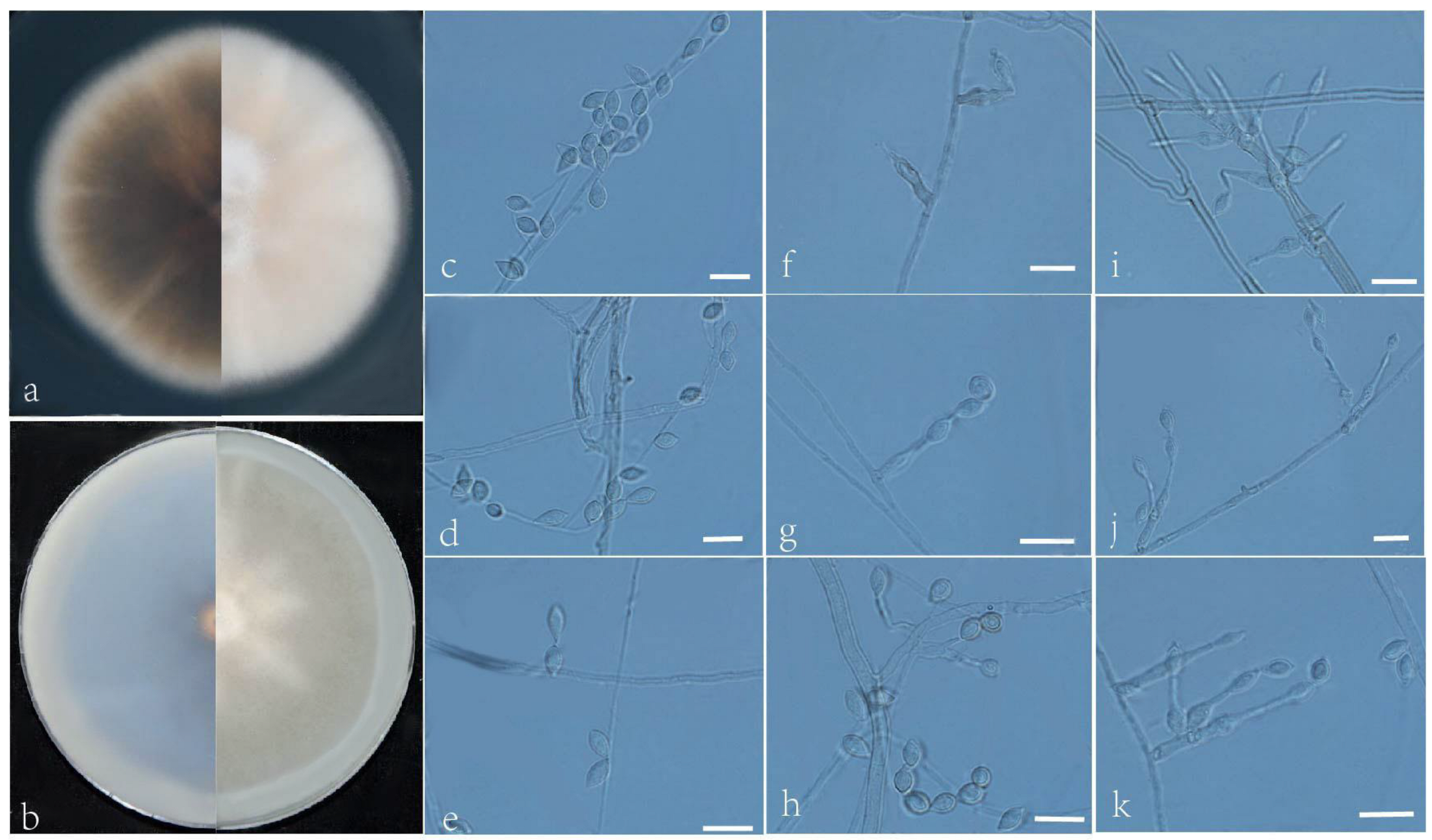
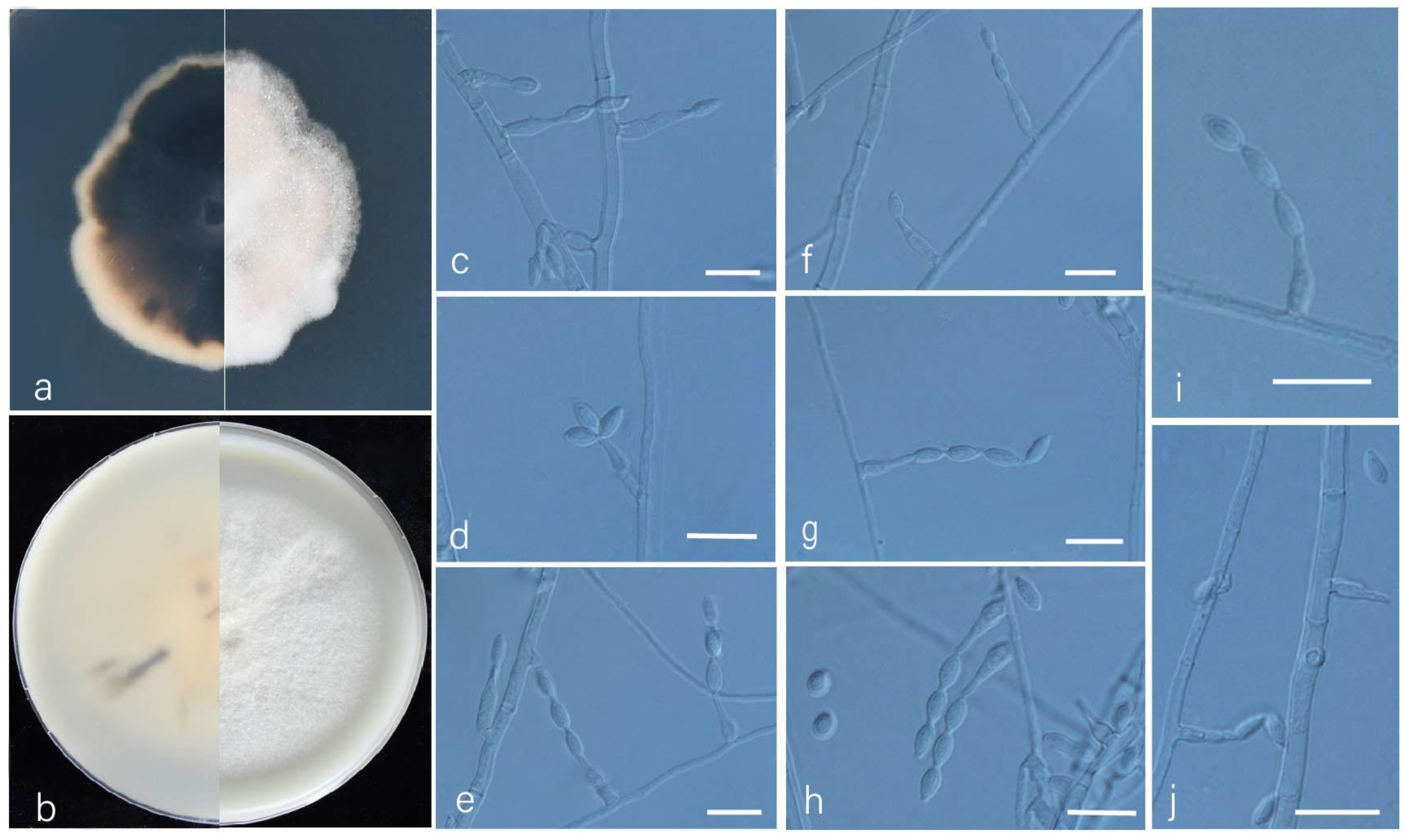
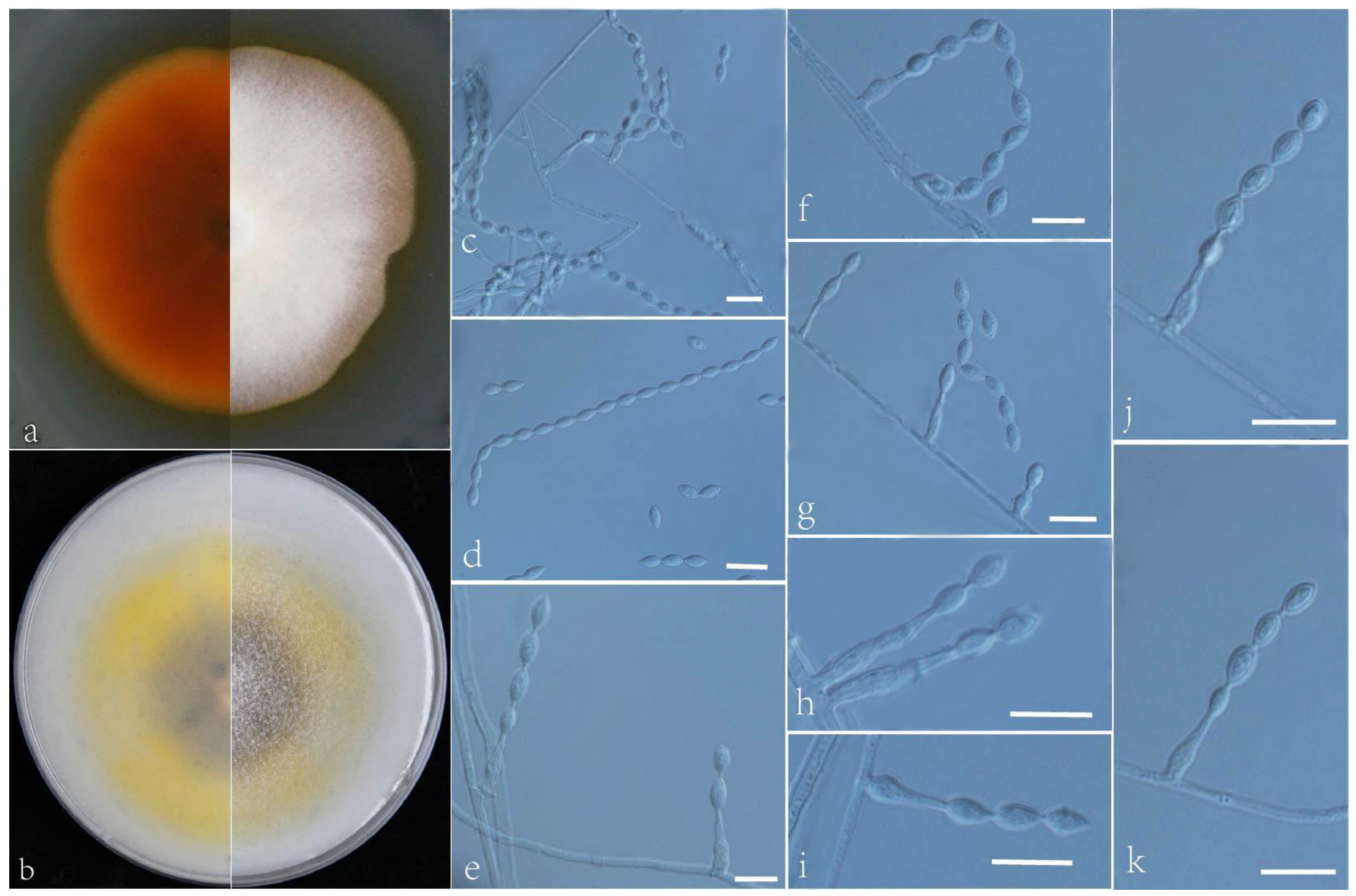
2.1. Sampling, Fungal Isolation, and Morphology

Soil samples were collected in August 2013 from Guangdong, Yunnan, Fujian, and Hainan provinces, southern China, by Y.F. Han. Fungi of the soil samples were isolated according to the method described by Wang et al. [15]. The purified isolates were transferred to potato dextrose agar (PDA) and oatmeal agar (OA) at 35 °C for 7 days in the dark to observe the macroscopic and morphological development of the colonies [17]. The characterization and measurement of the fungal microscopic characteristics were performed in 25% lactic acid. Images were obtained using an optical microscope (BX53, Olympus, Tokyo, Japan) with differential interference contrast (DIC). Colonies on PDA were incubated after 7 days at 35 °C, and the cultures were placed in 50 °C to produce the dried holotypes. The dried holotypes were conserved in the Mycological Herbarium of the Institute of Microbiology, Chinese Academy of Sciences, Beijing, China (HMAS). Ex-type strains were conserved at the China General Microbiological Culture Collection Center (CGMCC) and the Institute of Fungus Resources, Guizhou University (GZUIFR), and other living strains were conserved at the Institute of Fungus Resources, Guizhou University (GZUIFR, GZAC). The morphological descriptions and names of the new taxa were introduced and deposited in MycoBank (https://www.mycobank.org/, asscessed on 3 December 2022).

2.2. DNA Extraction, PCR Amplifications, and Sequencing

The genomic DNA was extracted using the BioTeke Fungus Genomic DNA Extraction Kit (DP2032, BioTeke, Beijing, China), following the manufacturer’s instructions. The ITS region, tub2, the D1/D2 domains of the 28S nrDNA (LSU), and partial RNA polymerase II second largest subunit (RPB2) genes were amplified by PCR, as described by White et al., Glass and Donaldson, Vilgalys and Sun, and Liu et al., respectively [24,25,26,27]. The purified PCR products were obtained and sequencing was performed by a commercial sequencing service provider (Quintarabio, Wuhan, China). In this study, the new sequences mentioned were uploaded to GenBank (Table 1).

2.3. Phylogenetic Analyses

The new sequences were uploaded to BLAST and searched in the GenBank database to determine their most probable related taxa. Acrophialophora species and outgroup Chrysocorona lucknowensis (CBS 727.71) sequence data (ITS, LSU, tub2 and RPB2) were employed to identify the new species. These sequence data were obtained from GenBank (Table 1). The gene regions were concatenated and aligned for phylogenetic analysis. Lasergene software (version 6.0, DNASTAR) was applied for the assembling and editing of the DNA sequences in this study. The consensus sequences from different primers were trimmed using MEGA7. The maximum likelihood (ML) and Bayesian inference (BI) approaches were carried out for the phylogenetic analyses using PhyloSuite v. 1.16 [28]. The maximum likelihood (ML), the best-fit model of substitution for each locus, was estimated using IQ-TREE’s ModelFinder function based on the corrected Akaike information criterion (AICc) [29,30]. The best-fit evolutionary models of ML and BI analyses of each locus are listed in Table 2. The resulting phylogenetic trees were visualized in FigTree v. 1.1.2 and subsequently edited in Adobe Illustrator 2020.

3. Results

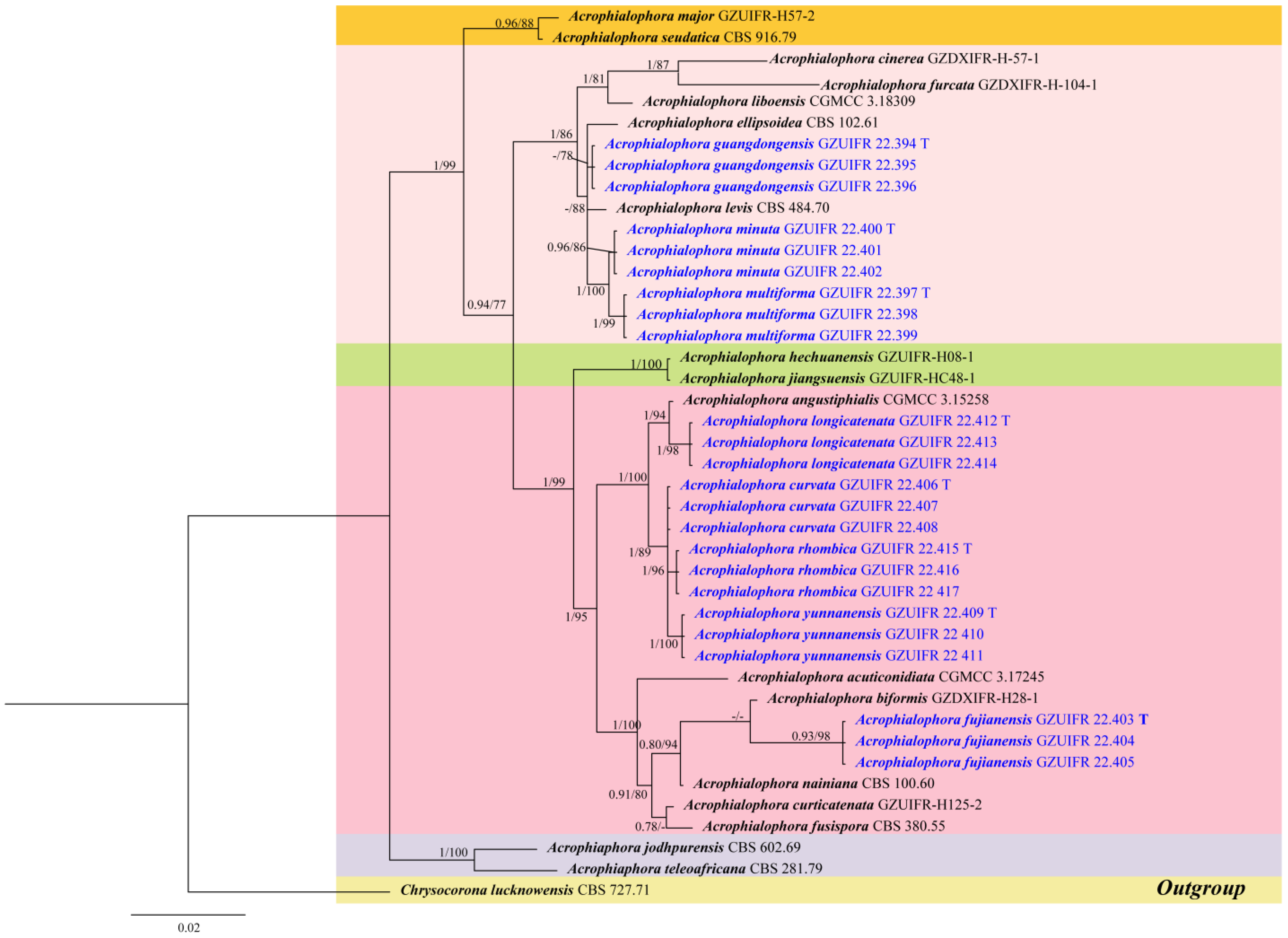
3.1. Phylogeny Analysis
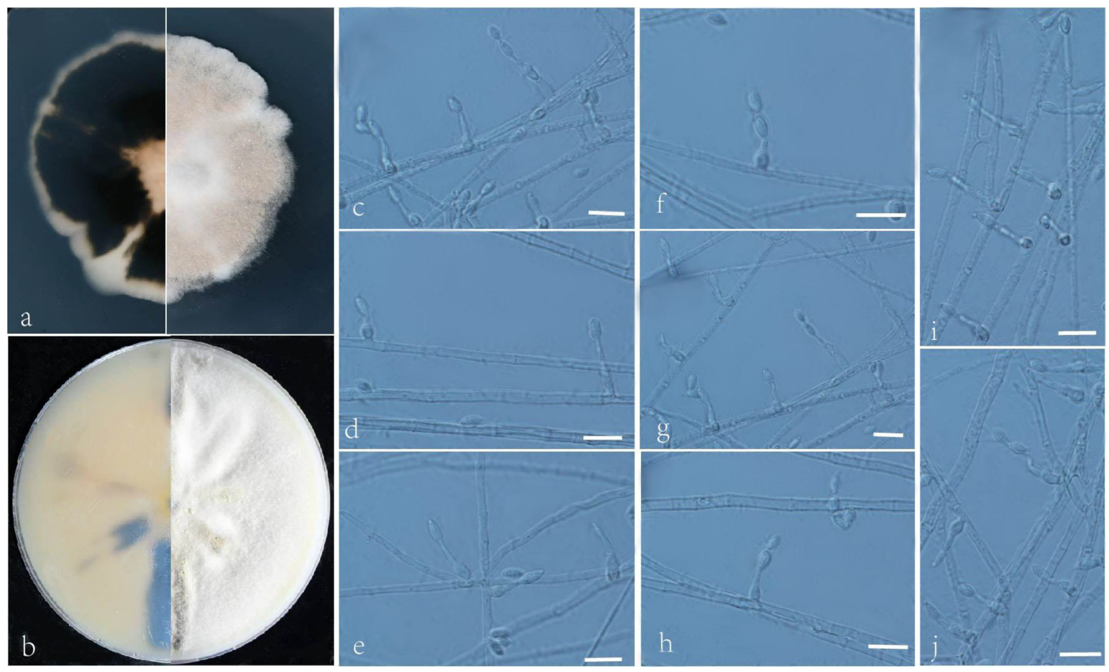
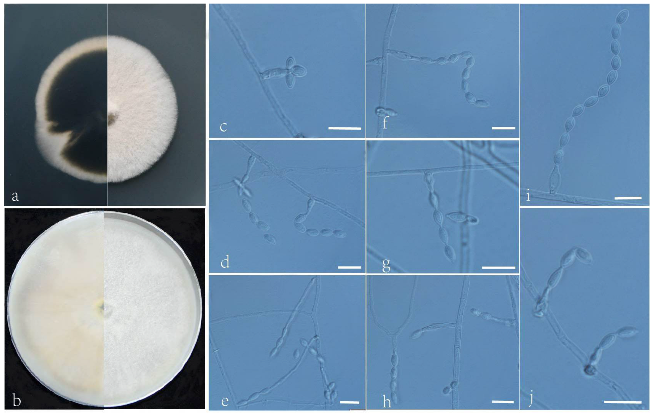
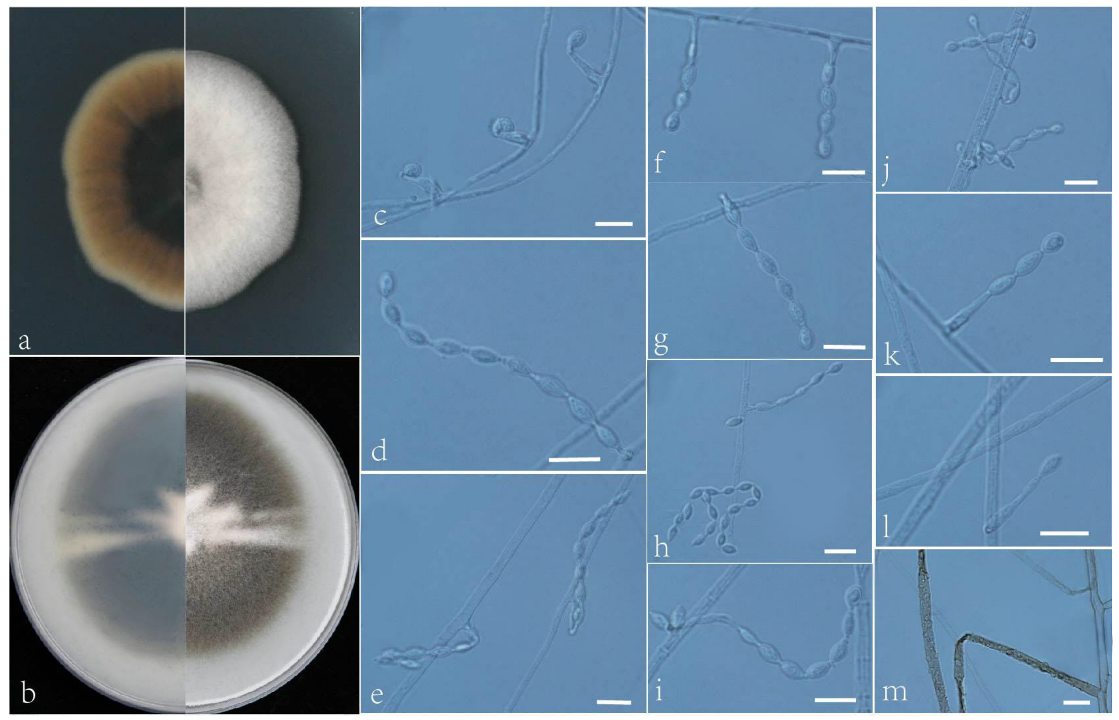
Based on a BLAST search (https://blast.ncbi.nlm.nih.gov/Blast.cgi, accessed on 30 July 2022) using the ITS sequences, our isolates were identified as belonging to the genus Acrophialophora. To determine the phylogenetic positions of these strains, we performed a multilocus (ITS+tub2+LSU+RPB2) phylogenetic analysis. The dataset was composed of the ITS (1–530 bp), tub2 (531–905 bp), LSU (906–1454), and RPB2 (906–1614 bp) genes, comprising a total of 2163 characters (including gaps). The results showed that the new species belong to the genus Acrophialophora (Figure 1). The topologies of the two trees for ML and BI analyses were nearly identical, so we chose BI trees, incorporating ML trees to show the phylogeny. In the phylogenetic tree, eight new species form a well-supported clade separated from other species in Acrophialophora, A. fujiangensis sp. nov. (0.93 PP/98% BS); A. curvata sp. nov. (1 PP/89% BS); A. yunnanensis sp. nov. (1 PP/100% BS); A. longicatenata sp. nov. (1PP/98% BS); A. rhombica sp. nov. (1 PP/96% BS); A guangdongensis sp. nov. (- PP/78% BS); A. multiforma sp. nov. (1 PP/99% BS); and A. minuta sp. nov. (0.96 PP/86% BS).

3.2. Taxonomy

The morphological characteristics of eight new species and their illustrations are mentioned below.
Acrophialophora curvata L. Peng and Y.F. Han sp. nov. Figure 2
MycoBank: 845781
Etymology: Refers to a frequent bend between conidia and phialides.
Holotype: China, Yunnan, Dai Autonomous Prefecture of Xishuangbanna (N 21°40′, E 101°50′), soil, dried holotype HMAS 255324, ex-type culture CGMCC 3.24167 = GZUIFR 22.406. GenBank accession numbers: OP454351 (ITS), OP547306 (tub2), OP454363 (LSU), OP802834 (RPB2).
Micromorphology: Somatic hyphae hyaline, smooth-walled, septate, and 1–4 μm wide. Conidiophores usually reduced to conidiogenous cells. Conidiogenous cells, phialidic, single, arising laterally from the hyphae, with an obclavate swollen basal portion, tapering into a narrow neck, sometimes proliferating in two furcations, 5–24.5 × 2–2.5 μm. Conidia one-celled, formed in basipetal chains, often composed of one to two conidia, hyaline, smooth, fusiform or ellipsoidal, 4–6.5 × 2–3.5 μm. Sexual morph not observed.
Culture characteristics: Colonies on PDA reaching 34–36 mm diam after 7 d at 35 °C, irregular in the margin, flat, dense, fluffy, gray to white, reverse center to edge, dark, pale brown to gray. Colonies on OA reaching 63–65 mm diam after 7 d at 35 °C, alternating brown and white, bronzing floccose due to aerial mycelium, reverse brown and white.
Geographical distribution: China.
Additional material examined: China, Yunnan, Dai Autonomous Prefecture of Xishuangbanna (N 21°40′, E 101°50′), soil, and our other living strains: GZUIFR 22.407, GZUIFR 22.408. GenBank accession numbers: OP454352 (ITS), OP547307 (tub2), OP454364 (LSU), OP802835 (RPB2) and OP454353 (ITS), OP547308 (tub2), OP454365 (LSU), and OP802836 (RPB2).
Notes: Phylogenetically, Acrophialophora curvata is closely related to A. rhombica (Figure 1); however, the sequence similarity of the RPB2 between the two species was 98.5%. The features of A. curvata are that its phialides frequently proliferate into two, the necks between the phialides and conidia are often curved, and the conidial chain consists of only one or two conidia. These features distinguish it from other species of the genus.
Figure 2. Acrophialophora curvata (ex-type CGMCC 3.24167). (a,b) The reverse and obverse colonies on PDA and OA after 7 d at 35 °C. (ck) Hyphae, phialidic conidiogenous cells, and conidia. Scale bars: (ck) = 10 μm.
Jof 09 00645 g002
Acrophialophora fujianensis L. Peng & Y.F. Han sp. nov. Figure 3
MycoBank: 845782
Etymology: Refers to Fujian Province, where the isolate was collected.
Holotype: China, Fujian, Fuzhou, Fujian Normal University (N 26°03′, E 119°21′), soil, dried holotype HMAS 352279, ex-type culture CGMCC 3.24166 = GZUIFR 22.403. GenBank accession numbers: OP454345 (ITS), OP536984 (tub2), OP454372 (LSU), and OP820579 (RPB2).
Micromorphology: Somatic hyphae hyaline, smooth-walled, septate, 1–4.5 μm wide. Conidiophores usually reduced to conidiogenous cells. Conidiogenous cells born apically in whorls or in verticils on conidiophores or arising laterally from the hyphae, phialides single, smooth, hyaline, cylindrical or slightly clavate, swollen near the base, tapering to a narrow neck, often proliferating, 8–25 × 2–2.5 μm. Conidia one-celled, formed in basipetal chains, often composed of one to three conidia, hyaline, smooth, fusiform, ellipsoidal or ovoid, 4–9 × 2.5–4.5 μm. Sexual morph not observed.
Culture characteristics: Colonies on PDA reaching 60–65 mm diam after 7 d at 35 °C, mostly regular in the margin, flat, dense, fluffy, beige to white, reverse pale olive, edge white. Colonies on OA reaching 78–80 mm diam after 7 d at 35 °C, flat, fluffy, gray, reverse dark. Sexual morph not observed.
Geographical distribution: China.
Additional material examined: China, Fujian, Fuzhou, Fujian Normal University (N 26°03′, E 119°21′), soil, and our other living strains: GZUIFR 22.404, GZUIFR 22.405. GenBank accession numbers: OP454346 (ITS), OP536985 (tub2), OP454373 (LSU), OP820580 (RPB2) and OP454347 (ITS), OP536986 (tub2), OP454374 (LSU), and OP820581 (RPB2).
Notes: Phylogenetically, Acrophialophora fujianensis sp. nov. is closely related to A. biformis (Figure 1); however, the sequence similarity of ITS between the two species was 98.6%. Morphologically, they can be distinguished by their conidial chains: A. fujianensis is often composed of one to three conidia, while A. biformis often forms long chains.
Figure 3. Acrophialophora fujianensis (ex-type CGMCC 3.24166). (a,b) The reverse and obverse colonies on PDA and OA after 7 d at 35 °C. (ck) Hyphae, phialidic conidiogenous cells, and conidia. Scale bars: (ck) = 10 μm.
Jof 09 00645 g003
Acrophialophora guangdongensis L. Peng & Y.F. Han sp. nov. Figure 4
MycoBank: 845776
Etymology: Refers to Guangdong Province, where the isolate was collected.
Holotype: China, Guangdong, Guangzhou, (N 23°8′, E 113°17′), soil, dried holotype HMAS 352277, ex-type culture CGMCC 3.24163 = GZUIFR 22.394. GenBank accession numbers: OP454339 (ITS), OP547315 (tub2), OP454369 (LSU), and OP491393 (RPB2).
Micromorphology: Somatic hyphae hyaline, smooth-walled, short septate, 1.5–4 μm wide. Conidiophores reduced to conidiogenous cells. Conidiogenous cells arising laterally from the hyphae, phialidic, solitary, smooth-walled, swollen near the base, cylindrical or flask-shaped, tapering to a narrow neck, 6–21 × 2–3.5 μm. Conidia one-celled, formed in basipetal chains, sometimes apical capitate, hyaline, smooth, fusiform, ellipsoidal or ovoid, 4.5–7 × 2.5–3 μm. Sexual morph not observed.
Culture characteristics: Colonies on PDA reaching 35–40 mm diam after 7 d at 35 °C, irregular in the margin, flat, dense, fluffy, pale wheat, reverse dark, edge pale yellow. Colonies on OA reaching 60–62 mm diam after 7 d at 35 °C, irregular in the margin, flat, dense, fluffy, white, reverse pale yellow.
Geographical distribution: China.
Additional materials examined: China, Guangdong, Guangzhou, (N 23°8′, E 113°17′), soil, and our other living strains: GZUIFR 22.395, GZUIFR 22.396. GenBank accession numbers: OP454340 (ITS), OP547315 (tub2), OP454369 (LSU), OP491393 (RPB2) and OP454340 (ITS), OP547316 (tub2), OP454370 (LSU), and OP491394 (RPB2).
Notes: Phylogenetically, this species is closely related to A. ellpsoidea and A. levis (Figure 1); however, the sequence similarities of the ITS between these species were 98.6% and 97.7%, respectively. Morphologically, the conidia of A. guangdongensis are sometimes apical capitate. This feature is similar to A. multiforma, but A. guangdongensis can be distinguished by its shorter conidial chains.
Figure 4. Acrophialophora guangdongensis (ex-type CGMCC 3.24163). (a,b) The reverse and obverse colonies on PDA and OA after 7 d at 35 °C. (cj) Hyphae, phialidic conidiogenous cells, and conidia. Scale bars: (cj) = 10 μm.
Jof 09 00645 g004
Acrophialophora longicatenata L. Peng & Y.F. Han sp. nov. Figure 5
MycoBank: 845783
Etymology: Refers to longer conidial chain.
Holotype: China, Yunnan, Dai Autonomous Prefecture of Xishuangbanna (N 21°40′, E 101°50′), soil, dried holotype HMAS 255325, ex-type culture CGMCC 3.24169 = GZUIFR 22.412. GenBank accession numbers: OP454357 (ITS), OP547309 (tub2), OP454378 (LSU), and OP834081 (RPB2).
Micromorphology: Somatic hyphae hyaline, smooth-walled, septate, 1–4 μm wide. Conidiophores simple, phialidic, single, arising laterally from the hyphae, swollen basal portion, obclavate or flask-shaped, tapering into a distinct neck, 5–20 × 2–3.5 μm. Conidia one-celled, formed in basipetal chains, often more than 10 conidia, smooth, fusiform or ellipsoidal, 3–6 × 2–4 μm. Sexual morph not observed.
Culture characteristics: Colonies on PDA reaching 55–56 mm diam after 7 d at 35 °C, mostly irregular in the margin, flat, center dense and fluffy and edge sparse, white, reverse sandy brown. Colonies on OA reaching 60–62 mm diam after 7 d at 35 °C, center brown, edge bright yellow, white floccose due to aerial mycelium, reverse center brown, edge bright yellow.
Geographical distribution: China.
Additional material examined: China, Yunnan, Dai Autonomous Prefecture of Xishuangbanna (N 21°40′, E 101°50′), soil, and our other living strains: GZUIFR 22.413, GZUIFR 22.414. GenBank accession numbers: OP454358 (ITS), OP547310 (tub2), OP454379 (LSU), OP834082 (RPB2) and OP454359 (ITS), OP547311 (tub2), OP454380 (LSU), and OP834083 (RPB2).
Notes: Phylogenetically, Acrophialophora longicatenata is closely related to A. angustiphialis (Figure 1); however, the sequence similarity of the tub2 between the two species was 97.8%. Morphologically, A. longicatenata and A. angustiphialis have smooth, ellipsoidal, and fusiform conidia [17]. However, they can be distinguished by their conidial chains; the conidial chain of A. longicatenata is usually more than 10 conidia.
Figure 5. Acrophialophora longicatenata (ex-type CGMCC 3.24169). (a,b) The reverse and obverse colonies on PDA and OA after 7 d at 35 °C. (ck) Hyphae, phialidic conidiogenous cells, and conidia. Scale bars: (ck) = 10 μm.
Jof 09 00645 g005
Acrophialophora minuta L. Peng & Y.F. Han sp. nov. Figure 6
MycoBank: 845778
Etymology: Refers to its smaller conidia and phialides.
Holotype: China, Fujian, Fuzhou, Fujian Normal University (N 26°03′, E 119°21′), soil, Y. F. Han, dried holotype HMAS 352278, ex-type culture CGMCC 3.24165 = GZUIFR 22.400. GenBank accession numbers: OP454342 (ITS), OP547318 (tub2), OP454366 (LSU), and OP880249 (RPB2).
Micromorphology: Somatic hyphae hyaline, smooth-walled, septate, 1–4 μm wide. Conidiophores single, phialidic, arising laterally from the hyphae, swollen basal portion, tapering into a distinct neck, 4–16 × 2–3 μm. Conidia one-celled, formed in basipetal chains, short conidia chain, often composed of one to three conidia, hyaline, smooth, fusiform or ellipsoidal, 4–6 × 2–3 μm. Sexual morph not observed.
Culture characteristics: Colonies on PDA reaching 45–50 mm diam after 7 d at 35 °C, irregular in the margin, flat, dense, fluffy, white to pale brown, reverse center to edge, pale yellow, dark to beige. Colonies on OA reaching 80–82 mm diam after 7 d at 35 °C, raised, dense, fluffy, white, reverse pale yellow and dark.
Geographical distribution: China.
Additional materials examined: China, Fujian Province, Fuzhou City, Fujian Normal University (N 26°03′, E 119°21′), soil, and our other living strains: GZUIFR 22.401, GZUIFR 22.402. GenBank accession numbers: OP454343 (ITS), OP547319 (tub2), OP454367 (LSU), OP880250 (RPB2) and OP454344 (ITS), OP547320 (tub2), OP454368 (LSU), and OP880251 (RPB2).
Notes: Phylogenetically, this species is closely related to A. multiforma (Figure 1); however, the sequence similarities of the ITS between the two species were 98.5%. Morphologically, A. minuta can be distinguished from A. multiforma by its smaller phialides (4–15 × 2–3 μm vs. 5–25 × 2–3.5 μm) and conidia (4–5.5 × 2–3 μm vs. 4.5–7 × 2–3.5 μm).
Figure 6. Acrophialophora minuta (ex-type CGMCC 3.24165). (a,b) the reverse and obverse colonies on PDA and OA after 7 d at 35 °C. (cj) Hyphae, phialidic conidiogenous cells, and conidia. Scale bars: (cj) = 10 μm.
Jof 09 00645 g006
Acrophialophora multiforma L. Peng & Y.F. Han sp. nov. Figure 7
MycoBank: 845779
Etymology: Refers to the variety of phialides.
Holotype: China, Yunnan, Baoshan, Tengchong County (N 24°50′, E 98°39′), soil, dried holotype HMAS 255321, ex-type culture CGMCC 3.24164 = GZUIFR 22.397. GenBank accession numbers: OP454336 (ITS), OP547321 (tub2), OP454360 (LSU), and OP880243 (RPB2).
Micromorphology: Somatic hyphae hyaline, smooth-walled, septate, 1–4 μm wide. Conidiophores absent or reduced to conidiogenous cells. Conidiogenous cells arising laterally from the hyphae, phialidic, solitary, smooth-walled, swollen near the basal portion, flask-shaped, obclavate, tapering to a narrow neck, sometimes awl-shaped or linear, 5–25 × 2–3.5 μm. Conidia one-celled, formed in basipetal chains, long chain, sometimes apical capitate, hyaline, smooth, fusiform or ellipsoidal, 4.5–7 × 2–3.5 μm. Sexual morph not observed.
Culture characteristics: Colonies on PDA reaching 44–46 mm diam after 7 d at 35 °C, mostly regular in the margin, flat, dense, fluffy, beige, reverse center dark, edge beige. Colonies on OA reaching 78–80 mm diam after 7 d at 35 °C, irregular in the margin, flat, dense, fluffy, white, reverse pale yellow.
Geographical distribution: China.
Additional materials examined: China, Yunnan, Baoshan, Tengchong County (N 24°50′, E 98°39′), soil, and our other living strains: GZUIFR 22.398, GZUIFR 22.399. GenBank accession numbers: OP454337 (ITS), OP547322 (tub2), OP454361(LSU), OP880244 (RPB2) and OP454338 (ITS), OP547323 (tub2), OP454362 (LSU), and OP880245 (RPB2).
Notes: This species is characterized by the variety of phialides and conidia, which are sometimes apical capitate. This feature distinguishes the species from the other species of Acrophialophora.
Figure 7. Acrophialophora multiforma (ex-type CGMCC 3.24164). (a,b) The reverse and obverse colonies on PDA and OA after 7 d at 35 °C. (cj) Hyphae, phialidic conidiogenous cells, and conidia. Scale bars: (cj) = 10 μm.
Jof 09 00645 g007
Acrophialophora rhombica L. Peng & Y.F. Han sp. nov. Figure 8
MycoBank: 845784
Etymology: Refers to nearly rhombic conidia.
Holotype: China, Hainan, Sanya, (N 18°20′, E 109°50′), soil, dried holotype HMAS 352281, ex-type culture CGMCC 3.24170 = GZUIFR 22.415. GenBank accession numbers: OP454354 (ITS), OP731571 (tub2), OP454381 (LSU), and OP834084 (RPB2).
Micromorphology: Somatic hyphae hyaline, smooth-walled, septate, 1–4.5 μm wide. Conidiophores absent or simple, phialidic, single, arising laterally from the hyphae, with an awl-shaped swollen basal portion, tapering gradually or abruptly to a narrow neck, 5–18 × 1.5–4 μm. Conidia one-celled, formed in basipetal chains, long chain, smooth, fusiform, long ellipsoidal or rhombic, 5–9 × 2–5 μm. Sexual morph not observed.
Culture characteristics: Colonies on PDA reaching 35–40 mm diam after 7 d at 35 °C, mostly irregular in the margin, thick, spongy, cream, reverse brown, edge orange. Colonies on OA reaching 78–80 mm diam after 7 d at 35 °C, brown, white floccose due to aerial mycelium, reverse yellow-brown.
Geographical distribution: China.
Additional material examined: China, Hainan, Sanya, (N 18°20′, E 109°50′), soil, our other living strains: GZUIFR 22.416, GZUIFR 22.417. GenBank accession numbers: OP454355 (ITS), OP731572 (tub2), OP454382 (LSU), OP834085 (RPB2) and OP454356 (ITS), OP731573 (tub2), OP454383 (LSU), and OP834086 (RPB2).
Notes: Morphologically, this species is similar to Acrophialophora acuticonidiata; however, it can be distinguished by its rhombic and long ellipsoidal conidia. In addition, the conidia of A. rhombica are smooth, while those of A. acuticonidiata are rough [17].
Figure 8. Acrophialophora rhombica (ex-type CGMCC 3.24170). (a,b) I reverse and obverse colonies on PDA and OA after 7 d at 35 °C. (ci) Hyphae, phialidic conidiogenous cells, and conidia. Scale bars: (ci) = 10 μm.
Jof 09 00645 g008
Acrophialophora yunnanensis L. Peng & Y.F. Han sp. nov. Figure 9
MycoBank: 845785
Etymology: Refers to Yunnan Province, where the isolate was collected.
Holotype: China, Yunnan, Dai Autonomous Prefecture of Xishuangbanna (N 21°40′, E 101°50′), soil, dried holotype HMAS 352280, ex-type culture CGMCC 3.24168 = GZUIFR 22.409. GenBank accession numbers: OP454348 (ITS), OP547312 (tub2), OP454375 (LSU), and OP880246 (RPB2).
Micromorphology: Somatic hyphae hyaline or brown, smooth-walled or rough, septate, 1–4.5 μm wide. Conidiophores absent or usually reduced to conidiogenous cells. Conidiogenous cells, single, phialidic, arising laterally from the hyphae, swollen basal portion, tapering into a distinct neck, awl-shaped, or forming a thin and short neck, 4.5–20 × 2–2.5 μm. Conidia 1-celled, formed in basipetal chains, long chain, smooth, fusiform or ellipsoidal to long ellipsoidal, 3.5–8 × 1.5–4 μm. Sexual morph not observed.
Culture characteristics: Colonies on PDA reaching 35–40 mm diam after 7 d at 35 °C, irregular in the margin, flat, dense, fluffy, light gray, reverse center to edge, dark, golden olive to light yellow. Colonies on OA reaching 65–68 mm diam after 7 d at 35 °C, puce to white, white floccose due to aerial mycelium, reverse dark to white.
Geographical distribution: China.
Additional material examined: China, Yunnan, Dai Autonomous Prefecture of Xishuangbanna (N 21°40′, E 101°50′), soil, and our other living strains: GZUIFR 22.410, GZUIFR 22.411. GenBank accession numbers: OP454349 (ITS), OP547313 (tub2), OP454376 (LSU), OP880247 (RPB2) and OP454350 (ITS), OP547314 (tub2), OP454377 (LSU), and OP880248 (RPB2).
Notes: Phylogenetically, this species is closely related to Acrophialophora rhombica (Figure 1); however, the sequence similarity of the ITS between the two species was 98.6%, and the sequence similarity of the RPB2 was 98.3%. Morphologically, it can be distinguished by its conidial shape and somatic hyphae. A. rhombica has nearly rhombic conidia as well as hyaline hyphae that are smooth-walled. However, the somatic hyphae of A. yunnanensis are sometimes brown and rough-walled.
Figure 9. Acrophialophora yunnanensis (ex-type CGMCC 3.24168). (a,b) the reverse and obverse colonies on PDA and OA after 7 d at 35 °C. (cm) Hyphae, phialidic conidiogenous cells, and conidia. Scale bars: (cm) = 10 μm.
Jof 09 00645 g009

4. Discussion

In this study, the ITS sequences of eight new species were uploaded to preliminary BLAST searches, and the results indicated that they were most closely related to the genus Acrophialophora. We then established the phylogenetic tree of Acrophialophora and showed that eight new species were indeed located in Acrophialophora (Figure 1). The species of the genus Acrophialophora were found to form five distinct subclades. Originally, only asexual states were known for the genus Acrophialophora, and subsequently, A. teleoafricana and A. jodhpurense, as one of the subclades with sexual structures, were accommodated in the genus [20]. In addition, there were three Acrophialophora species: A. ampullaris, A. ampullaphora, and A. berberidis, which were not discussed in this study due to a lack of sequence support. However, according to the morphological description and illustration of Han et al. and Matushima [14,31,32], A. ampulliphora has rough conidia, and A. ampullaris has subglobose conidia, which can be distinguished from the eight new species with smooth and fusiform or ellipsoidal conidia, respectively. Acrophialophora berberidis is similar to A. guangdongensis and A. multiforma in having both capitate and catenulate conidia, but the phialide bases of A. guangdongensis are cylindrical swollen, of A. multiforma are multiform, and of A. berberidis are ellipsoidal. Therefore, the three species are different from our eight new species. However, the taxonomic statuses of these three species still need further study.
Some genera of Chaetomiaceae have certain morphological similarities to Acrophialophora, including Botryotrichum and Humicola. Some species of Botryotrichum and Humicola also directly produce phialides and conidial chains on the hyphae. However, these fungi are characterized by simple, short conidiophores and brown subglobose or globose conidia [13]. Monocillium, Acremonium, and Phialemonium are also somewhat similar to these genera in morphology, but they do not belong to Chaetomiaceae. Monocillium has a similar conidiogenous structure, which is distinguished by the inflated or thickened middle part of the phialide [33]. However, the Acremonium species can be distinguished by their typical awl-shaped phialides [34]. Phialemonium inflatum is similar in morphology, but it is not thermotolerant [16].
Chaetomiaceae includes mostly soilborne cellulose decomposers and thermotolerant opportunistic pathogens [18]. In the same way, almost all species of Acrophialophora were proven to be thermotolerant. Studies have shown that some species can produce useful thermostable enzymes. For example, A. major and A. biformis can produce laccase, and A. cinerea and A. hechuanensis can produce ligninase and cellulase [10,35,36]. However, Sandoval et al. first studied the clinical significance of Acrophialophora, and they considered that the genus is a rare opportunistic human and animal pathogen and includes common species isolated from human clinical samples, such as Acrophialophora levis [18]. The study also mentioned that one clinical isolate, FMR 8888, which caused corneal infection, was confirmed as Acrophialophora fusispora, while another, FMR 6662, which was isolated from sputum, was reidentified as Acrophialophora levis. Therefore, the potential of these fungi in applied research and their pathogenicity to humans need to be further studied.

Author Contributions

Writing—original draft preparation, L.P.; writing—review and editing, Y.-F.H.; methodology, L.P., Y.-F.H. and H.-Y.W.; software, L.P., W.-H.C. and C.-B.D.; sampling, Y.-F.H. and Y.-W.Z.; data curation, L.P. and Y.-F.H.; visualization and funding acquisition, Y.-F.H. and Y.-W.Z.; supervision, C.-B.D. and J.-D.L.; project administration, Y.-F.H. All authors have read and agreed to the published version of the manuscript.

Funding

This work was financially supported by the Natural Science Foundation of China (No. 32160007, 32060011, 32260003), the “Hundred” Talent Projects of Guizhou Province (Qian Ke He [2020] 6005), and the Construction Program of Biology First-Class Discipline in Guizhou (GNYL [2017] 009).

Institutional Review Board Statement

Not applicable.

Informed Consent Statement

Not applicable.

Data Availability Statement

All newly generated sequences were deposited in GenBank (https://www.ncbi.nlm.nih.gov/genbank/, (accessed on 13 September 2022) Table 1). All new taxa were deposited in MycoBank (https://www.mycobank.org/ (accessed on 3 December 2022)).

Acknowledgments

We appreciate MDPI for the English language editing of the whole manuscript. We are thankful to Qiu-Yu Shao, Xin Li, Yu-Lian Ren, and Qing-Qing Zhang for providing comments that improved the quality of the manuscript and their kind help for this study.

Conflicts of Interest

The author declares that there are no conflict of interest.

References

  1. Edward, J.C. A new genus of the Moniliaceae. Mycologia 1959, 51, 781–786. [Google Scholar] [CrossRef]
  2. Barron, G.L.; Peterson, J.L. The Genera of Hyphomycetes from Soil; Williams & Wilkins Co.: Baltimore, MD, USA, 1968; p. 477. [Google Scholar]
  3. Dal Vesco, G.; Peyronel, B. Funghi isolati dal suolo di due isole del Pacifico meridionale. Allionia 1968, 14, 31–49. [Google Scholar]
  4. Samson, R.A.; Mahmood, T. The genus Acrophialophora (fungi, moniliales). Acta Bot. Neerl. 1970, 19, 804–808. [Google Scholar] [CrossRef]
  5. Luangsa-Ard, J.J.; Hywel-Jones, N.L.; Samson, R.A. The polyphyletic nature of Paecilomyces sensu lato based on 18S-generated rDNA phylogeny. Mycologia 2004, 96, 773–780. [Google Scholar] [CrossRef]
  6. Brown, A.H.S.; Smith, G. The genus Paecilomyces Bainier and its perfect stage Byssochlamys Westling. Trans. Br. Mycol. Soc. 1957, 40, 17–89. [Google Scholar] [CrossRef]
  7. Onions, A.H.S.; Barron, G.L. Monophialidic species of Paecilomyces. Mycological 1967, 107, 1–25. [Google Scholar]
  8. Gams, W. Cephalosporium-Artige Schimmelpilze (Hyphomycetes); Gustav Fischer: Stuttgart, Germany, 1971; pp. 38–130. [Google Scholar]
  9. Liang, Z.Q.; Han, Y.F.; Chu, H.L. Studies on the genus Paecilomyces in China. IV. Two new species of Paecilomyces with monophialides. Mycotaxon 2006, 97, 13–20. [Google Scholar]
  10. Liang, Z.Q.; Han, Y.F.; Chu, H.L. A new thermotolerant Paecilomyces species which produces laccase and a biform sporogenous structure. Fungal Divers. 2007, 27, 95–102. [Google Scholar]
  11. Han, Y.F.; Liang, Z.Q.; Chu, H.L. A new thermophilic species of Paecilomyces, Paecilomyces curticatenatus. Mycosystema 2007, 26, 13–16. [Google Scholar]
  12. Chu, H.L.; Liang, Z.Q.; Han, Y.F. A thermotolerant Paecilomyces inflatus var. major Liang Z Q, Chu H L et Han Y F var. nov. Which Produces Laccase. J. Fungal Res. 2004, 2, 43–46. [Google Scholar]
  13. Liang, Z.Q.; Han, Y.F.; Chu, H.L.; Fox, R.T. Studies on the genus Paecilomyces in China, V. Taifanglania gen. nov. for some monophialidic species. Fungal Divers. 2009, 34, 69–77. [Google Scholar]
  14. Han, Y.F.; Liang, J.D.; Liang, Z.Q.; Zou, X.; Dong, X. Two new Taifanglania species identified through DELTA-assisted phenetic analysis. Mycotaxon 2010, 112, 325–333. [Google Scholar] [CrossRef]
  15. Wang, Y.; Wang, Y.R.; Han, Y.F.; Liang, Z.Q. A new thermotolerant species of Taifanglania. Mycosystem 2015, 34, 345–349. [Google Scholar]
  16. Perdomo, H.; García, D.; Gené, J.; Cano, J.; Sutton, D.A.; Summerbell, R.; Guarro, J. Phialemoniopsis, a new genus of Sordariomycetes, and new species of Phialemonium and Lecythophora. Mycologia 2013, 105, 398–421. [Google Scholar] [CrossRef]
  17. Zhang, Y.; Liu, F.; Wu, W.; Cai, L. A phylogenetic assessment and taxonomic revision of the thermotolerant hyphomycete genera Acrophialophora and Taifanglania. Mycologia 2015, 107, 768–779. [Google Scholar] [CrossRef]
  18. Sandoval-Denis, M.; Gené, J.; Sutton, D.A.; Wiederhold, N.P.; Guarro, J. Acrophialophora, a poorly known fungus with clinical significance. J. Clin. Microbiol. 2015, 53, 1549–1555. [Google Scholar] [CrossRef] [Green Version]
  19. Zhang, Y.W.; Wang, Y.; Zeng, G.P.; Chen, W.H.; Zou, X.; Han, Y.F.; Qiu, S.Y.; Liang, Z.Q. A new species of Acrophialophora from Guizhou Province, China. Phytotaxa 2017, 302, 266–272. [Google Scholar] [CrossRef] [Green Version]
  20. Wang, X.W.; Bai, F.Y.; Bensch, K.; Meijer, M.; Sun, B.D.; Han, Y.F.; Crous, P.W.; Samson, R.A.; Yang, F.Y.; Houbraken, J. Phylogenetic re-evaluation of Thielavia with the introduction of a new family Podosporaceae. Stud. Mycol. 2019, 93, 155–252. [Google Scholar] [CrossRef]
  21. Wang, X.W.; Han, P.J.; Bai, F.Y.; Luo, A.; Bensch, K.; Meijer, M.; Kraak, B.; Han, D.Y.; Sun, B.D.; Crous, P.W.; et al. Taxonomy, phylogeny and identification of Chaetomiaceae with emphasis on thermophilic species. Stud. Mycol. 2022, 101, 121–243. [Google Scholar] [CrossRef]
  22. Hendy, M.H.; Hashem, A.H.; Sulieman, W.B.; Sultan, M.H.; Abdelraof, M. Purification, characterization and anticancer activity of l-methionine γ-lyase from thermo-tolerant aspergillus fumigatus. Microb. Cell Fact. 2023, 22, 8. [Google Scholar] [CrossRef]
  23. Barros, R.R.O.; Oliveira, R.A.; Gottschalk, L.M.F.; Bon, E.P.S. Production of cellulolytic enzymes by fungi Acrophialophora nainiana and Ceratocystis paradoxa using different carbon sources. Appl. Biochem. Biotechnol. 2010, 161, 448–454. [Google Scholar] [CrossRef]
  24. White, T.J.; Bruns, T.; Lee, S.; Taylor, J. Amplification and direct sequencing of fungal ribosomal RNA genes for phylogenetics. In PCR Protocols: A Guide to Methods and Applications; Innis, M.A., Gelfand, D.H., Sninsky, J.J., White, T.J., Eds.; Academic Press: San Diego, CA, USA, 1990; pp. 315–322. [Google Scholar]
  25. Glass, N.L.; Donaldson, G.C. Development of primer sets designed for use with the PCR to amplify conserved genes from filamentous ascomycetes. Appl. Environ. Microbiol. 1995, 61, 1323–1330. [Google Scholar] [CrossRef] [Green Version]
  26. Vilgalys, R.; Sun, B.L. Ancient and recent patterns of geographic speciation in the oyster mushroom Pleurotus revealed by phylogenetic analysis of ribosomal DNA sequences. Proc. Natl. Acad. Sci. USA 1994, 91, 4599–4603. [Google Scholar] [CrossRef] [Green Version]
  27. Liu, Y.J.; Whelen, S.; Hall, B.D. Phylogenetic relationships among ascomycetes: Evidence from an RNA polymerse II subunit. Mol. Biol. Evol. 1999, 16, 1799–1808. [Google Scholar] [CrossRef] [Green Version]
  28. Zhang, D.; Gao, F.L.; Jakovlić, I.; Zou, H.; Zhang, J.; Li, W.X. PhyloSuite: An integrated and scalable desktop platform for streamlined molecular sequence data management and evolutionary phylogenetics studies. Mol. Ecol. Resour. 2020, 20, 348–355. [Google Scholar] [CrossRef]
  29. Nguyen, L.T.; Schmidt, H.A.; Haeseler, A.V.; Minh, B.Q. IQ-TREE: A fast and effective stochastic algorithm for estimating maximum-likelihood phylogenies. Mol. Biol. Evol. 2015, 32, 268–274. [Google Scholar] [CrossRef]
  30. Ronquist, F.; Teslenko, M.; Van, D.M.P. MrBayes 3.2: Efficient Bayesian phylogenetic inference and model choice across a large model space. Syst. Biol. 2012, 61, 539–542. [Google Scholar] [CrossRef] [Green Version]
  31. Matushima, T. Microfungi of the Solomon Islands and Papua-New Guinea; Matushima: Kobe, Japan, 1971; p. 42. [Google Scholar]
  32. Matushima, T. Icones Microfungorum a Matushima Lectorum; Matushima: Kobe, Japan, 1975; p. 104. [Google Scholar]
  33. Arx, J.A. The Genera of Fungi Sporulating in Pure Culture; Cramer: Vaduz, Liechtenstein, 1981; pp. 283–331. [Google Scholar]
  34. Samson, R.A. Paecilomyces and some allied hyphomycetes. Stud. Mycol. 1974, 6, 1–119. [Google Scholar]
  35. Zhang, Y.W.; Chen, W.H.; Wang, Y.R.; Han, Y.F.; Liang, Z.Q. Screening on cellulase from thermophilic Taifanglania spp. Chin. Brew. 2013, 3, 45–47. [Google Scholar]
  36. Wang, Y.; Han, Y.F.; Liang, Z.Q. Rice straw degradation and enzyme production of two Taifanglania strains. Mycosystema 2017, 36, 598–603. [Google Scholar]
Figure 1. Phylogenetic tree of the genus Acrophialophora constructed from ITS, tub2, LSU, and RPB2 gene region alignment. Maximum likelihood bootstrap values ≥ 75% and posterior probabilities ≥ 0.90 are shown above internal branches. The “-” indicates a lack of statistical support. The eight new species are shown in blue. The scale bar shows the expected number of changes per site. Type strains are marked with “T” after the culture number.
Jof 09 00645 g001
Table 1. Names, strain numbers, and corresponding GenBank accession numbers of the taxa used in the phylogenetic analyses of this study.
SpeciesStrainsGenBank Accession Numbers
ITStub2LSURPB2
Acrophialophora acuticonidiataCGMCC 3.17245KJ026975KJ147441
Acrophialophora angustiphialisCGMCC 3.15258KJ026972KJ147438
Acrophialophora biformisGZDXIFR-H28-1DQ191963
Acrophialophora cinereaGZDXIFR-H-57-1DQ243694KP143110OP419973OP886702
Acrophialophora curticatenataGZUIFR-H125-2EU004811
Acrophialophora curvataGZUIFR 22.406OP454351OP547306OP454363OP802834
Acrophialophora curvataGZUIFR 22.407OP454352OP547307OP454364OP802835
Acrophialophora curvataGZUIFR 22.408OP454353OP547308OP454365OP802836
Acrophialophora ellipsoideaCBS 102.61MK926786MK926886MK926786MK876748
Acrophialophora fujianensisGZUIFR 22.403OP454345OP536984OP454372OP820579
Acrophialophora fujianensisGZUIFR 22.404OP454346OP536985OP454373OP820580
Acrophialophora fujianensisGZUIFR 22.405OP454347OP536986OP454374OP820581
Acrophialophora furcataGZDXIFR-H-104-1DQ243695KP143113OP456145OP886703
Acrophialophora fusisporaCBS 380.55MK926788MK926888MK926788MK876750
Acrophialophora guangdongensisGZUIFR 22.394 TOP454339OP547315OP454369OP491393
Acrophialophora guangdongensisGZUIFR 22.395OP454340OP547316OP454370OP491394
Acrophialophora guangdongensisGZUIFR 22.396OP454341OP547317OP454371OP491395
Acrophialophora hechuanensisGZUIFR-H08-1MK926789MK926889MK926789MK876751
Acrophialophora jiangsuensisGZUIFR HC48.1KF719171KP143112OP456146OP491392
Acrophialophora jodhpurensisCBS 602.69MK926790MK926890MK926790MK876752
Acrophialophora levisCBS 484.70KP233038KP233044KM995840
Acrophialophora liboensisCGMCC 3.18309KP192127KP999978OP456147OP886704
Acrophialophora longicatenataGZUIFR 22.412 TOP454357OP547309OP454378OP834081
Acrophialophora longicatenataGZUIFR 22.413OP454358OP547310OP454379OP834082
Acrophialophora longicatenataGZUIFR 22.414OP454359OP547311OP454380OP834083
Acrophialophora majorGZUIFR-H57-2MK926792MK926892MK926792MK876754
Acrophialophora minutaGZUIFR 22.400 TOP454342OP547318OP454366OP880249
Acrophialophora minutaGZUIFR 22.401OP454343OP547319OP454367OP880250
Acrophialophora minutaGZUIFR 22.402OP454344OP547320OP454368OP880251
Acrophialophora multiformaGZUIFR 22.397 TOP454336OP547321OP454360OP880243
Acrophialophora multiformaGZUIFR 22.398OP454337OP547322OP454361OP880244
Acrophialophora multiformaGZUIFR 22.399OP454338OP547323OP454362OP880245
Acrophialophora nainianaCBS 100.60MK926793MK926893MK926793MK876755
Acrophialophora rhombicaGZUIFR 22.415 TOP454354OP731571OP454381OP834084
Acrophialophora rhombicaGZUIFR 22.416OP454355OP731572OP454382OP834085
Acrophialophora rhombicaGZUIFR 22.417OP454356OP731573OP454383OP834086
Acrophialophora seudaticaCBS 916.79LN736030LN736032LN736031
Acrophialophora teleoafricanaCBS 281.79MK926795MK926895MK926795MK876757
Acrophialophora yunnanensisGZUIFR 22.409 TOP454348OP547312OP454375OP880246
Acrophialophora yunnanensisGZUIFR 22.410OP454349OP547313OP454376OP880247
Acrophialophora yunnanensisGZUIFR 22.411OP454350OP547314OP454377OP880248
Chrysocorona lucknowensisCBS 727.71 TMK926813MK926913MK926813MK876773
Table 2. The best-fit evolutionary model phylogenetic analyses.
ITSLSUtub2RPB2
ML analysisK3P+G4F81+F+IHKY+F+G4GTR+F+G4
BI analysisK2P+G4F81+F+IHKY+F+G4GTR+F+G4
Disclaimer/Publisher’s Note: The statements, opinions and data contained in all publications are solely those of the individual author(s) and contributor(s) and not of MDPI and/or the editor(s). MDPI and/or the editor(s) disclaim responsibility for any injury to people or property resulting from any ideas, methods, instructions or products referred to in the content.

