Next Article in Journal
Effect of Microbial Consortium Constructed with Lignolytic Ascomycetes Fungi on Degradation of Rice Stubble
Previous Article in Journal
Identification, Culture Characteristics and Whole-Genome Analysis of Pestalotiopsis neglecta Causing Black Spot Blight of Pinus sylvestris var. mongolica
 
 
Correction published on 13 July 2023, see J. Fungi 2023, 9(7), 743.
Article

Distinct Roles of Ena ATP Family Proteins in Sodium Accumulation, Invasive Growth, and Full Virulence in Colletotrichum gloeosporioides

1
Co-Innovation Center for Sustainable Forestry in Southern China, Nanjing Forestry University, Nanjing 210037, China
2
College of Forestry, Nanjing Forestry University, Nanjing 210037, China
*
Author to whom correspondence should be addressed.
J. Fungi 2023, 9(5), 566; https://doi.org/10.3390/jof9050566
Submission received: 6 February 2023 / Revised: 14 April 2023 / Accepted: 23 April 2023 / Published: 13 May 2023 / Corrected: 13 July 2023

Abstract

:
Colletotrichum gloeosporioides, a significant fungal pathogen of crops and trees, causes large economic losses worldwide. However, its pathogenic mechanism remains totally unclear. In this study, four Ena ATPases (Exitus natru-type adenosine triphosphatases), homology of yeast Ena proteins, were identified in C. gloeosporioides. Gene deletion mutants of ΔCgena1, ΔCgena2, ΔCgena3, and ΔCgena4 were obtained through the method of gene replacement. First, a subcellular localization pattern indicated that CgEna1 and CgEna4 were localized in the plasma membrane, while the CgEna2 and CgEna3 were distributed in the endoparasitic reticulum. Next, it was found that CgEna1 and CgEna4 were required for sodium accumulation in C. gloeosporioides. CgEna3 was required for extracellular ion stress of sodium and potassium. CgEna1 and CgEna3 were involved in conidial germination, appressorium formation, invasive hyphal development, and full virulence. The mutant of ΔCgena4 was more sensitive to the conditions of high concentrations of ion and the alkaline. Together, these results indicated that CgEna ATPase proteins have distinct roles in sodium accumulation, stress resistance, and full virulence in C. gloeosporioides.

1. Introduction

In living cells, sodium and potassium are strictly controlled within certain concentration range [1]. Maintaining appropriate ion concentration requires the participation of P-type ATPases [2,3]. P-type ATPases are a large family of membrane proteins that play important roles in ion transport across the plasma membrane and form a typical phosphatase intermediate which hydrolyzes ATP to resist the electrochemical gradient [4,5]. Two P-types of ATPases, H+-ATPase and Ena ATPase, were reported in fungi, which are involved in maintaining the ion balance inside and outside the cell [3]. H+-ATPase, an ATP-driven proton pump, forms an electrochemical gradient on the plasma membrane which coordinates the absorption of K+ and the extrusion of Na+ through the secondary transport systems, resulting in the asymmetric distribution of K+ and Na+ [6,7]. Ena ATPase can combine ATP hydrolysis with cation (K+, Na+ and Li+) transports to regulate the electrochemical potential gradient [8,9]. Although both P-type ATPases are complementary, the deletion of ATPase genes suggests that the Ena ATPases are the major determinant for sodium tolerance under standard growth conditions in Saccharomyces cerevisiae [10].
An Ena ATPase was first identified in S. cerevisiae [11,12]. The efflux of sodium, lithium and potassium was found [10,13]. There are different copies of ENA genes which usually encode the same or similar proteins in fungi as well as bryophytes and protozoa [3]. In S. cerevisiae, five Ena ATPase genes (ENA1, ENA2, ENA3, ENA4, and ENA5) were identified [14]. Different Ena ATPases have specificity for the ion transport of K+ and Na+ [12]. When S. cerevisiae were exposed to high Na+ stress, the expression of ScEna1 was dramatically increased. The deletion of gene ScENA1 affects the efflux of Na+. Deletion of ScENA2 led to more sensitive to high pH, and the decreased efflux of K+, however, it did not alter the tolerance to Na+ [12,15]. Ena21 and Ena22 of the Ena family were identified in Candida albicans and Candida dubliniensis, respectively. Both were identified as being involved in the regulation of salt tolerance [16]. CnEna1 played a role in virulence of Cryptococcus neoformans [17]. In Aspergillus fumigatus, the expression of AfEnaA was rapidly up-regulated after exposure to the alkaline condition. Additionally, the mycelial growth of the gene deletion mutant was significantly depressed under the high concentration of sodium, divalent manganese ions and alkaline conditions [18]. Although Ena proteins in yeast and filamentous fungi were extensively studied, the function of Ena proteins in plant pathogenic fungi remain largely unknown.
Colletotrichum gloeosporioides is a semi-biotrophic fungus that infects a variety of plant hosts [19]. The C. gloeosporioides genome data were released, and provides a reference for functional gene identification and control target discovery [20]. In C. gloeosporioides, potassium transporter-related protein CgSat4 was identified in previous studies. CgSat4 was involved in K+ uptake by regulating the localization and expression of the potassium transporter CgTrk1, and is required in cell wall integrity, hyperoxide stress response, and pathogenicity [21]. The ΔCgppz1 mutant was hypersensitive to osmotic stresses, cell wall stressors and oxidative stress [22]. However, despite these advances, it remains unknown whether the Ena ATPase proteins participate in the accumulation of Na+ and K+, and plant infection in C. gloeosporioides.
This study investigated the subcellular localization of four Ena ATPase proteins CgEna1, CgEna2, CgEna3, and CgEna4 in C. gloeosporioides. CgEna1 and CgEna4 were required for the accumulation of sodium, rather than potassium. CgEna3 was required for the extracellular ion stress resistance of sodium and potassium. CgEna1 and CgEna3 were involved in conidial germination, appressorium development, invasive hyphal growth, and full virulence. CgEna4 was required for cation stress resistance under non-acidic conditions. These findings can reveal the potential mechanisms of ion transport and the roles of Ena proteins in the phytopathogenic fungi.

2. Results

2.1. Ena Family Proteins in C. gloeosporioides and Their Localization Pattern

Four Ena family protein homologues in C. gloeosporioies were identified with the S. cerevisiae Ena family proteins as the reference to search the C. gloeosporioides genome database, and were named CgEna1, CgEna2, CgEna3, and CgEna4. CgEna1, CgEna2, CgEna3, and CgEna4 shared a high conserved sequence and contained a homologous ATPase catalytic domain (Figure S1A,B).
To examine the localization pattern of CgEna1, CgEna2, CgEna3, and CgEna4, the GFP gene was fused to the C-terminal of each gene. Each construct of the four genes was transformed into the corresponding gene deletion mutant, which was used for localization pattern analysis of the target gene. Observation with a microscope showed that the GFP signal of CgEna1-GFP and CgEna4-GFP was strongly deposited on the plasma membrane of C. gloeosporioides (Figure 1A). For excluding the interference of the cell wall, a similar localization pattern was observed in the plasma membrane in their protoplasts. These results indicated that both CgEna1 and CgEna4 were expressed in the plasma membrane of C. gloeosporioides. However, the GFP signal of CgEna2-GFP was mainly localized in the endoplasmic reticulum and CgEna3-GFP completely in the endoplasmic reticulum. To further confirm subcellular localization, the fusion plasmid of CgLHS1-RFP was transferred into C. gloeosporioides in which CgEna2-GFP or CgEna3-GFP was expressed. Lhs1 has been employed as a marker protein of the endoplasmic reticulum in phytopathogenic fungi [23]. The co-expression of CgLhs1-RFP was merged with CgEna2-GFP or CgEna3-GFP, which were distributed in an endoplasmic reticulum localization pattern (Figure 1B). These results indicated that CgEna1, CgEna2, CgEna3, and CgEna4 had different localizations.

2.2. Ena Family Proteins Expressed at the Early Stage of Plant Infection

The transcription analysis showed that, besides CgENA1, the expression level of CgENA2, CgENA3 and CgENA4 in conidia was significantly higher than mycelia (Figure S2). The expression levels of CgENA1, CgENA2, CgENA3 and CgENA4 was significantly induced during the early interaction of C. gloeosporioides and its host plant. At the initiated infected phase of 4 to 8 hpi, CgENA1 exhibited 5- to 8-fold greater expression than the mycelium stage. Furthermore, transcripts of CgENA2, CgENA3, and CgENA4 were significantly increased in most invasion periods (Figure S2). To investigate the function of CgEna1, CgEna2, CgEna3, and CgEna4, we obtained the gene deletion mutants of ΔCgena1, ΔCgena2, ΔCgena3, ΔCgena4 by replacing their coding region with the hygromycin resistance cassette (HPH) (Figure S3). These deletion mutants have been confirmed by PCR (Figure S3). These corresponding complemented strains were generated by reintroducing the target gene ORF and its native promoter into the corresponding gene deletion mutants. Vegetative growth assays showed that no significant difference was observed among the mutants of ΔCgena1, ΔCgena2, ΔCgena3, or ΔCgena4, compared to wild type SMCG1#C and the corresponding complemented strains (Figure S4A,B). These results suggested that CgEna1, CgEna2, CgEna3 and CgEna4 play crucial roles at the early stage of plant infection, while they were not indispensible for vegetative growth in C. gloeosporioides.

2.3. CgEna1 and CgEna4 Are Involved in Sodium Accumulation

In the yeast S. cerevisiae, Ena proteins are cationic pumps that transport sodium to the outside of cells and transport potassium into cells [8]. In the study, the concentration of sodium and potassium in the mycelia of the wild type and mutants of ΔCgena1, ΔCgena2, ΔCgena3, and ΔCgena4, were measured. This result showed that deletion of CgENA1, CgENA2, CgENA3 and CgENA4 had no significant effects on the content of mycelial potassium (Figure 2A). The sodium concentration of the mutants of ΔCgena2 and ΔCgena3 were similar to that of the wild type. However, there was significantly higher sodium content in the mycelial in the ΔCgena1 and ΔCgena4, compared to the wild type (Figure 2B). In addition, the extracellular ion stress response of the strains was examined. Interestingly, only the growth of the ΔCgena3 mutant was significantly inhibited when exposed to extracellular Na+ or K+ (Figure 3A,B). These results suggest that CgEna1 and CgEna4 are involved in regulating sodium accumulation in C. gloeosporioides.

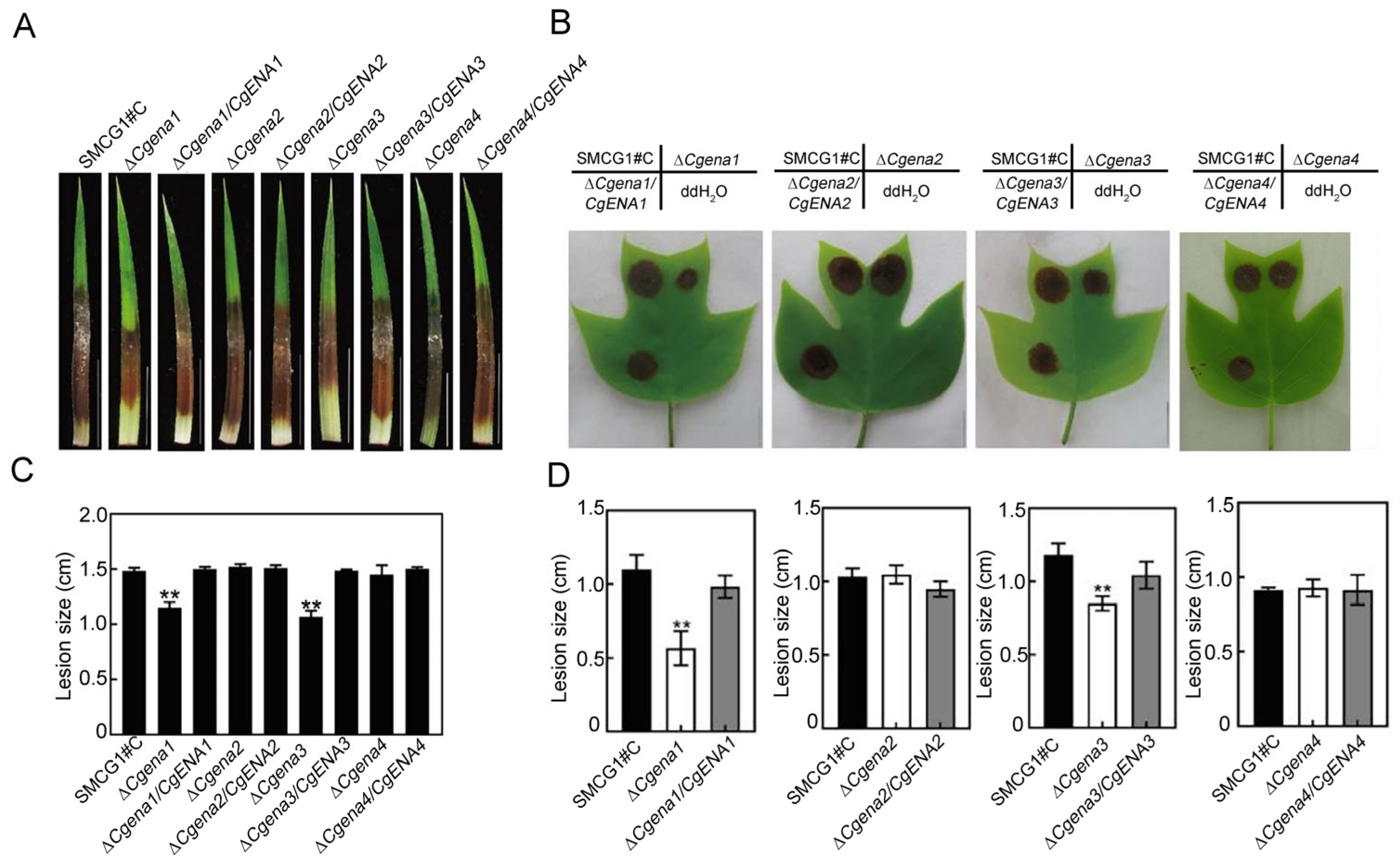
2.4. CgEna1 and CgEna3 Indispensable for Full Virulence

The pathogenicity of the wild type, ΔCgena1, ΔCgena2, ΔCgena3, ΔCgena4, and corresponding complemented strains were measured on healthy leaves of C. lanceolata and L. chinense × tulipifera, respectively. At 5 or 7 days post inoculation(dpi), there was no significant difference among the diameter of lesions caused by ΔCgena2, ΔCgena4, and the wild type. However, the lesions caused by ΔCgena1 and ΔCgena3 mutants were significantly smaller than those of other tested strains (Figure 4A,C). Similarly, the ΔCgena1 and ΔCgena3 mutants were also less virulent on L. chinense × tulipifera (Figure 4B,D). These results suggest that CgEna1 and CgEna3 are required for the full virulence of C. gloeosporioides.

2.5. CgEna1 and CgEna3 Regulate Conidial Germination, Appressorium Formation and Invasive Hypha Development

Conidial germination of the wild type, ΔCgena1, ΔCgena2, ΔCgena3, ΔCgena4, and corresponding complemented strains were measured at 2 and 4 hpi. These data showed that there was no significant difference among the wild type, ΔCgena2, and ΔCgena4. However, compared with the wild type, the conidial germination ratio of ΔCgena1 was significantly decreased at 2 hpi (Figure 5A). In the mutant of ΔCgena3, the conidial germination ratio was significantly lower than that of the wild type (Figure 5A). These data indicate that the deletion of the CgENA1 and CgENA3 significantly delayed conidial germination at the early stage. Similarly, the appressorium formation ratio of the mutants of ΔCgena1 and ΔCgena3 was also decreased significantly, while no changes happened to ΔCgena2 and ΔCgena4 mutants (Figure 5B).
The invasive hyphae (IH), developed on the onion epidermis, was further observed 24 h after inoculation. The data showed that the most IH of the wild type belonged to type III and IV. Compared to wild type strain SMCG1#C, the mutants of ΔCgena1 and ΔCgena3 produced more IH of type I and II and less IH of type III and IV proportionally (Figure 5C). Collectively, all results indicated that CgEna1 and CgEna3 are indispensable for full virulence by involving in the development of the invasive structure in C. gloeosporioides.

2.6. CgEna4 Is Required for Ion Stress Response under Certain Extracellular Conditions

It has been reported that the expression of ENA genes was induced by alkaline conditions in the yeast [10]. In this study, it was found that the deletion of CgENA1, CgENA2, CgENA3, andCgENA4, respectively, had no significant effects on the vegetative growth on the CM (complete medium) plates. Under the sole stress, like NaCl, KCl, or non-acidic conditions, the growth of ΔCgena4 wasn’t inhibited (Figure 6A,B,D,E). However, when exposed to 0.7 M NaCl under the pH 7 or 10 conditions, the vegetative growth of the ΔCgena4 mutant was significantly depressed compared to the wild type (Figure 6C,F). Interestingly, under the condition of same pH, the growth inhibition in the ΔCgena4 mutant wasn’t observed when the NaCl was replaced by KCl (Figure 6C,F). These results indicated that CgEna4 plays an important role in the growth of C. gloeosporioides under the non-acidic condition combined with sodium ion stress.

3. Materials and Methods

3.1. Fungal Strains and Culture Conditions

The C. gloeosporioides strain SMCG1#C [20], and mutant strains of ΔCgena1, ΔCgena2, ΔCgena3, ΔCgena4, and the corresponding complemented strains were cultured on CM medium at 25 °C [24]. The fungal mycelia were cultured in a liquid CM medium for DNA extraction, protein extraction and protoplasts preparation, as described previously [22].

3.2. Gene Deletion of CgENA1, CgENA2, CgENA3, and CgENA4

The complete gene sequences of Ena ATPases, CgENA1, CgENA2, CgENA3 and CgENA4 of C. gloeosporioides were obtained from the genome database of GenBank (https://www.ncbi.nlm.nih.gov/). The upstream and downstream fragments of each gene were amplified from the wild type gDNA, and ligated to two ends of the hygromycin phosphotransferase (HPH) gene, respectively, using the overlapping PCR method [21]. The fusion fragments were amplified and transformed into the protoplasts of the wild type as described previously [25,26]. The putative gene deletion transformants were confirmed by PCR assays.

3.3. Complementation of Targeted Gene Mutants

To obtain the complemented strains of the genes mutants, the fragments containing the entire ORF (open reading frame) of each targeted gene and its native promoter were amplified and inserted into a pYF11 vector and fused to the GFP reporter gene [27]. The resulting construct of each gene was verified by sequencing and reformatting it into the corresponding gene mutant. The candidate complementation transformants were verified by detecting the GFP signals, as described previously [28].

3.4. RNA Isolation and Quantitative Real-Time PCR (qRT-PCR)

To determine the relative abundance of CgENA1, CgENA2, CgENA3, and CgENA4 at the early infection stage, the onion epidermis, inoculated with conidia of the WT, was harvested at 2, 4, 6, 8, 12, 16, 24 and 32 hpi for RNA extraction. Total RNA was extracted through a RNA Extraction kit according to the manufacturer’s instructions (Conway Reagent, Nanjing, China). The first strand of cDNA was synthesized through an RNA reverse transcription reagent (Novozan Biotechnology Co., Nanjing, China). RT-PCRs were carried out according to the method described by [29], and the house-keeping gene ACTIN was used as the internal control. The relative expression level of each gene was calculated through the 2−ΔΔCT method as described in [30]. The experiment was carried out three times in each treatment. All used primers are listed in Supplementary Table S1.

3.5. Assays for Vegetative Growth

Mycelial plugs (2 mm × 2 mm in size) of the wild type, mutants of ΔCgena1, ΔCgena2, ΔCgena3, ΔCgena4, and the corresponding complemented strains were inoculated on PDA (Potato dextrose medium), OMA (Oatmeal agar), MM (Minimal medium) and Mathur’s medium plates, respectively, and cultured at 25 °C in the dark for 5 days. The diameter of fungal colonies was measured as described previously [24]. Three independent biological experiments were performed.

3.6. Assays of Stress Resistance and Sodium/Potassium Accumulation

For mycelial sodium and potassium content assays, the wild type, mutants of ΔCgena1, ΔCgena2, ΔCgena3, ΔCgena4, and their corresponding complemented strains, were cultured in a liquid CM medium for 2 days as described previously [21,29]. The mycelial pellets were collected through a miracloth (EMD Millipore, Bellerica, MA, USA) to remove the filtrate. Fungal mycelia were harvested and dried in a freeze dryer. The dried mycelia were digested with sulfuric acid. The potassium and sodium content was then detected by a flame spectrophotometer, respectively, as described previously [21]. Three independent biological experiments were performed.
For stress resistance analysis, the wild type, mutants of ΔCgena1, ΔCgena2, ΔCgena3, ΔCgena4, and their corresponding complemented strains were inoculated on CM plates supplemented with 0.7 M NaCl and 0.7 M KCl, respectively, and cultured at 25 °C in the dark. At 5 dpi, the diameter of fungal colonies was measured [6]. Three independent biological experiments were performed.

3.7. Pathogenicity Tests

For pathogenicity tests, the conidium of the wild type, mutants of ΔCgena1, ΔCgena2, ΔCgena3, ΔCgena4, and their corresponding complemented strains were collected, and the concentration of the conidial suspensions of each strain was adjusted to 1 × 10⁵ spores/ml [25]. A total of ten µL of conidial suspensions were inoculated on the healthy leaves of Liriodendron chinense × tulipifera. An amount of five µl of the conidial suspension of each strain was inoculated on the healthy leaves of C. lanceolata. The inoculated leaves were kept in a moist chamber at 25 °C. The size of the lesions was measured and photographed at 7 dpi or 5 dpi, respectively, as described previously [25].

3.8. Observation of Conidial Germination, Appressorium Formation and Invasive Hyphal Development

Conidium of the wild type, mutants of ΔCgena1, ΔCgena2, ΔCgena3, ΔCgena4 and their corresponding complemented strains were prepared by the method described above. The concentration of conidium was adjusted to 2 × 10⁴ conidia/mL. A total of ten µl of conidial suspensions of each strain were dropped on hydrophobic glass slides (Fisher brand) and placed in an incubator at 25 °C [24]. Conidial germination and appressorium formation was observed under a compound microscope (Carl Zeiss, Jena, Germany) at 2, 4, 8, and 12 h hpi. The experiment was conducted three times, with at least 120 conidium counted per strain.
Invasive hyphae (IH) were induced on the onion epidermal strip according to the method as described previously [25]. IH were divided into four different types: type I, no IH development, type II, IH with one branch, type III, IH with at least two branches, but limited expansion and type IV, IH with numerous branching and extensive hyphal growth. The experiment was performed three times, and at least 50 invasive structures were observed in each treatment.

3.9. Statistical Analysis

The mean standard deviation (SD) was calculated for each treatment. The statistical analysis of significant among difference among treatments in this study was performed with a one-way analysis of variance (ANOVA) followed by least significant differences multiple range tests. Statistical analyses were carried out by the SPSS 19.0 software program (SPSS Inc., Chicago, IL, USA).

4. Discussion

In many fungal genomes, there are two or more genetically divergent ENA genes [3,30]. In this study, four Ena ATPase homologs: CgEna1, CgEna2, CgEna3, and CgEna4 were identified in C. gloeosporioides by blastp searches through S. cerevisiae Ena ATPase sequences against C. gloeosporioides. These four Ena proteins shared high sequence identity with corresponding proteins found in other fungi, particularly within the ATPase domain. It was found that these Ena proteins play critical roles at the early stage of plant infection, while they are not involved in vegetative growth. Among them, CgEna2 was located in the endoplasmic reticulum and was not involved in the regulation of pathogenicity. CgEna1 and CgEna3 were required for the full virulence of C. gloeosporioides. CgEna3 was also involved in the response to extracellular Na+ and K+ stress. CgEna1 and CgEna4 regulated the sodium accumulation. Interestingly, CgEna4 played a critical role in the growth of C. gloeosporioides under Na+ stress and non-acidic conditions.
The main function of Ena ATPases in fungi has been proven to pump cation [31]. ScEna1, a dual functional protein, is responsible for the extracellular transport of sodium and also accumulation transport of potassium into the cell [15]. Studies of the roles of Ena2 in Debaryomyces occidentals showed that the mutation of Ena2 led to the reduction of intracellular potassium accumulation. In Neurospora crassa, Ena1 is only involves in efflux Na+, while others Ena ATPases regulate the efflux of Na+ and K+ [3]. In contrast, the deletion of ENA genes was found to have no significant effect on potassium among the wild type and all mutants, and higher intracellular sodium content was tested in the mutants of ΔCgena1 and ΔCgena4, indicating that CgEna1 and CgEna4 might be involved in the regulation of sodium in C. gloeosporioides. This finding further enhances the understanding of the role of Ena proteins in sodium efflux.
The different localization pattern of Ena proteins may determine the functional differentiation [15,30]. In Aspergillus nidulans, EnaB located in the endoplasmic reticulum that greatly improved its tolerance to highly toxic lithium [32]. In this study, CgEna2 and CgEna3 were found to be both located in the endoplasmic reticulum, but only ΔCgena3 showed hypersensitivity to Na+ and K+ stresses, indicating that CgEna3 was involved in extracellular ion stress response. A similar function of EnaB was observed in A. nidulans [32]. In S. cerevisiae, Ena1 was located in the plasma membrane and seems to be a sodium pump. [30]. In agreement with that, CgEna1 and CgEna4 was observed to locate on the plasma membrane in hypha and protoplasts of C. gloeosporioide. ΔCgena1 and ΔCgena4 significantly increased the concentration of Na+ in C. gloeosporioides. CgEna1 and CgEna4 may regulate the accumulation of sodium.
In the interaction between fungi and their hosts, the fungi have the ability to grow in a complex pH environment. In most cases, Ena ATPases are not required in acidic environments, as neutral Na+/H+ and K+/H+ antiporters can replace Ena ATPase [14,32,33]. However, when the extracellular pH increased, the ScENA genes were strongly in up-regulation, which causes the loss of the function of the anti-transporter ScNha1, resulting in the inability to correctly excrete sodium and potassium [34,35]. In general, the excessive increase of the intracellular concentration of sodium is harmful for an organism [36]. Under usual growth conditions, the Ena proteins are the main determinant of sodium detoxification tolerance to alkaline ambient pH in A. nidulans, [32]. Large amounts of sodium accumulation in the cytosol tend to destroy essential and sensitive enzymes [37]. In this study, the lack of CgENA4 may affect the maintenance of the redox homeostasis of the transport of sodium in C. gloeosporioides. The deletion of CgENA4 significantly inhibited the mycelial growth under combined conditions of 0.7 M NaCl and pH 7, or pH 10, indicating that non-acidic pH may alter the ion resistance mediated by CgEna4 in C. gloeosporioides.
In addition, four Ena proteins were investigated for their potential roles in the pathogenicity of C. gloeosporioides. Inoculation assays manifested on mutants of ΔCgena1 and ΔCgena3, relative to the wild-type SMCG1#C, developed smaller disease lesion on the leaves of C. lanceolata and L. chinense × tulipifera leaves. The invasive structures of appressorium and invasive hyphae (IH) were also found to be delayed in mutants of ΔCgena1 and ΔCgena3. Therefore, it is suggested that CgEna1 and CgEna3 are required for the full virulence of the regulated invasive structure that was developed.
In summary, four Ena proteins were identified in C. gloeosporioides, and investigated their potential roles in ion transport, appressorium formation, invasive hyphae development and pathogenicity. These findings help reveal the potential mechanisms of ion transport and the roles of Ena proteins in plant infection in C. gloeosporioides.

Supplementary Materials

The following supporting information can be downloaded at: https://www.mdpi.com/2309-608X/9/5/566/s1, Figure S1: Protein structure of the CgEna1/2/3/4 with its homologs in other fungi; Figure S2: The expression of CgEna1/2/3/4 in mycelium and different stages of infection; Figure S3: Acquisition and verification of mutants; Figure S4: CgEna family proteins are not involved in the regulation of vegetative growth; Table S1: Primer sequence of CgEna1/2/3/4.

Author Contributions

L.H. and J.-Y.Y. designed the research; T.-C.D., M.-L.S., Y.-Z.Z. and Y.-T.P. performed the experiments; L.H. contributed to the new reagents/analytical tools; T.-C.D., Y.-Z.Z. and Y.-T.P. analysed the data; and T.-C.D. and J.-Y.Y. wrote the manuscript. All authors have read and agreed to the published version of the manuscript.

Funding

This study was supported by the National Science Foundation of China (31870631), the National Key R & D Program of China (2017YFD0600102), the Qing Lan Project, and the Priority Academic Program Development of Jiangsu Higher Education Institutions (PAPD).

Data Availability Statement

The data presented in this study are available on request from the corresponding author.

Conflicts of Interest

The authors declare that they have no conflict of interest.

References

  1. Clausen, M.J.; Poulsen, H. Sodium/Potassium homeostasis in the cell. Met. Ions Life Sci. 2013, 12, 41–67. [Google Scholar] [PubMed]
  2. Adler, V.; Yin, Z.; Fuchs, S.Y.; Benezra, M.; Rosario, L.; Tew, K.D.; Pincus, M.R.; Sardana, M.; Henderson, C.J.; Wolf, C.R.; et al. Regulation of JNK signaling by GSTp. EMBO J. 1999, 18, 1321–1334. [Google Scholar] [CrossRef]
  3. Rodríguez-Navarro, A.; Benito, B. Sodium or potassium efflux ATPase a fungal, bryophyte, and protozoal ATPase. Biochim. Biophys. Acta 2010, 1798, 1841–1853. [Google Scholar] [CrossRef] [PubMed] [Green Version]
  4. Catty, P.; de Kerchove d Exaerde, A.; Goffeau, A. The complete inventory of the yeast Saccharomyces cerevisiae P-type transport ATPases. FEBS Lett. 1997, 409, 325–332. [Google Scholar] [CrossRef] [PubMed] [Green Version]
  5. Riedelsberger, J.; Obando, P.A.; Gonzalez, W. Yeast strain Saccharomyces cerevisiae BYT45 lacking the cation extrusion systems ENA1-5 and NHA1 is suitable for the characterization of TASK-3 potassium channel antagonists. FEMS Yeast Res. 2019, 19, 44–52. [Google Scholar] [CrossRef]
  6. Zhang, M.; Fang, Y.; Liang, Z.; Huang, L. Enhanced expression of vacuolar H+-ATPase subunit E in the roots is associated with the adaptation of Broussonetia papyrifera to salt stress. PLoS ONE 2012, 7, 481–483. [Google Scholar] [CrossRef] [Green Version]
  7. Eaton, A.F.; Merkulova, M.; Brown, D. The H+-ATPase (V-ATPase): From proton pump to signaling complex in health and disease. Am. J. Physiol. Cell Physiol. 2021, 320, 392–414. [Google Scholar] [CrossRef]
  8. Benito, B.; Garciadeblas, B.; Rodrıguez-Navarro, A. Potassium- or sodium-efflux ATPase, a key enzyme in the evolution of fungi. Microbiology 2002, 148, 933–941. [Google Scholar] [CrossRef] [Green Version]
  9. Ma, Q.; Jin, K.; Peng, G.; Xia, Y. An Ena ATPase, MaEna1, of metarhizium acridum influences the Na+, thermo and UV tolerances of conidia and is involved in multiple mechanisms of stress tolerance. Fungal Genet. Biol. 2015, 83, 68–77. [Google Scholar] [CrossRef]
  10. Ruiz, A.; Ariño, J. Function and regulation of the Saccharomyces cerevisiae Ena sodium ATPase system. Eukaryot. Cell 2007, 6, 2175–2183. [Google Scholar] [CrossRef] [PubMed] [Green Version]
  11. Rudolph, H.K.; Antebi, A.; Fink, G.R.; Buckley, C.M.; Moir, D.T. The yeast secretory pathway is perturbed by mutations in PMR1, a member of a Ca2+ ATPase family. Cell 1989, 58, 133–145. [Google Scholar] [CrossRef] [PubMed]
  12. Fraile-Escanciano, A.; Garciadeblás, B.; Rodríguez-Navarro, A.; Benito, B. Role of Ena ATPase in Na+ efflux at high pH in bryophytes. Plant. Mol. Biol. 2009, 71, 599–608. [Google Scholar] [CrossRef] [PubMed]
  13. Haro, R.; Garciadeblas, B.; Rodríguez-Navarro, A. A novel P-type ATPase from yeast involved in sodium transport. FEBS Lett. 1991, 291, 189–191. [Google Scholar] [CrossRef] [PubMed] [Green Version]
  14. Garciadeblas, B.; Rubio, F.; Quintero, F.J.; Bañuelos, M.A.; Haro, R.; Rodríguez-Navarro, A. Differential expression of two genes encoding isoforms of the ATPase involved in sodium efflux in Saccharomyces cerevisiae. Mol. Gen. Genet. 1993, 236, 363–368. [Google Scholar] [CrossRef]
  15. Banuelos, M.A.; Rodriguez-Navarro, A. P-type ATPases Mediate Sodium and Potassium Effluxes in Schwanniomyces occidentalis. J. Biol. Chem. 1998, 273, 1640–1646. [Google Scholar] [CrossRef] [PubMed] [Green Version]
  16. Enjalbert, B.; Moran, G.P.; Vaughan, C.; Yeomans, T.; Maccallum, D.M.; Quinn, J.; Coleman, D.C.; Brown, A.J.P.; Sullivan, D.J. Genome-wide gene expression profiling and a forward genetic screen show that differential expression of the sodium ion transporter Ena21 contributes to the differential tolerance of Candida albicans and Candida dubliniensis to osmotic stress. Mol. Microbiol. 2009, 72, 216–228. [Google Scholar] [CrossRef]
  17. Idnurm, A.; Walton, F.J.; Floyd, A.; Reedy, J.L.; Heitman, J. Identification of ENA1 as a virulence gene of the human pathogenic fungus Cryptococcus neoformans through signature-tagged insertional mutagenesis. Eukaryot. Cell 2009, 8, 315–326. [Google Scholar] [CrossRef] [PubMed] [Green Version]
  18. Rinyu, E.; Varga, J.; Ferenczy, L. Phenotypic and genotypic analysis of variability in Aspergillus fumigatus. J. Clin. Microbiol. 1995, 33, 2567–2575. [Google Scholar] [CrossRef] [Green Version]
  19. Lee, H.B.; Park, J.Y.; Jung, H.S. First report of leaf anthracnose caused by Colletotrichum boninense on spindle trees. Plant Pathol. 2010, 54, 254. [Google Scholar] [CrossRef]
  20. Huang, L.; Kim, K.T.; Yang, J.Y.; Song, H.; Choi, G.; Jeon, J.; Cheong, K.; Ko, J.; Xu, H.; Lee, Y.H. A High-Quality draft genome sequence of Colletotrichum gloeosporioides sensu stricto SMCG1#C, a causal agent of anthracnose on Cunninghamia lanceolata in China. Mol. Plant Microbe Interact. 2019, 32, 139–141. [Google Scholar] [PubMed] [Green Version]
  21. Yang, J.Y.; Fang, Y.L.; Wang, P.; Ye, J.R.; Huang, L. Pleiotropic Roles of ChSat4 in Asexual Development, Cell Wall Integrity Maintenance, and Pathogenicity in Colletotrichum higginsianum. Front. Microbiol. 2018, 9, 2311. [Google Scholar] [CrossRef] [PubMed]
  22. Zhang, Y.Z.; Li, B.; Pan, Y.T.; Fang, Y.L.; Li, D.W.; Huang, L. Protein phosphatase CgPpz1 regulates potassium uptake, stress responses, and plant infection in Colletotrichum gloeosporioides. Phytopathology 2022, 112, 820–829. [Google Scholar] [CrossRef] [PubMed]
  23. Yi, M.; Chi, M.H.; Khang, C.H.; Park, S.Y.; Kang, S.; Valent, B.; Lee, Y.H. The ER chaperone Lhs1 is involved in asexual development and rice infection by the blast fungus Magnaporthe oryzae. Plant Cell 2009, 21, 681–695. [Google Scholar] [CrossRef] [PubMed] [Green Version]
  24. Fang, Y.L.; Xia, L.M.; Wang, P.; Zhu, L.H.; Ye, J.R.; Huang, L. The MAPKKK CgMck1 is required for cell wall integrity, appressorium development, and pathogenicity in Colletotrichum gloeosporioides. Genes 2018, 9, 543–552. [Google Scholar] [CrossRef] [PubMed] [Green Version]
  25. Wang, P.; Li, B.; Pan, Y.T.; Zhang, Y.Z.; Li, D.W.; Huang, L. Calcineurin-responsive transcription factor CgCrzA is required for cell wall integrity and infection-related morphogenesis in Colletotrichum gloeosporioides. Plant Pathol. J. 2020, 36, 385–394. [Google Scholar] [CrossRef]
  26. Li, B.; Liu, L.; Li, Y.; Dong, X.; Zhang, H.; Chen, H.; Zheng, X.; Zhang, Z. The FgVps39-FgVam7-FgSso1 complex mediates vesicle trafficking and is important for the development and virulence of Fusarium graminearum. Plant Microbe Interact. 2017, 30, 410–422. [Google Scholar] [CrossRef] [PubMed] [Green Version]
  27. Liu, W.; Xu, J.R.; Xu, Q.; Ding, S.; Wang, Y.; Wang, C.; Zhou, X. A MADS-box transcription factor MoMcm1 is required for male fertility, microconidium production and virulence in Magnaporthe oryzae. Mol. Microbiol. 2011, 80, 33–53. [Google Scholar]
  28. Yue, C.; Su, Z.; Yi, S.; Li, M.; Zhang, Z. MoTup1 is required for growth, conidiogenesis and pathogenicity of Magnaporthe oryzae. Mol. Plant Pathol. 2015, 16, 799–810. [Google Scholar]
  29. Pan, Y.T.; Li, L.; Yang, J.Y.; Li, B.; Zhang, Y.Z.; Wang, P.; Huang, L. Involvement of protein kinase CgSat4 in potassium uptake, cation tolerance, and full virulence in Colletotrichum gloeosporioides. Front. Plant Sci. 2022, 13, 773–898. [Google Scholar] [CrossRef]
  30. Benito, B.; Garciadeblás, B.; Pérez-Martín, J.; Rodríguez-Navarro, A. Growth at high pH and sodium and potassium tolerance in media above the cytoplasmic pH depend on Ena ATPases in Ustilago maydis. Eukaryot. Cell 2009, 8, 821–829. [Google Scholar] [CrossRef] [Green Version]
  31. Lanza, M.; Haro, R.; Conchillo, L.B.; Benito, B. The endophyte Serendipita indica reduces the sodium content of Arabidopsis plants exposed to salt stress: Fungal Ena ATPases are expressed and regulated at high pH and during plant co-cultivation in salinity. Environ. Microbiol. 2019, 21, 3364–3378. [Google Scholar] [CrossRef] [PubMed]
  32. Markina-Iñarrairaegui, A.; Spielvogel, A.; Etxebeste, O.; Ugalde, U.; Espeso, E.A. Tolerance to alkaline ambient pH in Aspergillus nidulans depends on the activity of Ena proteins. Sci. Rep. 2020, 10, 1–16. [Google Scholar] [CrossRef]
  33. Brett, C.L.; Donowitz, M.; Rao, R. Evolutionary origins of eukaryotic sodium/proton exchangers. Am. J. Physiol. Cell Physiol. 2005, 288, 223–239. [Google Scholar] [CrossRef] [PubMed] [Green Version]
  34. Serrano, R.; Ruiz, A.; Bernal, D.; Chambers, J.R.; Ariño, J. The transcriptional response to alkaline pH in Saccharomyces cerevisiae: Evidence for calcium-mediated signalling. Mol. Microbiol. 2002, 46, 1319–1333. [Google Scholar] [CrossRef]
  35. Dibalova-Culakova, H.; Alonso-Del-Real, J.; Querol, A.; Sychrova, H. Expression of heterologous transporters in Saccharomyces kudriavzevii: A strategy for improving yeast salt tolerance and fermentation performance. Int. J. Food Microbiol. 2018, 268, 27–34. [Google Scholar] [CrossRef]
  36. Spielvogel, A.; Findon, H.; Arst, H.N.; Araújo-Bazán, L.; Hernández-Ortíz, P.; Stahl, U.; Meyer, V.; Espeso, E.A. Two zinc finger transcription factors, CrzA and SltA, are involved in cation homoeostasis and detoxification in Aspergillus nidulans. Biochem. J. 2008, 414, 419–429. [Google Scholar] [CrossRef] [PubMed] [Green Version]
  37. Hirasaki, M.; Horiguchi, M.; Numamoto, M.; Sugiyama, M.; Kaneko, Y.; Nogi, Y.; Harashima, S. Saccharomyces cerevisiae protein phosphatase Ppz1 and protein kinases Sat4 and Hal5 are involved in the control of subcellular localization of Gln3 by likely regulating its phosphorylation state. J. Biosci. Bioeng. 2011, 111, 249–254. [Google Scholar] [CrossRef]
Figure 1. Localization pattern of Ena family proteins C. gloeosporioides. (A) Subcellular localization of CgEna1, CgEna2, CgEna3, CgEna4 in the hypha and protoplast of C. gloeosporioides. The fused constructs of CgEna1-GFP, CgEna2-GFP, CgEna3-GFP, CgEna4-GFP were transformed into the corresponding gene deletion mutants. The subcellular localization of CgEna1, CgEna2, CgEna3, and CgEna4 was observed in the hypha and protoplast by using a microscope (Zeiss, Oberkochen, Germany). (B) Proteins of CgEna2-GFP, CgEna3-GFP, and the endoplasmic reticulum maker protein of Lhs1-RFP were co-expressed in the conidia of C. gloeosporioides. Scale bars = 10 μm.
Jof 09 00566 g001
Figure 2. CgEna1 and CgEna4 are involved in sodium accumulation. The concentration of potassium (A) and sodium (B) in the mycelia of the wild type, ΔCgena1, ΔCgena2, ΔCgena3, and ΔCgena4. Error bars represent the SD (standard deviation) and “**” indicate a significant difference at p < 0.01. Three independent experiments were performed and similar results were obtained.
Jof 09 00566 g002
Figure 3. CgEna3 is involved in ion stress resistance. (A) Colonies of the wild type, ΔCgena1, ΔCgena2, ΔCgena3, ΔCgena4, and their corresponding complemented strains, cultured on CM medium supplemented with 0.7 M NaCl and KCl. (B) The growth inhibition rate of the tested strains exposed to stress conditions indicated in (A). Error bars represent the SD. “**” indicate significant differences at p < 0.01. Three independent experiments were performed, and similar results obtained.
Jof 09 00566 g003
Figure 4. CgEna1 and CgEna3 are required for full virulence on host plants. Lesions developed on leaves of C. lanceolata (A) and L. chinensis × tulipifera (B) after inoculation by the wild type, ΔCgena1, ΔCgena2, ΔCgena3, ΔCgena4, and their corresponding complemented strains. Pictures were photographed at 5 dpi or 7 dpi. ddH2O was used as the negative control. (D) Average diameter of lesions developed on leaves of C. lanceolata (C) and L. chinensis × tulipifera. Error bars represent the SD. “**” indicate significant differences at p < 0.01. Three independent biological experiments were performed and yielded similar results.
Jof 09 00566 g004
Figure 5. CgEna1 and CgEna3 regulate conidial germination, appressorium formation, and invasive hyphal development. (A) Conidial germination ratio of the wild type, ΔCgena1, ΔCgena2, ΔCgena3 and ΔCgena4 at 2 and 4 hpi. (B) Appressoria formation ratio of the wild type, ΔCgena1, ΔCgena2, ΔCgena3 and ΔCgena4 at 4, 8 and 12 hpi. (C) Proportion of different types of invasive hypha (IH) developed by the wild type, ΔCgena1, ΔCgena2, ΔCgena3 and ΔCgena4. Error bars represent the SD. “**” indicate significant difference at p < 0.01. Three independent biological experiments were performed and yielded similar results.
Jof 09 00566 g005
Figure 6. Roles of CgEna4 on sodium stress response under non-acidic condition. (A) Colonies of the wild type, ΔCgena4, and complementary strain ΔCgena4/CgENA4 inoculated on CM medium supplemented with 0.7 M NaCl or 0.7 M KCl under normal pH. (B) Colonies of the wild type, ΔCgena4, and complementary strain ΔCgena4/CgENA4 inoculated on the CM medium under different pH (7 or 10). (C) Colonies of the wild type, ΔCgena4, and complementary strain ΔCgena4/CgENA4 inoculated on CM medium under different combination conditions of pH and sodium stress. (DF) The inhibition ratio of vegetative growth of the above tested strains indicated in (AC), respectively. Error bars represent the SD. “**” indicate significant difference at p < 0.01. Three independent biological experiments were performed.
Jof 09 00566 g006
Disclaimer/Publisher’s Note: The statements, opinions and data contained in all publications are solely those of the individual author(s) and contributor(s) and not of MDPI and/or the editor(s). MDPI and/or the editor(s) disclaim responsibility for any injury to people or property resulting from any ideas, methods, instructions or products referred to in the content.

