Review

Fungal Aeroallergens—The Impact of Climate Change

by
Monika Sztandera-Tymoczek
and
Agnieszka Szuster-Ciesielska
*
Department of Virology and Immunology, Institute of Biological Sciences, Maria Curie-Skłodowska University, Akademicka 19, 20-033 Lublin, Poland
*
Author to whom correspondence should be addressed.
J. Fungi 2023, 9(5), 544; https://doi.org/10.3390/jof9050544
Submission received: 14 April 2023 / Revised: 2 May 2023 / Accepted: 5 May 2023 / Published: 7 May 2023

Abstract

:
The incidence of allergic diseases worldwide is rapidly increasing, making allergies a modern pandemic. This article intends to review published reports addressing the role of fungi as causative agents in the development of various overreactivity-related diseases, mainly affecting the respiratory tract. After presenting the basic information on the mechanisms of allergic reactions, we describe the impact of fungal allergens on the development of the allergic diseases. Human activity and climate change have an impact on the spread of fungi and their plant hosts. Particular attention should be paid to microfungi, i.e., plant parasites that may be an underestimated source of new allergens.

1. Introduction

Allergies are a group of complex diseases characterized by an inappropriate or overreactive immune response of the organism to common environmental substances named allergens. Most allergens, e.g., house dust mites, pollen, fungal or mold spores, food (especially milk, eggs, fruit, wheat, soybeans, seafood, and nuts), insect venoms (e.g., wasps and bees), some medications, latex, and household chemicals, are harmless to humans. Upon contact with these agents, a hypersensitivity reaction develops and antibodies are produced in predisposed subjects [1,2]. Depending on the cause, i.e., the type of contact with the allergen, the following conditions are distinguished: (1) respiratory system allergies, i.e., allergic rhinitis with conjunctivitis and asthma with wheezing, coughing, and shortness of breath; (2) contact (skin) allergies, among which the most common are atopic and contact dermatitis, manifested mainly in rashes; (3) food allergies, and (4) insect venom allergies, which cause a wide range of symptoms and can be life-threatening in some cases (anaphylaxis) [3,4,5].

2. Allergy Epidemiology

The incidence of allergic diseases in the world is rapidly increasing, and according to the estimates of the World Allergy Organization (WAO), it ranges from 10–40%, depending on the country. In most developed countries, allergy affects over 20% of the population [6]. While allergy was regarded as a rare disease in the early 20th century, the last few decades have witnessed a dramatic increase in its prevalence. Currently, more than 150 million Europeans suffer from chronic allergic diseases, and 20% struggle with severe and debilitating forms. By 2025, half the European Union population is estimated to suffer from allergies. The prevalence of allergic diseases is increasing together with the progress in urbanization, industrialization, pollution, and climate change, factors that are not likely to change quickly. Moreover, these data are considered to be underestimations, because many patients do not report their symptoms or are misdiagnosed, with potentially 45% of patients never receiving an allergy diagnosis [7].
Respiratory allergies are the most common allergy type in Europe and worldwide [8,9]. Data in the White Book of Allergy published in 2013 by the WAO confirm that the prevalence of allergic rhinitis and asthma is increasing worldwide [10]. Allergic rhinitis with conjunctivitis is the most common non-infectious rhinitis, affecting approximately 400 million people worldwide [11,12]. Asthma is one of the most common chronic diseases, affecting about 339 million people worldwide, and its prevalence is increasing, especially among children [13,14]. The most common asthma phenotype is allergic asthma. It is estimated that up to 89% of childhood asthma and more than 50% of adult asthma cases may have an allergic component [15]. According to the European Federation of Allergy and Airways Diseases Patients’ Associations (EFA), allergic rhinitis affects 4–40% of Europe’s population [16]. In comparison, the prevalence of asthma in the European Union is 8.2% in adults and 9.4% in children [17]. In 2019, Portugal and Sweden were the EU countries with the highest prevalence of asthma, with approximately 9.9% and 8.2% of their populations suffering from the disease, respectively [18].

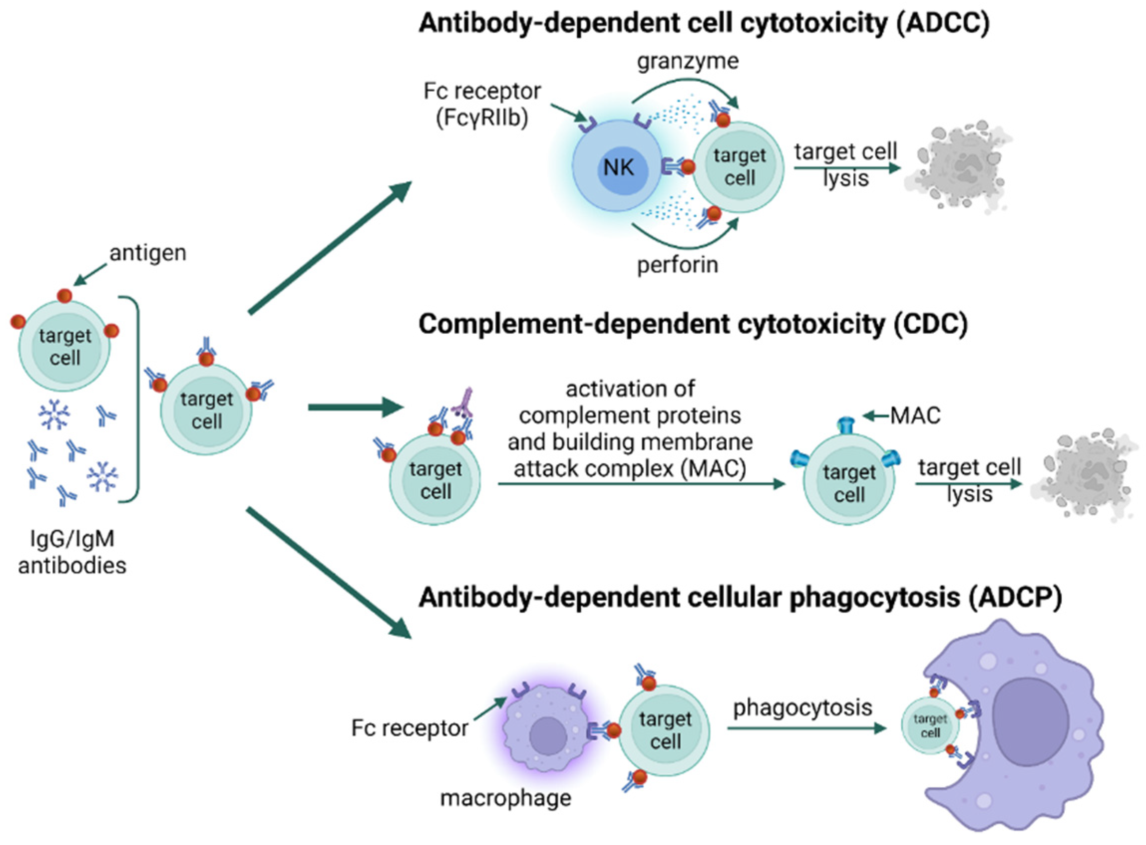
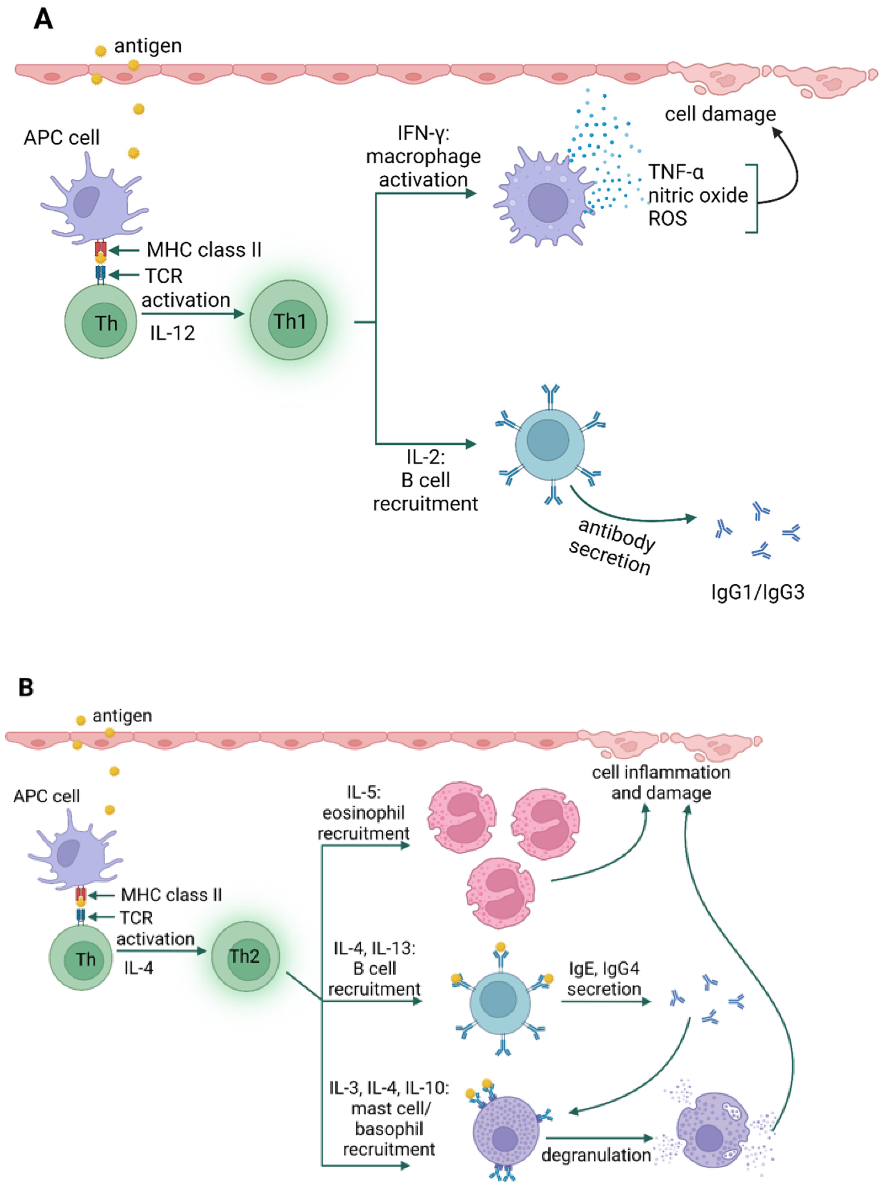
3. Types of Hypersensitivity Reactions

One of the functions of the human immune system is to provide protection against such microorganisms as bacteria, viruses, and fungi. Exaggerated immune responses and hypersensitivity reactions may lead to disease development. In 1963, Gell and Combs, who investigated the effector mechanism responsible for cell and tissue damage and the type of immune response, created the basis for the classification of hypersensitivity reactions, dividing them into four distinct types: type I (immediate or IgE-dependent), type II (cytotoxic or dependent on IgG/IgM), type III (immune complexes), and type IV (delayed or dependent on T lymphocytes) [19]. However, in clinical practice, the categories of hypersensitivity may overlap, because patients exhibit simultaneous coexistence of symptoms characteristic of specific hypersensitivity reactions [20,21].
In type I hypersensitivity, type 2 helper T cells (Th2) and their mediators initiate a change in the isotype of B cells, producing allergen-specific IgE. IgE antibodies bind to receptors (FcεRI) present on the surface of mast cells and basophils [21]. Upon re-exposure, the allergen is bound by IgE, resulting in an immediate type I hypersensitivity reaction in two phases. The early phase occurs within minutes of allergen exposure and is triggered by histamine, proteases, lysosomal enzymes, and other mediators released after the degranulation of mast cells and basophils. In addition, mast cells produce lipid mediators, including prostaglandin D2 and leukotriene D4, from arachidonic acid, and release them into the circulation. The late phase begins 4 to 8 h after allergen exposure and is mediated by cytokines IL-1, IL-4, IL-5, IL-13, tumor necrosis factor (TNF-α), and granulocyte-monocyte colony-stimulating factor (GM-CSF) [21,22,23] (Figure 1). Immediate hypersensitivity reactions include anaphylaxis, bronchial asthma, and urticaria [21,24].
Unfortunately, antibodies can sometimes bind to self-antigens, directing a cytotoxic response against the host. This is the basis of type II hypersensitivity, a cytotoxic reaction characterized by IgG/IgM antibodies recognizing self-antigens usually found on circulating blood cells (erythrocytes, neutrophils, platelets), epithelial cells of glands and mucous membranes, or basement membranes [21]. Type II reactions are divided into two subtypes: IIa and IIb. Type IIa refers to reactions characterized by cytolytic destruction of target cells. IgG/IgM binding on the cell surface triggers cytotoxicity via three mechanisms [25]. In the first one (antibody-dependent cell cytotoxicity, ADCC), IgG binds to the Fc gamma IIb receptor (FcγRIIb) on NK cells and macrophages, which leads to their degranulation and release of perforin and granzyme directly destroying the cells. In the second mechanism (complement-dependent cytotoxicity, CDC), antigen-antibody complexes on the cell surface activate the complement via the classical pathway to form the C5–C9 membrane attack complex (MAC), which induces lysis of the target cell. The third mechanism (antibody-dependent cellular phagocytosis, ADCP) initiates phagocytosis by binding IgG and IgM to Fc receptors on phagocytes and activating them (Figure 2). Type IIb reactions refer to the direct stimulation of cells by autoantibodies and the development of disease; for example, in Graves’ disease, antibodies directed against thyrotropin receptors stimulate the thyroid gland to produce excessive amounts of the hormone [27,28].
In the type III immune response, class G and M immunoglobulins bind to antigens, forming immune complexes that, deposited in tissues, activate complement system components. This leads to the recruitment and activation of neutrophils, mast cells, and basophils, which cause inflammation and tissue damage [21,25] (Figure 3). The type and nature of symptoms occurring in type III reactions depend on the site of deposition of immune complexes rather than on the source of the allergen [27,31]. The antigens involved in the type III response may be identified as self, as in the case of autoimmune diseases (e.g., lupus), or non-self [27].
Type IV hypersensitivity reactions are referred to as delayed reactions. The primary effector cells are T lymphocytes that can cause injury directly (cytotoxic T lymphocytes, Tc) or activate other leukocytes (macrophages, neutrophils, and eosinophils) to damage tissues by releasing reactive oxygen species, lysosomal enzymes, and inflammatory cytokines (helper T cells) [33]. The relatively recent identification of T cell subsets has allowed the categorization of type IV hypersensitivity reactions into four subtypes, based on the type of cells involved, pathogenesis, and cytokine profile [21]. Type IVa is a reaction involving type 1 helper T cells (Th1), which activate and stimulate macrophages to produce such cytokines as interferon γ (IFN-γ) and TNF-α (e.g., contact dermatitis) (Figure 4A). In type IVb reactions, Th2 cells produce IL-4, IL-5, and IL-13, which initiate the production of IgE by B lymphocytes and the inactivation of macrophages (e.g., DRESS syndrome) [34]. Moreover, reactions of this type may also be involved in the late phase of allergic bronchitis or rhinitis (i.e., asthma and allergic rhinitis) (Figure 4B). IVc hypersensitivity is mainly mediated by Tc-released mediators, such as perforins, granulysin, and granzyme B to kill target cells (e.g., Stevens-Johnson syndrome) (Figure 4C). Type IVd reactions cause tissue damage due to the production of CXCL-8 (IL-8) by T lymphocytes, which promotes the recruitment of neutrophils to sites of inflammation (e.g., Behçet’s disease) (Figure 4D) [27,33,35].

4. Allergy Mechanism

The immune system responds to an allergen in two phases: early (sensitization) and late (effector) [39]. During the early phase, which may occur many years before the onset of clinical symptoms, sensitization to a specific allergen is initiated [26,40]. Allergens are recognized by major histocompatibility class II (MHC II) molecules on the surface of antigen-presenting cells (APCs). Antigen-MHC complexes are detected by Th lymphocytes as foreign, which results in the differentiation and activation of Th2 lymphocytes [41]. The activation of these cells leads to the production of inflammatory cytokines, such as interleukin 5 (essential for eosinophilic inflammation), interleukin 9 (stimulates mast cell proliferation), and interleukins 4 and 13, which stimulate B lymphocytes to switch the class of immunoglobulins to type E (IgE) [42]. Allergen-specific IgE antibodies bind to FcεRI receptors present on the surface of mast cells and basophils. Re-exposure to the allergen causes cross-linking of the IgE-FcεRI complexes, which in turn stimulates degranulation of mast cells and basophils, releasing inflammatory mediators responsible for development of specific allergic symptoms within a few minutes (Figure 5) [43,44]. Upon allergen activation, group 2 innate lymphoid cells (ILC2s) reside in mucosal tissues and secrete copious amounts of IL-5 and IL-13. IL-5 induces eosinophil recruitment, while IL-13 causes smooth muscle contraction and subepithelial fibrosis, resulting in tissue remodeling and airway hyperresponsiveness [45]. Mast cells and basophils deliver two types of mediators. Histamine, serotonin, and tryptase are constitutive mediators released by exocytosis. Other mediators, i.e., prostaglandins and leukotrienes, are synthesized de novo, and when released from the cell, they act as pro-inflammatory signaling molecules [46]. Recruitment of inflammatory cells, including eosinophils, basophils, and T cells, enhances the release of histamine and leukotrienes as well as other compounds, including pro-inflammatory cytokines and chemokines, sustaining the allergic response and promoting a late-phase reaction that may occur 6-9 h after primary exposure to an allergen [47,48].

5. Fungal Aeroallergens

Airborne fungal spores are now considered one of the leading causes of respiratory allergies [49,50,51]. Their concentration (230–106 spores/m3) exceeds the pollen concentration in the atmosphere by 100–1000 times [52]. The prevalence of respiratory allergy to fungi is not fully known. Still, it is estimated to affect about 20–30% of sensitive subjects (predisposed to allergies), or up to 6% of the general population [49]. The list of fungal allergens officially approved by the Subcommittee on Nomenclature of the International Union of Immunological Societies (IUIS) includes 105 isoallergens and variants from 25 fungal species belonging to the Ascomycota and Basidiomycota [51]. However, the number of fungal proteins capable of causing type I hypersensitivity reactions described in the literature is much higher, even if many of these allergens need to be better characterized. The catalog of defined fungal allergens [53] lists 174 allergens from the phylum Ascomycota and 30 from Basidiomycota. However, this list only includes a few fungal allergens partially characterized in their primary sequence [51]. Hypersensitivity to fungal spores is mainly induced by representatives of the genera Alternaria, Cladosporium, Aspergillus, Penicillium, and Fusarium [50].
The occurrence of fungal spores in the air is seasonal. Their peak concentration is recorded mainly in summer due to the availability of nutrients in the soil, favorable temperature, and humidity and in early autumn when rainy days are followed by sunny, dry, and windy days [54,55]. Many spores occur not only in the external environment but also indoors. Fungal spores present in the environment enter buildings with air or are carried by humans and animals. A high concentration of spores indoors is particularly detected in conditions of increased humidity, poor ventilation, or air-conditioning systems [55,56].

6. Clinical Manifestations of Fungal Hypersensitivity

Allergy to fungi manifests itself as immediate type I hypersensitivity mediated by immunoglobulin E, and as type II, III, and IV reactions. There is often an interaction between mechanisms in the pathogenesis of hypersensitivity reactions, which is reflected primarily in allergy to fungal spores [50,51]. The vast spectrum of clinical symptoms caused by fungal allergens includes rhinitis, conjunctivitis, urticaria, or atopic dermatitis [57]. In addition, the small size of the spores, usually not exceeding 10 µm (Aspergillus fumigatus 3.5–5.0 µm; Aspergillus niger 3.0–4.5 µm; Cladosporium macrocarpum 5–8 µm; Penicillium brevicompactum 7–17 µm), allows penetration into the lower respiratory tract, which in turn often leads to the development of such allergic reactions as asthma and allergic alveolitis [50,57].

6.1. Allergic Rhinitis (AR)

Many species of fungi induce AR, with Alternaria, Aspergillus, Bipolaris, Cladosporium, Curvularia, and Penicillium [52] as the most important factors. T helper lymphocytes are crucial in initiating the allergic immune response in AR through secretion of cytokines IL-5, IL-10, IL-13, and IL-4, which induces switching the classes of antibodies produced by B lymphocytes. Cross-linking allergens with their specific antibodies IgE on the surface of mast cells leads to rapid release of pre-formed inflammatory mediators, such as histamine, triggering early nasal allergic reaction symptoms, such as itching, sneezing, rhinitis, nasal congestion, and sometimes hyposmia, within minutes [21,58]. Histamine, TNF-α, and newly synthesized lipid mediators (leukotriene C4 and prostaglandin D2) contribute to the influx of inflammatory cells, i.e., eosinophils, basophils, and Th cells. The influx of these cells characterizes the onset of the late-phase allergic reaction, with nasal obstruction, hyposmia, and nasal hyperreactivity as the main symptoms [59]. The presence of allergen-specific IgE and eosinophilic rhinitis are typical features of AR, distinguishing it from other forms of this disorder [60,61]. Risk factors for AR include family predisposition, ethnic origin, high socioeconomic status, environmental pollution, and exposure to allergens or alcohol abuse [62,63]. Allergic rhinitis is classified as seasonal or annual, depending on sensitization to cyclic or year-round allergens, respectively [64,65].
In many cases, AR decreases the quality of life, which is manifested by poorer sleep quality, distraction, fatigue, irritability, and emotional disorders [59]. Much evidence points to a link between allergic rhinitis and asthma. Epidemiological studies have shown that these conditions often coexist in patients. At least 60% of patients with asthma suffer from rhinoconjunctivitis, while 20% to 30% of patients with allergic rhinitis have asthma symptoms [11,61]. Allergic rhinitis is the most critical risk factor for asthma and usually precedes its onset, which largely depends on the duration and severity of allergic rhinitis [66].
Moreover, allergic rhinitis enhances bronchial Th2-driven inflammation and the development of asthma [67]. There is also evidence of a link between sinus disease and allergic rhinitis. Some 25–30% of patients with acute sinusitis suffer from AR, as do 40–67% of subjects with unilateral chronic sinusitis, and up to 80% with bilateral chronic sinusitis [68]. The coexistence of AR and allergic conjunctivitis is equally shared, characterized by intense itching and hyperemia of the eye, lacrimation, and periorbital edema [69]. It occurs in about 50–70% of allergic rhinitis patients and is a symptom that best differentiates AR from other forms of this disease [59].

6.2. Allergic Fungal Rhinosinusitis (AFSR)

Allergy to fungi is associated with chronic rhinosinusitis (CRS) [51]. Allergic fungal rhinosinusitis (AFSR) is a distinct syndrome accounting for 5 to 10% of chronic rhinosinusitis [70]. Safirstein reported the first case of AFSR as a symptom of allergic bronchopulmonary aspergillosis (ABPA) in 1976 [71]. The diagnostic criteria for AFSR, in addition to the presence of chronic rhinosinusitis with nasal polyposis, include the presence of “allergic mucin” (also called “eosinophilic mucin”) [72,73]. It is a thick, viscous secretion characterized by substantial accumulation of eosinophils, often showing signs of degranulation (Charcot–Leyden crystals) and presephae [70,74]. The causality of AFSR is mainly attributed to Aspergillus species and fungi responsible for phaeohyphomycosis infection, including Cladosporium, Bipolaris, Curvularia, Exserohilum, and Alternaria [75,76].

6.3. Allergic Bronchopulmonary Mycosis (ABPM)

Most often, ABPM results from the spread of fungi in the respiratory tract [77]. An example of ABPM is allergic bronchopulmonary aspergillosis (ABPA) characterized by a severe hypersensitivity reaction to Aspergillus fumigatus antigens released during airway colonization [78]. Immunologically, ABPA is a mixed type I, type III, and type IV hypersensitivity caused by A. fumigatus colonization of the bronchi, with symptoms ranging from asthma exacerbation to severe, possibly fatal, lung damage [79]. Patients suffering from cystic fibrosis and asthma are particularly exposed to ABPA (ABPA occurs in about 7–22% of asthmatics) [77,79,80]. The symptoms of ABPA are primarily wheezing and pulmonary infiltrates, which may lead to pulmonary fibrosis and/or bronchitis [50].
Moreover, the exaggerated immune response induced by A. fumigatus is associated with a constellation of immune manifestations, including elevated serum IgE, severe pulmonary and peripheral blood eosinophilia, increased mucus production, and bronchiectasis [35,81]. Although it is speculated that A. fumigatus conidia and mycelium may remain in the airways long enough to release potent antigens that inhibit ciliary movement and/or initiate lung architecture remodeling, the exact mechanism of A. fumigatus-induced allergic respiratory disease is not known [82]. The pathophysiology of ABPA involves structural abnormalities in the airway epithelium that allow spores to break through the immune barrier and germinate into hyphae. During germination, spores secrete proteases that can disrupt the integrity of the epithelial barrier [83,84]. Several A. fumigatus allergens have been characterized [85,86], and studies of the Asp f1 allergen showed its strong homology with the mitogillin toxin (the consistency of protein sequences was equal to 95%) [87]. Candida albicans, Curvularia, Geotrichum, and Helminthosporium can also initiate the onset of symptoms characteristic of ABPA. However, their occurrence frequency is much lower than ABPA, and there are only a few documented cases [53,80].

6.4. Allergic Asthma

Asthma is characterized by the activation of mast cells and the release of mediators that initiate bronchial smooth muscle contraction, thereby leading to airway obstruction [88,89,90]. The ongoing inflammatory process is accompanied by damage to the airway epithelium, high levels of serum IgE, increased mucus production, influx and activation of eosinophils, and production of cytokines, mainly by Th2 lymphocytes [91]. Asthma manifests itself by wheezing, shortness of breath, chest tightness, coughing, and hyperreactivity [92]. During the asthmatic process, the airways develop inflammation involving a set of cells: eosinophils, lymphocytes, mast cells, and neutrophils. Epithelial cells, fibroblasts, myofibroblasts, and smooth muscle cells also play an essential role [93,94].
Asthmatic airway inflammation is characterized by infiltration of eosinophils, manifested by an increase in their number in the bronchoalveolar fluid, with a decreasing number of peripheral eosinophils. IL-13, histamine, prostaglandin type 2, and chemokines (RANTES, eotaxins, MCP-4) are responsible for the recruitment of eosinophils to the respiratory tract [95,96]. Neutrophils are mainly found in the airways of severe asthmatics [97]. These cells can release mediators, such as platelet-activating factor (PAF), thromboxanes, and leukotrienes, which initiate bronchial hyperreactivity and airway inflammation. Neutrophils secrete proteases and free radicals, thereby causing tissue damage [95]. Dendritic cells act as antigen-presenting cells and are stimulated directly or indirectly by allergens or by mediators (IL-25, IL-33, GM-CSF) of airway epithelial cells, respectively. It has also been found that dendritic cells can recruit eosinophils at the antigen presentation site and influence the differentiation of T lymphocytes [96,98].
Upon re-exposure to an allergen, mast cells become activated by cross-linking IgE Fc receptors on their surface, or by such stimuli as complement components C5a and C3a, releasing mediators responsible for bronchoconstriction and perpetuating the underlying inflammation. Mast cells are a source of histamine, cysteinyl leukotrienes, prostaglandins, cytokines, and PAF [95,99]. The inflammatory response in asthmatic airways is a complex interaction between the respiratory epithelium and the immune system. The quest for a chronic inflammatory response begins with the production of bioactive mediators from the airway epithelium that attract, activate, and recruit inflammatory cells to the lung airways. Infiltrated cells intensify the inflammatory response by releasing other biochemical mediators. The inflammatory mediators released by these cells are effectors of chronic inflammation, including (a) Th2 cytokines, (b) pro-inflammatory cytokines that promote and enhance the inflammatory response, (c) chemokines that are chemoattractants for leukocytes, and (d) growth factors [100,101]. Cytokines produced by Th2 cells, also called lymphokines, play an essential role in immunoregulation. They act on target cells, having a wide range of cellular functions such as activation, proliferation, chemotaxis, immunomodulation, releasing inflammatory mediators, cell growth and differentiation, and apoptosis. In contrast to acute and subacute inflammatory reactions, cytokines dominate in maintaining chronic inflammation [92,94,95,102].
IL-4, IL-5, and IL-13 are key pathophysiologic cytokines in asthma [96,103]. The primary cellular sources of IL-5 include Th2 lymphocytes, group 2 innate lymphoid cells (ILC2), mast cells, and eosinophils. Interleukin 5 is the main factor regulating the activation, proliferation, and maturation of eosinophils and promoting their migration from the bloodstream to the respiratory tract [104,105]. IL-4 and IL-13 are produced by various inflammatory cells, including activated Th2 cells, mast cells, basophils, and eosinophils. They are involved in differentiation of Th cells into Th2 cells, switching B cells to IgE production, airway remodeling, and mucus overproduction [90,103]. Another group of cytokines to be considered are pro-inflammatory cytokines, such as IL-1, TNF-α, IL-6, IL-11, granulocyte and macrophage colony-stimulating factor (GM-CSF), and stem cell factor (SCF). They may play a role in disease severity and resistance to anti-inflammatory therapy in asthma [94]. The pleiotropic activities of these cytokines include pro-inflammatory actions, such as leukocyte recruitment through increased expression of adhesion molecules on vascular endothelial cells, and induction of cytokine and chemokine synthesis [106]. GM-CSF is essential for the production and differentiation of macrophages and neutrophils and their survival in the airways [94,107]. Elevated levels of IL-1β, characteristic of severe asthma, are associated with macrophage activation and neutrophilic inflammation, similar to the role of TNF-α [108]. In addition, as shown by in vitro studies conducted in 1998, TNF-α plays a vital role in bronchial hyperresponsiveness and airway remodeling in asthmatics [109], which was confirmed by later experiments [110]. Lung alveolar macrophages and airway endothelial cells are the primary source of chemokines that directly contribute to the development of asthma. In asthma, chemokines that have a chemotactic effect on eosinophils deserve special attention [111]. The chemokines RANTES (CCL5), MCP-3 (CCL7), and MCP-4 (CCL113), which recruit eosinophils via CCR3 receptors, have been identified in the airways of asthmatics [111,112,113]. In addition, the CCR3 receptor can be activated by eotaxin-2 and eotaxin-3, resulting in eosinophil degranulation and release of damaging epithelial proteins [114,115].
Moreover, the same chemokines affect basophils and Th2 helper lymphocytes [112,116]. Growth factors are involved in the proliferation and differentiation of smooth muscle cells derived from various tissues, including the vascular system and the respiratory tract. They potentially contribute to an increase in the airway smooth muscle mass, which is observed in patients with chronic severe asthma, by stimulating the proliferation of airway smooth muscle cells. The different growth factors involved in the pathophysiology of asthma include PAF, transforming growth factor (TGF-β), nerve growth factor (NGF), fibroblast growth factor (FGF), epidermal growth factor (EGF), and insulin-like growth factor 1 (IGF-1) [109,117,118,119].
Dysfunction in the airway epithelial barrier plays a crucial role in sensitization to allergens and asthma pathogenesis [120,121]. Pattern recognition receptors (PRRs), Toll-like receptors (TLRs), retinoic acid-inducible gene-like 1 receptors (RIG-1), NOD-like receptors, C-type lectin receptors, protease-activated receptors (PARs), and purinergic receptors are expressed on airway epithelial cells recognizing pathogen-associated molecular patterns (PAMPs) and molecular pattern damage-associated proteins (DAMPs) released from dying or damaged cells [122,123]. This results in the release of pro-inflammatory cytokines/chemokines, such as IL-6, IL-8, CCL20, CCL17, TSLP, IL-25, IL-33, and GM-CSF, which can activate the effector cells of the innate and adaptive immune systems [124,125]. Exposure to allergens is associated with pathological structural changes in the epithelial barrier, manifested by the release of growth factors, e.g., EGF and TGF-β, which activate fibroblasts and myofibroblasts [118,126]. Its consequence is the excessive deposition of extracellular matrix components, leading to subepithelial fibrosis, thickening of the airway wall, and increased smooth muscle mass [127,128]. Vascular endothelial growth factor (VEGF) released by airway cells promotes angiogenesis and increases the size of airway wall vessels [129]. Features indicating a lack of integrity of the airway epithelial layer include detached ciliary cells, creoles in the sputum, and increased allergen permeability [130,131]. One of the critical features of epithelial remodeling in asthma is the loss of proteins responsible for mechanical cell–cell coupling, ensuring tight barrier integrity. The intercellular connections include apically located tight junctions, adjacent junctions, and basolaterally arranged (hemi)desmosomes [125]. E-cadherin is a protein specific for adherent junctions. Its extracellular domain connects neighboring cells, and the intracellular environment is linked with the actin elements of the cytoskeleton via α-, β-catenin, and p120 protein. E-cadherin is believed to be vital in forming other intercellular connections, and its disruption contributes to delocalization of proteins forming tight connections [132,133]. Proteins typical of tight junctions are also zonula occludens-1, i.e., ZO-1, occludins, and claudins; their role is to regulate epithelial permeability [127,134]. Disrupted expression of E-cadherin, β-catenin, ZO-1, and occluding, resulting in impaired barrier function, has been observed in the airway epithelium of asthmatics [130,131,135,136,137]. Moreover, loss of E-cadherin causes epithelial denudation with specific loss of ciliary cells and proliferation of goblet cells with inhibition of their differentiation and promotion of epithelial-mesenchymal transition (EMT) cells [104,138,139]. The consequences of the inability to restore the epithelial barrier’s function include increased allergen permeability, hyperreactivity, and remodeling of the airways, resulting from pro-inflammatory reactions and disrupted repair processes in the airways [109,125,126].

6.5. Severe Asthma with Fungal Sensitization (SAFS)

SAFS is characterized by severe asthma and sensitization to allergens of Alternaria, Cladosporium, Candida albicans, or Aspergillus fungi [140]. SAFS differs from ABPA in the absence of pulmonary infiltrates, bronchiectasis, and mucus retention in the airways. According to the WHO, the disease affects up to 33.9 million people worldwide, making it the most common respiratory disorder associated with A. fumigatus [141].

7. Impact of Climate Change on Fungal Aeroallergens

Climate change, mainly caused by increased concentrations of carbon dioxide and other greenhouse gases in the atmosphere, manifests in increased temperature and humidity and changes in the amount and distribution of atmospheric precipitation [142,143]. Extreme weather events, such as heat waves, heavy rainfall, and storms, will increase over the next few decades. These climate-related factors can affect the physiology and distribution of living organisms, such as plants and fungi. In this context, there is evidence that climate change has an impact on pollen and spore production by plants and fungi and on various phenological events [144,145]. This is reflected in the production, distribution, dispersion, and content of aeroallergens in the air, which may result in changes in the incidence of allergic diseases and/or the severity of their symptoms [146]. Increases in temperature change the phenology of many living organisms, including fungi. The reaction of airborne spores to temperature changes is difficult to predict because their concentration in the air is affected by a set of meteorological parameters [147]. Studies conducted so far indicate an extension of the fruiting seasons of allergenic fungi, which is the cumulative effect of higher air temperature and lower rainfall [140,148]. Climate change has also been found to induce morphological changes in fungal spores. Kauserud et al. found that spores produced at the beginning of autumn exhibited higher water accumulation, which increased their size, while spores produced at the end of autumn were smaller [145]. In terms of allergy, changes in the spore size are important because a smaller size makes spores more accessible and inhalable; hence, they are more likely to be deposited deeper in the human respiratory system. In addition, spore enlargement was observed in early autumn, during a period of elevated average air temperature and lower rainfall [140].
Rapid weather changes, such as floods, storms, and hurricanes, can disperse fungi, bringing very rare or unknown fungal species into new areas. Additionally, extreme weather events can increase the number of spores in the air. For instance, in the aftermath of Hurricane Katrina in New Orleans, USA, high indoor and outdoor fungal counts were noted [149,150]. Thunderstorms and the occurrence of asthma are correlated with a doubling of fungal spores in the environment [147,151]. There have been many documented cases of asthma attacks during thunderstorms, not only in asthmatics but also in subjects who previously only suffered from allergic rhinitis. So-called “thunderstorm asthma” is characterized by the onset of asthmatic symptoms possibly caused by the extensive dispersion of inhaled allergenic spore particles due to osmotic rupture [152,153]. Since the phenomenon was first reported in the UK in 1985 [144], several parts of the world have experienced successive episodes characterized by increased emergency room visits and hospitalizations that correlate with thunderstorm seasons and high airborne spore concentrations [154]. According to current climate change scenarios, the intensity and frequency of heavy rainfall episodes, including thunderstorms, will increase over the next several decades, which can be expected to be associated with an increase in the number and severity of asthma attacks [155,156]. Climate change also leads to changes in seasonality (e.g., later winters or earlier springs). This may affect the appearance of fungi at different times than before, leading to an increase in fungal allergens in the air in new seasons. In the southern Indian region, Priyamvada et al. found a large seasonal variation in the occurrence of three allergenic fungi: Cladosporium cladosporioides, Aspergillus fumigatus, and Alternaria alternata [157]. A change in air quality can affect fungal allergens, as fungi growing in polluted air can produce more allergens. In addition, particulate matter (PM) can interact with airborne allergens, such as fungal spores, increasing the risk of sensitization and worsening asthma and hay fever symptoms [158,159,160].

Fungal Plant Parasites as a Source of New Allergens—Impact of Climate Change

Humans have transported various plants to new lands, for food, medicine, or ornamental purposes, for centuries. Some species find a new niche in which they can grow, develop, and even become dominant. Sometimes, such invasive plant species harm their new ecosystem. Their fungal parasites may also have a harmful effect. An example is the accidental import of Cryphonectria parasitica from Asia, which has destroyed many tall chestnut trees in forests on the east coast of the United States [161].
As a result of climate change, plants can also inhabit new areas and introduce fungal spores, exposing humans to contact with as yet unencountered novel allergens. Although Alternaria, Aspergillus, Cladosporium, Penicillium, and Fusarium are the most critical allergenic fungi, it cannot be excluded that very common native and invasive phytopathogenic microfungi causing mass plant infestations are also a source of allergens. Phytopathogenic microfungi are plant parasites commonly found in the human environment [162,163]. They cause mass plant infestation and are responsible for reducing yields and deteriorating the quality of plant products and the decorative value of ornamental plants. The physiology and distribution of plants and fungi, as well as pollen and spore production, depends on geographical location, air quality, human activity, and the local source of vegetation [144,151]. Besides native phytopathogenic microfungi, over the last few years, massive plant infestation of invasive microfungi has been observed, e.g., in Poland, mainly from Asia, North America, and even Australia. These microfungi primarily belong to the Erysiphales and Puccinales orders [164,165,166,167].
The arguments for the high probability of induction of allergic reactions by phytopathogenic fungi are as follows:
plant parasites are commonly found in the human environment;
large numbers of spores are produced on the surface of infected plant organs and in the air. In favorable environmental conditions, spores are released into the air in enormous numbers. Estimates indicate that the number of fungal spores on the surface of infected plant organs and in the air is comparable to the amount of vascular plant pollen [168];
microfungal spores and fruiting bodies are microscopic, readily carried by air currents, and can be inhaled into the human respiratory tract. The dimensions of conidia, urediniospores and teliospores of microfungi are within the limits of 16–63 × 12–25 µm [169,170]. The fruiting bodies are more extensive, with a diameter of 105–270 µm. However, such sizes do not exclude their penetration into the respiratory tract of humans and animals;
the presence of chitin in the cell wall (in species from the kingdom Fungi) can induce severe allergic reactions [50];
representatives of the kingdom Chromista can cause an allergic response in humans [171]. Our preliminary studies have indicated that Peronospora lamii produces large amounts of arachidonic acid, which is responsible for development of human inflammation (unpublished data).
Although many existing and potentially invasive plant species spread into new areas as stowaways and on cargo ships, they must find favorable conditions for settlement. Thus, climate change may favor the colonization of new regions by invasive plant species, along with their fungal parasites. This may be associated with introduction of new allergens to these territories [149,172].

8. Conclusions

Fungi are essential but still underestimated sources of allergens. The increasing incidence of allergic respiratory diseases suggests the need for extension of diagnostics to include new species of fungi. Phytopathogenic microfungi that parasitize many common crops, ornamental plants, and weeds may be such unique potential allergens. In addition, climate change may contribute to expanding the range of many plants and their fungal pathogens.

Author Contributions

M.S.-T.: Writing—Original Draft Preparation, Visualization, Writing—Review and Editing, A.S.-C.: Conceptualization, Writing—Original Draft Preparation, Writing—Review and Editing. All authors have read and agreed to the published version of the manuscript.

Funding

This work received funding from the National Science Centre (Poland), project OPUS No 2019/35/B/NZ6/00472.

Data Availability Statement

No new data were created in the manuscript.

Conflicts of Interest

The authors declare no conflict of interest.

References

  1. Del Moral, M.G.; Martinez-Naves, E. The Role of Lipids in Development of Allergic Responses. Immune Netw. 2017, 17, 133–143. [Google Scholar] [CrossRef]
  2. Justiz Vaillant, A.A.; Vashisht, R.; Zito, P.M. Immediate Hypersensitivity Reactions; StatPearls: Treasure Island, FL, USA, 2023. [Google Scholar]
  3. Aldakheel, F.M. Allergic Diseases: A Comprehensive Review on Risk Factors, Immunological Mechanisms, Link with COVID-19, Potential Treatments, and Role of Allergen Bioinformatics. Int. J. Environ. Res. Public Health 2021, 18, 12105. [Google Scholar] [CrossRef] [PubMed]
  4. Mohd Adnan, K. A review on Respiratory allergy caused by insects. Bioinformation 2018, 14, 540–553. [Google Scholar] [CrossRef] [PubMed]
  5. Xu, G.; Liu, B.; Yang, W.; Snetselaar, L.G.; Chen, M.; Bao, W.; Strathearn, L. Association of Food Allergy, Respiratory Allergy, and Skin Allergy with Attention Deficit/Hyperactivity Disorder among Children. Nutrients 2022, 14, 474. [Google Scholar] [CrossRef] [PubMed]
  6. Global Initiative for Asthma. Pocket Guide for Asthma Management and Prevention. 2022. Available online: https://kinderpneumologie.ch/wp-content/uploads/2022/11/gina-2022-pocket-guide-wms.pdf (accessed on 4 April 2023).
  7. (EAACI) Tackling the Allergy Crisis in Europe—Concerted Policy Action Needed 2015. Available online: https://www.veroval.info/-/media/diagnostics/files/knowledge/eaaci_advocacy_manifesto.pdf (accessed on 4 April 2023).
  8. Payandeh, P.; Fadaee, J.; Jabbari Azad, F.; Bakhshaii, M.; Sistani, S. Allergens Prevalence among Patients with Respiratory Allergies in Mashhad, Iran. Tanaffos 2019, 18, 133–141. [Google Scholar] [PubMed]
  9. Mazur, M.; Czarnobilska, M.; Dyga, W.; Czarnobilska, E. Trends in the Epidemiology of Allergic Diseases of the Airways in Children Growing Up in an Urban Agglomeration. J. Clin. Med. 2022, 11, 2188. [Google Scholar] [CrossRef]
  10. AO White Book on Allergy 2013 Update. Available online: https://www.worldallergy.org/wao-white-book-on-allergy (accessed on 4 April 2023).
  11. Compalati, E.; Ridolo, E.; Passalacqua, G.; Braido, F.; Villa, E.; Canonica, G.W. The link between allergic rhinitis and asthma: The united airways disease. Expert. Rev. Clin. Immunol. 2010, 6, 413–423. [Google Scholar] [CrossRef]
  12. Nur Husna, S.M.; Tan, H.T.; Md Shukri, N.; Mohd Ashari, N.S.; Wong, K.K. Allergic Rhinitis: A Clinical and Pathophysiological Overview. Front. Med. 2022, 9, 874114. [Google Scholar] [CrossRef] [PubMed]
  13. 2020. Available online: https://www.who.int/news-room/facts-in-pictures/detail/asthma (accessed on 4 April 2023).
  14. 2022. Available online: https://www.who.int/news-room/fact-sheets/detail/asthma (accessed on 4 April 2023).
  15. Lieberman, P.; Nicklas, R.A.; Randolph, C.; Oppenheimer, J.; Bernstein, D.; Bernstein, J.; Ellis, A.; Golden, D.B.; Greenberger, P.; Kemp, S.; et al. Anaphylaxis—A practice parameter update 2015. Ann. Allergy Asthma Immunol. 2015, 115, 341–384. [Google Scholar] [CrossRef] [PubMed]
  16. 2023. Available online: https://www.efanet.org/inform/patient-evidence/rhinitis (accessed on 4 April 2023).
  17. Selroos, O.; Kupczyk, M.; Kuna, P.; Łacwik, P.; Bousquet, J.; Brennan, D.; Palkonen, S.; Contreras, J.; Fitzgerald, M.; Hedlin, G.; et al. National and regional asthma programmes in Europe. Eur. Respir. Rev. 2015, 24, 474–483. [Google Scholar] [CrossRef]
  18. 2019. Available online: https://www.statista.com/statistics/1296610/asthma-prevalence-in-the-eu/ (accessed on 4 April 2023).
  19. Celik, G.E.P.W.; Adkinson, N.F., Jr. Drug allergy. In Middleton’s Allergy: Principles and Practice; Adkinson, N.F., Jr., Bochner, B.S., Burks, A.W., Busse, W.W., Holgate, S.T., Lemanske, R.F., O’Hehir, R.E., Eds.; Elsevier Saunders: Philadelphia, PA, USA, 2014. [Google Scholar]
  20. Chinen, J.F.T.; Shearer, W.T. The Immune system: An overview. In Middleton’s Allergy Principles & Practice; Szczeklik, A., Nizankowska-Mogilnicka, E., Sanak, M., Adkinson, N.F., Bochner, B.S., Busse, W.W., Holgate, S.T., Lemanske, R.F., Simons, F.E.R., Eds.; Mosby: Philadelphia, PA, USA, 2009; pp. 3–17. [Google Scholar]
  21. Abbas, M.; Moussa, M.; Akel, H. Type I Hypersensitivity Reaction; StatPearls: Treasure Island, FL, USA, 2023. [Google Scholar]
  22. León, B.; Ballesteros-Tato, A. Modulating Th2 Cell Immunity for the Treatment of Asthma. Front. Immunol. 2021, 12, 637948. [Google Scholar] [CrossRef] [PubMed]
  23. Pritchard, D.I.; Falcone, F.H.; Mitchell, P.D. The evolution of IgE-mediated type I hypersensitivity and its immunological value. Allergy 2021, 76, 1024–1040. [Google Scholar] [CrossRef] [PubMed]
  24. Gulsen, A.; Wedi, B.; Jappe, U. Hypersensitivity reactions to biologics (part I): Allergy as an important differential diagnosis in complex immune-derived adverse events. Allergo J. Int. 2020, 29, 97–125. [Google Scholar] [CrossRef] [PubMed]
  25. Dispenza, M.C. Classification of hypersensitivity reactions. In Allergy & Asthma Proceedings; OceanSide Publications, Inc.: Cocoa Beach, FL, USA, 2019; Volume 40, pp. 470–473. [Google Scholar] [CrossRef]
  26. Barnes, P.J. Pathophysiology of allergic inflammation. Immunol. Rev. 2011, 242, 31–50. [Google Scholar] [CrossRef]
  27. Uzzaman, A.; Cho, S.H. Chapter 28: Classification of hypersensitivity reactions. In Allergy & Asthma Proceedings; OceanSide Publications, Inc.: Cocoa Beach, FL, USA, 2012; Volume 33, (Suppl. 1), pp. 96–99. [Google Scholar] [CrossRef]
  28. Carr, T.F.; Saltoun, C.A. Chapter 21: Urticaria and angioedema. In Allergy & Asthma Proceedings; OceanSide Publications, Inc.: Cocoa Beach, FL, USA, 2012; Volume 33, (Suppl. 1), pp. 70–72. [Google Scholar] [CrossRef]
  29. van De Donk, N.W.C.J.; Moreau, P.; Plesner, T.; Palumbo, A.; Gay, F.; Laubach, J.P.; Malavasi, F.; Avet-Loiseau, H.; Mateos, M.-V.; Sonneveld, P.; et al. Clinical efficacy and management of monoclonal antibodies targeting CD38 and SLAMF7 in multiple myeloma. Blood 2016, 127, 681–695. [Google Scholar] [CrossRef]
  30. Ballanti, E.; Perricone, C.; Greco, E.; Ballanti, M.; Di Muzio, G.; Chimenti, M.S.; Perricone, R. Complement and autoimmunity. Immunol. Res. 2013, 56, 477–491. [Google Scholar] [CrossRef]
  31. Abbas, A.K.L.A.; Pillai, S. (Eds.) Immediate hypersensitivity. In Cellular and Molecular Immunology, 6th ed.; Saunders: Philadelphia, PA, USA, 2007; pp. 441–461. [Google Scholar]
  32. Usman, N.; Annamaraju, P. Type III Hypersensitivity Reaction; StatPearls: Treasure Island, FL, USA, 2023. [Google Scholar]
  33. Marwa, K.; Kondamudi, N.P. Type IV Hypersensitivity Reaction; StatPearls: Treasure Island, FL, USA, 2023. [Google Scholar]
  34. Kang, S.-Y.; Kim, J.; Ham, J.; Cho, S.-H.; Kang, H.-R.; Kim, H.Y. Altered T cell and monocyte subsets in prolonged immune reconstitution inflammatory syndrome related with DRESS (drug reaction with eosinophilia and systemic symptoms). Asia Pac. Allergy 2020, 10, e2. [Google Scholar] [CrossRef]
  35. Pichler, W.J. Delayed drug hypersensitivity reactions. Ann. Intern. Med. 2003, 139, 683–693. [Google Scholar] [CrossRef]
  36. Romagnani, S. Th1/Th2 Cells. Inflamm. Bowel Dis. 1999, 5, 285–294. [Google Scholar] [CrossRef]
  37. Liu, Y.; Ng, K.-Y.; Lillehei, K.O. Cell-Mediated Immunotherapy: A New Approach to the Treatment of Malignant Glioma. Cancer Control. 2003, 10, 138–147. [Google Scholar] [CrossRef]
  38. Feldmeyer, L.; Heidemeyer, K.; Yawalkar, N. Acute Generalized Exanthematous Pustulosis: Pathogenesis, Genetic Background, Clinical Variants and Therapy. Int. J. Mol. Sci. 2016, 17, 1214. [Google Scholar] [CrossRef]
  39. Holt, P.G.; Thomas, W.R. Sensitization to airborne environmental allergens: Unresolved issues. Nat. Immunol. 2005, 6, 957–960. [Google Scholar] [CrossRef]
  40. Calzada, D.; Baos, S.; Cremades-Jimeno, L.; Cárdaba, B. Immunological Mechanisms in Allergic Diseases and Allergen Tolerance: The Role of Treg Cells. J. Immunol. Res. 2018, 2018, 6012053. [Google Scholar] [CrossRef]
  41. Noertjojo, K.; Dimich-Ward, H.; Obata, H.; Manfreda, J.; Chan-Yeung, M. Exposure and sensitization to cat dander: Asthma and asthma-like symptoms among adults. J. Allergy Clin. Immunol. 1999, 103 Pt 1, 60–65. [Google Scholar] [CrossRef] [PubMed]
  42. Pramod, S.N. Immunological basis for the development of allergic diseases-prevalence, diagnosis and treatment strategies. In Cell Interaction-Molecular and Immunological Basis for Disease Management; IntechOpen: London, UK, 2021. [Google Scholar]
  43. Turcanu, V.; Stephens, A.C.; Chan, S.M.H.; Rancé, F.; Lack, G. IgE-mediated facilitated antigen presentation underlies higher immune responses in peanut allergy. Allergy 2010, 65, 1274–1281. [Google Scholar] [CrossRef]
  44. Kay, A.B. Allergy and allergic diseases. First of two parts. N. Engl. J. Med. 2001, 344, 30–37. [Google Scholar] [CrossRef]
  45. Mathä, L.; Martinez-Gonzalez, I.; Steer, C.A.; Takei, F. The Fate of Activated Group 2 Innate Lymphoid Cells. Front. Immunol. 2021, 12, 671966. [Google Scholar] [CrossRef]
  46. Howarth, P.H.; Salagean, M.; Dokic, D. Allergic rhinitis: Not purely a histamine-related disease. Allergy 2000, 55 (Suppl. 64), 7–16. [Google Scholar] [CrossRef]
  47. Greiner, A.N.; Hellings, P.W.; Rotiroti, G.; Scadding, G.K. Allergic rhinitis. Lancet 2011, 378, 2112–2122. [Google Scholar] [CrossRef] [PubMed]
  48. Canonica, G.W.; Bousquet, J.; Mullol, J.; Scadding, G.K.; Virchow, J.C. A survey of the burden of allergic rhinitis in Europe. Allergy 2007, 62 (Suppl. 85), 17–25. [Google Scholar] [CrossRef] [PubMed]
  49. Horner, W.E.; Helbling, A.; Salvaggio, J.E.; Lehrer, S.B. Fungal allergens. Clin. Microbiol. Rev. 1995, 8, 161–179. [Google Scholar] [CrossRef] [PubMed]
  50. Żukiewicz-Sobczak, W.A. The role of fungi in allergic diseases. Adv. Dermatol. Allergol. 2013, 30, 42–45. [Google Scholar] [CrossRef] [PubMed]
  51. Crameri, R.; Garbani, M.; Rhyner, C.; Huitema, C. Fungi: The neglected allergenic sources. Allergy 2014, 69, 176–185. [Google Scholar] [CrossRef] [PubMed]
  52. Simon-Nobbe, B.; Denk, U.; Pöll, V.; Rid, R.; Breitenbach, M. The Spectrum of Fungal Allergy. Int. Arch. Allergy Immunol. 2008, 145, 58–86. [Google Scholar] [CrossRef]
  53. Agarwal, R.; Gupta, D. Severe asthma and fungi: Current evidence. Med. Mycol. 2011, 49 (Suppl. 1), S150–S157. [Google Scholar] [CrossRef]
  54. Anees-Hill, S.; Douglas, P.; Pashley, C.H.; Hansell, A.; Marczylo, E.L. A systematic review of outdoor airborne fungal spore seasonality across Europe and the implications for health. Sci. Total Environ. 2022, 818, 151716. [Google Scholar] [CrossRef]
  55. Nolard, N.; Beguin, H.; Chasseur, C. Mold allergy: 25 years of indoor and outdoor studies in Belgium. Allerg. Immunol. 2001, 33, 101–102. [Google Scholar]
  56. Li, X.; Liu, D.; Yao, J. Aerosolization of fungal spores in indoor environments. Sci. Total Environ. 2022, 820, 153003. [Google Scholar] [CrossRef]
  57. Kurup, V.P.; Shen, H.-D.; Banerjee, B. Respiratory fungal allergy. Microbes Infect. 2000, 2, 1101–1110. [Google Scholar] [CrossRef]
  58. Zoabi, Y.; Levi-Schaffer, F.; Eliashar, R. Allergic Rhinitis: Pathophysiology and Treatment Focusing on Mast Cells. Biomedicines 2022, 10, 2486. [Google Scholar] [CrossRef]
  59. Fokkens, W.; Lund, V.; Mullol, J. European Position Paper on R, Nasal Polyps g. European position paper on rhinosinusitis and nasal polyps 2007. Rhinol. Suppl. 2007, 20, 1–136. [Google Scholar]
  60. Liva, G.A.; Karatzanis, A.D.; Prokopakis, E.P. Review of Rhinitis: Classification, Types, Pathophysiology. J. Clin. Med. 2021, 10, 3183. [Google Scholar] [CrossRef]
  61. Bousquet, J.; Anto, J.M.; Bachert, C.; Baiardini, I.; Bosnic-Anticevich, S.; Canonica, G.W.; Melén, E.; Palomares, O.; Scadding, G.K.; Togias, A.; et al. Allergic rhinitis. Nat. Rev. Dis. Prim. 2020, 6, 95. [Google Scholar] [CrossRef]
  62. Smurthwaite, L.; Durham, S.R. Local ige synthesis in allergic rhinitis and asthma. Curr. Allergy Asthma Rep. 2002, 2, 231–238. [Google Scholar] [CrossRef] [PubMed]
  63. Chong, S.N.; Chew, F.T. Epidemiology of allergic rhinitis and associated risk factors in Asia. World Allergy Organ. J. 2018, 11, 17. [Google Scholar] [CrossRef]
  64. Wise, S.K.; Lin, S.Y.; Toskala, E.; Orlandi, R.R.; Akdis, C.A.; Alt, J.A.; Azar, A.; Baroody, F.M.; Bachert, C.; Canonica, G.W.; et al. International Consensus Statement on Allergy and Rhinology: Allergic Rhinitis. Int. Forum Allergy Rhinol. 2018, 8, 108–352. [Google Scholar] [CrossRef] [PubMed]
  65. Chai, W.; Zhang, X.; Lin, M.; Chen, Z.; Wang, X.; Wang, C.; Chen, A.; Wang, C.; Wang, H.; Yue, H.; et al. Allergic rhinitis, allergic contact dermatitis and disease comorbidity belong to separate entities with distinct composition of T-cell subsets, cytokines, immunoglobulins and autoantibodies. Allergy Asthma Clin. Immunol. 2022, 18, 10. [Google Scholar] [CrossRef] [PubMed]
  66. Tohidinik, H.R.; Mallah, N.; Takkouche, B. History of allergic rhinitis and risk of asthma; a systematic review and meta-analysis. World Allergy Organ. J. 2019, 12, 100069. [Google Scholar] [CrossRef]
  67. Panzner, P.; Malkusová, I.; Vachová, M.; Liska, M.; Brodská, P.; Růžičková, O.; Malý, M. Bronchial inflammation in seasonal allergic rhinitis with or without asthma in relation to natural exposure to pollen allergens. Allergol. Immunopathol. 2015, 43, 3–9. [Google Scholar] [CrossRef] [PubMed]
  68. Bendtsen, P.; Grnbk, M.; Kjær, S.K.; Münk, C.; Linneberg, A.; Tolstrup, J.S. Alcohol consumption and the risk of self-reported perennial and seasonal allergic rhinitis in young adult women in a population-based cohort study. Clin. Exp. Allergy 2008, 38, 1179–1185. [Google Scholar] [CrossRef]
  69. Scadding, G.K.; Durham, S.R.; Mirakian, R.; Jones, N.S.; Leech, S.C.; Farooque, S.; Ryan, D.; Walker, S.M.; Clark, A.T.; Dixon, T.A.; et al. BSACI guidelines for the management of allergic and non-allergic rhinitis. Clin. Exp. Allergy 2008, 38, 19–42. [Google Scholar] [CrossRef] [PubMed]
  70. Hamilos, D.L. Allergic Fungal Rhinitis and Rhinosinusitis. Proc. Am. Thorac. Soc. 2010, 7, 245–252. [Google Scholar] [CrossRef] [PubMed]
  71. Safirstein, B.H. Allergic Bronchopulmonary Aspergillosis with Obstruction of the Upper Respiratory Tract. Chest 1976, 70, 788–790. [Google Scholar] [CrossRef] [PubMed]
  72. Morpeth, J.F.; Bent, J.P.; Kuhn, F.A.; Rupp, N.T.; Dolen, W.K. Fungal Sinusitis: An Update. Ann. Allergy Asthma Immunol. 1996, 76, 128–140; quiz 39–40. [Google Scholar] [CrossRef]
  73. Dykewicz, M.S.; Rodrigues, J.M.; Slavin, R.G. Allergic fungal rhinosinusitis. J. Allergy Clin. Immunol. 2018, 142, 341–351. [Google Scholar] [CrossRef]
  74. Meltzer, E.O.; Hamilos, D.L. Rhinosinusitis Diagnosis and Management for the Clinician: A Synopsis of Recent Consensus Guidelines. Mayo Clin. Proc. 2011, 86, 427–443. [Google Scholar] [CrossRef]
  75. Schubert, M.S. Allergic fungal sinusitis. Clin. Allergy Immunol. 2007, 20, 263–271. [Google Scholar]
  76. Hoyt, A.E.; Borish, L.; Gurrola, J.; Payne, S. Allergic Fungal Rhinosinusitis. J. Allergy Clin. Immunol. Pract. 2016, 4, 599–604. [Google Scholar] [CrossRef]
  77. Chaudhary, N.; Marr, K.A. Impact of Aspergillus fumigatus in allergic airway diseases. Clin. Transl. Allergy 2011, 1, 4. [Google Scholar] [CrossRef]
  78. Ghosh, S.; Hoselton, S.A.; Schuh, J.M. Allergic Inflammation in Aspergillus fumigatus-Induced Fungal Asthma. Curr. Allergy Asthma Rep. 2015, 15, 59. [Google Scholar] [CrossRef]
  79. Denning, D.W.; Pleuvry, A.; Cole, D.C. Global burden of allergic brochopulmonary aspergillosis with asthma and its complication chronic pulmonary aspergillosis in adults. Med. Mycol. 2013, 51, 361–370. [Google Scholar] [CrossRef] [PubMed]
  80. Crameri, R.; Hemmann, S.; Mayer, C.; Appenzeller, U.; Blaser, K. Molecular aspects and diagnostic value of fungal allergens. Mycoses 1998, 41 (Suppl. 1), 56–60. [Google Scholar] [CrossRef]
  81. Denning, D.W.; Pashley, C.; Hartl, D.; Wardlaw, A.; Godet, C.; Del Giacco, S.; Delhaes, L.; Sergejeva, S. Fungal allergy in asthma–state of the art and research needs. Clin. Transl. Allergy 2014, 4, 14. [Google Scholar] [CrossRef]
  82. Chen, J.-C.; Chuang, J.-G.; Su, Y.-Y.; Chiang, B.-L.; Lin, Y.S.; Chow, L.-P. The Protease Allergen Pen c 13 Induces Allergic Airway Inflammation and Changes in Epithelial Barrier Integrity and Function in a Murine Model. J. Biol. Chem. 2011, 286, 26667–26679. [Google Scholar] [CrossRef]
  83. Greenberger, P.A.; Patterson, R. Allergic bronchopulmonary aspergillosis and the evaluation of the patient with asthma. J. Allergy Clin. Immunol. 1988, 81, 646–650. [Google Scholar] [CrossRef]
  84. Rowley, J.; Namvar, S.; Gago, S.; Labram, B.; Bowyer, P.; Richardson, M.D.; Herrick, S.E. Differential Proinflammatory Responses to Aspergillus fumigatus by Airway Epithelial Cells In Vitro Are Protease Dependent. J. Fungi 2021, 7, 468. [Google Scholar] [CrossRef] [PubMed]
  85. Kurup, V.P. Fungal Allergy. In Handbook of Fungal Biotechnology; Arora, N., Ed.; Dekker: New York, NY, USA, 2003; pp. 515–525. [Google Scholar]
  86. Agarwal, R.; Chakrabarti, A.; Shah, A.; Gupta, D.; Meis, J.F.; Guleria, R.; Moss, R.; Denning, D.W.; ABPA complicating asthma ISHAM working group. Allergic bronchopulmonary aspergillosis: Review of literature and proposal of new diagnostic and classification criteria. Clin. Exp. Allergy 2013, 43, 850–873. [Google Scholar] [CrossRef] [PubMed]
  87. Arruda, L.K.; Platts-Mills, T.A.; Fox, J.W.; Chapman, M. Aspergillus fumigatus allergen I, a major IgE-binding protein, is a member of the mitogillin family of cytotoxins. J. Exp. Med. 1990, 172, 1529–1532. [Google Scholar] [CrossRef]
  88. Holgate, S.T. A look at the pathogenesis of asthma: The need for a change in direction. Discov. Med. 2010, 9, 439–447. [Google Scholar]
  89. Sinyor, B.; Concepcion Perez, L. Pathophysiology of Asthma; StatPearls: Treasure Island, FL, USA, 2023. [Google Scholar]
  90. Wu, A.Y.; Sur, S.; Grant, J.A.; Tripple, J.W. Interleukin-4/interleukin-13 versus interleukin-5: A comparison of molecular targets in biologic therapy for the treatment of severe asthma. Curr. Opin. Allergy Clin. Immunol. 2019, 19, 30–37. [Google Scholar] [CrossRef]
  91. Laing, K.J.; Secombes, C.J. Chemokines. Dev. Comp. Immunol. 2004, 28, 443–460. [Google Scholar] [CrossRef] [PubMed]
  92. Desai, D.; Brightling, C. Cytokine and anti-cytokine therapy in asthma: Ready for the clinic? Clin. Exp. Immunol. 2009, 158, 10–19. [Google Scholar] [CrossRef] [PubMed]
  93. Wills-Karp, M.; Karp, C.L. Biomedicine. Eosinophils in asthma: Remodeling a tangled tale. Science 2004, 305, 1726–1729. [Google Scholar] [CrossRef] [PubMed]
  94. Barnes, P.J. The cytokine network in asthma and chronic obstructive pulmonary disease. J. Clin. Investig. 2008, 118, 3546–3556. [Google Scholar] [CrossRef] [PubMed]
  95. Parulekar, A.D.; Kao, C.C.; Diamant, Z.; Hanania, N.A. Targeting the interleukin-4 and interleukin-13 pathways in severe asthma: Current knowledge and future needs. Curr. Opin. Pulm. Med. 2018, 24, 50–55. [Google Scholar] [CrossRef] [PubMed]
  96. Lambrecht, B.N.; Hammad, H.; Fahy, J.V. The Cytokines of Asthma. Immunity 2019, 50, 975–991. [Google Scholar] [CrossRef] [PubMed]
  97. Salazar, F.; Ghaemmaghami, A.M. Allergen recognition by innate immune cells: Critical role of dendritic and epithelial cells. Front. Immunol. 2013, 4, 356. [Google Scholar] [CrossRef] [PubMed]
  98. Morianos, I.; Semitekolou, M. Dendritic Cells: Critical Regulators of Allergic Asthma. Int. J. Mol. Sci. 2020, 21, 7930. [Google Scholar] [CrossRef] [PubMed]
  99. Mendez-Enriquez, E.; Hallgren, J. Mast Cells and Their Progenitors in Allergic Asthma. Front. Immunol. 2019, 10, 821. [Google Scholar] [CrossRef] [PubMed]
  100. Bartemes, K.R.; Kita, H. Dynamic role of epithelium-derived cytokines in asthma. Clin. Immunol. 2012, 143, 222–235. [Google Scholar] [CrossRef]
  101. Wang, Y.; Bai, C.; Li, K.; Adler, K.B.; Wang, X. Role of airway epithelial cells in development of asthma and allergic rhinitis. Respir. Med. 2008, 102, 949–955. [Google Scholar] [CrossRef] [PubMed]
  102. Barnes, P.J. Immunology of asthma and chronic obstructive pulmonary disease. Nat. Rev. Immunol. 2008, 8, 183–192. [Google Scholar] [CrossRef] [PubMed]
  103. Pelaia, C.; Heffler, E.; Crimi, C.; Maglio, A.; Vatrella, A.; Pelaia, G.; Canonica, G.W. Interleukins 4 and 13 in Asthma: Key Pathophysiologic Cytokines and Druggable Molecular Targets. Front. Pharmacol. 2022, 13, 851940. [Google Scholar] [CrossRef]
  104. Pelaia, C.; Paoletti, G.; Puggioni, F.; Racca, F.; Pelaia, G.; Canonica, G.W.; Heffler, E. Interleukin-5 in the Pathophysiology of Severe Asthma. Front. Physiol. 2019, 10, 1514. [Google Scholar] [CrossRef]
  105. Greenfeder, S.; Umland, S.P.; Cuss, F.M.; Chapman, R.W.; Egan, R.W. Th2 cytokines and asthma. The role of interleukin-5 in allergic eosinophilic disease. Respir. Res. 2001, 2, 71–79. [Google Scholar] [CrossRef] [PubMed]
  106. Van Hove, C.L.; Maes, T.; Joos, G.F.; Tournoy, K.G. Chronic inflammation in asthma: A contest of persistence vs resolution. Allergy 2008, 63, 1095–1109. [Google Scholar] [CrossRef] [PubMed]
  107. Nobs, S.P.; Pohlmeier, L.; Li, F.; Kayhan, M.; Becher, B.; Kopf, M. GM-CSF instigates a dendritic cell-T-cell inflammatory circuit that drives chronic asthma development. J. Allergy Clin. Immunol. 2021, 147, 2118–2133.e3. [Google Scholar] [CrossRef]
  108. Wenzel, S.E.; Wang, L.; Pirozzi, G. Dupilumab in persistent asthma. N. Engl. J. Med. 2013, 369, 1276. [Google Scholar] [CrossRef]
  109. Hirata, N.; Kohrogi, H.; Iwagoe, H.; Goto, E.; Hamamoto, J.; Fujii, K.; Yamaguchi, T.; Kawano, O.; Ando, M. Allergen Exposure Induces the Expression of Endothelial Adhesion Molecules in Passively Sensitized Human Bronchus: Time Course and the Role of Cytokines. Am. J. Respir. Cell. Mol. Biol. 1998, 18, 12–20. [Google Scholar] [CrossRef]
  110. Huang, Y.; Qiu, C. Research advances in airway remodeling in asthma: A narrative review. Ann. Transl. Med. 2022, 10, 1023. [Google Scholar] [CrossRef]
  111. Lukacs, N.W. Role of chemokines in the pathogenesis of asthma. Nat. Rev. Immunol. 2001, 1, 108–116. [Google Scholar] [CrossRef]
  112. Bao, Y.; Zhu, X. Role of Chemokines and Inflammatory Cells in Respiratory Allergy. J. Asthma Allergy 2022, 15, 1805–1822. [Google Scholar] [CrossRef] [PubMed]
  113. Zou, H.; Fang, Q.H.; Ma, Y.M.; Wang, X.Y. Analysis of growth factors in serum and induced sputum from patients with asthma. Exp. Ther. Med. 2014, 8, 573–578. [Google Scholar] [CrossRef] [PubMed]
  114. Badewa, A.P.; Hudson, C.E.; Heiman, A.S. Regulatory effects of eotaxin, eotaxin-2, and eotaxin-3 on eosinophil degranulation and superoxide anion generation. Exp. Biol. Med. 2002, 227, 645–651. [Google Scholar] [CrossRef]
  115. Kampen, G.T.; Stafford, S.; Adachi, T.; Jinquan, T.; Quan, S.; Grant, J.A.; Skov, P.S.; Poulsen, L.K.; Alam, R. Eotaxin induces degranulation and chemotaxis of eosinophils through the activation of ERK2 and p38 mitogen-activated protein kinases. Blood 2000, 95, 1911–1917. [Google Scholar] [CrossRef]
  116. Berghi, N.O.; Dumitru, M.; Vrinceanu, D.; Ciuluvica, R.C.; Simioniuc-Petrescu, A.; Caragheorgheopol, R.; Tucureanu, C.; Cornateanu, R.S.; Giurcaneanu, C. Relationship between chemokines and T lymphocytes in the context of respiratory allergies (Review). Exp. Ther. Med. 2020, 20, 2352–2360. [Google Scholar] [CrossRef]
  117. Kardas, G.; Daszynska-Kardas, A.; Marynowski, M.; Brzakalska, O.; Kuna, P.; Panek, M. Role of Platelet-Derived Growth Factor (PDGF) in Asthma as an Immunoregulatory Factor Mediating Airway Remodeling and Possible Pharmacological Target. Front. Pharmacol. 2020, 11, 47. [Google Scholar] [CrossRef]
  118. Doherty, T.; Broide, D. Cytokines and growth factors in airway remodeling in asthma. Curr. Opin. Immunol. 2007, 19, 676–680. [Google Scholar] [CrossRef]
  119. Han, Y.Y.; Yan, Q.; Chen, W.; Forno, E.; Celedon, J.C. Serum insulin-like growth factor-1, asthma, and lung function among British adults. Ann. Allergy Asthma Immunol. 2021, 126, 284–291.e2. [Google Scholar] [CrossRef]
  120. Xiao, C.; Puddicombe, S.M.; Field, S.; Haywood, J.; Broughton-Head, V.; Puxeddu, I.; Haitchi, H.M.; Vernon-Wilson, E.; Sammut, D.; Bedke, N.; et al. Defective epithelial barrier function in asthma. J. Allergy Clin. Immunol. 2011, 128, 549–556.e12. [Google Scholar] [CrossRef] [PubMed]
  121. Gon, Y.; Hashimoto, S. Role of airway epithelial barrier dysfunction in pathogenesis of asthma. Allergol. Int. 2018, 67, 12–17. [Google Scholar] [CrossRef]
  122. Hammad, H.; Lambrecht, B.N. The basic immunology of asthma. Cell 2021, 184, 1469–1485. [Google Scholar] [CrossRef] [PubMed]
  123. Calven, J.; Ax, E.; Radinger, M. The Airway Epithelium-A Central Player in Asthma Pathogenesis. Int. J. Mol. Sci. 2020, 21, 8907. [Google Scholar] [CrossRef] [PubMed]
  124. Hackett, T.-L.; Singhera, G.K.; Shaheen, F.; Hayden, P.; Jackson, G.R.; Hegele, R.G.; Van Eeden, S.; Bai, T.R.; Dorscheid, D.R.; Knight, D.A. Intrinsic Phenotypic Differences of Asthmatic Epithelium and Its Inflammatory Responses to Respiratory Syncytial Virus and Air Pollution. Am. J. Respir. Cell. Mol. Biol. 2011, 45, 1090–1100. [Google Scholar] [CrossRef] [PubMed]
  125. Heijink, I.H.; Kuchibhotla, V.N.S.; Roffel, M.P.; Maes, T.; Knight, D.A.; Sayers, I.; Nawijn, M.C. Epithelial cell dysfunction, a major driver of asthma development. Allergy 2020, 75, 1902–1917. [Google Scholar] [CrossRef]
  126. Lee, T.H.; Song, H.J.; Park, C.S. Role of inflammasome activation in development and exacerbation of asthma. Asia Pac. Allergy 2014, 4, 187–196. [Google Scholar] [CrossRef]
  127. Nawijn, M.C.; Hackett, T.L.; Postma, D.S.; van Oosterhout, A.J.; Heijink, I.H. E-cadherin: Gatekeeper of airway mucosa and allergic sensitization. Trends Immunol. 2011, 32, 248–255. [Google Scholar] [CrossRef]
  128. Bergeron, C.; Tulic, M.K.; Hamid, Q. Airway remodelling in asthma: From benchside to clinical practice. Can. Respir. J. 2010, 17, e85–e93. [Google Scholar] [CrossRef]
  129. Hartsock, A.; Nelson, W.J. Adherens and tight junctions: Structure, function and connections to the actin cytoskeleton. Biochim. Biophys. Acta 2008, 1778, 660–669. [Google Scholar] [CrossRef]
  130. Fehrenbach, H.; Wagner, C.; Wegmann, M. Airway remodeling in asthma: What really matters. Cell Tissue Res. 2017, 367, 551–569. [Google Scholar] [CrossRef]
  131. Lee, C.G.; Ma, B.; Takyar, S.; Ahangari, F.; DeLaCruz, C.; He, C.H.; Elias, J.A. Studies of Vascular Endothelial Growth Factor in Asthma and Chronic Obstructive Pulmonary Disease. Proc. Am. Thorac. Soc. 2011, 8, 512–515. [Google Scholar] [CrossRef]
  132. de Boer, W.I.; Sharma, H.S.; Baelemans, S.M.; Hoogsteden, H.C.; Lambrecht, B.N.; Braunstahl, G.J. Altered expression of epithelial junctional proteins in atopic asthma: Possible role in inflammation. Can. J. Physiol. Pharmacol. 2008, 86, 105–112. [Google Scholar] [CrossRef]
  133. Hackett, T.-L.; de Bruin, H.G.; Shaheen, F.; Berge, M.V.D.; van Oosterhout, A.J.; Postma, D.S.; Heijink, I.H. Caveolin-1 Controls Airway Epithelial Barrier Function. Implications for Asthma. Am. J. Respir. Cell Mol. Biol. 2013, 49, 662–671. [Google Scholar] [CrossRef] [PubMed]
  134. Tunggal, J.A.; Helfrich, I.; Schmitz, A.; Schwarz, H.; Günzel, D.; Fromm, M.; Kemler, R.; Krieg, T.; Niessen, C.M. E-cadherin is essential for in vivo epidermal barrier function by regulating tight junctions. EMBO J. 2005, 24, 1146–1156. [Google Scholar] [CrossRef] [PubMed]
  135. Heijink, I.H.; Brandenburg, S.M.; Noordhoek, J.A.; Postma, D.S.; Slebos, D.J.; van Oosterhout, A.J. Characterisation of cell adhesion in airway epithelial cell types using electric cell-substrate impedance sensing. Eur. Respir. J. 2010, 35, 894–903. [Google Scholar] [CrossRef]
  136. Post, S.; Heijink, I.H.; Hesse, L.; Koo, H.K.; Shaheen, F.; Fouadi, M.; Kuchibhotla, V.N.S.; Lambrecht, B.N.; Van Oosterhout, A.J.M.; Hackett, T.L.; et al. Characterization of a lung epithelium specific E-cadherin knock-out model: Implications for obstructive lung pathology. Sci. Rep. 2018, 8, 13275. [Google Scholar] [CrossRef]
  137. Ceteci, F.; Ceteci, S.; Zanucco, E.; Thakur, C.; Becker, M.; El-Nikhely, N.; Fink, L.; Seeger, W.; Savai, R.; Rapp, U.R. E-cadherin Controls Bronchiolar Progenitor Cells and Onset of Preneoplastic Lesions in Mice. Neoplasia 2012, 14, 1164–1177. [Google Scholar] [CrossRef]
  138. Hackett, T.L. Epithelial-mesenchymal transition in the pathophysiology of airway remodelling in asthma. Curr. Opin. Allergy Clin. Immunol. 2012, 12, 53–59. [Google Scholar] [CrossRef]
  139. Barnes, P.J. Targeting cytokines to treat asthma and chronic obstructive pulmonary disease. Nat. Rev. Immunol. 2018, 18, 454–466. [Google Scholar] [CrossRef] [PubMed]
  140. Kauserud, H.; Heegaard, E.; Halvorsen, R.; Boddy, L.; Hoiland, K.; Stenseth, N.C. Mushroom’s spore size and time of fruiting are strongly related: Is moisture important? Biol. Lett. 2011, 7, 273–276. [Google Scholar] [CrossRef]
  141. Prasad, R.; Kazmi, S.A.; Kacker, R.; Gupta, N. Severe asthma with fungal sensitization. Indian J. Allergy Asthma Immunol. 2021, 35, 3–7. [Google Scholar] [CrossRef]
  142. Solomon, S.; Plattner, G.K.; Knutti, R.; Friedlingstein, P. Irreversible climate change due to carbon dioxide emissions. Proc. Natl. Acad. Sci. USA 2009, 106, 1704–1709. [Google Scholar] [CrossRef]
  143. Lindsey, R. Climate Change: Atmospheric Carbon Dioxide 2023. Available online: https://www.climate.gov/news-features/understanding-climate/climate-change-atmospheric-carbon-dioxide (accessed on 2 April 2023).
  144. Reid, C.E.; Gamble, J.L. Aeroallergens, allergic disease, and climate change: Impacts and adaptation. Ecohealth 2009, 6, 458–470. [Google Scholar] [CrossRef] [PubMed]
  145. Kauserud, H.; Stige, L.C.; Vik, J.O.; Okland, R.H.; Hoiland, K.; Stenseth, N.C. Mushroom fruiting and climate change. Proc. Natl. Acad. Sci. USA 2008, 105, 3811–3814. [Google Scholar] [CrossRef] [PubMed]
  146. Wills-Karp, M. Allergen-specific pattern recognition receptor pathways. Curr. Opin. Immunol. 2010, 22, 777–782. [Google Scholar] [CrossRef]
  147. Kasprzyk, I.; Kaszewski, B.M.; Weryszko-Chmielewska, E.; Nowak, M.; Sulborska, A.; Kaczmarek, J.; Szymanska, A.; Haratym, W.; Jedryczka, M. Warm and dry weather accelerates and elongates Cladosporium spore seasons in Poland. Aerobiologia 2016, 32, 109–126. [Google Scholar] [CrossRef] [PubMed]
  148. D’Amato, G.; Cecchi, L.; Liccardi, G. Thunderstorm-related asthma: Not only grass pollen and spores. J. Allergy Clin. Immunol. 2008, 121, 537–538. [Google Scholar] [CrossRef]
  149. Nnadi, N.E.; Carter, D.A. Climate change and the emergence of fungal pathogens. PLoS Pathog. 2021, 17, e1009503. [Google Scholar] [CrossRef]
  150. Barbeau, D.N.; Grimsley, L.F.; White, L.E.; El-Dahr, J.M.; Lichtveld, M. Mold exposure and health effects following hurricanes Katrina and Rita. Annu. Rev. Public. Health 2010, 31, 165–178. [Google Scholar] [CrossRef]
  151. Cecchi, L.; D’amato, G.; Ayres, J.G.; Galan, C.; Forastiere, F.; Forsberg, B.; Gerritsen, J.; Nunes, C.; Behrendt, H.; Akdis, C.; et al. Projections of the effects of climate change on allergic asthma: The contribution of aerobiology. Allergy 2010, 65, 1073–1081. [Google Scholar] [CrossRef]
  152. Pulimood, T.B.; Corden, J.M.; Bryden, C.; Sharples, L.; Nasser, S.M. Epidemic asthma and the role of the fungal mold Alternaria alternata. J. Allergy Clin. Immunol. 2007, 120, 610–617. [Google Scholar] [CrossRef] [PubMed]
  153. Dales, R.E.; Cakmak, S.; Judek, S.; Dann, T.; Coates, F.; Brook, J.R.; Burnett, R.T. The Role of Fungal Spores in Thunderstorm Asthma. Chest 2003, 123, 745–750. [Google Scholar] [CrossRef] [PubMed]
  154. Celenza, A.; Fothergill, J.; Kupek, E.; Shaw, R.J. Thunderstorm associated asthma: A detailed analysis of environmental factors. BMJ 1996, 312, 604–607. [Google Scholar] [CrossRef]
  155. Packe, G.E.; Ayres, J.G. Asthma outbreak during a thunderstorm. Lancet 1985, 2, 199–204. [Google Scholar] [CrossRef]
  156. Kevat, A. Thunderstorm Asthma: Looking Back and Looking Forward. J. Asthma Allergy 2020, 13, 293–299. [Google Scholar] [CrossRef]
  157. Priyamvada, H.; Singh, R.K.; Akila, M.; Ravikrishna, R.; Verma, R.S.; Gunthe, S.S. Seasonal variation of the dominant allergenic fungal aerosols—One year study from southern Indian region. Sci. Rep. 2017, 7, 11171. [Google Scholar] [CrossRef] [PubMed]
  158. Lang-Yona, N.; Shuster-Meiseles, T.; Mazar, Y.; Yarden, O.; Rudich, Y. Impact of urban air pollution on the allergenicity of Aspergillus fumigatus conidia: Outdoor exposure study supported by laboratory experiments. Sci. Total Environ. 2016, 541, 365–371. [Google Scholar] [CrossRef]
  159. Martins, C.; Varela, A.; Leclercq, C.C.; Núñez, O.; Větrovský, T.; Renaut, J.; Baldrian, P.; Pereira, C.S. Specialisation events of fungal metacommunities exposed to a persistent organic pollutant are suggestive of augmented pathogenic potential. Microbiome 2018, 6, 208. [Google Scholar] [CrossRef]
  160. Lam, H.C.Y.; Jarvis, D.; Fuertes, E. Interactive effects of allergens and air pollution on respiratory health: A systematic review. Sci. Total Environ. 2021, 757, 143924. [Google Scholar] [CrossRef]
  161. Rossman, A.Y. A Special Issue on Global Movement of Invasive Plants and Fungi. BioScience 2001, 51, 93–94. [Google Scholar] [CrossRef]
  162. Jensen, M.; Ale-Agha, N.; Brassmann, M. Survey of microfungi in the Kleinwalsertal (Austrian alps). Commun. Agric. Appl. Biol. Sci. 2008, 73, 135–145. [Google Scholar]
  163. Hirsch, G.; Braun, U. Communities of parasitic microfungi. In Fungi in Vegetation Science Handbook of Vegetation Science; Winterhoff, W., Ed.; Springer: Dordrecht, The Netherlands, 1992. [Google Scholar]
  164. Mułenko, W.P.M.; Wołczańska, A.; Kozłowska, M.; Ruszkiewicz-Michalska, M. Plant parasitic fungi introduced to Poland in modern times. Alien and invasive species. Biol. Invasions Pol. 2010, 1, 49–71. [Google Scholar]
  165. Wołczańska, A. First report of Erysiphe carpinicola (perfect state) in Poland. Plant. Pathol. 2007, 56, 354. [Google Scholar] [CrossRef]
  166. Wołczańska, A. First report of Puccinia bornmuelleri causing rust disease of lovage in Poland. Plant Pathol. 2010, 59, 1176. [Google Scholar] [CrossRef]
  167. Wołczańska, A. Puccinia lojkaiana: A rust fungus new for Poland. Pol. Bot. J. 2012, 57, 479–482. [Google Scholar]
  168. Lacey, J. Spore dispersal—Its role in ecology and disease: The British contribution to fungal aerobiology. Mycol. Res. 1996, 100, 641–660. [Google Scholar] [CrossRef]
  169. Kochman, J. Flora Polska. In Grzyby (Mycota) 4. Glonowce (Phycomycetes), Wroślikowe (Peronosporales); PWN: Kraków, Poland; Warszawa, Poland, 1970. [Google Scholar]
  170. Majewski, T. Flora Polska. In Grzyby (Mycota) 9: Basidiomycetes, Uredinales; PWN: Kraków, Poland; Warszawa, Poland, 1977. [Google Scholar]
  171. Rossin, G.; Villalta, D.; Martelli, P.; Cecconi, D.; Polverari, A.; Zoccatelli, G. Grapevine Downy Mildew Plasmopara viticola Infection Elicits the Expression of Allergenic Pathogenesis-Related Proteins. Int. Arch. Allergy Immunol. 2015, 168, 90–95. [Google Scholar] [CrossRef] [PubMed]
  172. Poland, T.M.; Patel-Weynand, T.; Finch, D.M.; Miniat, C.F.; Hayes, D.C.; Lopez, V.M. Effects of climate change on invasive species. In Invasive Species in Forests and Rangelands of the United States; Poland, T.M., Patel-Weynand, T., Finch, D.M., Miniat, C.F., Hayes, D.C., Lopez, V.M., Eds.; Springer: Berlin/Heidelberg, Germany, 2021. [Google Scholar]
Figure 1. Type I hypersensitivity [25,26] [created with BioRender.com accessed on 8 April 2023].
Jof 09 00544 g001
Figure 2. Type II hypersensitivity; type IIa ADCC, type IIa CDC, type IIa ADCP [25,29,30] [created with BioRender.com accessed on 8 April 2023].
Jof 09 00544 g002
Figure 3. Type III hypersensitivity [25,32] [created with BioRender.com accessed on 8 April 2023].
Jof 09 00544 g003
Figure 4. Type IV hypersensitivity; type IVa (A), type IVb (B), type IVc (C), type IVd (D) [35,36,37,38] [created with BioRender.com accessed on 8 April 2023].
Jof 09 00544 g004aJof 09 00544 g004b
Figure 5. Allergy mechanism [25,40,41,43,44] [created with BioRender.com accessed on 8 April 2023].
Jof 09 00544 g005
Disclaimer/Publisher’s Note: The statements, opinions and data contained in all publications are solely those of the individual author(s) and contributor(s) and not of MDPI and/or the editor(s). MDPI and/or the editor(s) disclaim responsibility for any injury to people or property resulting from any ideas, methods, instructions or products referred to in the content.

