Article

Cytochalasans from the Endophytic Fungus Aspergillus sp. LE2: Their Structures, Antibacterial and NO Production Inhibitory Activities

1
School of Life Science and Technology, Henan Institute of Science and Technology, Xinxiang 453003, China
2
Henan International Joint Laboratory of Plant Genetic Improvement and Soil Remediation, Xinxiang 453003, China
3
School of Food Science, Henan Institute of Science and Technology, Xinxiang 453003, China
*
Author to whom correspondence should be addressed.
J. Fungi 2023, 9(3), 374; https://doi.org/10.3390/jof9030374
Submission received: 13 February 2023 / Revised: 15 March 2023 / Accepted: 17 March 2023 / Published: 19 March 2023

Abstract

:
Six new cytochalasans—namely, aspergicytochalasins A–F (16)—together with five known analogs were isolated and characterized from the endophytic fungus Aspergillus sp. from the medicinal plant Lonicera japonica. The structures of the new compounds were established by NMR and MS methods as well as single crystal X-ray diffractions. Compounds 3 and 4 showed weak antibacterial activities to Staphylococcus aureus, with MIC values of 128 and 64 μg/mL, respectively. Compounds 1, 3, 5 and 6 showed inhibitory activities on NO production, with IC50 values less than 40 μM.

1. Introduction

Endophytic fungi have established special functions that can produce a number of bioactive metabolites due to the process of co-evolution with host plants in a long history; these metabolites are widely used in healthcare and pharmaceutical areas [1]. Therefore, the discovery of bioactive substances and even lead compounds from endophytic fungi has become one of the important contents in the study of natural products of fungi.
Lonicera japonica Thunb. is a famous medicinal plant with a wide range of pharmacological activities, including antibacterial, anti-inflammatory, antiviral and anticancer, which has been utilized in most East Asian countries [2]. Its dried bud or the initial blooming flower, called honeysuckle, is used as a tea for daily healthcare in China [3]. As a widely used medicinal herb, L. japonica has been extensively studied on its chemical composition, which has led to identification of a number of flavonoids, iridoids, phenolic acids and saponins with a wide range of biological activities [4,5,6,7,8,9,10,11]. However, the endophytic fungi system of L. japonica and the chemical components have not been studied. Several endophytic strains were isolated from the plant of L. japonica, which exhibited potential plant growth improvement [12,13,14,15]. Therefore, it is necessary to carry out a systematical study on the endophytic fungi of L. japonica and their metabolites.
In our study, sixteen fungal species were identified from both the stem and flower of L. japonica. One of the strains, identified as Aspergillus sp. LE2, exhibited certain antibacterial and anti-inflammatory activities; therefore, we carried out a chemical study on its secondary metabolites. In the current study, six previously undescribed cytochalasans—namely, aspergicytochalasins A–F (16)—together with five known analogs, were obtained from the cultural broth of Aspergillus sp. LE2 (Figure 1). The structures of the new compounds were identified. The new compounds were evaluated for their antibacterial activity against Escherichia coli and Staphylococcus aureus as well as for their anti-inflammatory activities by inhibiting nitric oxide (NO) production in LPS-induced RAW 264.7 macrophages. Herein, the isolation, structure elucidation and their biological activities are reported.

2. Materials and Methods

2.1. General Experimental Procedures

To acquire the infrared (IR) spectra, we used a Shimadzu Fourier Transform Infrared spectrometer with KBr pellets. The ultraviolet (UV) spectra were acquired using a double beam spectrophotometer (UH5300). The optical rotations were acquired using a Rudolph Autopol IV polarimeter (Hackettstown, NJ, USA). High-resolution electrospray ionization mass spectra (HRESIMS) were obtained using an Agilent 6200 Q-TOF MS system or a Thermo Scientific Q Exactive Orbitrap MS system. Nuclear magnetic resonance (NMR) spectra were obtained using a Bruker Avance III 600 MHz spectrometer (Bruker, Karlsruhe, Germany). Column chromatography (CC) was performed using a Sephadex LH-20 (GE Healthcare) with silica gel (80–100 and 200–300 mesh) and RP-18 gel (20–45 μm; FuJi). High-performance liquid chromatography (HPLC) was performed using an Agilent 1260 liquid chromatography system equipped with Zorbax SB-C18 columns (5 μm; 9.4 mm × 150 mm or 21.2 mm × 150 mm).

2.2. Fungal Material

The fungus Aspergillus sp. LE2 was isolated from the fresh stems and flowers of Lonicera japonica Thunb. that were collected from Xinxiang, Henan Province, China. The fungal species was identified by ITS sequencing with the accession number KM051392.1. A voucher specimen (LJEF20210513-LE2) was deposited at the Department of Life Science and Technology, Henan Institute of Science and Technology.

2.3. Fermentation, Extraction and Isolation

The rice culture medium consisted of 5% yeast, 6% glucose, 0.15% pork peptone, 0.04% KH2PO4 and 0.06% MgSO4. The initial pH was adjusted to 6.5.
This fungal strain was cultured in a potato dextrose agar medium at 24 °C for 7 days to be used as the seed. The seed was then transferred to a rice medium and incubated at 20 °C in a dark place for 30 days. The rice medium in each bottle (500 mL) contained rice (70 g) and water (70 mL). A total of 50 bottles were used in this work.
The solid rice culture broth (3.5 kg) was extracted five times with EtOAc to obtain a crude extract (92 g). The crude extract was separated by CC over silica gel (80–100 mesh) to obtain six fractions (A–F), as eluted with a solvent system of CHCl3/MeOH (from 1:0 to 0:1 v/v). Fraction B (12 g) was separated by MPLC over RP-18 silica gel eluted with MeOH/H2O (from 0:100 to 100:0) to obtain five subfractions, B1–B5. Fraction B2 (1.8 g) was separated by CC over silica gel (200–300 mesh) eluted with CHCl3/acetone (10:1) to obtain six subfractions, B21–B26. Fraction B23 (320 mg) was purified by prep-HPLC eluted with CH3CN/H2O (from 20:80 to 60:40 for 30 min) to obtain compounds 4 (1.8 mg) and 5 (6.3 mg). Fraction B24 (400 mg) was purified by prep-HPLC eluted with CH3CN/H2O (from 20:80 to 60:40 for 30 min) to produce 2 (2.6 mg) and 6 (4.5 mg). Fraction B25 (900 mg) was further separated by CC over silica gel (200–300 mesh) eluted with petroleum ether/acetone (7:1) to obtain 9 (12 mg), 10 (7 mg) and a mixture. The mixture was dissolved in MeOH to obtain crystal 8 (330 mg). Fraction D (16 g) was fractionated by MPLC over RP-18 silica gel eluted with MeOH/H2O (from 0:100 to 100:0) to obtain 12 subfractions (D1–D12). Fraction D3 (800 mg) was separated by CC over silica gel (200–300 mesh) eluted with CHCl3/acetone (12:1) to obtain compound 6 (4.5 mg) and a mixture. The colorless crystal 7 (19 mg) was deposited from the mixture. Fraction D5 (1.2 g) was separated by CC (silica gel, 200–300 mesh) to produce compounds 3 (3.8 mg) and 1 (3.1 mg), as eluted with petroleum ether/acetone (about 8:1).
Aspergicytochalasin A (1): colorless crystals (MeOH), mp: 218–220 °C; α 25 D + 76.4 (c 0.12, MeOH); UV (MeOH) λmax (log ε) 248 (3.01) and 210 (3.76) nm; IR (KBr) νmax 3423, 1688, 1651, 1277, 1102, 1016, 974 and 703 cm−1; 1H- (600 MHz) and 13C-NMR (150 MHz) data (methanol-d4), see Table 1; HRESIMS m/z 502.25627 [M + Na]+ (calcd for C29H37NO5Na+: 502.25639).
Aspergicytochalasin B (2): colorless oil; α 25 D + 88.8 (c 0.64, MeOH); UV (MeOH) λmax (log ε) 210 (3.85) nm; 1H-NMR (600 MHz) and 13C-NMR (150 MHz) data (methanol-d4), see Table 1; HR-ESI-MS m/z 518.25122 [M + Na]+ (calcd for C29H37NO6Na+: 518.25131).
Aspergicytochalasin C (3): colorless oil; α 25 D   + 39.3 (c 0.18, MeOH); UV (MeOH) λmax (log ε) 250 (3.02) and 210 (3.76) nm; 1H- (600 MHz) and 13C-NMR (150 MHz) data (methanol-d4), see Table 1; HRESIMS m/z 516.23566 [M + Na]+ (calcd for C29H35NO6Na+: 516.23566).
Aspergicytochalasin D (4): colorless oil; α 25 D   + 25.6 (c 0.21, MeOH); 1H- (600 MHz) and 13C-NMR (150 MHz) data (methanol-d4), see Table 2; HRESIMS m/z 518.25116 [M + Na]+ (calcd for C29H37NO6Na+: 518.25131).
Aspergicytochalasin E (5): colorless oil; α 25 D + 100.7 (c 0.23, MeOH); 1H- (600 MHz) and 13C-NMR (150 MHz) data (methanol-d4), see Table 2; HRESIMS m/z 496.26947 [M + H]+ (calcd for C29H38NO6+: 496.26936).
Aspergicytochalasin F (6): colorless oil; α 25 D − 34.0 (c 0.14, MeOH); 1H- (600 MHz) and 13C-NMR (150 MHz) data (methanol-d4), see Table 2; HRESIMS m/z 520.26679 [M + Na]+ (calcd for C29H39NO6Na+: 520.26696).
X-ray Crystallographic Data for aspergicytochalasin A (1): C29H37NO5·H2O, M = 497.61. Cell constants of a = 9.8035(6) Å, b = 12.4869(8) Å, c = 11.0745(7) Å, α = 90.00°, β = 98.951(2)°, γ = 90.00°, V = 1339.18(15) Å3 and T = 150(2) K. Space group P1 21 1, Z = 2 and μ(CuKα) = 1.54178. Goodness of fit: 1.054. Flack parameter: 0.08(4). CCDC number: 2240408 (data are available at https://www.ccdc.cam.ac.uk, accessed on 12 February 2023). For details of the crystallographic data, see the Supplementary Materials.

2.4. Antibacterial Assay

Bacterial strains Escherichia coli ATCC25922 and Staphylococcus aureus subsp. aureus ATCC29213, purchased from the China General Microbiological Culture Collection Center, were cultured in Mueller Hinton broth (MHB) (Guangdong Huankai Microbial Sci. &Tech. Co., Ltd., Guangzhou, China) at 37 °C overnight with shaking at 200 rpm. The assay was carried out by following the method reported in the literature [16]. Penicillin G sodium salt (Biosharp) was used as the positive inhibitor control.

2.5. Nitric Oxide Production in RAW 264.7 Macrophages

This assay was carried out by following the method reported in the literature [17]. Murine monocytic RAW 264.7 macrophages and a RPMI-1640 medium were acquired from HyClone. Griess reagents, including reagent A and reagent B, were purchased from Sigma. The absorbance of samples was measured at 570 nm with a 2104 Envision multilabel plate reader (Perkin-Elmer Life Sciences, Inc., Boston, MA, USA). S-methylisothiourea sulfate (SMS) was used as a positive control.

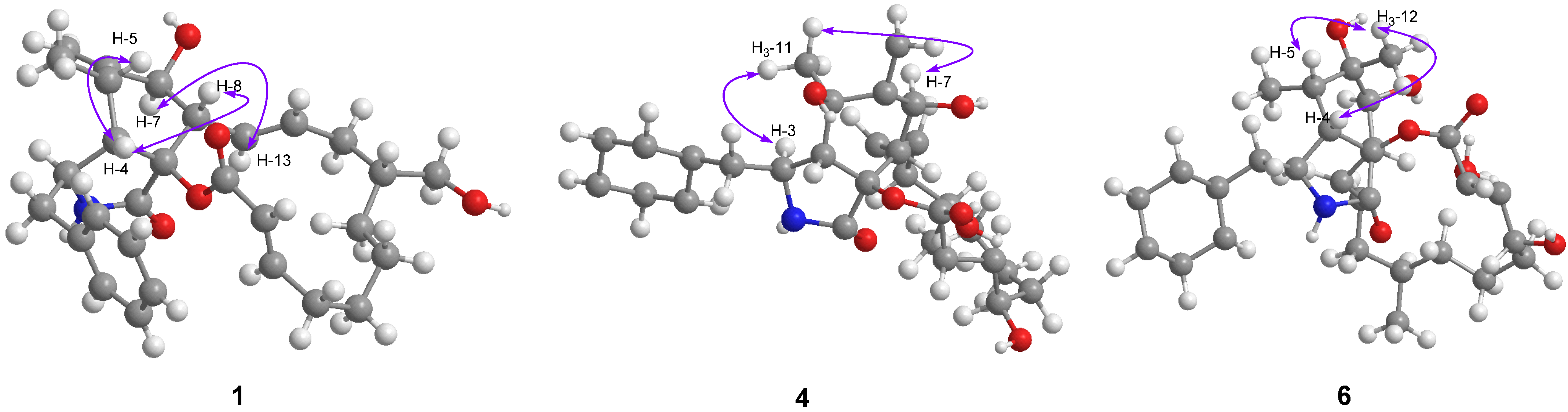
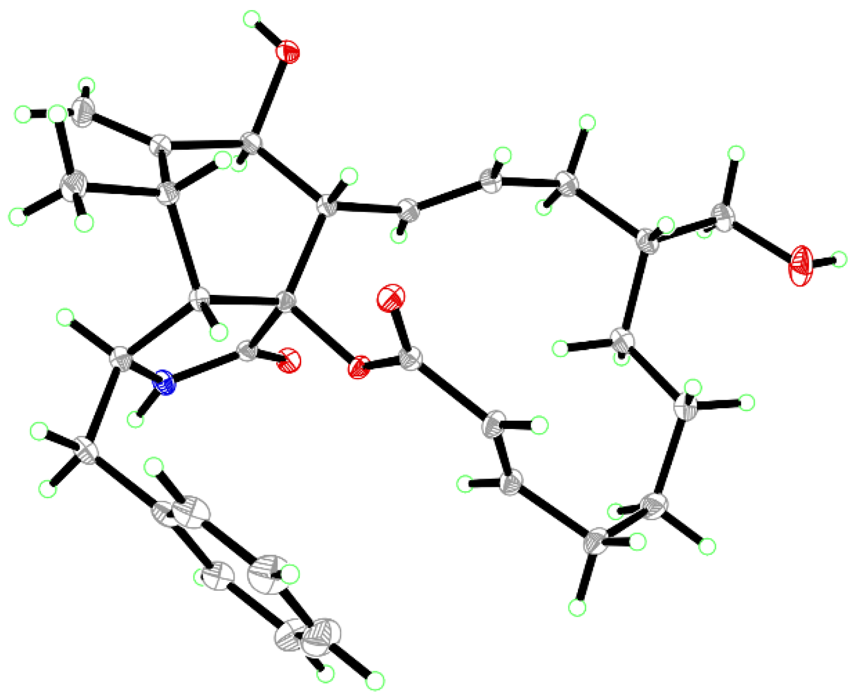
3. Results and Discussion

Compound 1 was isolated as colorless crystals (MeOH). Its molecular formula was identified as C29H37NO5 by HRESIMS (measured at m/z 502.25627 [M + Na]+; calcd for C29H37NO5Na+: 502.25639), corresponding with 12 degrees of unsaturation. In the 1H-NMR spectrum (Table 1), five olefinic protons at δH 7.14 (2H, d, J = 7.4 Hz, H-2ʹ and H-6ʹ), 7.26 (2H, t, J = 7.4 Hz, H-3ʹ and H-5ʹ) and 7.18 (1H, t, J = 7.4 Hz, H-4ʹ) revealed the presence of a mono-substituted phenyl group. Four olefinic protons at δH 5.86 (1H, dd, J = 15.0, 10.1 Hz, H-13), 5.29 (1H, m, H-14), 5.63 (1H, d, J = 15.5 Hz, H-22) and 7.02 (1H, m, H-21) indicated two E-form double bonds whereas the protons at δH 5.30 and 5.08 (each 1H, s, H2-12) suggested one terminal double bond. In addition, two protons at δH 3.59 and 3.27 suggested the presence of one oxygenated methylene group. 13C-NMR displayed 29 carbon resonances (Table 1), which could be classified as 2 CH3, 7 CH2, 15 CH and 5 non-protonated carbons by the DEPT method and HSQC data. Of them, one phenyl group and three double bonds could be readily identified, whilst the oxygenated methylene group could be observed at δC 65.9 (t). A preliminary analysis of these data suggested that 1 should be a cytochalasan product with a structure identical to that of cytochalasin B [18], a known cytochalasan isolated in this study. A further analysis of the NMR data suggested that 1 should be an isomer of cytochalasin B, with the loss of the hydroxy group at C-20 and the presence of oxygenated CH2 at δC 65.9 (t). The HMBC correlations from δH 3.59 and 3.27 (each 1H) with δC 42.4 (d, C-16), 37.5 (t, C-15) and 30.7 (t, C-17) revealed that the oxygenated CH2 should be placed at C-24 (Figure 2). It was supported by the 1H-1H COSY data, as shown in Figure 2. A detailed analysis of the 1H-1H COSY and HMBC data suggested that the other parts of 1 were the same as those of cytochalasin B (Figure 2). In the ROESY spectrum, the cross peaks of H-4/H-5, H-4/H-8 and H-7/H-13 suggested that 1 should have the same stereochemistry as cytochalasin B (Figure 3). Fortunately, the single crystal X-ray diffraction not only established the planar structure, but also determined the absolute configuration of 1, as shown in Figure 4 (Flack parameter = 0.08(4); CCDC: 2240408). Finally, the structure of 1 was established and trivially named aspergicytochalasin A.
Compound 2 was isolated as a colorless oil. Its molecular formula was identified as C29H35NO6 by HRESIMS (measured at m/z 518.25122 [M + Na]+; calcd for C29H37NO6Na+: 518.25131). All the 1H- and 13C-NMR data (Table 1) of 2 showed similarities to those of 1 and cytochalasin B. A further analysis of the NMR data suggested that 2 should be 24-hydroxy-cytochalasin, as supported by the HMBC correlations from δH 3.60 and 3.26 (each 1H, H2-24) with δC 42.6 (d, C-16), 37.6 (t, C-15) and 31.1 (t, C-17) (Figure 2). It was further supported by the 1H-1H COSY data, as shown in Figure 2. Therefore, the structure of compound 2 was established and named aspergicytochalasin B.
Compound 3 was isolated as a colorless oil. Its molecular formula was identified as C29H35NO6 by HRESIMS (measured at m/z 516.23566 [M + Na]+; calcd for C29H35NO6Na+: 516.23566). The analysis of the 1H- and 13C-NMR data (Table 1) suggested that 3 should also be a cytochalasan, possessing a structure with a high similarity to that of cytochalasin B. However, a carbonyl carbon at δC 205.2 (s, C-15) suggested that there was one keto group in 3. In the HMBC spectrum, the correlations from δH 0.99 (3H, d, J = 6.6 Hz, H3-24), 2.68 (1H, dd, J = 12.7 and 6.6 Hz, H-16) and 6.32 (1H, d, J = 15.3 Hz, H-14) with δC 205.2 indicated that the keto group should be placed at C-15, which allowed H-14 to be a doublet in the 1H-NMR spectrum (Figure 2). A comprehensive analysis of the HMBC, 1H-1H COSY and ROESY data suggested that the other parts of 3 were the same as those of cytochalasin B. Therefore, the structure of compound 3 was constructed and named aspergicytochalasin C.
Compound 4 was isolated as a colorless oil. The molecular formula of C29H37NO6 was determined based on HRESIMS (measured at m/z 518.25116 [M + Na]+; calcd for C29H37NO6Na+: 518.25131), corresponding with 12 degrees of unsaturation. Similarly, the 1H- and 13C-NMR data (Table 2) of 4 displayed high similarities with those of cytochalasin B. The main difference was that one non-protonated carbon at δC 74.3 (s, C-5) appeared in 4. These data, as well as the analysis of the MS data, suggested that 4 should be a hydroxy product of cytochalasin B. In the HMBC spectrum, the correlations between δH 1.34 (3H, s, H3-11) and δC 74.3 (s), 55.6 (d, C-4) and 152.6 (s, C-6) supported that the OH should be placed at C-5 (Figure 2). A detailed analysis of the 1H-1H COSY and HMBC data suggested that the other parts of 3 were the same as those of cytochalasin B (Figure 2). In the ROESY spectrum, cross peaks of H3-11/H-3 and H3-11/H-7 were observed (Figure 3), which indicated that the OH at C-5 should be β-oriented. Based on the same biogenetic pathway for cytochalasin B and 4, the absolute configuration of 4 was established, as shown in Figure 1. Finally, the structure of 4 was figured out and named aspergicytochalasin D.
Compound 5 was isolated as a colorless oil. According to HRESIMS (measured at m/z 496.26947 [M + H]+; calcd for C29H38NO6+: 496.26936), the molecular formula was identified as C29H37NO6, indicating 12 degrees of unsaturation. The 1H- and 13C-NMR data (Table 2) of 5 were closely related to those of cytochalasin B. A further analysis of these data suggested that 4 should have one additional OH group at the phenyl group, with respect to that of cytochalasin B. Four aromatic protons at δH 6.53 (1H, d, J = 1.8 Hz, H-2ʹ), 6.57 (1H, dd, J = 7.8 and 1.8 Hz, H-4ʹ), 6.55 (1H, d, J = 7.8 Hz, H-6ʹ) and 7.03 (1H, t, J = 7.8 Hz, H-5ʹ) could readily identify a 1,3-disubstituted phenyl moiety. The HMBC correlations from H-2ʹ to C-4ʹ and C-6ʹ further supported that the OH should be placed at C-3ʹ in 5. A comprehensive analysis of the HMBC, 1H-1H COSY and ROESY data suggested that the other parts of 5 should be the same as those of cytochalasin B. Therefore, compound 5 was identified as 3ʹ-hydroxy-cytochalasin B and trivially named aspergicytochalasin E.
Compound 6 was isolated as a colorless oil. Its molecular formula was identified as C29H39NO6 by HRESIMS (measured at m/z 520.26679 [M + Na]+; calcd for C29H39NO6Na+: 520.26696), corresponding with 11 degrees of unsaturation. The 1H- and 13C-NMR data (Table 2) of 6 were similar to those of cytochalasin B. An analysis of these data indicated that the double bond between C-6 and C-12 in cytochalasin B had disappeared, instead of a methyl group at C-12 (δH 1.18; δC 22.3) and one non-protonated carbon at C-6 (δC 77.4) in 6. It was supported by the key HMBC correlations from δH 1.18 (3H, s, H3-12) with δC 77.4 (s, C-6), 38.1 (d, C-5) and 76.3 (d, C-7) as well as the HMBC correlations from H-5 and H-7 with C-6 (Figure 2). A comprehensive analysis of the HMBC and 1H-1H COSY data suggested that the other parts of 5 were the same as those of cytochalasin B (Figure 2). In the ROESY spectrum, the cross peaks of H3-12/H-4 and H3-12/H-5 suggested that the OH at C-6 should be α-oriented, with respect to that of cytochalasin B (Figure 3). Therefore, the structure of 6 was established as 6-hydroxy-cytochalasin B and trivially named aspergicytochalasin F.
The structural characteristics of compounds 16 showed that they were similar to cytochalasin B, which was one of a large number of compounds found in this study. These results indicated that compounds 16 were likely to be obtained by the oxidation of cytochalasin B via p450, so the absolute configurations of compounds 16 should be consistent with that of cytochalasin B. The single crystal X-ray diffraction data for 1 also supported this conclusion.
In addition to the new compounds, the five known cytochalasans were identified as cytochalasin A (7) [18], cytochalasin B (8) [18], dihydrocytochalasin B (9) [19], cytochalasin Z2 (10) [20] and cytochalasin Z4 (11) [18] by a comparison of the spectroscopic data with those reported in the literature.
Compounds 16 were evaluated for their antibacterial activities against Staphylococcus aureus. Compounds 3 and 4 showed certain inhibitory activities, with MIC values of 128 and 64 μg/mL, respectively. In addition, compounds 16 were evaluated for their anti-inflammatory activities by inhibiting NO production in LPS-induced RAW 264.7 macrophages. As a result, compounds 1, 3, 5 and 6 showed inhibitory activities on NO production, with IC50 values less than 40 μM (Table 3).

4. Conclusions

In summary, a total of eleven cytochalasans, including six new ones, were isolated from the honeysuckle endophytic fungus Aspergillus sp. LE2. The new compounds exhibited antibacterial and anti-inflammatory activities. Cytochalasans are a class of important natural products with a rich structural diversity and biological activity [21,22,23,24,25,26]. At the same time, cytochalasans can also reflect the chemical relationship between microorganisms and hosts, and are an important bridge molecule in the study of medicinal plants and their functions. To the best of our knowledge, this is the first chemical study on honeysuckle endophytic fungi. Therefore, the new cytochalasans found in this study can provide certain information for further investigations on this plant and its endophytic fungal system.

Supplementary Materials

The following are available online at https://www.mdpi.com/article/10.3390/jof9030374/s1. NMR and mass spectra for 16 and X-ray crystallographic data for 1.

Author Contributions

Conceptualization, Y.-C.L.; methodology, J.Y., Q.-D.M., B.-G.L. and J.-L.S.; resources, Y.-Z.Z. and Y.-C.L.; data curation, J.Y., Q.-D.M., B.-G.L. and J.-L.S.; writing, Y.-C.L.; project administration, Y.-C.L.; funding acquisition, Y.-C.L. All authors have read and agreed to the published version of the manuscript.

Funding

This research was funded by the Key R&D projects in Henan Province (No. 221111110300, granted to Y.C.L.); Scientific and technological project in Henan Province (No. 212102310876).

Institutional Review Board Statement

Not applicable.

Informed Consent Statement

Not applicable.

Data Availability Statement

The supporting information of this study is available from the corresponding author (Y.-C.L.). The CIF of 1 is available free of charge at https://www.ccdc.cam.ac.uk.

Acknowledgments

The authors thank the Analytical & Measuring Centre, South-Central University for Nationalities, for the spectra measurements.

Conflicts of Interest

The authors declare no conflict of interest.

References

  1. Wen, J.; Okyere, S.K.; Wang, S.; Wang, J.; Xie, L.; Ran, Y.; Hu, Y. Endophytic fungi: An effective alternative source of plant-derived bioactive compounds for pharmacological studies. J. Fungi 2022, 8, 205. [Google Scholar] [CrossRef] [PubMed]
  2. Flora Reipblicae Popularis Sinicae; China Science Publishing & Media Ltd.: Beijing, China, 1988; Volume 72, p. 236.
  3. Shang, X.F.; Pan, H.; Li, M.X.; Miao, X.L.; Ding, H. Lonicera japonica Thunb.: Ethnopharmacology, phytochemistry and pharmacology of an important traditional Chinese medicine. J. Ethnopharmacol. 2011, 138, 1–21. [Google Scholar] [CrossRef] [PubMed]
  4. Son, K.H.; Jung, K.Y.; Chang, H.W.; Kim, H.P.; Kang, S.S. Triterpenoid saponins from the aerial parts of Lonicera japonica. Phytochemistry 1994, 35, 1005–1008. [Google Scholar] [CrossRef] [PubMed]
  5. Tomassini, L.; Cometa, M.F.; Serafini, M.; Nicoletti, M. Isolation of secoiridoid artifacts from Lonicera japonica. J. Nat. Prod. 1995, 58, 1756–1758. [Google Scholar] [CrossRef]
  6. Machida, K.; Sasaki, H.; Iijima, T.; Kikuchi, M. Studies on the constituents of Lonicera species. XVII. New iridoid glycosides of the stems and leaves of Lonicera japonica THUNB. Chem. Pharm. Bull. 2002, 50, 1041–1044. [Google Scholar] [CrossRef] [Green Version]
  7. Lin, L.M.; Zhang, X.G.; Zhu, J.J.; Gao, H.M.; Wang, Z.M.; Wang, W.H. Two new triterpenoid saponins from the flowers and buds of Lonicera japonica. J. Asian Nat. Prod. Res. 2008, 10, 925–929. [Google Scholar] [CrossRef]
  8. Ge, L.L.; Li, J.M.; Wan, H.Q.; Zhang, K.D.; Wu, W.G.; Zou, X.T.; Wu, S.P.; Zhou, B.P.; Tian, J.; Zeng, X.B. Novel flavonoids from Lonicera japonica flower buds and validation of their anti-hepatoma and hepatoprotective activity in vitro studies. Ind. Crops Prod. 2018, 125, 114–122. [Google Scholar] [CrossRef]
  9. Fan, Z.L.; Li, L.; Bai, X.L.; Zhang, H.; Liu, Q.R.; Zhang, H.; Fu, Y.J.; Moyo, R. Extraction optimization, antioxidant activity, and tyrosinase inhibitory capacity of polyphenols from Lonicera japonica. Food Sci. Nutr. 2019, 7, 1786–1794. [Google Scholar] [CrossRef] [Green Version]
  10. Yang, R.; Hao, H.; Li, J.; Xuan, J.; Xia, M.F.; Zhang, Y.Q. Three new secoiridoid glycosides from the flower buds of Lonicera japonica. Chin. J. Nat. Med. 2020, 18, 70–74. [Google Scholar] [CrossRef]
  11. Kashiwada, Y.; Omichi, Y.; Kurimoto, S.; Shibata, H.; Miyake, Y.; Kirimoto, T.; Takaishi, Y. Conjugates of a secoiridoid glucoside with a phenolic glucoside from the flower buds of Lonicera japonica Thunb. Phytochemistry 2013, 96, 423–429. [Google Scholar] [CrossRef]
  12. Sun, L.; Lu, Z.; Bie, X.; Sun, D.; Yang, S. Isolation and identification of an antimicrobial endophytic strain EJH-2. Zhongguo Weishengtaixue Zazhi 2006, 18, 23–26. [Google Scholar]
  13. Liu, Y.L.; Zhang, J.; Yang, Z.R.; Wang, C. Isolation and identification of a chlorogenic acid-producing endophytic bacterium from flos Lonicerae japonicae. Sichuan Daxue Xuebao Ziran Kexueban 2014, 51, 603–608. [Google Scholar]
  14. Zhao, L.; Xu, Y.; Lai, X.-H.; Shan, C.; Deng, Z.; Ji, Y. Screening and characterization of endophytic Bacillus and Paenibacillus strains from medicinal plant Lonicera japonica for use as potential plant growth promoters. Braz. J. Microbiol. 2015, 46, 977–989. [Google Scholar] [CrossRef]
  15. Gupta, H.; Saini, R.V.; Pagadala, V.; Kumar, N.; Sharma, D.K.; Saini, A.K. Analysis of plant growth promoting potential of endophytes isolated from Echinacea purpurea and Lonicera japonica. J. Soil Sci. Plant Nutr. 2016, 16, 558–577. [Google Scholar] [CrossRef]
  16. Liu, Y.P.; Dai, Q.; Wang, W.X.; He, J.; Li, Z.H.; Feng, T.; Liu, J.K. Psathyrins: Antibacterial diterpenoids from Psathyrella candolleana. J. Nat. Prod. 2020, 83, 1725–1729. [Google Scholar] [CrossRef]
  17. Feng, T.; Cai, J.L.; Li, X.M.; Zhou, Z.Y.; Li, Z.H.; Liu, J.K. Chemical constituents and their bioactivities of mushroom Phellinus rhabarbarinus. J. Agric. Food Chem. 2016, 64, 1945–1949. [Google Scholar] [CrossRef]
  18. Evidente, A.; Andolfi, A.; Vurro, M.; Zonno, M.C.; Motta, A. Cytochalasins Z4, Z5, and Z6, three new 24-oxa[14]cytochalasans produced by Phoma exigua var. heteromorpha. J. Nat. Prod. 2003, 66, 1540–1544. [Google Scholar] [CrossRef]
  19. Lin, D.C.; Lin, S. High affinity binding of [3H]-dihydrocytochalasin B to peripheral membrane proteins related to the control of cell shape in the human red cell. J. Biol. Chem. 1978, 253, 1415–1419. [Google Scholar] [CrossRef]
  20. Evidente, A.; Andolfi, A.; Vurro, M.; Zonno, M.C.; Motta, A. Cytochalasins Z1, Z2 and Z3, three 24-oxa[14]cytochalasans produced by Pyrenophora semeniperda. Phytochemistry 2002, 60, 45–53. [Google Scholar] [CrossRef]
  21. Zhu, H.; Chen, C.; Xue, Y.; Tong, Q.; Li, X.-N.; Chen, X.; Wang, J.; Yao, G.; Luo, Z.; Zhang, Y. Asperchalasine A, a cytochalasan dimer with an unprecedented decacyclic ring system, from Aspergillus flavipes. Angew. Chem. Int. Ed. 2015, 54, 13374–13378. [Google Scholar] [CrossRef]
  22. Zhu, H.; Chen, C.; Tong, Q.; Li, X.-N.; Yang, J.; Xue, Y.; Luo, Z.; Wang, J.; Yao, G.; Zhang, Y. Epicochalasines A and B: Two bioactive merocytochalasans bearing caged epicoccine dimer units from Aspergillus flavipes. Angew. Chem. Int. Ed. 2016, 55, 3486–3490. [Google Scholar] [CrossRef]
  23. Zhu, H.; Chen, C.; Tong, Q.; Yang, J.; Wei, G.; Xue, Y.; Wang, J.; Luo, Z.; Zhang, Y. Asperflavipine A: A cytochalasan heterotetramer uniquely defined by a highly complex tetradecacyclic ring system from Aspergillus flavipes QCS12. Angew. Chem. Int. Ed. 2017, 56, 5242–5246. [Google Scholar] [CrossRef] [PubMed]
  24. Wang, W.-X.; Li, Z.-H.; Feng, T.; Li, J.; Sun, H.; Huang, R.; Yuan, Q.-X.; Ai, H.-L.; Liu, J.-K. Curtachalasins A and B, two cytochalasans with a tetracyclic skeleton from the endophytic fungus Xylaria curta E10. Org. Lett. 2018, 20, 7758–7761. [Google Scholar] [CrossRef] [PubMed]
  25. Wang, W.-X.; Lei, X.; Ai, H.-L.; Bai, X.; Li, J.; He, J.; Li, Z.-H.; Zheng, Y.-S.; Feng, T.; Liu, J.-K. Cytochalasans from the endophytic fungus Xylaria cf. curta with resistance reversal activity against fluconazole-resistant Candida albicans. Org. Lett. 2019, 21, 1108–1111. [Google Scholar] [CrossRef] [PubMed]
  26. Wang, W.-X.; Lei, X.; Yang, Y.-L.; Li, Z.-H.; Ai, H.-L.; Li, J.; Feng, T.; Liu, J.-K. Xylarichalasin A, a halogenated hexacyclic cytochalasan from the fungus Xylaria cf. curta. Org. Lett. 2019, 21, 6957–6960. [Google Scholar] [CrossRef]
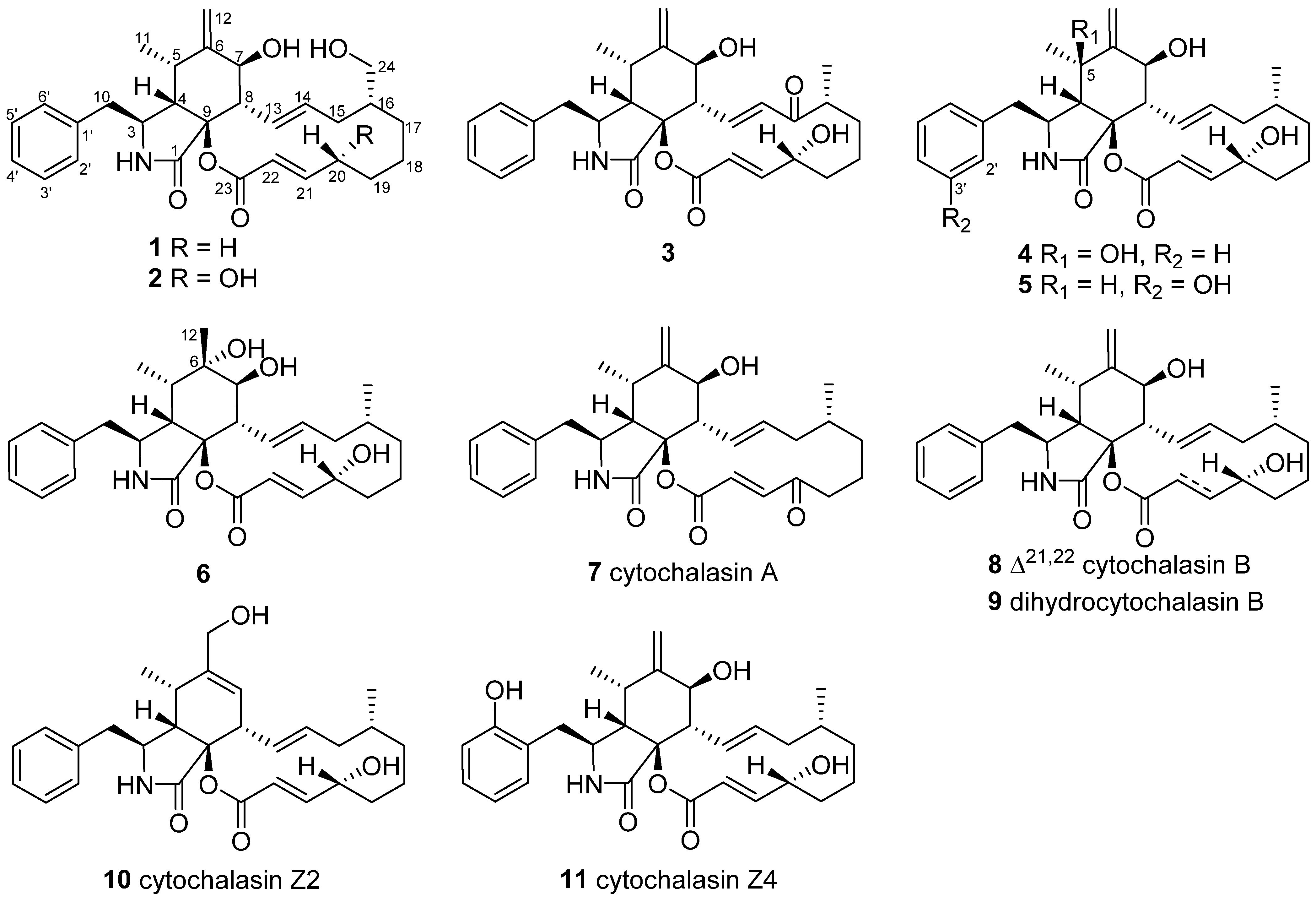
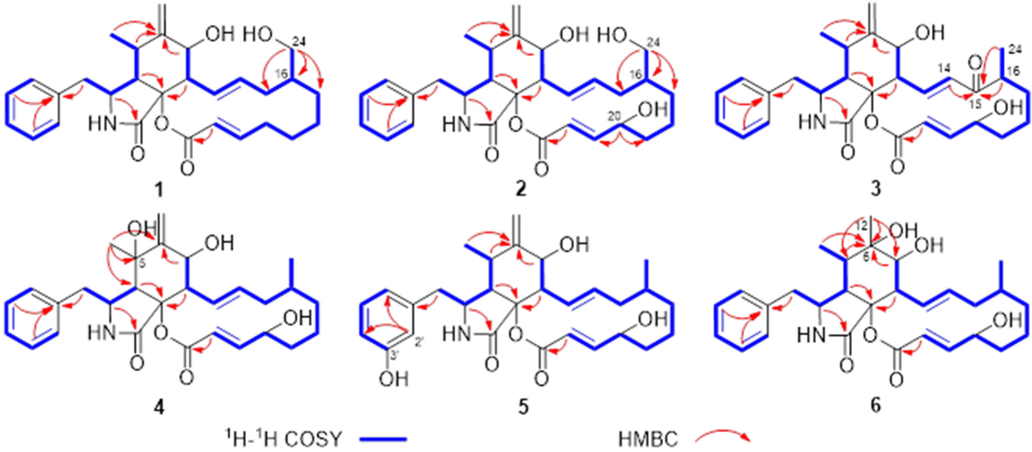
Figure 1. Structures of chemicals 111.
Jof 09 00374 g001
Figure 2. Key 2D NMR correlations of 16.
Jof 09 00374 g002
Figure 3. Key ROESY correlations of 1, 4 and 6.
Jof 09 00374 g003
Figure 4. ORTEP diagram of 1 showing absolute configurations.
Jof 09 00374 g004
Table 1. 1H (600 MHz) and 13C (150 MHz) NMR spectroscopic data for 13 in methanol-d4.
No.123
δCδH (J in Hz)δCδH (J in Hz)δCδH (J in Hz)
1173.9, C 174.0, C 173.0, C
355.0, CH3.36, m54.8, CH3.40, m55.3, CH3.40, td (6.1, 2.8)
448.9, CH2.80, m48.5, CH2.85, dd (5.0, 2.4)48.9, CH2.84, m
532.9, CH3.15, m32.8, CH3.21, m33.1, CH3.17, m
6151.5, C 151.4, C 151.1, C
771.1, CH3.80, d (11.1)71.5, CH3.80, d (10.8)70.8, CH4.02, d (11.1)
849.9, CH3.30, dd (11.1, 10.1)49.2, CH3.40, dd (10.8, 10.1)49.0, CH3.47, dd (11.1, 9.9)
984.9, C 85.4, C 86.1, C
1044.0, CH22.84, m
2.84, m
44, CH22.80, m43.9, CH22.86, m
1114.3, CH30.83, d (6.7)14.1, CH30.85, d (6.7)14.3, CH30.88, d (6.7)
12114.3, CH25.08, s
5.30, s
114.4, CH25.28, s; 5.09, s114.6, CH25.35, s; 5.13, s
13129.1, CH5.86, dd (15.0, 10.1)129.2, CH5.88, dd (14.7, 10.1)145, CH7.09, dd (15.3, 9.9)
14136.5, CH5.29, m136.5, CH5.24, m134.5, CH6.32, d (15.3)
1537.5, CH22.40, m; 1.69, m37.6, CH22.38, m; 1.66, m205.2, C
1642.4, CH1.35, m42.6, CH1.29, m46.0, CH2.68, dd (12.7, 6.6)
1730.7, CH21.60, m; 0.86, m31.1, CH21.53, m; 0.84, m35.1, CH21.75, m; 1.29, m
1827.6, CH21.70, m; 1.07, m21.2, CH21.47, m; 1.29, m22.0, CH21.48, m; 1.13, m
1927.3, CH21.81, m; 1.35, m35.4, CH21.92, m; 1.53, m36.2, CH21.74, m; 1.66, m
2035.2, CH22.37, m; 2.18, m70.9, CH4.46, m71.5, CH4.25, m
21154.0, CH7.02, m154.4, CH6.94, dd (15.6, 4.3)153.1, CH6.91, dd (15.7, 6.6)
22122.0, CH5.63, d (15.5)119.6, CH5.79, dd (15.6)121.7, CH5.84, d (15.7)
23166.2, C 166.3, C 166.4, C
2465.9, CH23.59, dd (10.8, 3.6)
3.27, dd (10.8, 7.5)
65.9, CH23.60, dd (10.9, 3.4);
3.26, dd (10.9, 7.4)
15.1, CH30.99, d (6.6)
1′138.5, C 138.2, C 138.4, C
2′,6′130.9, CH7.14, d (7.4)131.0, CH7.13, d (7.4)131.0, CH7.15, d (7.3)
3′,5′129.6, CH7.26, t (7.4)129.6, CH7.27, t (7.4)129.6, CH7.27, t (7.3)
4′127.8, CH7.18, t (7.4)127.9, CH7.19, t (7.4)127.9, CH7.23, t (7.3)
Table 2. 1H (600 MHz) and 13C (150 MHz) NMR spectroscopic data for 46 in methanol-d4.
No.456
δCδH (J in Hz)δCδH (J in Hz)δCδH (J in Hz)
1173.3, C 174.0, C 173.7, C
357.2, CH3.25, m54.8, CH3.32, m55.0, CH4.18, m
455.6, CH2.86, d (4.2)48.6, CH2.81, dd (5.2, 2.6)50.1, CH2.58, m
574.3, C 32.9, CH3.16, m38.1, CH2.27, m
6152.6, C 151.5, C 77.4, C
769.2, CH3.94, d (12.2)71.5, CH3.76, d (10.9)76.3, CH3.45, d (12.3)
851.3, CH3.83, dd (12.2, 10.2)49.3, CH3.34, dd (10.9, 9.8)49.4, CH2.86, dd (12.3, 10.2)
985.2, C 85.4, C 86.7, C
1044.0, CH23.02, d (6.6)44.1, CH22.68, m45.0, CH22.79, dd (13.7, 7.9)
2.93, dd (13.7, 4.4)
1126.5, CH31.34, s14.1, CH30.84, d (6.5)13.0, CH30.99, d (7.1)
12114.4, CH25.51, s; 5.47, s114.3, CH25.24, s; 5.05, s22.3, CH31.18, s
13128.8, CH5.62, dd (14.2, 10.2)128.7, CH5.79, dd (15.0, 9.8)128.0, CH5.64, dd (14.4, 10.2)
14137.1, CH5.41, m136.7, CH5.20, m136.5, CH5.25, m
1542.7, CH22.19, m; 1.73, m43.2, CH22.06, m; 1.61, m42.8, CH22.14, m; 1.70, m
1633.9, CH1.46, m34.5, CH1.18, m34.3, CH1.35, m
1735.7, CH21.83, m; 0.65, m36.6, CH21.68, m; 0.53, m36.1, CH21.77, m; 0.62, m
1821.8, CH21.53, m; 1.42, m21.5, CH21.41, m; 1.23, m21.7, CH21.41, m
1935.3, CH21.85, m; 1.61, m35.4, CH21.87, m; 1.52, m35.4, CH21.86, m; 1.58, m
2072.8, CH4.29, m71.2, CH4.42, m72.0, CH4.34, m
21153.7, CH7.04, dd (15.6, 6.3)154.6, CH6.92, dd (15.7, 5.4)154.1, CH6.99, dd (15.6, 6.4)
22121.2, CH5.79, d (15.6)119.4, CH5.74, d (15.7)120.3, CH5.75, d (15.6)
23166.5, C 166.4, C 166.3, C
2420.7, CH30.90, d (6.5)20.7, CH30.84, d (6.5)20.7, CH30.88, d (6.6)
1′139.2, C 139.7, C 139.0, C
2′130.5, CH7.20, d (7.5)117.8, CH6.53, (1.8)130.7, CH7.19, d (7.4)
3′129.8, CH7.30, t (7.5)158.5, C 129.6, CH7.27, t (7.4)
4′127.9, CH7.22, t (7.5)114.9, CH6.57, dd (7.8, 1.8)127.7, CH7.19, t (7.4)
5′129.8, CH7.30, t (7.5)130.5, CH7.03, t (7.8)129.6, CH7.27, t (7.4)
6′130.5, CH7.20, d (7.5)122.2, CH6.55, d (7.8)130.7, CH7.19, d (7.4)
Table 3. NO production inhibitions of Compounds 16.
EntryIC50 (μM)
126.8 ± 2.69
2>40
317.1 ± 1.27
4>40
531.2 ± 1.02
612.7 ± 1.85
SMS7.3 ± 0.34
Disclaimer/Publisher’s Note: The statements, opinions and data contained in all publications are solely those of the individual author(s) and contributor(s) and not of MDPI and/or the editor(s). MDPI and/or the editor(s) disclaim responsibility for any injury to people or property resulting from any ideas, methods, instructions or products referred to in the content.

