Article

Survey of Edible Amanita in Northern Thailand and Their Nutritional Value, Total Phenolic Content, Antioxidant and α-Glucosidase Inhibitory Activities

1
Research Center of Microbial Diversity and Sustainable Utilization, Chiang Mai University, Chiang Mai 50200, Thailand
2
Department of Biology, Faculty of Science, Chiang Mai University, Chiang Mai 50200, Thailand
3
Biology Program, Faculty of Science and Technology, Pibulsongkram Rajabhat University, Phitsanulok 65000, Thailand
4
Academy of Science, The Royal Society of Thailand, Bangkok 10300, Thailand
*
Author to whom correspondence should be addressed.
J. Fungi 2023, 9(3), 343; https://doi.org/10.3390/jof9030343
Submission received: 7 February 2023 / Revised: 8 March 2023 / Accepted: 8 March 2023 / Published: 10 March 2023
(This article belongs to the Special Issue Edible and Medicinal Macrofungi, 2nd Edition)

Abstract

:
Edible wild mushrooms are extremely popular among consumers and are highly valued for their potential economic benefits in northern Thailand. In this present study, a total of 19 specimens of edible Amanita were collected during investigations of wild edible mushrooms in northern Thailand during the period from 2019 to 2022. Their morphological characteristics and the phylogenetic analyses of the internal transcribed spacer (ITS) and partial large subunit (nrLSU) of ribosomal RNA, RNA polymerase II second-largest subunit (rpb2) and partial translation elongation factor 1-alpha (tef-1) indicated that the collected specimens belonged to A. hemibapha, A. pseudoprinceps, A. rubromarginata, A. subhemibapha, and Amanita section Caesareae. This is the first report of A. pseudoprinceps and A. subhemibapha from Thailand. Full descriptions, illustrations and a phylogenetic placement of all specimens collected in this study are provided. Subsequently, the nutritional composition and total phenolic content, as well as the antioxidant and α-glucosidase inhibitory activities, of each species were investigated. The results indicate that the protein contents in both A. pseudoprinceps and A. subhemibapha were significantly higher than in A. hemibapha and A. rubromarginata. The highest total phenolic content was found in the extract of A. pseudoprinceps. In terms of antioxidant properties, the extract of A. pseudoprinceps also exhibited significantly high antioxidant activity by 2,2-azino-bis(3-ethylbenzothiazoline-6-sulfonic acid (ABTS), 2,2-diphenyl-1-picrylhydrazyl (DPPH) and ferric reducing antioxidant power (FRAP) assays. However, the extract of A. rubromarginata had the lowest total phenolic content and level of antioxidant activity. Additionally, α-glucosidase inhibitory activity varied for different Amanita species and the highest level of α-glucosidase inhibitory activity was found in the extract of A. pseudoprinceps. This study provides valuable information on the nutrient content, phenolic content and the antioxidant and α-glucosidase inhibitory potential of edible Amanita species found in northern Thailand.

1. Introduction

The genus Amanita Pers. was first introduced in 1797 by Persoon [1] with A. muscaria (L.) Lam. as the type species. This genus is one of several large genera with approximately 650 species distributed throughout tropical, subtropical and temperate regions around the world [2,3,4,5,6]. Amanita is a member of the family Amanitaceae, order Agaricales [3,4,5,6]. Generally, Amanita is characterized by agaricoid basidiomata having free lamellae, white spore prints, hyaline and smooth basidiospores, as well as the presence of volval remnants (universal veil) and the presence of annulus (partial veil) on the stem [1,7,8,9]. Currently, taxonomic studies have divided this genus into three subgenera (Amanita subg. Amanita, Amanitina, and Lepidella) and eleven sections based on multi-gene phylogenetic analyses [4,10,11]. Most of the Amanita species are known to be ectomycorrhizal fungi that form mutualistic symbioses with more than ten families of plants (including Betulaceae, Caesalpiniaceae, Casuarinaceae, Dipterocarpaceae, Fabaceae, Myrtaceae, Pinaceae, and Salicaceae) and are known to grow on the ground in forests [3,4,9,12]. However, the Amanita species in Amanita sect. Lepidella have been reported as saprobes that grow in grasslands [4,9,13,14,15]. Notably, Amanita contains both edible and lethal species. The most toxic species are in Amanita sect. Phalloideae [e.g., A. exitialis Zhu L. Yang & T.H. Li, A. phalloides (Vaill. ex Fr.) Link, A. verna Bull. ex Lam. and A. virosa Bertill.], while most of the edible species belong to Amanita sect. Caesareae [2,4,16,17,18]. The most famous edible Amanita species are A. caesarea (Scop.) Pers., A. chepangiana Tulloss & Bhandary, A. flammeola Pegler & Piearce, A. franchetii (Boud.) Fayod, A. fulva Fr., A. hemibapha (Berk. & Broome) Sacc., A. jacksonii Pomerl., A. manginiana Har. & Pat., A. loosii Beeli, A. pseudoporphyria Hongo, A. princeps Corner & Bas, A. rubescens Pers., A. tuza Guzmán, A. vaginata (Bull.) Lam., and A. zambiana Pegler & Piearce [3,4,5,6,17,18,19,20].
Several edible wild mushrooms are known to be a good source of essential dietary minerals, nutrients, and vitamins, which makes them an important source of food for humans [17,20,21]. These mushrooms have also been recognized as a source of many bioactive compounds (e.g., immunomodulatory compounds, phenolic compounds, polysaccharides, terpenoids and tocopherols) that exhibit various beneficial biological activities including anticancer, antidiabetic, anti-inflammatory, antimicrobial, antioxidant, cholesterol-reducing, immunomodulatory and neuroprotective properties [22,23]. Additionally, ethnomycologists have recorded vital information on the relevant consumption patterns and applications of wild edible mushrooms for medicinal purposes [24,25]. Thailand, a Southeast Asian country, has many species of edible wild mushroom that are particularly abundant during the rainy season (mid-May to October) each year. Generally, wild edible mushrooms are collected by local farmers for consumption and sale in local, roadside or city markets (Figure 1).
Preliminary investigations of edible wild mushrooms in northern Thailand have revealed the existence of many genera, e.g., Amanita, Astraeus, Boletus, Cantharellus, Lactarius, Phlebopus, Russula, and Termitomyces [26,27,28]. Edible Amanita species are the most popular variety of edible wild mushrooms in northern Thailand because of their palatable texture and flavor. However, the number of lethal and edible Amanita species that have been found in Thailand has remained a controversial issue due to the absence of comprehensive herbarium reference material, accurate descriptions and available molecular data [29].
During our ongoing studies of edible wild mushrooms in northern Thailand, we have collected specimens of edible Amanita species from natural forests, roadsides and local markets. Therefore, the present study aimed to identify the collected specimens based on their morphological characteristics and multi-gene phylogeny using the sequence data of ITS, nrLSU, rpb2, and tef-1. A full description, color photographs, illustrations and a phylogenetic tree of the collected specimens are provided. Moreover, the nutritional composition, total phenolic content, and antioxidant and α-glucosidase inhibitory activities of collected edible Amanita were investigated.

2. Materials and Methods

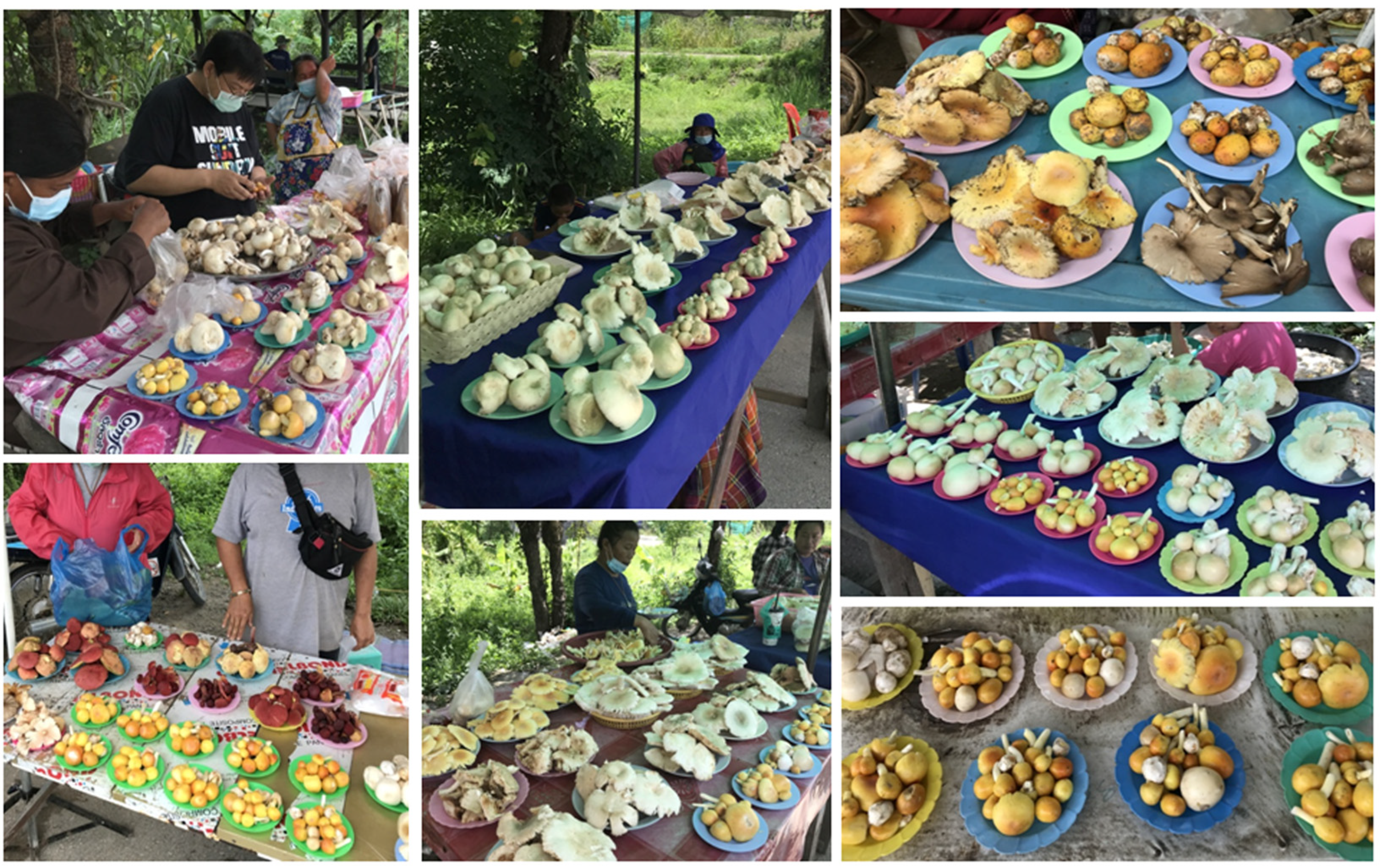
2.1. Sample Collection

The edible Amanita were surveyed and collected from natural forests, roadsides and local markets in Chiang Mai and Lamphun Provinces in northern Thailand during the rainy seasons of the years 2019 to 2022. Basidiomata were kept in plastic boxes and taken to the laboratory. Specimens were dried in a hot air oven at 45 °C until they were completely dry. After that, the dried specimens were kept in a plastic Ziplock bag and deposited in the Herbarium of Sustainable Development of Biological Resources (SDBR-CMU), Faculty of Science, Chiang Mai University, Thailand.

2.2. Identification of the Edible Amanita

2.2.1. Morphological Observations

Fresh specimens were used to describe macromorphological data. Color names and codes were followed by Kornerup and Wanscher [30]. The dried specimens were examined for micromorphological data. Dried specimens were mounted in 5% aqueous KOH, Melzer’s reagent, or 1% aqueous Congo red solution. A light microscope (Nikon Eclipse Ni U, Tokyo, Japan) was used to examine micromorphological features. Each microscopic structure’s size data were derived from at least 50 measurements using the Tarosoft (R) Imaging Frame Work program. The terminology for microscopic features followed Largent et al. [31] and Bas [32]. Basidiospore statistics are expressed as (a–) b–c (–d), where ‘a’ and ‘d’ are the extreme values and ‘b–c’ is the range comprising 90% of all values. The Q value represents ratio of the length divided by the width of each basidiospore and Qm is the average Q of all specimens ± standard deviation.

2.2.2. DNA Extraction, Amplification, Sequencing, and Phylogenetic Analyses

A Genomic DNA Extraction Mini-Kit (FAVORGEN, Ping-Tung, Taiwan) was used to extract DNA from fresh tissue of each specimen. The ITS, nrLSU, rpb2, and tef-1 regions were amplified by polymerase chain reaction (PCR) using ITS5/ITS4 [33], LR0R/LR5 [34], Am6F/Am7R [35], and EF1-983F/EF1-1567R [36] primers, respectively. The PCR for these four domains was performed in separate PCR reactions on a peqSTAR thermal cycler (PEQLAB Ltd., Fareham, UK). The PCR programs of ITS, nrLSU, rpb2, and tef-1 genes were established by following the methods employed by Liu et al. [15] and Cai et al. [36]. PCR products were directly sequenced by the Sanger sequencing method at 1st Base Company (Kembangan, Malaysia).
Sequence analysis was performed by a similarity search using the BLAST program available at NCBI (http://blast.ncbi.nlm.nih.gov, accessed on 12 November 2022). Sequences from this study, previous studies, and the GenBank database were selected and listed in Table 1. The combined dataset of ITS, nrLSU, rpb2, and tef-1 was used for the phylogenetic analysis. MUSCLE [37] was used to perform multiple sequence alignments, and BioEdit v. 6.0.7 [38] was used to make any necessary improvements. Maximum likelihood (ML) and Bayesian inference (BI) methods were used to construct phylogenetic trees. The best substitution models were GTR+I+G for ITS, nrLSU and tef-1 and HKY+I+G for rpb2 from the Akaike Information Criterion (AIC) in jModeltest 2.1.10 [39]. The GTRCAT model with 25 categories was subjected to ML analysis using RAxML v7.0.3 and 1000 bootstrap replications [40,41]. MrBayes v3.2.6 [42] was used for the BI analysis, which evaluated the posterior probabilities (PP) using Markov chain Monte Carlo sampling (MCMC). Six simultaneous Markov chains were run from random trees for one million generations and trees were sampled every 100th generation. The first 25% of trees were discarded and the remaining trees were used for calculating PP value in the majority rule consensus tree. FigTree v1.4.0 [43] was used to visualize the tree topologies.

2.3. Nutritional Analysis

A total of six samples of edible Amanita (SDBR-CMUNK0775, SDBR-CMUNK0776, SDBR-CMUNK0780, SDBR-CMUNK0853, SDBR-CMUNK0855, and SDBR-CMUNK0857) obtained in this study were used in the analyses of nutrition, antioxidant, and α-glucosidase inhibitory activities because their dry weights were sufficient for testing. A Waring blender (New Hartford, CT, USA) was used to grind each dried sample. The nutritional composition (including ash, carbohydrate, fat, fiber, and protein) of each dried sample was determined using a method developed by the Association of Official Analytical Chemists (AOAC) [44] at the Central Laboratory Company Limited (Chiang Mai, Thailand).

2.4. Preparation of Mushroom Extracts

Ten grams (10 g) of each ground mushroom sample was extracted with 100 mL of absolute ethanol at 25 °C and 150 rpm for 24 h, as described by Kaewnarin et al. [45]. After that, each extract was placed in an ultrasonic bath (Elma Transsonic Digital, Singen, Germany) at 60 °C for 3 h. Whatman’s No. 1 filter paper was used to filter the samples. The residue was then re-extracted twice with absolute ethanol as mentioned above. The ethanolic extract was then dried using rotary evaporation at 40 °C. The extract was dissolved in 100 mL absolute ethanol and kept at 4 °C until further determination.

2.5. Determination of Total Phenolic Content

The method of Thitilertdecha et al. [46] was modified slightly to determine the total phenolic content. Folin-Ciocalteu reagent at 0.5 mL was mixed with 2.5 mL deionized water and 0.25 mL mushroom extract. After 5 min, 0.5 mL of Na2CO3 (20% w/v) was added. The mixture was incubated for 1 h in the dark at 25 °C. Measurements of absorbance at 760 nm were used to investigate the total phenolic content. The total phenolic content of the samples was calculated using a standard curve of gallic acid. Results were expressed as milligrams of gallic acid equivalents per gram of dry weight (mg GAE/g dw). Each sample extract was analyzed in five replicates.

2.6. Antioxidant Assay

2.6.1. ABTS Scavenging Assay

The procedure of Re et al. [47] with slight modifications was used to determine the 2,2-azino-bis(3-ethylbenzothiazoline-6-sulfonic acid) (ABTS) radical scavenging activity. The stock solution of ABTS cation chromophore was prepared by facilitating a reaction between 100 mL of 2.45 mM K2S2O8 and 100 mL of 7.0 mM ABTS solution. The solution was kept for 16 h in a dark place at room temperature. The ABTS solution was diluted with phosphate buffer (50 mM, pH 7.4) before use to yield an absorbance value of 0.70 ± 0.2 at 734 nm. A quantity of 2.9 mL of ABTS solution was mixed with 0.1 mL of each sample extract. The mixtures were incubated in the dark for 30 min at room temperature. A mixture of absolute ethanol and ABTS solution was used as the control. After incubation, the absorbance of each mixture was measured spectrophotometrically at 734 nm. Trolox was used as a reference compound. The ABTS scavenging activity was expressed as the Trolox equivalent antioxidant capacity per gram of dry weight (TE/g dw). Each sample extract was subjected to five replications.

2.6.2. DPPH Scavenging Assay

The method developed by Gülçin et al. [48] was used to determine the 2,2-diphenyl-1-picrylhydrazyl (DPPH) radical scavenging activity. Initially, 1.5 mL of the 0.1 mM DPPH solution in methanol was combined with 0.5 mL of the sample extract. The mixtures were incubated at room temperature in the dark for 30 min. Then, the absorbance of each mixture was determined using spectrophotometry at 517 nm. Trolox was used as a reference compound. The DPPH scavenging activity was expressed as the TE/g dw. Five replicates were performed for each sample extract.

2.6.3. FRAP Assay

The method developed by Li et al. [49] was used to determine the ferric reducing antioxidant power (FRAP) activity. The FRAP reagent was prepared using a mixture containing 20 mL of 20 mM ferric (III) chloride, 10 mM 2,4,6-tripyridyl-s-triazine solution in 20 mL of 40 mM HCl, and 5 mL of 300 mM acetate buffer (pH 3.6). A quantity of 1.5 mL of FRAP reagent and 1.4 mL of acetate buffer (300 mM, pH 3.6) were mixed with 0.1 mL of each sample extract. Then, the mixture was incubated in the dark for 30 min at room temperature. Trolox was used as a reference compound. A mixture of absolute ethanol and FRAP solution was used as the control. After incubation, the absorbance of each mixture was measured spectrophotometrically at 593 nm. Trolox was used as a reference compound and the FRAP value was expressed as the TE/g dw. Five replicates were performed for each sample extract.

2.7. Determination of α-Glucosidase Inhibitory Activity

The procedure of Oki et al. [50] was modified to prepare the α-glucosidase solution from rat intestinal acetone powder. A quantity of 3 mL of 0.9% NaCl solution was mixed with 100 mg of intestinal acetone powder (Sigma-Aldrich Chemical Co., Saint Louis, MO, USA), homogenized by sonication, and stored in an ice bath. The enzyme mixture was centrifuged at 4 °C for 30 min at 8000 rpm. The supernatant was maintained in an ice bath and directly subjected to inhibitory assay. The α-glucosidase inhibitory assay was followed the procedure of Tanruean et al. [51] with some modifications. Each extracted sample (10 μL) was mixed with α-glucosidase solution (30 μL) and incubated at 37 °C for 15 min. Later, 70 μL of 37 mM D-maltose was then added and incubated at 37 °C for 15 min. The reaction was stopped after 10 min in boiling water. A glucose oxidase assay was used to determine the released glucose concentration of the reaction mixture. The peroxidase-glucose oxidase (PGO) reagent (900 µL) containing 1 capsule of PGO enzymes to 100 mL of water and 1.6 mL of o-dianisidine solution was added to the reaction mixture and it was then mixed for 30 min at 37 °C in a water bath. The absorbance of α-glucosidase activity was measured at 450 nm. The percentage of inhibition was calculated according to the formula: Percentage of inhibition = (Ao−As/Ao) × 100, where Ao is the absorbance of the control and As is the absorbance of the mixture containing the test compound. Acarbose (a standard synthetic inhibitor of α-glucosidase) was used for standard compound. Each sample extract was analyzed in five replicates.

2.8. Statistical Analysis

Statistical differences between treatments were assessed using one-way analysis of variance (ANOVA) with the SPSS program version 16.0 for Microsoft Windows. Significant differences at the p < 0.05 level were determined using Tukey’s test. The Pearson correlation coefficients (r) of the total phenolic content with antioxidant and α-glucosidase inhibitory activities of extract samples were analyzed using the SPSS program at a significance level of p < 0.05.

3. Results and Discussion

3.1. Sample Collection and Morphological Observations

In this study, a total of 19 edible Amanita specimens were obtained (Table 2). These specimens were initially classified into four Amanita species, namely A. hemibapha (4 specimens), A. pseudoprinceps (7 specimens), A. rubromarginata (4 specimens), and A. subhemibapha (4 specimens), based on their morphological characteristics. Subsequently, multi-gene phylogenetic analysis further confirmed their identification.

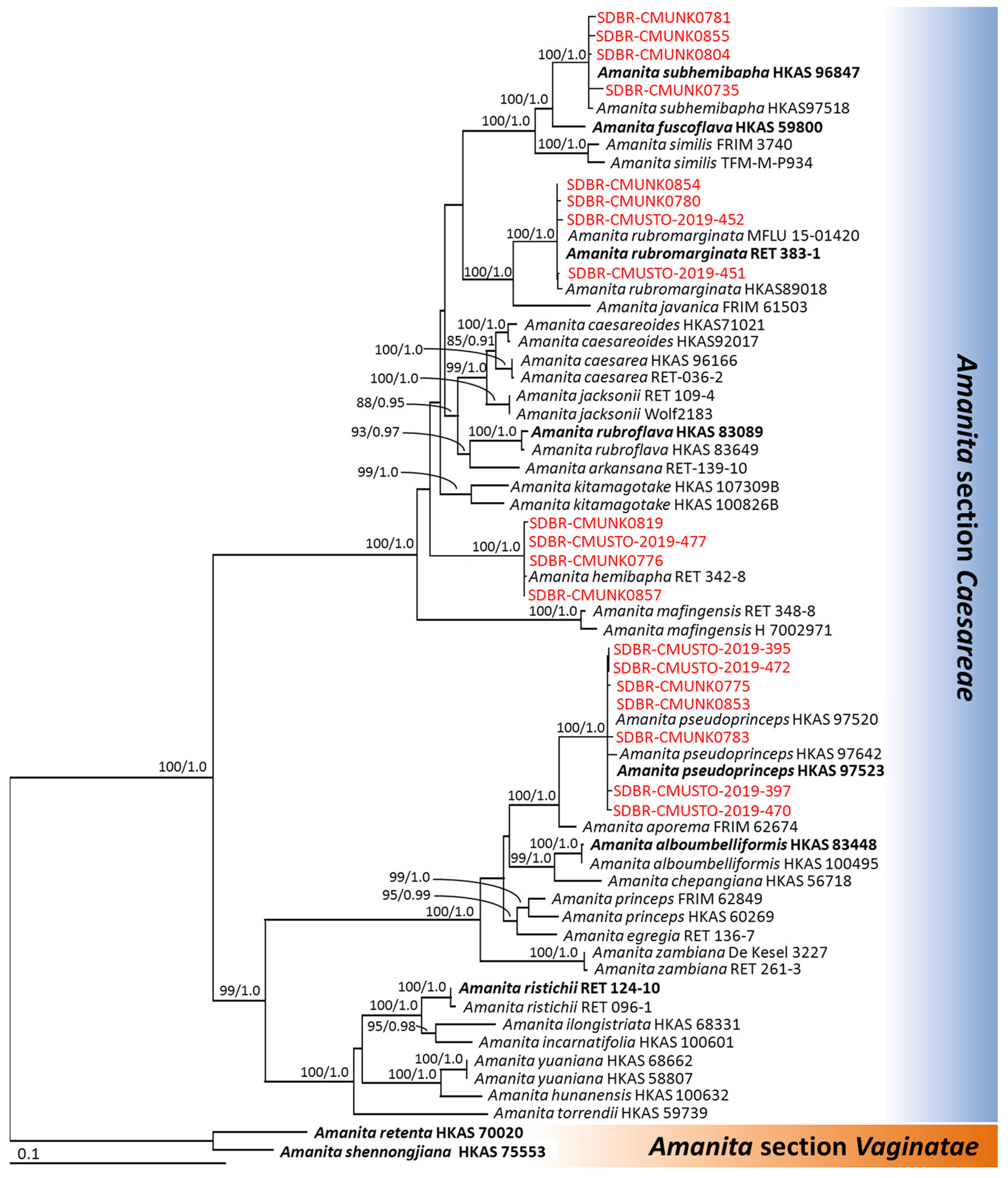
3.2. Phylogenetic Analyses

The aligned dataset of the combined ITS, nrLSU, rpb2, and tef-1 sequences consisted of 2831 characters including gaps (ITS: 1–903, nrLSU: 904–1676, rpb2: 1677–2316, and tef-1: 2317–2831). The matrix had 1192 different alignment patterns and 23.41% gaps or undetermined characters. A final ML Optimization Likelihood value of −15972.8506 was the best-scoring RAxML tree. For BI analysis, the final average standard deviation value of the split frequencies at the end of the total MCMC generations was calculated as 0.00723. The topology of the phylogenetic trees from the ML and BI analyses were similar. A phylogenetic tree obtained from the ML analysis is represented in Figure 2. Our phylogenetic tree was constructed with the aim of having similar outcomes to previous phylogenetic studies [4,52,53,54]. The phylogenetic tree consisted of 62 specimens of Amanita sect. Caesareae and two specimens of Amanita sect. Vaginatae (the outgroup). The phylogenetic tree clearly separated the 19 specimens obtained in this study into four species clades, namely A. hemibapha (4 specimens), A. pseudoprinceps (7 specimens), A. rubromarginata (4 specimens), and A. subhemibapha (4 specimens) in Amanita sect. Caesareae with high supported values (BS = 100% and PP = 1.0).

3.3. Morphological Descriptions

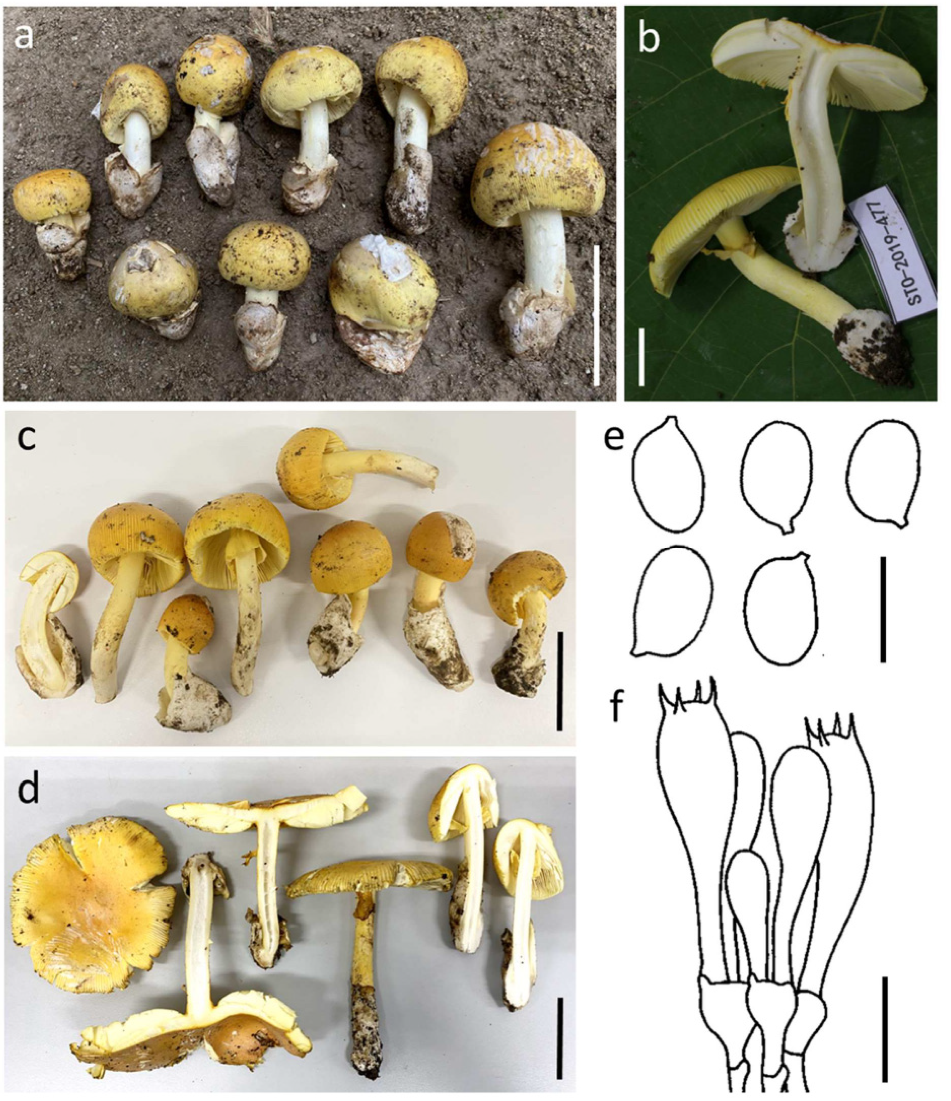
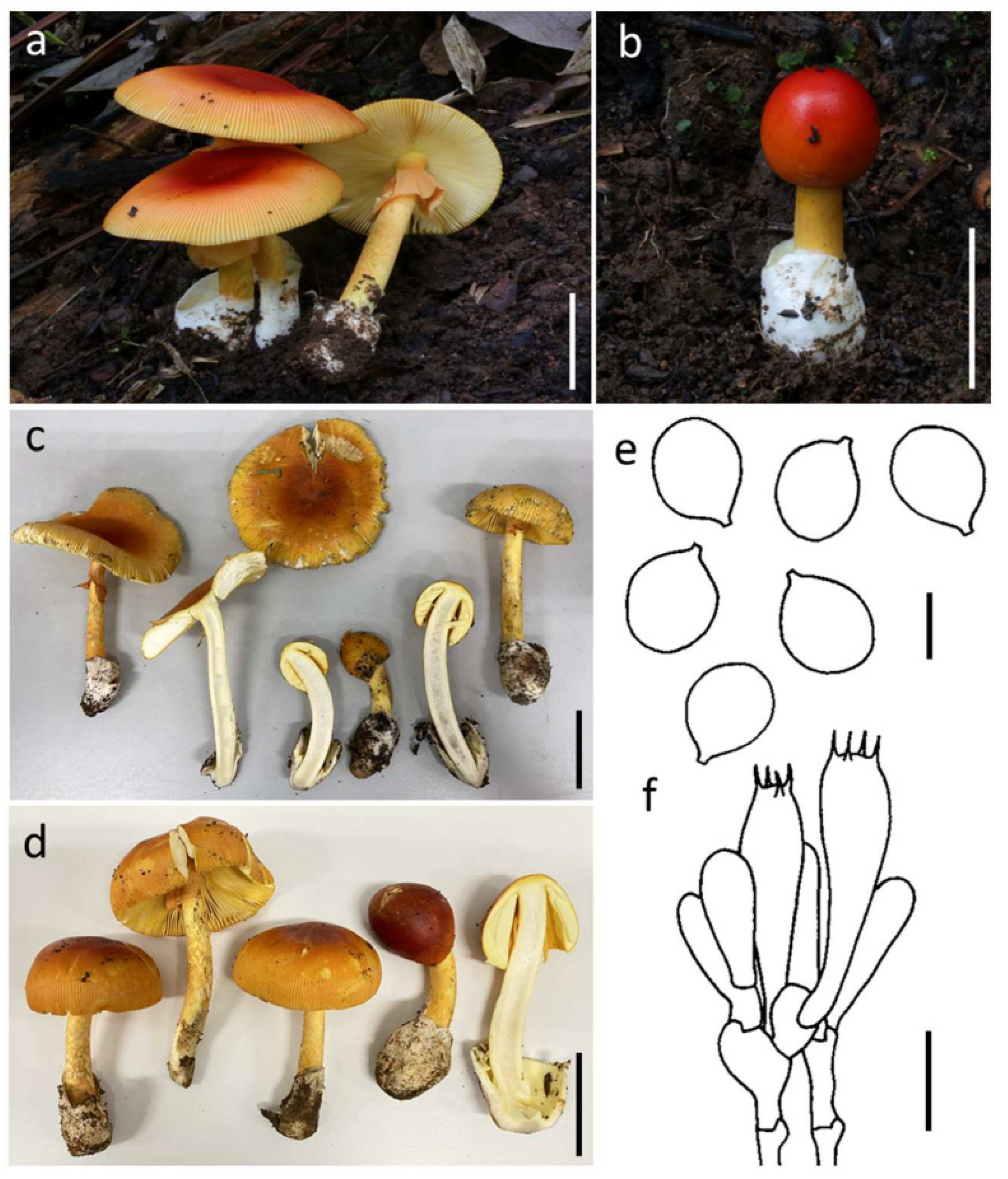
3.3.1. Amanita hemibapha (Berk. & Broome) Sacc., Syll. Fung. (Abellini) 5: 13 (1887) (Figure 3)

Basidioma medium-sized. Pileus 6–12 cm diam., plano-convex with the center slightly depressed, orange-red (6A6–8) to lemon yellow (3B8) at center, and becoming vivid yellow (3A7–8) to pale yellow (3A3–4) towards the margin; universal veil on pileus white patch; margin striate (0.3 R), non-appendiculate; context 5 mm wide, white (1A1) to yellowish white (2A2), unchanging. Lamellae free, crowded, cream white (1A1–2); lamellulae truncate. Stipe 7–10.5 × 0.5–1.5 cm, cylindrical, covered by light yellow to vivid yellow (2A5–8) fibrous squamules; context broadly fistulose, white (1A1). Bulb absent. Universal veil on stipe base saccate, membranous, up to 4 cm high, white (1A1). Partial veil subapical, fragile, vivid yellow (3A7–8).
Figure 3. Amanita hemibapha SDBR-CMUNK0776 (a), SDBR-CMUSTO-2019-477 (b), SDBR-CMUNK0857 (c) and SDBR-CMUNK0819 (d). Basidiomata (ad). Basidiospores (e). Basidia (f). Scale bars: (a,c,d) = 5 cm; (b) = 1 cm; (e) = 10 μm; (f) = 15 μm.
Jof 09 00343 g003
Lamellar trama bilateral, divergent; mediostratum 25–40 μm wide, filamentous hyphae abundant, 2–8 μm wide; clavate to ellipsoidal inflated cells 70–98 × 20–23 μm; vascular hyphae scarce. Subhymenium 20–35 μm thick in 2–3 layers, with subglobose to ellipsoidal or irregular cells, 6–25 × 5–15 μm. Basidia 32–50 × 8–12 μm, clavate, 4-spored with sterigmata 3–5 μm long; clamps present at base. Basidiospores (7.0–) 7.5–11.0 (–12.0) × 5.5–7.0 μm, Q = 1.23–1.64 (–1.71) μm, Qm = 1.44 ± 0.13, broadly ellipsoid to ellipsoid, sometimes elongate, inamyloid, hyaline, thin-walled, smooth; apiculus small. Lamellar edge sterile; filamentous hyphae 1–5 μm wide, hyaline, thin-walled; inflated cells, with subglobose, ovoid to ellipsoidal, 14–40 × 12–30 μm, single and terminal or in chains of 2–3, hyaline, thin-walled. Pileipellis 60–110 μm thick; 2-layered, upper layer 15–35 μm thick, filamentous hyphae 1–5 μm wide, weakly gelatinized, branching, thin-walled, hyaline; lower layer 50–85 μm thick, filamentous hyphae 2–7 μm wide, branching, thin-walled, hyaline to light yellow; vascular hyphae rare. Inner surface of universal veil on stipe base filamentous hyphae dominant 1–9 μm wide, hyaline to light yellow, thin-walled, branching; inflated cells, with subglobose, pyriform to clavate, 30–85 × 20–54 μm, hyaline, thin-walled; vascular hyphae rare. Outer surface of universal veil on stipe base similar to structure of inner part, but presenting more abundant inflated cells. Stipe trama longitudinally acrophysalidic; filamentous, undifferentiated hyphae 1–12 μm wide, thin-walled, frequently branching; acrophysalides 65–190 × 25–65 μm, thin-walled; vascular hyphae rare. Partial veil filamentous hyphae very abundant, 2–9 μm wide, hyaline, thin-walled; inflated cells scarce to locally abundant, globose, subglobose to clavate, 22–70 × 9–25 μm, hyaline to light yellow, thin-walled; vascular hyphae rare. Clamp connections present in all tissues of basidioma.
Habitat: Solitary to scattered on soil in tropical deciduous forests dominated by Dipterocarpus and Shorea.
Distribution: known from China [3,4,8,10], India [55,56], Sri Lanka [57], and Thailand [5], this study.
Specimens examined: Thailand, Chiang Mai Province, Mae Taeng District, 19°07′45″ N 98°45′51″ E, alt. 1421 m, 9 August 2019, Yuan S.L., STO-2019-477 (SDBR-CMUSTO-2019-477); Doi Saket District, 18°53′2″ N 99°9′17″ E, alt. 343 m, 26 July 2020, Kumla J. and Suwannarach N., CMUNK0819 (SDBR-CMUNK0819), 11 August 2020, Kumla J. and Suwannarach N., CMUNK0857 (SDBR-CMUNK0857); Lumphun Province, Mueang District, Chiang Mai University Haripunchai Campus, 18°30′10″ N 99°8′25″ E, alt. 400 m, 25 July 2020, Suwannarach N., CMUNK0776 (SDBR-CMUNK0776).
Remarks: The remarkable features of A. hemibapha include the fact that this species has a reddish yellow or orange-red tone in the center of its pileus that becomes vivid yellow or pale yellow towards the edges. This species is also known to have a yellow annulus. Amanita hemibapha was firstly reported from Sri Lanka, and then found in China, India and Thailand [3,4,5,8,10,55,56,57]. Morphologically, A. hemibapha is easily confused with A. caesareoides Lj. N. Vassiljeva, A. kitamagotake N. Endo & A. Yamada, A. rubroflava Y.Y. Cui, Q. Cai & Zhu L. Yang and A. subhemibapha Zhu L. Yang, Y.Y. Cui & Q. Cai. However, A. caesareoides, A. kitamagotake, and A. rubroflava differ from A. hemibapha by having a distinctly umbonate pileus, a much darker and reddish tone in the pileus center and relatively broader basidiospores [3,4,58]. Amanita subhemibapha, originally reported from China, differs from A. hemibapha by having a lighter yellowish tone pileus and relatively broader basidiospores (8.0–11.0 × 6.0–8.0 μm) [4]. According to illustrations of the Thai specimens (Figure 3), most of them are orange-red to lemon yellow at the center of the pileus. This feature was different from the original description of A. hemibapha due to the presence of red to orange-red in the center of the pileus. This may be influenced by the phenotypic variability that exists across a wide geographic range. However, the sizes of basidiomata, other macroscopic and microscopic features of the Thai specimens agree well with descriptions of previous studies [3,4,5,10,55,56,57]. Hence, we identify our specimens as A. hemibapha using a combination of morphological and molecular data.

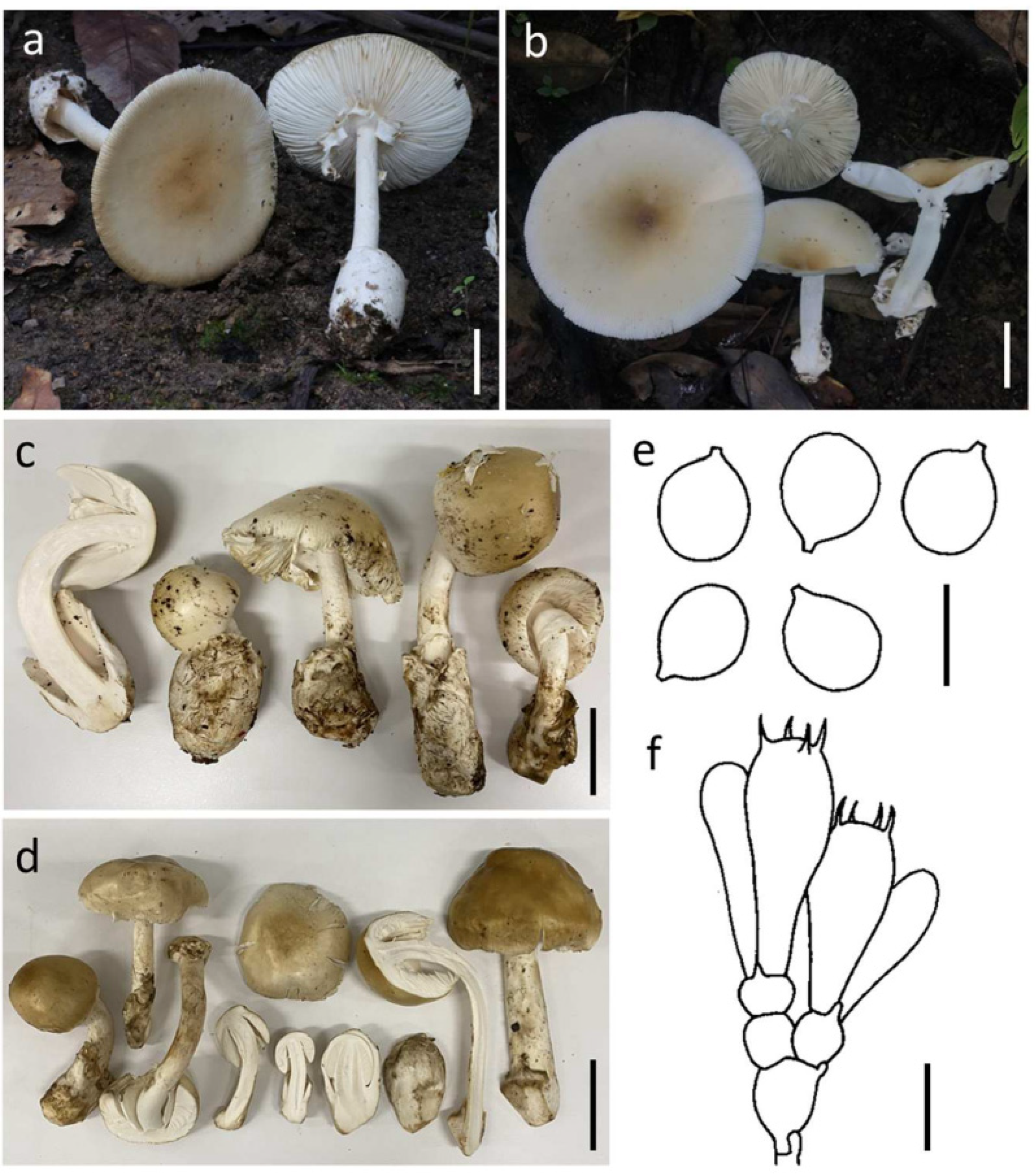
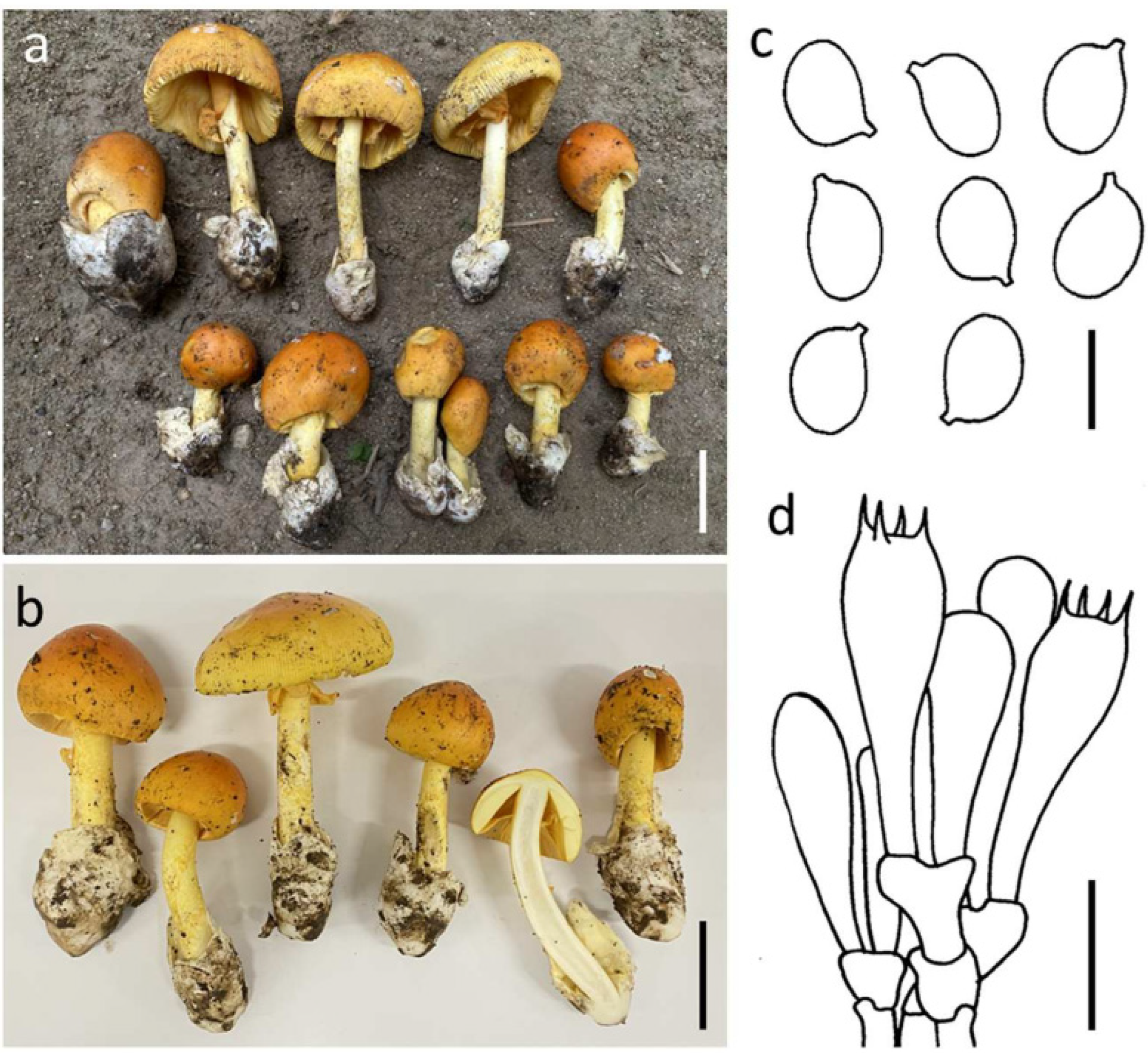
3.3.2. Amanita pseudoprinceps Y.Y. Cui, Q. Cai & Zhu L. Yang, Fungal Divers. 91: 59 (2018) (Figure 4)

Basidioma medium-sized to very large. Pileus 8.5–16 cm diam., hemispherical, convex to applanate with age, light yellow (4A5–6) to greyish orange (5B3–4) or sometime golden yellow (5B7–8) at center, and becoming yellow white (4A2–3) to white (4A1) towards the margin; universal veil on pileus absent; margin striate (0.1–0.3 R), non-appendiculate; context 9.5–13.5 mm wide, white (1A1), unchanging. Lamellae free, crowded, white to cream white (1A1–2); lamellulae truncate. Stipe 11.5–17.2 × 1.1–1.9 cm, subcylindrical with slightly tapering upwards and apex slightly expanded, white, covered by minute, white (1A1) fibrous squamules; context fistulose to broadly fistulose, white (1A1), Bulb absent. Universal veil on stipe base saccate, membranous, up to 7 cm high, white (1A1). Partial veil apical, membranous, white (1A1), becoming fragile or disappear with age.
Figure 4. Amanita pseudoprinceps SDBR-CMUSTO-2019-472 (a), SDBR-CMUNK0775 (b), SDBR-CMUNK0853 (c) and SDBR-CMUNK0783 (d). Basidiomata (ad). Basidiospores (e). Basidia (f). Scale bars: (ad) = 5 cm; (e) = 10 μm; (f) = 15 μm.
Jof 09 00343 g004
Lamellar trama bilateral, divergent; mediostratum 40–75 μm wide, filamentous hyphae abundant, 2–12 μm wide; fusiform to ellipsoidal inflated cells 85–213 × 12–26 μm; vascular hyphae scarce to locally abundant. Subhymenium 30–50 μm thick in 2–3 layers, with subglobose to ellipsoidal or irregular cells, 12–33 × 10–26 μm. Basidia 36–53 × 12–18 μm, clavate, 4-spored with sterigmata 3–6 μm long; clamps present at base. Basidiospores (9.0–) 9.5–12.5 (–13.0) × (8.0–) 8.5–12.0 (–12.5) μm, Q = 1.00–1.20 (1.22) μm, Qm = 1.10 ± 0.06, globose to subglobose, sometimes broadly ellipsoid, inamyloid, hyaline, thin-walled, smooth; apiculus small. Lamellar edge sterile; filamentous hyphae 2–6 μm wide, hyaline, thin-walled; inflated cells, with subglobose to ellipsoidal, 12–35 × 8–34 μm, single and terminal or in chains of 2–3, hyaline, thin-walled. Pileipellis 85–160 μm thick; 2-layered, upper layer 45–80 μm thick, filamentous hyphae 2–5 μm wide, gelatinized, branching, thin-walled, hyaline; lower layer 40–80 μm thick, filamentous hyphae 2–8 (–15) μm wide, branching, thin-walled, hyaline to light yellow; vascular hyphae rare. Inner surface of universal veil on stipe base filamentous hyphae dominant 1–8 μm wide, hyaline to light yellow, thin-walled, branching; inflated cells, with subglobose, fusiform to clavate, 50–93 × 15–52 μm, hyaline, thin-walled, mostly terminal or sometimes in chains of 2–3; vascular hyphae rare. Outer surface of universal veil on stipe base similar to structure of inner part, but presenting more abundant inflated cells. Stipe trama longitudinally acrophysalidic; filamentous, undifferentiated hyphae 2–7 μm wide, thin-walled, frequently branching; acrophysalides 100–233 × 23–45 μm, thin-walled; vascular hyphae rare. Partial veil filamentous hyphae very abundant, 1–7 μm wide, hyaline, thin-walled; inflated cells scarce to locally anundant, globose, subglobose to ellipsoidal, 12–70 × 12–35 μm, hyaline to light yellow, thin-walled; vascular hyphae rare. Clamp connections present in all tissues of basidioma.
Habitat: Solitary to scattered on soil in tropical deciduous forests dominated by Dipterocarpus and Shorea.
Distribution: known from China [4] and Thailand (this study).
Specimens examined: Thailand, Chiang Mai Province, Mae Taeng District, 19°05′38.2″ N 98°52′44.4″ E, alt. 1105 m, 7 August 2019, Yuan S.L., STO-2019-395 (SDBR-CMUSTO-2019-395); Yuan S.L., STO-2019-397 (SDBR-CMUSTO-2019-397); 19°07′45.0″ N 98°45′51.0″ E, alt. 1421 m, 9 August 2019, Yuan S.L., STO-2019-470 (SDBR-CMUSTO-2019-470); Yuan S.L., STO-2019-472 (SDBR-CMUSTO-2019-472). Doi Saket District, 18°53′2″ N 99°9′17″ E, alt. 343 m, 26 July 2020, Kumla J. and. Suwannarach N., CMUNK0783 (SDBR-CMUNK0783), 2 August 2022, Kumla J. and. Suwannarach N., CMUNK0853 (SDBR-CMUNK0853); Lumphun Province, Mae Tha District, 18°27′41″ N 99°10′30″ E, alt. 427 m, 25 July 2020, Kumla J. and. Suwannarach N., CMUNK0775 (SDBR-CMUNK0775).
Remarks: Morphologically, A. pseudoprinceps resembles A. princeps Corner & Bas by having a similar yellowish-brown pileus and margin striates (about 0.2–0.3 R). However, A. princeps presents the larger basidiomata, as well as an outer layer of volval remnants on the stipe cracks and peels in pale buff thin patches [4,54,59]. According to the phylogenetic analysis, our seven samples cluster together with three other samples of A. pseudoprinceps and form a well-supported clade that presents a sister clade with A. aporema Boedijn. Meanwhile, these two species possess a similar brown tone pileus. However, A. aporema has a smaller (6–10 cm) but much darker pileus, as well as obviously longer margin striates (0.5–0.6 R) [4,54,60].

3.3.3. Amanita rubromarginata Har. Takah., Mycoscience 45: 372 (2004) (Figure 5)

Basidioma medium-sized to large. Pileus 6.0–10.0 cm diam., convex to plano-convex with the center depressed, reddish orange (7B7) over disk, or sometime orange red (8B7–8) at center and becoming light orange (5A4–5) towards the margin; universal veil on pileus absent; margin striate (0.4–0.5 R), non-appendiculate; context 4.5–8.0 mm wide, yellowish white (3A2), unchanging. Lamellae free, crowded, pale yellow to light yellow (4A3–4), with lamellar edges reddish orange (7B7–8); lamellulae truncate. Stipe 13.7–20.0 × 1.0–1.8 cm, subcylindrical with slightly tapering upwards, yellow (3A6–7), densely covered by reddish yellow to deep yellow (4A7–8) squamules; context broadly fistulose, yellowish white (3A2) to white (3A1). Bulb absent. Universal veil on stipe base saccate, membranous, up to 5 cm high, white (1A1). Partial veil subapical to apical, membranous, dark orange (5A7–8) to orange (6A6–7).
Figure 5. Amanita rubromarginata SDBR-CMUSTO-2019-451 (a), SDBR-CMUSTO-2019-452 (b), SDBR-CMUNK0780 (c) and SDBR-CMUNK0854 (d). Basidiomata (ad). Basidiospores (e). Basidia (f). Scale bars: (ad) = 5 cm; (e) = 5 μm; (f) = 15 μm.
Jof 09 00343 g005
Lamellar trama bilateral, divergent; mediostratum 20–25 μm wide, filamentous hyphae abundant, 2–11 μm wide; fusiform to ellipsoidal inflated cells 60–153 × 15–27 μm; vascular hyphae scarce. Subhymenium 25–30 μm thick in 1–3 layers, with subglobose to ellipsoidal or irregular cells, 8–18 × 5–13 μm. Basidia 32–46 × 8–13 μm, clavate, 4-spored with sterigmata 3–4 μm long; clamps present at base. Basidiospores 7.0–9.5 (–10.0) × 6.0–7.0 (–8.0) μm, Q = (1.08–) 1.13–1.50 μm, Qm = 1.28 ± 0.11, subglobose to broadly ellipsoid or ellipsoid, inamyloid, hyaline, thin-walled, smooth; apiculus small. Lamellar edge sterile; filamentous hyphae 3–7 μm wide, hyaline, thin-walled; inflated cells, with globose, pyriform to clavate, 15–46 × 12–27 μm, hyaline, thin-walled. Pileipellis 80–130 μm thick; 2-layered, upper layer 25–40 μm thick, filamentous hyphae 1–6 μm wide, gelatinized, branching, thin-walled, hyaline; lower layer 50–100 μm thick, filamentous hyphae 3–8 μm wide, branching, thin-walled, hyaline to light yellow; vascular hyphae rare. Inner part of universal veil on stipe base filamentous hyphae dominant 2–11 μm wide, hyaline to light yellow, thin-walled, branching; inflated cells, with subglobose, ovoid to clavate, 30–72 × 10–70 μm, hyaline, thin-walled; vascular hyphae rare. Outer surface of universal veil on stipe base similar to structure of inner part, but presenting more abundant inflated cells. Stipe trama longitudinally acrophysalidic; filamentous, undifferentiated hyphae 1–6 μm wide, thin-walled, frequently branching; acrophysalides 150–295 × 33–53 μm, thin-walled; vascular hyphae rare. Partial veil filamentous hyphae very abundant, 2–11 μm wide, hyaline, thin-walled; inflated cells scarce to locally abundant, ellipsoidal to clavate, 45–110 × 10–16 μm, hyaline to light yellow, thin-walled; vascular hyphae rare. Clamp connections present in all tissues of basidioma.
Habitat: Solitary to scattered on soil in tropical deciduous forests dominated by Dipterocarpus and Shorea.
Distribution: known from China [4], Japan [61,62], and Thailand [52], this study.
Specimens examined: Thailand, Chiang Mai Province, Mae Taeng District, 19°06′53.3″ N 98°44′22.7″ E, alt. 1718 m, 8 August 2019, Yuan S. Liu, STO-2019-451 (SDBR-CMUSTO-2019-451); STO-2019-452 (SDBR-CMUSTO-2019-452); Doi Saket District, 18°53′2″ N 99°9′17″ E, alt. 343 m, 2 August 2022, Kumla J. and Suwannarach N., CMUNK0854 (SDBR-CMUNK0854); Lumphun Province, Mae Tha District, 18°27′41″ N 99°10′30″ E, alt. 427 m, 25 July 2020, Kumla J. and Suwannarach N., CMUNK0780 (SDBR-CMUNK0780).
Remarks: Morphologically, A. rubroflava is easily confused with A. rubromarginata. However, A. rubroflava differs from A. rubromarginata by having a distinctly umbonate pileus and larger basidiospores (8.0–10.0 × 6.5–8.0 μm) [4]. Phylogenetically, Amanita javanica (Corner & Bas) Oda, Tanaka & Tsuda is closely related to A. rubromarginata. Meanwhile, both these two species share similar characteristics, such as an orange-red tone pileus and reddish yellow squamules covering their stipes. However, A. javanica has a distinctly umbonate pileus, while A. rubromarginata does not appear to display this characteristic [4,54,59,62].

3.3.4. Amanita subhemibapha Zhu L. Yang, Y.Y. Cui & Q. Cai, Fungal Divers. 91: 65 (2018) (Figure 6)

Basidioma medium-sized to large. Pileus 6.0–10.0 cm diam., convex to plano-convex, lacking an umbo at center, purely orange (5B5–8) when young, but becoming orange (5B5–8) at center and yellow (4A6–8) to yellowish (3A3–6) at margin when mature; universal veil on pileus absent; margin striate (0.25–0.3 R), non-appendiculate; context 4.5–5.0 mm wide, yellow (4A6–8) to yellowish (3A3–6), unchanging. Lamellae free, crowded, white (1A1) to cream (1A4–6), with lamellar edges yellow (4A6–8); lamellulae truncate. Stipe 5–15 × 0.7–1.5 cm, subcylindrical with slightly tapering upwards, with apex slightly expanded, yellow (4A6–8) to orange (5B5–8), with its surface covered with concolorous, snakeskin-shaped squamules; context white (1A1), hollow in center. Bulb absent. Universal veil on stipe base saccate, membranous, up to 5 cm high 3, white (1A1). Partial veil apical to subapical, yellow (4A6–8) to orange (5B5–8).
Figure 6. Amanita subhemibapha SDBR-CMU0781 (a) and SDBR-CMU0855 (b). Basidiomata (a,b). Basidiospores (c). Basidia (d). Scale bars: (a,d) = 5 cm; (c) = 10 μm; (d) = 15 μm.
Jof 09 00343 g006
Lamellar trama bilateral, divergent; mediostratum 25–70 μm wide, filamentous hyphae abundant, 2–7 μm wide; ellipsoid, fusiform to clavate inflated cells 30–80 × 10–27 μm; vascular hyphae scarce. Subhymenium 30–50 μm thick in 2–3 layers, with subglobose to ellipsoid cells, 10–25 × 8–20 μm. Basidia 40–50 × 9–12 μm, clavate, 4-spored with sterigmata 3–5 μm long; clamps present at base. Basidiospores (7.0–) 8.0–11.0 × 6.5–8.5 (–9.0) μm, Q = 1.15–1.53 (–1.65), Qm = 1.34 ± 0.08, broadly ellipsoid to ellipsoid, inamyloid, hyaline, thin-walled, smooth; apiculus small. Lamellar edge sterile; filamentous hyphae 2–4 μm wide, hyaline, thin-walled; inflated cells, with subglobose to ellipsoid or sphaeropedunculate, 8–45 × 8–20 μm, single and terminal or in chains of 2–3, hyaline, thin-walled. Pileipellis 90–170 μm thick; 2-layered, upper layer 30–145 μm thick, filamentous hyphae 2–5 μm wide, gelatinized, branching, thin-walled, hyaline; lower layer 30–55 μm thick, filamentous hyphae 3–8 (–10) μm wide, branching, thin-walled, hyaline to light yellow; vascular hyphae scarce. Inner surface of universal veil on stipe base filamentous hyphae dominant 2–10 μm wide, hyaline to light yellow, thin-walled, branching; inflated cells, with subglobose, fusiform to ellipsoid, 55–100 × 20–70 μm, hyaline, thin-walled, mostly terminal or sometimes in chains of 2–3; vascular hyphae rare. Outer surface of universal veil on stipe base similar to structure of inner part, but presenting more abundant inflated cells. Stipe trama longitudinally acrophysalidic; filamentous, undifferentiated hyphae 2–10 (–15) μm wide, thin-walled, frequently branching; acrophysalides 60–260 × 25–65 μm, thin-walled; vascular hyphae rare. Partial veil filamentous hyphae very abundant, 2–10 μm wide, hyaline, thin-walled; inflated cells scarce to locally abundant, subglobose, fusiform to clavate, 20–100 × 10–35 μm, hyaline to light yellow, thin-walled; vascular hyphae rare. Clamp connections present in all tissues of basidioma.
Habitat: Solitary to scattered on soil in subtropical broad-leaved or mixed forests with Dipterocarpaceae, Fagaceae, and Pinaceae.
Distribution: known from China [4] and Thailand (this study).
Specimens examined: Thailand, Chiang Mai Province, Doi Saket District, 18°53′2″ N 99°9′17″ E, alt. 343 m, 26 July 2021, Kumla J. and Suwannarach N., CMUNK0804 (SDBR-CMUNK0804); 18°53′2″ N 99°9′17″ E, alt. 343 m, 2 August 2022, Kumla J. and Suwannarach N., CMUNK0855 (SDBR-CMUNK0855); Lumphun Province, Mueang District, Chiang Mai University Haripunchai Campus, 18°32′34″ N 99°9′231″ E, alt. 450 m, 25, July, 2020, Suwannarach N., CMUNK0781 (SDBR-CMUNK0781); Mae Tha District, 18°27′41″ N 99°10′30″ E, alt. 427 m, 27 August 2020, Kumla J. and Suwannarach N., CMUNK0735 (SDBR-CMUNK0735).
Remarks: Morphologically, A. subhemibapha is easily confused with A. hemibapha, A. javanica and A. kitamagotake. Morphological comparisons of A. hemibapha and A. subhemibapha have been included in our remarks pertaining to A. hemibapha. Amanita javanica differs from A. subhemibapha by having a broadly umbonate and much darker yellow tone in the center of the pileus, longer tuberculate striates (0.4–0.5 R) on the margins and smaller basidiospores (7.5–9.0 × 5.8–7.0 μm) [54]. Alternatively, A. kitamagotake differs from A. subhemibapha by having an umbonate pileus and narrower basidiospores (9.0–13.5 × 6.5–8.5 μm) [58]. Based on multigene phylogeny, A. subhemibapha forms a sister clade with A. fuscoflava Zhu L. Yang, Y.Y. Cui & Q. Cai. However, A. fuscoflava has a dark brown tone in the pileus center, much longer margin striates (0.5–0.7 R) and relatively narrower basidiospores (8.5–10.5 × 6.0–7.0 μm) [4].
Traditionally, morphological characteristics have been the primary basis for the identification of Amanita species [7,8,11,32]. However, identification can be difficult due to the high phenotypic variability that is influenced by differing environmental conditions and geographic distributions. Therefore, it is crucial to identify the Amanita species using DNA-based methods. The current classification of the genus Amanita is based on combined data on their morphological characteristics and molecular data. Moreover, multi-gene molecular phylogeny has provided researchers with a powerful tool for the identification of the Amanita species [4,14,36,52,53,54,58]. In this present study, specimens of the edible Amanita species collected in northern Thailand were identified as A. hemibapha, A. pseudoprinceps, A. rubromarginata, and A. subhemibapha based on morphological characteristics and multi-gene phylogenetic analyses. The results of morphological comparisons of four edible Amanita species in this study are presented in Table 3. Morphologically, the color of the pileus and the larger spore size found in A. pseudoprinceps clearly differentiate it from those other three species. Additionally, the yellow annulus and narrow spores in A. hemibapha clearly distinguish it from A. rubromarginata and A. subhemibapha. Remarkably, A. rubromarginata has a redder and more of an orange-red-shaded pileus and annulus than A. subhemibapha. The multi-gene phylogenetic analysis also supports the determination that A. hemibapha, A. pseudoprinceps, A. rubromarginata, and A. subhemibapha are different species. Four Amanita species obtained from natural forests, roadsides, and local markets in this study belonged to the Amanita section Caesareae. This section is a highly regarded edible mushroom in the genus Amanita [4,16,17,18,19]. Prior to this study, the toxicological analysis of A. hemibapha showed that no amatoxins and phallotoxins had been discovered and that it should be regarded as an edible species [63]. However, further research is required to fully understand the edibility and safety of A. pseudoprinceps, A. rubromarginata, and A. subhemibapha based on their toxicological studies. As a result, our study should be considerably important and highly valuable in terms of stimulating deeper investigations of edible macrofungi in Thailand. It will also help researchers in understanding the distribution and ecology of Amanita.

3.4. Nutritional Analysis

A total of six samples of four edible Amanita species (namely A. hemibapha, A. pseudoprinceps, A. rubromarginata, and A. subhemibapha) obtained in this study have been included in the experiments. In this study, the fruiting bodies of edible Amanita were analyzed for their nutritional composition, which included ash, carbohydrate, protein, fat and fiber. The results are presented in Table 4. The results indicate that the protein contents in A. pseudoprinceps and A. subhemibapha were significantly higher than A. hemibapha and A. rubromarginata. The highest content of fiber was found in A. pseudoprinceps. It was determined that A. rubromarginata had the highest ash content. In addition, the carbohydrate content in A. hemibapha was significantly higher than the other Amanita species. The highest fat content was obtained in A. rubromarginata, but this value was not found to be significantly different from the fat content of A. hemibapha. These results were consistent with previous studies, which reported that edible wild mushrooms to be natural sources of nutrients for human diets (high-protein and low-fat contents), while the nutritional composition of each mushroom is dependent upon the mushroom species [20,22,64,65]. The amounts of ash, carbohydrate, protein, fat, and fiber of the four edible Amanita species in this study were within the ranges mentioned in previous reports of edible Amanita. Accordingly, the ash (0.11–11.82% dry weight), carbohydrate (22.16–61.70% dry weight), protein (10.11–45.65% dry weight), fat (0.17–17.52% dry weight) and fiber (1.18–30.30% dry weight) contents were found in various edible Amanita species, namely A. caesarea, A. calyptroderma, A. fulva, A. hemibapha, A. princeps, A. rubescens, and A. zambiana [66,67,68,69,70,71,72,73,74,75]. When compared to the findings of other previously published reports, the protein content of the Amanita species obtained in this study was relatively higher than those of A. calyptroderma [75] and A. loosei [69]. With regard to the outcomes of this study, this is the first comprehensive report on the nutritional composition of A. pseudoprinceps, A. rubromarginata, and A. subhemibapha.

3.5. Determination of Total Phenolic Content

The total phenolic content of each extract of Amanita in this study is presented in Table 5. It was found that the total phenolic contents ranged from 0.94–1.62 mg GAE/g dw. The highest value of total phenolic content was found in the extract of A. pseudoprinceps, followed by the extracts of A. subhemibapha and A. hemibapha. The lowest value of total phenolic content was found in the extract of A. rubromarginata. Previous findings support the results of this study in that the amount of phenolic contents of edible wild mushrooms varied within different ranges and was dependent upon the various mushroom species [45,76,77,78]. According to our results, the amounts of total phenolic content obtained in this study were within the previously reported ranges of phenolic content found in edible wild mushrooms and varied from 0.39–38.44 mg GAE/g dw [76,77,78,79]. The total phenolic contents in the methanolic extracts of A. caesarea [79], A. fulva [74], A. hemibapha [80], A. javanica [81], A. ovoidea [82], A. princeps [80,81], and A. zambiana [73] were reported as 0.64, 0.39, 8.5, 18.01, 0.50, 14.29–16.80 and 8.76 mg GAE/g dw, respectively. Additionally, the total phenolic contents in the ethanolic extracts of A. javanica and A. princeps were 12.79 and 16.52 mg GAE/g dw, respectively [81]. When compared to the results of previously published reports, the phenolic contents of the ethanolic extracts of A. hemibapha, A. pseudoprinceps, A. rubromarginata, and A. subhemibapha obtained in this study have been found to be relatively higher than those of methanolic extracts of A. caesarea, A. fulva and A. ovoidea [74,79,82], while they were relatively lower than extracts of A. javanica, A. princeps and A. zambiana [73,81]. However, the phenolic content of A. hemibapha obtained in this study was lower than that of the previous report of Butkhup et al. [80]. It can be concluded from our experiments that, similarly to the results of previous studies, the total content of phenolic can be influenced by different phenolic compounds found in mushroom extracts, along with the extractability of the different solvents used in the preparation process [45,81,83,84]. According to several previous studies, catechin, р-coumaric acid, gallic acid, hydroxycinnamic acid, quercetin, protocatechuic acid, rosmarinic acid, and syringic acid were found to be the major phenolic components in the ethanolic extracts of edible wild mushrooms [45,85,86,87]. Some previous investigations revealed that the Folin–Ciocalteu assay, a method typically used for detection and quantification of total phenolic content, might be unsuited for total phenolic content measurement in complex biological samples due to high interference from various reducing compounds contained in samples [88,89,90]. The effectiveness of the Folin–Ciocalteu assay is also hampered by its limited suitability for some phenolic compounds [89,90]. Therefore, the measurement of total phenolic content in this study will still be assessed using other techniques such as high-performance liquid chromatography (HPLC) or liquid chromatography–mass spectrometer mass spectrometry (LC-MS) for further studies to characterize and identify the phenolic compounds contained in mushroom extracts.

3.6. Antioxidant Assay

A single method cannot fully determine the antioxidant activity of mushroom extracts. Thus, in this study, three methods, namely ABTS, DPPH, and FRAP assays, were used to determine the antioxidant activity of the ethanolic extracts of different samples of edible Amanita species. The ABTS and DPPH values were determined by evaluating the scavenging abilities of ABTS and DPPH radicals, respectively (by measuring the decrease in ABTS and DPPH radical absorption after exposure to radical scavengers) [91,92]. The FRAP assay was used to measure the conversion of the ferric form (Fe3+) to the ferrous form (Fe2+) [92]. In this study, the highest values of DPPH activity were observed in the extract of the A. pseudoprinceps, followed by the extracts of A. hemibapha and A. subhemibapha (Table 5). The lowest value of DPPH activity was observed in the extract of A. rubromarginata. Furthermore, the results indicated that all extracts exhibited positive results in terms of the ABTS and FRAP assays, while the ABTS values varied from 0.56 to 1.00 mg TE/g dw (Table 5). The highest ABTS value was observed in the extract of A. pseudoprinceps, followed by the extracts of A. hemibapha, A. subhemibapha, and A. rubromarginata. In the FRAP system, the extract of A. pseudoprinceps had significantly higher FRAP values than the extracts from the other samples (Table 5). The results from the ABTS, DPPH, and FRAP assays were similar and demonstrated that the extract of A. pseudoprinceps exhibited significantly high antioxidant activity. The lowest level of antioxidant activity was found in the extract of A. rubromarginata. According to Pearson correlation (p < 0.05), the total phenolic content of mushroom extract samples showed a significant strong positive correlation with DPPH (r = 0.975) and FRAP (r = 0.948) activities (Table 6). However, the positive correlation between the total phenolic content and ABTS activity (r = 0.762) was not statistically significant.
All extracts of the four edible Amanita species exhibited antioxidant activities. These results are consistent with those of previous studies which reported that the extracts of wild mushrooms (e.g., genera Amanita, Boletus, Cantharellus, Lactarius, and Russula) exhibited antioxidant activities that varied according to the mushroom species [45,66,78,80,81,82,83]. Furthermore, recent research has indicated that wild mushrooms contain dietary ingredients that are alternative sources of natural antioxidants [45,77,93]. In this study, A. pseudoprinceps exhibited the highest level of antioxidant activity due to the fact that it possesses high total polyphenol content. This determination is supported by the results of previous studies, which reported that high phenolic content is responsible for the high antioxidant activity [45,83,94]. Prior to this present study, the antioxidant activities of A. caesarea, A. calyptroderma, A. hemibapha, A. javanica, A. loosei, A. ovidea, and A. princeps have been reported from a variety of assays employing different mechanisms including lipid peroxidation, metal chelation, reducing power and scavenging activity, among others [69,75,79,80,81]. However, variations in the assays themselves, and the results they express, make it difficult to compare the outcomes obtained in this study with those of previous studies.

3.7. Determination of α-Glucosidase Inhibitory Activity

Importantly, α-glucosidase is one of the key enzymes related to hyperglycemia by leading to an increase in blood glucose levels [95,96]. Therefore, inhibition of the function of this enzyme can reduce and control the risk of hyperglycemia. In this study, the α-glucosidase inhibition activity of the extracts of each edible Amanita species was investigated in terms of the inhibition percentage. The results were then compared with those of acarbose (anti-diabetic drug). The results then revealed that all extract samples exhibited α-glucosidase inhibition activity, while the value of the inhibition percentage varied according to the differences in the extract samples (Table 5). The value of α-glucosidase inhibition activity in the extract samples varied from 19.26% to 31.44% inhibition. However, all mushroom extracts were found to be less effective than acarbose, a synthetic standard Inhibitor of α-glucosidase (44.06% inhibition at concentration of 1 mg/mL). These results are supported by those of previous studies, which reported that the extracts of certain edible wild mushrooms (e.g., Amanita, Astraeus, Boletus, Lactarius, Phlebopus, Russula, Suillus, and Tylopilus) have potential as natural α-glucosidase inhibitors. Accordingly, the α-glucosidase inhibition activity varied from 9.72–78.75% for each different mushroom species [45,97,98]. In this study, the amounts of α-glucosidase inhibitory activity obtained in this study were within the ranges reported from previous studies. Compared with the outcomes of a report conducted by Pongkunakorn et al. [97], the α-glucosidase inhibitory activity of the methanolic extracts of A. hemibapha (19.26 and 20.37%) and A. rubromarginata (20.28%) obtained in this study were lower than the α-glucosidase inhibitory activity of the water extracts of A. hemibapha and A. princeps, which were reported at 22.66% and 25.54%, respectively. Interestingly, the α-glucosidase inhibitory activity of the methanolic extracts of A. pseudoprinceps obtained in this study was higher than the α-glucosidase inhibitory activity of the water extracts of both A. hemibapha and A. princeps [97]. Several previous studies have reported that the use of different solvents resulted in different patterns of active compounds in mushroom extracts, which were related to biological activities including α-glucosidase inhibitory activity [83,84,97,98]. Importantly, this study is the first report on the α-glucosidase inhibition activities of A. pseudoprinceps, A. rubromarginata, and A. subhemibapha. This study found that the extracts of A. pseudoprinceps displayed a high level of α-glucosidase inhibition activity over the other extracts, which could be related to their high total phenolic content. Additionally, the total phenolic content of all mushroom extracts and α-glucosidase inhibitory activity were shown to be significantly correlated by Pearson correlation (p < 0.05) (Table 6). These results were similar to those of previous studies [45,99,100], which revealed that the α-glucosidase inhibitory activity of natural substances is strongly correlated with the phenolic compound content.

4. Conclusions

The edible Amanita specimens collected in northern Thailand were identified as A. hemibapha, A. pseudoprinceps, A. rubromarginata, and A. subhemibapha based on the relevant morphological characteristics and multi-gene phylogenetic analyses. These four Amanita species were selected for further experiments, wherein their nutritional composition, total phenolic content, antioxidant activities, and α-glucosidase inhibitory activities were evaluated. All Amanita species were high in protein and carbohydrate but low in fat content. Additionally, the methanolic extracts of these four Amanita species contained varied amounts of total phenolic content and exhibited varied results in terms of their antioxidant and α-glucosidase inhibitory activities. The highest levels of antioxidant and α-glucosidase inhibitory activities were found in the methanolic extract of A. pseudoprinceps. The findings of this investigation provide valuable information on the nutrient content, total phenolic content, and the antioxidant and α-glucosidase inhibitory potential of the edible Amanita species found in northern Thailand. Therefore, our results suggest that these four edible Amanita species can be representative of an alternative food source. These species are also a good source of natural antioxidants and exhibit potential to naturally inhibit α-glucosidase for human health benefits. However, future studies should be implemented to conduct a comprehensive mineral analysis and to identify the phenolic profiles present in each edible Amanita species.

Author Contributions

Conceptualization, J.K. and N.S.; methodology, J.K., N.S., Y.S.L. and K.T.; software, J.K. and N.S.; validation, J.K., Y.S.L., K.T. and S.L.; formal analysis, J.K. and K.T.; investigation, J.K., N.S., Y.S.L. and K.T.; resources, J.K., N.S. and Y.S.L.; data curation, J.K., N.S., Y.S.L. and K.T.; writing—original draft preparation, J.K., N.S., Y.S.L. and K.T.; writing—review and editing, J.K., N.S., Y.S.L., K.T. and S.L.; visualization, J.K., N.S. and S.L.; supervision, J.K., N.S. and S.L.; project administration, J.K. and S.L.; funding acquisition, J.K., N.S. and S.L. All authors have read and agreed to the published version of the manuscript.

Funding

This work was financially supported by Fundamental Fund 2022 (FF65/070), Chiang Mai University, Thailand and National Research Council of Thailand (N42A650198).

Institutional Review Board Statement

Not applicable.

Informed Consent Statement

Not applicable.

Data Availability Statement

The DNA sequence data obtained from this study have been deposited in GenBank under the accession numbers; ITS (OQ199032–OQ199048); nrLSU (OQ187796– OQ187813); rpb2 (OQ200073–OQ200091); tef-1 (OQ200092–OQ200110).

Acknowledgments

The authors are grateful to Russell Kirk Hollis for the English correction of this manuscript.

Conflicts of Interest

The authors declare no conflict of interest.

References

  1. Persoon, C.H. Tentamen Dispositionis Methodicae Fungorum; Apud Petrum Philippum Wolf: Leipzig, Germany, 1797. [Google Scholar]
  2. Bresinsky, A.; Besl, H. A Colour Atlas of Poisonous Fungi: A Handbook for Pharmacists, Doctors, and Biologists; CRC Press: London, UK, 1990. [Google Scholar]
  3. Yang, Z.L. Atlas of the Chinese Species of Amanitaceae; Science Press: Beijing, China, 2015; pp. 1–213. [Google Scholar]
  4. Cui, Y.Y.; Cai, Q.; Tang, L.P.; Liu, J.W.; Yang, Z.L. The family Amanitaceae: Molecular phylogeny, higher-rank taxonomy and the species in China. Fungal Divers. 2018, 91, 5–230. [Google Scholar] [CrossRef]
  5. Sanmee, R.; Tulloss, R.E.; Lumyong, P.; Dell, B.; Lumyong, S. Studies on Amanita (Basidiomycetes: Amanitaceae) in Northern Thailand. Fungal Divers. 2008, 3, 97–123. [Google Scholar]
  6. Tulloss, R.E. Amanita-distribution in the Americas, with comparison to eastern and southern Asia and notes on spore character variation with latitude and ecology. Mycotaxon 2005, 93, 189–231. [Google Scholar]
  7. Yang, Z.L.; Oberwinkler, F. Die Fruchtköperentwicklung von Amanita muscaria (Basidiomycetes). Nova Hedwig. 1999, 68, 441–468. [Google Scholar] [CrossRef]
  8. Yang, Z.L. Die Amanita-Arten von Südwestchina. Bibl. Mycol. 1997, 170, 1–240. [Google Scholar]
  9. Zhang, L.F.; Yang, J.B.; Yang, Z.L. Molecular phylogeny of eastern Asian species of Amanita (Agaricales, Basidiomycota): Taxonomic and biogeographic implications. Fungal Divers. 2004, 17, 219–238. [Google Scholar]
  10. Yang, Z.L. Amanitaceae. Flora Fungorum Sinicorum 27; Science Press: Beijing, China, 2005. [Google Scholar]
  11. Wood, A. Studies in the genus Amanita (Agaricales) in Australia. Aust. Syst. Bot. 1997, 10, 723–854. [Google Scholar] [CrossRef]
  12. Zhang, P.; Tang, L.P.; Cai, Q.; Xu, J.P. A review on the diversity, phylogeography and population genetics of Amanita mushrooms. Mycology 2015, 6, 86–93. [Google Scholar] [CrossRef] [Green Version]
  13. Gilbert, E.J. Notules sur les Amanites III–XI. Bull. Trimest. Soc. Mycol. Fr. 1925, 41, 287–309. [Google Scholar]
  14. Tulloss, R.E.; Kuijper, T.W.M.; Vellinga, E.C.; Yang, Z.L.; Halling, R.E.; Geml, J.; Sánchez-Ramírez, S.; Gonçalves, S.C.; Hess, J.; Pringle, A. The genus Amanita should not be split. Amanitaceae 2016, 1, 1–16. [Google Scholar]
  15. Liu, Y.S.; Kumla, J.; Suwannarach, N.; Sysouphanthong, P.; Lumyong, L. Three species of Amanita section Lepidella (Amanitaceae, Agaricales) from northern Thailand. Phytotaxa 2022, 570, 16–028. [Google Scholar] [CrossRef]
  16. Roberts, D.M.; Hall, M.J.; Falkland, M.M.; Strasser, S.I.; Buckley, N.A. Amanita phalloides poisoning and treatment with silibinin in the Australian Capital Territory and New South Wales. Med. J. Aust. 2013, 198, 43–47. [Google Scholar] [CrossRef] [PubMed] [Green Version]
  17. Pegler, D.N. Useful fungi of the world: Caesar’s mushroom and the Christmas mushroom. Mycologist 2002, 16, 140–141. [Google Scholar] [CrossRef]
  18. Greeshma, A.A.; Sridhar, K.R.; Pavithra, M. Nutritional perspectives of an ectomycorrhizal edible mushroom Amanita of the southwestern India. Curr. Res. Environ. Appl. Mycol. 2018, 8, 54–68. [Google Scholar] [CrossRef]
  19. Sánchez-Ramírez, S.; Tulloss, R.E.; Amalfi, M.; Moncalvo, J.M. Palaeotropical origins, boreotropical distribution and increased rates of diversification in a clade of edible ectomycorrhizal mushrooms (Amanita section Caesareae). J. Biogeogr. 2015, 42, 351–363. [Google Scholar] [CrossRef]
  20. Li, H.; Tian, Y.; Menolli, N.; Ye, L.; Karunarathna, S.C.; Perez-Moreno, J.; Rahman, M.M.; Rashid, M.H.; Phengsintham, P.; Rizal, L.; et al. Reviewing the world’s edible mushroom species: A new evidence-based classification system. Compr. Rev. Food Sci. Food Saf. 2021, 20, 1982–2014. [Google Scholar] [CrossRef]
  21. Cheung, P.C. Nutritional value and health benefits of mushrooms. Mushrooms Funct. Foods 2008, 2, 71–109. [Google Scholar]
  22. Anusiya, G.; Gowthama, P.U.; Yamini, N.V.; Sivarajasekar, N.; Rambabu, K.; Bharath, G.; Banat, F. A review of the therapeutic and biological effects of edible and wild mushrooms. Bioengineered 2021, 12, 11239–11268. [Google Scholar] [CrossRef]
  23. Kumar, K.; Mehra, R.; Guiné, R.P.F.; Lima, M.J.; Kumar, N.; Kaushik, R.; Ahmed, N.; Yadav, A.N.; Kumar, H. Edible mushrooms: A comprehensive review on bioactive compounds with health benefits and processing aspects. Foods 2021, 10, 2996. [Google Scholar] [CrossRef]
  24. Panda, M.K.; Tayung, K. Documentation and ethnomedicinal knowledge on wild edible mushrooms among ethnic tribes of Northern Odisha, India. Asian J. Pharm. Clin. Res. 2015, 8, 139–143. [Google Scholar]
  25. Atri, N.S.; Mridu, C. Mushrooms-Some ethnomycological and sociobiological aspects. Kavaka 2018, 51, 11–19. [Google Scholar]
  26. Sanmee, R.; Dell, B.; Lumyong, P.; Izumori, K.; Lumyong, S. Nutritive value of popular wild edible mushroom from Northen Thailand. Food Chem. 2003, 82, 527–532. [Google Scholar] [CrossRef]
  27. Watling, R. Foray in Thailand. Fungi 2013, 6, 45–46. [Google Scholar]
  28. Ruksawong, P.; Flegel, T.W. Thai Mushrooms and other Fungi; National Center for Genetic Engineering and Biotechnology (BIOTEC), National Science and Technology Development Agency: Bangkok, Thailand, 2001. [Google Scholar]
  29. Suwannarach, N.; Kumla, J.; Khuna, S.; Wannathes, N.; Thongklang, N.; Sysouphanthong, P.; Luangharn, T.; Wongkanoun, S.; Karunarathna, S.C.; Liu, Y.S.; et al. History of Thai mycology and resolution of taxonomy for Thai macrofungi confused with Europe and American names. Chiang Mai J. Sci. 2022, 49, 654–683. [Google Scholar] [CrossRef]
  30. Kornerup, A.; Wanscher, J.H. Methuen Handbook of Colour, 3rd ed.; Eyre Methuen: London, UK, 1978; pp. 1–243. [Google Scholar]
  31. Largent, D.L.; Johnson, D.; Watling, R. How to Identify Mushrooms to Genus III: Microscopic Features; Mad River Press Inc.: Eureka, CA, USA, 1977; pp. 1–148. [Google Scholar]
  32. Bas, C. Morphology and subdivision of Amanita and a monograph of its section Lepidella. Persoonia 1969, 5, 285–579. [Google Scholar]
  33. White, T.J.; Bruns, T.; Lee, S.; Taylor, J.W. Amplification and direct sequencing of fungal ribosomal RNA genes for phylogenetics. In PCR Protocols: A Guide to Methods and Applications; Innis, M.A., Gelfand, D.H., Sninsky, J.J., White, T.J., Eds.; Academic Press: San Diego, CA, USA, 1990; Volume 38, pp. 315–322. [Google Scholar]
  34. Vilgalys, R.; Hester, M. Rapid genetic identification and mapping of enzymatically amplified ribosomal DNA from several Cryptococcus species. J. Bacteriol. 1990, 172, 4238–4246. [Google Scholar] [CrossRef] [Green Version]
  35. Rehner, S.A.; Buckley, E. A Beauveria phylogeny inferred from nuclear ITS and EF1-a sequences: Evidence for cryptic diversification and links to Cordyceps teleomorphs. Mycologia 2005, 97, 84–98. [Google Scholar] [CrossRef]
  36. Cai, Q.; Tulloss, R.E.; Tang, L.P.; Tolgor, B.; Zhang, P.; Chen, Z.H.; Yang, Z.L. Multi-locus phylogeny of lethal amanitas: Implications for species diversity and historical biogeography. BMC Evol. Biol. 2014, 14, 143. [Google Scholar] [CrossRef] [Green Version]
  37. Edgar, R.C. MUSCLE: A multiple sequence alignment method with reduced time and space complexity. BMC Bioinform. 2004, 5, 113. [Google Scholar] [CrossRef] [Green Version]
  38. Hall, T.A. BioEdit: A user-friendly biological sequence alignment editor and analysis program for Windows 95/98/NT. Nucleic Acids Symp. Ser. 1999, 41, 95–98. [Google Scholar]
  39. Darriba, D.; Taboada, G.L.; Doallo, R.; Posada, D. jModelTest 2: More models, new heuristics and parallel computing. Nat. Methods 2012, 9, 772. [Google Scholar] [CrossRef] [PubMed] [Green Version]
  40. Stamatakis, A. Raxml-vi-hpc: Maximum likelihood-based phylogenetic analyses with thousands of taxa and mixed models. Bioinformatics 2006, 22, 2688–2690. [Google Scholar] [CrossRef] [PubMed] [Green Version]
  41. Felsenstein, J. Confidence intervals on phylogenetics: An approach using bootstrap. Evolution 1985, 39, 783–791. [Google Scholar] [CrossRef]
  42. Ronquist, F.; Teslenko, M.; Van Der Mark, P.; Ayres, D.L.; Darling, A.; Hoehna, S.; Larget, B.; Liu, L.; Suchard, M.A.; Huelsenbeck, J.P. MrBayes 3.2: Efficient bayesian phylogenetic inference and model choice across a large model space. Syst. Biol. 2012, 61, 539–542. [Google Scholar] [CrossRef] [Green Version]
  43. Rambaut, A. FigTree Tree Figure Drawing Tool Version 131, Institute of Evolutionary 623 Biology, University of Edinburgh. Available online: http://treebioedacuk/software/figtree/ (accessed on 25 December 2022).
  44. AOAC. Official Methods of Analysis of AOAC International, 16th ed.; AOAC International: Gaithersburg, MD, USA, 1996. [Google Scholar]
  45. Kaewnarin, K.; Suwannarach, N.; Kumla, J.; Lumyong, S. Phenolic profile of various wild edible mushroom extracts from Thailand and their antioxidant properties, anti-tyrosinase and hyperglycaemic inhibitory activities. J. Funct. Foods 2016, 27, 352–364. [Google Scholar] [CrossRef]
  46. Thitilertdecha, N.; Teerawutgulrag, A.; Rakariyatham, N. Antioxidant and antimicrobial activities of Nephelium lappacium L. extracts. LWT Food Sci. Technol. 2008, 41, 2029–2035. [Google Scholar] [CrossRef]
  47. Re, R.; Pellegrini, N.; Proteggente, A.; Pannala, A.; Yang, M.; Rice-Evans, C. Antioxidant activity applying an improved ABTS radical cation decolorization assay. Free Radic. Biol. Med. 1999, 26, 1231–1237. [Google Scholar] [CrossRef]
  48. Gülçın, İ.; Oktay, M.; Kıreçcı, E.; Küfrevıoǧlu, Ö.İ. Screening of antioxidant and antimicrobial activities of anise (Pimpinella anisum L.) seed extracts. Food Chem. 2003, 83, 371–382. [Google Scholar] [CrossRef]
  49. Li, Y.; Guo, C.; Yang, J.; Wei, J.; Xu, J.; Cheng, S. Evaluation of antioxidant properties of pomegranate peel extract in comparison with pomegranate pulp extract. Food Chem. 2006, 96, 254–260. [Google Scholar] [CrossRef]
  50. Oki, T.; Matsui, T.; Osajima, Y. Inhibitory effect of α-glucosidase inhibitors varies according to its origin. J. Agric. Food Chem. 1999, 47, 550–553. [Google Scholar] [CrossRef]
  51. Tanruean, K.; Kaewnarin, K.; Suwannarach, N.; Lumyong, S. Comparative evaluation of phytochemicals, and antidiabetic and antioxidant activities of Cuscuta reflexa grown on different hosts in northern Thailand. Nat. Prod. Commun. 2017, 12, 51–54. [Google Scholar] [CrossRef] [Green Version]
  52. Thongbai, B.; Tulloss, R.E.; Miller, S.L.; Hyde, K.D.; Chen, J.; Zhao, R.L.; Raspé, O. A new species and four new records of Amanita (Amanitaceae; Basidiomycota) from Northern Thailand. Phytotaxa 2016, 286, 211–231. [Google Scholar] [CrossRef] [Green Version]
  53. Mu, M.; Huang, H.Y.; Zhang, W.H.; Tang, L.P. A new species with pink lamellae of Amanita section Caesareae from China. Phytotaxa 2021, 478, 141–150. [Google Scholar] [CrossRef]
  54. Tang, L.P.; Lee, S.S.; Zeng, N.K.; Cai, Q.; Zhang, P.; Yang, Z.L. Notes on Amanita section Caesareae from Malaysia. Mycologia 2017, 109, 557–567. [Google Scholar] [CrossRef]
  55. Vrinda, K.B.; Pradeep, C.K.; Kumar, S.S. Occurrence of a lesser known edible Amanita in the Western Ghats of Kerala. Mushroom Res. 2005, 14, 5–8. [Google Scholar]
  56. Upadhyay, R.; Semwal, K.; Tripathi, A.; Kumari, D. Three taxa of Amanita hemibapha from North-Western Himalaya (India). Mushroom Res. 2008, 17, 1–7. [Google Scholar]
  57. Pegler, D.N. Agaric flora of Sri Lanka. Kew Bull. Add. Ser. 1986, 12, 1–519. [Google Scholar]
  58. Endo, N.; Fangfuk, W.; Kodaira, M.; Sakuma, D.; Hadano, E.; Hadano, A.; Murakami, Y.; Phosri, C.; Matsushita, N.; Fukuda, M.; et al. Re-evaluation of Japanese Amanita section Caesareae species with yellow and brown pileus with descriptions of Amanita kitamagotake and A. chatamagotake spp. nov. Mycoscience 2017, 58, 457–471. [Google Scholar] [CrossRef]
  59. Corner, E.J.H.; Bas, C. The genus Amanita in Singapore and Malaya. Persoonia 1962, 2, 241–304. [Google Scholar]
  60. Boedijn, K.B. Notes on Indonesian fungi: The genus Amanita. Sydowia 1951, 5, 317–327. [Google Scholar]
  61. Imazeki, R.; Otani, Y.; Hongo, T. Fungi of Japan (Revisied and Enlarged Edition); Yama-kei Publishers Co.: Tokyo, Japan, 2011. [Google Scholar]
  62. Takahashi, H. Two new species of Agaricales from southwestern islands of Japan. Mycoscience 2004, 45, 372–376. [Google Scholar] [CrossRef]
  63. Parnmen, S.; Sikaphan, S.; Leudang, S.; Boonpratuang, T.; Rangsiruji, A.; Naksuwankul, K. Molecular identification of poisonous mushroom using nuclear ITS region and peptide toxins: A retrospective study on fetal cases in Thailand. J. Toxical. Sci. 2016, 41, 65–76. [Google Scholar] [CrossRef] [PubMed] [Green Version]
  64. Tafere, D.A. Chemical composition and uses of wild edible mushrooms—A review. EC Nutr. 2022, 17, 8. [Google Scholar]
  65. Boa, E. Wild Edible Fungi: A Global Overview of Their Use and Importance to People; The Food and Agriculture Organization of the United Nations: Rome, Italy, 2004. [Google Scholar]
  66. Dimopoulou, M.; Kolonas, A.; Mourtakos, S.; Androutsos, O.; Gortzi, O. Nutritional composition and biological properties of sixteen edible mushroom species. Appl. Sci. 2022, 12, 8074. [Google Scholar] [CrossRef]
  67. Giri, A.; Rana, P. Ethnomycological knowledge and nutritional analysis of some wild edible mushrooms of sagarmatha national park (snp), Nepal. J. Nat. Hist. Mus. 2008, 23, 65–77. [Google Scholar] [CrossRef] [Green Version]
  68. Kalač, P. Chemical composition and nutritional value of European species of wild growing mushrooms: A review. Food Chem. 2009, 113, 9–16. [Google Scholar] [CrossRef]
  69. Kissanga, R.; Liberal, Â.; Diniz, I.; Rodrigues, A.S.B.; Baptista-Ferreira, J.L.; Batista, D.; Ivanov, M.; Sokovi’c, M.; Ferreira, I.C.F.R.; Fernandes, Â.; et al. Biochemical and molecular profiling of wild edible mushrooms from Huila, Angola. Foods 2022, 11, 3240. [Google Scholar] [CrossRef]
  70. León-Guzmón, M.F.; Silva, I.; López, M.G. Proximate chemical composition, free amino acid contents, and free fatty acid contents of some wild edible mushrooms from Querétaro, Mexico. J. Agric. Food Chem. 1997, 45, 4329–4332. [Google Scholar] [CrossRef]
  71. Raphael, O.; Izuchukwu, D.U.; Sandra, U.I. Proximate composition and mineral profiles of selected edible mushroom consumed in northern part of Nigeria. Acade. J. Sci. Res. 2017, 5, 349–364. [Google Scholar]
  72. Ravikrishnan, V.; Sanjeev, G.; Rajashekhar, M. Compositional and nutritional studies on two wild mushrooms from Western Ghat forests of Karnataka, India. Int. Food Res. J. 2017, 24, 679–684. [Google Scholar]
  73. Reid, T.; Munyanyi, M.; Mduluza, T. Effect of cooking and preservation on nutritional and phytochemical composition of the mushroom Amanita zambiana. Food Sci. Nutr. 2017, 5, 538–544. [Google Scholar] [CrossRef] [PubMed]
  74. Sharma, S.K.; Gautam, N. Chemical, bioactive, and antioxidant potential of twenty wild culinary mushroom species. BioMed Res. Int. 2015, 2015, 346508. [Google Scholar] [CrossRef] [PubMed] [Green Version]
  75. Srikram, A.; Supapvanich, S. Proximate compositions and bioactive compounds of edible wild and cultivated mushrooms from Northeast Thailand. Arg. Nat. Resour. 2016, 50, 432–436. [Google Scholar] [CrossRef]
  76. Nowacka, N.; Nowak, R.; Drozd, M.; Olech, M.; Los, R.; Malm, A. Antibacterial, antiradical potential and phenolic compounds of thirty-one polish mushroom. PLoS ONE 2015, 10, e0140355. [Google Scholar] [CrossRef] [PubMed]
  77. Kosanić, M.; Ranković, B.; Dašić, M. Mushrooms as possible antioxidant and antimicrobial agents. Iran J. Pharm. Res. 2012, 11, 1095–1102. [Google Scholar] [PubMed]
  78. Rameah, C.; Pattar, M.G. Antimicribial properties, antioxidant activity and bioactive compounds from six wild edible mushrooms of western ghats of Karnataka, India. Pharm. Res. 2010, 2, 107–112. [Google Scholar]
  79. Doğan, H.H.; Akbas, G. Biological activity and fatty acid composition of Caesar’s mushroom. Pharm. Biol. 2013, 51, 863–871. [Google Scholar] [CrossRef] [Green Version]
  80. Butkhup, L.; Samappito, W.; Jorjong, S. Evaluation of bioactivities and phenolic contents of wild edible mushrooms from northeastern Thailand. Food Sci. Biotechnol. 2018, 27, 193–202. [Google Scholar] [CrossRef]
  81. Champatasi, L.; Chamnantap, N.; Saisong, A.; Naksuwankul, K. The evaluation of potentials of antioxidant activities, total phenolic, flavonoid, and tannin contents from selected species in Amanita crude extract. J. Thai Trad. Alt. Med. 2022, 20, 282–294. [Google Scholar]
  82. Doğan, H.H. Evaluation of phenolic compounds, antioxidant activities and fatty acid composition of Amanita ovoidea (Bull.) Link. in Turkey. J. Food Compost. Anal. 2013, 31, 87–93. [Google Scholar] [CrossRef]
  83. Smolskaitė, L.; Venskutonis, P.R.; Talou, T. Comprehensive evaluation of antioxidant and antimicrobial properties of different mushroom species. LWT Food Sci. Technol. 2015, 60, 462–471. [Google Scholar] [CrossRef] [Green Version]
  84. Abugria, D.A.; McElhenneyb, W.H. Extraction of total phenolic and flavonoids from edible wild and cultivated medicinal mushrooms as affected by different solvents. J. Nat. Prod. Plant Resour. 2013, 3, 37–42. [Google Scholar]
  85. Khatua, S.; Acharya, K. Exploring the chemical composition and bioactivity of ethanol extract from Russula pseudocyanoxantha (Agaricomycetes), a novel mushroom of Tribal preference in India. Int. J. Med. Mushrooms 2022, 24, 87–93. [Google Scholar] [CrossRef]
  86. Yildiz, O.; Can, Z.; Laghari, A.B.; Sahin, H.; Malkoc, M. Wild edible mushrooms as a natural source of phenolics and antioxidants. J. Food Biochem. 2015, 39, 148–154. [Google Scholar] [CrossRef]
  87. Vamanu, E.; Nita, S. Antioxidant capacity and the correlation with major phenolic compounds, anthocyanin, and tocopherol content in various extracts from the wild edible Boletus edulis mushroom. Biomed Res. Int. 2013, 2013, e313905. [Google Scholar] [CrossRef] [PubMed] [Green Version]
  88. Sanchez-Rangel, J.C.; Benavides, J.; Heredia, J.B.; Cisneros-Zevallos, L.; Jacobo-Velazquez, A. The Folin–Ciocalteu assay revisited: Improvement of its specificity for total phenolic content determination. Anal. Methods 2013, 5, 5990. [Google Scholar] [CrossRef]
  89. Samara, M.; Nasser, A.; Mingelgrin, U. Critical examination of the suitability of the Folin-Ciocalteu reagent assay for quantitative analysis of polyphenols–the case of olive-mill wastewater. Am. J. Analyt. Chem. 2022, 13, 476–493. [Google Scholar] [CrossRef]
  90. Martins, G.R.; Monteiro, A.F.; do Amaral, F.R.L.; da Silva, A.S. A validated Folin-Ciocalteu method for total phenolics quantification of condensed tannin-rich açaí (Euterpe oleracea Mart.) seeds extract. J. Food Sci. Technol. 2021, 58, 4693–4702. [Google Scholar] [CrossRef]
  91. Gulcin, I. Antioxidants and antioxidant methods: An updated overview. Arch. Toxicol. 2020, 94, 651–715. [Google Scholar]
  92. Munteanu, I.G.; Apetrei, C. Analytical methods used in determining antioxidant activity: A review. Int. J. Mol. Sci. 2021, 22, 3380. [Google Scholar] [CrossRef]
  93. Ferreira, I.C.F.R.; Barros, L.; Abreu, R.M.V. Antioxidants in wild mushrooms. Curr. Med. Chem. 2009, 16, 1543–1560. [Google Scholar] [CrossRef] [PubMed] [Green Version]
  94. Lin, S.; Ching, L.T.; Chen, J.; Cheung, P.C.K. Antioxidant and anti-angiogenic effects of mushroom phenolics-rich fractions. J. Funct. Foods 2015, 17, 802–815. [Google Scholar] [CrossRef]
  95. Joshi, S.R.; Standl, E.; Tong, N.; Shah, P.; Kalra, S.; Rathod, R. Therapeutic potential of α-glucosidase inhibitors in type 2 diabetes mellitus: An evidence-based review. Expert Opi. Pharmacother. 2015, 16, 1959–1981. [Google Scholar] [CrossRef] [PubMed]
  96. Bischoff, H. Pharmacology of alpha-glucosidase inhibition. Eur. J. Clin. Invest. 1994, 3, 3–10. [Google Scholar]
  97. Pongkunakorn, T.; Watcharachaisoponsiri, T.; Chupeerach, C.; On-nom, N.; Suttisansanee, U. Inhibitions of key enzymes relevant to obesity and diabetes of Thai local mushroom extracts. Curr. Appl. Sci. Technol. 2017, 17, 181–190. [Google Scholar]
  98. Kim, J.H.; Lee, E.J.; Seok, S.J. Fibrinolytic and α-glucosidase inhibition activities of wild mushroom methanol extracts. Korean J. Mycol. 2007, 35, 128–132. [Google Scholar]
  99. Nagarani, G.; Abirami, A.; Siddhuraju, P. A comparative study on antioxidant potentials, inhibitory activities against key enzymes related to metabolic syndrome, and anti-inflammatory activity of leaf extract from different Momordica species. Food Sci. Hum. Wellness 2014, 3, 36–46. [Google Scholar] [CrossRef] [Green Version]
  100. Shodehinde, S.A.; Oboh, G. In vitro antioxidant activities and inhibitory effects of aqueous extracts of unripe plantain pulp (Musa paradisiaca) on enzymes linked with type 2 diabetes and hypertension. J. Toxicol. Environ. Health Sci. 2012, 4, 65–75. [Google Scholar]
Figure 1. The local and roadside markets in northern Thailand sell a variety of edible wild mushrooms, including Amanita species. Photo credit by Kumla, J.
Jof 09 00343 g001
Figure 2. The phylogenetic tree derived from maximum likelihood analysis of 64 specimens of the combined ITS, nrLSU, rpb2, and tef-1 genes. The tree is rooted with A. retenta and A. shennongjiana. Numbers above branches are the bootstrap percentages (left) and Bayesian posterior probabilities (right). Bootstrap values ≥ 75% and Bayesian posterior probabilities ≥ 0.90 are shown. The scale bar displays the expected number of nucleotide substitutions per site. Sequences derived in this study are shown in red. Type species are shown in bold.
Jof 09 00343 g002
Table 1. Sequence information used in the molecular phylogenetic analyses.
Amanita SpeciesStrain/VoucherCountryGenBank Accession Number
ITSnrLSUrpb2tef-1
A. alboumbelliformisHKAS 83448 TChinaMH486635MH486085MH508892
A. alboumbelliformisHKAS 100495ChinaMH486634MH486084MH508891
A. aporemaFRIM 62674MalaysiaKU714575KU714551KU714593KU714538
A. arkansanaRET-139-10USAJX844675KF877195KF877036KP724416
A. caesareaHKAS 96166ItalyMH508283MH486418MH485898MH508705
A. caesareaRET-036-2ItalyJX844687KF877205KF877042KP724491
A. caesareoidesHKAS92017ChinaMH508286MH486422MH485902MH508709
A. caesareoidesHKAS71021JapanMH508284MH486419MH485899MH508706
A. chepangianaHKAS 56718ChinaKU714569KU714545KU714588KU714534
A. egregiaRET 136-7AustraliaJX844707KF877227KF877052KF877119
A. fuscoflavaHKAS 59800 TChinaMH508372MH486557MH486023MH508827
A. hemibaphaRET 342-8IndiaKF877233KF877055KF877124
A. hemibaphaSDBR-CMUNK0776ThailandOQ199032OQ187796OQ200073OQ200092
A. hemibaphaSDBR-CMUNK0819ThailandOQ199033OQ187797OQ200074OQ200093
A. hemibaphaSDBR-CMUNK0857ThailandOQ199034OQ187798OQ200075OQ200094
A. hemibaphaSDBR-CMUSTO-2019-477ThailandOQ199035OQ187799OQ200076OQ200095
A. hunanensisHKAS 100632ChinaMH508396MH486588MH486050MH508856
A. incarnatifoliaHKAS 100601ChinaMH508403MH486597MH486059MH508865
A. jacksoniiRET 109-4USAKF877247KF877064KP724551
A. jacksoniiWolf2183USAMH486606MH486063MH508872
A. javanicaFRIM 61503MalaysiaKU714572KU714548KU714536
A. kitamagotakeHKAS 100826BChinaMW258868MW258920MW324494
A. kitamagotakeHKAS 107309BChinaMW258874MW258921MW324495
A. longistriataHKAS 68331ChinaMH508428MH486631MH486081MH508888
A. mafingensisRET 348-8ZambiaJX844729KF877259KF877148
A. mafingensisH 7002971TanzaniaJF710834JF710802JF710822
A. princepsFRIM 62849MalaysiaKU714576KU714552KU714594KU714539
A. princepsHKAS 60269ChinaMH486766MH486184MH508993
A. pseudoprincepsHKAS 97523 TChinaMH508527MH486788MH486202
A. pseudoprincepsHKAS 97642ChinaMH486789MH486203MH509015
A. pseudoprincepsHKAS 97520ChinaMH508526MH486787MH486201
A. pseudoprincepsSDBR-CMUNK0775ThailandOQ199036OQ187800OQ200077OQ200096
A. pseudoprincepsSDBR-CMUNK0783ThailandOQ199037OQ187801OQ200078OQ200097
A. pseudoprincepsSDBR-CMUNK0853ThailandOQ199038OQ187802OQ200079OQ200098
A. pseudoprincepsSDBR-CMUSTO-2019-395ThailandOQ187803OQ200080OQ200099
A. pseudoprincepsSDBR-CMUSTO-2019-397ThailandOQ199039OQ187804OQ200081OQ200100
A. pseudoprincepsSDBR-CMUSTO-2019-470ThailandOQ199040OQ200082OQ200101
A. pseudoprincepsSDBR-CMUSTO-2019-472ThailandOQ187805OQ200083OQ200102
A. ristichiiRET 124-10 TUSAJX844737KF877277
A. ristichiiRET 096-1CanadaJX844738JX844738KF877075KF877162
A. rubroflavaHKAS 83089 TChinaMH508568MH486827MH486238MH509054
A. rubroflavaHKAS 83649ChinaMH508569MH486828MH486239MH509055
A. rubromarginataRET 383-1 TJapanJX844739KF877279KF877164
A. rubromarginataMFLU 15-01420ThailandKU904822KU877538
A. rubromarginataHKAS89018ChinaMH508573MH486832MH486243MH509059
A. rubromarginataSDBR-CMUNK0780ThailandOQ199041OQ187806OQ200084OQ200103
A. rubromarginataSDBR-CMUNK0854ThailandOQ199042OQ187807OQ200085OQ200104
A. rubromarginataSDBR-CMUSTO-2019-451ThailandOQ199043OQ187808OQ200086OQ200105
A. rubromarginataSDBR-CMUSTO-2019-452ThailandOQ199044OQ187809OQ200087OQ200106
A. similisFRIM 3740MalaysiaKU714566JF710796KU714531
A. similisTFM-M-P934IndonesiaKU714568JF710798KU714533
A. subhemibaphaHKAS 96847 TChinaMH486906MH486307MH509127
A. subhemibaphaHKAS97518ChinaMH508621MH486907MH486308
A. subhemibaphaSDBR-CMUNK0735ThailandOQ199045OQ187810OQ200088OQ200107
A. subhemibaphaSDBR-CMUNK0781ThailandOQ199046OQ187811OQ200089OQ200108
A. subhemibaphaSDBR-CMUNK0804ThailandOQ199047OQ187812OQ200090OQ200109
A. subhemibaphaSDBR-CMUNK0855ThailandOQ199048OQ187813OQ200091OQ200110
A. torrendiiHKAS 59739SpainKU714578KU714555KU714591KU714540
A. yuanianaHKAS 58807ChinaMH508653MH486954MH486347MH509174
A. yuanianaHKAS 68662ChinaMH508654MH486957MH486350MH509177
A. zambianaDe Kesel 3227BeninKF877307KF877093
A. zambianaRET 261-3BurundiKF877311KF877096KF877193
A. retentaHKAS 70020 TChinaMH508543MH486802MH486215MH509028
A. shennongjianaHKAS 75553 TChinaMH508590MH486862MH486270MH509085
Superscript “T” represents type species. “–” represents the absence of sequence data in GenBank database.
Table 2. The initial identification and sources of edible Amanita obtained in this study.
Intitial IdentificationSourceNo. of CollectionSpecimen Voucher SDBR-CMU
A. hemibaphaNatural forest2STO-2019-477 and NK0776
Roadside market2NK0819 and NK0857
A. pseudoprincepsNatural forest5STO-2019-395, STO-2019-397, STO-2019-470, STO-2019-472, and NK0775
Roadside market2NK0783 and NK0853
A. rubromarginataNatural forest2STO-2019-451 and STO-2019-452
Roadside market2NK0780 and NK0854
A. subhemibaphaNatural forest1NK0781
Roadside market3NK0735, NK0804, and NK0855
Table 3. Comparison of morphological characteristics of edible Amanita species obtained in this study.
Amanita SpeciesPileusAnnulusBasidia (μm)Basidiospores (μm)
A. hemibapha6–12 cm diam., orange to yellow at center, and yellow to pale yellow at marginYellow32–50 × 8–128.0–12.0 × 5.5–7.0
A. pseudoprinceps8.5–16 cm diam., brownish, yellow-brown to brown at center, and cream to white towards marginWhite to cream36–53 × 12–189.0–13.0 × 8.0–12.5
A. rubromarginata6–10 cm diam., red to orange-red at center, becoming reddish orange, orange-yellow to yellow towards marginReddish
to orange-red
32–46 × 8–137.0–10.0 × 6.0–8.0
A. subhemibapha5–10 cm diam., orange at center, and yellow to yellowish at marginOrange to yellow40–50 × 9–127.0–11.0 × 6.5–9.0
Table 4. Nutritional value on a dry basis of different edible Amanita species in this study.
Amanita Species/
Specimen Voucher SDBR
Nutritional Value (% Dry Weight) *
AshCarbohydrateFatFiberProtein
A. hemibapha/CMUNK077614.39 ± 0.16 b34.67 ± 0.22 a9.94 ± 0.44 a10.03 ± 0.43 b23.30 ± 0.40 c
A. hemibapha/CMUNK085714.10 ± 0.22 b35.17 ± 0.38 a9.71 ± 0.28 a9.13 ± 0.25 c24.37 ± 0.52 c
A. pseudoprinceps/CMUNK077012.29 ± 0.23 c30.10 ± 0.26 d6.05 ± 0.42 c12.11 ± 0.50 a27.97 ± 0.43 a
A. pseudoprinceps/CMUNK085312.11 ± 0.61 c30.03 ± 0.64 d6.11 ± 0.06 c12.29 ± 0.38 a28.07 ± 0.59 a
A. rubromarginata/CMUNK078017.84 ± 0.65 a31.45 ± 0.26 c10.24 ± 0.81 a7.75 ± 0.13 e26.88 ± 0.19 b
A. subhemibapha/CMUNK085511.99 ± 0.44 c33.84 ± 0.16 b9.30 ± 0.08 b8.74 ± 0.51 d27.87 ± 0.67 a
* Results are expressed as mean ± standard deviation. According to Tukey’s test (p < 0.05), distinct letters within the same column are regarded as statistically different.
Table 5. Total phenolic content, antioxidant and α-glucosidase inhibitory activities of different edible Amanita species in this study.
Amanita Species/
Specimen Voucher SDBR
TPC
(mg GAE/g dw)
DPPH Assay
(mg TE/g dw)
ABTS Assay
(mg TE/g dw)
FRAP Assay
(mg TE/g dw)
AGI
(% Inhibition)
A. hemibapha/CMUNK07761.03 ± 0.03 b0.69 ± 0.01 b0.87 ± 0.04 c0.45 ± 0.03 b20.37 ± 0.99 d
A. hemibapha/CMUNK08571.07 ± 0.02 b0.66 ± 0.04 b0.89 ± 0.02 c0.49 ± 0.03 b19.26 ± 0.34 d
A. pseudoprinceps/CMUNK07701.51 ± 0.03 a1.54 ± 0.01 a0.95 ± 0.04 b0.63 ± 0.02 a29.14 ± 0.71 b
A. pseudoprinceps/CMUNK08531.62 ± 0.10 a1.57 ± 0.05 a1.00 ± 0.03 a0.60 ± 0.04 a31.44 ± 0.71 b
A. rubromarginata/CMUNK07800.94 ± 0.02 c0.44 ± 0.12 d0.56 ± 0.01 e0.38 ± 0.03 c20.28 ± 0.23 d
A. subhemibapha/CMUNK08551.09 ± 0.08 b0.49 ± 0.02 c0.70 ± 0.05 d0.47 ± 0.01 b23.90 ± 1.10 c
Standard Compound: AcarboseNTNTNTNT44.06 ± 0.78 a
TPC = total polyphenol content, AGI = α-glucosidase inhibitory assay, and NT = Not Tested. Results are expressed as mean ± standard deviation. According to Tukey’s test (p < 0.05), distinct letters within the same column are regarded as statistically different.
Table 6. Pearson correlation coefficient (r) of the total phenolic content with antioxidant and α-glucosidase inhibitory activities of the sample extracts.
ParameterAntioxidant ActivityAGI Activity
DPPH ActivityABTS ActivityFRAP Activity
Total phenolic content0.975 *0.7620.948 *0.959 *
p-valuep < 0.01p = 0.78p = 0.04p = 0.02
“*” indicates a significant positive correlation at a significance level of p < 0.05. AGI = α-glucosidase inhibitory assay.
Disclaimer/Publisher’s Note: The statements, opinions and data contained in all publications are solely those of the individual author(s) and contributor(s) and not of MDPI and/or the editor(s). MDPI and/or the editor(s) disclaim responsibility for any injury to people or property resulting from any ideas, methods, instructions or products referred to in the content.

