Article

Expanding the Toolbox for Functional Genomics in Fonsecaea pedrosoi: The Use of Split-Marker and Biolistic Transformation for Inactivation of Tryptophan Synthase (trpB) Gene

by
Luísa Dan Favilla
1,2,
Tatiana Sobianski Herman
1,3,
Camila da Silva Goersch
1,4,
Rosangela Vieira de Andrade
5,
Maria Sueli Soares Felipe
5,
Anamélia Lorenzetti Bocca
1,2,3 and
Larissa Fernandes
1,4,6,*
1
Laboratory of Applied Immunology, Institute of Biology, Campus Darcy Ribeiro, University of Brasília, Asa Norte, Federal District, Brasilia 70910-900, Brazil
2
Graduate Program in Molecular Biology, Institute of Biology, Campus Darcy Ribeiro, University of Brasília, Asa Norte, Federal District, Brasilia 70910-900, Brazil
3
Graduate Program in Molecular Patology, Faculty of Medicine, Campus Darcy Ribeiro, University of Brasília, Asa Norte, Federal District, Brasilia 70910-900, Brazil
4
Graduate Program in Microbial Biology, Institute of Biology, Campus Darcy Ribeiro, University of Brasília, Asa Norte, Federal District, Brasilia 70910-900, Brazil
5
Graduate Program of Genomic Sciences and Biotechnology, Catholic University of Brasilia, Campus Asa Norte, Asa Norte, Federal District, Taguatinga 70790-160, Brazil
6
Centro Metropolitano, Faculty of Ceilândia, Campus UnB Ceilândia, University of Brasília, Ceilândia Sul, Federal District, Brasilia 72220-275, Brazil
*
Author to whom correspondence should be addressed.
J. Fungi 2023, 9(2), 224; https://doi.org/10.3390/jof9020224
Submission received: 3 January 2023 / Revised: 27 January 2023 / Accepted: 31 January 2023 / Published: 8 February 2023
(This article belongs to the Special Issue Young Investigators of Human Pathogenic Fungi)

Abstract

:
Chromoblastomycosis (CBM) is a disease caused by several dematiaceous fungi from different genera, and Fonsecaea is the most common which has been clinically isolated. Genetic transformation methods have recently been described; however, molecular tools for the functional study of genes have been scarcely reported for those fungi. In this work, we demonstrated that gene deletion and generation of the null mutant by homologous recombination are achievable for Fonsecaea pedrosoi by the use of two approaches: use of double-joint PCR for cassette construction, followed by delivery of the split-marker by biolistic transformation. Through in silico analyses, we identified that F. pedrosoi presents the complete enzymatic apparatus required for tryptophan (trp) biosynthesis. The gene encoding a tryptophan synthase trpB —which converts chorismate to trp—was disrupted. The ΔtrpB auxotrophic mutant can grow with external trp supply, but germination, viability of conidia, and radial growth are defective compared to the wild-type and reconstituted strains. The use of 5-FAA for selection of trp- phenotypes and for counter-selection of strains carrying the trp gene was also demonstrated. The molecular tools for the functional study of genes, allied to the genetic information from genomic databases, significantly boost our understanding of the biology and pathogenicity of CBM causative agents.

1. Introduction

Chromoblastomycosis (CBM) is a chronic, progressive subcutaneous endemic mycosis prevalent in tropical and subtropical countries, caused by different black yeast-like fungi [1,2,3,4,5]. The predominant etiological agents change from one region to another; however, Fonsecaea pedrosoi is considered the most prevalent one worldwide [3,6,7,8,9,10,11]. After mechanical implantation of fungal saprophytic propagules into the subcutaneous tissue, the host develops CBM [1,2,4]. If lesions are undiagnosed and untreated, chronicity can be observed, leading to tissue fibrosis and lymphatic circulation impairment. In addition to the loss of limb functional capacity, malignant skin tumors and secondary infections can occur [1,2,5,12,13,14]. CBM is a difficult-to-treat disease with slow progression, challenging the usual antifungal therapies and leading to frequent relapses, as the causative agents do not respond well to the drugs. To bypass those limitations, alternative physical methods are required to remove the lesions [14].
For human pathogens, the capacity to infect and induce the disease depends on expression of virulence factors and secretion of metabolic products in host tissue. The best-described virulence factor in Fonsecaea species is melanin [15,16,17,18,19,20,21]. Several studies reported that this pigment plays a variety of roles in host–pathogen interactions, improving fungal survival, and promoting resistance to host immune response [22,23,24,25]. The mechanisms that F. pedrosoi elicits to infect and maintain the granulomatous inflammatory reaction in the host are still under investigation [26,27,28,29,30]. Inside infected tissue, this pathogen is able to differentiate into sclerotic cells, which are large, spherical, highly pigmented, thick-walled, and resistant to immune response [1,30,31,32,33]. The dimorphic transition is probably the main morphogenetic event required for CBM; the molecular mechanism and host factors that promote cell differentiation are not yet clear [1,34].
In recent years, our research group has worked to develop molecular tools for the functional study of genes in F. pedrosoi. We were the first to publish two genetic transformation systems in detail (biolistics and Agrobacterium-mediated transformation), as well as vectors carrying drug-resistance genes as markers of dominant selection for the construction of deletion cassettes [35]. Our advances have laid the foundation for the scientific community to explore the biology and the mechanisms of pathogenicity used by F. pedrosoi. In the present work, we took a step further in developing a useful and versatile molecular toolbox to perform genetic studies of F. pedrosoi.
Several organisms are capable of synthesizing tryptophan, including bacteria, plants, and fungi [36,37,38,39]. For all living organisms, tryptophan is not just an amino acid that builds proteins. It is also necessary for various physiological processes, serving as a substrate for secondary metabolite synthesis. Its role has recently been explored as a modulator of the immune response and maintenance of the human gut microbiota [40,41].
The enzymatic machinery for tryptophan anabolism is absent in animals and humans; however, they share a similar organization among the studied species so far [42,43,44,45,46,47]. Usually, it comprises four or five different enzymes in fungi, some of them with multifunctional characteristics, depending on the species. Chorismic acid from the shikimate pathway is the initial substrate to generate L-tryptophan [37,47,48]. The last reaction is catalyzed by tryptophan synthase enzyme [43,47,48]. In this work, we identified and in silico analyzed the putative enzymes for L-tryptophan biosynthesis by F. pedrosoi. Of the genes involved in the tryptophan biosynthetic pathway, none have been previously studied in F. pedrosoi; thus, we chose trpB (a putative tryptophan synthase gene) to generate a null mutant. In addition, we described in detail the construction of a deletion cassette in a quick and easy way (by double-joint PCR) [49] that can be directly delivered by biolistic transformation, without cloning steps, into conidia. Finally, we characterized the trpB null mutant phenotype and proposed new molecular tools for genetic manipulation of F. pedrosoi. Taken together, our data evidence that gene replacement by biolistic transformation is a feasible approach for functional genetic studies to advance the knowledge of this human fungal pathogen and other closely related species.

2. Materials and Methods

2.1. Strains and Growth Conditions of F. pedrosoi

F. pedrosoi CBS 271.37 was kindly provided by Dr. Vania A. Vicente from the University of Paraná and Paraná Network of Biological Collections. It was used in all experiments and maintained on Sabouraud dextrose agar (SAB) (for 1L: 10 g peptone, 40 g glucose, 15 g agar; pH: 5.6; autoclaved at 120 °C/15 min) at 28 °C for 7 days. Mutants carrying the trpB deletion were cultivated on the same media with the addition of 500 µg/mL of L-tryptophan (L-trp) (Sigma-Aldrich, St. Louis, MO, USA) from a 10 mg/mL stock solution prepared in ultrapure water, 0.45 μm filtered, and sterilized. For conidia purification, F. pedrosoi was grown in potato dextrose (PD) broth (Difco) for 10 days at 28 °C under 200 rpm of agitation. The culture was vortexed for 1 min, filtered through sterile glass wool, and centrifuged at 4000 rpm for 5 min. The pelleted conidia were resuspended in saline solution (0.9% NaCl), and the density was adjusted after counting in a Neubauer chamber. Potato dextrose agar (PDA) consisted of the PD broth with an addition of 1.5% of bacteriological agar. LB medium (1% peptone, 1% yeast extract, and 0.5% sodium chloride) was used for growing Escherichia coli DH5α in cloning experiments.

2.2. In Silico Search for Enzymes Related to Tryptophan Biosynthesis in F. pedrosoi and Evaluation of The trp Gene’s Expression

We analyzed the F. pedrosoi tryptophan anabolic pathway based on data provided by other studies of Aspergillus fumigatus [48], Cryptococcus neoformans [50], and Saccharomyces cerevisiae [47]. We used the BLASTp tool to search for F. pedrosoi putative enzyme sequences on EnsemblFungi (https://fungi.ensembl.org/index.html, accessed on 20 June 2020). We also calculated the percentage of identity between A. fumigatus and F. pedrosoi amino acid sequences using http://imed.med.ucm.es/Tools/sias.html (accessed on 20 June 2020). To identify the conserved domains of F. pedrosoi tryptophan biosynthesis putative enzymes, Pfam search was employed (https://pfam.xfam.org/search/sequence, accessed on 20 June 2020) and, based on KEGG (https://www.genome.jp/kegg/, accessed on 20 June 2020), the enzymatic nomenclature (EC) was applied. Multiple alignment of the sequences was performed by ClustalW (https://www.genome.jp/tools-bin/clustalw, accessed on 20 June 2020) to identify the connecting region between α and β catalytic domains of TrpB. We evaluated the expression of the genes after the growth of a WT strain on minimal medium (Czapeck dox-CD) [31] and CD+L-trp (500 µg/mL) as control by real-time PCR using the ΔΔCT method and gapdh (Glyceraldehyde-3-Phosphate Dehydrogenase, Z517_04938, from F. pedrosoi genomic database) as housekeeping normalizer. Briefly, the WT strain was grown in PD broth for 7 days at 28 °C under agitation (150 rpm). After this period, the fungal mass was collected, washed twice with saline solution, and inoculated in CD and CD+L-trp for 3 h at 28 °C/150 rpm. The fungal biomass was precipitated by centrifugation at 4000 rpm for 5 min and the pellet was washed twice with saline solution. The supernatant was discarded, the lysis solution (RLT) from the RNEasy® Plant RNA Extraction Kit (Qiagen) and 100 µL of acid-washed glass beads (600 µm) were added to the pellet, which was subjected to lysis using the Precellys equipment (Bertin) in three cycles of 6500 rpm for 1 min intercalated by incubation on ice for 2 min. Then, the total RNA extraction followed as per the manufacturer’s instructions. cDNA was generated using a high-capacity cDNA kit (Thermo Fisher Scientific, Waltham, MA, USA) and real-time PCR was performed using SYBR Green kit Master Mix (Thermo Fisher Scientific) in a 7500 Fast Real-time PCR System. At least one primer of all targets spanned an exon–exon junction.

2.3. Construction of trpB Deletion Cassette by Double-Joint PCR (DJ-PCR)

The trpB deletion cassette was constructed using double-joint PCR (DJ-PCR), described by Kim and colleagues [49]. All oligonucleotides used in this work are described in Table 1. Firstly, regular PCR was used to amplify four fragments of the cassette: 5′ and 3′ flanking ORF regions of trpB, using, respectively, Fp128 + Fp129 (915 bp), Fp130 + Fp131 (947 bp), and genomic DNA from the wild-type (WT) strain (CBS 271.37) as template (Figure S1A). The oligonucleotides Fp90 + Fp15 were used to amplify the 5′ fragment of the Hygromycin B (HygB)-resistance marker (HygR) and the combination Fp16 + Fp91 for 3′ HygR using pSilent-1 [51] as the template for the reactions. The products 5′ and 3′ HygR were 1109 bp and 987 bp, respectively (amplicons not shown in Figure S1). The second step consisted of two independent overlapping PCRs using a combination of previously obtained products: 5′ trpB flanking was fused to 5′ HygR to produce the 5′ trpB + 5′ HygR of 2024 bp (Figure S1A,B). The other reaction yielded a product of 1934 bp corresponding to 3′ trpB + 3′ HygR (Figure S1A,B). The OneTaq 2 × Master Mix with Standard Buffer (NEB) was used for all amplifications, following the supplier’s recommendations. Once the two fragments were obtained, they were directly used for genetic transformation of F. pedrosoi.

2.4. Biolistics Transformation and Selection of trpB Mutant

The biolistics transformation was performed as previously described by [35]. Five micrograms of each fragment constructed above were precipitated on tungsten particles M10 (0.7 μm diameter, Biorad, Hercules, CA, USA). Next, 1 × 10⁹ conidia of F. pedrosoi were spread on SAB agar supplied with tryptophan (1000 µg/mL) and were genetically transformed using the Biolistics PDS-1000/He Particle Delivery System (Biorad), following the manufacturer’s instructions. Bombarded plates were wrapped with aluminum foil and incubated at 25 °C for 24 h. On the next day, cells were scraped and inoculated on SAB agar with L-trp (1000 μg/mL) and HygB (50 μg/mL) (Invitrogen, Waltham, MA, USA). After 21 days of incubation at 25 °C, the colonies were replicated on a series of four selective media plates: SAB agar, SAB agar with 50 μg/mL of HygB (SAB + HygB), SAB agar supplemented with 1000 μg/mL of L-trp (SAB + trp), and SAB agar containing both L-trp (1000 μg/mL) and HygB (50 μg/mL) (SAB + L-trp + HygB). To evaluate mitotic stability, WT and ΔtrpB strains were cultured on the same medium used for the selection of transformants (SAB + L-trp + HygB) and incubated at 25 °C. Every seven days, the colonies that had grown in the non-selective plate (SAB + L-trp) were transferred to new plates of SAB + L-trp + HygB and SAB + L-trp. The procedure was repeated until four passages were completed. Hygromycin B and L-trp levels were 50 µg/mL and 1000 µg/mL, respectively. The transformants that were mitotically stable and were unable to grow in the absence of tryptophan were selected for genomic DNA extraction and PCR confirmation of trpB deletion.

2.5. PCR Confirmation of trpB Deletion

A PCR was used to confirm trpB gene deletion of chosen transformants. Reaction Fp120 + Fp121, using genomic DNA from the WT strain as template, was the positive control, the product of which is a 570 bp of trpB. The combinations Fp119 + Fp15 and Fp118 + Fp16 amplify, respectively, the 5′ region (2.5 kb for ΔtrpB and no amplification for WT) and 3′ region of the trpB locus replaced by the HygR cassette (2.4 kb for ΔtrpB and no amplification for WT). The trpB full-length locus was accessed by Fp118 + Fp119 reaction (4.6 kb for ΔtrpB and 5.1 kb for WT) (Figure S2). gDNA of F. pedrosoi WT was used as a control for the reactions.

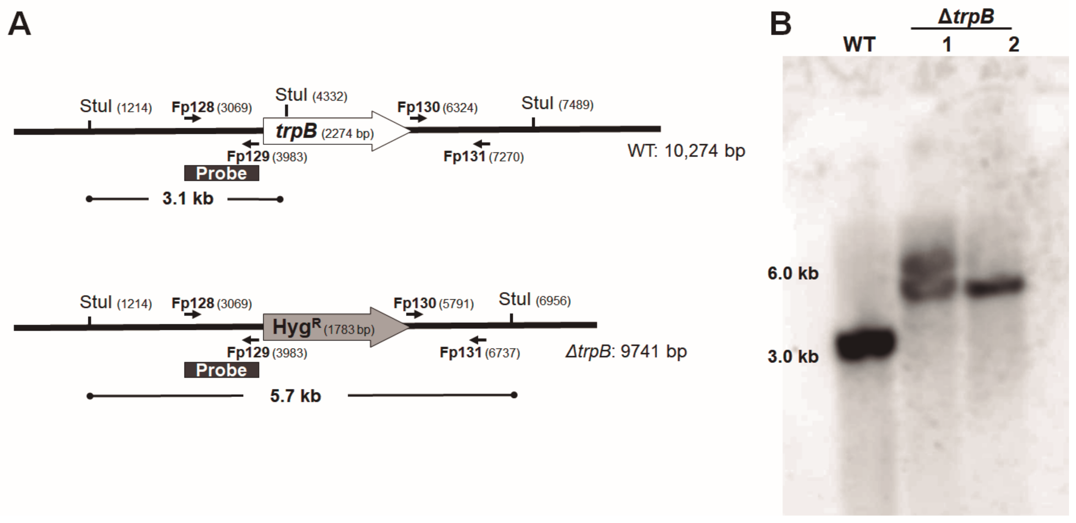
2.6. Southern Blotting

Two transformants in which the trpB deletion was confirmed by the PCR and the WT strain of F. pedrosoi had their gDNA extracted. Twenty micrograms of gDNA were overnight restriction digested with StuI (3U/μg of gDNA) at 37 °C and electrophoresed in a 1.0% TAE 1 × agarose gel. The gel was blotted onto a charged nylon membrane (GE Healthcare) by capillary transfer. The probe was amplified with oligonucleotides Fp128 + Fp129 (corresponding to the 5′ flanking region of the trpB deletion cassette) producing a 915 bp fragment labeled with digoxigenin, following the supplier’s instructions (PCR DIG Probe Synthesis Kit, Roche, Basel, Switzerland). The hybridization, washing, and detection procedures were all done according to the manufacturer’s instructions (DIG Easy Hyb Granules, DIG Wash and Block Solution Set, Anti-Digoxigenin-AP, Fab fragments and CDP-Star ready-to-use, Roche). ImageQuant LAS 4000 (GE) equipment was used to read the signal. A 5.7 kb hybridization fragment was expected for ΔtrpB and a 3.1 kb for the undeleted control.

2.7. Evaluation of Tryptophan Concentration Required for ΔtrpB Growth

The minimum concentration of L-tryptophan required to promote the trpB auxotrophic mutants’ growth was determined by incubating both WT and mutant strains (2.ΔtrpB) on SAB agar supplemented with different concentrations of the amino acid (0, 20, 50, 100, 250, 500, and 1000 μg/mL), at 28 °C for 10 days. The radial growth of the colonies was used to compare the strains.

2.8. Radial Growth Measurement

To quantify colony growth, 2.∆trpB, 22.ΔtrpB + trpB, 25.ΔtrpB + trpB, and WT strains were point-inoculated with same amount of conidia (103) onto the surface of SAB agar containing 500 μg/mL of L-trp (SAB + L-trp). The plates were incubated at 28 °C or 37 °C. Colonies’ diameters were measured every 3 days for 3 weeks. Two independent experiments were carried out in triplicate. Data collected were analyzed using the program GraphPad Prism. Mixed-effects analysis and Dunnett’s multiple comparisons test were used to compare variations between the control (WT) and test groups.

2.9. Evaluation of Conidia Germination

After growth on PD broth, conidia from 2.∆trpB, 22.ΔtrpB + trpB, 25.ΔtrpB + trpB, and WT strains were collected, counted on a Neubauer chamber, and then inoculated in 10 mL of SAB + L-trp broth (500 μg/mL of L-trp) at the concentration of 10⁶ conidia/mL. The cultures were incubated under continuous agitation (200 rpm) at 28 °C or 37 °C for 24 and 48 h. After incubation, the conidial suspension was vortexed and 100 µL were collected for microscopic inspection. One hundred randomly selected conidia from the samples of the same culture were counted to distinguish between germinated and non-germinated conidia. Conidia with a germ tube were considered germinated, and swollen ones were considered non-germinated. The experiment was performed in triplicate, repeated twice, and the data were analyzed using the program GraphPad Prism. Two-way ANOVA and Dunnett’s multiple comparisons test were used to compare variations between the control (WT) and test groups.

2.10. Conidial Viability Assay

For the viability assay, strains (2.∆trpB, 22.ΔtrpB + trpB, 25.ΔtrpB + trpB, and WT) previously grown on PD broth had the conidia collected and counted in a Neubauer chamber. Five hundred conidia of each strain were spread on three independent SAB + L-trp agar plates (500 μg/mL of L-trp), incubated at 28 or 37 °C for 10 days, and then the number of colonies was counted (CFU). The experiment was performed in triplicate and repeated twice. Data were analyzed using the program GraphPad Prism. Two-way ANOVA and Dunnett’s multiple comparisons test were used to compare variations between the control (WT) and test groups.

2.11. Determination of 5-Fluoroanthranilic Acid (5-FAA) Concentration for Selection of trp- and Counter-Selection of trp+

Culture media were supplemented with 5-fluoroanthranilic acid (5-FAA) (Sigma-Aldrich) for tryptophan pathway counter-selection. The stock solution (100 mg/mL) was prepared in absolute ethanol. Since the concentration used for counter-selection of auxotrophic tryptophan mutants is very variable among different fungal species [52,53,54,55], we decided to test the interval from 0.25 to 3.0 mg/mL for F. pedrosoi. Conidia from the WT and 2.∆trpB strains were obtained as previously described, and density was adjusted. Five microliters of each serial dilution (1 × 10⁷ to 1 × 10³ /mL) were spotted on SAB agar containing 500 μg/mL L-trp and 5-FAA. The same procedure was done to evaluate the susceptibility of reconstituted strains to 5-FAA. Fungal growth was observed after 13 days of incubation at 25 °C. The experiment was repeated four times. In order to simulate the use of 5-FAA as agent to select trp- mutants, a high density of conidia (5 × 10⁵) of the WT and ΔtrpB strains was spread on a set of conditions. The plates SAB agar, SAB + L-trp (500 µg/mL), SAB + L-trp + HygB (500 µg/mL of L-trp and 100 µg/mL of HygB), and SAB + L-trp + 5-FAA (500 µg/mL of L-trp and 0.5 mg/mL of 5-FAA) were incubated at 25 °C and inspected every 3 days until the 22nd day of growth.

2.12. Gene Replacement Strategy

Construction of plasmid pFpNAT + trpB for reconstitution of 2.ΔtrpB was carried out as follows: First, the 5.1 kb fragment corresponding to the trpB gene was amplified from the WT genome with primers Fp119 and Fp118 (Figure S3A). The product was subjected to double digestion by NdeI and SacI to generate a 4.7 kb fragment. This fragment was then cloned into the plasmid pFpNAT [35], previously opened with NdeI and SacI, using the enzyme T4 DNA ligase (Invitrogen) (Figure S3B). The ligation was transformed into thermo-competent E. coli DH5α, and colonies that grew in LB medium containing 100 µg/mL of Ampicillin were selected for plasmid extraction. Cloning of the 4.7 kb fragment corresponding to the trpB gene was confirmed by NdeI / SacI digestion (Figure S3) and the plasmid was named pFPNAT+trpB (8.9 kb). The resulting flanking regions of trpB ORF were 923 and 1,263 bp for 5′ and 3′, respectively. Non-linearized pFPNAT+trpB was transformed on 2.ΔtrpB conidia to obtain the reconstituted strains. The conditions for biolistic transformation were the same as those previously described for obtaining the trpB mutant [35,56]. To select trp+ transformants, SAB agar plates were supplied with 500 µg/mL of L- trp and 100 µg/mL of Nourseotricin (Nat). After 21 days of growth at 28 °C, colonies were transferred to SAB + Nat agar plates to detect the tryptophan prototrophic transformants, which were selected for gDNA extraction. PCR amplification with Fp120 and Fp121 that produced a 570 bp fragment of trpB was performed (Figure S3C). We also tested whether the HygR marker used for the gene deletion was preserved in the trpB locus after the transformation of pFpNAT+trpB by plating the colonies on SAB agar + 100 µg/mL Nat + 50 µg/mL HygB.

3. Results

3.1. Biosynthesis of Tryptophan in F. pedrosoi—An In Silico Analysis

By an in silico search for the enzyme sequences of the tryptophan biosynthesis pathway, we propose how this process may occur in F. pedrosoi (Figure 1). In the F. pedrosoi genome database, we found four genes encoding enzymes that work in the five-step tryptophan biosynthesis (trpB, trpC, trpD, and trpE) as shown in Table 1. By KEGG analysis, we observed well-conserved pathway components among the species F. pedrosoi, S. cerevisiae [37,47], A. fumigatus [48], and C. neoformans [50]. We observed a high identity percentage between the primary sequences of F. pedrosoi TrpB, TrpC, TrpD, and TrpE and those of A. fumigatus (>63%). In addition, using the Pfam search tool, we detected all conserved domains of each enzyme in the tryptophan pathway (Figure S4). We evaluated the expression of the genes after incubation of WT strain for 3 h on minimal medium in comparison to the minimal medium supplemented with 500 µg/mL of L-trp (control) by real-time PCR; however, we did not detect a genetic modulation of trp genes in response to the conditions analyzed (Figure S5).
In all tryptophan-producing species, biosynthesis begins with chorismate, which is the product of the shikimate pathway. The first step is carried out by TrpE/TrpC, in which chorismate and glutamine are converted to anthranilate. Anthranilate, in turn—in the presence of phosphoribosyl phosphate—is converted to phosphoribosyl anthranilate by TrpD (Anthranilate phosphoribosyl transferase). In the third step, carboxyphenyl-amino deoxyribose phosphate synthesis occurs [37]. In S. cerevisiae, phosphoribosyl-anthranilate isomerase is encoded by TRP1 [47]. However, in F. pedrosoi, the TRP1 orthologue is not found, and this step is performed by Trp3TrpC, as is the case for C. neoformans and A. fumigatus [48,50]. TrpC is a trifunctional enzyme found in several other fungal species [37,57], and it participates in three steps of this pathway. In the F. pedrosoi TrpC sequence, we found the three catalytic domains required for indole 3-glycerolphosphate synthesis: N- terminal glutamine amidotransferase class-I (GATase), Indole-3-glycerol phosphate synthase (IGPS), and C- terminal phosphoribosyl-anthranilate isomerase (PRAI) (Figure S6). The last two stages of tryptophan biosynthesis are performed by tryptophan synthase (Trp5TrpB) [37,43,47]. In the F. pedrosoi TrpB putative sequence, we found two conserved signature domains: the tryptophan synthase α chain (positions 8–258 aa) and tryptophan synthase β chain pyridoxal-phosphate attachment site (positions 389–712 aa). Between those domains, there is a poorly conserved connector region of 83 aa, located between a residue of tyrosine (Y) and proline (P) that forms the channel necessary to couple the reactions (Figure S6).

3.2. Gene Disruption and Reconstitution in F. pedrosoi

To expand and make available a more versatile molecular toolbox for further genetic functional studies in F. pedrosoi, we chose the trpB gene to construct the deletion cassette. The disruption cassette was obtained using the approach described by [49] called double-joint PCR. It consists of successive PCR steps for fusing flanking fragments of the gene of interest to the 5′ and 3′ fragments related to the selective marker. Two products were obtained as expected (Figure S1B), and were readily co-transformed into F. pedrosoi spores (strain CBS271.37) by biolistics according to the parameters we previously defined.
Three independent experiments were conducted. Firstly, from 49 transformants resistant to HygB, 4 were unable to grow in the absence of tryptophan (SAB). The second transformation generated 19 mutants HygR, and 2 mutants did not grow in SAB. These two mutants, named 1 and 2.ΔtrpB, were chosen for PCR and Southern blotting (Figure S2 and Figure 2). In the third experiment, we recovered 146 colonies of HygR, and 9 mutants showed the tryptophan auxotrophic phenotype. Thus, we observed that the rate of homologous recombination for trpB in F. pedrosoi varied from 6 to 11%. All trp auxotrophic mutants exhibited mitotic stability after three passages in the non-selective medium (SAB + L-trp).
Gene replacement was confirmed initially by PCR using two combinations of oligonucleotides. For both auxotrophic tryptophan mutants (1 and 2.ΔtrpB), we amplified the expected products (Figure S2). To confirm there was no ectopic integration of the deletion cassette on those two mutants’ genome, a Southern blot was performed. Through a trpB-specific probe (1 + 2 flanking region of the deletion cassette), we visualized the expected hybridization pattern after StuI restriction digestion of gDNA on the 2.ΔtrpB mutant (5.7 kb) and WT (3.1 kb) strains. Despite 1.ΔtrpB displaying the 5.7 kb fragment, a 6.0 kb hybridization fragment was also detected, indicating a genomic rearrangement after the HygR insertion (Figure 2). After confirming 2.ΔtrpB is a null mutant for the trpB allele, it was used in the following experiments. To ensure the observed tryptophan auxotrophic phenotype was due to trpB deletion, as a proof-of-concept, we reintroduced the trpB gene into the ΔtrpB mutant genome (Figure S3). Of 360 recovered Nat-resistant transformants, 12 recovered the ability to produce tryptophan, and were designated ΔtrpB + trpB reconstituted strains. Reinsertion of trpB was also confirmed by PCR, using oligonucleotides that amplified a 570 bp fragment of the gene (Figure S3C). As we selected the phenotype trp+ HygR NatR (Figure S3D), we considered trpB insertion to have not occurred in the original locus. All the strains (2.∆trpB, 22.ΔtrpB + trpB, 25.ΔtrpB + trpB, and WT) used on the following experiments had the presence of selective markers and the auxotroph for tryptophan confirmed after growth in SAB agar supplemented with antibiotics (Nat and Hyg) and L-trp (500 µg/mL) (Figure 3).

3.3. External Supply of Tryptophan Is Required for Proper Growth of F. pedrosoi ΔtrpB

To define the lowest amount of tryptophan necessary to support the growth of the F. pedrosoi auxotrophic mutant, various concentrations were tested. The minimum L-trp required by different trp mutants from other fungi are very variable, from 10 µg/mL for S. cerevisiae [52] to 2000 µg/mL for Nodulisporium [58]. We evaluated growth of ΔtrpB in SAB agar supplemented with 20, 50, 100, 250, 400, 500, and 1000 µg/mL of L-trp. After 10 days of incubation at 28 °C, we detected darkly pigmented colonies in all L-trp-supplied plates above 250 µg/mL. Thus, F. pedrosoi ΔtrpB requires intermediate levels of L-trp to grow and synthesize melanin (Figure 4) in comparison to other fungi, but is unable to grow in the absence of an external source of this amino acid.

3.4. Deletion of trpB Causes Growth Delay and Viability Defects

To assess whether trpB deletion affects the F. pedrosoi life cycle, we measured the radial growth of colonies in SAB agar supplemented with 500 µg/mL of L-trp over 21 days at 28 and 37 °C. The mutant (2.∆trpB) showed a growth delay in relation to WT and reconstituted strains (22.ΔtrpB + trpB and 25.ΔtrpB + trpB) at 28 °C; however, at 37 °C this growth retardation was not so evident over the evaluated period (Figure 5A,B). Notably, the growth rate of all strains (WT, ΔtrpB, and reconstituted strains) is slower at 37 °C than observed at 28 °C. We also determined if ΔtrpB conidia had the ability to produce germ tubes and if they are viable (Figure 5C,D). After 24 h at 28 °C on SAB broth, about 25% of WT and reconstituted strains’ conidia were germinated, while ΔtrpB failed to reach 20% of conidia germination. After 48 h of incubation at 28 °C, the germination defect remained for the mutant in comparison to the other strains (WT and 25.ΔtrpB + trpB), but the percentage of germ tubes increased to close to 38% (Figure 5C,D). Differently from what we observed at 28 °C, we did not detect statistical differences in conidia germination among mutant and the other strains at 37 °C. Finally, we analyzed the ability of conidia to produce viable colonies (CFU) on SAB agar supplemented with 500 µg/mL tryptophan. As can be seen in Figure 5E, we detected a statistical difference in the number of colonies recovered on SAB agar when compared ΔtrpB to the other strains at both temperatures tested. At 28 °C, an average of 103% of WT conidia resulted in viable colonies compared to 51% of ΔtrpB, 86% of 22.ΔtrpB + trpB, and 77% of 25.ΔtrpB + trpB. The difference in viability rate of ΔtrpB was also detected at 37 °C, in which only 41% of CFU were recovered for ΔtrpB; meanwhile, the WT and reconstituted strains were in the range of 60% (Figure 5E). Taken together, our data indicate that ΔtrpB has a radial growth delay and a reduced conidial germination rate at 28 °C. We also show that decreased viability of ΔtrpB conidia is not a temperature-dependent phenotype. The reconstituted strains displayed phenotypes similar to the WT in the evaluated tests, indicating that the defects detected in the mutant are related to the absence of trpB.

3.5. trpB Deletion Induces 5-FAA Resistance in F. pedrosoi

In yeasts, mutants for tryptophan biosynthesis genes are resistant to 5-FAA [52,53,54,55]. This antimetabolite, when assimilated, is converted into the toxic product 5-fluorotryptophan. In our work, we decided to evaluate whether trpB deletion in F. pedrosoi confers resistance to 5-FAA, in order to expand the genetic manipulation tools for selection and counter-selection of mutants. For this purpose, we spotted serial dilutions of WT and trpB strains on SAB + L-trp plates supplied by increasing amounts of 5-FAA (0.25; 0.5; 1.0; 2.0; and 3.0 mg/mL). The F. pedrosoi trpB mutant has higher resistance to 5-FAA compared to the WT strain. After 13 days of incubation at 28 °C, we observed that the trpB mutant tolerates 0.5 mg/mL of 5-FAA at low conidial density (102), while the WT grows only on 105 and 104 conidial spots. None of the strains grew on plates with >0.75 mg/mL of 5-FAA (Figure 6A). The reintroduction of trpB allele restored the ΔtrpB susceptibility to the WT level (Figure 6B).
In order to analyze the use of 5-FAA as an agent to select tryptophan auxotrophic mutants, we plated WT and trpB mutant strains on different agar plates as follows: SAB agar, SAB + L-trp, SAB + L-trp + HygB, and SAB + L-trp + 5-FAA. Furthermore, to simulate a genetic transformation and evaluate the rate spontaneous mutants for trp auxotrophy, a high density of conidia (5 × 10⁵) was spread on the plates. As expected, in the SAB plates only the WT strain was able to grow, while in the SAB + L-trp + HygB condition only the ΔtrpB strain grew, since resistance to HygB is guaranteed by the presence of the hph gene. Interestingly, in the condition of SAB + L-trp + 5-FAA, only trp- colonies were observed and no WT colonies were detected on those plates (Figure S7). This experiment demonstrates that it is possible to use 0.5 mg/mL of 5-FAA as the sole selective agent for identification of tryptophan auxotrophic mutants, extending the range of applications related to trpB locus and establishing it as a potential genetic transformation marker. Taken together, our results indicate that the 5-FAA-supplemented medium can be an interesting approach for tryptophan auxotrophic mutant selection. In addition, 5-FAA can be applied as a counter-selector of transformants in the case of trp- as a recipient strain in combination with the use of trpB as the selective marker in genetic transformation of F. pedrosoi.

4. Discussion

We first evaluated the enzymatic machinery in silico for tryptophan biosynthesis in F. pedrosoi. We identified that the enzymes in F. pedrosoi are displayed in a similar way to other filamentous fungi [48,50]. Unlike S. cerevisiae, F. pedrosoi has four of the five enzymes required to transform chorismate into tryptophan [47]. As reported for other ascomycetes and basidiomycetes, a single TRP1 ORF encoding a monofunctional PRA isomerase (PRAI) was not found in F. pedrosoi. Instead, it is fused to trpCTRP3, generating a trifunctional enzyme with the arrangement from N- to C- terminals of glutamine amidotransferase (GATase), Indole-3-glycerol phosphate synthase (IGPS), and PRAI [47,48,50,59,60]. Our analyses showed that F. pedrosoi encodes all catalytic domains required for the synthesis of tryptophan; this finding opens prospects and launches new tools for a detailed and functional exploration of this essential pathway for fungal survival. Interestingly, in C. neoformans, TRP3trpC and TRP5trpB are essential genes [50], but the same cannot be applied to other fungi in which auxotrophic mutants were previously isolated [53,58,61,62,63]. We detected the four transcripts of the trp biosynthetic pathway (trpB, trpC, trpD, and trpE) by RT-PCR after WT strain biomass was incubated for 3 h in minimal medium and minimal medium supplemented with L-trp. It was not possible to detect differential expression of these genes depending on the culture media and incubation time used; however, the mechanisms of regulation of the tryptophan pathway deserve to be elucidated in future work. Because trp genes are suitable as selective markers for genetic transformation in other fungi, associated to the fact that their knockout mutants are easily detected on genetic transformation plates, we decided to evaluate the DJ-PCR approach to construct the deletion cassette and the biolistic approach to deliver the DNA in F. pedrosoi. Then, as a proof of concept of the methodology, we functionally characterized the tryptophan synthase (trpB) putative gene in this human fungal pathogen.
Tryptophan synthase is conserved among bacteria, archaea, fungi, and plants, and is absent in mammals; however, only fungi present the two subunits fused into a single ORF in an α–β order [37,64]. For this reason, in those microorganisms, the enzyme is a homodimer, unlike in bacteria, where it is a tetrameric enzyme with a β–α organization [45]. The F. pedrosoi TrpB putative protein sequence presents both α and β catalytic domains: tryptophan synthase alpha chain, pyridoxal-phosphate-dependent enzyme, and the region that interconnects them [45]. FpTrpB possesses the longest connector region (83 aa) located between a residue of tyrosine (Y) and proline (P), described so far among fungal species (ranging from 40 to 69 aa) [44,63,64,65]. Definition of the three-dimensional structure of tryptophan synthases occurred after the completion of X-ray crystallography studies of the S. thyphimurium enzyme [66]. The connector region forms a channel required for the mechanical transfer of indole between the two catalytic sites, and in fungi, it is longer than in other organisms to allow proper folding of α and β subunits and, thus, catalyze their respective reactions [46,67]. The deletion of an 18 amino acid segment of this region in S. cerevisiae inactivates the enzyme, and reinsertion of an unrelated fragment restores its activity [67]. TrpB from F. pedrosoi shares all features already described for other fungi; however, a more detailed analysis of its three-dimensional structure may elucidate if a longer length of tunnel region promotes any effect in enzymatic catalytic properties.
In 2018, our group reported—through two efficient techniques, biolistic and Agrobacterium mediated transformation—the insertion and expression of exogenous DNA into the F. pedrosoi genome. We also explored three drug-resistance markers (NAT, HYG, and NEO) for mutant selection. Continuing our efforts to make available and improve new molecular tools for genetic manipulation of a CBM-causing agent, in this study, we developed a stable, fast, and efficient approach for achieving targeted gene disruption by biolistic transformation into F. pedrosoi. We utilized the HygR marker to replace trpB and constructed a deletion cassette using the double-joint PCR technique. This technique has already been successfully employed for other fungi [49,68,69,70]; however, this work shows for the first time its use in constructing cassettes for F. pedrosoi gene disruption. DJ-PCR is based on only two successive rounds of DNA amplifications and does not require long and time-consuming vector construction steps, as is the case for Agrobacterium-mediated transformation in which the insertion of the deletion cassette into the T-DNA-carrying vector is required. Another advantage of constructing gene-disruption cassettes by DJ-PCR is that amplified products can be directly used in the genetic transformation of the fungus by electroporation or biolistics. Since homologous recombination inside the selective marker fragments is also required for mutant recovery, the chance of obtaining mitotically unstable transformants is reduced with the usage of the split-marker strategy, avoiding undesired genomic insertion [69]. Considering the non-homologous recombination mediated by the NHEJ pathway is more common in filamentous fungi than homologous recombination [71], this strategy contributes to generating a higher targeted-integration frequency. To our knowledge, this is the first report of the use of the DJ-PCR method followed by biolistic transformation to obtain null mutants in F. pedrosoi.
Three independent experiments yielded an efficiency of 6–11% triple-crossover recombination to promote the trpB replacement. Two other genes were successfully disrupted in our laboratory, with an HR rate of 2 and 5%. Our data are in accordance with those previously described for other filamentous fungi, in which a low efficiency of target integration into a specific locus is observed [71]. For other ascomycetes, this rate is quite variable: A. fumigatus has an HR efficiency of 5–10% [72,73], while in N. crassa it ranges from 2 to 23% [74,75]. Even within the same species, variable efficiency can be observed, as we have observed for F. pedrosoi. This fact can be attributed to the gene locus context, the size of the flanking region chosen to construct the deletion cassette, and the DNA delivery method [71]. For many fungi, especially ascomycetes, several former works inactivated the NHEJ machinery responsible for random integration of foreign DNA at ectopic genomic sites to circumvent this problem. The most common approach is to inactivate the ku70 or ku80 genes, creating NHEJ-deficient recipient strains that significantly improve the frequency of knockout mutants’ recovery [71,76]. To prevent ectopic integrations during complementation of null mutants from interfering with the virulence and other characteristics of the fungus, some authors have already reported genomic regions considered as safe for genetic integrations, such as the safe haven for C. neoformans [77,78] or RPS10 locus in Candida albicans [79]. Although we have not yet evaluated a potential safe region for gene reconstitution in Fonsecaea spp., this tool will be of great relevance when it was described.
trpB is not an essential gene in F. pedrosoi; however, its absence directly affects its life cycle. Our results show that the F. pedrosoi trpB mutant is extremely dependent on an external source of tryptophan. Supplementation of culture medium with >250 µg/mL of this amino acid is required to sustain mycelial vegetative growth and sporulation, but 500 µg/mL (level of L-trp used in all phenotype tests) was not enough to maintain germination, viability and growth rate as in the WT strain. We did not assess whether the WT phenotypes of ΔtrpB strain can be rescued at higher levels of L-trpB as reported for the A. nidulans [63]. In A. nidulans ΔtrpB, although 10 µM (2 µg/mL) of tryptophan can trigger conidial germination and 100 µM (20 µg/mL) maintains the vegetative growth in solid medium, only higher L-trp concentrations (10 mM/2000 µg/mL) induce conidiospore production. However, even 50 mM (10,000 µg/mL) of tryptophan is not enough to generate viable ascospores and mature cleistothecia [63].
Yeast trp auxotrophic mutants require low levels of exogenous tryptophan [for S. cerevisiae 20 µg/mL (0.1 mM), Hansenulla 30 µg/mL (0.15 mM) and Candida guilliermondii 40 µg/mL (0.2 mM)] [52,53,55]. On the opposite way, some filamentous fungi, as well as F. pedrosoi, demand 10 to 100 times higher amounts of this amino acid [Aspergillus niger (1 mM), Metarhizium anisopliae (5 mM) and Nodulosporium sp (10 mM)] [58,61,62,80]. Eckert and colleagues suggested that the increased demand for tryptophan might be due to the limited ability to uptake the amino acid from the external source [63]. It is widely known that the anabolic and catabolic machinery of tryptophan, permeases and transporters, are finely tuned by intracellular levels of aromatic amino acids [48,50,64,81]. In filamentous fungal species, the transcriptional factor Cpc (cross pathway control) regulates the synthesis of aromatic amino acids, including tryptophan [59,63,82,83,84]. The high availability of exogenous tryptophan can also promote feedback repression of the initial steps of aromatic amino acids biosynthesis, and impair trp- mutants’ growth due to decreased synthesis of phenylalanine and tyrosine. To circumvent this defect, simultaneous tryptophan and phenylalanine supply improved the growth of N. crassa trp mutants [85]. Further investigation can clarify whether CpcA plays the same role in F. pedrosoi and whether the addition of external sources of aromatic amino acids would benefit mycelial growth. By the way, we detected, through an in silico BLASTP search, a sequence similar to CpcA in the genome of F. pedrosoi opening prospects for its future characterization.
Auxotrophic mutants and the anabolic genes that complement biosynthesis defects have been widely used as powerful molecular tools in the fungal genetic transformation system, since the detection of the prototrophic transformants is quick and easy and does not require high-cost antibiotics [76,86,87]. The use of auxotrophic mutants has also disadvantages to be consider. In C. albicans URA3 auxotrophic mutant, the authors identified that ectopically integration of URA3 gene affects different characteristics of the fungus, including the virulence [79]. The trp encoding enzymes have been applied as selective markers of several fungi [53,54,59,61,80,88] and in this work, we propose the use of trpB as a new selective marker in F. pedrosoi. We showed the complementation of the F. pedrosoi ΔtrpB mutant with the wild type copy of gene, that in spite of had being inserted randomly into the genome, the mutants became prototrophic and recovery the WT traits.
We assessed the susceptibility of F. pedrosoi to 5-FAA. Our results showed this antimetabolite could be used as a marker of counter-selection in genetic transformation experiments using trp- as the recipient strain. This strategy is already widely used for yeasts [50,52,53,55]; however, as far as we know, there are no data for filamentous fungi. Due to the inability of the WT strain to grow on culture medium supplemented with 0.5 mg/mL of 5-FAA, it is a feasible and interesting tool for easily find trp- mutants (positive selection) and also for identification of transformants complemented by trpB (counter selection) in F. pedrosoi. The counter selection methodology is indeed useful for recycling the selective marker for multiple gene deletion strategies on the same host strain, and it can be employed to select point mutations in genes related to the biosynthesis pathway [88]. Considering all this, the trpB mutant, despite some defective traits observed in conditions we tested, is not unfeasible as a recipient strain and can be used to generate other knockout strains; in addition, the trpB gene can be an alternative to the antibiotics as a selective marker for functional genomics in F. pedrosoi.
As tryptophan is an essential amino acid for fungal development and survival, and mammals acquire it from dietary sources, some authors suggest its biosynthesis apparatus may be an attractive selective therapeutic target [89]. In C. neoformans, an anthranilate synthase inhibitor (6-diazo-5-oxo-L-norleucine) was efficient in promoting in vitro cell death [50]. Although the role of TRP3 and TRP5 has not been evaluated in the pathogenicity of C. neoformans [50], in A. fumigatus, null mutants of aroC (chorismate mutase) and trpA play important roles in pulmonary and systemic aspergillosis infection models [90]. Considering that F. pedrosoi requires higher levels of tryptophan to survive (250 µg/mL) than that achieved in human serum (about 14 µg/mL) [91], and that mammals do not produce this amino acid, the search for specific inhibitors of tryptophan biosynthesis enzymes becomes a potential strategy to be explored against Fonsecaea spp. and other microbial pathogens [89,92,93].

5. Conclusions

In conclusion, to our knowledge, our work represents the first successful gene inactivation of the main CBM causative agent, F. pedrosoi. We demonstrate the use of split-markers for homologous integration allied to biolistic transformation were feasible to obtain ΔtrpB mutants. The trpB gene can be exploited as an alternative and attractive selective marker for auxotrophic complementation of the trpB mutant. In addition, we showed the employment of 5-FAA as positive selection of trp- mutants and as a new approach to facilitate identification of null mutants’ complementation in F. pedrosoi. Our data broaden the molecular toolbox by the use of traditional techniques for functional genome studies to improve the knowledge of not only F. pedrosoi biology but also of other CBM etiological agents. Notably, the functional studies of genes in the fungi of the genus Fonsecaea spp. are far behind compared to other fungal species, further contributing to the fact that many gaps in our knowledge of the disease remain, and CBM continues to be treated as a neglected tropical disease that affects many people around of the world.

Supplementary Materials

The following supporting information can be downloaded at: https://www.mdpi.com/article/10.3390/jof9020224/s1. Table S1. Oligonucleotides used in this work; Figure S1. Construction of trpB disruption cassette by DJ-PCR/HygR split marker strategy; Figure S2. PCR checking for homologous recombination of the trpB disruption cassette; Figure S3. Strategy for obtaining and confirming the complementation of F. pedrosoi trpB mutant; Figure S4. Conserved signature domains of F. pedrosoi putative enzymes required for tryptophan biosynthesis; Figure S5. Real-time PCR to evaluate the expression of the tryptophan biosynthetic genes: trpB, trpC, trpD, and trpE of F. pedrosoi in response to incubation in minimal medium (Czapeck Dox) for 3 h at 28 °C; Figure S6. Multiple sequence alignment of tryptophan synthases protein sequences; Figure S7. Evaluation of the use of 5-FAA at the concentration of 0.5 mg/mL as a selection agent for trp- mutants, and for counter-selection of strains that present active tryptophan synthesis pathway at high cell density (5 × 10⁵) of WT and ΔtrpB.

Author Contributions

Conceptualization, L.F. and L.D.F.; methodology, L.D.F., C.d.S.G., T.S.H. and R.V.d.A.; formal analysis, L.F. and L.D.F.; investigation, L.D.F.; resources, L.F., A.L.B., R.V.d.A. and M.S.S.F.; writing—original draft preparation, L.F. and L.D.F.; writing—review and editing, L.F.; supervision, L.F. and A.L.B.; project administration, L.F.; funding acquisition, L.F., A.L.B. and M.S.S.F. All authors have read and agreed to the published version of the manuscript.

Funding

This research was funded by Fundação de Apoio à Pesquisa do Distrito Federal (FAP-DF), Conselho Nacional de Desenvolvimento Científico e Tecnológico (CNPq) and Coordenação de Aperfeiçoamento de Pessoal de Nível Superior (CAPES). The grants are FAP-DF-DEMANDA ESPONTÂNEA 00193-00000180/2019-91 to LF, FAP-DF-PRONEX 193.001.533/2016 to MSSF and FAP-DF-DEMANDA ESPONTÂNEA 193.000.805/2015 to ALB. LDF acknowledges the support given by the CNPq for Ms. Scholarship. The APC was funded by Fundação Universidade de Brasília (FUB).

Institutional Review Board Statement

Not applicable.

Informed Consent Statement

Not applicable.

Data Availability Statement

Not applicable.

Acknowledgments

We thank Vania A. Vicente from the University of Paraná and the Paraná Network of Biological Collections for kindly providing F. pedrosoi CBS 271.37 strain.

Conflicts of Interest

The authors declare no conflict of interest.

References

  1. Santos, A.L.S.; Palmeira, V.F.; Rozental, S.; Kneipp, L.F.; Nimrichter, L.; Alviano, D.S.; Rodrigues, M.L.; Alviano, C.S. Biology and Pathogenesis of Fonsecaea pedrosoi, the Major Etiologic Agent of Chromoblastomycosis. FEMS Microbiol. Rev. 2007, 31, 570–591. [Google Scholar] [CrossRef] [PubMed]
  2. Queiroz-Telles, F.; Esterre, P.; Perez-Blanco, M.; Vitale, R.; Salgado, C.G.; Bonifaz, A. Chromoblastomycosis: An Overview of Clinical Manifestations, Diagnosis and Treatment. Med. Mycol. 2009, 47, 3–15. [Google Scholar] [CrossRef] [PubMed]
  3. Coelho, R.A.; Brito-Santos, F.; Figueiredo-Carvalho, M.H.G.; Silva, J.V.D.S.; Gutierrez-Galhardo, M.C.; do Valle, A.C.F.; Zancopé-Oliveira, R.M.; Trilles, L.; Meyer, W.; Freitas, D.F.S.; et al. Molecular Identification and Antifungal Susceptibility Profiles of Clinical Strains of Fonsecaea spp. Isolated from Patients with Chromoblastomycosis in Rio de Janeiro, Brazil. PLoS Negl. Trop. Dis. 2018, 12, 1–15. [Google Scholar] [CrossRef] [PubMed]
  4. Brito, A.C.D.; Bittencourt, M.D.J.S. Chromoblastomycosis: An Etiological, Epidemiological, Clinical, Diagnostic, and Treatment Update. An. Bras. Dermatol. 2018, 93, 495–506. [Google Scholar] [CrossRef]
  5. Santos, D.W.C.L.; de Azevedo, C.d.M.P.E.S.; Vicente, V.A.; Queiroz-Telles, F.; Rodrigues, A.M.; de Hoog, G.S.; Denning, D.W.; Colombo, A.L. The Global Burden of Chromoblastomycosis. PLoS Negl. Trop. Dis. 2021, 15, 1–26. [Google Scholar] [CrossRef]
  6. Bonifaz, A.; Carrasco-Gerard, E.; Saul, A. Chromoblastomycosis: Clinical and Mycologic Experience of 51 Cases. Mycoses 2001, 44, 1–7. [Google Scholar] [CrossRef]
  7. Esterre, P.; Andriantsimahavandy, A.; Ramarcel, E.R.; Pecarrere, J.-L. Forty Years of Chromoblastomycosis in Madagascar: A Review. Am. J. Trop. Med. Hyg. 1996, 55, 45–47. [Google Scholar] [CrossRef]
  8. Agarwal, R.; Singh, G.; Ghosh, A.; Verma, K.K.; Pandey, M.; Xess, I. Chromoblastomycosis in India: Review of 169 Cases. PLoS Negl. Trop. Dis. 2017, 11, e0005534. [Google Scholar] [CrossRef]
  9. Attapattu, M.C. Chromoblastomycosis—A Clinical and Mycological Study of 71 Cases from Sri Lanka. Mycopathologia 1997, 137, 145–151. [Google Scholar] [CrossRef]
  10. Gomes, R.R.; Vicente, V.A.; Azevedo, C.M.P.S.d.; Salgado, C.G.; da Silva, M.B.; Queiroz-Telles, F.; Marques, S.G.; Santos, D.W.C.L.; de Andrade, T.S.; Takagi, E.H.; et al. Molecular Epidemiology of Agents of Human Chromoblastomycosis in Brazil with the Description of Two Novel Species. PLoS Negl. Trop. Dis. 2016, 10, 1–20. [Google Scholar] [CrossRef] [Green Version]
  11. Silva, J.P.; De Souza, W.; Rozental, S. Chromoblastomycosis: A Retrospective Study of 325 Cases on Amazonic Region (Brazil). Mycopathologia 1998, 143, 171–175. [Google Scholar] [CrossRef]
  12. Queiroz-Telles, F.; de Hoog, S.; Santos, D.W.C.L.; Salgado, C.G.; Vicente, V.A.; Bonifaz, A.; Roilides, E.; Xi, L.; de Maria Pedrozo e Silva Azevedo, C.; da Silva, M.B.; et al. Chromoblastomycosis. Clin. Microbiol. Rev. 2017, 30, 233–276. [Google Scholar] [CrossRef]
  13. Gimenes, V.M.F.; Criado, P.R.; Martins, J.E.C.; Almeida, S.R. Cellular Immune Response of Patients with Chromoblastomycosis Undergoing Antifungal Therapy. Mycopathologia 2006, 162, 97–101. [Google Scholar] [CrossRef]
  14. Ameen, M. Chromoblastomycosis: Clinical Presentation and Management. Clin. Exp. Dermatol. 2009, 34, 849–854. [Google Scholar] [CrossRef]
  15. Alviano, C.S.; Farbiarz, S.R.; De Souza, W.; Angluster, J.; Travassos, L.R. Characterization of Fonsecaea pedrosoi Melanin. J. Gen. Microbiol. 1991, 137, 837–844. [Google Scholar] [CrossRef]
  16. Alviano, D.S.; Franzen, A.J.; Travassos, L.R.; Holandino, C.; Rozental, S.; Ejzemberg, R.; Alviano, C.S.; Rodrigues, M.L. Melanin from Fonsecaea pedrosoi Induces Production of Human Antifungal Antibodies and Enhances the Antimicrobial Efficacy of Phagocytes. Society 2004, 72, 229–237. [Google Scholar] [CrossRef]
  17. Cunha, M.M.L.; Franzen, A.J.; Alviano, D.S.; Zanardi, E.; Alviano, C.S.; De Souza, W.; Rozental, S. Inhibition of Melanin Synthesis Pathway by Tricyclazole Increases Susceptibility of Fonsecaea pedrosoi against Mouse Macrophages. Microsc. Res. Tech. 2005, 68, 377–384. [Google Scholar] [CrossRef]
  18. Cunha, M.M.; Franzen, A.J.; Seabra, S.H.; Herbst, M.H.; Vugman, N.V.; Borba, L.P.; de Souza, W.; Rozental, S. Melanin in Fonsecaea pedrosoi: A Trap for Oxidative Radicals. BMC Microbiol. 2010, 10, 80. [Google Scholar] [CrossRef]
  19. Bocca, A.L.; Brito, P.P.M.S.; Figueiredo, F.; Tosta, C.E. Inhibition of Nitric Oxide Production by Macrophages in Chromoblastomycosis: A Role for Fonsecaea pedrosoi Melanin. Mycopathologia 2006, 161, 195–203. [Google Scholar] [CrossRef]
  20. Pinto, L.; Granja, L.F.Z.; Alviano, D.S.; da Silva, M.H.; Alviano, C.S.; Ejzemberg, R. Activation of the Human Complement System by Pigmented and Hypopigmented Mycelia of the Fungus Fonsecaea pedrosoi. Mycoses 2011, 54, 474–480. [Google Scholar] [CrossRef]
  21. Pinto, L.; Granja, L.F.Z.; Almeida, M.A.d.; Alviano, D.S.; Silva, M.H.D.; Ejzemberg, R.; Rozental, S.; Alviano, C.S. Melanin Particles Isolated from the Fungus Fonsecaea pedrosoi Activates the Human Complement System. Mem. Inst. Oswaldo Cruz 2018, 8, 113. [Google Scholar] [CrossRef] [PubMed]
  22. Gómez, B.L.; Nosanchuk, J.D. Melanin and Fungi. Curr. Opin. Infect. Dis. 2003, 16, 91–96. [Google Scholar] [CrossRef] [PubMed]
  23. Chongkae, S.; Nosanchuk, J.D.; Pruksaphon, K.; Laliam, A.; Pornsuwan, S.; Youngchim, S. Production of Melanin Pigments in Saprophytic Fungi in Vitro and during Infection. J. Basic Microbiol. 2019, 59, 1092–1104. [Google Scholar] [CrossRef] [PubMed]
  24. Xiao, X.; Li, Y.; Lan, Y.; Zhang, J.; He, Y.; Cai, W.; Chen, Z.; Xi, L.; Zhang, J. Deletion of PksA Attenuates the Melanogenesis, Growth and Sporulation Ability and Causes Increased Sensitivity to Stress Response and Antifungal Drugs in the Human Pathogenic Fungus Fonsecaea monophora. Microbiol. Res. 2021, 244, 126668. [Google Scholar] [CrossRef]
  25. Li, M.; Huang, H.; Liu, J.; Zhang, X.; Li, Q.; Li, D.; Luo, M.; Wang, X.; Zeng, W.; Sun, J.; et al. Deletion C-Terminal Thioesterase Abolishes Melanin Biosynthesis, Affects Metabolism and Reduces the Pathogenesis of Fonsecaea monophora. PLoS Negl. Trop. Dis. 2022, 16, 1–25. [Google Scholar] [CrossRef]
  26. Siqueira, I.M.; Wüthrich, M.; Li, M.; Wang, H.; Las-Casas, L.d.O.; de Castro, R.J.A.; Klein, B.; Bocca, A.L. Early Immune Response against Fonsecaea Pedrosoi Requires Dectin-2-Mediated Th17 Activity, Whereas Th1 Response, Aided by Treg Cells, Is Crucial for Fungal Clearance in Later Stage of Experimental Chromoblastomycosis. PLoS Negl. Trop. Dis. 2020, 14, e0008386. [Google Scholar] [CrossRef]
  27. Wüthrich, M.; Wang, H.; Li, M.; Lerksuthirat, T.; Hardison, S.E.; Brown, G.D.; Klein, B. Fonsecaea pedrosoi -Induced Th17-Cell Differentiation in Mice Is Fostered by Dectin-2 and Suppressed by Mincle Recognition. Eur. J. Immunol. 2015, 45, 2542–2552. [Google Scholar] [CrossRef]
  28. Castro, R.J.A.D.; Siqueira, I.M.; Jerônimo, M.S.; Basso, A.M.M.; Veloso Junior, P.H.D.H.; Magalhães, K.G.; Leonhardt, L.C.; Oliveira, S.A.M.D.; Bürgel, P.H.; Tavares, A.H.; et al. The Major Chromoblastomycosis Etiologic Agent Fonsecaea pedrosoi Activates the NLRP3 Inflammasome. Front. Immunol. 2017, 8, 1572. [Google Scholar] [CrossRef]
  29. Machado, A.P.; Regis Silva, M.R.; Fischman, O. Local Phagocytic Responses after Murine Infection with Different Forms of Fonsecaea pedrosoi and Sclerotic Bodies Originating from an Inoculum of Conidiogenous Cells. Mycoses 2011, 54, 202–211. [Google Scholar] [CrossRef]
  30. Siqueira, I.M.; de Castro, R.J.A.; Leonhardt, L.C.d.M.; Jerônimo, M.S.; Soares, A.C.; Raiol, T.; Nishibe, C.; Almeida, N.; Tavares, A.H.; Hoffmann, C.; et al. Modulation of the Immune Response by Fonsecaea pedrosoi Morphotypes in the Course of Experimental Chromoblastomycosis and Their Role on Inflammatory Response Chronicity. PLoS Negl. Trop. Dis. 2017, 11, e0005461. [Google Scholar] [CrossRef] [Green Version]
  31. Alviano, C.S.; Farbiarz, S.R.; Travassos, L.R.; Angluster, J.; de Souza, W. Effect of Environmental Factors on Fonsecaea pedrosoi Morphogenesis with Emphasis on Sclerotic Cells Induced by Propranolol. Mycopathologia 1992, 119, 17–23. [Google Scholar] [CrossRef]
  32. Alviano, D.S.; Kneipp, L.F.; Lopes, A.H.; Travassos, L.R.; Meyer-Fernandes, J.R.; Rodrigues, M.L.; Alviano, C.S. Differentiation of Fonsecaea pedrosoi Mycelial Forms into Sclerotic Cells Is Induced by Platelet-Activating Factor. Res. Microbiol. 2003, 154, 689–695. [Google Scholar] [CrossRef]
  33. Dong, B.; Tong, Z.; Li, R.; Chen, S.C.A.; Liu, W.; Liu, W.; Chen, Y.; Zhang, X.; Duan, Y.; Li, D.; et al. Transformation of Fonsecaea pedrosoi into Sclerotic Cells Links to the Refractoriness of Experimental Chromoblastomycosis in BALB/c Mice via a Mechanism Involving a Chitin-Induced Impairment of IFN-γ Production. PLoS Negl. Trop. Dis. 2018, 12, 1–31. [Google Scholar] [CrossRef]
  34. Las-Casas, L.d.O.; Marina, C.L.F.; de Castro, R.J.A.; Coelho, L.C.; Báo, S.N.; de Hoog, G.S.; Vicente, V.A.; Fernandes, L.; Bocca, A.L. Pathogenicity and Growth Conditions Modulate Fonsecaea Extracellular Vesicles’ Ability to Interact With Macrophages. Front. Cell. Infect. Microbiol. 2022, 12, 1–14. [Google Scholar] [CrossRef]
  35. Florencio, C.S.; Brandão, F.A.S.; Teixeira, M.d.M.; Bocca, A.L.; Felipe, M.S.S.; Vicente, V.A.; Fernandes, L. Genetic Manipulation of Fonsecaea pedrosoi Using Particles Bombardment and Agrobacterium Mediated Transformation. Microbiol. Res. 2018, 207, 269–279. [Google Scholar] [CrossRef]
  36. Lynch, J.H.; Dudareva, N. Aromatic Amino Acids: A Complex Network Ripe for Future Exploration. Trends Plant Sci. 2020, 25, 670–681. [Google Scholar] [CrossRef]
  37. Hutter, R.; Niederberger, P.; DeMoss, J.A. Tryptophan Biosynthetic Genes in Eukaryotic Microorganisms. Annu. Rev. Microbiol. 1986, 40, 55–77. [Google Scholar] [CrossRef]
  38. Bentley, R.; Haslam, E. The Shikimate Pathway—A Metabolic Tree with Many Branche. Crit. Rev. Biochem. Mol. Biol. 1990, 25, 307–384. [Google Scholar] [CrossRef]
  39. Herrmann, K.M.; Weaver, L.M. The Shikimate Pathway. Annu. Rev. Plant Physiol. Plant Mol. Biol. 1999, 50, 473–503. [Google Scholar] [CrossRef]
  40. Comai, S.; Bertazzo, A.; Brughera, M.; Crotti, S. Tryptophan in Health and Disease. Adv. Clin. Chem. 2020, 95, 165–218. [Google Scholar] [CrossRef]
  41. Ren, W.; Bin, P.; Yin, Y.; Wu, G. Impacts of Amino Acids on the Intestinal Defensive System. Adv. Exp. Med. Biol. 2020, 1265, 133–151. [Google Scholar] [CrossRef] [PubMed]
  42. Yanofsky, C. Comparison of Regulatory and Structural Regions of Genes of Tryptophan Metabolism. Mol. Biol. Evol. 1984, 1, 143–161. [Google Scholar] [CrossRef] [PubMed]
  43. Burns, D.M.; Horn, V.; Paluh, J.Y.C. Evolution of the Tryptophan Synthetase of Fungi. Analysis of Experimentally Fused Escherichia Coli Tryptophan Synthetase Alpha and Beta Chains. Biochemistry 1990, 265, 2060–2069. [Google Scholar] [CrossRef]
  44. Burns, D.M.; Yanofsky, C. Nucleotide Sequence of the Neurospora Crassa Trp-3 Gene Encoding Tryptophan Synthetase and Comparison of the Trp-3 Polypeptide with Its Homologs in Saccharomyces Cerevisiae and Escherichia Coli. J. Biol. Chem. 1989, 264, 3840–3848. [Google Scholar] [CrossRef] [PubMed]
  45. Dunn, M.F.; Niks, D.; Ngo, H.; Barends, T.R.M.; Schlichting, I. Tryptophan Synthase: The Workings of a Channeling Nanomachine. Trends Biochem. Sci. 2008, 33, 254–264. [Google Scholar] [CrossRef]
  46. Fleming, J.R.; Schupfner, M.; Busch, F.; Baslé, A.; Ehrmann, A.; Sterner, R.; Mayans, O. Evolutionary Morphing of Tryptophan Synthase: Functional Mechanisms for the Enzymatic Channeling of Indole. J. Mol. Biol. 2018, 430, 5066–5079. [Google Scholar] [CrossRef]
  47. Braus, G.H. Aromatic Amino Acid Biosynthesis in the Yeast Saccharomyces cerevisiae: A Model System for the Regulation of a Eukaryotic Biosynthetic Pathway. Microbiol. Rev. 1991, 55, 349–370. [Google Scholar] [CrossRef]
  48. Choera, T.; Zelante, T.; Romani, L.; Keller, N.P. A Multifaceted Role of Tryptophan Metabolism and Indoleamine 2,3-Dioxygenase Activity in Aspergillus Fumigatus-Host Interactions. Front. Immunol. 2018, 8, 1–11. [Google Scholar] [CrossRef]
  49. Kim, M.S.; Kim, S.Y.; Yoon, J.K.; Lee, Y.W.; Bahn, Y.S. An Efficient Gene-Disruption Method in Cryptococcus neoformans by Double-Joint PCR with NAT-Split Markers. Biochem. Biophys. Res. Commun. 2009, 390, 983–988. [Google Scholar] [CrossRef]
  50. Fernandes, J.D.S.; Martho, K.; Tofik, V.; Vallim, M.A.; Pascon, R.C. The Role of Amino Acid Permeases and Tryptophan Biosynthesis in Cryptococcus neoformans Survival. PLoS ONE 2015, 10, 1–22. [Google Scholar] [CrossRef] [Green Version]
  51. Nakayashiki, H.; Hanada, S.; Quoc, N.B.; Kadotani, N.; Tosa, Y.; Mayama, S. RNA Silencing as a Tool for Exploring Gene Function in Ascomycete Fungi. Fungal Genet. Biol. 2005, 42, 275–283. [Google Scholar] [CrossRef]
  52. Toyn, J.H.; Gunyuzlu, P.L.; Hunter White, W.; Thompson, L.A.; Hollis, G.F. A Counterselection for the Tryptophan Pathway in Yeast: 5-Fluoroanthranilic Acid Resistance. Yeast 2000, 16, 553–560. [Google Scholar] [CrossRef]
  53. Foureau, E.; Clastre, M.; Millerioux, Y.; Simkin, A.J.; Cornet, L.; Dutilleul, C.; Besseau, Ś.; Marais, E.; Melin, Ć.; Guillard, J.; et al. A TRP5/5-Fluoroanthranilic Acid Counter-Selection System for Gene Disruption in Candida guilliermondii. Curr. Genet. 2012, 58, 245–254. [Google Scholar] [CrossRef]
  54. Cheon, S.A.; Han, E.J.; Kang, H.A.; Ogrydziak, D.M.; Kim, J.Y. Isolation and Characterization of the TRPI Gene from the Yeast Yarrowia lipopolytica and Multiple Gene Disruption Using a TRP Blaster. Yeast 2003, 20, 677–685. [Google Scholar] [CrossRef]
  55. Cheon, S.A.; Choo, J.; Ubiyvovk, V.M.; Park, J.-N.; Kim, M.W.; Oh, D.-B.; Kwon, O.; Sibirny, A.A.; Kim, J.-Y.; Kang, H.A. New Selectable Host-Marker Systems for Multiple Genetic Manipulations Based on TRP1, MET2 and ADE2 in the Methylotrophic Yeast Hansenula polymorpha. Yeast 2009, 26, 507–521. [Google Scholar] [CrossRef]
  56. Abe, F.; Hiraki, T. Mechanistic Role of Ergosterol in Membrane Rigidity and Cycloheximide Resistance in Saccharomyces cerevisiae. Biochim. Biophys. Acta Biomembr. 2009, 1788, 743–752. [Google Scholar] [CrossRef]
  57. Käfer, E. The Anthranilate Synthetase Enzyme Complex and the Trifunctional Trpc Gene of Aspergillus. Can. J. Genet. Cytol. 1977, 19, 723–738. [Google Scholar] [CrossRef]
  58. Ireland, C.; Peekhaus, N.; Lu, P.; Sangari, R.; Zhang, A.; Masurekar, P.; An, Z. The Tryptophan Synthetase Gene TRP1 of Nodulisporium Sp.: Molecular Characterization and Its Relation to Nodulisporic Acid a Production. Appl. Microbiol. Biotechnol. 2008, 79, 451–459. [Google Scholar] [CrossRef]
  59. Dörnte, B.; Kües, U. Paradoxical Performance of Tryptophan Synthase Gene Trp1 + in Transformations of the Basidiomycete Coprinopsis cinerea. Appl. Microbiol. Biotechnol. 2016, 100, 8789–8807. [Google Scholar] [CrossRef]
  60. Yelton, M.M.; Hamer, J.E.; de Souza, E.R.; Mullaney, E.J.; Timberlake, W.E. Developmental Regulation of the Aspergillus nidulans TrpC Gene. Proc. Natl. Acad. Sci. USA 1983, 80, 7576–7580. [Google Scholar] [CrossRef] [Green Version]
  61. Goosen, T.; van Engelenburg, F.; Debets, F.; Swart, K.; Bos, K.; van den Broek, H. Tryptophan Auxotrophic Mutants in Aspergillus niger: Inactivation of the TrpC Gene by Cotransformation Mutagenesis. MGG Mol. Gen. Genet. 1989, 219, 282–288. [Google Scholar] [CrossRef] [PubMed]
  62. Horng, J.S.; Linz, J.E.; Pestka, J.J. Cloning and Characterization of the TrpC Gene from an Aflatoxigenic Strain of Aspergillus parasiticus. Appl. Environ. Microbiol. 1989, 55, 2561–2568. [Google Scholar] [CrossRef] [PubMed]
  63. Eckert, S.E.; Kübler, E.; Hoffmann, B.; Braus, G.H. The Tryptophan Synthase-Encoding TrpB Gene of Aspergillus nidulans Is Regulated by the Cross-Pathway Control System. Mol. Gen. Genet. 2000, 263, 867–876. [Google Scholar] [CrossRef]
  64. Zalkin, H.Y.C. Yeast Gene TRP5: Structure, Function, Regulation. J. Biol. Chem. 1982, 257, 1491–1500. [Google Scholar] [CrossRef] [PubMed]
  65. Skrzynia, C.; Binninger, D.M.; Alspaugh, J.A.; Pukkila, P.J. Molecular Characterization of TRP1, a Gene Coding for Tryptophan Synthetase in the Basidiomycete Coprinus Cinereus. Gene 1989, 81, 73–82. [Google Scholar] [CrossRef] [PubMed]
  66. Hyde, C.C.; Ahmed, S.A.; Padlan, E.A.; Miles, E.W.; Davies, D.R. Three-Dimensional Structure of the Tryptophan Synthase Alpha 2 Beta 2 Multienzyme Complex from Salmonella typhimurium. J. Biol. Chem. 1988, 263, 17857–17871. [Google Scholar] [CrossRef]
  67. Crawford, I.P.; Clarke, M.; Van Cleemput, M.; Yanofsky, C. Crucial Role of the Connecting Region Joining the Two Functional Domains of Yeast Tryptophan Synthetase. J. Biol. Chem. 1987, 262, 239–244. [Google Scholar] [CrossRef]
  68. Liang, L.; Li, J.; Cheng, L.; Ling, J.; Luo, Z.; Bai, M.; Xie, B. A High Efficiency Gene Disruption Strategy Using a Positive-Negative Split Selection Marker and Electroporation for Fusarium oxysporum. Microbiol. Res. 2014, 169, 835–843. [Google Scholar] [CrossRef]
  69. Kück, U.; Hoff, B. New Tools for the Genetic Manipulation of Filamentous Fungi. Appl. Microbiol. Biotechnol. 2010, 86, 51–62. [Google Scholar] [CrossRef]
  70. Shen, B.; Xiao, J.; Dai, L.; Huang, Y.; Mao, Z.; Lin, R.; Yao, Y.; Xie, B. Development of a High-Efficiency Gene Knockout System for Pochonia chlamydosporia. Microbiol. Res. 2015, 170, 18–26. [Google Scholar] [CrossRef]
  71. Krappmann, S. Gene Targeting in Filamentous Fungi: The Benefits of Impaired Repair. Fungal Biol. Rev. 2007, 21, 25–29. [Google Scholar] [CrossRef]
  72. Da Silva Ferreira, M.E.; Kress, M.R.V.Z.; Savoldi, M.; Goldman, M.H.S.; Härtl, A.; Heinekamp, T.; Brakhage, A.A.; Goldman, G.H. The AkuBKU80 Mutant Deficient for Nonhomologous End Joining Is a Powerful Tool for Analyzing Pathogenicity in Aspergillus fumigatus. Eukaryot. Cell 2006, 5, 207–211. [Google Scholar] [CrossRef]
  73. Krappmann, S.; Sasse, C.; Braus, G.H. Gene Targeting in Aspergillus Fumigatus by Homologous Recombination Is Facilitated in a Nonhomologous End- Joining-Deficient Genetic Background. Eukaryot. Cell 2006, 5, 212–215. [Google Scholar] [CrossRef]
  74. Ishibashi, K.; Suzuki, K.; Ando, Y.; Takakura, C.; Inoue, H. Nonhomologous Chromosomal Integration of Foreign DNA Is Completely Dependent on MUS-53 (Human Lig4 Homolog) in Neurospora. Proc. Natl. Acad. Sci. USA 2006, 103, 14871–14876. [Google Scholar] [CrossRef]
  75. Ninomiya, Y.; Suzuki, K.; Ishii, C.; Inoue, H. From The Cover: Highly Efficient Gene Replacements in Neurospora Strains Deficient for Nonhomologous End-Joining. Proc. Natl. Acad. Sci. USA 2004, 101, 12248–12253. [Google Scholar] [CrossRef]
  76. Jiang, D.; Zhu, W.; Wang, Y.; Sun, C.; Zhang, K.-Q.; Yang, J. Molecular Tools for Functional Genomics in Filamentous Fungi: Recent Advances and New Strategies. Biotechnol. Adv. 2013, 31, 1562–1574. [Google Scholar] [CrossRef]
  77. Arras, S.D.M.; Chitty, J.L.; Blake, K.L.; Schulz, B.L.; Fraser, J.A. A Genomic Safe Haven for Mutant Complementation in Cryptococcus neoformans. PLoS ONE 2015, 10, 1–16. [Google Scholar] [CrossRef]
  78. Fan, Y.; Lin, X. An Intergenic “Safe Haven” Region in Cryptococcus neoformans Serotype D Genomes. Fungal Genet. Biol. 2020, 144, 103464. [Google Scholar] [CrossRef]
  79. Brand, A.; MacCallum, D.M.; Brown, A.J.P.; Gow, N.A.R.; Odds, F.C. Ectopic Expression of URA3 Can Influence the Virulence Phenotypes and Proteome of Candida albicans but Can Be Overcome by Targeted Reintegration of URA3 at the RPS10 Locus. Eukaryot. Cell 2004, 3, 900–909. [Google Scholar] [CrossRef]
  80. Staats, C.C.; Junges, A.; Fitarelli, M.; Furlaneto, M.C.; Vainstein, M.H.; Schrank, A. Gene Inactivation Mediated by Agrobacterium tumefaciens in the Filamentous Fungi Metarhizium anisopliae. Appl. Microbiol. Biotechnol. 2007, 76, 945–950. [Google Scholar] [CrossRef]
  81. Wang, P.-M.; Choera, T.; Wiemann, P.; Pisithkul, T.; Amador-Noguez, D.; Keller, N.P. TrpE Feedback Mutants Reveal Roadblocks and Conduits toward Increasing Secondary Metabolism in Aspergillus fumigatus. Fungal Genet. Biol. 2016, 89, 102–113. [Google Scholar] [CrossRef] [PubMed]
  82. Carsiotis, M.; Jones, R.F. Cross-Pathway Regulation: Tryptophan-Mediated Control of Histidine and Arginine Biosynthetic Enzymes in Neurospora crassa. J. Bacteriol. 1974, 119, 889–892. [Google Scholar] [CrossRef] [PubMed]
  83. Tian, C.; Kasuga, T.; Sachs, M.S.; Glass, N.L. Transcriptional Profiling of Cross Pathway Control in Neurospora crassa and Comparative Analysis of the Gcn4 and CPC1 Regulons. Eukaryot. Cell 2007, 6, 1018–1029. [Google Scholar] [CrossRef] [PubMed]
  84. Braus, G.H.; Sasse, C.; Krappmann, S. Amino Acid Acquisition, Cross-Pathway Control, and Virulence in Aspergillus. Med. Mycol. 2006, 44, 91–94. [Google Scholar] [CrossRef] [PubMed]
  85. Perkins, D.D.; Radford, A.; Newmeyer, D.; Björkman, M. Chromosomal Loci of Neurospora crassa. Microbiol. Rev. 1982, 46, 426–570. [Google Scholar] [CrossRef]
  86. Prabha, V.L.; Punekar, N.S. Genetic Transformation in Aspergilli: Tools of the Trade. Indian J. Biochem. Biophys. 2004, 41, 205–215. [Google Scholar]
  87. Meyer, V. Genetic Engineering of Filamentous Fungi—Progress, Obstacles and Future Trends. Biotechnol. Adv. 2008, 26, 177–185. [Google Scholar] [CrossRef]
  88. Olmedo-Monfil, V.; Cortés-Penagos, C.; Herrera-Estrella, A. Three Decades of Fungal Transformation: Key Concepts and Applications. Methods Mol. Biol. 2004, 267, 297–313. [Google Scholar] [CrossRef]
  89. Amich, J.; Bignell, E. Amino Acid Biosynthetic Routes as Drug Targets for Pulmonary Fungal Pathogens: What Is Known and Why Do We Need to Know More? Curr. Opin. Microbiol. 2016, 32, 151–158. [Google Scholar] [CrossRef]
  90. Sasse, A.; Hamer, S.N.; Amich, J.; Binder, J.; Krappmann, S. Mutant Characterization and in Vivo Conditional Repression Identify Aromatic Amino Acid Biosynthesis to Be Essential for Aspergillus fumigatus Virulence. Virulence 2016, 7, 56–62. [Google Scholar] [CrossRef]
  91. Geisler, S.; Mayersbach, P.; Becker, K.; Schennach, H.; Fuchs, D.; Gostner, J.M. Serum Tryptophan, Kynurenine, Phenylalanine, Tyrosine and Neopterin Concentrations in 100 Healthy Blood Donors. Pteridines 2015, 26, 31–36. [Google Scholar] [CrossRef]
  92. Dias, M.V.B.; Canduri, F.; da Silveira, N.J.F.; Czekster, C.M.; Basso, L.A.; Palma, M.S.; Santos, D.S.; de Azevedo, W.F. Molecular Models of Tryptophan Synthase from Mycobacterium tuberculosis Complexed with Inhibitors. Cell Biochem. Biophys. 2006, 44, 375–384. [Google Scholar] [CrossRef]
  93. Naz, S.; Farooq, U.; Ali, S.; Sarwar, R.; Khan, S.; Abagyan, R. Identification of New Benzamide Inhibitor against α -Subunit of Tryptophan Synthase from Mycobacterium tuberculosis through Structure-Based Virtual Screening, Anti-Tuberculosis Activity and Molecular Dynamics Simulations. J. Biomol. Struct. Dyn. 2019, 37, 1043–1053. [Google Scholar] [CrossRef] [Green Version]
Figure 1. Putative tryptophan biosynthesis pathway in F. pedrosoi. The corresponding transcript IDs are indicated in parenthesis below their protein names. The Enzyme Commission (EC) numbers are depicted on the left side of each step of the pathway.
Jof 09 00224 g001
Figure 2. trpB deletion by homologous recombination of HygR selective marker and confirmation by Southern blotting. (A) StuI restriction pattern of trpB locus in the genetic background of WT and mutant. (B) Southern blotting of the gDNA extracted from: WT, 1.∆trpB, and 2.∆trpB digested with restriction enzyme StuI probed with a 914 bp DIG-labeled PCR fragment. The annealing region of the probe is indicated as a solid line. The indications 1.∆trpB and 2.∆trpB correspond to two different null mutants for trpB, while the WT is the CBS 271.37.
Jof 09 00224 g002
Figure 3. Radial growth of F. pedrosoi tryptophan auxotrophic mutant (2.ΔtrpB), reconstituted transformants (22.ΔtrpB + trpB and 25.ΔtrpB + trpB), and WT on Sabouraud agar (SAB) supplemented or not with 500 µg/mL L-tryptophan (L-trp) and 50 µg/mL Hygromycin B (HygB). The 2.∆trpB corresponds to the null mutant for trpB, while 22.ΔtrpB + trpB and 25.ΔtrpB + trpB are two independent reconstituted strains for trpB and the WT is the CBS 271.37. The plates were incubated at 28 °C for 7 days.
Jof 09 00224 g003
Figure 4. Determination of the minimum tryptophan level required for growth of F. pedrosoi trpB mutant. The 2.ΔtrpB and WT strains were inoculated on SAB agar, supplemented or not with different amounts of tryptophan (L-trp) (0, 20, 50, 100, 250, 400, 500, and 1000 µg/mL). The WT is the CBS 271.37. The plates were incubated at 28 °C for 10 days.
Jof 09 00224 g004
Figure 5. Deletion of trpB affects growth, viability, and conidial germination of F. pedrosoi. Colony diameter of WT, ΔtrpB, and reconstituted strains was measured after inoculation on SAB agar. The radial growth of the colonies was measured every seven days at 25 (A) and 37 °C (B). The statistical analyses were mixed-effects analysis and Dunnett’s multiple comparisons post-test. p < 0.0332, p < 0.0021, and p < 0.0002 are indicated as *, **, and ***, respectively. Percentage of germinated conidia of WT, ΔtrpB, and reconstituted strains at 28 (C) and 37 °C (D) after incubation on SAB+L-trp broth for 24 and 48 h. (E) Colony Forming Units (CFU) were counted after 500 conidia of each strain were plated on SAB L-trp and incubated for 10 days at 28 or 37 °C. The statistical tests were two-way ANOVA and Dunnett’s multiple comparisons post-test; **** indicates p < 0.0001. All experiments used culture medium supplemented with L-tryptophan (500 µg/mL). Averages of three independent experiments with intra-experiment triplicates were plotted on the graphics. The bars represent the standard error of each group.
Jof 09 00224 g005
Figure 6. Deletion of F. pedrosoi trpB induces resistance to 5-FAA. The strains were grown on PD broth supplemented with 500 µg /mL of tryptophan (L-trp) for 7 days at 28 °C. The conidia were purified, counted, and serial diluted to 2 × 10⁸ − 103/mL. Five µL of each dilution were spotted on SAB + 500 µg /mL of L-trp, supplemented with increasing concentrations of 5-FAA. (A) Serial dilutions of 2.∆trpB and the WT (CBS 271.37) conidia were spotted and plates were incubated at 28 °C for 13 days. The trpB mutant tolerates 0.5 mg/mL of 5-FAA at low conidia density (102), while WT grows only on 105 and 104 spots. None of the strains were able to grow at 5-FAA concentrations above 0.75 mg/mL. (B) Susceptibility to 5-FAA requires a functional tryptophan biosynthesis pathway. Reconstituted transformants (22.∆trpB + trpB and 25.∆trpB + trpB) recovered the sensitivity to 5-FAA of ∆trpB to WT levels after incubation for 7 days at 28 °C. The pictures are representative of three independent experiments.
Jof 09 00224 g006
Table 1. F. pedrosoi tryptophan biosynthesis genes and putative protein function in comparison to other fungi.
Enzymatic FunctionProtein Name /Gene IDF. pedrosoi transcript ID e% Amino acid identity f
S. cerevisiaeaC. neoformansbA. fumigatuscF. pedrosoid
Anthranilate synthase component I-Chorismate aminase
[EC:4.1.3.27]
Trp2
YER090W
Trp2
CNAG_06679
TrpE
Afu6g12580
TrpEZ517_08726KIW7888770.98
Anthranilate synthase component II-Glutamine amidotransferase/Phosphoribosyl-anthranilate isomerase/Indoleglycerol phosphate synthase
[EC:4.1.3.27 4.1.1.48]
Trp3
YKL211C
Trp3
CNAG_04501
TrpC
Afu1g13090
TrpCZ517_05805-KIW7919366.49
Anthranilate phosphoribosyl transferase
[EC:2.4.2.18]
Trp4
YDR354W
Trp4
CNAG_00811
TrpD
Afu4g11980
TrpDZ517_01180-KIW8578863.84
Phosphoribosylanthranilate isomerase
[EC:5.3.1.24]
Trp1
YDR007W
Trp3
CNAG_04501
TrpC
Afu1g13090
TrpCZ517_05805-KIW7919366.49
Tryptophan synthase
[EC:4.2.1.20]
Trp5
YGL026C
Trp5
CNAG_00649
TrpB
Afu2g13250
TrpBZ517_07968-KIW7813578.39
a Sequence ID from S. cerevisiae genome database: https://www.yeastgenome.org accessed on 20 June 2020; b sequence ID from C. neoformans H99 strain: https://fungi.ensembl.org accessed on 20 June 2020; c sequence ID from A. fumigatus Af293 strain: http://www.aspergillusgenome.org accessed on 20 June 2020; d sequence ID from F. pedrosoi CBS271.37 strain https://fungi.ensembl.org accessed on 20 June 2020; e transcript ID from F. pedrosoi CBS271.37 strain https://fungi.ensembl.org accessed on 20 June 2020; f percentage of identity between A. fumigatus and F. pedrosoi protein sequences: pairwise alignments were done in https://www.ebi.ac.uk accessed on 20 June 2020 and identity calculation was performed at http://imed.med.ucm.es/Tools/sias.html accessed on 20 June 2020.
Disclaimer/Publisher’s Note: The statements, opinions and data contained in all publications are solely those of the individual author(s) and contributor(s) and not of MDPI and/or the editor(s). MDPI and/or the editor(s) disclaim responsibility for any injury to people or property resulting from any ideas, methods, instructions or products referred to in the content.

